Preprint
Review

This version is not peer-reviewed.

The Promising Role of Artificial Intelligence in Aorta Aneurysms: An Aid not a Surrogate in Clinical Management

Submitted:

07 January 2026

Posted:

09 January 2026

You are already at the latest version

Abstract

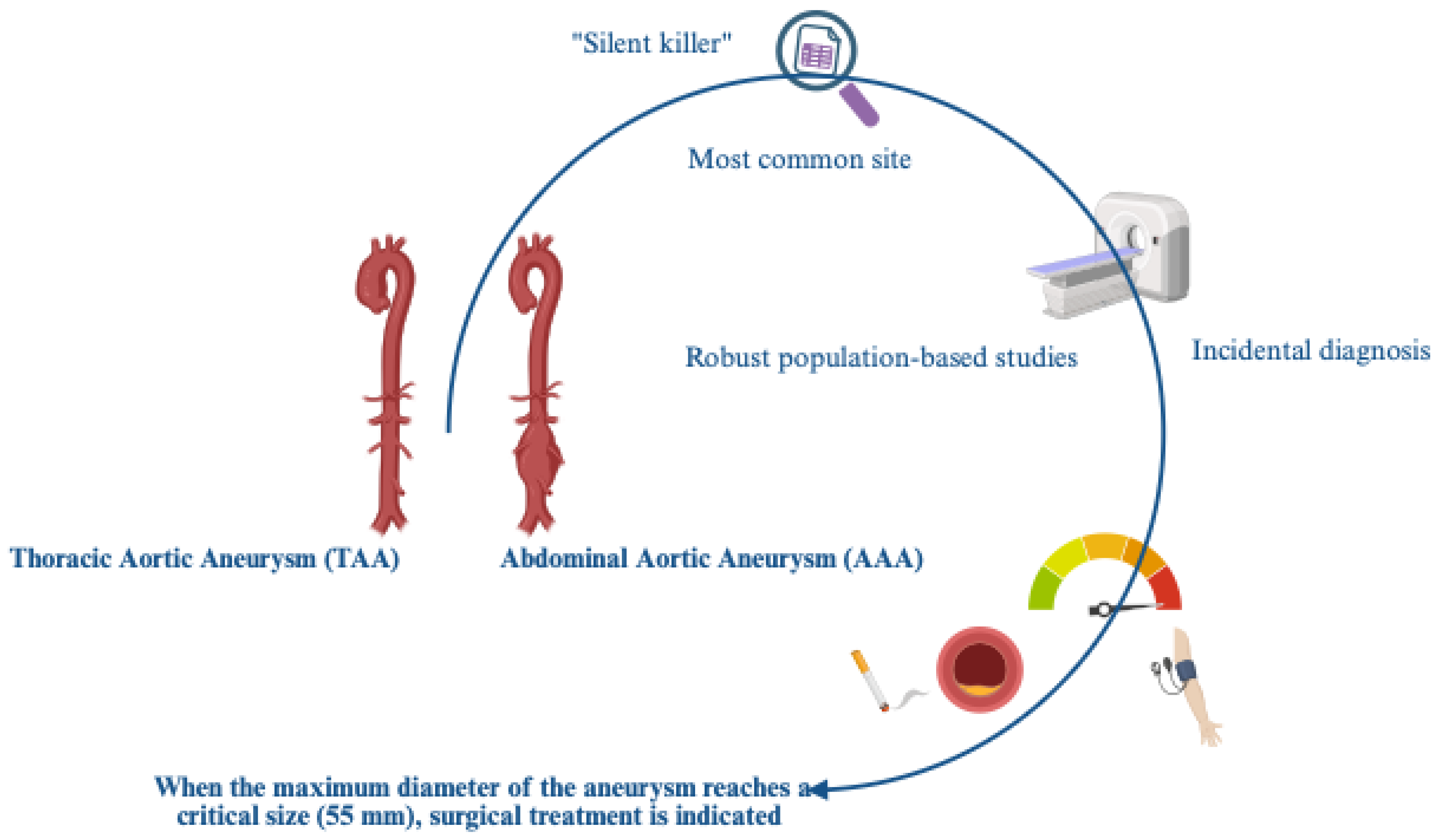
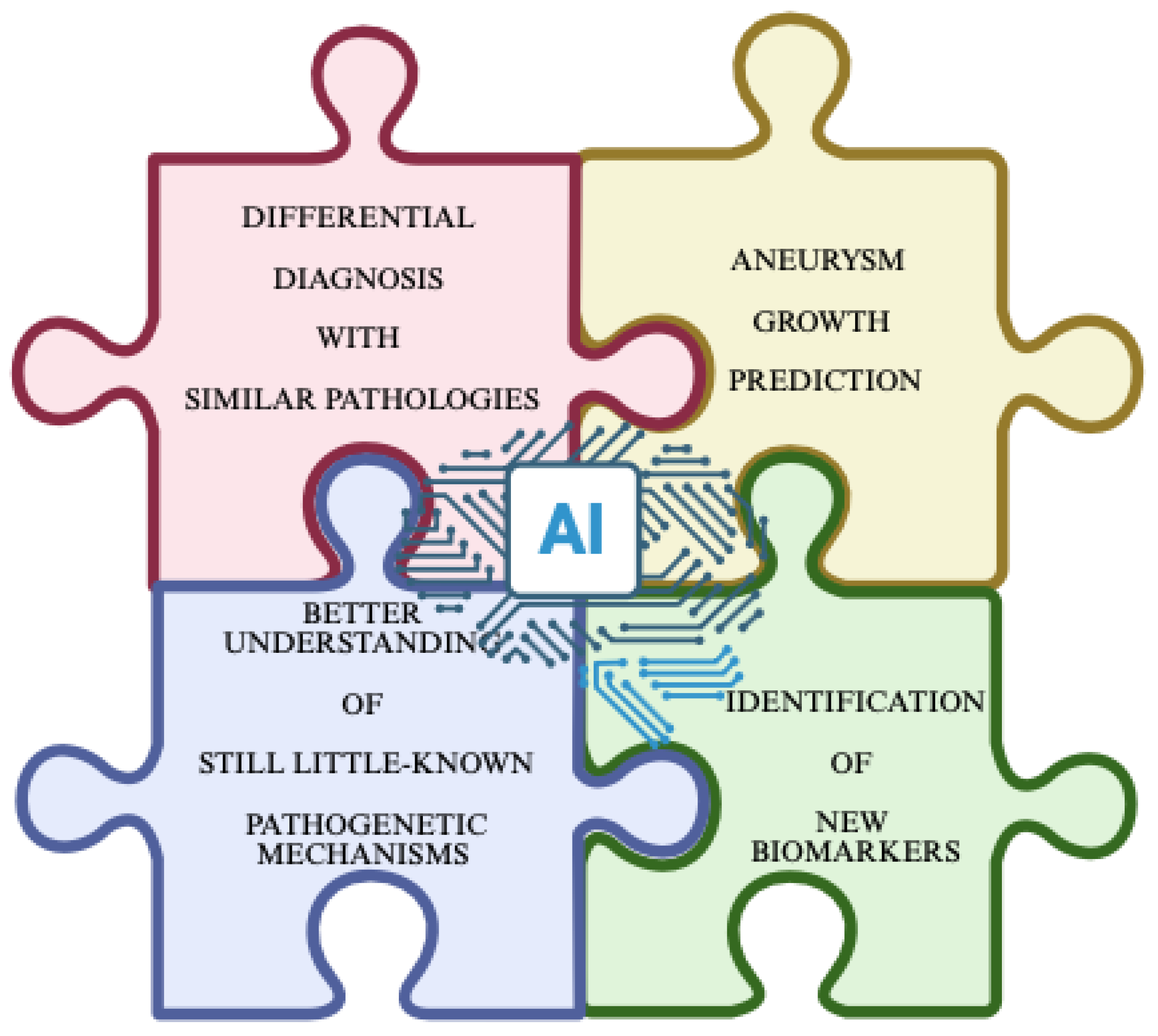
Aortic aneurysms (AAs), both abdominal and thoracic, remain one of the most fatal cardiovascular emergencies, with a growing prevalence and incidence, especially for sporadic forms in our populations, which are primarily comprised of elderly individuals. A high mortality risk also appears to be linked to managerial delays, despite advances in imaging techniques that facilitate the difficult diagnosis, and in surgical procedures. This is the result of the clinical decision-making approach, which is unfortunately still based, as per guidelines, on the maximum aortic diameter. This parameter, as suggested, fails to capture the biological and biomechanical complexity underlying these pathological conditions, which are influenced, among other things, by entirely individual factors (genetics, gender, lifestyle, etc.). With the emergence of network medicine and omics sciences, diverse and complex sets of clinical, imaging, and biomarker data are now available. These could be precisely processed by artificial intelligence (AI), enabling accurate prediction of AA risk and facilitating its complex management. Therefore, AI could represent an excellent tool for AAs, showing the potential not only to refine AA risk prediction but also to radically transform the way we understand, monitor, and manage AA patients, despite some limitations. These aspects are the subject of this review, as are their therapeutic applications.

Keywords: 
;  ;  ;  ;  ;  ;  ;  

1. Introduction

In recent decades, the use of network medicine is revolutionizing the clinical management of human diseases, particularly of highly complex ones, such as aortic aneurysms (AA), by bringing advances in both diagnostic processes and their simplification, as well as in prognosis and outcomes, clinical decision-making, and research, which could benefit the healthcare management of affected patients [1]. This evolution is being achieved thanks to the application of artificial intelligence (AI), based on deep learning techniques, such as machine learning (ML), which allows for the analysis of complex multidisciplinary data sets, recognizing patterns, and creating accurate evidence, and consequently supporting early disease’s diagnosis and evidence-based therapeutic or surgical treatments with an accuracy comparable to that of expert specialists, but who are not replaceable, as will be highlighted [2,3].
In medical research, AI is enabling the discovery of blood biomarkers useful for easier diagnosis, prognosis, and disease outcomes, as well as the development of new drugs, thanks to the analysis of multi-omics models and the optimization of clinical trials by mining large datasets to identify new therapeutic targets [4,5]. In healthcare management, AI has been displayed to improve resource distribution, streamline hospital operations, and facilitate patient interaction, for example through telemedicine [6,7,8,9,10,11]. Despite the many resources and applications of AI, and the resulting advances, several challenges remain [12,13,14,15]. Data privacy issues remain, algorithmic biases must be considered, a lack of transparency in the use of AI models, and finally, poor acceptance by physicians. Ethical integration requires explainable AI, along with robust regulatory frameworks and interdisciplinary collaboration [11,12,14,16]. However, medicine is still evolving toward precision medicine, and AI is proving to be a very promising tool in this field, as well as in robotic surgery and medical education itself [17]. AI can also offer personalized treatments, as well as suggesting and achieving better surgical outcomes, and even serving as adaptive learning tools for students. Consequently, its application in medicine depends essentially on acceptance and trust among healthcare professionals. The key lies in thoughtful and innovative use, which can transform healthcare into a more accurate, efficient, and patient-centered system worldwide.
In this review, we provide a summary of the above concepts, illustrating the success of this approach in addressing biomedical complexity and its current challenges. We focus specifically on the application of AI in AA management.

2. AI: What Is it? Its Features, Advantages, Limits, and Instances from Deep Learning Techniques to Digital Twins

Artificial Intelligence refers to a system that can simulate human-like complex cognitive functions, such as learning, problem-solving, and decision-making[18]. Within this domain, machine learning (ML) refers to a subset of algorithms that learn directly from data, such as anonymized patient records, biomarker features, and medical images[19], and use them to make predictions, such as aneurysm growth, rupture risk, or postoperative complications. Unlike traditional statistics, which test specific pre-defined hypotheses, ML models optimize predictive accuracy by identifying multi-dimensional, non-linear relationships. These algorithms can learn by training data in which a domain expert associates a specific output with a particular input (supervised learning). Common supervised ML methods used in aneurysm research include Random Forests, Gradient Boosting Machines, Support Vector Machines, and Naïve Bayes classifiers. Moreover, multiple individual models can be aggregated into a single, stronger predictor in techniques such as Extreme Gradient Boosting (XGBoost), Random Survival Forests, and hybrid frameworks like SHAP-FIRE or PSO-ELM-XGBoost. This is particularly effective in aneurysm research, where datasets contain many variables related to anatomical, physiological, and biochemical information but relatively few patients. ML methods also include unsupervised techniques, such as clustering of proteomic or transcriptomic profiles, which help uncover disease subtypes or molecular patterns. Recent efforts emphasized not only prediction but also interpretability. Tools such as LASSO regression and Permutation Importance help identify the variables most strongly associated with the prediction. SHAP (Shapley Additive Explanations) is increasingly used because it conveys information about the relative contribution of each model’s feature to the prediction.
Deep learning (DL), a further specialization within ML, employs interconnected nodes, called neurons, that process and transmit information from the input to the output layer [18,19,20]. DL can automatically learn, extract and categorize complex spatial features from raw imaging data [18,19,20], making it highly suited for 3D aortic imaging. Common architectures, such as U-Net, nnU-Net, ResNet, VNet, AlexNet, and VGG-16, are based on Convolutional Neural Networks (CNNs).

3. An AI Clinical Application in the Complex Case of Aortic Aneurysms? The Initial Panoramic

Aortic aneurysm (AA) is defined as aorto-pathy characterized by a permanent, localized arterial dilation of the human aorta, greater than 1.5 times the normal diameter of the vessel, that involves all three layers of the aortic wall (different from pseudoaneurysm) [21]. The AA occurrence is associated with various epidemiological factors, such as age, gender, race, family history, and smoking [22]. AA is often asymptomatic in early stage, and a life-threatening condition and associated with a high risk of rupture and death. Its treatment depends on surgical repair, which can be achieved through open surgery or endovascular aneurysm repair (EVAR) [23].
AAs may develop at any level of the aorta and are commonly divided into thoracic aorta aneurysms (TAA) and abdominal aortic aneurysms (AAA)[24,25,26] Although TAA and AAA are abnormalities found in the same vessel, they appear to have different pathogenesis and etiologies, as reflected by the histopathological differences in the aortic wall components, when comparing thoracic and abdominal aorta[22,23,24,26]. AAA is the most common site for aneurysm formation, interesting particularly the infrarenal segment of abdominal aorta [27,28]. AAA is notably more prevalent than TAA, occurring five times as often [25,27]. However, it is worth noting that epidemiological data on TAA are confounded by its asymptomatic nature and diagnostic difficulties [25]. It is indeed called a “silent killer”, and based on its natural history, TAA usually grows asymptomatically until rupture, with a mortality rate ranging from 94% to 100% [25]. Furthermore, its onset in the thorax makes diagnosis more difficult, which is usually made incidentally via echocardiogram, chest CT, or autopsy [25]. This is the opposite of the AAA, which is easy to screen and currently well-studied with robust population-based studies. Available evidence suggests that the prevalence of TAA and treatment outcomes vary by race, gender, and socioeconomic factors. Furthermore, a characteristic increase in incidence is observed among older adults [29]. On the other hand, in both sexes, increasing age and body surface area (BSA) are risk factors for AAs of any anatomical location. Whereas arterial hypertension is a risk factor for TAAs, hypercholesterolemia and smoking are risk factors for AAAs [30] (Figure 1).
Currently, [31] When the maximum diameter of the aneurysm reaches a critical size (55 mm), surgical treatment is indicated [32]. However, assessing the true size of the aorta can be difficult. In clinical practice, specific sources of error are regularly encountered, including obliquity, asymmetry, and mismatched sites. This leads to the use of both echocardiography and computed tomography/magnetic resonance imaging (CT/MRI) for a comprehensive assessment. In addition, it has been highlighted that approximately 60% of patients experience complications before reaching this size. Therefore, maximum diameter is not a reliable predictor and does not reflect aneurysm-related risk. Several factors increase the risk of aneurysm rupture, including biochemical/biomechanical factors, sex/gender, genetic factors [33], and connective tissue diseases, such as Marfan syndrome and Ehlers-Danlos syndrome [31]. For example, our group and others have shown that arterial age and premature vascular aging, but not chronological age, are significantly associated with faster aneurysm growth [34]. Consequently, leukocyte telomere wear and the potential for altered telomerase activity are optimal biomarkers for predicting the onset and progression of AA. Similarly, increased expression[34] of certain pro-inflammatory pathways, such as TGF-β, TLR-4, interferon-γ, chemokines, and interferon-γ, correlated with specific pro-inflammatory allelic profiles, significantly accelerates aortic dilation and growth [35,36,37,38,39]. Biomechanical factors can modulate aortic wall dilation and are assessed using a computer modeling technique to simulate stress distribution in a theoretical aortic aorta model. Several imaging modalities can be used to assess the risk of aortic rupture, including ultrasound elastography (EEL), computed tomography (CT) angiography, and magnetic resonance imaging (MRI). However, CTA is the gold standard technique in surgical planning for assessing the extent and morphology of the aortic valve and identifying any coexisting occlusive disease. Despite this, traditional screening methods, however, face challenges, including difficulty in detecting small or atypical aneurysms.

3.1. The Challenge and Unpromising Solutions

The need for a reliable approach to integrate diameter criteria into complication risk assessment and elective repair timing motivates current research into new risk stratification criteria for aortic aneurysms. The identification of circulating and tissue biomarkers and computational biomechanics could represent potential solutions. The identification of biomarkers related to the onset and poor prognosis of aortic aneurysms has currently led to the identification of promising molecules and genes with genetic variants or mutations associated with both familial and sporadic forms. Biomechanical indicators based on computational modeling of blood flow dynamics and wall mechanics have also been proposed, but their clinical utility is hampered by the high computational cost, the complexity/assumptions/idealizations of the model, and the technical expertise of the end user. Furthermore, it is often not possible to combine information from different sources, from the molecular to the organ level. A major challenge lies in the complex multifactorial nature of the disease, which encompasses molecular and cellular mechanisms, biomechanics, function, and pathophysiology of the aorta.

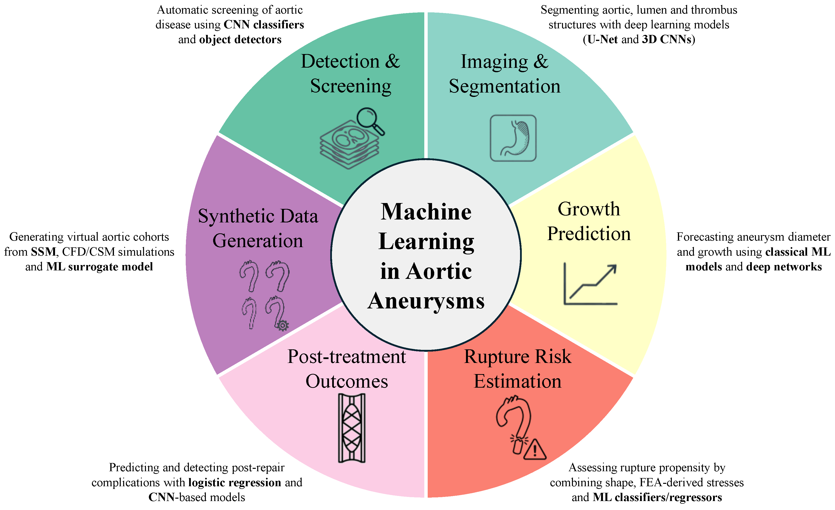
4. AI as Potential Solution of AA and Literature Evidence

4.1. Deep Learning Techniques as Aid in Evaluating the Aorta Size

In identifying the exact dimensions of the aorta, visualization techniques are used to enable medical experts to better interpret imaging data [31,32]. State-of-the-art imaging data are three-dimensional (3D) and require appropriate visualization techniques to model and represent the required information with high accuracy. 3D visualization can provide a complete anatomical view of the area of clinical interest, the aorta wall in our case, and facilitates inspection of the data from angles that are impossible to achieve with typical two-dimensional (2D) imaging systems. 3D visualization, in the specific case of structural and anatomical analysis of the aorta, appears advantageous, as it offers an effective and non-invasive way to examine the areas of interest and allows the evaluation and identification of aortic pathologies, such as aortic aneurysms [40,41] . The application of visualization techniques, however, involves extracting anatomical areas of interest from the rest of the imaging data. This is achieved using an image segmentation method, which is the most important step in computer-aided visualization techniques in digital image processing and analysis. Generally, segmentation depends on various characteristics of processed imaging data, such as color or texture, and allows a digital image to be divided into continuous, homogeneous subgroups called image segments. From a technical standpoint, segmentation allows for a reduction in image complexity and allows for further processing or analysis of each segment. In our case, the target of segmentation is the aorta, which we can consider an organ comprising several parts: ascending aorta, aortic arch, descending aorta, and abdominal aorta. Each of these parts can develop characteristic pathological conditions, such as aneurysms. Consequently, aortic segmentation is crucial for assessing these conditions [42] as it allows for the application of several more advanced assessment and follow-up techniques, based on the segmented clinical area of interest [43]. Basic and generic methods (i.e., such as flood-fill, graph-cut-based methods, growcut and watershed) of deep learning for aortic segmentation have been used, but they have shown several limitations. However, in recent years, the performance of image segmentation techniques based on deep learning has improved significantly. As a result, many of these techniques can be easily used in clinical practice for processing medical imaging data.
Lareyre and coworkers projected a fully automated pipeline for detecting AAA by using image segmentation. In this study, a threshold-based contour detection method was successfully utilized to automatically detect key AAA features, such as the distance from the renal and iliac arteries and the aneurysm’s location within the aorta [44]. López-Linares and colleagues attempted to perform fully automated detection and segmentation of abdominal aortic thrombus in postoperative computational tomography /angiography (CTA) images using deep convolutional neural networks (CNNs) [45] Furthermore, Larsson et al. proposed a fully automated method for segmenting abdominal organs using deep convolutional neural networks. This specific method initially operates as a robust target organ localizer and then activates the use of a deep convolutional neural network (CNN) to perform pixel-by-pixel classification [46] Noothout et al. proposed an automatic segmentation method for detecting the thoracic aorta in low-dose chest CT using a dilated CNN analyzing the axial, coronal, and sagittal planes. Data from each image plane is combined to provide a final segmentation estimate [47] Furthermore, Fantazzini and colleagues suggested an automatic 3D segmentation for aortic CTA data by using a combination of 2D multi-view convolutional neural networks. This study applies a fully automatic procedure for preoperative aortic segmentation by firstly applying a CNN based on the brightness of the CTA data, followed by additional CNNs to assess the final segmented regions [48] .
In addition to CT or CTA, other imaging techniques have been used for aortic segmentation. Müller-Eschner et al. attempted to compare native and contrast-enhanced magnetic resonance (MR) angiography (MRA) sequences through automated 3D lumen segmentation. This method allowed a quantitative evaluation (evaluation of differences in aortic lumen diameters and lengths) for both native and contrast-enhanced MR angiography acquisition techniques [49]
George K. Matsopoulos’s group recently proposed a new, fully automated 3D segmentation method, combining basic image processing techniques and more advanced machine learning algorithms, to detect and model the aorta in 3D CT imaging data [50]. The proposed methodology was applied to 16 3D CT data sets, and the extracted aortic segments were reconstructed as 3D models. This method achieved superior segmentation performance compared to all the segmentation techniques described above, in terms of the accuracy of the extracted 3D aortic model. Therefore, the proposed segmentation scheme could be used in clinical practice.
As AI-based semantic segmentation of the aneurysmal aorta becomes increasingly available in clinical practice, standardization of aortic diameter measurements is expected to improve substantially. Recent EACTS guidelines[51] emphasize the need for automated aortic diameter measurements within clinical workflows to reduce intra- and inter-observer variability and minimize missed aneurysms. Once the aortic wall is segmented, automated pipelines can be implemented not only to extract standardized diameter measurements at predefined anatomical locations but also to derive novel geometric metrics, such as the aortic length. Remarkably, morphological analyses of aortic length performed by the Yale group[52] revealed an association with dissection events for lengths exceeding 11 cm, and the combined use of aortic length and diameter demonstrated superior predictive capability for adverse events compared with diameter alone. The “Aorta Report v1.0” tool is an open-source, web-based application that automatically computes aortic diameters at multiple anatomical landmarks using a trained nnU-Net model. Users can upload anonymized CT scans and obtain vessel diameter measurements without the need for specific hardware. Though not intended for direct clinical use, such tools may represent a game changer in the next future for the diagnosis of aortic aneurysms.

4.1.1. Considerations

Progresses in imaging techniques have revolutionized the diagnosis and management of AA. Thus, it is possible to underline that ultrasound techniques represent the primary screening tool. CTA and MRA, and AI-assisted imaging techniques show an elevated power to improve both risk stratification and early detection. Accordingly, population-based screening trials conducted in diverse countries, including UK, Denmark, and Australia, are confirming a significant decrease in mortality related to AAA, supported by use of such imaging techniques [53]However, these encouraging data are undermined by the fact that a more targeted screening approach should be used in these studies, giving priority to high-risk subjects. In addition, standardization of imaging protocols is also crucial. The application of AI in 3D intravenous ultrasound (IVUS) and PET-CT of the aorta could also be considered. This might ensure accurate measurement of the aneurysm and optimal timing of intervention. Furthermore, promising and subsequently consolidated data must be incorporated into guidelines. These must align with technological innovations to optimize patient outcomes while maintaining cost-effectiveness. They must also consider suggesting a precision-based multidisciplinary approach that can help shape the evolving landscape of AA management.

4.2. Deep Learning Techniques as Aid in Evaluating Biomechanical Indicators

Current research addresses the improvement of the assessment of the risk of complications and the optimization of the timing of elective repair through mechanistically grounded biomechanical indicators, such as those based on wall shear stress (WSS) and peak mechanical wall stress (MWS) [54,55]. However, although high-fidelity computational simulations currently represent the most reliable way to obtain these biomechanical indicators, their routine clinical use remains limited because conventional physics-based approaches—typically computational fluid dynamics (CFD) and finite element analysis—are computationally expensive, time-consuming, and require specialized expertise.
Several works have demonstrated the potential of AI-based methods to accelerate the estimation of these biomechanical quantities. Liang and coworkers [56] showed that deep neural networks (DNNs) trained on CFD data can directly and rapidly estimate steady-state pressure and velocity distributions within the thoracic aorta, delivering results in less than one second with low average errors. Pajaziti et al. [57] used large-scale CFD simulations on 3,000 synthetic aortic geometries to train an ML model capable of faithfully reproducing full 3D pressure and velocity fields in a fast and fully automatic manner. Similarly, Du et al. [58] reported an AI framework that, after training on 1,000 synthetic aortic shapes, could recover detailed 3D hemodynamics—including pressure, velocity, and WSS patterns—from anatomy alone in a few milliseconds, thus offering a highly scalable tool for patient-specific hemodynamic assessment. Deep learning surrogate models have also been proposed for the prediction of WSS patterns across the cardiac cycle in AAAs.[59]
AI-based approaches have been also explored for the estimation of structural properties of the aortic wall. A first line of work has focused on direct structural wall-stress prediction: for example, Chung and colleagues[60] introduced an automated pipeline that integrates DL-based segmentation, mesh generation, and ML-driven MWS estimation, substantially accelerating conventional preprocessing and simulation workflows. Beyond direct MWS prediction, AI has been leveraged to generate inputs needed for biomechanical analysis, including the estimation of the aortic zero-pressure configuration and the development of data-driven surrogate constitutive models [61]. These surrogate models have been shown to outperform classical phenomenological formulations in capturing ascending thoracic aortic aneurysm (ATAA) tissue behavior and to enable derivation of patient-specific material properties[62] . Taken together, these advances indicate that AI can act as a powerful accelerator and enabler for biomechanically informed risk assessment in aortic aneurysm disease.

4.3. Advanced Prediction Models and Synthetic Data Generation

To develop an AI-based model for risk stratification of aortic aneurysms, accurate prediction of the long-term biomechanical response of the aneurysm is essential to determine when rupture may occur and thus guide prophylactic intervention. Because aneurysm diameter alone is not a reliable predictor of rupture risk, novel approaches combining baseline medical imaging with computational modeling or biomarkers have been proposed, including those by Pasta et al. [63]. First, in silico assessment of aneurysm biomechanics relies on clinical imaging modalities such as CT or MRI, which are not routinely performed during post–in-hospital follow-up. Second, large datasets are required to train machine-learning models, making multicenter studies necessary. Consequently, only a limited number of studies have attempted to develop predictive models capable of describing aneurysm growth and rupture over time. In the context of the abdominal aorta, Lee et al. [64] developed a nonlinear support vector regression model that combines the basal diameter of the AAA with a functional measure of flow-mediated dilation to predict aneurysm expansion at 12 months from in-hospital admission, with the goal of reducing surveillance imaging. Rengarajan et al. [65] trained eight different classifiers on 53 geometric descriptors and biomarkers derived from computational analysis in 150 AAAs, highlighting that maximum transverse diameter, mean wall thickness, and mean wall stress are more informative risk factors than maximum diameter alone within 1-6 months of follow up. Therefore, further studies are needed to develop robust predictive tools for aneurysm growth and remodeling.
Despite these achievements, one of the most critical aspects remains the determination of rupture stress in patients who have not yet undergone surgical repair. While wall stress can be estimated using routine clinical imaging combined with computational modeling, the ultimate tensile strength must currently be determined through ex vivo material testing. Therefore, an AI-based predictive tool should not only quantify the stress acting on the aneurysm wall but also estimate the maximum strength at which the aneurysm fails (i.e., the rupture strength). For these reasons, several researchers have attempted to develop predictive models to estimate rupture risk in patients undergoing routine clinical imaging. In this setting, Luo and colleagues[66] trained a random forest on over 11,000 stress-strain curves obtained from bulge inflation tests on ascending aorta samples, using the pre-rupture response to classify the samples into ruptured versus non-ruptured tissues. Later, He et al.[67] proposed a two-stage procedure where a machine learning model was initially trained on stress–strain data collected from ex vivo inflation tests for automatic identification of aneurysm wall regions of rapid stress accumulation, and later the model was used to predict the local ultimate strength in these areas. For a patient study group, the rupture index maps achieved estimates comparable to those of experiments in 13 out of 15 samples, surpassing simpler criteria such as maximum principal stress or the diameter rule alone. Finally, Liu and colleagues[68] used a surrogate machine learning model trained on computational models of ascending aortic aneurysms to identify regions most susceptible to rupture. The model was able to estimate local safety indices with greater accuracy than criteria based on aortic size.
Machine learning enables the generation of synthetic datasets, which may serve as the basis for in silico trials. In the context of aortic aneurysms, synthetic datasets of patient-specific aneurysm geometries may help overcome the lack of large cohorts required to train robust machine-learning models. To achieve this goal, statistical shape modeling represents a powerful technique for extrapolating geometric features that, when combined with machine-learning approaches, can also provide insights into the mechanistic relationship between shape and function. For ascending aortic aneurysms, Liang et al.[69] developed a statistical shape model of aortic morphology based on a limited patient cohort and used this tool to generate hundreds of synthetic geometries. Finite element analyses were then performed on these geometries to derive rupture risk indices, which were subsequently used to train classification and regression model. Geronzi et al.[70] used principal component analysis and partial least squares regression as the basis for predictive models, demonstrating that aneurysms located closer to the aortic root and those with larger initial diameters exhibit higher growth rates. More recently, an increasingly popular approach has been the direct generation of aneurysm geometries using deep learning models. These approaches offer the advantage of capturing nonlinear correlations among shape features that principal component analysis is unable to represent. In this context, Fabbri et al. [71] combined a graph convolutional network with a β-variational autoencoder to sample realistic aortic geometries from an extremely limited dataset, effectively transforming a cohort of 60 patients into a large virtual AAA database. Similarly, Tenderini et al.[72] introduced a diffeomorphic auto-decoder framework for the generative development of new aneurysm anatomies, enabling the incorporation of realistic geometric variability into physical process simulations. Using implicit neural network Gasparovici et al.[73] represented and generated new aortic anatomies while explicitly controlling the degree of deviation from the original dataset. Collectively, these synthetic data generation strategies have enabled the creation of large databases of anatomically consistent aortic geometries for systematic rupture risk analysis, calibration of supervised machine-learning models using enriched datasets, and, ultimately, the development of in silico trials for evaluating cardiovascular device efficacy.
Figure 2 provides an overview of the main applications of machine learning across the aortic aneurysm clinical and research pipeline, spanning detection and imaging to growth modeling, rupture risk estimation, and synthetic data generation. This shows how machine learning serves as a unifying framework to support risk stratification, biomechanical understanding, and the development of in silico trials for the management of patients with aortic aneurysms.

5. Biomarkers and AI in Aortic Aneurysm

The pathogenesis of AA involves a multitude of complex mechanisms: genetic predisposition, specific genetic mutations, vascular inflammatory processes caused by the infiltration of T lymphocytes and macrophages, resulting in alterations of the extracellular matrix (ECM) and progressive weakening of the vascular wall, with a greater tendency towards the development of aneurysmal pathology. Considering these AAA features, it is clear how the use of AI, and particularly of machine learning techniques, can constitute a fundamental contribution to the complex diagnostic process of such diseases. They constitute a system that allows us to integrate all the pathways and related biomarkers currently recognized in the pathogenesis of AA. In addition, AI would represent a significant breakthrough in clinical practice, in addition to the significant role in the development of targeted therapies. For this reason, studies investigating the correlation between biomarkers and pathogenic processes by using AI and machine learning are increasingly numerous. Currently, in our opinion, AI would represent a future prospect for scientific research. The main ones addressed in our discussion are summarized in Table 1. The large number of currently known biomarkers (and therefore variables) far exceeds the number of available subjects; and this is why machine learning is so successfully applied. Statistical analysis alone, in fact, focuses on inference: once a specific model is developed, the extent to which the association with the studied effect is studied is studied. In contrast, machine learning focuses on prediction using generic learning algorithms, identifying patterns within complex data [74].
The complex etiopathogenesis of AA makes it equally challenging to identify precise biomarkers that can aid in the diagnostic process. Therefore, the real challenge currently is to create an integrated model that can guide the clinical process. However, much of this research undoubtedly focuses primarily on the etiopathogenesis of abdominal aneurysms. We therefore believe that review work can highlight the evidence currently achieved, and, consequently, it can be imperative. Momenzadeh A et al. proposed a hypothesis for the differential diagnosis between descending thoracic aortic aneurysms and type B dissections by comparing statistical analysis with machine learning algorithms. Of the 1,549 peptides and 198 proteins analyzed by the authors, statistical analysis revealed only one significant correlation, hemopexin (HPX), between the two different pathologies. Using machine learning, the authors first subdivided quantitatively similar proteins using hierarchical clustering analysis and subsequently analyzed them using six different classification algorithms. The five proteins with the highest Permutation Importance (PI) scores were immunoglobulin heavy variable 6-1 (IGHV6-1), lecithin-cholesterol acyltransferase (LCAT), coagulation factor 12 (F12), HPX, and immunoglobulin heavy variable 4-4 (IGHV4-4). Furthermore, proteins involved in complement activation, humoral immune response, and blood coagulation were associated with significantly more frequent pathways in the plasma of patients with type B dissection compared to those with descending thoracic aortic aneurysms. Therefore, machine learning, compared to statistical analysis alone, allowed the identification of a specific plasma proteome that could aid in the differential diagnosis between these two very similar pathological conditions [75].
Another aspect of fundamental importance in the clinical management of aneurysms is the prediction of growth in patients undergoing follow-up. Even in managing this aspect, the use of AI has shown its potential benefits. In this sense, Forneris A et al. used three different functional biomarkers (time-averaged wall-shear stress, in vivo principal strain, and intra-luminal thrombus thickness) and managed to obtain, using the binary Extra Trees classifier algorithm, area under the curve=0.92, demonstrating an excellent performance in predicting relevant aortic growth, greater than geometric models. In particular, the authors highlighted a statistically significant difference for the time-averaged wall-shear stress: patients with a basal diameter > 50 mm showed a lower value than patients with a basal diameter < 50 mm (0.59 ± 0.37 Pa vs 0.78 ± 0.48 Pa; P < 0.001). This data further strengthens the predictive power of this biomarker on aortic weakening and therefore aneurysmal growth over time [76].
The ability to handle such a vast amount of data, combined with the training of specific learning models, has also allowed for a further understanding of still poorly understood pathogenic mechanisms, such as the precise role of T lymphocytes. Li D. et al. investigated the T cell imbalance in aortic wall infiltration. Specifically, the authors identified eight different T cell phenotypes, each associated with different gene expression patterns and phenotype-specific markers. Using two distinct learning models, four key biomarkers were identified: FOSB, JUNB, cystatin F (CST7), and TBC1 domain family member 4 (TBC1D4) from four independent datasets. Dysregulation of these biomarkers was highlighted in the abdominal aortic wall and from the analysis of the ROC curves, with AUC for FOSB, JUNB, CST7 and TBC1D4, a significant role of FOSB and JUNB emerged with curve values of 0.911 and 0.917 respectively, similarly in all datasets [77]. Similarly, by combining multi-omics systems and machine learning algorithms, Yong X. et al. studied the mechanisms of cellular infiltration (macrophages and CD8-positive alpha-beta T cells) underlying the pathogenic process of abdominal aortic aneurysms, leading, thanks to machine learning methods, to the identification of two specific biomarkers: CCR7 and CBX6. Specifically, the authors highlighted a positive correlation between CCR7 and naive B cells, demonstrating that CCR7 overexpression may play a role in the immune response of naive B cells in the pathogenesis of abdominal aortic aneurysms. Conversely, the inverse correlation between CBX6 and neutrophils could indicate a suppressive role of the aforementioned protein on neutrophil activity, thus potentially reducing the aneurysmal process [78]. This research was applied with a similar methodology by Xiong T. et al., demonstrating the association between G0S2 expression and neutrophils, activated and quiescent mast cells, M0 and M1 macrophages, regulatory T cells (Treg), quiescent dendritic cells and quiescent CD4 memory T cells, further confirming the crucial role of inflammatory cell infiltration in the etiopathogenesis of aneurysms [79].
In addition to confirming pathological processes that are already partially known, machine learning algorithms also lead to the discovery of new biomarkers, such as those associated with the mechanisms of cuproptosis and ferroptosis in AAA. While it is well known that iron and copper ion metabolism plays a key role in regulating cell death, the pathogenic mechanisms linking these metabolic alterations to the development of aneurysmal processes are not yet fully understood. In this regard, Han Z. et al., starting from three scRNA-seq datasets from different mouse models and a bulk RNA-seq dataset from human Peripheral Blood Mononuclear Cells (PBMCs), and analyzing them using four machine learning algorithms, were able to identify the role of PIM1 upregulation in cuproptosis and ferroptosis [80].
Although machine learning systems have experienced rapid growth in recent years, they still require training of diverse models to further advance the application of AI in clinical practice. Figure 1 summarizes the current main fields of application of machine learning algorithms in the management of biomarkers implicated in the etiopathogenesis of aortic aneurysms (Figure 3).

6. AI and Prognosis in Aortic Aneurysms

The growing use of AI in the management of aneurysmal disease is also finding application in the development of prognostic prediction models, with higher success rates than currently used models. [49] Predicting prognostic risk plays a fundamental role in the clinical management of aneurysmal disease. Given a disease that, in most cases, is asymptomatic, determining the timing of surgical intervention has always been a clinical challenge requiring an accurate prediction of the risk of dilation and/or rupture, a prediction that must necessarily consider the patient’s individual characteristics. When managing such large and complex data, it is easy to see how the application of AI can be extremely useful. The Danish team led by Skovbo JS et al. [81], starting from the analysis of a population of 637 individuals that included all cases of AAA rupture available between January 2009 and December 2016 and those undergoing elective surgery with a ratio of 1:2, proposed a SHAPFire (SHapley Additive exPlanations Feature Importance Rank Ensembling) AI tool. This model, starting from 68 different characteristics, identified 20 key factors in assessing progression risk, obtaining a more favorable AUC and Youden index than using aortic diameter alone. These 20 characteristics included, in addition to the evaluation of dimensional parameters by CT (maximum transverse diameter, maximal luminal area, distance between iliac bifurcatures, distance between lowest renal artery to aortic bifurcation, maximum diameter of the right iliac artery, distance between the right and left iliac bifurcation from the aortic bifurcation, distance between the aortic bifurcation and the sacrum, transvers outer-to-outer diameter of L3), the evaluation of anterior wall thickness, clinical factors (age, smoking habit, pulse pressure values and hypertension, BSA, use of statins and platelet inhibitors), the detection of UniCa score at maximum size over 15 mm and UniCa score in thrombus and the presence of suprarenal mural thrombus.
Another interesting prognostic classification model, the APC (aneurysm prognosis classifier), comes from the University of Pittsburgh [82]. Specifically, data from 381 patients with AAA with a known clinical outcome (aneurysm repair or rupture) were divided into three different categories: clinical, biomechanical, and morphological. Subsequently, machine learning algorithms were trained on this data to create a single final classification model that could advise clinicians on the appropriate timing of surgery, demonstrating the fundamental role of imaging-based biomechanical and morphological quantification, evidence already demonstrated by Lindquist Liljeqvist M et al. also in small aneurysms [83]. Other models have been proposed for assessing prognostic risk, although several have significant limitations due to the small populations included [84,85].
Therefore, we believe that the application of AI and machine learning mechanisms can play a fundamental role in the prognosis of such a complex and multifactorial pathological process. However, achieving this validity requires the creation of models that include a significant number of patients, thus overcoming the limitations associated with statistical biases.

7. AI as Aid in Predicting Risk Assessment of AA, and in Selecting the Appropriate Therapeutic Strategy: A Particular Focus on TAA, Being Less Studied than AAA

It is well recognized that the actual therapy in case of TAA consists in two principal approaches: a) the open surgery, representing the gold standard in case of TAA involving the ascending and arch aorta, and consisting of diseased aortic segment’s replacement with a Dacron woven graft, often performed with cardiopulmonary bypass, and ensuring maximal durability; b) endovascular approach (TEVAR - Thoracic Endovascular Aortic Repair), a minimally invasive technique, excluding the aneurysmal sac from the circulation via a stent-graft insertion. TEVAR is selected in the case of TAA involving descending aorta, as it reduces perioperative morbidity, especially in frail patients. Accordingly, therapeutic management of TAA demands careful risk stratification of dissection or rupture. Consequently, therapeutic management of AAT requires careful risk stratification. In this context, AI is emerging as a fundamental tool. Thanks to machine learning and radiomics analysis, AI can extract subclinical information from CT images (such as wall stiffness or hemodynamic forces) that complement simple dimensional data. Thus, AI enables more personalized and accurate risk stratification, and it consequently supports the physicians in selecting one of two abovementioned approaches. AI helps distinguish patients ideally suited for open surgery from those who are better candidates for TEVAR by assessing complex anatomical constraints and predicting postoperative outcomes and complications associated with each procedure. Accordingly, AI improves the prediction of individual rupture risk, by informing when to intervene.
Recently, AI, in conjunction with multi-omics approaches [86], is also identifying molecular targets (i.e., TGF-β signaling (e.g., ACTA2, SMAD2; [36,87]) pathway and molecules of pro-fibrotic events (e.g., CTGF, MMPs; [88,89]), or proteins involved in cell/extracellular structures (e.g., contractile/ECM proteins) or molecules involved in inflammation (e.g., TLR4 pathway, [90,91]) that may respond to specific pharmacotherapies, by providing a scientific basis to support or further develop the pharmacological option as an alternative to surgery strategies [92]

7.1. Experimental Evidence

Table 2 lists many studies that emphasize the use of AI models in TAA risk assessment. Some of these are described below.
Kennedy et al. explored the efficacy of a Machine Learning (ML) model in correlating routine patient clinical data with the intrinsic biomechanical function of the aortic tissue. Precisely, the study analyzed resected TAA tissue obtained from 158 patients undergoing surgery [93]. Tissue samples were subjected to biaxial tensile testing, and the calculated energy loss was utilized as the output variable for the ML model. The input parameters for algorithm training (including Gaussian Process Regression, GPR) were derived from comprehensive clinical assessments, encompassing medical imaging reports and genetic paneling data. The impact of including an echocardiogram-derived stiffness metric (CCPM) as an additional input was also evaluated in a patient’s subgroup. The most performant ML model, based on GPR, provided the evidence of a significantly superior correlation with tissue energy loss R2=0.63 compared to traditional methods relying solely on aortic diameter R2=0.26 or indexed aortic diameter R2=0.32. Patient ‘s age emerged as a variable displaying the strongest relationship with energy loss. Furthermore, the integration of the echocardiographic stiffness metric notably improved the model’s accuracy within a subgroup, raising the correlation from R2=0.46 to R2=0.62. These results, although preliminary, indicate that a machine learning (ML)-based approach may facilitate achieving significantly improved prediction of TAA tissue mechanical function compared to diameter measurements alone. The developed model offers a tool both to integrate a wide range of clinical and biometric data and to refine risk stratification for AAT, suggesting a likely advance over current diameter-based guidelines for identifying subjects at high risk of aortic complications.
Ben Li et al. developed ML models to predict one-year outcomes after TEVAR and complex endovascular aneurysm repair (EVAR) procedures [94]. These intervention procedures are increasingly common treatment options, but they carry a significant risk of major complications such as death, stroke, permanent paralysis, or new-onset dialysis, and existing risk-prediction tools remain limited in accuracy. The researchers utilized the Vascular Quality Initiative (VQI) database, including 10,738 patients who underwent elective TEVAR or complex EVAR for non-infrarenal aortic aneurysms between 2012 and 2023. A total of 172 variables were analyzed: 93 preoperative (demographic and clinical), 46 intraoperative (technical and procedural), and 33 postoperative (complications and hospital course). The primary outcome was a one-year ThoracoAbdominal Aortic aneurysm Life-altering Event (TALE), defined as death, stroke, permanent paralysis, or new chronic dialysis. Six ML algorithms were tested, including Extreme Gradient Boosting (XGBoost), Random Forest, Support Vector Machine, Artificial Neural Network, Naïve Bayes, and Logistic Regression, with the dataset divided into 70% for training and 30% for testing, and 10-fold cross-validation applied. The XG-Boost model outperformed all others, achieving excellent discrimination with an AUC of 0.96 using preoperative data, 0.97 when intraoperative variables were added, and 0.98 when postoperative variables were included, compared with 0.70 for traditional logistic regression. Overall accuracy reached 89%, with good calibration (Brier score improving from 0.09 to 0.05). The most important predictors of adverse outcomes included proximal landing zone, procedure type (arch or extensive type I–III thoracoabdominal repairs), preoperative functional status, chronic obstructive pulmonary disease, heart failure, history of stroke, procedure duration, non-home discharge, and absence of aspirin at discharge. Patients who experienced adverse events were generally older, had greater comorbidity burden (cardiovascular and pulmonary diseases), larger aneurysm diameters, and underwent more extensive or technically demanding repairs. Many of these risk factors were identifiable in the preoperative phase, underscoring the value of ML tools in patient selection and perioperative optimization. Compared with conventional regression models, ML algorithms demonstrated superior ability to capture complex, nonlinear interactions among variables, maintaining strong performance across demographic and clinical subgroups (age, sex, ethnicity, and procedure type) without evidence of bias. Clinically, these models can assist in preoperative risk assessment, optimization of comorbidities, intraoperative planning, and postoperative monitoring of high-risk patients. The authors highlight the potential integration of these models into vascular centers, especially within the VQI network, where the required data are routinely collected, thus facilitating personalized care and potentially reducing post-TEVAR/EVAR morbidity. In conclusion, this study demonstrates that machine learning models, particularly XGBoost, enable highly accurate prediction of one-year outcomes after TEVAR and complex EVAR, substantially outperforming traditional logistic regression. These models represent a significant advancement toward personalized risk stratification and decision-making in patients undergoing complex endovascular aortic interventions.
Huang et al. provided a comprehensive overview about the application of deep learning (DL) in clinical for improving the diagnosis, treatment, and management of AA [95]. Precisely, they analyzed the role of DL in AA by focusing their interest on screening, image segmentation, surgical assistance, and prognosis prediction. To this aim, the authors conducted a broad literature review covering applications of convolutional neural networks (CNNs) and related DL architectures such as U-Net, ResNet, and Generative Adversarial Networks (GANs) in CT, MRI, and angiographic datasets. These models were evaluated for their ability to enhance accuracy, efficiency, and reproducibility compared with traditional radiological and statistical methods. In the screening and diagnostic domain, DL models achieved high performance in identifying both AAA and TAA aneurysms, even from non-contrast CT (NCCT) scans. CNN-based architectures such as ResNet, VGG-16, and AlexNet reached areas under the ROC curve (AUC) up to 0.97, demonstrating near-radiologist-level accuracy. GAN-based methods allowed the conversion of NCCT into contrast-enhanced CTA-like images, offering diagnostic alternatives for patients with contraindications to contrast media. In large population studies, DL tools like AI-Rad (Siemens Healthineers) successfully screened over 18,000 CT scans, identifying missed TAAs with 97% accuracy, underscoring their clinical feasibility for large-scale aneurysm detection. DL has also been widely applied in aneurysm segmentation, a crucial step for accurate surgical planning. Models such as U-Net, VNet, and ResNetMed supported excellent performance, achieving Dice Similarity Coefficients above 0.95 on both contrast-enhanced and non-contrast CT images. These systems automatically delineate aneurysm contours, thrombus volume, and aortic geometry, allowing for precise and reproducible measurements essential for endovascular repair. Hybrid models combining expert systems with DL improved thrombus segmentation accuracy, highlighting the synergy between rule-based and data-driven methods. In the field of surgical assistance, DL demonstrated to contribute to preoperative planning, intraoperative support, and postoperative assessment. CNN- and U-Net–based algorithms can automatically measure aortic diameters and volumes and provide consistent results and minimizing human variability. DL models also successfully employed to identify optimal stent-graft landing zones, evaluate stent deployment, and assess postoperative end leaks after EVAR and TEVAR. For instance, automated stent-graft segmentation on DSA images achieved Dice scores up to 0.96, enabling accurate intraoperative guidance. In addition, Deep Belief Networks (DBN) integrated with vascular growth and remodeling models predicted aneurysm expansion with high precision, suggesting their potential in optimizing the timing of surgical interventions. DL also showed substantial promise in prognosis and disease progression prediction. CNNs analyzing morphological, hemodynamic, and thrombus-related features predicted aneurysm growth with AUCs around 0.89 and overall accuracies above 80%. Multimodal and radiomic-based models outperformed purely morphological approaches in forecasting serious adverse events (SAEs) after EVAR, achieving AUCs up to 0.93. Fully automated software such as PRAEVAORTA can evaluate aneurysm sac evolution after EVAR with segmentation accuracy (Dice 0.95) and ninefold faster processing than manual review, significantly improving clinical workflow. Moreover, DL models using cine-MRI data accurately assessed aortic compliance and stiffness, offering new insights into the risk of dissection in TAA patients. Despite these advances, the authors highlighted several limitations that currently hinder its full clinical implementation. The lack of large, high-quality, multicenter datasets reduces the generalizability of most models. The intrinsic “black box” nature of DL algorithms limits interpretability and raises concerns regarding transparency in clinical decision-making. Furthermore, many models remain insufficiently validated across diverse populations and imaging modalities, and there is still a lack of standardization and regulatory guidance for AI-based diagnostic tools. Thus, the authors proposed several strategies to overcome these challenges, including fostering data-sharing collaborations across institutions, developing explainable AI frameworks to clarify model decision processes, expanding multimodal research integrating CT, MRI, and DSA imaging, and ensuring continuous model updating with new clinical data. They also emphasized areas requiring further research, such as automated quantification of aortic calcification, optimization of stent placement planning, and real-time risk prediction for aneurysm rupture or postoperative complications. Nevertheless, DL technologies demonstrate to have an exceptional potential in advancing the imaging-based diagnosis, treatment planning, and follow-up of aortic aneurysms. By automating complex image analysis tasks and predicting disease behavior with high accuracy, DL models can improve diagnostic efficiency, surgical precision, and patient outcomes. Although important limitations remain, continued development, validation, and integration of these algorithms are expected to transform vascular medicine, ushering in an era of personalized, data-driven management for patients with aortic aneurysm.
Zhang et al. described the development and validation of an advanced ML- based predictive model to estimate the 30-day mortality risk in patients with acute type A aortic dissection (ATAAD) who underwent total aortic arch replacement (TAR) combined with frozen elephant trunk (FET) implantation [96]. The retrospective study included 640 patients operated on at a single Chinese center (Fuwai Hospital) between 2015 and 2020, reporting a perioperative mortality rate of 5.78% (37 cases). To overcome the limitations of standard ML models on small and imbalanced datasets, the authors developed and optimized a hybrid algorithm, named PSO- ELM-FLXGBoost (combining Particle Swarm Optimization, Extreme Learning Machine, and Focal Loss with XGBoost). This optimized model demonstrated the highest predictive performance, achieving an AUC value of 0.8687, a significant improvement over the basic XGBoost model (AUC 0.6981). The SHAP interpretability analysis identified the 10 most influential factors for mortality, which included: age, length of ICU stays, ALT (Alanine Aminotransferase), cardiopulmonary bypass (CPB) time, D-Dimer, CREA (Creatinine) or eGFR, BMI, LDH (Lactate Dehydrogenase), gender and red blood cell transfusion. Specifically, advanced age, prolonged CPB time, and elevated levels of ALT and D- Dimer were found to be the strongest risk predictors. The authors conclude that integrating advanced ML techniques successfully established robust and stable predictive models for ATAAD mortality, offering clinicians a simple and accurate tool for risk stratification and optimizing postoperative treatment strategies for high-risk patients.
Li K et al. described the development and validation of ML predictive models to estimate the risk of requiring Continuous Renal Replacement Therapy (CRRT) following surgical repair of Acute Type A Aortic Dissection (ATAAD) [97] The retrospective, single-center study included 588 patients who underwent total arch replacement with frozen elephant trunk (TAR + FET). The incidence of postoperative CRRT was 10.9% (64 patients). Following feature selection using Lasso regression, seven ML models were evaluated, with the XGBoost model demonstrating superior performance, achieving an AUC of 0.96 and an F1 score of 0.79. The six key variables identified and incorporated into the final model included: peak intraoperative lactate, red blood cell transfusion volume, renal artery involvement (due to dissection), myoglobin, cystatin C (CysC), and creatine kinase MB (CK-MB). The SHAP interpretability analysis revealed that peak intraoperative lactate was the most significant predictor of CRRT risk, with higher levels increasing the likelihood of the unfavorable outcome. The authors conclude that the XGBoost model effectively predicts the requirement for CRRT after ATAAD surgery, enabling early risk identification and timely intervention to optimize perioperative management and improve patient outcomes.
Luo H. et al. described the development and validation of a preoperative predictive model, based on a ML ensemble, to estimate the risk of Major Adverse Outcomes (MAO) in patients with ATAAD undergoing total arch replacement (TAR) combined with frozen elephant trunk (FET) [98]. The retrospective, single-center study analyzed data from 635 patients who underwent TAR + FET surgery between 2018 and 2023. MAOs, defined according to the International Aortic Arch Surgery Study Group, were observed in 25.2% of patients (160 cases). The authors trained 190 machine learning models using 66 initial variables. The combination of Random Survival Forest (RSF) and Gradient Boosting Machine (GBM) models was identified as the best predictive model (ensemble model), demonstrating superior performance compared to single models, particularly in the Precision-Recall Curve (PRC) area. A simplified model, created using the 11 strongest predictors identified via the SHAP method, maintained high accuracy. The key predictors of MAO included: advanced age, reduced left ventricular ejection fraction (LVEF), time from symptom onset to surgery, innominate artery involvement (in malperfusion), preoperative liver dysfunction, preoperative cardiogenic shock, need for urgent reintervention due to bleeding (which relies on early intra- and postoperative data, but was considered in the context of the preoperative model, presumably as a potential necessity), renal artery involvement, history of diabetes, hypertension, and extended Stanford Aortic Dissection in the abdominal/thoracic aorta. The authors concluded that the RSF- and GBM-based ML ensemble model provides a highly accurate preoperative prediction of MAO. This tool can assist clinicians in identifying ultra-high-risk patients, guiding critical decisions on surgical strategies, preoperative optimization, and postoperative preparation, with the goal of improving the quality of survival.
Cai et al. described the development of a reliable and interpretable prognostic model for long-term survival in patients with ATAAD, utilizing a Support Vector Machine (SVM) ML algorithm [99]. The retrospective study reviewed clinical data of ATAAD patients who underwent open surgical repair between September 2017 and April 2020 at two hospital centers. LASSO Cox regression analysis was performed to identify significant prognostic factors from a broad set of preoperative blood markers, medical history, and intraoperative conditions. The prognostic model based on the SVM algorithm was subsequently developed and evaluated. The SVM model demonstrated notable performance, achieving an AUC of 0.816 for long-term survival prediction, outperforming other tested logistic or Cox regression models. The interpretability analysis (presumably via SVM coefficients or related methods) identified six key variables as the most significant predictors of long-term survival: coronary artery (CA) involvement, common carotid artery (CCA) involvement, atrial fibrillation (AF), heart failure (HF), diabetes mellitus (DM), and selective cerebral ischemia time. The authors highlight that the integration of these factors into the SVM model provides a clinically relevant prognostic framework, suggesting that carotid and coronary involvement are the most influential predictors. In conclusion, the study establishes that the SVM algorithm is effective in constructing a robust predictive model for long-term survival after TAAD, offering clinicians an interpretable tool for improved risk stratification and guiding therapeutic strategies.
Kano et al. described the use of ML- based Decision Tree Analysis (DTA) to identify risk factors associated with early mortality (within 2 years) in patients undergoing TEVAR for degenerative TAA [100]. The retrospective observational study analyzed data from 79 patients who underwent elective TEVAR. The dataset included 36 variables covering age, sex, nutritional status, comorbidities, inflammation, immune status, and surgical details. The decision tree classifier was developed and validated using Python. DTA identified octogenarian status as the strongest predictor of early mortality, followed by poor nutritional status (assessed via specific scoring systems) and the performance of associated debranching procedures (presumably for arch-involving TAA). The generated decision tree model offered a high degree of clinical interpretability, allowing for patient stratification based on a sequential risk pathway. The authors concluded that ML-based DTA is an effective tool for predicting early post-TEVAR mortality. This AI-driven approach facilitates the identification of ultra-high-risk patient subgroups, particularly the elderly with nutritional impairment, supporting clinicians in patient selection, preoperative optimization, and planning personalized surveillance strategies to improve outcomes.
Han et al. reported the development and validation of an individualized prediction model, based on statistical regression and validated with ML, to estimate the risk of Postoperative Hepatic Dysfunction (HD) in patients undergoing surgery for ATAAD [101]. The retrospective study analyzed data from ATAAD patients treated surgically between January 2020 and March 2024. The dataset was split into training and validation cohorts (7:3), grouping patients into HD and non-HD categories based on postoperative liver function. LASSO regression and multivariate logistic regression were used to identify independent predictive factors for postoperative HD, which formed the basis of a nomogram prediction model. The key risk factors identified included: chronic kidney disease (CKD), preoperative creatinine, International Normalized Ratio (INR), red blood cell (RBC) transfusion volume, peak intraoperative lactate, and aortic cross-clamping time. The model’s performance was assessed using the C-statistics, which demonstrated excellent discriminatory ability (C-statistic > 0.8), and calibration was excellent. The authors concluded that the developed predictive nomogram, integrating six crucial risk factors, is effective and well-calibrated in predicting postoperative HD. This tool provides clinicians with a means to identify high-risk patients before or immediately after surgery, enabling targeted preventative interventions and optimized postoperative management to improve the prognosis for ATAAD patients.

8. Discussion and Considerations

The field of CVD, such as AA, is undergoing significant changes, reflecting the development of new trends in its management and treatments based on AI. AI is expected to revolutionize clinical strategies for these diseases and their modeling in patients, each manifesting a unique onset, progression, and resolution, as well as response to therapy (i.e., essentially surgery). These changes include the application of a wide range of omics technological advances and combined projects, that are expected to revolutionize the field of CVD management [102] Accordingly, the employment of big data by combining genomics, transcriptomics, and proteomics in AI models, has led and is leading to new progress into the management of AA and the creation of successful programs of both prevention and treatment. Although AI has gained traction in AA management, its real role remains limited. One reason is, for example, that ML models do not perform better than traditional logistic regression in predicting adverse clinical outcomes [103]. Model validation procedures are often poorly performed or interpreted, and consequently, an adequate comparison in real-world case studies is urgently needed [104]. Another reason concerns the time required to train DLs [103]. An evaluation of costs and effectiveness represents another limitation. The high costs of imaging data storage, omics assessments, and the use of graphics processing units should also be contemplated [103]. Likely, appropriate stakeholders may be necessary for conducting cost-effectiveness estimation and determine the impact on healthcare economics. This requires large-scale studies to assess whether common implementation of AI methods could exhibit involuntary consequences. Another limitation is the limited size of existing datasets suitable for training and validating AA studies. They are often provided by a single organization and typically lack external validation across multiple populations, imaging devices, platforms, and institutions, resulting in algorithm overfitting.
Based on the observations described above and recent evidence, AI is expected to have great potential in AA management in the future. It could be used in imaging to improve quantification, as well as a tool for notification, diagnosis, and risk prediction for treatment, primarily surgical. For AA management, AI offers several approaches: a) an automatic and consistent pre-screening triage system for allowing to radiologists and emergency physicians to identify on patients at highest risk eventual adverse complications; b) automatic discovery and intelligent outcome prediction; c) successive prediction of treatment strategies such as open surgery or alternative treatments; and 4) automatic and intelligent assessment of de novo aneurysm formation in other tissues, such as intracranial aneurysms, or recurrence after treatment and prediction of recurrence risk. Researchers should increase their research to develop innovative DL algorithms for resolving multifaceted difficulties, such as extracting information about vascular wall inflammation from high-resolution MRI images of the vascular wall, an emerging area of research in recent years [105]. Additionally, AI tools able to perform multiple tasks are essential. For example, algorithms able of automatically assessing and classifying the diverse types of aneurysms, and their diverse features, might be imperative. A solution might be achieved with the combined use of convolutional residual networks, active learning, one-shot learning, and generative adversarial networks [106] However, such areas are promising and need additional investigation. Based on our point of view and experience, an efficient AI tool for AA might be represented as a combined tool having multiple tasks that can facilitate AA detection, types, features, prediction, prognosis, and outcomes. Certainly, this deserves further study and is expected to discover and develop an easy tool. As a result, growing evidence is emerging on AI for human clinical applications with potential solutions that can improve model performance and act as gatekeepers for clinical decisions. They might be also applied for complex aneurysms and not easy to identify, such as intracranial aneurysms [107]. Four criteria for validating the clinical performance of AI algorithms in real-world clinical practice have been recently proposed: 1) external validation must be obtained; 2) a diagnostic cohort study must be used; 3) it must be derived from multicentric studies; and 4) it must be performed prospectively. These recommendations should be used to develop a fully reliable, effective AI tool for aneurysm management that can distinguish truly positive cases with high reliability, which requires an extremely large number of annotated imaging and omics studies before widespread implementation in real-world clinical practice can be envisioned. It can therefore be said that the road is still long, but all the conditions are in place to be able to travel it quickly.

Author Contributions

Conceptualization: C.R.B. Data curation: L.A.; S.N. and D.T; Writing-original draft: C.R.B.; L.A.; S.N.; C.P.; S.N.; D.T.; Writing-review and editing: C.R.B. Supervision: C.R.B; Funding: D.G.; S.P.; C.R.B.

Funding

This work was supported by the ASSOCIATE - ARTIFICIAL INTELLIGENCE-POWERED SUPPORT SYSTEM FOR ASCENDING AORTA ANEURYSMS” COD. 2022L7KK7L_002 CUP B53D23006200006 FUNDED UNDER PUBLIC NOTICE FROM THE MINISTRY OF UNIVERSITY AND RESEARCH (MUR) D.D. N. 104 OF 02/02/2022 - PRIN 2022 CALL.

Data Availability Statement

No new data were generated or analyzed in support of this research.

Conflicts of Interest

none declared.

References

  1. Altucci, L; Badimon, L; Balligand, JL; et al. Artificial Intelligence and Network Medicine: Path to Precision Medicine. NEJM AI 2025, 2(9). [Google Scholar] [CrossRef]
  2. Singla B, Afridi S, Vayolipoyil S, et al. The Evolving Role of Artificial Intelligence in Medical Science: Advancing Diagnostics, Clinical Decision-Making, and Research. Cureus. Published online September 3, 2025. [CrossRef]
  3. Chong, PL; Vaigeshwari, V; Mohammed Reyasudin, BK; et al. Integrating artificial intelligence in healthcare: applications, challenges, and future directions. Future Sci OA 2025, 11(1). [Google Scholar] [CrossRef]
  4. Kant S, Deepika, Roy S. Integrative Multi-Omics and Artificial Intelligence: A New Paradigm for Systems Biology. OMICS. Published online November 7, 2025. [CrossRef]
  5. Wörheide, MA; Krumsiek, J; Kastenmüller, G; Arnold, M. Multi-omics integration in biomedical research – A metabolomics-centric review. Anal Chim Acta 2021, 1141, 144–162. [Google Scholar] [CrossRef]
  6. Alghareeb, E; Aljehani, N. AI in Health Care Service Quality: Systematic Review. JMIR AI 2025, 4, e69209–e69209. [Google Scholar] [CrossRef]
  7. Luo, X; Li, Y; Xu, J; Zheng, Z; Ying, F; Huang, G. AI in Medical Questionnaires: Scoping Review. J Med Internet Res. 2025, 27, e72398. [Google Scholar] [CrossRef] [PubMed]
  8. Abdelmohsen, SA; Al-jabri, MM. Artificial Intelligence Applications in Healthcare: A Systematic Review of Their Impact on Nursing Practice and Patient Outcomes. Journal of Nursing Scholarship 2025, 57(6), 957–966. [Google Scholar] [CrossRef]
  9. Wan, Y; Nakayama, M; Aldana, CF; Alvino, F. Persona-driven generative AI in pharmaceuticals. Drug Discov Today 2025, 30(12), 104520. [Google Scholar] [CrossRef] [PubMed]
  10. Loriga, B; Baglivo, F; Bellini, V; et al. Top three priorities for artificial intelligence integration into emergency, critical, and perioperative medicine: an interdisciplinary clinical expert consensus. Journal of Anesthesia, Analgesia and Critical Care 2025, 5(1), 77. [Google Scholar] [CrossRef]
  11. Maleki Varnosfaderani, S; Forouzanfar, M. The Role of AI in Hospitals and Clinics: Transforming Healthcare in the 21st Century. Bioengineering 2024, 11(4), 337. [Google Scholar] [CrossRef]
  12. Tudor, S; Bhatia, R; Liem, M; Wani, TA; Boyd, J; Raza Khan, U. Opportunities and Challenges of Using Artificial Intelligence in Predicting Clinical Outcomes and Length of Stay in Neonatal Intensive Care Units: Systematic Review. J Med Internet Res. 2025, 27, e63175. [Google Scholar] [CrossRef] [PubMed]
  13. Fahim, YA; Hasani, IW; Kabba, S; Ragab, WM. Artificial intelligence in healthcare and medicine: clinical applications, therapeutic advances, and future perspectives. Eur J Med Res. 2025, 30(1), 848. [Google Scholar] [CrossRef] [PubMed]
  14. Wubineh, BZ; Deriba, FG; Woldeyohannis, MM. Exploring the opportunities and challenges of implementing artificial intelligence in healthcare: A systematic literature review. Urologic Oncology: Seminars and Original Investigations 2024, 42(3), 48–56. [Google Scholar] [CrossRef]
  15. Younis, HA; Eisa, TAE; Nasser, M; et al. A Systematic Review and Meta-Analysis of Artificial Intelligence Tools in Medicine and Healthcare: Applications, Considerations, Limitations, Motivation and Challenges. Diagnostics 2024, 14(1), 109. [Google Scholar] [CrossRef] [PubMed]
  16. Younis, HA; Eisa, TAE; Nasser, M; et al. A Systematic Review and Meta-Analysis of Artificial Intelligence Tools in Medicine and Healthcare: Applications, Considerations, Limitations, Motivation and Challenges. Diagnostics 2024, 14(1), 109. [Google Scholar] [CrossRef] [PubMed]
  17. Daemi, A; Kalami, S; Tahiraga, RG; et al. Revolutionizing personalized medicine using artificial intelligence: a meta-analysis of predictive diagnostics and their impacts on drug development. Clin Exp Med. 2025, 25(1), 255. [Google Scholar] [CrossRef]
  18. Janiesch, C; Zschech, P; Heinrich, K. Machine learning and deep learning. Electronic Markets 2021, 31(3), 685–695. [Google Scholar] [CrossRef]
  19. Olczak, J; Pavlopoulos, J; Prijs, J; et al. Presenting artificial intelligence, deep learning, and machine learning studies to clinicians and healthcare stakeholders: an introductory reference with a guideline and a Clinical AI Research (CAIR) checklist proposal. Acta Orthop. 2021, 92(5), 513–525. [Google Scholar] [CrossRef]
  20. LeCun, Y; Bengio, Y; Hinton, G. Deep learning. Nature 2015, 521(7553), 436–444. [Google Scholar] [CrossRef]
  21. Mian, O; Santi, N; Boodhwani, M; et al. Arterial Age and Early Vascular Aging, But Not Chronological Age, Are Associated With Faster Thoracic Aortic Aneurysm Growth. J Am Heart Assoc. 2023, 12(16). [Google Scholar] [CrossRef]
  22. Paneni, F; Costantino, S; Cosentino, F. Molecular pathways of arterial aging. Clin Sci. 2015, 128(2), 69–79. [Google Scholar] [CrossRef]
  23. Pham, MHC; Sigvardsen, PE; Fuchs, A; et al. Aortic aneurysms in a general population cohort: prevalence and risk factors in men and women. Eur Heart J Cardiovasc Imaging 2024, 25(9), 1235–1243. [Google Scholar] [CrossRef]
  24. Ruddy, JM; Jones, JA; Ikonomidis, JS. Pathophysiology of Thoracic Aortic Aneurysm (TAA): Is It Not One Uniform Aorta? Role of Embryologic Origin. Prog Cardiovasc Dis. 2013, 56(1), 68–73. [Google Scholar] [CrossRef]
  25. Berger, T; Dumfarth, J; Kreibich, M; Minatoya, K; Ziganshin, BA; Czerny, M. Thoracic aortic aneurysm. Nat Rev Dis Primers 2025, 11(1), 34. [Google Scholar] [CrossRef]
  26. Mian, O; Santi, N; Boodhwani, M; et al. Arterial Age and Early Vascular Aging, But Not Chronological Age, Are Associated With Faster Thoracic Aortic Aneurysm Growth. J Am Heart Assoc. 2023, 12(16). [Google Scholar] [CrossRef]
  27. Guirguis-Blake, JM; Beil, TL; Senger, CA; Coppola, EL. Primary Care Screening for Abdominal Aortic Aneurysm. JAMA 2019, 322(22), 2219. [Google Scholar] [CrossRef]
  28. Freischlag, JA. Updated Guidelines on Screening for Abdominal Aortic Aneurysms. JAMA 2019, 322(22), 2177. [Google Scholar] [CrossRef]
  29. Gouveia E Melo R SDGLAAMCDFEFRMPL. Incidence and Prevalence of Thoracic Aortic Aneurysms: A Systematic Review and Meta-analysis of Population-Based Studies. Semin Thorac Cardiovasc Surg. Published online 2022.
  30. Kolijn, D; Pabel, S; Tian, Y; et al. Empagliflozin improves endothelial and cardiomyocyte function in human heart failure with preserved ejection fraction via reduced pro-inflammatory-oxidative pathways and protein kinase Gα oxidation. Cardiovasc Res. 2021, 117(2), 495–507. [Google Scholar] [CrossRef] [PubMed]
  31. Isselbacher, EM; Preventza, O; Hamilton Black, J; et al. 2022 ACC/AHA Guideline for the Diagnosis and Management of Aortic Disease: A Report of the American Heart Association/American College of Cardiology Joint Committee on Clinical Practice Guidelines. Circulation 2022, 146(24). [Google Scholar] [CrossRef] [PubMed]
  32. Zettervall, SL; Schanzer, A. ESVS 2024 Clinical Practice Guidelines on the Management of Abdominal Aorto-iliac Artery Aneurysms: A North American Perspective. European Journal of Vascular and Endovascular Surgery 2024, 67(2), 187–189. [Google Scholar] [CrossRef]
  33. Wagner CM, Marway PS, Ferrel MN, et al. Sex differences in ascending aortic diameter at the time of acute type A aortic dissection. Heart. Published online October 12, 2025:heartjnl-2025-325984. [CrossRef]
  34. Balistreri, CR; Pisano, C; Martorana, A; et al. Are the leukocyte telomere length attrition and telomerase activity alteration potential predictor biomarkers for sporadic TAA in aged individuals? Age (Omaha) 2014, 36(5), 9700. [Google Scholar] [CrossRef]
  35. Scola, L; Di Maggio, FM; Vaccarino, L; et al. Role of TGF- β Pathway Polymorphisms in Sporadic Thoracic Aortic Aneurysm: rs900 TGF- β 2 Is a Marker of Differential Gender Susceptibility. Mediators Inflamm. 2014, 2014, 1–8. [Google Scholar] [CrossRef] [PubMed]
  36. Pisano, C; Terriaca, S; Scioli, MG; et al. The Endothelial Transcription Factor ERG Mediates a Differential Role in the Aneurysmatic Ascending Aorta with Bicuspid or Tricuspid Aorta Valve: A Preliminary Study. Int J Mol Sci. 2022, 23(18), 10848. [Google Scholar] [CrossRef] [PubMed]
  37. Ruvolo, G; Pisano, C; Candore, G; et al. Can the TLR-4-Mediated Signaling Pathway Be “A Key Inflammatory Promoter for Sporadic TAA”? Mediators Inflamm 2014, 2014, 1–14. [Google Scholar] [CrossRef] [PubMed]
  38. Pisano C RBCFTOAVRG. Acute Type A Aortic Dissection: Beyond the Diameter. J Heart Valve Dis. Published online 2016.
  39. Pisano, C; Balistreri, CR; Merlo, D; et al. CRT-724 Can the Aortic Wall Communicate with Us? JACC Cardiovasc Interv. 2014, 7(2), S59. [Google Scholar] [CrossRef]
  40. Zhou L, Fan M, Hansen C, Johnson CR, Weiskopf D. A Review of Three-Dimensional Medical Image Visualization. Health Data Science. 2022;2022. [CrossRef]
  41. Ma, Y; Ding, P; Li, L; et al. Three-dimensional printing for heart diseases: clinical application review. Biodes Manuf. 2021, 4(3), 675–687. [Google Scholar] [CrossRef]
  42. Mazzolai, L; Teixido-Tura, G; Lanzi, S; et al. 2024 ESC Guidelines for the management of peripheral arterial and aortic diseases. Eur Heart J. 2024, 45(36), 3538–3700. [Google Scholar] [CrossRef] [PubMed]
  43. Gasser, TC. Aorta. In Biomechanics of Living Organs; Elsevier, 2017; pp. 169–191. [Google Scholar] [CrossRef]
  44. Lareyre, F; Adam, C; Carrier, M; Dommerc, C; Mialhe, C; Raffort, J. A fully automated pipeline for mining abdominal aortic aneurysm using image segmentation. Sci Rep. 2019, 9(1), 13750. [Google Scholar] [CrossRef]
  45. López-Linares, K; Aranjuelo, N; Kabongo, L; et al. Fully automatic detection and segmentation of abdominal aortic thrombus in post-operative CTA images using Deep Convolutional Neural Networks. Med Image Anal. 2018, 46, 202–214. [Google Scholar] [CrossRef]
  46. Larsson M et al. Deepseg: Abdominal Organ Segmentation Using Deep Convolutional Neural Networks. Seattle:Semantic Scholar. Published online 2016.
  47. Sasabe, H; Wada, T; Hosoda, M; et al. Photonics and organic nanostructures; Khanarian, G, Ed.; 1990; Volume 62. [Google Scholar] [CrossRef]
  48. Fantazzini, A; Esposito, M; Finotello, A; et al. 3D Automatic Segmentation of Aortic Computed Tomography Angiography Combining Multi-View 2D Convolutional Neural Networks. Cardiovasc Eng Technol. 2020, 11(5), 576–586. [Google Scholar] [CrossRef]
  49. Yeh, CH; Chou, YJ; Tsai, TH; et al. Artificial-Intelligence-Assisted Discovery of Genetic Factors for Precision Medicine of Antiplatelet Therapy in Diabetic Peripheral Artery Disease. Biomedicines 2022, 10(1), 116. [Google Scholar] [CrossRef]
  50. Mavridis, C; Economopoulos, TL; Benetos, G; Matsopoulos, GK. Aorta Segmentation in 3D CT Images by Combining Image Processing and Machine Learning Techniques. Cardiovasc Eng Technol. 2024, 15(3), 359–373. [Google Scholar] [CrossRef]
  51. Sidik AI, Al-Ariki MK, Shafii AI, et al. Advances in Imaging and Diagnosis of Abdominal Aortic Aneurysm: A Shift in Clinical Practice. Cureus. Published online March 27, 2025. [CrossRef]
  52. Pasta, S; Gentile, G; Raffa, GM; et al. In Silico Shear and Intramural Stresses are Linked to Aortic Valve Morphology in Dilated Ascending Aorta. European Journal of Vascular and Endovascular Surgery 2017, 54(2), 254–263. [Google Scholar] [CrossRef]
  53. De Nisco, G; Tasso, P; Calò, K; et al. Deciphering ascending thoracic aortic aneurysm hemodynamics in relation to biomechanical properties. Med Eng Phys. 2020, 82, 119–129. [Google Scholar] [CrossRef] [PubMed]
  54. Liang, L; Mao, W; Sun, W. A feasibility study of deep learning for predicting hemodynamics of human thoracic aorta. J Biomech. 2020, 99, 109544. [Google Scholar] [CrossRef]
  55. Pajaziti, E; Montalt-Tordera, J; Capelli, C; et al. Shape-driven deep neural networks for fast acquisition of aortic 3D pressure and velocity flow fields. PLoS Comput Biol. 2023, 19(4), e1011055. [Google Scholar] [CrossRef]
  56. Du, P; Zhu, X; Wang, JX. Deep learning-based surrogate model for three-dimensional patient-specific computational fluid dynamics. Physics of Fluids 2022, 34(8). [Google Scholar] [CrossRef]
  57. Bzdok, D; Altman, N; Krzywinski, M. Statistics versus machine learning. Nat Methods 2018, 15(4), 233–234. [Google Scholar] [CrossRef]
  58. Momenzadeh A, Kreimer S, Guo D, et al. Differentiation between Descending Thoracic Aortic Diseases using Machine Learning and Plasma Proteomic Signatures. Preprint posted online April 28, 2023. [CrossRef]
  59. Forneris, A; Beddoes, R; Benovoy, M; Faris, P; Moore, RD; Di Martino, ES. AI-powered assessment of biomarkers for growth prediction of abdominal aortic aneurysms. JVS Vasc Sci. 2023, 4, 100119. [Google Scholar] [CrossRef] [PubMed]
  60. Li, D; Zhang, G; Du, P; et al. Machine learning combined with omics-based approaches reveals T-lymphocyte cellular fate imbalance in abdominal aortic aneurysm. BMC Biol. 2025, 23(1), 280. [Google Scholar] [CrossRef] [PubMed]
  61. Yong, X; Hu, X; Kang, T; et al. Identification of CCR7 and CBX6 as key biomarkers in abdominal aortic aneurysm: Insights from multi-omics data and machine learning analysis. IET Syst Biol. 2024, 18(6), 250–260. [Google Scholar] [CrossRef]
  62. Xiong, T; Lv, XS; Wu, GJ; et al. Single-Cell Sequencing Analysis and Multiple Machine Learning Methods Identified G0S2 and HPSE as Novel Biomarkers for Abdominal Aortic Aneurysm. Front Immunol 2022, 13. [Google Scholar] [CrossRef]
  63. Han, Z; Lu, X; He, Y; et al. Integration of bulk/scRNA-seq and multiple machine learning algorithms identifies PIM1 as a biomarker associated with cuproptosis and ferroptosis in abdominal aortic aneurysm. Front Immunol 2024, 15. [Google Scholar] [CrossRef]
  64. Skovbo, JS; Andersen, NS; Obel, LM; et al. Individual risk assessment for rupture of abdominal aortic aneurysm using artificial intelligence. J Vasc Surg. 2025, 81(3), 613–622.e5. [Google Scholar] [CrossRef]
  65. Chung, TK; Gueldner, PH; Aloziem, OU; Liang, NL; Vorp, DA. An artificial intelligence based abdominal aortic aneurysm prognosis classifier to predict patient outcomes. Sci Rep. 2024, 14(1), 3390. [Google Scholar] [CrossRef] [PubMed]
  66. Lindquist Liljeqvist, M; Bogdanovic, M; Siika, A; Gasser, TC; Hultgren, R; Roy, J. Geometric and biomechanical modeling aided by machine learning improves the prediction of growth and rupture of small abdominal aortic aneurysms. Sci Rep. 2021, 11(1), 18040. [Google Scholar] [CrossRef]
  67. Kontopodis, N; Klontzas, M; Tzirakis, K; et al. Prediction of abdominal aortic aneurysm growth by artificial intelligence taking into account clinical, biologic, morphologic, and biomechanical variables. Vascular 2023, 31(3), 409–416. [Google Scholar] [CrossRef] [PubMed]
  68. Jiang, Z; Do, HN; Choi, J; Lee, W; Baek, S. A Deep Learning Approach to Predict Abdominal Aortic Aneurysm Expansion Using Longitudinal Data. Front Phys. 2020, 7. [Google Scholar] [CrossRef]
  69. Magro, D; Venezia, M; Rita Balistreri, C. The omics technologies and liquid biopsies: Advantages, limitations, applications. Medicine in Omics 2024, 11, 100039. [Google Scholar] [CrossRef]
  70. Scola, L; Di Maggio, FM; Vaccarino, L; et al. Role of TGF- β Pathway Polymorphisms in Sporadic Thoracic Aortic Aneurysm: rs900 TGF- β 2 Is a Marker of Differential Gender Susceptibility. Mediators Inflamm. 2014, 2014, 1–8. [Google Scholar] [CrossRef]
  71. Scola, L; Giarratana, RM; Marinello, V; et al. Polymorphisms of Pro-Inflammatory IL-6 and IL-1β Cytokines in Ascending Aortic Aneurysms as Genetic Modifiers and Predictive and Prognostic Biomarkers. Biomolecules 2021, 11(7), 943. [Google Scholar] [CrossRef]
  72. Balistreri, CR; Pisano, C; Candore, G; Maresi, E; Codispoti, M; Ruvolo, G. Focus on the unique mechanisms involved in thoracic aortic aneurysm formation in bicuspid aortic valve versus tricuspid aortic valve patients: clinical implications of a pilot study. European Journal of Cardio-Thoracic Surgery 2013, 43(6), e180–e186. [Google Scholar] [CrossRef]
  73. Balistreri, CR; Ruvolo, G; Lio, D; Madonna, R. Toll-like receptor-4 signaling pathway in aorta aging and diseases: “its double nature”. J Mol Cell Cardiol. 2017, 110, 38–53. [Google Scholar] [CrossRef]
  74. Balistreri, CR. Genetic contribution in sporadic thoracic aortic aneurysm? Emerging evidence of genetic variants related to TLR-4-mediated signaling pathway as risk determinants. Vascul Pharmacol. 2015, 74, 1–10. [Google Scholar] [CrossRef]
  75. Rega, S; Farina, F; Bouhuis, S; et al. Multi-omics in thoracic aortic aneurysm: the complex road to the simplification. Cell Biosci. 2023, 13(1), 131. [Google Scholar] [CrossRef]
  76. Kennedy, L; Bates, K; Therrien, J; et al. Thoracic Aortic Aneurysm Risk Assessment. JACC: Advances 2023, 2(8), 100637. [Google Scholar] [CrossRef] [PubMed]
  77. Li, B; Eisenberg, N; Beaton, D; et al. Using Machine Learning to Predict Outcomes Following Thoracic and Complex Endovascular Aortic Aneurysm Repair. J Am Heart Assoc. 2025, 14(5). [Google Scholar] [CrossRef]
  78. Huang, L; Lu, J; Xiao, Y; et al. Deep learning techniques for imaging diagnosis and treatment of aortic aneurysm. Front Cardiovasc Med 2024, 11. [Google Scholar] [CrossRef] [PubMed]
  79. Zhang, S; Li, L; Wang, J; et al. An AI-driven machine learning approach identifies risk factors associated with 30-day mortality following total aortic arch replacement combined with stent elephant implantation. Ann Med. 2025, 57(1). [Google Scholar] [CrossRef]
  80. Li, K; Li, Y; Gao, Q; et al. Machine Learning in Risk Prediction of Continuous Renal Replacement Therapy After Surgical Repair of Acute Type A Aortic Dissection. J Cardiothorac Vasc Anesth. 2025, 39(10), 2739–2747. [Google Scholar] [CrossRef]
  81. Luo, H; Liu, X; Yang, Y; et al. Preoperative prediction of major adverse outcomes after total arch replacement in acute type A aortic dissection based on machine learning ensemble. Sci Rep. 2025, 15(1), 20930. [Google Scholar] [CrossRef]
  82. Cai, H; Shao, Y; Liu, X; yu; et al. Interpretable prognostic modeling for long-term survival of Type A aortic dissection patients using support vector machine algorithm. Eur J Med Res. 2025, 30(1), 277. [Google Scholar] [CrossRef] [PubMed]
  83. Kano, M; Nishibe, T; Iwasa, T; et al. Predicting Early Mortality after Thoracic Endovascular Aneurysm Repair: A Machine Learning-Based Decision Tree Analysis. Ann Vasc Dis. 2025, 18(1), oa.25–00009. [Google Scholar] [CrossRef] [PubMed]
  84. Han, X; Wang, W; Gu, T; Shi, E. Development and validation of a predictive model for postoperative hepatic dysfunction in Stanford type A aortic dissection. Sci Rep. 2025, 15(1), 22126. [Google Scholar] [CrossRef] [PubMed]
  85. Karatzia, L; Aung, N; Aksentijevic, D. Artificial intelligence in cardiology: Hope for the future and power for the present. Front Cardiovasc Med. 2022, 9. [Google Scholar] [CrossRef] [PubMed]
  86. Christodoulou, E; Ma, J; Collins, GS; Steyerberg, EW; Verbakel, JY; Van Calster, B. A systematic review shows no performance benefit of machine learning over logistic regression for clinical prediction models. J Clin Epidemiol. 2019, 110, 12–22. [Google Scholar] [CrossRef]
  87. Lee, JG; Jun, S; Cho, YW; et al. Deep Learning in Medical Imaging: General Overview. Korean J Radiol. 2017, 18(4), 570. [Google Scholar] [CrossRef]
  88. Pisano, C; Balistreri, CR; Ricasoli, A; Ruvolo, G. Cardiovascular Disease in Ageing: An Overview on Thoracic Aortic Aneurysm as an Emerging Inflammatory Disease. Mediators Inflamm. 2017, 2017, 1–8. [Google Scholar] [CrossRef]
  89. Hesamian, MH; Jia, W; He, X; Kennedy, P. Deep Learning Techniques for Medical Image Segmentation: Achievements and Challenges. J Digit Imaging 2019, 32(4), 582–596. [Google Scholar] [CrossRef]
  90. Joo, B. Methodological Challenges in Deep Learning-Based Detection of Intracranial Aneurysms: A Scoping Review. Neurointervention 2025, 20(2), 52–65. [Google Scholar] [CrossRef]
Figure 1. TAA and AAA with risk factors and diagnosis and parameter for surgical treatment.
Preprints 193413 g001
Figure 2. An overview of the main applications of machine learning across the aortic aneurysm clinical and research pipeline, spanning detection and imaging to growth modeling, rupture risk estimation, and synthetic data generation. This shows how machine learning serves as a unifying framework to support risk stratification, biomechanical understanding, and the development of in silicon trials for the management of patients with aortic aneurysms.
Preprints 193413 g002
Figure 3. The e current main fields of application of machine learning algorithms in the management of biomarkers implicated in the etiopathogenesis of aortic aneurysms.
Preprints 193413 g003
Table 1. Recent evidence about AA biomarkers and AI.
Title Author, year Conclusions
Differentiation between Descending Thoracic Aortic Diseases using Machine Learning and Plasma Proteomic Signatures Momenzadeh A. et al., 2023 Proteins involved in complement activation, humoral immune response, and blood coagulation were associated with significantly more frequent pathways in the plasma of patients with type B dissection compared to those with descending thoracic aortic aneurysms.
AI-powered assessment of biomarkers for growth prediction of abdominal aortic aneurysms Forneris A. et al., 2023 Significant difference for the time-averaged wall-shear stress: patients with a basal diameter > 50 mm showed a lower value than patients with a basal diameter < 50 mm.
Machine learning combined with omics-based approaches reveals T-lymphocyte cellular fate imbalance in abdominal aortic aneurysm Li D. et al., 2025 Dysregulation of FOSB and JUNB was highlighted in the abdominal aortic wall.
Identification of CCR7 and CBX6 as key biomarkers in
abdominal aortic aneurysm: Insights from multi-omics data and
machine learning analysis
Yong X. et al., 2024 CCR7 expression is upregulated, whereas CBX6 expression is downregulated, both showing significant correlations with immune cell infiltration.
Single-Cell Sequencing Analysis and Multiple Machine Learning Methods Identified G0S2 and HPSE as Novel Biomarkers for Abdominal Aortic Aneurysm Xiong T. et al., 2022 Association between G0S2 expression and neutrophils, activated and quiescent mast cells, M0 and M1 macrophages, regulatory T cells (Treg), quiescent dendritic cells and quiescent CD4 memory T cells.
Integration of bulk/scRNA-seq and multiple machine learning algorithms identifies PIM1 as a biomarker associated with cuproptosis and ferroptosis in abdominal aortic aneurysm Han Z. et al., 2024 The combined results of our bioinformatics and
Machine Learning Models Highlighted PIM1 as a valid biomarker for AAA.
Table 2. Studies about AI models in TAA risk assessment.
Authors, Years Method Results and Conclusion
Legang Huang et al., 2024 A comprehensive literature review was conducted, analyzing studies that utilized deep learning models such as Convolutional Neural Networks (CNNs) in various aspects of aortic aneurysm (AA) management. The review covered applications in screening, segmentation, surgical planning, and prognosis prediction, with a focus on how these models improve diagnosis and treatment outcomes. Deep learning models demonstrated significant advancements in AA management. For screening and diagnosis, models like ResNet achieved high accuracy in identifying AA in non-contrast CT scans. In segmentation, techniques like U-Net provided precise measurements of aneurysm size and volume, crucial for surgical planning. Deep learning also assisted in surgical procedures by accurately predicting stent placement and postoperative complications. Furthermore, models were able to predict AA progression and patient prognosis with high accuracy.
Yuan-Lin Luo, et al., 2025 This retrospective study included 100 patients who underwent abdominal aortic CTA between January 2018 and October 2023, meeting specific inclusion criteria. Vessel and calcification segmentation were manually scored by two physicians, and an convolutional neural network (nnU-Net) deep learning model was developed to automate calcification measurement. Model performance was assessed using Dice scores. Agreement between manual and model-based scoring was assessed using Spearman rank correlation and Bland-Altman analysis. The nnU-Net model achieved median Dice scores of 93.60% for blood vessels and 81.06% for calcification. Average Dice scores were 92.37 ± 4.87% for blood vessel segmentation and 81.03 ± 5.11% for calcified plaque. The model’s Agatston scores correlated closely with manual scores (Spearman’s ρ = 0.969), with a mean difference of -229.51 (95% limits of agreement: -6003.92 to 5544.90). The model’s evaluation time was also shorter than manual scoring (112 ± 4.4 s vs. 3796 ± 6.6 s, p < 0.001). The nnU-Net-based model shows potential as an automated tool for accurately segmenting and quantifying abdominal aortic calcification, offering comparable results to manual scoring with significantly reduced evaluation time. This approach may assist in more efficient assessment of AAA rupture risk, supporting clinical decision-making in patient management.
Ben Li et al., 2025 The Vascular Quality Initiative database was used to identify patients who underwent elective TEVAR and complex EVAR for noninfrarenal aortic aneurysms between 2012 and 2023. They extracted 172 features from the index hospitalization, including 93 preoperative (demographic/clinical), 46 intraoperative (procedural), and 33 postoperative (in- hospital course/complications) variables. The primary outcome was 1- year thoracoabdominal aortic aneurysm life- altering event, defined as new permanent dialysis, new permanent paralysis, stroke, or death. The data were split into training (70%) and test (30%) sets. They trained 6 machine learning models using preoperative features with 10 fold cross validation. Model robustness was evaluated using calibration plots and Brier scores. Overall, 10 738 patients underwent TEVAR or complex EVAR, with 1485 (13.8%) experiencing 1-year thoracoabdominal aortic aneurysm life altering event. Extreme Gradient Boosting was the best preoperative prediction model. The Extreme Gradient Boosting model maintained excellent performance at the intra and postoperative stages. Calibration plots indicated good agreement between predicted/observed event probabilities. Therefore, Machine learning models can accurately predict 1-year outcomes following TEVAR and complex EVAR, performing better than logistic regression.
Zhang Shuai et al., 2025 In this study, the authors looked back at a large group of 640 patients who had undergone total aortic arch replacement combined with a frozen elephant trunk procedure for acute Type A aortic dissection. These cases, treated between 2015 and 2020, were analyzed to understand which factors might predict death within 30 days of surgery. To do this, the researchers collected a wide range of clinical, laboratory, and intraoperative data from medical records. They started with 55 possible variables and gradually narrowed these down to the 10 most relevant predictors using correlation analysis and XGBoost feature selection. To improve the accuracy and handle issues such as imbalanced data and missing values, they combined several advanced techniques—including Extreme Learning Machine, particle swarm optimization, and focal loss—to enhance the standard XGBoost model. Finally, they used SHAP analysis to understand how each variable contributed to the model’s predictions. The enhanced PSO-ELM-FLXGBoost model performed the best, showing a markedly higher accuracy than the conventional XGBoost model. Its area under the ROC curve reached 0.8687, indicating strong predictive power. The SHAP analysis helped clarify how each factor influenced the risk, confirming the importance of both traditional clinical variables and a few more novel markers. The resulting model could help clinicians identify high-risk patients earlier and tailor perioperative management more effectively.
Kunyu Li et al., 2025 In this study, the authors looked back at several years of clinical data from patients who underwent emergency surgery for acute Type A aortic dissection at a major national cardiovascular center. They began by collecting detailed preoperative, intraoperative, and laboratory information for each patient. After applying clear inclusion and exclusion criteria, they assembled a final dataset of 588 patients, all of whom had undergone total arch replacement with a frozen elephant trunk. Feature selection was performed using LASSO regression, which ultimately highlighted a small group of variables most strongly associated with requiring postoperative continuous renal replacement therapy (CRRT). Next, the team trained a range of machine learning models using fivefold cross-validation to reduce overfitting. Each model was tuned through grid search to optimize performance. After comparing their accuracy, sensitivity, specificity, AUC, and precision-recall characteristics, the authors chose the best-performing model for further interpretation. They then used SHAP analysis to visualize and understand how each predictor contributed to the final model’s decisions. Among the 588 patients who underwent surgical repair for acute Type A aortic dissection, 64 (11%) required CRRT after surgery. These patients showed clear differences compared to the rest of the cohort, including higher levels of kidney and muscle injury markers, greater transfusion needs, and higher peak intraoperative lactate. After feature selection, six predictors emerged as the most important contributors to CRRT risk. When the research team compared multiple machine learning models, XGBoost consistently performed the best, achieving an AUC of 0.96 in the validation set. SHAP analysis confirmed that peak lactate, red blood cell transfusion volume, renal artery involvement, and injury markers played the largest roles in the model’s predictions. The study shows that machine learning—especially an XGBoost-based approach—can accurately identify patients at high risk of needing CRRT after Type A aortic dissection surgery. By focusing on a small group of meaningful predictors, the model provides clinicians with a practical and interpretable tool for early risk assessment. The authors suggest that implementing such models could help guide perioperative management and improve outcomes, though external validation is still needed to confirm its broader applicability.
Hao Cai et al., 2025 The researchers carried out a retrospective, two-center study including patients who underwent surgical repair for Type A aortic dissection between 2018 and 2021. After applying clear inclusion and exclusion criteria, they assembled a dataset of 244 patients. A broad collection of demographic, clinical, laboratory, and operative variables was extracted from medical records. To identify the most meaningful predictors of long-term mortality, the authors combined several feature-selection techniques, including LASSO Cox regression, univariate analysis, and expert clinical judgment. This process reduced the large pool of variables to a focused set of seven. Using these predictors, the study trained a support vector machine (SVM) model to estimate long-term survival. To enhance clinical interpretability, the authors employed SHAP analysis, which allowed them to quantify and visualize how each variable contributed to the SVM’s individual predictions. Over the follow-up period, 53 deaths occurred. Several clinical and intraoperative variables differed noticeably between survivors and non-survivors, suggesting meaningful prognostic patterns. Through stepwise feature selection the authors narrowed the predictors down to seven key variables: operation time, CPB duration, ACC time, age, creatinine, white blood cell count, and plasma transfusion volume. These variables captured both the patient’s underlying condition and the intensity of surgical stress. When evaluated across training, internal, and external test sets, the SVM model consistently performed well. AUC values ranged from 0.85 to 0.91, indicating strong discriminatory ability without signs of overfitting. Accuracy, precision, recall, and Brier scores were similarly stable across datasets, supporting the model’s robustness. Importantly, SHAP analysis confirmed that longer operative and bypass times, together with older age and markers of renal or inflammatory stress, contributed most strongly to higher predicted risk. Patients classified as high-risk by the SVM model showed significantly worse long-term survival on Kaplan–Meier analysis.
Masaki Kano et al., 2025 The study reviewed clinical records from patients who underwent elective TEVAR for degenerative thoracic aortic aneurysm over an eight-year period. After applying the exclusion criteria the final dataset included 79 patients. For each patient, the authors collected a broad set of variables reflecting demographics, comorbidities, nutritional and immune status, aneurysm characteristics, and operative details. To identify factors linked to early mortality, they first performed a univariable analysis to screen for significant predictors. These variables were then fed into a machine learning decision tree model built using the CART algorithm. The model applied pruning, limits on tree depth, minimum node sizes, and grid-search tuning to avoid overfitting. Fivefold cross-validation was used to check the model’s stability, and feature importance was calculated using the Gini criterion to highlight the most influential predictors. Several factors differed significantly between survivors and non-survivors, including advanced age (particularly octogenarians), poor nutritional status, compromised immunity, PAD, and the need for debranching procedures. When these variables were incorporated into the decision tree, octogenarian status emerged as the top splitting factor, followed by nutritional status, debranching procedures, and immune markers. The model identified seven terminal risk groups, with early mortality rates ranging from 0% to nearly 78%, depending on the combination of factors. Overall, the model achieved moderate accuracy (65.8%), high sensitivity (81.0%), and lower specificity, making it most effective for identifying high-risk patients. The resulting model offers an intuitive, visually interpretable tool that may support risk stratification and shared decision-making. However, because of the modest accuracy and the study’s single-center, retrospective design, the authors note that the model should complement—not replace—clinical judgment and needs validation in larger, external cohorts.
Xiaotian Han et al., 2025 The authors performed a retrospective analysis of 273 patients who underwent surgery for acute Stanford Type A aortic dissection between 2020 and 2024. They collected detailed demographic, laboratory, and operative data and classified patients into hepatic dysfunction (HD) and non-HD groups based on postoperative AST/ALT levels. The cohort was randomly split into training and validation sets (7:3). To identify the most relevant predictors, the team applied LASSO regression to reduce the variable set and then performed multivariable logistic regression to determine independent risk factors. These variables were incorporated into a nomogram designed to estimate patient-specific HD risk. Model performance was assessed using ROC curves, calibration plots, and decision curve analysis, with 1000-fold bootstrap validation to test robustness and avoid overfitting. Among the 273 patients, 46 (16.8%) developed postoperative hepatic dysfunction. HD was associated with higher preoperative creatinine, elevated inflammatory markers, greater RBC transfusion, higher intraoperative lactate levels, and longer cross-clamp times. Logistic regression confirmed that RBC transfusion, peak lactate, prolonged aortic cross-clamping, and reoperation were independent predictors of HD. The nomogram built from these variables performed well: the AUC reached 0.856 in the training cohort and 0.958 in the validation cohort, indicating strong discrimination. Calibration curves showed close agreement between predicted and observed risks, and decision curve analysis demonstrated meaningful clinical benefit. Bootstrap resampling produced nearly identical AUC values, confirming the model’s stability. The study successfully developed a nomogram that integrates key preoperative and intraoperative factors to predict postoperative hepatic dysfunction in ATAAD patients. The model showed excellent discrimination, good calibration, and strong clinical utility, offering clinicians a practical tool for early identification of high-risk patients. Although external validation is needed, this individualized prediction approach may support earlier intervention and help improve postoperative outcomes.
Disclaimer/Publisher’s Note: The statements, opinions and data contained in all publications are solely those of the individual author(s) and contributor(s) and not of MDPI and/or the editor(s). MDPI and/or the editor(s) disclaim responsibility for any injury to people or property resulting from any ideas, methods, instructions or products referred to in the content.
Copyright: This open access article is published under a Creative Commons CC BY 4.0 license, which permit the free download, distribution, and reuse, provided that the author and preprint are cited in any reuse.
Prerpints.org logo

Preprints.org is a free preprint server supported by MDPI in Basel, Switzerland.

Subscribe

Disclaimer

Terms of Use

Privacy Policy

Privacy Settings

© 2026 MDPI (Basel, Switzerland) unless otherwise stated