Preprint
Review

This version is not peer-reviewed.

An Overview of Small Intestinal Bacterial Overgrowth and Gut Microbiota in Patients with Rosacea

Submitted:

29 December 2025

Posted:

30 December 2025

You are already at the latest version

Abstract

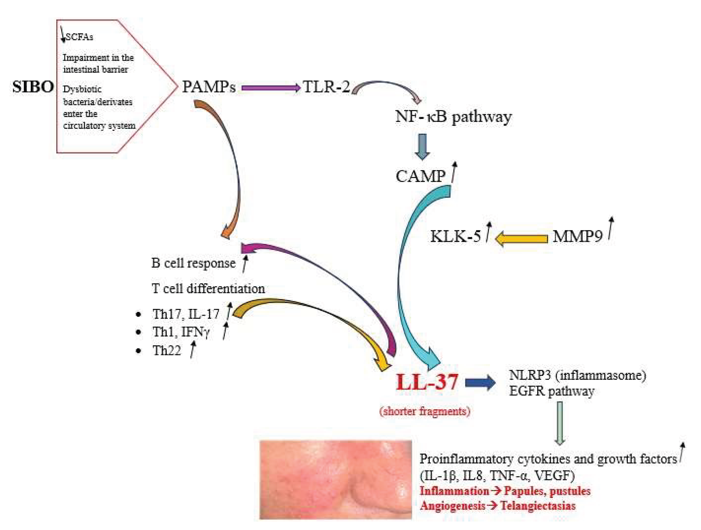
Rosacea is a chronic skin condition, characterized by persistent inflammation, manifesting primarily on the face and causing redness, papules, pustules, and phymatous changes. The etiology of rosacea is multifactorial, with immune system factors playing a crucial role in its pathogenesis. The scientific literature contains an increasing number of studies that suggest a correlation between rosacea and the gut microbiota. Small intestinal bacterial overgrowth (SIBO) is defined as an excessive proliferation of potentially pathogenic bacteria within the small intestine of the gastrointestinal system. Multiple factors have been posited to explain the pathogenesis of rosacea, and the presence of SIBO has been identified as a potential factor in its occurrence. A decrease in the Lactobacillus genus, Prevotella copri, Lachnospiraceae, and Faecalibacterium within the gut microbiota may initiate inflammation related to rosacea. These bacterial species are crucial for regulating the intestinal mucosa. The findings indicate that there is an increase of Bacteriodes, Acidaminococcus and Megasphaera, and Ruminococcus in the gut microbiome of patients with rosacea. Probiotics can be advantageous for managing the intestinal microbiome, while Rifaximin treatment has shown efficacy in addressing inflammatory rosacea lesions associated related to SIBO. The present review has been undertaken with the objective of enhancing our comprehension of SIBO in rosacea. The emphasis has been placed on the pathogenetic mechanisms and the shift in the gut microbiota that will lead to understanding probiotic benefits and therapy options in rosacea patients.

Keywords: 
;  ;  ;  ;  

1. Introduction

Rosacea, a prevalent dermatological condition, is estimated to influence between 1% and 20% of the global population [1]. The clinical characteristics of rosacea are typically characterized by recurring flare-ups of flushing, persistent redness, telangiectasia, papulo-pustules, or phymatous changes. Additionally, there is a multiphasic phenotype spectrum of disease, accompanied by burning, pain, or migraine-like symptoms [2]. The clinical classification of rosacea encompasses the following subtypes: erythematotelangiectatic rosacea (ETR), papulopustular rosacea (PPR), phymatous rosacea, and ocular rosacea. A recent clinical subtype known as neurogenic rosacea has also been documented [3]. Etiology is complex and multifactorial, with potential contributors including microbiological, neurovascular, immune, ultraviolet radiation, genetic, dietary, and psychological stress factors that may collectively contribute to the inflammatory process [4]. Due to the multiple etiological factors; there is considerable heterogeneity in the results of therapeutic interventions for rosacea. The primary approach to managing rosacea involves patient education, the establishment of a skin care routine, treatment with topical and oral medications to control flare-ups, and the use of lasers or light-based therapies. It is imperative that treatment be adapted to the individual’s specific needs, and a combination of various therapeutic modalities is frequently necessary to address the diverse manifestations of rosacea [5].
The significance of the skin-gut axis is becoming increasingly evident with the advancement of our understanding of the underlying pathophysiological mechanisms. Although the precise mechanisms of the gut-skin axis have yet to be fully elucidated, mounting evidence indicates that the probiotic intervention may confer benefits in the context of inflammatory cutaneous conditions [6]. Rosacea, as an immune-mediated inflammatory disease, has exhibited notable associations with small intestinal bacterial overgrowth (SIBO), a condition involving an imbalance in the composition of the gut microbiota, suggesting a potential underlying mechanism [7].
This narrative review explores the correlation between rosacea and SIBO, offering a theoretical framework for understanding how SIBO could have a potential effect in the immune mechanism affecting rosacea pathogenesis.

2. Small Intestinal Bacterial Overgrowth

The gut microbiota performs a pivotal and advantageous function in maintaining the normal physiological mechanisms of the human organism. Additionally, the microbiota provides protective benefits against pathogens through several mechanisms, including direct barrier functions, the secretion of antimicrobial peptides, and the stimulation of immune signalling pathways. Dysregulation of the microbiome, whether due to shifts in its location or composition, can have detrimental consequences on overall health [8,9]. Gut commensally bacteria have been shown to function as modulators in the mechanisms that underpin immune toleration [10]. Alterations in the structure of the gut microbiota may result in immunological imbalances in the organ systems external to the gastrointestinal system because almost 70% of lymphocytes are present in the lymphoid tissue of the gut [11].
SIBO is a medical phenomenon defined as the existence of elevated bacterial loads in the small intestine, resulting in gastrointestinal manifestations [12]. While the predominant localization of the gut microbiota is in the large and distal small intestine, there is a possibility of their infiltration into the mid and proximal small intestine, which can result and lead to the development of SIBO8. Predominant etiological factor of SIBO is the presence of dysmotility along the gastrointestinal system. The migrating motor complex is a contributing factor to the elimination of fecal matter within the gastrointestinal canal. It has been observed in a range of anatomical intestinal abnormalities or conditions, including intestinal obstructions, diabetic gastroparesis, diverticulosis, and scleroderma [13]. A shift in microbial composition or abundance, referred to as gut dysbiosis, has the potential to disrupt the body’s normal homeostasis. The condition known as dysbiosis has been linked to a number of intestinal and extraintestinal pathologies, including SIBO [9]. It is possible for dysbiosis to cause weakening of the mucosa of the intestines, as well as dysfunctions in the intestinal epithels and cellular junctions. This can lead to compromised intestinal barrier integrity and facilitate the migration of bacteria, noxious bacterial metabolites and compounds. In addition, bacterial components capable of trigger the immune response within the gastrointestinal tract to the systemic circulation [14].
The standard diagnostic approach for SIBO involves the administration of a lactulose or glucose breath test, a non-invasive and cost-effective method [8]. The definitive prevalence in the overall demographic remains indeterminate; though, extant researches suggest a range of 2–22%, classifying it as a reasonably prevalent ailment. In addition, there is an observable rise in the prevalence of the condition with increasing age, particularly among populations afflicted with comorbidities [15]. Rifaximin has emerged as a preferred treatment among clinicians for the management of SIBO [8].

3. Pathogenesis of Rosacea and Small Intestinal Bacteria Overgrowth

In rosacea etiology, both innate and adaptive immunity have been demonstrated to exert influence. Toll-like receptor (TLR)-2 is a pattern recognition receptor that can identify molecular patterns present in microbial compounds, thereby activating immune systems as a defense mechanism [16]. Elevated levels of TLR-2, Kallikrein-5 (KLK-5), cathelicidin, and matrix metalloproteinases (MMPs) expressions have been observed in the patients with rosacea [17]. Stimulation of TLR-2 results in the activation of the nuclear factor-kappa B (NF-κB) pathway, which leads to the synthesis of chemokines, cytokines and antimicrobial peptides (AMPs) in the proinflammatory cathelicidin pathway [18]. Moreover, activation of TLR-2 leads to the production of active human cathelicidine LL-37 via triggering KLK-5 which is a serine protease that cleaves CAMP into active fragments, including LL-37 [19,20]. KLK5 is expressed in an inactive form that is subsequently converted to an active form through cleavage performed by matrix metalloproteases (MMPs), especially MMP-9 [6].
The increased levels of CAMP and KLK5 can augment the levels of the shorter LL-37 fragment forms in rosacea skin [21,22]. These shorter LL-37 fragments have been demonstrated to induce symptoms consistent with rosacea, including erythema, vasodilatation, flushing, and telangiectasia in mice [21]. Furthermore, the activation of the nucleotide-binding oligomerization domain like receptors family pyrin domain containing 3 (NLRP3) inflammasome, has been determined to play a crucial role in LL-37-induced skin inflammation and rosacea pathogenesis [23]. The inflammasome has been defined as a multiprotein complex that is responsible for the stimulation of caspase-1 and activation of IL-1β. This leads to the activation of the IL-1 receptor in multiple cells and the infiltration of neutrophils [24].
Increased levels of LL-37 have been shown to induce the release of pro-inflammatory cytokines by neutrophils, including interleukin (IL)-8, IL-1β, and tumor necrosis factor (TNF)-α. Furthermore, LL-37 has been observed to stimulate the synthesis of vascular endothelial growth factor and activate the epidermal growth factor receptor signaling pathway [19].
In rosacea, T-cell immune responses are initiated through the actions of Th1 and Th17 cells, leading to elevated levels of interferon γ (IFN-γ) and IL-17 [6]. Th17 cells have been observed to produce IL-17, which may play a role in the effects on LL-37 through the expression of atypical forms that are characteristic of rosacea. IL-17 has been shown to induce angiogenesis through the VEGF pathway and to regulate the expression of LL-37 [25,26].
TLR activation is imperative for specific antigen-specific antibody responses in B cells, and TLR agonists have been shown to promote the proliferation of plasma cells from B cells [27]. Cleaved fragments of LL-37 robust B cell immune response accompanied by increased expression of Th17 and Th22 cytokines [6]. All these processes could be explained as contributing factors to the chronic inflammatory process and enhanced angiogenic growth that are characteristic for rosacea.
Pathogen-associated molecular patterns (PAMPs) are defined as biological stimulants that elicit a response in TLRs, including TLR-2 [28]. Short-chain fatty acids (SCFAs) have been demonstrated to facilitate the maintenance of intestinal barrier integrity. These acids are produced by healthy gut microbiota from complex polysaccharides [29]. The significance of SCFAs extends beyond their role in diminishing intestinal permeability, as they also modulate skin barrier integrity by inducing keratinocyte metabolism and differentiation [30]. Butyrate is a short-chain fatty acid and demonstrates a robust anti-inflammatory effect by suppressing immune reactions, via inhibiting cytokine production by inflammatory cells [31]. The presence of elevated PAMPs in the circulation system, along with a decline in butyrate of bacterial origin which is a protective factor for intestinal barrier, might suggest an increased responsiveness of B-cells and impairment in the differentiation of T-cells [32]. Despite the absence of evidence demonstrating the full complement of innate and adaptive pathways in the existing studies, the hypothesis that SIBO may induce analogous pathways is a plausible one. This is due to the fact that dysbiotic bacteria instigate similar pathways in both the gastrointestinal tract and the skin (Figure 1).

4. Preventive Effects of Probiotics on Rosacea

In a study employing a mouse model of rosacea, effectiveness of probiotics in addressing rosacea were evaluated by conducting an LL-37-induced model of the skin condition. The mixture of. Lacticaseibacillus salivarius 23-006 and Lacticaseibacillus paracasei 23-008 exhibited the utmost significance in its effect, thereby alleviating dermatological manifestations, reducing the presence of inflammatory cellular infiltrates, and decreasing the levels of inflammatory mediators in mice. The concomitant administration of these two strains resulted in a reduction in the synthesis of cathelicidin LL-37 and rosacea-associated factors. This effect was achieved by inhibiting the TLR2/MyD88/NF-κB pathway [33], which is responsible for the synthesis of chemokines and AMPs, as well as cytokines, within the proinflammatory cathelicidin cascade [18]. Another study involved 60 patients diagnosed with rosacea, who were randomly divided into three groups: probiotic, placebo, and control. The results demonstrated that the administration of probiotics resulted in improvements to facial skin conditions, a reduction in inflammation, and a decrease in facial skin microbiota diversity, concurrent with an enhancement of gut microbiota heterogeneity [34]. A multitude of studies and reports in the literature demonstrate the efficacy of probiotics in the treatment of rosacea and related skin disorders by virtue of their ability to inhibit inflammation and restore the skin barrier [35,36,37,38]. The present findings provide a theoretical base for the treatment of rosacea, thus offering a promising avenue for potential clinical applications.

5. The Intestinal Microbiota in Rosacea Patients

The studies presented herein demonstrate alterations in the intestinal microbiota that may be of particular relevance to patients diagnosed with rosacea (Table 1).
In a study, the gut microbiome of 15 patients diagnosed with PPR was compared with 15 healthy controls. Twelve patients diagnosed with rosacea were female and had a mean age of 36 years; 13 subjects who served as controls were also female and had an average age of 39 years. The results obtained via canonical correspondence analysis were found to be significantly different between the patients and controls. The implementation of a linear discriminant analysis effect size (LEfSe) analysis has led to the identification of an increase in the Syntrophomonadaceae family, Anaerovorax genus, Bacteroidales sp., Tyzzerella sp., Lachnospiraceaefamily, Akkermansiamuciniphila and Parabacteroides distasonis. It was determined that these bacteria were characterized as compositional elements indicative of PPR patients. The LEfSe analysis indicated a decline in the prevalence of Prevotella copri. The study delineates the intestinal microbiological profile of patients afflicted with inflammatory rosacea, thereby substantiating the notion of intestinal dysbiosis [39]. In a research study, the intestinal microbiota of 11 rosacea patients was compared with the microbiota of 110 age- and sex-matched individuals who were considered to be healthy. More than 90% of patients in both groups are female. %90.9 of patients diagnosed with rosacea were female, with a mean age of 49.9 ±11.3 years. Furthermore, 4 of the patients (36.3%) had ETR and 7 of them (63.7%) had PPR. A decline in abundance, though not in uniqueness of bacteria diversity, was demonstrated in rosacea patients. The incorporation of additional variables, including alcohol consumption, tea or yogurt intake, tobacco use, exercise habits, vegetarian diet, and rosacea subtype revealed no significant impact on the structure of the gut microbiota structure, as indicated by PCoA analysis. A comparative analysis of the gut microbiota revealed that both groups exhibited a predominance of Bacteroidetes, Firmicutes, and Proteobacteria. Nevertheless, a notable distinction was identified in the rosacea subjects, who exhibited an increased prevalence of Bacteroides and Fusobacterium, and a reduced prevalence of Prevotella and Sutterella, in contrast to the control group. The LEfSe analysis revealed substantial changes in the composition of the intestinal microbiome in patients with rosacea, characterized by increased abundance of Rhabdochlamydia, Bifidobacterium, Sarcina, and Ruminococcus, as well as reduction in levels of Lactobacillus, Megasphaera, Acidaminococcus, Haemophilus, Roseburia, Clostridium, and Citrobacter. Rosacea patients exhibit distinctive characteristics in their fecal microbiota, which may be associated with sulphur metabolism, cobalamin, and carbohydrate transport [40]. In a separate study, the relationship between the gut microbiota of 12 female rosacea patients and 251 female healthy controls were evaluated. Rosacea patients exhibited a range of subtypes, including 50% ETR, 17% PPR, and other subtypes. The study revealed no statistically significant disparities in diversity metrics between subjects with rosacea and those without rosacea, suggesting that the presence of this skin condition does not significantly impact biodiversity levels. Additionally, the study indicated that patients with rosacea exhibited distinct compositions of enteral microbiota, yet these compositions were comparable in abundance to control group. Nevertheless, substantial disparities were identified at the level of genera. Using metagenomeSeq for comparison of differentials in the subjects, there was a notable abundance of Acidaminococcus and Megasphaera, in contrast to the relative paucity of Peptococcaceae family, unknown genus, and Methanobrevibacter were observed in the patients in comparison with the control group [41]. A recent study examined the disparities in fecal microbial profiles, as analyzed with MiSeq 16S rRNA sequencing, among patients diagnosed with 54 cases of rosacea compared to 50 healthy controls. A decrease in microbial richness and diversity was identified in patients with rosacea in comparison with the control group. In rosacea, a significant decrease was observed in the levels of Faecalibacteriumprausnitzii, Lachnoospiraceae ND 3007 group sp, and Ruminococcaceae. Conversely, Oscillobacter sp., Flavonifractorplautii, and Ruminococccaceae UBA 1819 exhibited a marked increase in their abundance in cases of rosacea when compared to the control group. The present study found no statistically significant associations between the clinical severity levels of rosacea or the existence of gastrointestinal symptoms and intestinal microbiome characteristics [42].
Another study was conducted to ascertain the correlation between the gut microbiome and rosacea. The study did not include a control group, and it did not examine how the levels of intestinal microbiota species changed. In this study, a two-sample Mendelian randomization (MR) design was employed and data derived from the Genome-Wide Association Study focused on gut microbiota and the FinnGen biobank for rosacea. The study identified and analyzed 2078 single nucleotide polymorphisms (SNPs) that are linked to the gut microbiota. The study has indicated that two bacterial genera, Actinomyces and Butyrivibrio, may play a pivotal role in preventing the development of rosacea. The results of the MR analysis indicated the absence of pleiotropy, with a uniform distribution exhibited across a selected set of SNPs. Tests of directionality indicated a substantial causative pathway from gut microbiota to rosacea. However, the study’s findings did not differentiate among the subtypes of rosacea [43].

6. Discussion

The findings of the studies indicate that there is a substantial change in the gut microbiome in patients with rosacea, which is related to the inflammatory process of the skin. The study conducted by Chen et al. revealed a decline in the prevalence of the Lactobacillus genus. This decline may potentially contribute to the development of dysbiosis in patients with rosacea [40], which is compatible with the findings demonstrated the capacity of Lactobacillus Paracasei CNCMI-2116 (ST11) to impede substance P-induced skin inflammation. Furthermore, these bacteria were demonstrated to enhance the skin barrier recovery, thereby contributing to a reduction in skin irritability [35]. Moreover, Lactobacilli are known to play a vital role in defending against pathogenic microorganisms, modulating inflammation, managing gut flora, and preventing bacterial infections [44].
Morena et al.’s study demonstrated a decline in the levels of Prevotellacopri [39], has been shown to have a protective effect on the mucosal barrier and to reduce the likelihood of inflammation by producing SCFAs through the process of fiber metabolism [45]. In addition, Guertler et al.’s study revealed a decline in the abundance of Lachnospiraceae and Faecalibacterium [42]. These bacteria have been identified as key contributors to the production of butyrate, a short-chain fatty acid with recognized anti-inflammatory properties [46].
The findings indicate that there is an increase of Bacteriodes [39], Acidaminococcus and Megasphaera [41] and Ruminococcus [42] in the gut microbiome of patients with rosacea and it is plausible that these results may serve as a rationale for the development of inflammation in these patients. Accordingly, the substantial presence of the genera Bacteroides, Ruminococcus, Acidaminococcus, and Megasphaera in the gut microbiome was found to be significantly associated with the disease-related group [46]. These findings may suggest that there is an association between elevated levels of intestinal inflammation and rosacea. Therefore, it would be beneficial to define SIBO as an underlying factor. In addition to analyzing fecal microbiota, the diagnosis of SIBO can be facilitated by a lactulose or glucose breath test. These methods are particularly advantageous in cases of rosacea that exhibit inflammatory characteristics associated with the condition.
The administration of rifaximin could be recommended as a potential therapeutic intervention for the diagnosis of SIBO in rosacea patients, as evidenced by the findings of the conducted studies [8,47,48]. Given the documented increase in the prevalence of SIBO in patients with PPR compared to those with ETR [47], PPR can benefit from anti-inflammatory properties of probiotics, contingent upon the inflammatory etiology of the disease. The potential of probiotics in managing rosacea as the oral use of probiotics, such as Escherichia coli Nissle, a mixture of Bifidobacterium strains and a mixture of Bifidobacterium and Lactobacillus in combination with standard therapies of rosacea, resulted in more frequent clinical remission, substantial symptom improvement, and a reduction in relapses lasting up to six months after the initiation of therapy [49].
The present review has limitations due to the restricted number of studies that have been conducted on the microbial flora of patients with rosacea. However, the study’s strengths lie in its presentation of the increased potential for alterations in the gut microbiota in individuals diagnosed with rosacea.

7. Conclusion

The identification and management of an underlying SIBO in patients diagnosed with rosacea may offer a therapeutic avenue for reducing the frequency of rosacea flare-ups. Additionally, probiotics play a pivotal role in the regulation of gut flora thus highlighting their significant importance in the management of rosacea symptoms. Consequently, future studies should encompass large cohort and control studies that delineate the subtypes of bacteria before and after probiotic utilization in rosacea patients.

Author Contributions

Conceptualization, S.M.; methodology, S.M.; formal analysis, S.M.; investigation, S.M.; resources, S.M.; data curation, S.M.; writing—original draft preparation, S.M.; writing—review and editing, S.M.; visualization, S.M.; supervision, S.M. “All authors have read and agreed to the published version of the manuscript.”.

Funding

This research received no external funding.

Data Availability Statement

Data could be found in the “References”.

Conflicts of Interest

The authors declare no conflicts of interest.

References

  1. Gether, L.; Overgaard, L.K.; Egeberg. A.; Thyssen, J.P. Incidence and prevalence of rosacea: a systematic review and meta-analysis. Br J Dermatol 2018, 179, 282-289. [CrossRef]
  2. Sticchi, A.; Fiorito, F.; Kaleci, S.; Paganelli, A.; Manfredini, M.; Longo, C. Rosacea and treatment with retinoids: a systematic review and meta-analysis. Ther Adv Chronic Dis 2025, 16, 20406223251339964. [CrossRef]
  3. Ivanic, M.G.; Oulee, A.; Norden, A.; Javadi, S.S.; Gold, M.H.; Wu, J.J. Neurogenic Rosacea Treatment: A Literature Review. J Drugs Dermatol 2023, 22, 566-575. [CrossRef]
  4. Maden S. Rosacea: An Overview of Its Etiological Factors, Pathogenesis, Classification and Therapy Options. Dermato 2023, 3, 241-262.
  5. Sharma, A.; Kroumpouzos, G.; Kassir, M.; Galadari, H.; Goren, A.; Grabbe, S; Goldust M. Rosacea management: A comprehensive review. J Cosmet Dermatol 2022, 21, 1895-1904.
  6. Sánchez-Pellicer, P.; Eguren-Michelena, C.; García-Gavín, J.; Llamas-Velasco, M.; Navarro-Moratalla, L.; Núñez-Delegido, E.; Agüera-Santos, J.; Navarro-López, V. Rosacea, microbiome and probiotics: the gut-skin axis. Front Microbiol 2024, 14, 1323644. [CrossRef]
  7. Wang, F.Y.; Chi, C.C. Rosacea, Germs, and Bowels: A Review on Gastrointestinal Comorbidities and Gut-Skin Axis of Rosacea. Adv Ther 2021, 38, 1415-1424. [CrossRef]
  8. Achufusi, T.G.O.; Sharma, A.; Zamora, E.A.; Manocha, D. Small Intestinal Bacterial Overgrowth: Comprehensive Review of Diagnosis, Prevention, and Treatment Methods. Cureus 2020, 12, e8860.
  9. Zafar, H.; Jimenez, B.; Schneider, A. Small intestinal bacterial overgrowth: current update. Curr Opin Gastroenterol 2023, 39, 522-528. [CrossRef]
  10. Sachdev, A.H.; Pimentel, M. Gastrointestinal bacterial overgrowth: pathogenesis and clinical significance. Ther Adv Chronic Dis 2013, 4, 223-231. [CrossRef]
  11. Skrzydło-Radomańska, B.; Cukrowska, B. How to Recognize and Treat Small Intestinal Bacterial Overgrowth? J Clin Med 2022, 11, 6017. [CrossRef]
  12. Pimentel, M.; Saad, R.J.; Long, M.D.; Rao, S.S.C. ACG Clinical Guideline: Small Intestinal Bacterial Overgrowth. Am J Gastroenterol 2020, 115, 165-178. [CrossRef]
  13. Belkaid, Y.; Naik, S. Compartmentalized and systemic control of tissue immunity by commensals. Nat Immunol 2013, 14, 646-653. [CrossRef]
  14. Takiishi, T.; Fenero, C.I.M.; Câmara, N.O.S. Intestinal barrier and gut microbiota: Shaping our immune responses throughout life. Tissue Barriers 2017, 5, e1373208. [CrossRef]
  15. Kinashi, Y.; Hase, K. Partners in Leaky Gut Syndrome: Intestinal Dysbiosis and Autoimmunity. Front Immunol 2021, 12, 673708. [CrossRef]
  16. Oliveira-Nascimento, L.; Massari, P.; Wetzler, L.M. The role of TLR2 in infection and immunity. Front Immunol 2012, 3, 79. [CrossRef]
  17. Ahn, C.S.; Huang, W.W. Rosacea pathogenesis. Dermatol Clin 2018, 36, 81–86.
  18. Yamasaki, K.; Kanada, K.; Macleod, D.T.; Borkowski, A.W.; Morizane, S.; Nakatsuji, T.; Cogen, A.L.; Gallo, R.L. TLR2 expression is increased in rosacea and stimulates enhanced serine protease production by keratinocytes. J Invest Dermatol 2011, 131, 688-97. [CrossRef]
  19. Geng, R.S.Q.; Bourkas, A.N.; Mufti, A.; Sibbald, R.G. Rosacea: Pathogenesis and Therapeutic Correlates. J Cutan Med Surg 2024, 28, 178-189. [CrossRef]
  20. Meyer-Hoffert, U.; Schroder, J.M. Epidermal proteases in the pathogenesis of rosacea. J Investig Dermatol Symp Proc 2011, 15, 16-23.
  21. Yamasaki, K.; Di Nardo, A.; Bardan, A.; Murakami, M.; Ohtake, T.; Coda, A.; Dorschner, R.A.; Bonnart, C.; Descargues, P.; Hovnanian. A.; Morhenn, V.B.; Gallo, R.L. Increased serine protease activity and cathelicidin promotes skin inflammation in rosacea. Nat Med 2007, 13, 975-980.
  22. Muto, Y.; Wang, Z.; Vanderberghe, M.; Two, A.; Gallo, R.L.; Di Nardo, A. Mast cells are key mediators of cathelicidin-initiated skin inflammation in rosacea. J Invest Dermatol 2014, 134, 2728-2736. [CrossRef]
  23. Yoon, S.H.; Hwang, I.; Lee, E.; Cho, H.J.; Ryu, J.H.; Kim, T.G.; Yu, J.W. Antimicrobial Peptide LL-37 Drives Rosacea-Like Skin Inflammation in an NLRP3-Dependent Manner. J Invest Dermatol 2021, 141, 2885-2894.e5. [CrossRef]
  24. Tang, L.; Zhou, F. Inflammasomes in Common Immune-Related Skin Diseases. Front Immunol 2020, 11, 882.
  25. Gerber, P.A.; Buhren, B.A.; Steinhoff, M.; Homey, B. Rosacea: The cytokine and chemokine network. J Investig Dermatol Symp Proc 2011, 15, 40-47. [CrossRef]
  26. Sakabe, J.; Umayahara, T.; Hiroike, M.; Shimauchi, T.; Ito, T.; Tokura, Y. Calcipotriol increases hCAP18 mRNA expression but inhibits extracellular LL37 peptide production in IL-17/IL-22-stimulated normal human epidermal keratinocytes. Acta Derm Venereol. 2014 Sep;94(5):512-6. [CrossRef]
  27. Pasare, C.; Medzhitov, R. Control of B-cell responses by Toll-like receptors. Nature 2005, 438, 364–368.
  28. Kumar, V. Going, Toll-like receptors in skin inflammation and inflammatory diseases. EXCLI J 2021, 20, 52-79.
  29. Koh, A.; De Vadder, F.; Kovatcheva-Datchary, P.; Bäckhed, F. From Dietary Fiber to Host Physiology: Short-Chain Fatty Acids as Key Bacterial Metabolites. Cell 2016, 165, 1332-1345. [CrossRef]
  30. Trompette ,A.; Pernot, J.; Perdijk, O.; Alqahtani, R.A.A.; Domingo, J.S.; Camacho-Muñoz, D.; Wong, N.C.; Kendall, A.C.; Wiederkehr, A.; Nicod, L.P.; Nicolaou, A.; von Garnier, C.; Ubags, N.D.J.; Marsland, B.J. Gut-derived short-chain fatty acids modulate skin barrier integrity by promoting keratinocyte metabolism and differentiation. Mucosal Immunol 2022, 15, 908-926. [CrossRef]
  31. Salem, I.; Ramser, A.; Isham, N.; Ghannoum, M.A. The Gut Microbiome as a Major Regulator of the Gut-Skin Axis. Front Microbiol 2018, 9, 1459.
  32. Mahmud, M.R.; Akter, S.; Tamanna, S.K.; Mazumder, L.; Esti, I.Z.; Banerjee, S.; Akter, S.; Hasan, M.R.; Acharjee, M.; Hossain, M.S.; Pirttilä, A.M. Impact of gut microbiome on skin health: gut-skin axis observed through the lenses of therapeutics and skin diseases. Gut Microbes 2022, 14, 2096995.
  33. Qi, X.; Xiao, Y.; Zhang, X.; Zhu, Z.; Zhang, H.; Wei, J.; Zhao, Z.; Li, J.; Chen, T. Probiotics suppress LL37 generated rosacea-like skin inflammation by modulating the TLR2/MyD88/NF-κB signaling pathway. Food Funct 2024, 15, 8916-8934.
  34. Yu, J.; Duan, Y.; Zhang, M.; Li, Q.; Cao, M.; Song, W.; Zhao, F.; Kwok, L-Y.; Zhang, H.; Li, R.; Sun, Z. Effect of combined probiotics and doxycycline therapy on the gut-skin axis in rosacea. mSystems 2024, 9, e0120124. [CrossRef]
  35. Gueniche, A.; Benyacoub, J.; Philippe, D.; Bastien, P.; Kusy, N.; Breton, L.; Blum, S.; Castiel-Higounenc, I. Lactobacillus paracasei CNCM I-2116 (ST11) inhibits substance P-induced skin inflammation and accelerates skin barrier function recovery in vitro. Eur J Dermatol 2010, 20, 731-737. [CrossRef]
  36. Fortuna, M.C.; Garelli, V.; Pranteda, G.; Romaniello, F.; Cardone, M.; Carlesimo, M.; Rossi, A. A case of Scalp Rosacea treated with low dose doxycycline and probiotic therapy and literature review on therapeutic options. Dermatol Ther 2016, 29, 249-251. [CrossRef]
  37. Manzhalii, E.; Hornuss, D.; Stremmel, W. Intestinal-borne dermatoses significantly improved by oral application of Escherichia coli Nissle 1917. World J Gastroenterol 2016, 22, 5415-5421.
  38. Pinchuk, I.V.; Bressollier, P.; Verneuil, B.; Fenet, B.; Sorokulova, I.B.; Mégraud, F.; Urdaci, M.C. In vitro anti-Helicobacter pylori activity of the probiotic strain Bacillus subtilis 3 is due to secretion of antibiotics. Antimicrob Agents Chemother 2001, 45, 3156-3161. [CrossRef]
  39. Moreno-Arrones, O.M.; Ortega-Quijano, D.; Perez-Brocal, V.; Fernandez-Nieto, D.; Jimenez, N.; de Las Heras, E.; Moya, A.; Perez-Garcia, B. Dysbiotic gut microbiota in patients with inflammatory rosacea: another clue towards the existence of a brain-gut-skin axis. Br J Dermatol 2021, 185, 655-657. [CrossRef]
  40. Chen, Y.J.; Lee, W.H.; Ho, H.J.; Tseng, C.H.; Wu, C.Y. An altered fecal microbial profiling in rosacea patients compared to matched controls. J Formos Med Assoc 2021, 120, 256-264. [CrossRef]
  41. Nam, J.H.; Yun, Y.; Kim, H.S.; Kim, H.N.; Jung, H.J.; Chang, Y.; Ryu, S.; Shin, H.; Kim, H.L.; Kim, W.S. Rosacea and its association with enteral microbiota in Korean females. Exp Dermatol 2018, 27, 37-42. [CrossRef]
  42. Guertler, A.; Hering, P.; Pacífico, C.; Gasche, N.; Sladek, B.; Irimi, M.; French, L.E.; Clanner-Engelshofen, B.M.; Reinholz, M. Characteristics of Gut Microbiota in Rosacea Patients-A Cross-Sectional, Controlled Pilot Study. Life (Basel) 2024, 14, 585.
  43. Li, J.; Yang, F.; Liu, Y.; Jiang, X. Causal relationship between gut microbiota and rosacea: a two-sample Mendelian randomization study. Front Med (Lausanne) 2024, 11, 1322685. [CrossRef]
  44. Shah, A.B.; Baiseitova, A.; Zahoor, M.; Ahmad, I.; Ikram, M.; Bakhsh, A.; Shah, M.A.; Ali, I.; Idress, M.; Ullah, R.; Nasr, F.A.; Al-Zharani, M. Probiotic significance of Lactobacillus strains: a comprehensive review on health impacts, research gaps, and future prospects. Gut Microbes 2024, 16, 2431643.
  45. Bedarf, J.R.; Hildebrand, F.; Coelho, L.P.; Sunagawa, S.; Bahram, M.; Goeser, F.; Bork, P.; Wüllner, U. Functional implications of microbial and viral gut metagenome changes in early-stage L-DOPA-naïve Parkinson’s disease patients. Genome Med 2017, 9, 39.
  46. Manor, O.; Dai, C.L.; Kornilov, S.A.; Smith, B.; Price, N.D.; Lovejoy, J.C.; Gibbons, S.M.; Magis, A.T. Health and disease markers correlate with gut microbiome composition across thousands of people. Nat Commun 2020, 11, 5206.
  47. Parodi, A.; Paolino, S.; Greco, A.; Drago, F.; Mansi, C.; Rebora, A.; Parodi, A.; Savarino, V. Small intestinal bacterial overgrowth in rosacea: clinical effectiveness of its eradication. Clin Gastroenterol Hepatol 2008, 6, 759-764.
  48. Drago, F.; De Col, E.; Agnoletti, A.F.; Schiavetti, I.; Savarino, V.; Rebora, A.; Paolino, S.; Cozzani, E.; Parodi, A. The role of small intestinal bacterial overgrowth in rosacea: A 3-year follow-up. J Am Acad Dermatol 2016, 75, e113-5.
  49. Manfredini, M.; Barbieri, M.; Milandri, M.; Longo, C. Probiotics and Diet in Rosacea: Current Evidence and Future Perspectives. Biomolecules 2025, 15, 411. [CrossRef]
Figure 1. Possible mechanism of small intestinal bacterial overgrowth contributes to the pathogenesis of rosacea. SIBO: small intestinal bacterial overgrowth, SCFAs: short chain fatty acids, PAMP: pathogen-associated molecular patterns, TLR-2: toll-like receptor-2, NF-Κb: nuclear factor-kappa B, CAMP:cathelicidin antimicrobial peptide, KLK-5:kallikrein-5, MMP9: matrix metalloproteinase-9, LL-37: human cathelicidin, NLRP3: nucleotide-binding oligomerization domain like receptors family pyrin domain containing 3, EGFR: epidermal growth factor receptor;IL, interleukin, TNF-α: tumour necrosis factor, VEGF: vascular endothelial growth factor, Th: T-helper, IFN-γ: interferon γ.
Preprints 191904 g001
Table 1. Intestinal microbiota of patients with rosacea.
Authors and year Study design Increased levels of intestinal microbiota Decreased levels of intestinal microbiota Results
Moreno-Arrones et al. [39] 2021 15 PPR patients
12 (80%) females
15 controls
5 (33.3%) females
LEfSe analysis
Syntrophomonadaceae family,
Anaerovorax genus, Bacteroidales sp.,
Tyzzerella sp.,
Lachnospiraceae
family,
Akkermansiamuciniphila, Parabacteroides distasonis
Prevotella copri The study outlines intestinal dysbiosis in rosacea patients.
Chen et al. [40] 2021 11 patients
4 ETR %36.3
7 PPR %63.7
90.9% female
110 controls
90.9% female
LEfSe analysis
Rhabdochlamydia, CF231, Bifidobacterium,
Sarcina,
Ruminococcus
Lactobacillus,
Megasphaera, Acidaminococcus, Haemophilus,
Roseburia,
Clostridium,
Citrobacter
Faecal microbiota profiles in rosacea patients associated with sulfur metabolism, cobalamin, and carbohydrate transport.
Nam et al. [41] 2018 12 patients
251 controls
MetagenomeSeq
Acidaminococcus, Megasphaera, Peptococcaceae, Methanobrevibacter,
Patients diagnosed with rosacea exhibit a more abundant and distinct profile of enteral microbiota.
Guertler et al. [42] 2024 54 patients
39 females 15 males
50 controls
MiSeq 16S rRNA
sequencing
Oscillobacter sp.,
Flavonifractorplauti,Ruminococccaceae UBA1819
Faecalibacteriumprausnitzii Lachnoospiraceae ND 3007 group sp, Ruminococcaceae There is a decline in microbial richness and diversity in rosacea patients.
PPR: papulopustular rosacea, ETR:erythematotelengiectatic rosacea, LEfSe:linear discriminant analysis effect size.
Disclaimer/Publisher’s Note: The statements, opinions and data contained in all publications are solely those of the individual author(s) and contributor(s) and not of MDPI and/or the editor(s). MDPI and/or the editor(s) disclaim responsibility for any injury to people or property resulting from any ideas, methods, instructions or products referred to in the content.
Copyright: This open access article is published under a Creative Commons CC BY 4.0 license, which permit the free download, distribution, and reuse, provided that the author and preprint are cited in any reuse.
Prerpints.org logo

Preprints.org is a free preprint server supported by MDPI in Basel, Switzerland.

Subscribe

Disclaimer

Terms of Use

Privacy Policy

Privacy Settings

© 2026 MDPI (Basel, Switzerland) unless otherwise stated