Preprint
Article

This version is not peer-reviewed.

Metallothionein Is Inappropriate to Neutralise Excess Hepatic Copper in Wilson’s Disease

Submitted:

21 December 2025

Posted:

22 December 2025

You are already at the latest version

Abstract

Mutations in ATP7B impair biliary copper excretion and copper–ceruloplasmin release, causing hepatic copper overload in Wilson’s disease (WD). Metallothionein (MT), a key intracellular copper buffering agent, is overexpressed in WD liver. We evaluated the diagnostic significance of MT immunostaining in explanted livers from hepatic and neurological WD, including untreated cases and those receiving short- versus long-term chelation. Histochemical and immunohistochemical analyses were performed. Our results show that MT overexpression progresses to irreversible accumulation. Sustained copper-induced redox cycles promote MT polymerisation, aggregation, and resistance to proteolysis, accompanied by oxidative stress, lysosomal dysfunction, and hepatocellular injury. MT localises to bile canaliculus membranes, hepatocyte nuclei, lysosomes, and mesenchymal cells, suggesting discrete roles in disease mechanisms. Given the persistent MT accumulation despite prolonged chelation, we propose that MT fails to neutralise excess copper in WD; instead, polymerised MT traps Cu+, depleting the cytosolic copper pool destined to ATP7B-mediated ceruloplasmin loading, plasma secretion, and biliary export. Accordingly, MT dysregulation emerges as central to WD pathogenesis and supports MT immunohistochemistry as a sensitive diagnostic adjunct. These findings provide a rationale for developing more efficient chelators that mobilise hepatic copper stores without causing MT build-up, potentially improving outcomes.

Keywords: 
;  ;  ;  ;  

1. Introduction

The discovery of the ATP7B gene [1] has greatly advanced our understanding of intracellular copper trafficking and the regulation of copper homeostasis [2].
Mutations in ATP7B, either in homozygosity or compound heterozygosity, cause hepatic copper storage and disease [3,4]. Copper storage concurrent with ATP7B dysfunction is attributed to defective copper biliary excretion and defective incorporation of copper into ceruloplasmin (CP), which in turn is believed to result in low plasma levels of the protein [5].
Unfortunately, it remains difficult to link individual mutations to clinical manifestations and, although the underlying mechanisms are relatively well known, the precise modality by which copper is incorporated into CP is still debated [6]. Patients with WD require lifelong systemic chelation therapy, which is unsatisfactory in many cases.
The recent demonstration of metallothionein (MT) overexpression in liver tissue sections at any stage of the disease [7,8,9] led to the proposal of MT immunohistochemistry (MT-IHC) as a diagnostic tool comparable to genetic testing [8], and validated it as a highly sensitive screening test for the disease [9]. Thus far, MT has received more attention from structural biologists than from biomedical investigators. We therefore undertook a focused study to characterise the pathophysiologic and diagnostic meaning of the MT-IHC pattern in explanted WD livers, including neurological and hepatic WD, from untreated patients and from patients with short-term versus long-term copper chelation therapy.
In this article, we discuss the functional implications of novel pathological findings.

2. Materials and Methods

2.1. Patients

Demographic and clinical data are summarised in Table 1.

Patient 1 (Neurological Wilson’s Disease)

A 14-year-old Albanian female presented in May 2010 with neurological symptoms (asthenia, dysarthria, dysphagia, slowed speech) and thrombocytopenia. CP was 8.45 mg/dL (n.r. = > 20 mg/dL); 24-h cupruria was 580 µg/L (n.r.=170-700); a Kayser–Fleischer ring (KFR) was present. Sequential treatment with zinc and D-penicillamine was started without improvement. Liver transplantation was performed four months later.

Patient 2 (Cholangiocarcinoma Case)

A 34-year-old female was diagnosed with WD in July 1994. The original liver biopsy showed chronic active hepatitis with bridging fibrosis and steatosis. CP was 11 mg/dL (n.r. = > 20 mg/dL); 24-h cupruria was 1,100 µg/L (n.r. = 170-700). For several years, the patient had intention tremor of the upper limbs and gait instability. D-penicillamine was started immediately and interrupted in 2006 because of systemic lupus erythematosus (SLE), then immediately replaced with trientine, continuously until 2022. Two intermediate follow-up biopsies (1999 and 2003) showed a good response with progressive regression of elementary lesions. In 2013, brain MRI showed abnormalities at both globi pallidi. In 2020, abdominal ultrasound revealed a liver mass histologically diagnosed as cholangiocarcinoma (CCA). Chemotherapy and radiotherapy were ineffective, and liver transplantation was performed in 2022.

Patient 3 (Acute on Chronic Liver Failure)

A 49 years old Italian man with a known family history of WD was diagnosed at age 4 years when he was asymptomatic. CP was 10.5 µg/dL (n.r. = > 20; 24-h cupruria 1,013 µg/L (n.r.= 170-700). The patient was treated discontinuously with D-penicillamine for 42 years. Treatment was interrupted for two years in 2020 and restarted in 2022. Owing to decompensated cirrhosis, liver transplantation was performed in November 2023.

Patient 4 (Decompensated Cirrhosis)

A 9-year-old Italian female with a history of anisocoria at 6 months and speech therapy during school, presented with hepatosplenomegaly and oesophageal varices. CP was 0.7 mg/d (n.r. = > 20mg/dL; 24-h cupruria 1,015 µg/L n.r.= 170-700). No KFR. WD was diagnosed in February 2024. Treatment with D-penicillamine followed by zinc was started; under treatment, cupruria rose to 3,500 µg/L. Liver function deteriorated and liver transplantation was performed in March 2024.

Patient 5 (Decompensated Cirrhosis)

A 15-year-old girl born in Italy to non-consanguineous Romanian parents, presented in April 2023 with jaundice, raised transaminases, abnormal liver ultrasound, and oesophageal varices. CP was 0.16 mg/dL (n.r.= > 20 mg/dL); 24-h cupruria was 1,230 µg/L (n.r.= 170–700). Dry tissue copper content was 0.55 µg/mg (normal 0.02–0.06). No KFR. Zinc followed by D-penicillamine was started in November 2023; cupruria increased to 2,000 µg/L. Liver transplantation was performed in March 2024.

3. Methods

Specimens (2.5 × 2.5 cm) from each case were fixed in formalin and embedded in paraffin. Four-micron tick serial sections were stained with H&E, PAS, PAS–diastase (PAS-D), Masson, rhodanine, and orcein and, by immunohistochemistry, with the following antibodies: anti-metallothionein-1 (as previously described [8], cytokeratin 7 (CK7), and Hep-Par-1. The study was conducted in accordance with the Declaration of Helsinki and informed consent was obtained for the use of clinical data and for publishing this manuscript.

4. Results

Macroscopy

On macroscopic examination, four cases (n. 1,3,4 and 5) showed a fully established cirrhosis. The liver from case 2 was not cirrhotic. Discrete peculiarities were observed in cases 1 and 2.
Case 1 In addition to 2–3 cm sized nodules in both lobes, flat areas extending for 3 cm without nodularity were observed especially beneath the liver capsule. Samples were taken from both nodular and flat areas.
Case 2 On sectioning, the liver showed a stellate scar in the right lobe. The remaining tissue had a smooth, non-nodular surface. Samples were taken from tumor-peritumor and distant areas.

Histopathology

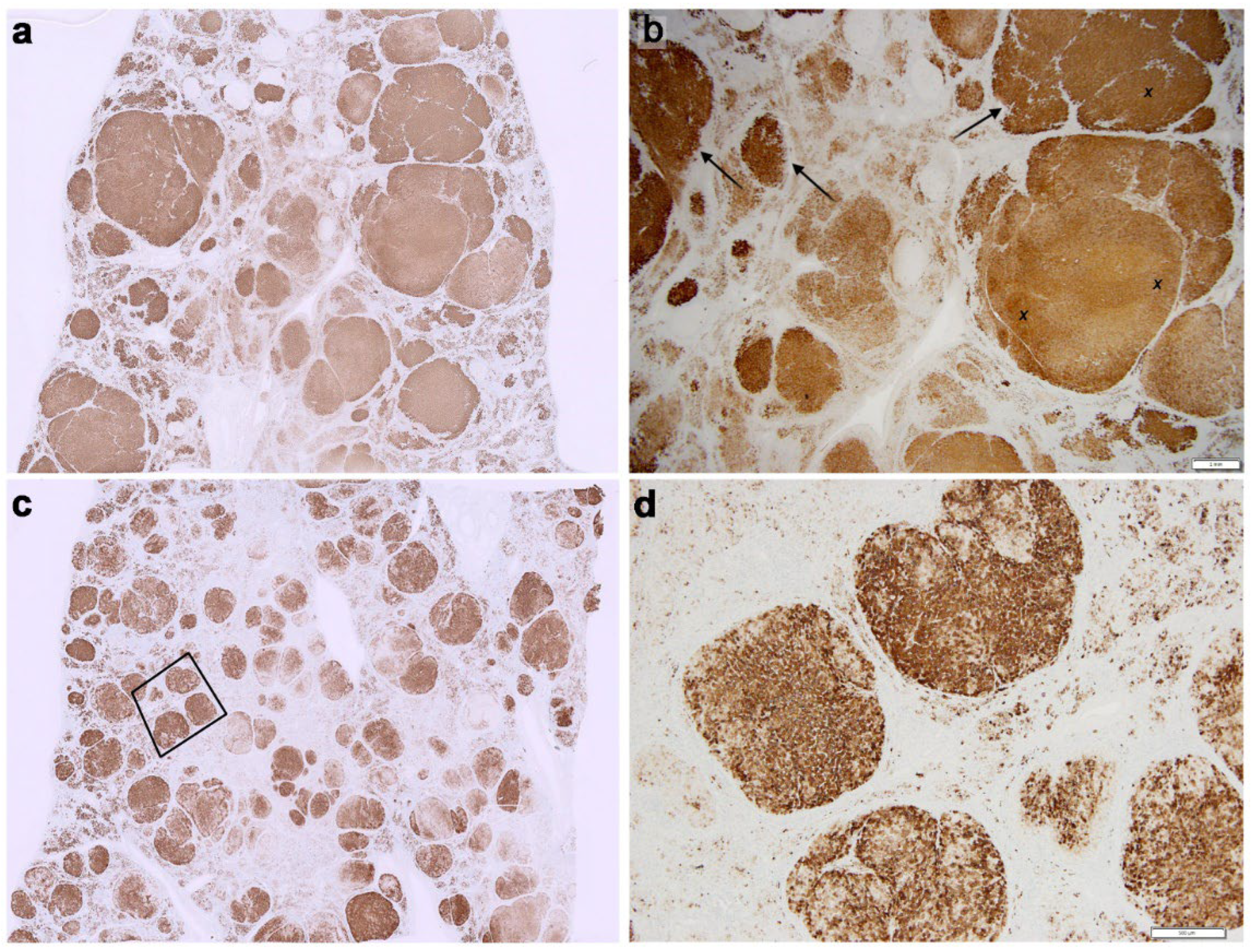
Case 1 (Figure 1, Panel 1)

The nodular liver showed a predominantly macronodular cirrhosis. The vast majority of nodules stained positively for MT (both cytoplasm and nuclei). The flat subcapsular parenchyma showed a preserved lobular architecture, i.e., normal portal tract–central vein ratio and topography, and monolayered liver cell plates. MT-IHC highlighted the normal structure, resulting in a map-like staining pattern that marked selectively periportal areas, while midlobular and centrilobular areas were negative. “Check-border” and most zone 1 hepatocytes showed a weak MT positivity, that made possible to visualize positive nuclei, linear staining of plasma and interface membranes, dot-like reactivity of the bile canaliculus, and round single or clustered megalysosome-like inclusions. All features are shown in Figure 1.
Adjacent S1. Larger nodules separated by mildly inflammatory thin septa, were negative for both MT and rhodanine (Supplementary Material, Figure S2).

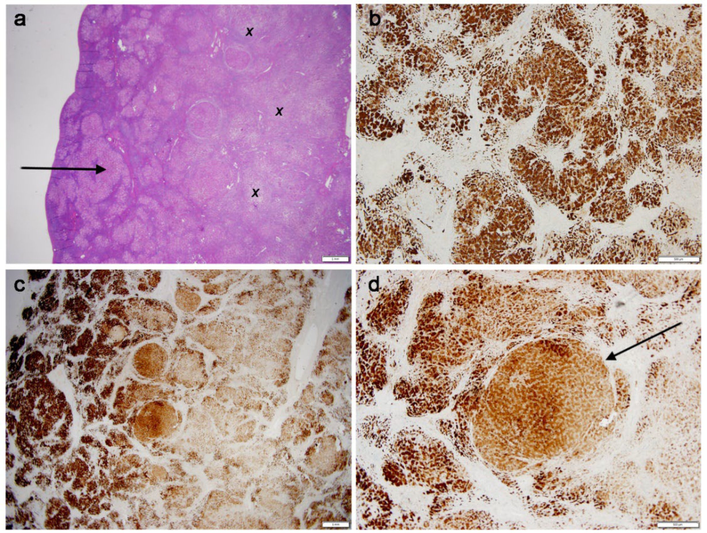
Case 2 (Figure 2, Panel 2)

The scar corresponded to an infiltrating sclerosing cholangiocarcinoma. Tumour cells were MT-negative (both cytoplasm and nuclei), CK7-positive, and Hep-Par-1-negative. The liver architecture was preserved, the only alterations being a mild portal fibrosis and a few periportal thin fibrous extensions without nodule formation. Large portion of normal peritumoral parenchyma showed diffuse strong nuclear and cytoplasmic MT staining, the latter was predominantly lacquer-painting-like. Rare monolayered rows or clusters of hepatocytes showed weaker MT positivity that marked the interface membranes between adjacent hepatocytes. Distant from the CCA, hepatocytes showed strong MT positivity in the form of cytoplasmic globules and negative nuclei. (Figure 2, Panel 2).
Rhodanine and orcein were substantially negative, except for rare hepatocytes containing a couple of cop-per granules (Supplementary Material Figure S3).

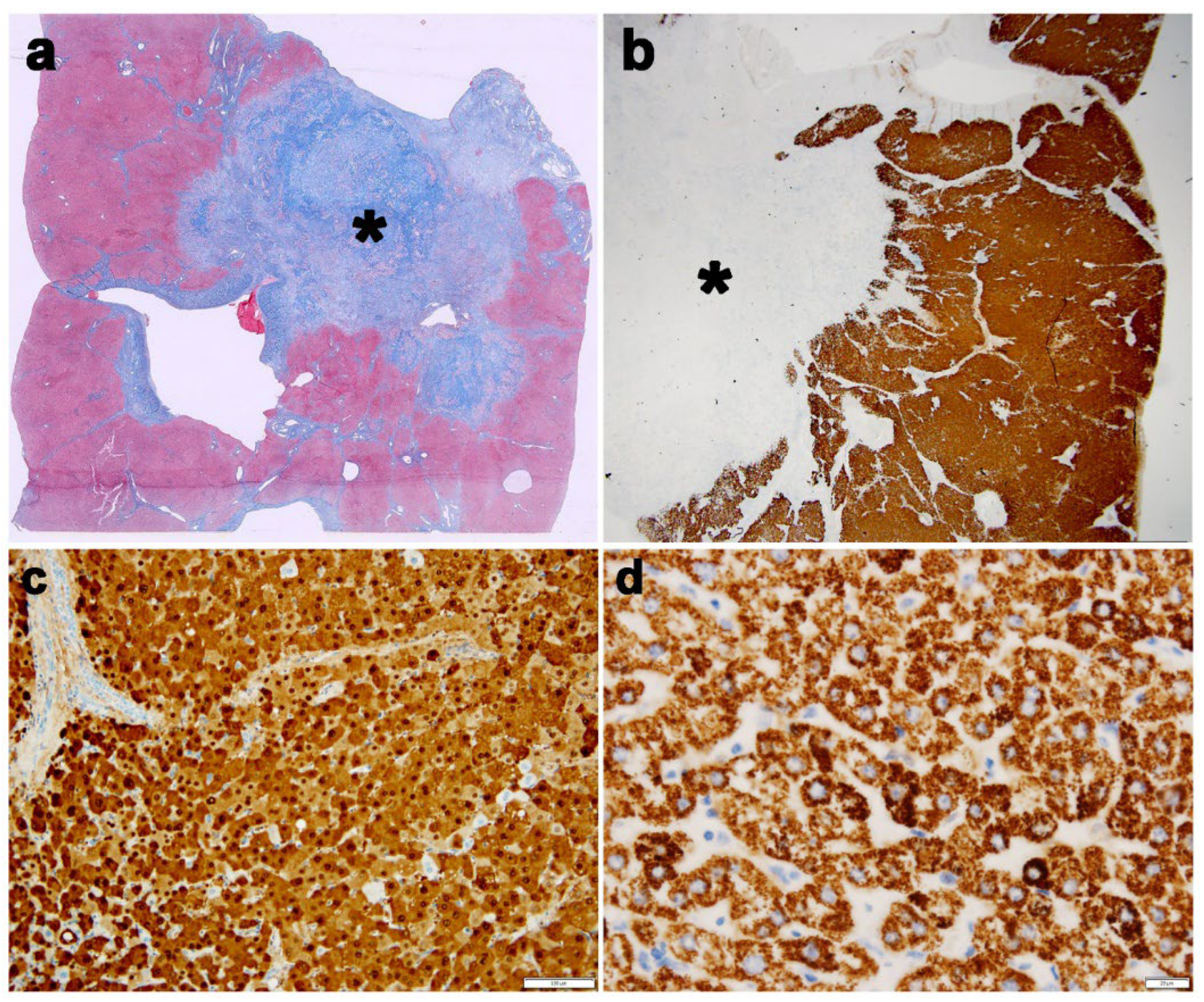
Cases 3 (Figure 3, Panel 3)

The liver showed a diffuse nodular transformation. Some nodules were surrounded by thin active septa with inflammatory infiltration penetrating into a MT positive parenchyma with severe hepatocellular damage, bile plugs and Mallory bodies. Adjacent nodules showed a variable degree of ischemic or lytic necrosis, resulting in their disintegration or liquefaction and extrusion of MT positive globular material. Polygonal interstitial cells with abundant cytoplasm were consistent with hepatocytes; round or spindle-shaped cells were interpreted as fibroblasts, myofibroblasts, macrophages, or inflammatory cells (Figure 3, Panel 3).
An occasional small nodule was entirely positive on rhodanine staining (Supplementary Material (Figure S4).

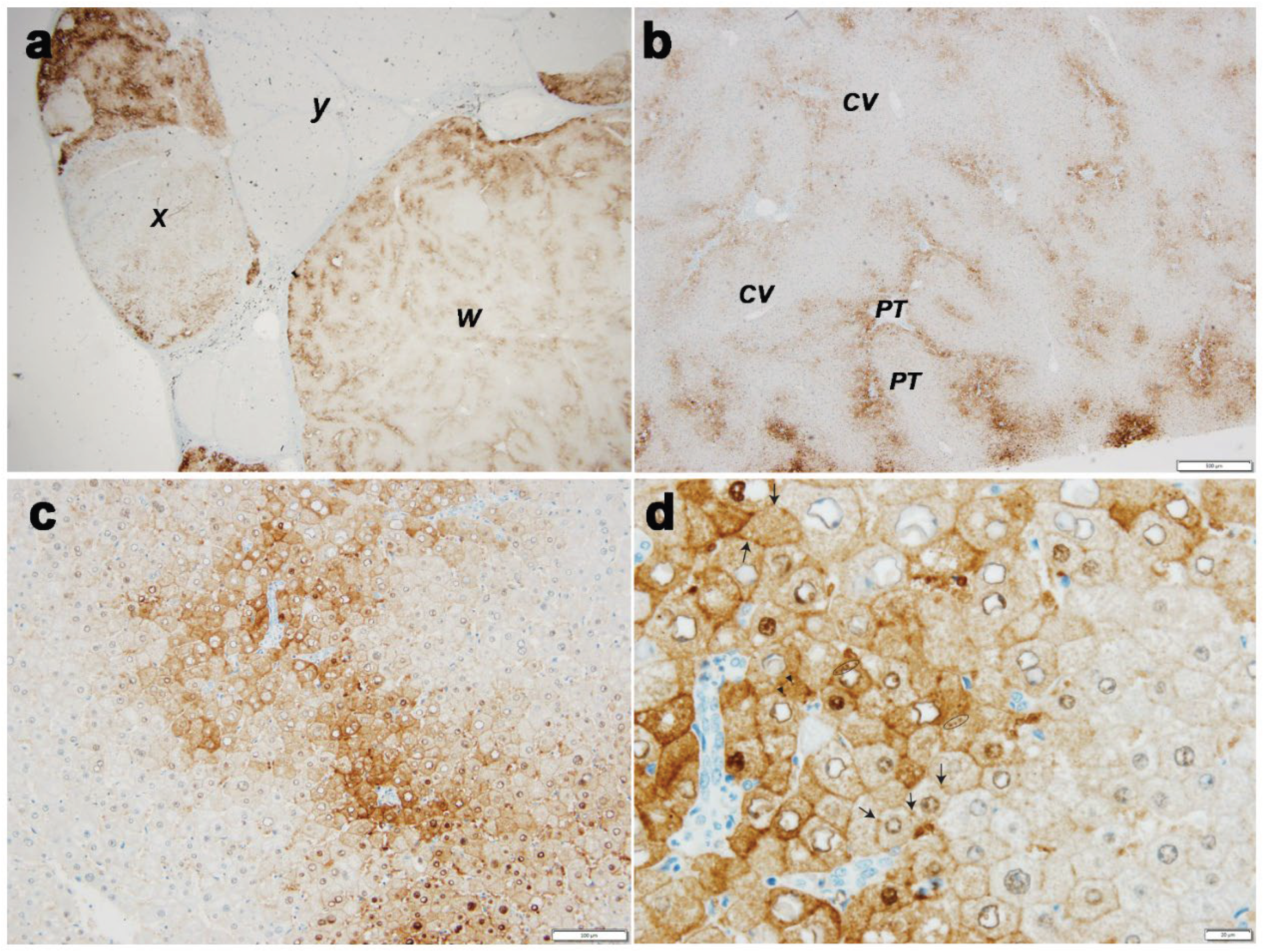
Case 4 (Figure 4, Panel 4 a,b)

The liver showed a macronodular cirrhosis characterized by nuclear and cytoplasmic MT positivity of the vast majority of hepatocytes. The cytoplasmic staining appeared either diffuse or in form of densely packaged granules. Interstitial cells were also MT positive (Figure 4, Panel 4a,b). Both rhodanine and orcein were positive in most nodules.

Case 5 (Figure 4, Panel 4 c,d)

The liver cirrhosis was mostly micronodular, Nodules were characterized by cytoplasmic MT positivity (granular/globular type) of most hepatocytes. Single or clusters of intranodular hepatocytes could remain unstained and display nuclear and lysosomal positivity. MT positive mesenchymal cells were present in the connective tissue septa (Figure 4, Panel 4 c,d).
Rhodanine and orcein were diffusely positive in several nodules (Supplementary Material Figure S5).

5. Discussion

Recent retrospective studies on WD liver tissue have shown MT overexpression and that MT-IHC can act as a biomarker for the disease [7,8,9]. However, in those studies, 10% of cases were MT-negative and a diagnostic requirement was >50% MT-positive hepatocytes in individual sections. These statistics warrant caution in prospective cases with low, intermediate, or high suspicion of WD, for which a comprehensive algorithm incorporating clinical features, copper metabolism measures, and DNA analysis has been recommended [9].
In the present study on explanted WD livers, we observed that MT overexpression unavoidably progresses to true accumulation, persisting beyond very long-term copper chelation, even when histopathological alterations regress (case n.2). Accordingly, all WDs should be MT-positive. Therefore, the distinction between MT-positive and MT-negative WD, seems inappropriate. In our series, we found a few MT-negative cirrhotic nodules adjacent to positive ones; this discrepancy requires explanation. Pathologists often invoke sampling error; in WD, this likely reflects copper and Cu–MT dynamic status and/or disease stage. Furthermore, it is known that copper is unevenly distributed especially in stage IV-WD [10,11], and that the available histochemical stainings for copper and copper binding protein (CBP) have limited specificity and sensitivity [8,9].
Beyond the inexorability of MT accumulation, we demonstrated new subcellular localisations of MT immunoreactivity that correspond to the results from biochemical MT quantitative studies in WD livers [12] and in the liver of WD animal models [13,14], as well as to the presence of MT in bile [15], nuclei [16], and lysosomes [17,18,19,20].
The accumulation process explains and strengthens the diagnostic value of MT-IHC, and simultaneously raises the question of whether it contributes to pathogenesis and progression of WD.
These novel findings allow further hypotheses concerning: (i) the link between defective copper biliary excretion and low plasma CP; and (ii) the link between low plasma CP and defective incorporation of copper into CP [21]. We discuss our interpretation within the available literature, under the following headings:
  • Mechanism of MT accumulation
  • Meaning of MT subcellular localisations under physiological conditions and in WD
  • Proposed role of copper and MT extra-hepatocytic deposits in WD diagnosis and progression
  • MT–CP relationship

5.1. Mechanism of MT Accumulation

Copper enters hepatocytes as Cu+ and is delivered by chaperones to functional sites where it serves as a prosthetic group for essential enzymes. The ATOX1 chaperone drives Cu+ to the trans-Golgi network (TGN), where the Wilson protein (ATP7Base) is located, believed to mediate incorporation of copper into apo-CP and export of copper to the bile canaliculus for biliary excretion [2].
Excess copper beyond homeostatic equilibrium is sequestered by MT.
MT is a cytoplasmic protein synthesised by non-membrane-bound polyribosomes and binds copper with very high affinity [22], providing major protection against cytosolic damage [23].
Several interdependent factors induce MT synthesis; zinc certainly does [6]. As MT affinity for Cu+ exceeds that for Zn2+, excess Cu+ displaces Zn2+, and the resulting free Zn2+ induces further MT synthesis [6]. D-penicillamine induces rat hepatic MT [24], and increases MT mRNA in mouse hepatocytes by removing copper from intermediary ligands for extracellular export, thereby making it available to induce additional MT synthesis [25]. Polymorphisms in MTs may also affect expression.
MTs are rich in cysteine residues. Depending on redox conditions, these cysteines interconvert between thiol and disulphide forms. Upon oxidation, MTs lose metal-coordinating capacity [26]. Reversible redox changes regulate metal binding and explain transfer of metals to other copper-binding proteins (CBPs) with lower affinity [26,27]. Metalloproteomic studies have expanded the CBP repertoire [28,29].
Disulphide formation occurs preferentially intra-molecularly, but with the higher MT concentrations the inter-molecular bonds increase, leading to MT polymerisation and aggregation [30], which can be reversed by reductants [31,32]. Higher-order MT aggregates may serve as physiological subcellular storage [30,31].
However, although glutathione is normally abundant, it reduces disulphides slowly and can also be lowered by oxidative stress [33]. Decreases in glutathione and glutathione reductase activity have been attributed to copper-mediated inhibition [34], whereas zinc treatment increases glutathione availability in WD [35].
Cytosolic oxidants (e.g., H2O2, oxidised glutathione, nitric oxide) induce Cu–MT to release copper into the cytosol. Normally, oxidation negatively regulates MT copper binding reversibly, but prolonged oxidation exhausts MT function and may upregulate MT expression. Reducing agents (e.g., reduced glutathione, ascorbic acid) reconvert Cu2+ to Cu+. Copper redox cycling generates reactive oxygen species (ROS) that can reach harmful levels when the metal is not adequately neutralised. MT is therefore synthesised continuously to ligate recurrently released free Cu+. MTs can act as both anti-oxidant and pro-oxidant [36]. The latter effect, occurring with elevated copper excess, is promoted by H2O2 and involves the hydroxyl radical generation that triggers cellular toxicity in WD [37,38]. Thus, copper—being redox-active—paradoxically compromises the very MTs mobilised to chelate it. Uncontrolled redox cycling targets MTs, leaving inactive, polymerised, abnormally aggregated proteins resistant to cytosolic and lysosomal proteolysis [39]. When ATP7B-mediated biliary copper excretion fails, MTs accumulate inexorably over time and persist despite long-term chelation. Copper–MT complexation is a double-edged sword: initially protective, ultimately promoting hepatocellular damage through hydroxyl radical production The mechanisms of MT inactivation, polymerisation, abnormal aggregation, and proteolytic resistance are schematically represented in Figure 5.

5.2. Meaning of MT Subcellular Localisations Under Physiological Conditions and in WD

Previous studies reported that full cytoplasmic staining in >50% of hepatocytes in formalin-fixed, paraffin-embedded liver specimens is diagnostic for WD, with positivity involving entire lobules up to 100% of hepatocytes [8]. In contrast, in normal livers, MT positivity, if any, has been reported as restricted to perivenular (zone 3) hepatocytes [7,8]. Owing to their abundant smooth endoplasmic reticulum and specific enzyme armamentarium (e.g., cytochrome P450), these hepatocytes are professional detoxifiers of endogenous/exogenous agents, including heavy metals. Pre-formed MT in zone 3 hepatocytes can therefore be considered physiological [8], providing a first-line defence. Notably, de novo MT synthesis is relatively slow compared with rapid re-localisation of Cu-ATPase (hours vs. minutes) [6].
In our study, large areas with preserved lobular architecture minimised sampling error. In these areas, the weak cytoplasmic MT positivity in zone 1 hepatocytes has made possible to identify the following subcellular localisations: nuclei, tight junctions and interface membranes, bile canaliculus, plasma membranes, megalysosomes, and interstitial mesenchymal cells within fibrous septa. The full and diffuse cytoplasmic positivity likely reflects cytosolic and organellar MT, including Cu–MT aggregates due to oxidation, newly synthesised apo-MT (upregulated), trafficking MT, and sequestered soluble MT [40].
Linear plasma-membrane positivity may reflect subplasmalemmal MT at sites of CTR1 receptors internalising circulating Cu+. Interface linear and dot-like positivity delineate the canalicular membrane and bile canaliculus lumen.
Nuclear positivity was nearly constant. In periportal (zone 1) hepatocytes—especially around terminal portal branches—nuclear positivity consistently co-occurred with bile canaliculus and lysosome positivity. Zone 1 hepatocytes, the first to receive portal venous blood, unexpectedly, showed the earliest simultaneous nuclear, bile canaliculus and lysosomal MT immunoreactivity. As copper enters nuclei early in WD [2,41], co-localisation of copper and MT is likely concurrent, and lysosomal MT seems to be also an early event.
The mechanism of Cu and MT nuclear entry is unknown. Within nuclei, copper inhibits nuclear receptors [2,16,41], remodels the transcriptome, and diminishes functions of genes such as LXR–RXR, causing lipid metabolism alterations and steatosis [2,16,41,42]. The early nuclear co-localisation suggests MT as a mediator of cross-talk between copper status and lipid metabolism [43,44]. Nuclear MT in viable hepatocytes is a new finding; in previous reports, only necrotic cells could stain positively [45,46].
Cytoplasmic round inclusions (lysosomes) in WD hepatocytes were originally described as lipofuscin-like inclusions shown to be lysosomes by acid phosphatase histochemistry [47], and later identified under the EM as electron-dense polymorphic bodies [48]. Chromatographic, luminescence, and electron probe X-ray microanalysis studies confirmed that the amino-acid composition of copper –containing particulate extractions from non-cytosolic regions is typical of Cu–MTs [17,18,19,20,49].
Under normal conditions and in experimental copper overload, lysosomes represent the major biliary excretory route via exocytosis [50]. In excess copper, Cu and MT stagnate within lysosomes, provoking membrane peroxidation, altered lipid composition, and decreased fluidity [51]. Copper-induced lysosomal damage in WD impairs lysosomal discharge and causes Cu and MT retention.
Immunoreactivity in bile canaliculi is interpreted as the morphological counterpart of physiological MT export into bile, as shown in normal animals [15] and in LEC rats, in which biliary MT is disproportionately low relative to massive intracellular MT accumulation [13]. In a WD rat model, the siderophore methanobactin has led to a prompt release of copper into bile and copper was shown to be associated with methanobactin [52], Moreover, in vitro experiments with liver cytosol, containing high copper-metallothionein, have demonstrated that methanobactin removes copper from MT, confirming its copper chelating activity [52]. In view of the newly described MT accumulation phenomenon in WD, these experiments would encourage to design alternative Cu chelators, capable of sequestering Cu more efficiently than MTs, detoxifying and promoting biliary excretion.
These observations lead to the conclusions that: (i) MT excretion under normal and copper-overload conditions can occur via an ATP7B-independent modality; and (ii) impaired lysosomal exocytosis in WD [53] and in animal models [13] indicates that the biliary export of both copper and MT is defective.
Among our five patients, two underwent long-term chelation. The CCA case (case 2) benefited from the initial 12 years of penicillamine, with regression of lesions; after SLE developed, timely trientine prevented harm from penicillamine interruption [54]. In contrast, patient n.3 received discontinuous penicillamine over 42 years. Moreover, the treatment was stopped for two years before the typical features of acute on chronic liver disease become manifested, clinical decompensation developed and transplantation was performed. That represents a common outcome when penicillamine is discontinued without alternative orphan drugs (trientine, zinc) [55].
Penicillamine’s chelating effect is rapid but normal hepatic values can be achieved only after long-term treatment (≥5 years) [56]. Thus, as under penicillamine treatment copper overload decreases, while MT accumulation increases, the trend of the two substances in WD is at least partially dissociated.
The diversion of excess copper to lysosomes may disrupt metal homeostasis beyond the primary ATP7B defect. MTs-laden lysosomes may be unable to degrade these proteins; while this may forestall sudden release of free copper, the lysosomal environment enriched in Cu+ and normally containing H2O2, and physiological reductants provides the necessary conditions for Fenton chemistry [57].
In summary, without ATP7B-mediated transport, copper stagnates and dysfunctional MTs build up gradually and continuously. Reliance on MTs alone cannot prevent intracellular toxic leakage, as well as diversion of copper-bound MTs into lysosomes, beyond organelle capacity, becomes a dead end culminating in poisoning [57,58].

5.3. Proposed Role of Copper and MT Extrahepatocytic Deposits in WD Diagnosis and Progression

The sequence above parallels that in Atp7b/ mouse hepatocytes [59]. Persistent redox cycling and ROS formation alter cytoskeleton and cell adhesion [6]. To mitigate damage, the liver reduces copper uptake, enhances alternative export mechanisms, and promotes absorption of extracellular free copper by interstitial cells (fibroblasts, myofibroblasts, and inflammatory cells) [41]. These cells are progressively recruited in stage II–IV WD as chronic hepatitis, fibrosis, and cirrhosis develop. Elevated copper accompanies tissue injury [60,61], and the interplay of injury–inflammation–fibrosis may lead to extracellular release of copper and MT, implicating interstitial cells [61].
Whether macrophages synthesise MT is unclear; they do take up Cu–MT complexes [61,62]. MT synthesis has been induced in cultured normal and Menkes fibroblasts [63]. Part of interstitial MT may also derive from circulating protein [61].

5.4. MT–CP Relationship

After the seminal report by Sass-Kortsak [64] of two WD patients with normal CP, further such cases have been described; however, most WD patients have low CP, which remains a component of the diagnostic Leipzig score [65].
Low serum levels of CP and copper with high 24-h urinary excretion are common to inherited copper disorders (WD, Menkes syndrome [66], MEDNIK disease [67]). Elevated hepatic copper is constant in WD and absent in Menkes; recently, high hepatic copper was reported in one MEDNIK patient responsive to zinc [68], suggesting overlapping features. In view of MT accumulation in WD, it would be informative to assess MT behaviour in these other disorders.
Low CP in WD and defective incorporation of copper into apo-CP remain poorly understood. Low CP and copper serum can also occur in conditions unrelated to inherited copper transport (e.g., congenital aceruloplasminaemia [69] and a variant of non-alcoholic non-diabetic steato-hepatitis with hyperferritinaemia [70,71].
In WD, low serum copper with high urinary excretion suggests interplay between intracellular and circulatory copper pools as a consequence of MT overexpression/accumulation. Excess MT may compete with ATOX1-driven Cu transport, diverting Cu+ from ATP7B and keeping cytosolic Cu+ below the level required to induce CP synthesis. Indeed, CP synthesis is regulated transcriptionally by nuclear Cu+ originating from the cytosolic pool [2,41,72]. It has also been speculated that apo-CP polymerises and is retained within the secretory pathway [21], potentially exerting feedback inhibition on CP synthesis.
Moreover, radio-copper studies suggesting defective incorporation [73] have been challenged by detection of holo-CP in WD liver tissue [21] and by the regular presence of apo- and holo-CP in bile of humans [21] and animals [74,75]. The bimodal route of CP export—plasma CP (pCP, ~132 kDa) and biliary CP (bCP, ~125 kDa) [21,76,77]—implies that in WD the hampered ATP7B-mediated biliary excretion and retention of holo-CP and copper follow a pathway distinct from that of Cu–MT.
So far, no study has conclusively linked defective biliary CP excretion with its reduced plasma levels. Although we did not perform CP-IHC, analogies with events in the endoplasmic reticulum (ER) in other diseases, such as mucolipidosis II (I-cell disease) and α-1-antitrypsin deficiency (AAT-D), suggest potential explanation about the mechanism. In I-cell disease [78], mis-sorting causes lysosomal enzymes to be secreted instead of delivered to lysosomes. If a similar defect affected bCP in WD, the protein might be retained in the ER or misdirected to blood. Retention could transiently inhibit synthesis, promote di-polymer formation [21], retro-translocation, ERAD degradation, and autophagy. This hypothesis is independent of other CP properties, such as inducible oxidative aggregation by hydrogen peroxide [79] and the ability to form stable complexes with multiple proteins [80].
In AAT-D, mutant Z protein is retained in ER globules which stain not only with anti-AAT but also with rhodanine and orcein [81], indicating the presence of AAT, copper, and a CBPs. As AAT is not itself a CBP, copper binding can be explained by disulphide formation between free thiol cysteines on AAT loops and thiols of other proteins [82].
Therefore, hepatic copper overload in WD, when ATP7B is compromised, can be due also to the defective biliary excretion of MT and bCP, and to the reduced secretion of pCP in blood, via an ATP7B-independent mechanism, as schematically represented in Figure 5.
Preprints 190810 i001

6. Conclusions

In WD, physiological MT overexpression progresses to true accumulation. This phenomenon is unavoidable and persists over decades of chelation therapy, explaining the diagnostic value of MT-IHC at all disease stages.
MT immunoreactivity in sinusoidal and interface membranes, bile canaliculus lumen, nuclei, lysosomes, and interstitial mesenchymal cells reflects discrete functions.
Chronic copper-driven redox cycles exhaust MT function, leading to polymerisation and aggregation into complexes resistant to cytosolic and lysosomal degradation. Stagnation of copper and Cu–MT in lysosomes induces lysosomal damage via Fenton chemistry, impairing the lysosome–bile canaliculus excretion route of copper and Cu–MT.
The whole above appear to add insights to the WD puzzle and would suggest strategies for more effective, specific chelators targeting hepatic copper storage and preventing its detrimental sequelae, including MT and CP accumulation. Further clinical and experimental research is warranted to validate these hypotheses.

Supplementary Materials

The following supporting information can be downloaded at: Preprints.org.

Author Contributions

FC and EB made the study design and prepared the manuscript; TL performed MT immunostaining and quality control; FC, PF, LB, MCG and LC interpreted the histopathology and provided tissue specimens; MZ, LDC, AWA provided the full clinical history.

Funding

The authors have no support or funding to report.

Institutional Review Board Statement

Authorization to publish the manuscript was obtained by the Director, Research and Innovation Chairperson CREC, CUHAS Directorate of Research and Innovation.

Informed Consent Statement

Written patients and parental consent is included in individual medical records.

Data Availability Statement

All relevant data are within the paper and its supporting files.

Acknowledgments

The authors wish to thank the Italian Non-profit Friends Raising Association for post- graduate education promotion in Tanzania, and Gabriele Bacile for preparing the microphotographs.

Conflicts of Interest

The authors declare that no conflict of interest exists.

References

  1. Bull, P.C.; Thomas, G.R.; Rommens, J.M.; Forbes, J.R.; Cox, D.W. The Wilson disease gene is a putative copper transporting P–type ATPase similar to the Menkes gene. Nat. Genet. 1993, 5, 327–337. [CrossRef]
  2. A Roberts, E.; Schilsky, M.L. Current and Emerging Issues in Wilson’s Disease. New Engl. J. Med. 2023, 389, 922–938. [CrossRef]
  3. Członkowska A, Litwin T, Dusek P, Ferenci P, Lutsenko S, Medici V, Rybakowski JK, Weiss KH, Schilsky ML. Wilson disease. Nat Rev Dis Primers. 2018;4:21.
  4. Lalioti, V.; Muruais, G.; Tsuchiya, Y.; Pulido, D.; Sandoval, I.V. Molecular mechanisms of copper homeostasis. Front. Biosci. 2009, 14, 4878–903. [CrossRef]
  5. Lutsenko, S. Dynamic and cell-specific transport networks for intracellular copper ions. J. Cell Sci. 2021, 134. [CrossRef]
  6. Delangle P. Mintz E: Chelation therapy in Wilson’s disease: from D-penicillamine to the design of selective bioinspired intracellular Cu chelators: Dalton Trans. 2012;41: 6359-70.
  7. Rowan, D.J.; Mangalaparthi, K.K.; Singh, S.; Moreira, R.K.; Mounajjed, T.; Lamps, L.; Westerhoff, M.; Cheng, J.; Bellizzi, A.M.; Allende, D.S.; et al. Metallothionein immunohistochemistry has high sensitivity and specificity for detection of Wilson disease. Mod. Pathol. 2022, 35, 946–955. [CrossRef]
  8. Wiethoff, H.; Mohr, I.; Fichtner, A.; Merle, U.; Schirmacher, P.; Weiss, K.H.; Longerich, T. Metallothionein: a game changer in histopathological diagnosis of Wilson disease. Histopathology 2023, 83, 936–948. [CrossRef]
  9. Stokes NL Patil A, et al. Validation of Metallothionein Immunohistochemistry as a High Sensitive Screening Test for Wilson Disease. Mod Pathol 2025;38:1-7.
  10. Stromeyer, F.W.; Ishak, K.G. Histology of the Liver in Wilson’s Disease: A Study of 34 Cases. Am. J. Clin. Pathol. 1980, 73, 12–24. [CrossRef]
  11. Faa, G.; Nurchi, V.; Demelia, L.; Ambu, R.; Parodo, G.; Congiu, T.; Sciot, R.; Van Eyken, P.; Silvagni, R.; Crisponi, G. Uneven hepatic copper distribution in Wilson's disease. J. Hepatol. 1995, 22, 303–308. [CrossRef]
  12. Mulder, T.; Janssens, A.; Verspaget, H.; van Hattum, J.; Lamers, C. Metallothionein concentration in the liver of patients with Wilson's disease, primary biliary cirrhosis, and liver metastasis of colorectal cancer. J. Hepatol. 1992, 16, 346–350. [CrossRef]
  13. Sugawara, N.; Sato, M.; Yuasa, M.; Sugawara, C. Biliary Excretion of Copper, Metallothionein, and Glutathione into Long-Evans Cinnamon Rats: A Convincing Animal Model for Wilson Disease. Biochem. Mol. Med. 1995, 55, 38–42. [CrossRef]
  14. Zhang CC, Volkman M, Tuma S, Stremmel W. Metallothionein is elevated in liver and duodenum of Atp7b (-/-) mice. Biometals 2018;31:617-625.
  15. Bremner, I.; Mehra, R.K.; Sato, M. Metallothionein in blood, bile, and urine. Experientia Suppl 1987;52:507-517.
  16. Cherian MG. The significance of the nuclear and cytoplasmic localization of metallothionein in human liver and tumor cells. Environ Health Perspect. 1994;102 Suppl 3(Suppl 3):131-5.
  17. Mehra, R.K.; Bremner, I. Species differences in the occurrence of copper-metallothionein in the particulate fractions of the liver of copper-loaded animals. Biochem. J. 1984, 219, 539–546. [CrossRef]
  18. Sternlieb, I. Hepatic lysosomal copper-thionein. Experientia Suppl 1987;52: 647-653.
  19. Sakurai, H.; Nakajima, K.; Kamada, H.; Satoh, H.; Otaki, N.; Kimura, M.; Kawano, K.; Hagino, T. Copper-Metallothionein Distribution in the Liver of Long-Evans Cinnamon Rats: Studies on Immunohistochemical Staining, Metal Determination, Gel Filtration and Electron Spin Resonance Spectroscopy. Biochem. Biophys. Res. Commun. 1993, 192, 893–898. [CrossRef]
  20. Hanaichi T, Kidokoro R, Hayashi H, Sakamoto N. Electron probe X-ray analysis on human hepatocellular lysosomes with copper deposits: copper binding to a thiol-protein in lysosomes. Lab Invest. 1984;51:592-7.
  21. Chowrimootoo, G.F.E.; Ahmed, H.A.; Seymour, C.A. New insights into the pathogenesis of copper toxicosis in Wilson's disease: evidence for copper incorporation and defective canalicular transport of caeruloplasmin. Biochem. J. 1996, 315, 851–855. [CrossRef]
  22. Koch, K.A.; Peña, M.M.O.; Thiele, D.J. Copper-binding motifs in catalysis, transport, detoxification and signaling. Chem. Biol. 1997, 4, 549–560. [CrossRef]
  23. Tapia, L.; González-Agüero, M.; Cisternas, M.F.; Suazo, M.; Cambiazo, V.; Uauy, R.; González, M. Metallothionein is crucial for safe intracellular copper storage and cell survival at normal and supra-physiological exposure levels. Biochem. J. 2004, 378, 617–624. [CrossRef]
  24. Heilmaier, H.; Jiang, J.; Greim, H.; Schramel, P.; Summer, K. D-penicillamine induces rat hepatic metallothionein. Toxicology 1986, 42, 23–31. [CrossRef]
  25. McArdle, H.J.; Kyriakou, P.; Grimes, A.; Mercer, J.F.; Danks, D.M. The effect of d-penicillamine on metallothionein mRNA levels and copper distribution in mouse hepatocytes. Chem. Interactions 1990, 75, 315–324. [CrossRef]
  26. Fabisiak, J.P.; Tyurin, V.A.; Tyurina, Y.Y.; Borisenko, G.G.; Korotaeva, A.; Pitt, B.R.; Lazo, J.S.; Kagan, V.E. Redox Regulation of Copper–Metallothionein. Arch. Biochem. Biophys. 1999, 363, 171–181. [CrossRef]
  27. Jiang LJ, Maret W, Vallee BL. The ATP-metallothionein complex. Proc Natl Acad Sci U.S.A. 1998;95:9146-9.
  28. A Roberts, E.; Schilsky, M.L. Current and Emerging Issues in Wilson’s Disease. New Engl. J. Med. 2023, 389, 922–938. [CrossRef]
  29. Blockhuys, S.; Celauro, E.; Hildesjö, C.; Feizi, A.; Stål, O.; Fierro-González, J.C.; Wittung-Stafshede, P. Defining the human copper proteome and analysis of its expression variation in cancers. Metallomics 2017, 9, 112–123. [CrossRef]
  30. Haase, H.; Maret, W. Partial oxidation and oxidative polymerization of metallothionein. Electrophoresis 2008, 29, 4169–4176. [CrossRef]
  31. Krizkova, S.; Adam, V.; Kizek, R. Study of metallothionein oxidation by using of chip CE. Electrophoresis 2009, 30, 4029–4033. [CrossRef]
  32. Křížková, S.; Masařík, M.; Eckschlager, T.; Adam, V.; Kizek, R. Effects of redox conditions and zinc(II) ions on metallothionein aggregation revealed by chip capillary electrophoresis. J. Chromatogr. A 2010, 1217, 7966–7971. [CrossRef]
  33. Jones, D.P. Redox potential of GSH/GSSG couple: assay and biological significance. Methods Enzymol. 2002;348:93-112.
  34. Rafter, G.W. The effect of copper on glutathione metabolism in human leukocytes. Biol. Trace Element Res. 1982, 4, 191–197. [CrossRef]
  35. Farinati, F.; Cardin, R.; D’inca, R.; Naccarato, R.; Sturniolo, G.C. Zinc treatment prevents lipid peroxidation and increases glutathione availability in Wilson’s disease. J. Lab. Clin. Med. 2003, 141, 372–377. [CrossRef]
  36. Fabisiak JP, Pearce LL, Borisenko GG, Tyhurina YY, Tyurin VA, Razzack J, Lazo JS, Pitt BR, Kagan VE. Bifunctional ani/prooxidant potential of metallothionein: redox signal of copper bindng and release Antioxid Redox Signal 1999;1:3-349.
  37. Suzuki, K.T.; Rui, M.; Ueda, J.; Ozawa, T. Production of hydroxyl radicals by copper-containing metallothionein: roles as prooxidant. Toxicol Appl Pharmacol.  1996, 141, 231–7.
  38. Zischka Hans, Lichtmannegger Josef. Cellular copper toxicity: a critical appraisal of Fenton-chemistry-based oxidative stress in Wilson disease. Wilson Disease, Pathogenesis, Molecular Mechanisms, Diagnosis, Treatment and Monitoring. 2019; pages 65-81. Edited by Karl Heinz Weissand Michel Schilsky.
  39. Cousins, R.J. Absorption, transport, and hepatic metabolism of copper and zinc: special reference to metallothionein and ceruloplasmin.. Physiol. Rev. 1985, 65, 238–309. [CrossRef]
  40. Hou, T.; Xu, X.; An, Y.; Ru, B.; Bi, R. Cysteine-independent polymerization of metallothioneins in solutions and in crystals. Protein Sci. 2000, 9, 2302–2312. [CrossRef]
  41. Ralle M, Huster D, Vogt S, Schirrmeister W, Burkhead JL, Capps TR, Gray L, Lai B, Maryon E, Lutsenko S. Wilson disease at a single cell level: intracellular copper trafficking activates compartment-specific responses in hepatocytes. J Biol Chem. 2010; 285:30875-83.
  42. Wooton-Kee, C.R.; Jain, A.K.; Wagner, M.; Grusak, M.A.; Finegold, M.J.; Lutsenko, S.; Moore, D.D. Elevated copper impairs hepatic nuclear receptor function in Wilson’s disease. J. Clin. Investig. 2015, 125, 3449–3460. [CrossRef]
  43. Dev, S.; Kruse, R.L.; Hamilton, J.P.; Lutsenko, S. Wilson Disease: Update on Pathophysiology and Treatment. Front. Cell Dev. Biol. 2022, 10, 871877. [CrossRef]
  44. Huster, D.; Lutsenko, S. Wilson disease: not just a copper disorder. Analysis of a Wilson disease model demonstrates the link between copper and lipid metabolism. Mol. Biosyst. 2007, 3, 816–824. [CrossRef]
  45. Elmes, M.E.; Clarkson, J.P.; Mahy, N.J.; Jasant, B. Metallothionein and copper in liver disease with copper retention—a histopathological study. J. Pathol. 1989, 158, 131–137. [CrossRef]
  46. NARTEY, N.; FREI, J.; CHERIAN, M. HEPATIC COPPER AND METALLOTHIONEIN DISTRIBUTION IN WILSONS-DISEASE (HEPATOLENTICULAR DEGENERATION). Lab Invest. 1987, 57, 397–401.
  47. GOLDFISCHER, S. LOCALIZATION OF COPPER IN PERICANALICULAR GRANULES (LYSOSOMES) OF LIVER IN WILSONS DISEASE (HEPATOLENTICULAR DEGENERATION). Am J Pathol. 1965, 46, 977–+.
  48. Jonas L, Fulda G, Salameh T, Schmidt W, Kroning G, Hoopt UT, Nizze H. Electron microscopic detection of copper in the liver of two patients with morbus Wilson by FELS and EDX. Ultrasct Pathol 2001;25:111-8.
  49. Beltramini, M.; Lerch, K. Luminescence properties of neurospora copper metallothionein. FEBS Lett. 1981, 127, 201–203. [CrossRef]
  50. Gross, J.B.; Myers, B.M.; Kost, L.J.; Kuntz, S.M.; LaRusso, N.F. Biliary copper excretion by hepatocyte lysosomes in the rat. Major excretory pathway in experimental copper overload.. J. Clin. Investig. 1989, 83, 30–39. [CrossRef]
  51. Myers, B.M.; Prendergast, F.G.; Holman, R.; Kuntz, S.M.; Larusso, N.F. Alterations in hepatocyte lysosomes in experimental hepatic copper overload in rats. Gastroenterology 1993, 105, 1814–1823. [CrossRef]
  52. Summer, K.H.; Lichtmannegger, J.; Bandow, N.; Choi, D.W.; DiSpirito, A.A.; Michalke, B. The biogenic methanobactin is an effective chelator for copper in a rat model for Wilson disease. J. Trace Elements Med. Biol. 2011, 25, 36–41. [CrossRef]
  53. Sternlieb, I.; Hamer, C.v.D.; Morell, A.G.; Alpert, S.; Gregoriadis, G.; Scheinberg, I.H. Lysosomal Defect of Hepatic Copper Excretion in Wilson's Disease (Hepatolenticular Degeneration). Gastroenterology 1973, 64, 99–105. [CrossRef]
  54. Schemberg JH, Jaffe ME, Sternilieb I. Thr use of trientine in preventing the effects of interrupting penicillamine therapy in Wilson’s disease. N Engl J Med. 1987;31:300-313.
  55. Ping, C.C.; Hassan, Y.; Aziz, N.A.; Ghazali, R.; Awaisu, A. Discontinuation of penicillamine in the absence of alternative orphan drugs (trientine?zinc): a case of decompensated liver cirrhosis in Wilson's disease. J. Clin. Pharm. Ther. 2007, 32, 101–107. [CrossRef]
  56. Marecek, Z.; Heyrovský, A.; Volek, V. The effect of long term treatment with penicillamine on the copper content in the liver in patients with Wilson's disease. Acta Hepato-Gastroenterologica 1975, 22, 292–6.
  57. Evering, W.; Haywood, S.; Bremner, I.; Wood, A.M.; Trafford, J. The protective role of metallothionein in copper-overload: II. Transport and excretion of immunoreactive MT-1 in blood, bile and urine of copper-loaded rats. Chem. Interactions 1991, 78, 297–305. [CrossRef]
  58. Kurz, T.; Eaton, J.W.; Brunk, U.T. Redox Activity Within the Lysosomal Compartment: Implications for Aging and Apoptosis. Antioxidants Redox Signal. 2010, 13, 511–523. [CrossRef]
  59. Lutsenko S. Atp7b-/- mice as a model for studies of Wilson’s disease. Biochen Soc trans 2008;36:123-128.
  60. Whitehouse, M.W.; Walker, W.R. Copper and inflammation. Inflamm. Res. 1978, 8, 85–90. [CrossRef]
  61. Milanino R, Conforti A, Franco L, Marrella M, Velo G. Copper and inflammation--a possible rationale for the pharmacological manipulation of inflammatory disorders. Agents Actions. 1985;16:504-513.
  62. White, C.; Lee, J.; Kambe, T.; Fritsche, K.; Petris, M.J. A Role for the ATP7A Copper-transporting ATPase in Macrophage Bactericidal Activity. J. Biol. Chem. 2009, 284, 33949–33956. [CrossRef]
  63. Chan, W.-Y.; Garnica, A.D.; Rennert, O.M. Inducibility of Metallothionein Biosynthesis in Cultured Normal and Menkes Kinky Hair Disease Fibroblasts: Effects of Copper and Cadmium. Pediatr. Res. 1979, 13, 197–203. [CrossRef]
  64. Sass-Kortsak A, Cherniak M, Geiger DW, Slater R. Observations on ceruloplasmin in Wilson’s disease. J Clin Invest 1959; 38:1672-82.
  65. European Association for the Study of the Liver EASL, clinical practice guidelines: Wilson’s disease. J Hepatol 2012;56: 671-685.
  66. Kaler SG. Menkes disease. Adv Pediatr. 1994;41:263-304.
  67. Alsaif HS, Alkuraya FS. IDEDNIK Syndrome. 2024 Nov 14. In: Adam MP, Bick S, Mirzaa GM, Pagon RA, Wallace SE, Amemiya A, editors. GeneReviews® [Internet]. Seattle (WA): University of Washington, Seattle; 1993–2025.
  68. Martinelli, D.; Travaglini, L.; Drouin, C.A.; Ceballos-Picot, I.; Rizza, T.; Bertini, E.; Carrozzo, R.; Petrini, S.; de Lonlay, P.; El Hachem, M.; et al. MEDNIK syndrome: a novel defect of copper metabolism treatable by zinc acetate therapy. Brain 2013, 136, 872–881. [CrossRef]
  69. Loréal, O.; Turlin, B.; Pigeon, C.; Moisan, A.; Ropert, M.; Morice, P.; Gandon, Y.; Jouanolle, A.-M.; Vérin, M.; Hider, R.C.; et al. Aceruloplasminemia: new clinical, pathophysiological and therapeutic insights. J. Hepatol. 2002, 36, 851–856. [CrossRef]
  70. Nunes, V.S.; Andrade, A.R.; Guedes, A.L.V.; A Diniz, M.; Oliveira, C.P.; Cançado, E.L.R. DISTINCT PHENOTYPE OF NON-ALCOHOLIC FATTY LIVER DISEASE IN PATIENTS WITH LOW LEVELS OF FREE COPPER AND OF CERULOPLASMIN. Arq. de Gastroenterol. 2020, 57, 249–253. [CrossRef]
  71. Wang Q, D, Wang M, Zhou M, Chem P, Liu L, Xinxin LM, Shen Z, Liu X, Chen T. A novel non- invasive approach based on serum ceruloplasmin for identifying non-alcoholic steato-hepatitis patients in the non-diabetic population. Front Med (Lausanne). 2022;9:900794.
  72. Evans, G.; Majors, P.F.; Cornatzer, W. Induction of ceruloplasmin synthesis by copper. Biochem. Biophys. Res. Commun. 1970, 41, 1120–1125. [CrossRef]
  73. Sternlieb, I.; Morell, A.G.; Tucker, W.D.; Greene, M.W.; Scheinberg, I.H. THE INCORPORATION OF COPPER INTO CERULOPLASMIN IN VIVO: STUDIES WITH COPPER64 AND COPPER67*. J. Clin. Investig. 1961, 40, 1834–1840. [CrossRef]
  74. Jeunet, F.; Richterich, R. [Bile and ceruloplasmin. Study in vitro by the aid of perfusion of the isolated rat liver]. J. Physiol. 1962, 54, 729–37.
  75. Aisen, P.; Morell, A.G.; Alpert, S.; Sternlieb, I. Biliary Excretion of Cæruloplasmin Copper. Nature 1964, 203, 873–874. [CrossRef]
  76. Puchkova, LV Aleĭnikova, TD Verbina, IA Zakharova, ET Pliss, MG Gaĭtskhoki, vs. Biosintez dvukh molekuliarnykh form tseruloplazmina v pecheni krysy i ikh poliarnaia sekretsiia v krovotok i v zhelch’ [Biosynthesis of two molecular forms of ceruloplasmin in rat liver and their polar secretion into the blood stream and bile]. Biokhimiia. 1993;58:1893-1901.
  77. Davis W, Chowrimootoo GFE, Seymour C A. Defective biliary copper excretion in Wilson’s disease: the role of ceruloplasmin. Eur J Clin Invest. 1996; 26:893-901.
  78. Sabatini, D.D.; Kreibich, G.; Morimoto, T.; Adesnik, M. Mechanisms for the incorporation of proteins in membranes and organelles.. J. Cell Biol. 1982, 92, 1–22. [CrossRef]
  79. M’hammed, A Pasquin J Furton, A Waldron, C Mateescu, MA. Oxidative aggregation of ceruloplasmin induced by hydrogen peroxide is prevented by pyruvate. Free Rad Res 2004;39:19-36.
  80. Vasiliyev, VB. Looking for a partner: ceruloplasmin-protein interactions. Biometals 2919;37:195-210.
  81. Callea, F, Ray, MB Desmet, VJ. Alpha-I-antitrypsin and copper in the liver. Histopathology. 1981;5:415-424.
  82. Griffiths, S.W.; King, J.; Cooney, C.L. The Reactivity and Oxidation Pathway of Cysteine 232 in Recombinant Human α1-Antitrypsin. J. Biol. Chem. 2002, 277, 25486–25492. [CrossRef]
Figure 1. (case 1 Panel 1): The WSI (a) shows coexistence of MT positive (x,x), and MT negative (y) cirrhotic nodules and a portion of subcapsular parenchyma with preserved lobular architecture, highlighted by the map-like pattern of MT immunostaining (w) as better demonstrated in (b), where portal tract (PT) and centrolobular veins (CV) are topographically identified: MT positivity is restricted to periportal areas and to hepatocytes around the terminal portal branches. These show glycogenated nuclei, steatosis vacuoles, and MT positive nuclei (c). Perivenular and zone 3 hepatocytes are negative for MT. In figure (d), the cytoplasm of zone 1 hepatocytes is weakly stained and MT immunoreactivity is appreciated at the cell membranes, bile canaliculus lumen (arrows), round cytoplasmic inclusions (encircled), in addition to nuclei.
Preprints 190810 g001
Figure 2. (case 2 Panel 2): The Masson staining shows a scar (*) with an infiltrating sclerosing CCA (a), whose cells are completely negative on MT staining * (b). The surrounding parenchyma looks normal and is entirely MT positive. The peritumoral liver tissue shows diffuse cytoplasmic and nuclear staining involving nearly all hepatocytes organized in monolayered muralia (c). Far away from the tumour, hepatocytes show MT positive globules filling up the entire cytoplasm while nuclei remain unstained (d).
Preprints 190810 g002
Figure 3. (case 3 Panel 3): The H.E. staining refers to a WSI (a), in which the left half shows cirrhotic nodules with chronic active hepatitis and severely damaged parenchyma (clear hepatocyte cytoplasm and Mallory bodies (arrow). In the central part of the figure, the liver structure is no longer appreciated due to the poor staining indicating confluent necrosis (x,x,x). (b) shows disintegration of MT positive cirrhotic parenchyma; (c) shows dissolution of nodules with extrusion of MT blocks into the interstitial tissue. (d) shows a nodule with rather healthy hepatocytes with nuclear and cytoplasmic MT positivity (arrow).
Preprints 190810 g003
Figure 4. (Panel 4): WSI (a) A prevalently macronodular cirrhosis (case n.4), shows nuclear and cytoplasmic MT positivity in all nodules and a variety of MT positive interstitial cells. The cytoplasmic positivity (b) appears as diffuse (x) or in the form of densely packaged granules or globules (arrows). WSI (c) A prevalently micronodular cirrhosis (case n.5), with intervening abundant connective tissue and a number of MT positive interstitial cells. MT positive and negative hepatocytes coexist in the same nodule (square). Fig.(d) highlights hepatocytes with poorly stained or unstained cytoplasm in which only nuclei and a few inclusions are positive for MT.
Preprints 190810 g004
Table 1. Clinical and epidemiological data of five Wilson’s disease (WD) patients. 1: Policlinico Gemelli, Rome; 2: Bambino Gesù, Rome; 3: Istituto Tumori, Milan; CP= ceruloplasmin; KFR= Kaiser-Fleisher Ring; PNA= penicillamine; ALF = acute liver failure; ACLD = Acute on Chronic Liver Failure; CLD= chronic liver disease; CCA = cholangiocarcinoma; ND: not determined.
Patient 1 2 3 4 5
Age 14 years 67 49 years 9 years 15 years
Sex F F M F F
Ethnics Albania Italy Italy Italy Romania
Diagnosis Neurological WD
&
liver cirrhosis
CCA, CLD & neurological symptoms ACLD ACLD ACLD
Age at diagnosis 14 years 34 years 4 years 9 years 16 years
Date of Tx 14 years 62 years 49 years 9 years 16 years
Interval between diagnosis and Tx 4 months 2 months 44 years 12 months 4 months
Tx Center 2 3 1 2 2
CP <8.45 11 10.5 7 16
Cu tissue 580 1.1 1.013 1.013 1.23
ATP7B mutation c.1772G>A(p.Gly591Asp); c.3207C>A (p.His1069Gln) H1069Q
D918N
ND c.3207C>A(p.His1069Gln) c.304dupC(p.Met769fsTer26) c.2293G>A(p.Asp765Asn);c.2906G>A(p.Asp969Gln)
KFR YES NO NO NO NO
Treatment NO PNA+ Trientine PNA, trientine PNA+Zinc Zinc+PNA
Treatment duration 4 months 28 years 42 years, discontinous 2 months 5 months
Disclaimer/Publisher’s Note: The statements, opinions and data contained in all publications are solely those of the individual author(s) and contributor(s) and not of MDPI and/or the editor(s). MDPI and/or the editor(s) disclaim responsibility for any injury to people or property resulting from any ideas, methods, instructions or products referred to in the content.
Copyright: This open access article is published under a Creative Commons CC BY 4.0 license, which permit the free download, distribution, and reuse, provided that the author and preprint are cited in any reuse.
Prerpints.org logo

Preprints.org is a free preprint server supported by MDPI in Basel, Switzerland.

Subscribe

Disclaimer

Terms of Use

Privacy Policy

Privacy Settings

© 2026 MDPI (Basel, Switzerland) unless otherwise stated