

Submitted:
30 September 2026
Posted:
01 October 2026
You are already at the latest version
Abstract
Background: Efficient transport of mesenchymal stem cell (MSC) products represents a major practical barrier to the clinical implementation of regenerative medicine. Clinically approved intravenous preservation fluids are not optimised for MSC preservation, and many standard hypothermic preservation solutions are not approved for direct injection, necessitating an additional washing step prior to administration. Additionally, maintaining the immunophenotypic identity of MSCs—as defined by the International Society for Cell and Gene Therapy (ISCT) minimal criteria—during transport is essential to confirm that clinically significant cellular characteristics are preserved. Methods: Human adipose-derived MSCs were suspended in normal saline, 5% dextran L (a Japanese-approved injectable formulation), and Lactated Ringer's solution. Samples were stored at refrigerated (4°C), room temperature (25°C), and body temperature (37°C) conditions. Viability was determined by trypan blue exclusion using an automated cell counter at hourly intervals from 0 to 4 hours. Immunophenotypic characterisation by flow cytometry (FCM) was performed on MSCs stored in the optimal (4°C dextran L) and worst (37°C normal saline) conditions at both 0 hours (pre-storage baseline) and 4 hours to assess preservation of ISCT surface marker criteria. Cellular membrane integrity at both timepoints was additionally confirmed by 7-aminoactinomycin D (7-AAD) exclusion staining. Results: Viability was not markedly affected across solutions and temperatures up to 2 hours; however, viability declined from 3 hours onwards. Storage at 37°C had a pronounced detrimental effect. FCM analysis confirmed full ISCT compliance in 4°C dextran L at 4 hours: CD73⁺ (99.96%), CD105⁺ (99.98%), CD90⁺ (99.47%), CD34⁻ (0.08%), CD45⁻ (0.003%), and HLA-DR⁻ (0.61%). In contrast, MSCs stored in 37°C normal saline for 4 hours exhibited two ISCT violations: CD105+ expression fell to 92.51% (below the ≥95% threshold) and CD34− expression rose to 12.49% (above the ≤2% threshold). 7-AAD staining confirmed near-complete membrane integrity at baseline in both conditions (0.000% and 0.061% 7-AAD⁺, respectively). In 4°C dextran L, 7-AAD positivity remained at 0.000% at 4 hours. In 37°C normal saline, 7-AAD positivity was unchanged at 0.061% at 4 hours despite a 49-percentage-point trypan blue viability decline, indicating that cellular deterioration under warm-storage conditions occurs predominantly through sub-lytic membrane compromise not captured by 7-AAD exclusion alone. Conclusions: Cell death begins within 2–3 hours following suspension in clinically approved solutions, indicating that transport conditions critically affect viability. MSCs stored in 4°C dextran L maintain full ISCT-compliant immunophenotypic profile at 4 hours with 100% 7-AAD-confirmed membrane integrity, suggesting that dextran L preserves both phenotypic identity and structural integrity beyond the limits detectable by trypan blue alone. Critically, MSCs stored in 37°C normal saline for 4 hours demonstrated two ISCT violations—CD34 upregulation (12.49%) and reduced CD105+ expression (92.51%)—alongside a marked trypan blue viability decline (Δ ≈ −49 pp) without a corresponding 7-AAD increase. This discordance indicates that warm-storage cellular deterioration proceeds predominantly via sub-lytic stress mechanisms, underscoring the complementary value of trypan blue and 7-AAD for MSC product quality control. Furthermore, cells replated after 4-hour storage in 4°C dextran L demonstrated normal plastic adherence at 24 hours post-replating, confirming preservation of this fundamental MSC functional property. Dextran L offers the practical advantage of obviating a post-storage wash step and may represent a pragmatic transport medium for clinical MSC workflows.
Keywords:
adipose‐derived mesenchymal stem cells
; transport
; MSC
1. Introduction
Human adipose-derived mesenchymal stem cells (MSCs) have emerged as one of the most clinically relevant cell types in regenerative medicine, particularly in longevity and anti-aging treatments where intravenous MSC infusions represent a major and growing therapeutic application worldwide [25]. Over the past decade, the conceptual basis for MSC therapy has shifted away from a simplistic "cell replacement" model towards a multifactorial paradigm in which therapeutic benefit is mediated primarily by immunomodulation, paracrine signalling, and the reprogramming of inflammatory microenvironments [1,2]. In this context, clinical efficacy depends not only on the number of cells delivered but critically on the condition of the cells at the moment of administration: cells must arrive at the target tissue with intact biophysical and membrane integrity, metabolic competence, and functional potency [3].
The ability to preserve MSC viability and phenotypic integrity during the interval between final laboratory preparation and patient administration—the "transport viability" problem—constitutes a decisive translational bottleneck. MSC therapies are exceptional among biologics in that manufacture, final preparation, and administration are rarely co-located. Even within a single health system, physical separation of GMP clean rooms and outpatient suites commonly necessitates temporary storage, transport, and on-site preparation of MSC suspensions [4]. On a global scale this challenge is amplified: many clinics rely on centralised GMP facilities and conduct injections at geographically remote satellite clinics, placing a premium on short-term transport solutions that preserve both cellular viability and functional identity [5,28]. Recent reports have demonstrated that GMP-manufactured MSC products can be successfully transported across continents while maintaining viability and phenotypic identity, confirming both the feasibility and the quality-management demands of centralised manufacturing workflows [28].
An accumulating body of evidence challenges the assumption that simple electrolyte solutions are innocuous carriers for MSCs. Several independent studies have reported that suspension of MSCs in normal saline can precipitate rapid declines in viability, loss of membrane integrity, up-regulation of apoptotic markers, and broader metabolic deterioration within hours after transfer from culture [6,7]. The abrupt environmental shift from nutrient-rich, buffered culture media to a simple ionic solution induces osmotic perturbation, loss of metabolic substrates, and heightened sensitivity to shear and oxidative stresses. Clinically, such losses are consequential: MSC potency—most notably paracrine immunoregulatory function—is tightly coupled to membrane and mitochondrial integrity, making pre-injection viability and phenotypic identity meaningful upstream surrogates for likely in vivo efficacy [9,10,11].
Polymeric colloids such as dextran represent a leading candidate class for an MSC-compatible transport carrier. Low molecular weight dextran 40 (dextran L; ≈40 kDa; commercially available as CellStore® S, Otsuka) is an established, marketed intravenous product whose physicochemical profile suggests plausible membrane-protective effects during short-term handling [8]. Comparative studies of clinical shipping vehicles for adipose-derived MSCs have demonstrated that carrier composition substantially affects post-transport viability and functional integrity, underscoring the importance of vehicle selection in clinical protocols [20]. Proposed mechanisms include osmotic buffering, steric and hydrodynamic effects that limit cell aggregation, formation of a hydrated polymer shell that attenuates membrane deformation under shear, and modulation of local microviscosity to dampen mechanical stress. These mechanisms are directly relevant to the biophysical vulnerabilities of MSCs removed from incubator conditions [9,10].
Critically, while trypan blue exclusion provides a rapid assessment of membrane integrity-based viability, it does not capture preservation of immunophenotypic surface markers—the CD73⁺/CD90⁺/CD105⁺ positive and CD14⁻/CD19⁻/CD34⁻/CD45⁻/HLA-DR⁻ negative expression profile defined by ISCT minimal criteria [13,23]. Cells that remain trypan blue-negative may nonetheless have lost key surface markers that define MSC identity and potency. Flow cytometric evaluation after transport storage therefore adds an essential orthogonal quality dimension.
This study addresses these gaps using human adipose-derived MSCs. We compare viability kinetics in three clinically relevant suspension vehicles—normal saline, 5% dextran L, and Lactated Ringer's solution—across temperatures reflecting practical handling scenarios (4°C, 25°C, and 37°C). In addition, we perform immunophenotypic characterisation by FCM following storage in the optimal condition (4°C dextran L at 4 hours) to assess whether ISCT-defined MSC surface marker expression is preserved beyond trypan blue-based viability. This study provides translationally oriented evidence to guide operational decisions for inter-facility MSC handling and patient safety during administration.
2. Methods
2.1. Cell Line, Reagents and Equipment
- Cells: Human mesenchymal stem cells from adipose tissue (hMSC-AT; PromoCell, Germany). The vendor verified that supplied cells conform to ISCT minimal criteria (CD73⁺/CD90⁺/CD105⁺, CD14⁻/CD19⁻/CD34⁻/CD45⁻/HLA-DR⁻).
- Media and reagents: OHC301T (custom MSC medium), penicillin–streptomycin (Gibco), TrypLE Express (Gibco), PBS(–) (Wako), Trypan Blue 0.4% (Thermo Fisher).
- Carrier media (test vehicles): Normal saline (0.9% NaCl; Otsuka), Lactated Ringer's solution, and low-molecular-weight dextran 40 (dextran L; commercially available as CellStore® S, Otsuka), evaluated at 5%. All fluids were used sterile and handled aseptically.
- Equipment: CO₂ incubator (37°C, 5% CO₂), laminar flow hood, centrifuge (300 × g), Countess II FL automated cell counter (Thermo Fisher), electronic temperature logger (1-minute interval), 50 mL sterile conical tubes (Falcon).
2.2. MSC Culture and Preparation
- Thawing and recovery: Cryopreserved vials were rapidly thawed in a 37°C water bath, immediately transferred to 20 mL pre-warmed OHC301T medium, centrifuged at 300 × g for 5 minutes, and resuspended in fresh medium. Cells were seeded into T25/T75 flasks for recovery at 37°C, 5% CO₂.
- Culture conditions: Medium was replaced every 24–48 hours; cells were passaged at 70–80% confluence using TrypLE Express, neutralised, centrifuged at 300 × g, and reseeded at 3,000–5,000 cells/cm². Passage 3 (P3) cells were used for all experiments.
- Quality control: Sterility and mycoplasma PCR testing were conducted prior to experiments. Baseline viability before experiments was ≥95%.
2.3. Final Preparation and Re-Suspension
Cells were detached using TrypLE Express, washed twice with medium, and centrifuged at 300 × g for 5 minutes. The pellet was resuspended in the pre-warmed carrier solution (normal saline, Lactated Ringer's, or dextran L) at a final concentration of 5.0 × 10⁶ cells/mL. Suspensions were gently homogenised by 8–10 slow up-and-down pipetting and dispensed as 10 mL aliquots into sterile 50 mL conical tubes (one tube per replicate per condition). Tubes were labelled with random alphanumeric codes to blind analytical personnel to the carrier identity.
2.4. Temperature Conditions and Storage
Each test group was stored under one of three temperature conditions: 4.0 ± 1.0°C (refrigerated), 25.0 ± 1.0°C (room temperature), and 37.0 ± 0.5°C (body temperature). An electronic temperature logger was placed in each storage rack to continuously monitor for deviations; log data were archived with each experimental record. Any run with confirmed temperature deviation was excluded and repeated.
All 9 conditions were run within the same experimental block. Total viability measurements: 225 (9 conditions × 5 timepoints × 5 replicates). Each condition used a dedicated 50 mL conical tube per replicate; tubes were assigned randomised alphanumeric codes prior to measurement. No validated commercial MSC preservation medium (e.g., PlasmaLyte A, CryoStor CS2, or equivalent) was included as a positive control benchmark; this is acknowledged as a limitation (see Section 4.5, Limitation 4).
2.5. Viability Measurement (Trypan Blue Exclusion)
- Sampling schedule: 0, 1, 2, 3, and 4 hours after re-suspension.
- Procedure: Immediately before each sampling timepoint, tubes were gently inverted three times to restore suspension. A 10 µL aliquot was mixed 1:1 with 0.4% trypan blue, incubated for ~1 minute, and loaded onto a Countess slide. Measurements were performed in bright-field mode on the Countess II FL (minimum cell size 7 µm, maximum 50 µm, threshold auto).
- Calculation: Viability (%) = (live cell count) / (live + dead cell count) × 100. Each biological replicate was measured in technical duplicate and the mean was reported.
2.6. Flow Cytometric Immunophenotyping
To assess preservation of MSC surface marker identity following transport storage, FCM analysis was performed on MSCs stored in 4°C dextran L at the 4-hour timepoint—representing the condition most favourable to viability among all tested conditions and therefore constituting the critical quality checkpoint for the transport window investigated in this study.
- Sample preparation: At 4 hours, the cell suspension was collected, washed once in PBS(–), and resuspended at approximately 1.0 × 10⁶ cells/mL in PBS(–) supplemented with 2% bovine serum albumin (BSA). Cells were incubated with fluorochrome-conjugated antibodies against the following ISCT-defined surface markers for 20 minutes at 4°C in the dark: CD73-FITC, CD90-PE-Cy7, CD105-APC, CD34-Alexa Fluor 700, CD45-PE, and HLA-DR-APC-eFluor 780. Cells were washed twice and resuspended in PBS(–)/BSA for acquisition.
- Instrument and gating: Cells were acquired on a flow cytometer with appropriate laser and detector configurations. A live cell gate (R3) was established based on forward and side scatter. A minimum of 10,000 events within the live gate was recorded per sample. Positive and negative populations were defined using fluorescence-minus-one (FMO) controls or isotype controls. To confirm baseline cellular viability prior to storage, 7-aminoactinomycin D (7-AAD) viability staining was performed at 0 hours (immediately after cell suspension, before any storage period) and at 4 hours post-storage for both the optimal condition (4°C dextran L) and the comparator condition (37°C normal saline). 7-AAD is excluded by cells with intact plasma membranes; cells emitting 7-AAD fluorescence (gate R9) indicate late-stage membrane disruption. The R9 fraction at each timepoint was used to confirm membrane integrity of the FCM sample before and after storage.
- Data analysis: Percentage of positive cells within the live gate was reported for each marker. Results were interpreted against ISCT minimal criteria (positive: CD73, CD90, CD105 ≥95%; negative: CD34, CD45, HLA-DR ≤2%).
2.7. Plastic Adherence Assessment
To assess preservation of a fundamental MSC functional property following transport storage, plastic adherence was evaluated in cells stored under the optimal condition (4°C dextran L, 4 hours). Following the 4-hour storage period, cells were collected, washed once with PBS(–), and replated at standard seeding density (3,000–5,000 cells/cm²) in OHC301T medium in a CO₂ incubator (37°C, 5% CO₂). At 24 hours post-replating, cells were assessed by bright-field microscopy for adherence to the tissue culture plastic surface and morphological characteristics (spindle-shaped fibroblastoid morphology typical of MSCs). This assessment was performed qualitatively and interpreted against the ISCT minimal criterion that MSCs must be plastic-adherent under standard culture conditions.
2.8. Statistical Analysis
Descriptive statistics (mean ± standard deviation, SD) were calculated for each condition and timepoint. For pairwise comparisons between conditions at defined timepoints, Welch's two-sample t-tests were used to accommodate unequal variances; degrees of freedom were estimated by the Welch–Satterthwaite equation. Effect sizes are reported as Cohen's d using pooled standard deviation. Mean differences are accompanied by 95% confidence intervals (CIs) computed from the t-distribution at the Welch degrees of freedom. Where multiple pairwise comparisons were performed within the same family (e.g., all three carrier pairs at a single temperature and timepoint), Bonferroni correction was applied. Statistical significance was defined as α = 0.05 (two-tailed). Given the sample size (N = 5 per condition), effect sizes and confidence intervals are emphasised alongside p-values as the primary inferential evidence, consistent with current reporting guidelines. All calculations were performed in Python 3.10 (scipy 1.10, pingouin 0.5). Biological replicates were N = 5, each performed in technical duplicate.
3. Results
3.1. Time-Course Viability Across Carriers and Temperatures
Viability was comparable across all nine conditions at baseline (0 h; range 90.3–98.7%). Divergence between carriers and temperatures emerged progressively from 1 hour onwards and was most pronounced at 3–4 hours (Figure 1 and Figure 2).
- Temperature as the dominant variable. Storage temperature exerted the largest independent effect on viability at 4 hours. For dextran L, viability at 4 h was 81.3 ± 4.5% at 4°C versus 36.3 ± 4.9% at 37°C, a difference of 45.0 percentage points [95% CI: 38.1, 51.9; t(7.9) = 15.045, p < 0.0001, d = 9.52]. For normal saline, 4°C (66.0 ± 8.0%) outperformed 37°C (49.7 ± 8.5%) with a mean difference of 16.3 pp [95% CI: 4.3, 28.4; t(8.0) = 3.128, p = 0.0141, d = 1.98], and for Lactated Ringer's, 4°C (64.3 ± 3.5%) versus 37°C (59.3 ± 2.5%) yielded a difference of 5.0 pp [95% CI: 0.5, 9.5; t(7.3) = 2.588, p = 0.0350, d = 1.64]. The temperature advantage was thus statistically and clinically most pronounced for dextran L; all three carriers showed a significant benefit from refrigeration at 4°C.
- Carrier comparisons at 4°C, 4 hours. At the clinically preferred refrigerated condition, dextran L (81.3 ± 4.5%) significantly outperformed both Lactated Ringer's (64.3 ± 3.5%; Δ = 17.0 pp; 95% CI: 11.0, 22.9; t(7.5) = 6.638, p = 0.0002, d = 4.20) and normal saline (66.0 ± 8.0%; Δ = 15.3 pp; 95% CI: 5.4, 25.2; t(6.3) = 3.725, p = 0.0089, d = 2.36). Normal saline and Lactated Ringer's did not differ significantly at 4°C/4 h (Δ = 1.7 pp; 95% CI: −8.1, 11.4; t(5.5) = 0.427, p = 0.686, d = 0.27).
- Carrier comparisons at 25°C and 37°C. At 25°C and 4 hours, dextran L (55.7 ± 4.0%) performed significantly worse than both normal saline (72.0 ± 6.1%; Δ = −16.3 pp; 95% CI: −24.1, −8.6; t(7.0) = −5.001, p = 0.0016, d = −3.16) and Lactated Ringer's (67.0 ± 4.4%; Δ = −11.3 pp; 95% CI: −17.5, −5.2; t(8.0) = −4.263, p = 0.0028, d = −2.70). Similarly, at 37°C/4 h, dextran L (36.3 ± 4.9%) showed significantly lower viability than Lactated Ringer's (59.3 ± 2.5%; Δ = −23.0 pp; 95% CI: −29.1, −16.9; t(6.0) = −9.287, p < 0.0001, d = −5.87) and normal saline (49.7 ± 8.5%; Δ = −13.3 pp; 95% CI: −23.9, −2.7; t(6.4) = −3.032, p = 0.0212, d = −1.92). Normal saline and Lactated Ringer's did not differ significantly at 37°C/4 h (Δ = −9.7 pp; 95% CI: −20.1, 0.7; t(4.7) = −2.437, p = 0.062, d = −1.54). These findings indicate that the viability advantage of dextran L is temperature-dependent and is observed exclusively under refrigerated conditions.
- Within-condition viability decline (0 h → 4 h). All nine conditions exhibited statistically significant viability declines over 4 hours (all p ≤ 0.0007). The smallest absolute decline was observed in 4°C dextran L (Δ = −17.0 pp; t(4.5) = 8.167, p = 0.0007, d = 5.17; 95% CI: 11.5, 22.5), while the largest was in 37°C dextran L (Δ = −62.0 pp; t(4.4) = 27.349, p < 0.0001, d = 17.30; 95% CI: 55.9, 68.0) and 37°C normal saline (Δ = −49.0 pp; t(4.7) = 12.361, p = 0.0001, d = 7.82; 95% CI: 38.6, 59.4). Time-resolved comparison of 4°C dextran L versus 4°C normal saline showed that the divergence was detectable as early as 1 hour (Δ = 7.0 pp; t(6.9) = 4.949, p = 0.0017, d = 3.13; 95% CI: 3.6, 10.4) and remained significant at 2 hours (Δ = 12.6 pp; t(5.6) = 5.673, p = 0.0016, d = 3.59; 95% CI: 7.1, 18.1), 3 hours (Δ = 12.0 pp; t(5.7) = 3.238, p = 0.0192, d = 2.05; 95% CI: 2.8, 21.2), and 4 hours (Δ = 15.3 pp; t(6.3) = 3.725, p = 0.0089, d = 2.36; 95% CI: 5.4, 25.2).
These findings indicate that even clinically approved intravenous solutions cannot fully sustain MSC viability beyond approximately 2 hours when cells are maintained at 25–37°C, and that 4°C refrigeration combined with dextran L provides the most favourable conditions within the constraints of this study.
Note: All p-values reported above are uncorrected for multiple comparisons unless stated. Given N = 5 per condition, results are well-powered for the primary comparisons; effect sizes and 95% CIs are reported alongside p-values as recommended by current reporting guidelines.
3.2. Immunophenotypic Characterisation After 4-Hour Storage in 4°C Dextran L
To determine whether MSC surface marker identity was preserved in the optimal storage condition—4°C dextran L—FCM analysis was performed at both 0 hours (baseline, immediately after cell suspension) and 4 hours post-storage. The results are presented in Figure 3 and summarised in Table 1.
Flow cytometric analysis demonstrated robust maintenance of the ISCT-defined MSC immunophenotype. Positive markers CD73, CD105, and CD90 were expressed on 99.96%, 99.98%, and 99.47% of gated live cells, respectively—all exceeding the 95% threshold required by ISCT criteria. Negative markers CD34, CD45, and HLA-DR were expressed on only 0.08%, 0.003%, and 0.61% of gated live cells, respectively—all well below the 2% threshold. These results confirm that the ISCT-defined phenotypic identity of MSCs is fully preserved after 4 hours of storage in 4°C dextran L.
Cells gated on live cell population (R3). ISCT: International Society for Cell and Gene Therapy.
- Cellular viability by 7-AAD staining. 7-AAD staining confirmed near-complete membrane integrity at both timepoints. At 0 hours (baseline), the proportion of 7-AAD⁺ cells (gate R9) was 0.000%, confirming that the starting material was 100% viable. At 4 hours post-storage, the 7-AAD⁺ fraction remained at 0.000%, indicating no detectable late-stage membrane disruption during the 4-hour refrigerated storage period. These results confirm that FCM surface marker analysis was performed on a population with intact membranes at both timepoints, and that storage in dextran L at 4°C does not induce terminal plasma membrane lysis within the transport window investigated.
Notably, the observation that ISCT surface marker expression is fully preserved at 4 hours in the 4°C dextran L condition—even though trypan blue viability has declined to approximately 80% at this timepoint—suggests that cells remaining viable at 4 hours retain their immunophenotypic identity. Furthermore, this result confirms that dextran L does not alter surface antigen expression in ways that would compromise MSC characterisation or clinical identity at the tested concentration and temperature.
3.3. Immunophenotypic Characterisation After 4-Hour Storage in 37°C Normal Saline
To determine whether the loss of viability observed under the worst transport condition (37°C normal saline, 4 hours) was accompanied by changes in ISCT-defined surface marker expression, FCM analysis was performed on MSCs stored under this condition at the 4-hour timepoint. The results are presented in Figure 4 and compared with the optimal condition (4°C dextran L) in Table 2 and Figure 5.
FCM analysis revealed two ISCT criterion violations under this condition. The positive marker CD105+ was expressed on only 92.51% of gated live cells—falling below the ISCT ≥95% threshold and constituting an ISCT criterion violation. The negative marker CD34 was expressed on 12.49% of gated live cells—exceeding the ISCT ≤2% threshold and constituting a second ISCT criterion violation. CD73+ (99.32%), CD90+ (99.96%), CD45− (0.90%), and HLA-DR− (0.65%) all remained compliant.
- Cellular viability by 7-AAD staining. 7-AAD staining at 0 hours (baseline) showed 0.061% 7-AAD⁺ cells (R9), confirming near-100% membrane integrity of the starting material prior to storage. At 4 hours post-storage in 37°C normal saline, the 7-AAD⁺ fraction remained unchanged at 0.061%. The absence of an increase in 7-AAD-positive cells at 4 hours indicates that the substantial viability loss observed by trypan blue (Δ ≈ −49 percentage points, 0h→4h) is not accompanied by detectable late-stage plasma membrane disruption at the 4-hour FCM timepoint. This finding is consistent with the known limitation of 7-AAD as a detector of only terminal membrane lysis, and does not exclude earlier-stage cellular stress (apoptosis or sub-lethal necrosis) occurring during storage at elevated temperature.
This finding demonstrates that suboptimal transport conditions do not merely reduce viability—they induce surface phenotypic changes in surviving MSCs, including CD34 upregulation and CD105 downregulation, which may compromise the identity and safety profile of cells at the moment of clinical administration.
3.4. Plastic Adherence After 4-Hour Storage in 4°C Dextran L
To assess functional preservation beyond viability and surface marker expression, cells stored in 4°C dextran L for 4 hours were replated and assessed for plastic adherence at 24 hours post-replating. Bright-field microscopic observation confirmed that cells adhered normally to the tissue culture plastic surface and displayed characteristic spindle-shaped fibroblastoid morphology indistinguishable from freshly passaged MSCs. No morphological abnormalities, rounding, or detachment were observed. These findings confirm that MSCs retain their plastic adherence capacity—a defining ISCT functional criterion—following 4-hour storage in 4°C dextran L.
4. Discussion
This study demonstrates that clinically realistic combinations of storage temperature and diluent formulation substantially affect pre-administration MSC viability, and that phenotypic identity as defined by ISCT minimal criteria is preserved at 4 hours under the optimal (4°C dextran L) condition.
4.1. Carrier and Temperature Interactions
Mild cooling to 4°C combined with 5% dextran L provided the greatest preservation of viability among all tested conditions. The mechanism of dextran-mediated protection likely involves osmotic buffering, steric limitation of cell-to-cell aggregation, and formation of a hydrated polymer shell that attenuates membrane deformation under shear [8,9,10]. Complementary evidence from hypothermic storage studies confirms that specialised carrier solutions—including novel osmolyte-based formulations—can extend MSC viability beyond what is achievable with simple physiological buffers, highlighting the importance of vehicle composition for cold-chain transport [21,22]. However, this protective effect is time-limited: approximately 20% cell death at 4 hours indicates that even the optimal carrier cannot fully mitigate the risk of prolonged transport. This finding underscores that short transport durations are paramount and cannot be fully compensated by carrier optimisation alone.
4.2. Preservation of ISCT Immunophenotype—Comparative Evidence
A key finding of this study is that MSCs stored in 4°C dextran L maintain full ISCT-compliant surface marker expression at 4 hours, whereas cells stored in 37°C normal saline exhibit two ISCT violations: CD34 upregulation (12.49%, exceeding the ≤2% threshold) and CD105+ reduction (92.51%, below the ≥95% threshold). This comparative phenotypic data provides critical translational context beyond viability measurements alone.
CD34 is a hematopoietic progenitor marker whose upregulation in MSCs under stress conditions has been reported in the context of hypoxia, oxidative stress, and inflammatory stimuli [14]. Concurrently, the reduction in CD105 (endoglin) expression—a TGF-β co-receptor critical for MSC immunomodulatory function—may reflect thermal stress-induced receptor shedding or transcriptional downregulation. Their co-occurrence in cells exposed to 37°C normal saline for 4 hours may reflect partial phenotypic reversion toward a progenitor-like state, membrane damage-related non-specific antigen changes, or an adaptive stress response. Regardless of mechanism, the clinical implication is significant: cells failing to meet ISCT positive or negative marker criteria at the point of administration no longer formally satisfy the regulatory definition of the product. This has direct relevance to patient safety, product release criteria, and regulatory compliance in Good Manufacturing Practice (GMP) environments.
Conversely, the finding that 4°C dextran L fully preserves CD34 negativity (0.08%) at 4 hours confirms that this carrier not only maintains viability but actively preserves phenotypic identity in a clinically meaningful way. The preservation of CD73⁺, CD90⁺, and CD105⁺ expression—markers associated with MSC immunomodulatory function—further supports the concept that dextran L maintains the functional identity of surviving cells (13,23,24). Quality assessment benchmarks for adipose-derived MSCs intended for therapeutic use confirm that post-processing ISCT compliance is a critical release criterion, and deviations such as those observed under 37°C normal saline warrant product rejection and root-cause investigation [23,24]. Future studies should pair FCM phenotyping with functional potency assays (e.g., IDO activity, PGE2 secretion, immunosuppression assays) to determine whether phenotypic preservation is accompanied by preserved immunomodulatory function.
4.2.1. Mechanistic Interpretation of the 7-AAD/Trypan Blue Discordance at 37°C Normal Saline
A notable and mechanistically informative finding of this study is the apparent discordance between trypan blue and 7-AAD viability measurements under the 37°C normal saline condition. Trypan blue exclusion demonstrated a large, statistically significant viability decline of approximately 49 percentage points over 4 hours (t(4.7) = 12.361, p = 0.0001, d = 7.82), whereas 7-AAD staining at the corresponding 4-hour FCM timepoint showed no increase in the 7-AAD⁺ fraction relative to the 0-hour baseline (R9: 0.061% at both timepoints). On the surface, this appears paradoxical — both assays are predicated on plasma membrane exclusion of an impermeant dye — but the discordance is explicable by methodological, kinetic, and biological factors that together illuminate the nature of MSC stress under thermal challenge.
- Methodological basis — scatter gate pre-selection. The most parsimonious explanation is the sequential gating strategy intrinsic to FCM analysis. In this study, as is standard practice, the 7-AAD panel was assessed within a live-cell scatter gate (R3) defined by forward scatter (FSC) and side scatter (SSC) characteristics. Cells undergoing thermal and metabolic stress characteristically lose cytoplasmic volume, undergo cytoskeletal reorganisation, and generate membrane blebs — all of which shift FSC and SSC properties and cause those cells to migrate outside the R3 scatter gate before 7-AAD staining is evaluated. Consequently, 7-AAD is effectively applied only to cells that already appear scatter-alive — a form of circular pre-selection that systematically excludes the very population that trypan blue identifies as compromised. Completely lysed cells fragment into sub-cellular debris and register neither as 7-AAD⁺ events nor within R3, making them invisible to the FCM viability readout entirely. This is a recognised limitation of scatter-gating-first FCM workflows and has been documented in the context of hyperthermia, oxidative stress, and nutrient deprivation in mammalian cell lines [17,18]; methods that place viability gates upstream of scatter gates (e.g., using a dump-channel strategy with a scatter-independent dead-cell marker) would be expected to recapitulate the trypan blue magnitude more closely [17,18,19].
- Kinetic basis — sub-lytic membrane compromise detected by trypan blue. A secondary, complementary explanation lies in the different sensitivities of the two dyes at the borderline of membrane integrity. Trypan blue (MW ~961 Da) can enter cells at early stages of membrane permeabilisation, including cells with transient aqueous pore formation or incipient cytoplasmic membrane disruption that is insufficient to permit passage of the larger 7-AAD molecule (MW ~1270 Da) [15,16]. In the context of 37°C storage, thermally driven increases in membrane fluidity, lipid bilayer disorder, and ATP-depletion–induced ion pump failure would be expected to progressively compromise membrane barrier function through sub-lytic mechanisms that may favour trypan blue uptake before full 7-AAD permeability is established.
- Biological implication — a sub-lytic thermal stress cascade. Together, these data suggest that the majority of MSC death at 37°C normal saline occurs not through rapid necrotic terminal lysis — which would be captured equally by both assays — but through a sub-lytic stress cascade in which cells undergo: (i) metabolic failure and ATP depletion due to active aerobic metabolism in the absence of exogenous energy substrates; (ii) cytoskeletal collapse and volumetric changes that alter scatter properties and remove cells from the R3 gate; and (iii) progressive, not instantaneous, membrane permeabilisation. This interpretation is mechanistically consistent with the concurrent ISCT phenotypic violations observed under the same condition. The upregulation of CD34 and the reduction of CD105 (endoglin) expression in cells still counted as scatter-gated viable events indicates that the surviving subpopulation is biochemically stressed and phenotypically remodelled — not simply depleted in number. CD34 re-expression in stressed MSCs is consistent with reports of hypoxia- and oxidative stress–induced reversion toward a more primitive progenitor phenotype, while CD105 reduction may reflect thermal stress–driven shedding or transcriptional suppression of this TGF-β co-receptor, which plays a central role in MSC immunomodulatory signalling. The co-occurrence of phenotypic instability and viability loss without detectable terminal membrane lysis thus supports a mechanistic model in which 37°C storage in normal saline drives a coordinated sub-lytic cellular deterioration that erodes both phenotypic and functional competence before cell death is complete.
- Methodological implications for quality testing. From a practical standpoint, this discordance has important implications for MSC product quality testing. Use of a scatter-gated 7-AAD FCM panel as the sole viability measure would substantially underestimate cell death under warm-storage conditions and could lead to the release of a product with a reported viability of >99% that is, in reality, substantially compromised [15,16]. For clinical quality control workflows, trypan blue performed on the raw undiluted suspension remains the more sensitive real-world viability check, and its use alongside — rather than as a replacement for — FCM-based viability staining is recommended [15,16,17,18,19]. Future studies employing a parallel time-course viability assessment using Annexin V/7-AAD co-staining with scatter-independent gating across all nine conditions would be required to formally characterise the relative contributions of apoptosis, sub-lytic necrosis, and terminal membrane lysis to the observed trypan blue viability decline.
4.3. Clinical Implications—The 2-Hour Rule
From a practical standpoint, our data support a working clinical guideline that MSCs should be administered within 2 hours of final laboratory preparation. This is especially important when using outsourced manufacturing, where transport, customs handling, and in-hospital processing can introduce additive delays. The 4°C × dextran L combination provides a meaningful safety margin and, given that dextran L is an approved intravenous solution in Japan, obviates the need for a post-storage wash step prior to patient administration—thereby simplifying bedside workflows and reducing handling-associated contamination risk.
4.4. Outsourcing Versus In-House Manufacturing
Outsourced manufacturing offers scale and standardisation advantages but introduces the quality risk of transport time. Centralised GMP manufacturing models for MSC therapies—including those employing bioreactor-based scale-up workflows for adipose-derived MSCs—are increasingly reported, further amplifying the logistical demands of vein-to-patient delivery [27,29]. Ideally, final re-suspension and administration would be completed in-house immediately prior to injection. Where transport is unavoidable, the combined approach of short transit time (<2 h), cooling to 4°C, and use of 5% dextran L should be implemented. The FCM data presented here provide confidence that when cells arrive within this window in this condition, their phenotypic identity—a critical quality attribute for regulatory and clinical purposes—remains intact.
4.5. Limitations
This exploratory study has several limitations. First, the sample size was N = 5 biological replicates, which, while adequate for the primary comparisons, provides limited power for secondary subgroup analyses; effect sizes and confidence intervals are therefore reported alongside p-values. Second, FCM immunophenotyping was performed at 0 hours (baseline) and 4 hours in two conditions (4°C dextran L and 37°C normal saline); comparative 7-AAD and full surface marker FCM data at the 2-hour timepoint, and across additional carriers and temperatures, are needed to fully characterise phenotypic preservation across the experimental matrix. Third, viability was assessed exclusively by trypan blue exclusion, which detects only late-stage membrane disruption and is considered less sensitive than fluorescent viability dyes; a parallel 7-AAD or Annexin V/PI time-course assay across all conditions would provide a more rigorous assessment of early cellular stress. Fourth, no validated commercial MSC preservation medium was included as a positive control benchmark. Proprietary isotonic preservation solutions such as PlasmaLyte A (Baxter), CryoStor CS2 (BioLife Solutions), or HypoThermosol FRS (BioLife Solutions) are commonly used as reference standards in the cell therapy field for assessing transport conditions; their inclusion in a future study would allow the performance of dextran L to be quantitatively contextualised relative to purpose-built preservation media [26]. Notably, international multicenter comparisons of DMSO-free and DMSO-containing cryoprotectant solutions have demonstrated that non-DMSO formulations can achieve comparable MSC preservation outcomes [26], suggesting that the search for clinically optimal, injection-ready carriers for short-term transport is a productive and timely research direction. Fifth, functional potency endpoints (cytokine secretion, immunosuppression assays, colony-forming capacity) were not assessed; viability and surface marker expression are necessary but not alone sufficient to establish potency. Sixth, transport simulation incorporating vibration and temperature fluctuation was not conducted. Seventh, the study used a single cell source (commercial hMSC-AT) and did not assess donor-to-donor variability, which can substantially affect MSC behaviour under stress conditions.
4.6. Ethics and Safety
Only commercially available human adipose-derived MSCs were used. No new human subjects, clinical interventions, or animal experiments were performed; institutional ethical approval was therefore not required. The cell supplier complied with donor screening, informed consent, and IRB governance. All procedures complied with institutional biosafety requirements.
5. Conclusion
Combinations of temperature and solution composition produce substantial differences in time-dependent MSC viability. Among all tested conditions, 4°C × dextran L preserved viability best, while FCM analysis confirmed that cells stored under this condition maintain full ISCT-compliant immunophenotypic identity at 4 hours. These findings suggest that dextran L may preserve functionally relevant cellular characteristics beyond what is captured by viability staining alone. Considering real-world clinical operations, the laboratory-to-patient interval should be within 2 hours; where external cell-processing facilities are used, rigorous management of transport time is pivotal to ensuring patient safety. Future studies incorporating functional potency assays, multi-donor variability, and realistic transport simulation conditions are needed to establish evidence-based operational guidelines for clinical MSC administration.
Author Contributions
Conceptualization, N.K.; Methodology, N.K., S.K. and T.M.; Formal Analysis, N.K.; Investigation, N.K., S.K. and T.M.; Resources, N.K.; Data Curation, N.K.; Writing — Original Draft Preparation, N.K.; Writing — Review & Editing, N.K., S.K. and T.M.; Visualization, N.K.; Supervision, N.K.; Project Administration, N.K. All authors have read and agreed to the published version of the manuscript.
Institutional Review Board Statement
Only commercially available human adipose-derived mesenchymal stem cells (hMSC-AT; PromoCell GmbH, Germany) were used. No new human subjects, clinical interventions, or animal experiments were performed. Ethical review and approval were waived for this study because it does not involve human participants or animals.
Informed Consent Statement
Not applicable.
Data Availability Statement
Raw Countess II output files, temperature logger records, and FCM acquisition data are available from the corresponding author upon reasonable request.
Conflicts of Interest
The authors declare no conflict of interest.
References
- Galipeau, J.; Sensebé, L. Mesenchymal stromal cells: compliance and standardisation of clinical trial reporting. Cytotherapy 2018, 20(2), 151–154. [Google Scholar]
- Moll, G.; Drzeniek, N.; Kamhieh-Milz, J.; et al. Mesenchymal stromal cells in regenerative medicine: immunomodulation and therapeutic mechanisms. Nat. Rev. Immunol. 2022, 22, 706–724. [Google Scholar] [CrossRef] [PubMed]
- Loebel, C.; Mauck, R.L. Mechanobiology of mesenchymal stromal cells: implications for cell preservation. Nat. Rev. Rheumatol. 2020, 16, 303–318. [Google Scholar]
- Bianco, P. Mesenchymal stromal cells: revisiting conventional concepts. Cell Stem Cell 2014, 14(3), 253–264. [Google Scholar]
- Kilpinen, L.; et al. Cryopreservation of human mesenchymal stromal cells: viability, recovery and functional characteristics. Stem Cell Res. Ther. 2013, 4(12), 148. [Google Scholar]
- De la Torre, P.; et al. Effect of diluent composition on MSC viability and morphological stability. Tissue Eng. Part C Methods 2021, 27(3), 133–142. [Google Scholar]
- Galipeau, J. The mesenchymal stromal cell dilemma: is there a cutting-edge standardisation framework? Stem Cells Transl. Med. 2021, 10(6), 861–867. [Google Scholar]
- Bahsoun, S.; et al. Effects of cryopreservation and post-thaw culture conditions on MSC phenotype. Front Cell Dev. Biol. 2020, 8, 148. [Google Scholar]
- Chinnadurai, R.; et al. Potency assays after cryopreservation and thawing of MSCs. Mol. Ther. 2016, 24(6), 1167–1177. [Google Scholar]
- Shou, Y.; et al. Influence of storage temperature and conditions on MSC viability and mitochondrial function. Cryobiology 2022, 106, 59–66. [Google Scholar]
- Niu, J.; et al. Optimisation strategies for short-term storage of MSCs intended for clinical injection. J. Transl. Med. 2023, 21, 412. [Google Scholar]
- Panés, J.; et al. Mesenchymal stromal cell therapy — challenges and future directions in clinical application. J. Clin. Med. 2022, 11(21), 6638. [Google Scholar]
- Dominici, M.; et al. Minimal criteria for defining multipotent mesenchymal stromal cells. The International Society for Cell and Gene Therapy position statement. Cytotherapy 2006, 8(4), 315–317. [Google Scholar] [CrossRef] [PubMed]
- Nekanti, U.; Dastidar, S.; Venugopal, P.; Totey, S.; Ta, M. Increased proliferation and analysis of differential gene expression in human Wharton's jelly-derived mesenchymal stromal cells under hypoxia. Int. J. Biol. Sci. 2010, 6(5), 499–512. [Google Scholar] [CrossRef] [PubMed]
- Cai, W.; Prochazkova, M.; Kim, B.; Jiang, K.; Ma, F. Assessment and comparison of viability assays for cellular products. Cytotherapy 2024, 26(4), 355–362. [Google Scholar] [CrossRef] [PubMed]
- Kim, K.M.; Huh, J.Y.; Kang, M.S.; Jung, S.H. Assessment of cell viability in umbilical cord blood by using 7-AAD/annexin V dual staining. Lab Med. Online 2014, 4(1), 1–6. [Google Scholar] [CrossRef]
- Ordoñez-Rueda, D.; Baying, B.; Pavlinic, D.; et al. Apoptotic cell exclusion and bias-free single-cell selection are important quality control requirements for successful single-cell sequencing applications. Cytom. Part A 2020, 97(2), 156–167. [Google Scholar] [CrossRef] [PubMed]
- Reardon, A.J.F.; Farrokhi, E.; Turner, G.; McGann, L.E.; Elliott, J.A.W. Fluorescence as an alternative to light-scatter gating strategies to identify frozen–thawed cells with flow cytometry. Cryobiology 2014, 69(2), 91–101. [Google Scholar] [CrossRef] [PubMed]
- Drescher, H.K.; Weiskirchen, S.; Weiskirchen, R. Flow cytometry: a blessing and a curse. Biomedicines 2021, 9(11), 1613. [Google Scholar] [CrossRef] [PubMed]
- Andreoli, V.; Berni, P.; Conti, V.; et al. Mesenchymal stromal cells derived from canine adipose tissue: evaluation of the effect of different shipping vehicles used for clinical administration. Int. J. Mol. Sci. 2024, 25(6), 3426. [Google Scholar] [CrossRef] [PubMed]
- Horcharoensuk, P.; Yang-en, S.; Narkwichean, A.; Rungsiwiwut, R. Proline-based solution maintains cell viability and stemness of canine adipose-derived mesenchymal stem cells after hypothermic storage. Preprint 2024. [Google Scholar] [CrossRef]
- Trufanova, N.; Trufanov, O.; Bozhok, G.A.; et al. Hypothermic storage of mesenchymal stromal cell-based spheroids at a temperature of 22°C. Probl. Cryobiol. Cryomed 2024, 34(3), 186–200. [Google Scholar] [CrossRef]
- Rytel, M.; Chodkowska-Michalowska, M.; Sulejczak, D.; et al. Enhancing the clinical potential: quality assessment of adipose tissue-derived mesenchymal stem/stromal cells for therapeutic use. Adv. Med. Sci. 2025. [Google Scholar] [CrossRef] [PubMed]
- Khan, A.A.; Amar, O.D.; Rathnayaka, M.S.; et al. In vitro MSC characterization of cryopreserved adipose stromal cells processed via Uvence workflow. Cell Transplant. 2026. [Google Scholar] [CrossRef] [PubMed]
- Rogers, C.; Harman, R.J.; Sheinkop, M.; et al. Clinical evaluation of safety and efficacy of a central cGMP laboratory produced autologous adipose-derived stromal vascular fraction cell therapy product for the treatment of knee osteoarthritis. Stem Cells Dev. 2024. [Google Scholar] [CrossRef] [PubMed]
- Mamo, T.; Cox, C.A.; Demorest, C.; et al. Cryopreservation of mesenchymal stem/stromal cells using a DMSO-free solution is comparable to DMSO-containing cryoprotectants: results of an international multicenter PACT/BEST collaborative study. Cytotherapy 2024. [Google Scholar] [CrossRef] [PubMed]
- Aabling, R.; Rusan, M.; Møller, A.M.J.; et al. A narrative review on manufacturing methods employed in the production of mesenchymal stromal cells for knee osteoarthritis therapy. Biomedicines 2025, 13(2), 509. [Google Scholar] [CrossRef] [PubMed]
- Kumar, A.; Ramesh, S.; Walther-Jallow, L.; et al. Successful transport across continents of GMP-manufactured and cryopreserved culture-expanded human fetal liver-derived mesenchymal stem cells for use in a clinical trial. Regen. Ther. 2024, 26, 324–333. [Google Scholar] [CrossRef] [PubMed]
- Costa, M.H.; Terrasso, A.P.; Crespo, I.E.; et al. Scaling the manufacture of adipose tissue-derived mesenchymal stromal cells: integrated bioreactor workflows from inoculation to harvest without the need of seed train. BMC Biotechnol. 2026. [Google Scholar] [CrossRef] [PubMed]
Figure 1.
Time-course cell viability of adipose-derived MSCs suspended in three storage solutions at each temperature (4°C, 25°C, and 37°C). Data points represent mean ± SD (N = 5 biological replicates). Statistical annotations (Welch's t-test) compare carriers at 3h and 4h timepoints vs. Dextran 40 (CellStore® S): p < 0.05, p < 0.01, p < 0.001.
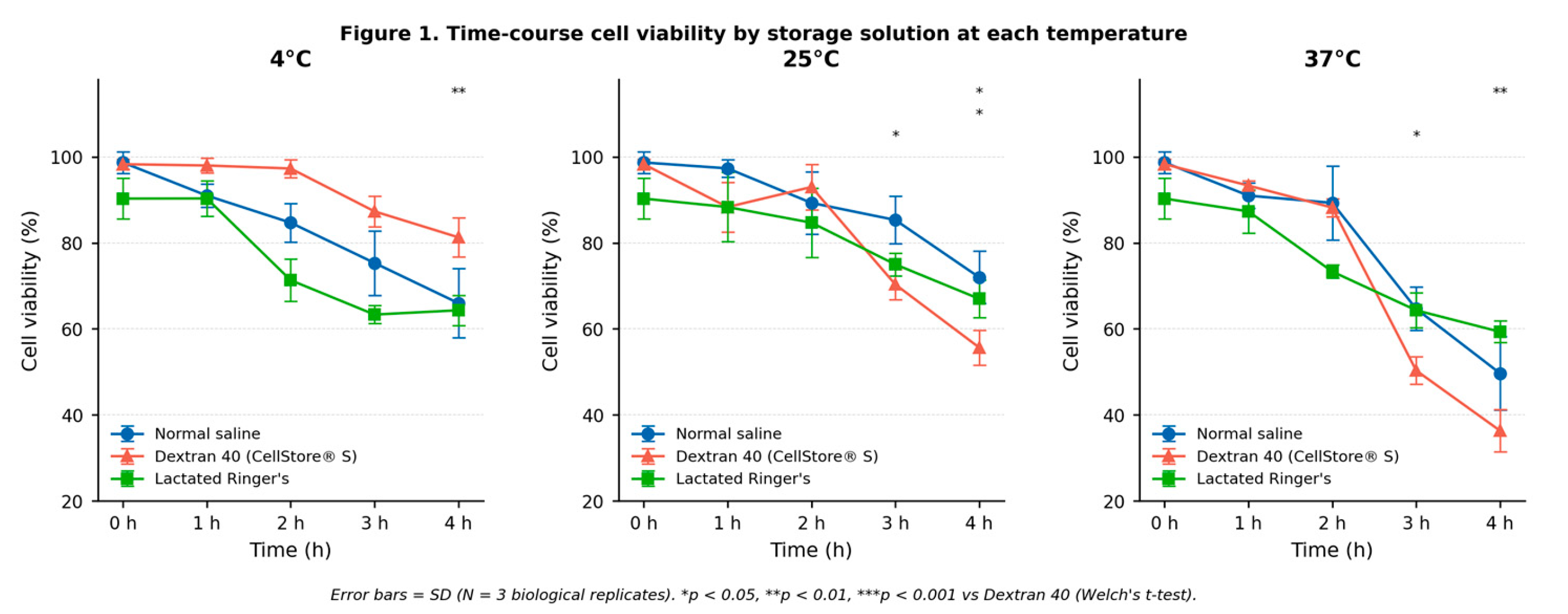
Figure 2.
Time-course cell viability of adipose-derived MSCs at each storage temperature (4°C, 25°C, and 37°C) grouped by carrier solution (Dextran 40 [CellStore® S], Normal saline, Lactated Ringer's). Data points represent mean ± SD (N = 5 biological replicates). Statistical annotations (Welch's t-test) compare temperatures at 3h and 4h timepoints vs. 4°C: p < 0.05, p < 0.01, p < 0.001.
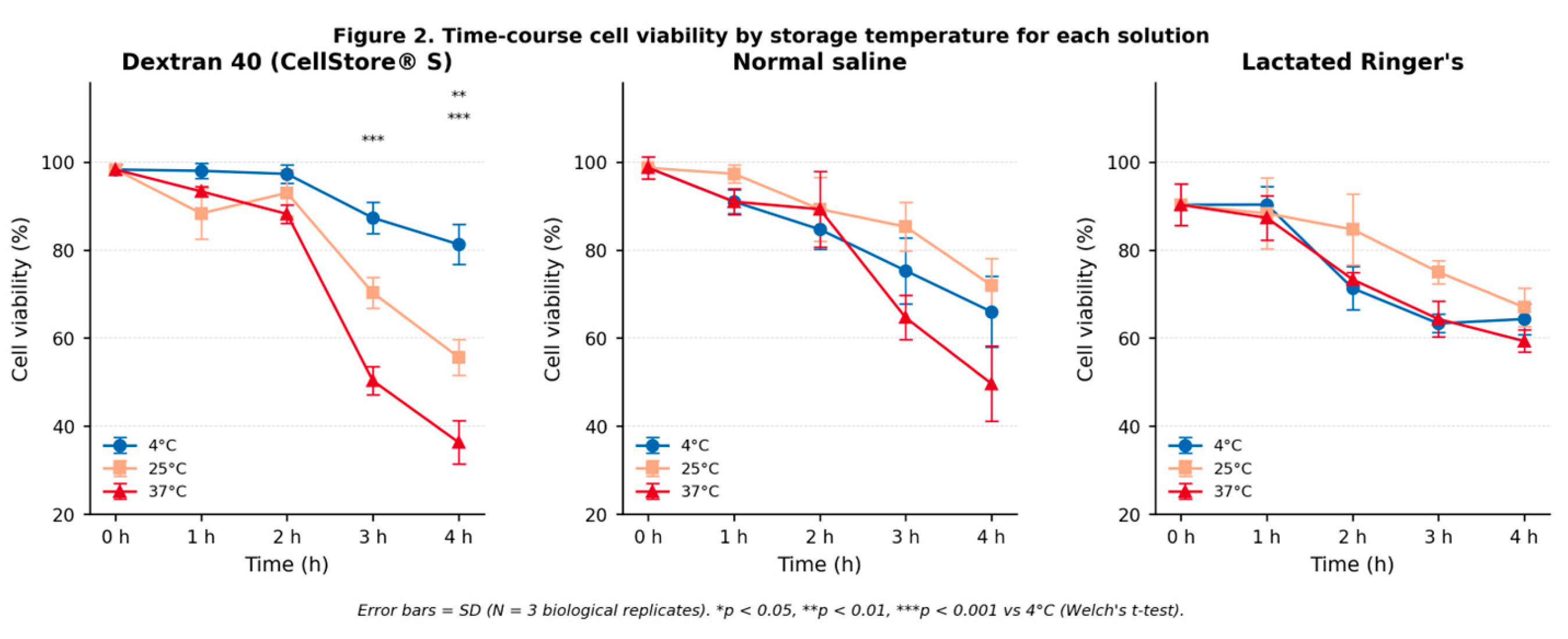
Figure 3.
Flow cytometric immunophenotyping of adipose-derived MSCs stored in 4°C dextran L (CellStore® S) for 4 hours. Panels are arranged in three rows: Row 1 (positive surface markers): CD73⁺ (99.96%), CD90⁺ (99.47%), CD105⁺ (99.98%); Row 2 (negative surface markers): CD34⁻ (0.08%), CD45⁻ (0.003%), HLA-DR⁻ (0.61%); Row 3 (cellular viability by 7-AAD exclusion): 0-hour baseline (R9: 0.000% dead, Sample 2) and 4-hour post-storage (R9: 0.000% dead, Sample 1), with ISCT compliance summary. Green borders denote 7-AAD viability panels. All six ISCT surface-marker criteria are met. Viability was 100% at both the pre-storage baseline and after 4 hours of storage.
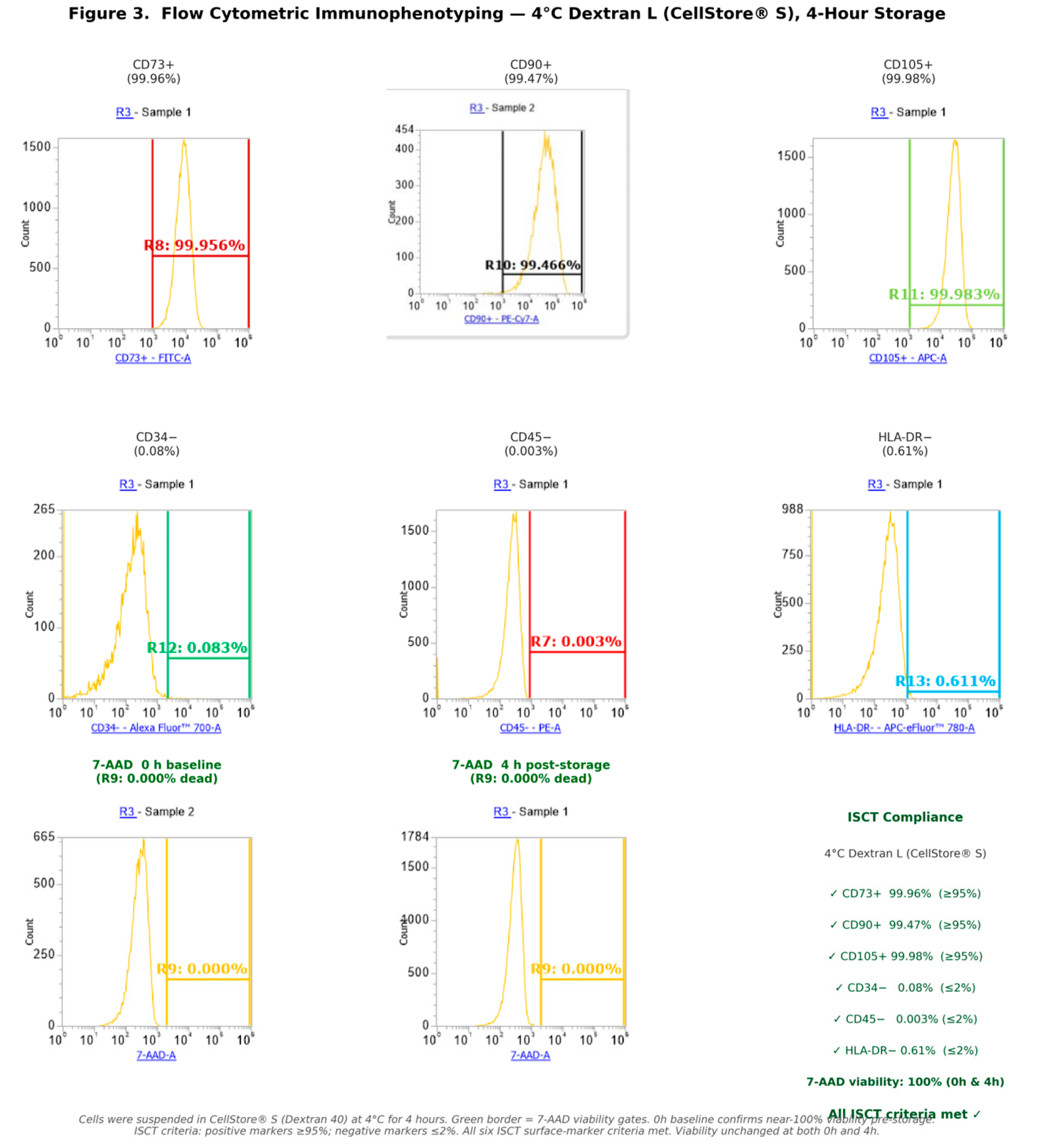
Figure 4.
Flow cytometric immunophenotyping of adipose-derived MSCs stored in 37°C normal saline for 4 hours. Panels are arranged in three rows: Row 1 (positive surface markers): CD73⁺ (99.32%), CD90⁺ (99.96%), CD105⁺ (92.51%); Row 2 (negative surface markers): CD34⁻ (12.49%), CD45⁻ (0.90%), HLA-DR⁻ (0.65%); Row 3 (cellular viability by 7-AAD exclusion): 0-hour baseline (R9: 0.061% dead, Sample #17098) and 4-hour post-storage (R9: 0.061% dead, Sample #14196), with ISCT compliance summary. Red borders and titles denote ISCT violations: CD105+ (92.51%) falls below the ≥95% threshold; CD34− (12.49%) exceeds the ≤2% threshold. Green borders denote 7-AAD viability panels. Data representative of Sample #14196 (surface markers).
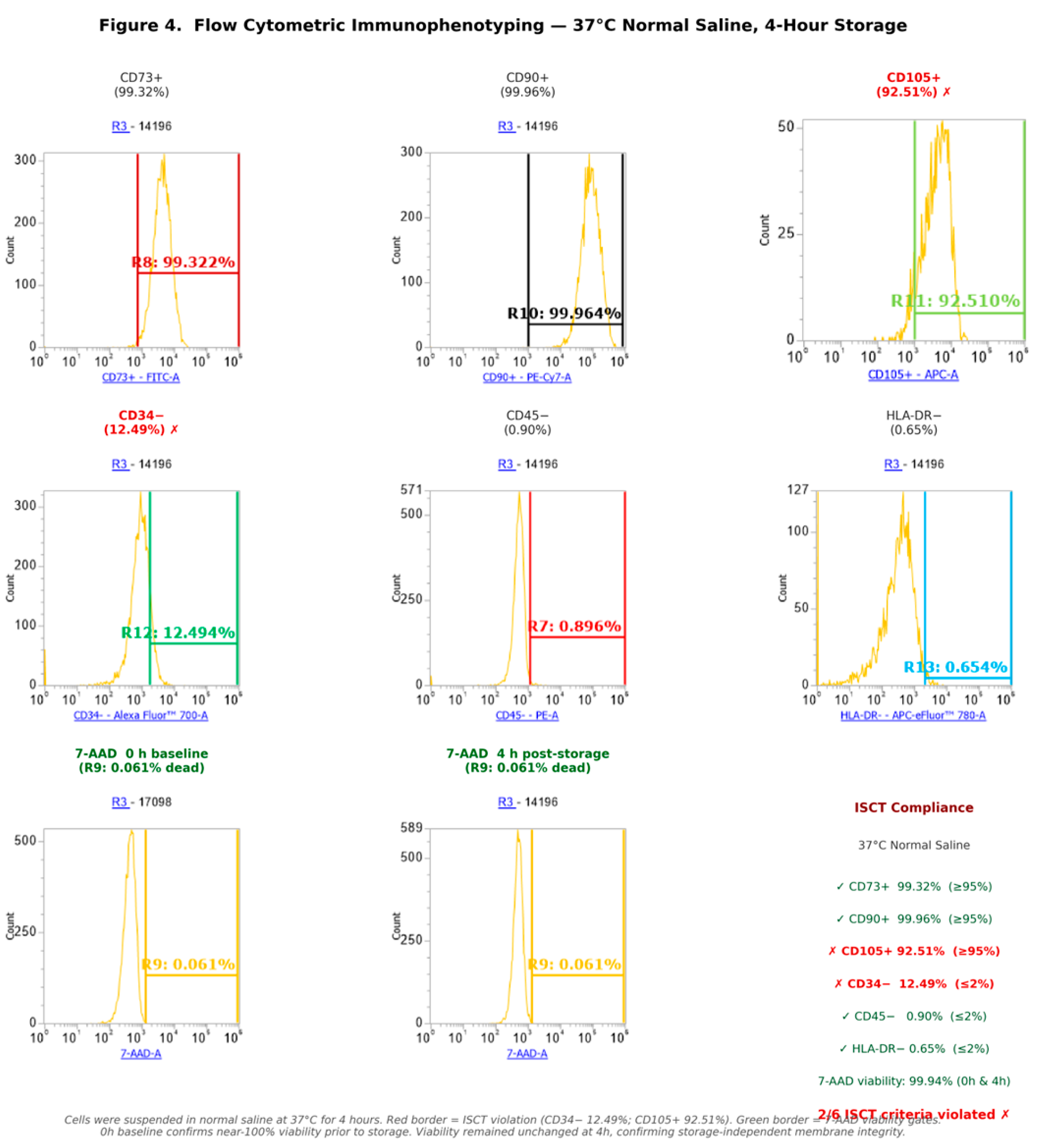
Figure 5.
Comparative ISCT surface marker expression between the optimal transport condition (4°C Dextran L, blue) and the worst transport condition (37°C Normal Saline, red) at 4 hours. ISCT thresholds are indicated by dashed lines (positive markers: ≥95%; negative markers: ≤2%). Two violations are observed under 37°C Normal Saline: CD105+ expression (92.51%) falls below the ≥95% threshold, and CD34− expression (12.49%) exceeds the ≤2% threshold. Both markers are fully compliant under 4°C Dextran L conditions.
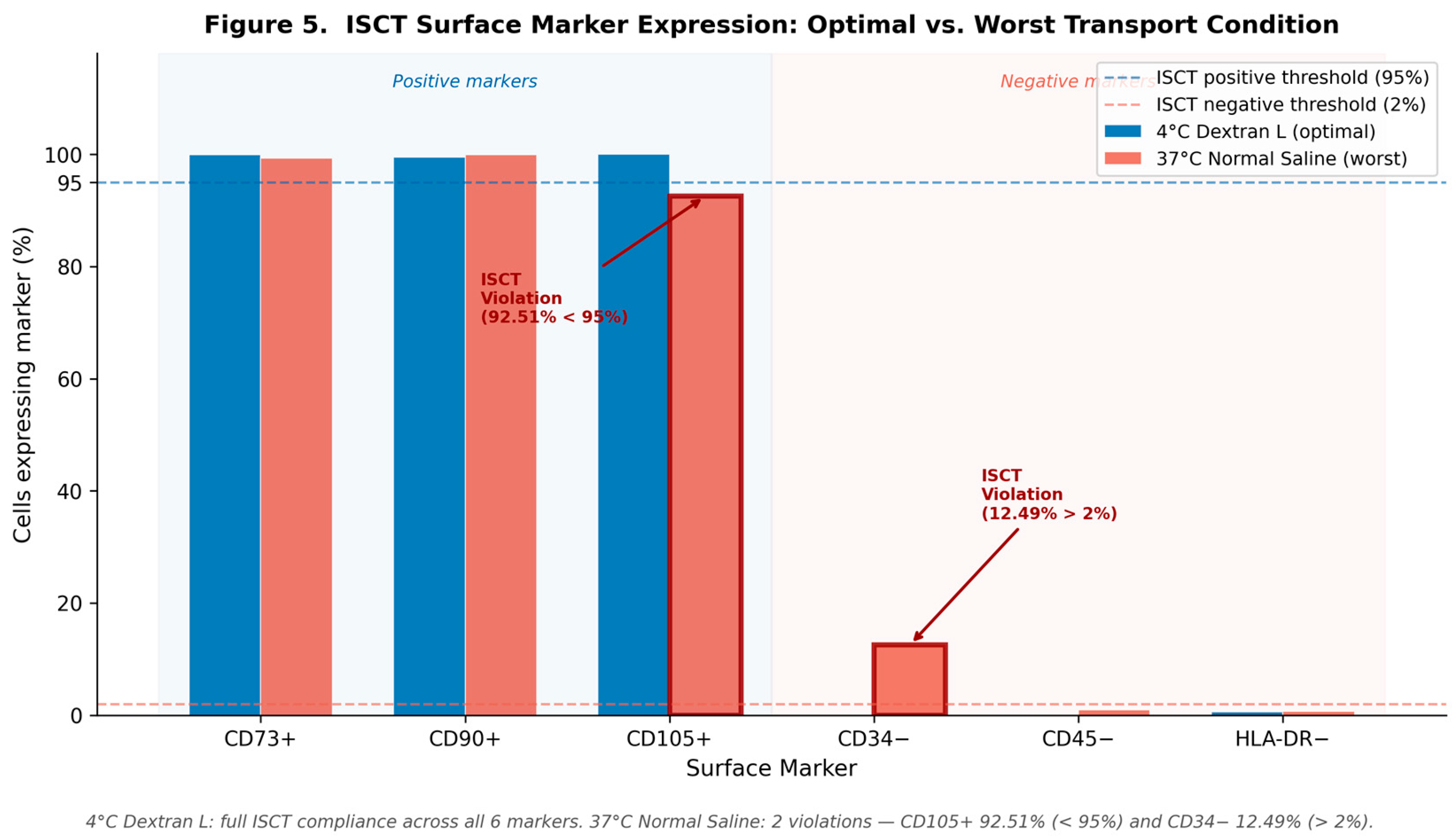

Table 1.
Summary of FCM Surface Marker Expression in MSCs Stored in 4°C Dextran L at 4 Hours.
| Marker | Fluorochrome | Measured (%) | ISCT Criterion | Compliance |
|---|---|---|---|---|
| CD73 | FITC | 99.96 | ≥95% positive | ✓ |
| CD105 | APC | 99.98 | ≥95% positive | ✓ |
| CD90 | PE-Cy7 | 99.47 | ≥95% positive | ✓ |
| CD34 | Alexa Fluor 700 | 0.08 | ≤2% negative | ✓ |
| CD45 | PE | 0.003 | ≤2% negative | ✓ |
| HLA-DR | APC-eFluor 780 | 0.61 | ≤2% negative | ✓ |
Table 2.
Comparative FCM Surface Marker Expression: Optimal vs. Worst Transport Condition.
| Marker | ISCT Criterion | 4°C Dextran L (4h) | 37°C Normal Saline (4h) | Δ |
|---|---|---|---|---|
| CD73+ | ≥95% | 99.96% ✓ | 99.32% ✓ | −0.64% |
| CD90+ | ≥95% | 99.47% ✓ | 99.96% ✓ | +0.49% |
| CD105+ | ≥95% | 99.98% ✓ | **92.510% ✗** | **−7.47%** |
| CD34− | ≤2% | 0.08% ✓ | **12.49% ✗** | **+12.41%** |
| CD45− | ≤2% | 0.003% ✓ | 0.90% ✓ | +0.90% |
| HLA-DR− | ≤2% | 0.61% ✓ | 0.65% ✓ | +0.04% |
Table 3.
Experimental conditions matrix.
| Temperature | Carrier | N (biological replicates) | Timepoints |
| 4°C (refrigerated) | Normal saline | 5 | 0, 1, 2, 3, 4 h |
| 4°C (refrigerated) | Dextran L (CellStore® S, 5%) | 5 | 0, 1, 2, 3, 4 h |
| 4°C (refrigerated) | Lactated Ringer's solution | 5 | 0, 1, 2, 3, 4 h |
| 25°C (room temperature) | Normal saline | 5 | 0, 1, 2, 3, 4 h |
| 25°C (room temperature) | Dextran L (CellStore® S, 5%) | 5 | 0, 1, 2, 3, 4 h |
| 25°C (room temperature) | Lactated Ringer's solution | 5 | 0, 1, 2, 3, 4 h |
| 37°C (body temperature) | Normal saline | 5 | 0, 1, 2, 3, 4 h |
| 37°C (body temperature) | Dextran L (CellStore® S, 5%) | 5 | 0, 1, 2, 3, 4 h |
| 37°C (body temperature) | Lactated Ringer's solution | 5 | 0, 1, 2, 3, 4 h |
Disclaimer/Publisher’s Note: The statements, opinions and data contained in all publications are solely those of the individual author(s) and contributor(s) and not of MDPI and/or the editor(s). MDPI and/or the editor(s) disclaim responsibility for any injury to people or property resulting from any ideas, methods, instructions or products referred to in the content. |
© 2026 by the authors. Licensee MDPI, Basel, Switzerland. This article is an open access article distributed under the terms and conditions of the Creative Commons Attribution (CC BY) license (http://creativecommons.org/licenses/by/4.0/).
Copyright: This open access article is published under a Creative Commons CC BY 4.0 license, which permit the free download, distribution, and reuse, provided that the author and preprint are cited in any reuse.