

Submitted:
13 July 2026
Posted:
15 July 2026
You are already at the latest version
Abstract
Autosomal dominant polycystic kidney disease (ADPKD) is a complex disorder characterized by the progressive development of renal cysts. Disease progression is linked to ADPKD-driven metabolic and mitochondrial reprogramming that exacerbates ADPKD pathophysiology by impairing central energetic pathways, including mitochondrial respiration and glucose, lipid, and amino acid metabolism. Caloric intake interventions can attenuate disease progression, but are difficult for patients to achieve and limited in application. Given the extensive alterations that impact ADPKD drug targets (i.e., mammalian target of rapamycin (mTOR) signaling pathway, peroxisome proliferator–activated receptor (PPAR) family, etc.) and drug metabolism (i.e., pharmacokinetics, cytochrome P450 (CYP450) family, etc.), improved understanding of the mechanisms driving metabolic and mitochondrial reprogramming may foster the identification of novel ADPKD severity-modifying drug targets and/or drugs. Such drugs could synergistically complement tolvaptan (the only U.S. Food and Drug Administration (FDA)-approved ADPKD therapeutic), or serve as alternatives, as tolvaptan is costly and associated with hepatotoxicity. Repurposing FDA-approved drugs could accelerate translation to clinical practice by leveraging prior work to improve the traditional drug development pipeline. To facilitate identification of repurposed drugs for ADPKD, we review ADPKD-driven metabolic and mitochondrial reprogramming from the perspective of drug target identification for repurposing ADPKD therapeutics.
Keywords:
autosomal dominant polycystic kidney disease
; ADPKD
; polycystic kidney disease
; PKD
; metabolic reprogramming
; mitochondrial dysfunction
; cystogenesis
; warburg effect
; oxidative phosphorylation
; drug repurposing
; drug repositioning
; drug development
; tolvaptan
; pharmacokinetics
; drug metabolism
; in silico
; drug targets
; precision medicine
Background
Autosomal dominant polycystic kidney disease (ADPKD), the fourth leading cause of renal replacement therapy, is projected to affect around 12 million individuals worldwide (Chapman et al., 2015). Its manifestations are characterized by the formation of renal cysts and a subsequent progressive decline in renal function. Cysts typically originate in the distal nephron, and as abnormal cystogenesis-promoting epithelial cells proliferate, they separate from the parent nephron, secrete chloride-rich fluid, and alter the extracellular matrix (Mahboob et al., 2025). The underlying cause of this process in most ADPKD cases involves a genetic defect in the PKD1 (~80% patients with ADPKD) or PKD2 (~15% patients with ADPKD) genes. These genes encode for polycystins 1 and 2 (PC1/PC2), respectively (Chapman et al., 2015). Many of their downstream pathways have been identified; however, so far, no single pathway has been broadly accepted as the principal driver of cystogenesis.
Pathways associated with metabolic and mitochondrial reprogramming have recently emerged as attractive candidates for elucidating the mechanistic underpinnings of renal cystogenesis. These pathways can directly modify ADPKD pathophysiology by impairing crucial processes central to normal healthy kidney function (Nowak & Hopp, 2020). In particular, ADPKD pathogenesis and progression revolve around energetic pathway alterations that impact glucose, amino acid, and lipid metabolism, along with mitochondrial respiration (Nowak & Hopp, 2020). Genetic defects appear to play a role in these alterations. PC1, for example, directly and indirectly affects mitochondrial structure and function (C.C. Lin et al., 2018). However, the mechanistic explanation is still not fully understood. Along with changes in other key cellular functions, these alterations can explain why ADPKD patients develop comorbidities associated with metabolic dysfunction, such as central obesity and insulin resistance (Mao et al., 2015), and the success of dietary interventions for symptomatic management (Nowak & Hopp, 2020). Understanding metabolic reprogramming is therefore crucial for advancing ADPKD patient care and treatment options.
Tolvaptan, originally indicated for hyponatremia, became the first and only U.S. Food and Drug Administration (FDA)-approved therapeutic for ADPKD in 2018 (Available Treatment, 2020). It is a vasopressin 2 receptor (V2R) antagonist, suggested to reduce the rate of renal function decline in ADPKD by ~30% on average. However, it is expensive (~$27,906 for a 30-day supply) (Cloutier et al., 2020), associated with idiosyncratic hepatotoxicity, and has a limited impact on extrarenal manifestations (Bellos, 2021; Chebib et al., 2018). Alternative candidate therapeutics to tolvaptan have entered preclinical and clinical trials, but none have successfully received FDA approval to date. Because de novo drug development is expensive and time-consuming, it may take many years for new ADPKD therapeutics to be approved. High levels of phenotypic, gene locus, and allelic heterogeneity contribute to ADPKD complexity (Claverie-Martin et al., 2015), further increasing drug development challenges and creating differences in drug biotransformation between individuals. Both of these challenges can be partially mitigated through drug repurposing. This approach leverages prior studies to reduce development costs and timelines, thereby increasing accessibility in drug research (reviewed in Al Khzem & Wali, 2025; Fisher et al., 2022; Jones et al., 2024). Given that the majority of drugs approved by the U.S. FDA in 2025 were repurposed drugs (Center for Drug Evaluation & Research, 2026), tolvaptan itself is a repurposed drug, and most drugs in clinical trials for ADPKD are already approved for another indication (Search ClinicalTrials.gov for: ADPKD, n.d.), drug repurposing offers a promising avenue to address the limitations in current ADPKD treatment options. To foster the identification of ADPKD therapeutics that may be used alone or in concert with nutritional interventions, we review evidence (see Supplementary File for review search terms) supporting metabolic and mitochondrial reprogramming as targets for ADPKD drug development, and its impact on identifying drug targets and repurposing ADPKD therapeutics.
Common Metabolic Diseases/Disorders Associated with ADPKD
Metabolic syndrome (MetS) describes a cluster of physiological, biochemical, clinical, and metabolic factors that notably increase the risk of cardiovascular diseases and type II diabetes mellitus (DM). Central obesity is the primary risk factor, but MetS diagnostic criteria also include insulin resistance, dyslipidemia, glucose intolerance, hypertension, inflammation, prothrombotic state, and microalbuminuria (Kaur, 2014). ADPKD patients have an increased association with multiple components of MetS, including increased waist circumference, fasting glucose levels, and dyslipidemia (Pietrzak-Nowacka et al., 2009; Veeramuthumari & Isabel, 2013). Given the risk MetS-associated conditions carry for the development of atherosclerotic cardiovascular disease, this increase in MetS components is critical to understand. It is likely they are contributing to the prevalent cardiovascular complications seen in ADPKD patients, including ischemic stroke, hypertension, hemorrhagic stroke, and atrial fibrillation (Hung et al., 2020).
Notably, ADPKD patients with DM exhibit worse disease progression than patients with ADPKD alone. These individuals have higher body mass index, greater total kidney volume (TKV), earlier onset of hypertension, increased renal failure risk, and reduced life expectancy (Chen et al., 2023). The risk of developing DM in ADPKD is substantially greater post-transplant, and patients have an approximate threefold increased risk of developing new-onset diabetes after transplantation within the first year compared to non-ADPKD recipients (de Mattos et al., 2005). ADPKD cohorts, however, appear to have a lower incidence of non-transplant-related DM (Pietrzak-Nowacka et al., 2006; Reed et al., 2012). Polycystic lesions may be responsible for the reduced risk by impairing renal gluconeogenesis and insulin degradation, causing hyperinsulinemic blood concentrations that lead to pancreatic beta cell fatigue, suppressing insulin production and release (Pietrzak-Nowacka et al., 2006). While studies have reported insulin resistance and hyperinsulinemia (Pietrzak-Nowacka et al., 2010; Vareesangthip et al., 1997), there remains controversy on whether it is ADPKD-specific or a consequence of general renal disease (Fliser et al., 1998). Recently, glucagon-like peptide-1 receptor agonists (GLP1-RAs), approved for chronic weight and type II DM management, have emerged as promising repurposing candidates for addressing metabolic reprogramming and cystogenesis in ADPKD. In two Pkd1 mouse models (Pkd1Rc/Rc and Pkd1nl/nl), Semaglutide significantly delayed cyst growth (p<0.001), decreased glucose uptake and glycolysis, and normalized mitochondrial function and morphology (Wu et al., 2024). Another GLP1-RA, tirzepatide, is currently in Phase II clinical trial (Table 1).
Dietary strategies have established benefits in ADPKD disease management. The prevalence of ADPKD-related metabolic disorders and reprogramming likely explains their efficacy (Nowak & Hopp, 2020). Daily caloric restriction (10-40%) in ADPKD mouse models (Pkd1RC/RC and PKDcond/cond:NesCre) slowed kidney growth, improved kidney function, and, notably, in the 40% caloric restriction group, reversed disease progression (Kipp et al., 2016; Warner et al., 2016). A clinical trial on weight loss using caloric restriction or intermittent fasting in overweight and obese ADPKD patients showed reduced kidney growth correlated with body weight and visceral adiposity loss. Notably, patients with clinically meaningful weight loss achieved kidney growth cessation (Hopp et al., 2022). Additionally, an exploratory randomized trial of 66 ADPKD patients found that the ketogenic diet group had improved kidney function compared to the control arm water fasting group (Cukoski et al., 2023). Overall, ADPKD is associated with metabolic derangements, and addressing them through dietary strategies, for example, is beneficial to disease progression.
Metabolic Reprogramming in ADPKD
Metabolic reprogramming, or alterations in physiological processes in response to disease and stress, is prevalent in ADPKD (Figure 1; and reviewed in Gao & Yu, 2025; Nowak & Hopp, 2020; Pagliarini & Podrini, 2021). Most notable is the Warburg effect, a preferential shift to glycolysis and lactic acid fermentation in the presence of oxygen that drives cystogenesis by promoting flux into biosynthetic pathways and, thus, uncontrolled cell proliferation (Liberti & Locasale, 2016). In silico analysis of human PKD1 renal cysts identified pathway alterations favoring glucose uptake and lactate production, with key glycolytic enzymes and regulatory genes upregulated, and mitochondrial energy metabolism and pyruvate oxidation pathways downregulated. Additionally, there was increased expression of anoctamin 1, indicating elevated oxidative damage and impairment of antioxidant pathways, such as glutathione metabolism (Song et al., 2024). A chronic progressive model of PKD using heterozygous Han:SPRD (Cy/+) rats also had glycolytic gene upregulation and gluconeogenesis-related gene downregulation in cystic kidneys. Treatment with 2-deoxyglucose, a glycolytic inhibitor, attenuated disease progression and improved both renal function and cyst index (Riwanto et al., 2016). Transcriptomic profiling of human PKD1 kidneys suggested cysts shuttle glucose through aerobic glycolysis and the pentose phosphate pathway (PPP) to sustain cell proliferation. They demonstrated upregulation in the rate limiting enzyme for the irreversible oxidative phase of the PPP and in a glycolytic-promoting aldolase isoform, along with downregulation in genes encoding enzymes for the reversible non-oxidative phase of the PPP and those shared with glycolysis/gluconeogenesis (Song et al., 2024). Perturbations within the PPP modulate reactive oxygen species (ROS) production and can exacerbate interstitial fibrosis and kidney injury (Subhash et al., 2024; TeSlaa et al., 2023).
ADPKD also dysregulates pathways related to mitochondrial respiration, particularly OXPHOS, further contributing to ROS generation and oxidative stress (Saxena et al., 2023). Seven out of the 10 most downregulated pathways in human PKD1 cystic renal tissue corresponded with mitochondrial bioenergetics: branched-chain amino acid degradation, pyruvate metabolism, FA metabolism, propanoate metabolism, butanoate metabolism, the TCA cycle, and OXPHOS (Song et al., 2024). Positron emission tomography demonstrated that ADPKD patients had reduced uptake rate of the acetyl coenzyme A (CoA) precursor, 11C-acetate, and thus decreased entrance into the TCA cycle, along with a reduction in average total kidney oxidative rates (Bjornstad et al., 2024). Cystic mice (Cys1cpk) and rats (Han:SPRD-Cy) had increased mRNA levels of the renoprotective and oxidative stress marker heme oxygenase (HO)-1, along with reduced antioxidant enzyme mRNAs, proteins, and activity levels (Maser et al., 2002). Another study found HO induction mitigated renal cystogenesis in Cys1cpk mice, while inhibition accelerated it, directly connecting oxidative stress to ADPKD pathophysiology (Zhou et al., 2012). Nitric oxide-related pathways may also increase levels of ROS and oxidative stress in ADPKD. Cystic kidneys of PCK rats exhibited elevated expression of NADPH-oxidase complex-4, likely leading to the observed reduction in endothelial nitric oxide synthase activity, along with upregulated ROS production, DNA damage, protein oxidation, and dysfunction in the mitochondria (Kahveci et al., 2020). Renal oxidative stress increases cell proliferation, extracellular matrix synthesis, inflammatory cell infiltration, and apoptosis (Nath et al., 1998). As such, oxidative stress is a likely measure of disease severity. This further highlights the pathogenic role of the Warburg effect in ADPKD cystogenesis and progression by impairing the pentose phosphate pathway (PPP) and mitochondrial respiration.
As noted above, amino acid and lipid metabolism are dysregulated in ADPKD patients. Glutamine (Gln), the most abundant amino acid in the human body, plays an important role in glutaminolysis and the TCA cycle in hyperproliferative cells (van den Heuvel et al., 2012). Similar to humans with ADPKD, mouse models of ADPKD demonstrated upregulation in glutaminase 1 (GLS1) GAC, a GLS1 isoform associated with glutaminolysis reliance in cancer, suggesting a similar phenomenon that supports hyperproliferation in ADPKD (Soomro et al., 2018). Gln and glutaminase are also important for shuttling ammonia in the urea cycle (Barmore et al., 2025), suggesting GLS1 upregulation in ADPKD may affect the urea cycle. Other key players in the urea cycle include argininosuccinate synthase 1 and arginine. Loss of argininosuccinate synthase 1 was found in both murine and human ADPKD tissue, along with an inverse relationship between arginine levels and cystogenesis (Trott et al., 2018), further highlighting urea cycle dysregulation in ADPKD. Given that the urea-to-plasma ratio in ADPKD patients is significantly associated with the rate of estimated glomerular filtration rate decline (P=0.02) (Heida et al., 2021), impairment in the urea cycle may have a pathogenic role in ADPKD progression. Lipid metabolism alterations are common in kidney disease and often present as increased fatty acid (FA) uptake, lipid accumulation, and the formation of lipid droplets (Saxena et al., 2023). For example, metabolomics identified acetylcarnitine, an important metabolite in mitochondrial β-oxidation, as a urine marker in Pkd1cko/cko mice specific to disease progression (Menezes et al., 2012; Podrini et al., 2020). Another study also demonstrated an intrinsic FA oxidation (FAO) defect in Pkd1cko/cko murine cells lacking glycolysis alterations (Menezes et al., 2016), suggesting ADPKD mitochondrial reprogramming may be related to mitochondrial dysfunction, rather than a preferential shift to aerobic glycolysis. Reduced FAO often occurs alongside increased de novo FA synthesis (FAS) and is associated with rapidly proliferating cells that require membrane production (Podrini et al., 2020). Pkd1 mouse embryonic fibroblasts and transcriptomic profiling of human PKD1 kidneys exhibited a similar phenomenon with increased FAS and preferential Gln use (Podrini et al., 2018). These findings suggest FAO impairment in ADPKD may not only highlight mitochondrial dysfunction, but additionally drive hyperproliferation in cystogenesis.
ADPKD mitochondrial reprogramming is likely influenced heavily by PC1. PC1 and PC2 are integral membrane proteins that form a heterotetrameric receptor-like/ion channel complex and are found in cellular locations like the endoplasmic reticulum (ER), cytoplasm, and primary cilia membranes. Together, PC1 and PC2 maintain cellular ion homeostasis and signal transduction, with PC1 as a transmembrane mechano-sensor receptor resembling G protein-coupled receptor function, and PC2 as a non-selective cationic channel permeable to calcium (Ferreira et al., 2015; Maser et al., 2022). In HEK293 and LLC-PK1 cell models with modulated PC1 and PC2 expression, oxygen levels controlled the subcellular location and channel activity of polycystin complexes by interacting with Egl-9 Family Hypoxia Inducible Factor 3, the oxygen-sensing prolyl hydroxylase domain containing protein that hydroxylates PC1. Cells lacking PC1 exhibited reduced oxygen consumption and mitochondrial calcium uptake, indicating that PC1 can directly regulate mitochondrial function in response to changes in cellular oxygen levels (Padovano et al., 2017). Given that ADPKD-associated cystogenesis aggravates regional hypoxia, pathogenic PKD1 variants impairing PC1 function may exacerbate hypoxia-related inflammation and ADPKD progression. One study found that tissue oxygenation is inversely correlated with height-adjusted total kidney volume (Buchholz & Eckardt, 2020), potentially supporting a connection between PC1 and hypoxia. In Pkd1ko/ko renal epithelial cells, a PC1 C-terminal fragment containing the cytoplasmic tail (CTT) translocated to the mitochondrial matrix. It restored some mitochondrial phenotypes, further confirming PC1 CTT interaction with both mitochondrial function and cellular metabolism. PC1 also modulates mitochondrial morphology, as demonstrated by studies in Pkd1 mouse models and PKD1 ADPKD patients, which found mitochondria had reduced elongation, mass, matrix electron density, and fusion proteins, along with increased mitochondrial network fragmentation, swelling with membrane whorls, and upregulated fission proteins (Cassina et al., 2020; C.C. Lin et al., 2018). PC2 potentially contributes to the changes in mitochondrial morphology as well, and one study found reduced PC2 expression affected mitochondrial calcium buffering, increasing mitochondrial network fragmentation. The authors proposed that PC2 regulates mitochondrial metabolism at mitochondrial-ER contacts (Kuo et al., 2019). In another study, truncating PKD2 mutations altered mitochondrial morphology, resulting in a rounded phenotype and dilated cristae, and resembling the effects of a PC1 deficiency (Chumley et al., 2019). Overall, it is clear that both PC1 and PC2 have a role in mitochondrial function and may exacerbate ADPKD pathophysiology and progression, as the alterations described are indicators of mitochondrial stress in ADPKD (Picard & McEwen, 2018), which directly enhances oxidative stress.
Established ADPKD Drug Targets
Drug development in ADPKD remains a formidable challenge as heterogeneity–both interindividually (e.g., variability in disease progression, penetrance/age of onset, and clinical manifestations) and intraindividually (e.g., differences in cyst severity and pathway activation)–complicates the design of universally effective therapies. Moreover, the full spectrum of functions for PKD1, PKD2, and other ADPKD-associated genes remains incompletely understood. For instance, PKD1/2 expression has been observed in multiple tissues and disease contexts, suggesting broader physiological roles that may influence drug response and safety. The approval of tolvaptan following the TEMPO (Torres et al., 2012) and REPRISE (Torres et al., 2017) trials marked a significant milestone in ADPKD treatment. V2R, the main target for tolvaptan, is upregulated in ADPKD and contributes to cell proliferation, fluid secretion, and cystogenesis. Its activation increases adenosine 3’,5 cyclic monophosphate (cAMP) and protein kinase A, which promote aquaporin 2 (AQP2) membrane localization, increasing water reabsorption and fluid secretion (Devlin et al., 2023). Tolvaptan antagonizes V2R, reducing cAMP levels and thereby inhibiting cyst epithelial cell proliferation and fluid secretion. However, benefits are largely restricted to patients with rapidly progressing ADPKD (Mayo Class ≥ 1C) or to chronic kidney disease (CKD) patients who begin treatment in an early stage (CKD Stage ≤ 3A) (Chebib et al., 2018). As such, alternative safe and effective therapeutics are desperately needed. Encouragingly, advances in understanding the metabolic underpinnings of ADPKD have created opportunities to explore potential drug targets and alleviate some of the challenges in ADPKD drug development. Here, we review both targets currently under development and those that could be studied in future analyses.
The mammalian target of rapamycin (mTOR) signaling pathway and its interactors are promising targets for addressing the consequences of the Warburg effect in ADPKD. The mTOR signaling pathway is hyperactivated in cystic lesions and promotes a metabolic shift from OXPHOS to glycolysis (Margaria et al., 2020). Increased mTOR activation is also suggested to increase insulin resistance and fat accumulation (Panwar et al., 2023), potentially contributing to the prevalence of MetS in ADPKD. Together, the effects of mTOR upregulation may explain why rapamycin inhibition of mTOR complex 1 (mTORC1) attenuated cystic progression in an ADPKD mouse model (Li et al., 2017). The phosphatidylinositol 3-kinase (PI3K)/protein kinase B pathway activates mTOR, initiating cellular processes, including metabolic reprogramming (Margaria et al., 2020). As such, dual PI3K/mTOR inhibition may have potential in ADPKD over only rapalog inhibition of mTOR (Liu et al., 2018). Activating adenosine monophosphate-activated protein kinase, which in turn inhibits mTOR signaling and cystic fibrosis transmembrane conductance regulator activity, is another strategy that may be able to reverse the Warburg-like metabolic shift in ADPKD cells and reduce cyst growth (Bais et al., 2022; Gwinn et al., 2008; Hallows et al., 2000; King et al., 2009). Wingless-related integration site (Wnt)/β-catenin signaling is proposed to coordinate metabolic flux consistent with the Warburg effect and activate mTORC1 through interactions with metabolic transcription factors such as cellular myelocytomatosis oncogene, peroxisome proliferator–activated receptor (PPAR) δ, and PPARγ (Sethi & Vidal-Puig, 2010). Transcriptomic profiling of PKD1 ADPKD renal cortical patient tissue samples demonstrated substantial Wnt pathway upregulation specific to cystic cells, and that the free CTT of PC1 physically interacts with β-catenin to inhibit Wnt signaling. The authors suggested that ADPKD-related loss of PC1-CTT β-catenin inhibition causes epithelial cells to revert to a less differentiated phenotype associated with proliferation and cyst formation (Lal et al., 2008), directly connecting aberrant Wnt/β-catenin signaling to cystogenesis. The ADPKD-associated Warburg effect can additionally be targeted using a glucose analog, 2-DG, along with dietary strategies, including the ketogenic diet and methionine restriction (Pagliarini & Podrini, 2021).
Autophagy interacts with many pathophysiological mechanisms such as oxidative stress, hypoxia, and the mTOR signalling pathway. Defective autophagy also has a demonstrated role in multiple kidney diseases, suggesting it as a potential ADPKD target (T.A. Lin et al., 2019). In vitro analysis found Pkd1-/- cells were unable to activate autophagic response (Rowe et al., 2013), and Pkd1 miRNA transgenic mice had significantly reduced renal mRNA expression of genes involved in autophagy and defective autophagy-lysosome pathway degradation of protein in vivo (Chou et al., 2018). An ADPKD zebrafish model (pkd1a-/-), as well as renal epithelial cells from both Pkd1-null mice and ADPKD patients, demonstrated impairment in autophagic flux, or fusion of the autophagosome and lysosome. The study also found that loss of the core autophagy protein, Atg5, promoted cystogenesis, and Beclin-1 activation of autophagy ameliorated cysts in the zebrafish model (Zhu et al., 2017). Researchers have additionally shown upregulation in early-stage autophagy regulators and activators, like p-Beclin-1, and downregulation in LC3-II, an autophagosome marker, suggesting the defect in autophagy occurs in later stages during autophagic flux (Atwood et al., 2020). Further, the mTOR signaling pathway is a known inhibitor of autophagy and may connect autophagy and cystogenesis (T.A. Lin et al., 2019; Nowak & Hopp, 2020). Increased mTOR signaling in Pkd1RC/RC mice resulted in suppressed autophagic flux (Atwood et al., 2020), and Pkd1-/- cells treated with rapamycin, an mTOR inhibitor, exhibited partially restored autophagy, increased cell survival under glucose deprivation, and decreased metabolic rates (Rowe et al., 2013). Mitophagy is a selective form of autophagy that facilitates the removal of damaged mitochondria (Ding & Yin, 2012). PINK1-mediated mitophagy can be triggered by various stressors present within ADPKD pathology, including hypoxia, ischemia, and oxidative stress, and cause increased ROS and mitochondrial damage. Cilia likely play an essential role in mitophagy activation, potentially linking PKD, one of the most common ciliopathies, to suppressed mitophagy (Nakashima et al., 2024). Abnormal mTOR activation could explain impaired mitophagy and defective cilia in ADPKD (Kou et al., 2019; Santoni et al., 2020), and rapalogs, or mTOR inhibitors, have shown potential in reducing cystic burden, restoring mitophagy and autophagy, and improving renal function, underscoring a potential pathogenic role of mitophagy in ADPKD progression (Li et al., 2017; Nakashima et al., 2024; Ramírez-Sagredo et al., 2021).
PPARα, a transcription factor vital for OXPHOS, is a potent target for addressing ADPKD-related mitochondrial dysfunction (Fan & Evans, 2015; Pagliarini & Podrini, 2021). Studies have noted reduced PPARɑ levels in CKD and aging kidneys impair FAO, leading to lipid accumulation in the kidney and worsened renal fibrosis (Chung et al., 2018; Kang et al., 2015). A similar phenotype is seen in ADPKD that augments cyst progression, pointing towards fenofibrate induction of PPARɑ as a potential therapeutic avenue (Lakhia, 2020). Researchers are additionally exploring inducing PPARγ signalling with compounds like thiazolidinediones to target lipid metabolism, glucose homeostasis, and oxidative stress (Mao et al., 2021). ADPKD-related lipid metabolism dysregulation can also be targeted through hydroxy-3-methylglutaryl-CoA reductase, the rate-limiting enzyme in cholesterol synthesis, and glycosphingolipids (Devlin et al., 2023), which accumulate in ADPKD (Natoli et al., 2020). PPARγ coactivator 1-ɑ (PGC-1ɑ), a key regulator of mitochondrial biogenesis and function, is downregulated in ADPKD and linked to several PKD and cilia-related genes (Chambers & Wingert, 2020). One study found that reduced PGC-1ɑ was associated with increased mitochondrial superoxide and reduced antioxidative effect, suggesting that inducing PGC-1ɑ activity may reduce oxidative stress in ADPKD (Ishimoto et al., 2017). Another avenue for rescuing mitochondrial function is the microRNA (miR)-17 family, which is upregulated in both human and murine ADPKD models (Lee et al., 2019). ADPKD mouse models have shown anti-miR-17 oligonucleotides can rescue OXPHOS defects and attenuate disease progression regardless of variant location (Pkd1 or Pkd2) or type (null or hypomorphic) (Hajarnis et al., 2017; Pagliarini & Podrini, 2021). Finally, the hormone somatostatin (SST) inhibits intracellular cAMP production and, in part, could regulate cellular energy turnover, which is heavily altered in ADPKD (Messchendorp et al., 2020). Targeting SST may additionally improve mitochondrial function, as cAMP signaling is involved in mitochondrial dynamics like fusion, fission, motility, and mitophagy (Di Benedetto et al., 2018). Reducing oxidative stress is critical in ADPKD as it may also reduce hypoxia, which is associated with cyst progression and stabilizing HIF-1ɑ, a potential accelerator of cyst growth (Kraus et al., 2018).
Recent experimental evidence has revealed altered amino acid metabolism in ADPKD as a potential target. Asparagine (Asn) synthetase (ASNS) is upregulated in ADPKD and has been implicated in mTOR activation and the Warburg effect through Asn, which can stimulate mTORC1 to boost glycolysis (Xu et al., 2021). ASNS inhibition in a slowly progressing ADPKD murine model drastically reduced TKV, proliferation, and cyst expansion, and rescued renal function (Clerici et al., 2024). Human and mouse ADPKD models have decreased arginosuccinate synthase 1 (ASS1) levels, resulting in arginine (Arg) auxotrophy (Gao & Yu, 2025). One study found Arg depletion increased ASS1 levels and reduced cystogenesis with minimal toxicity to non-cystic cells (Trott et al., 2018). Another demonstrated arginase-1 inhibition in Pkd1cond/cond mice slowed cyst growth, protected renal function, and improved survival rates (Yang et al., 2018). Gln metabolism may be a viable target given the ADPKD-associated derangements described above, and that glutaminolysis activates mTORC1 signaling (Durán et al., 2012). In one study, telaglenastat (CB-839) inhibition of glutamine metabolism by targeting GLS1-GAC had variable, but promising outcomes. In the less aggressive Aqp2-Cre; Pkd1fl/fl mouse model, telaglenastat slowed cyst growth and inhibited mTOR and mitogen-activated protein kinase kinase activation, but did not in the more aggressive Pkdh1-Cre; Pkd1fl/fl mouse model, likely due to compensatory metabolic pathways (Gao & Yu, 2025; Soomro et al., 2018). Dietary restriction of protein intake has shown benefits on renal function in experimental (Aukema et al., 1992) and interventional studies (Pahlavani & Azizi-Soleiman, 2023). A methionine (Met)-restricted diet may slow ADPKD progression as Met and S-adenosyl-L-Met levels are increased in ADPKD, ultimately activating c-Myc and cAMP pathways and accelerating cyst growth by inducing Mettl3 expression (Ling et al., 2023; Ramalingam et al., 2021). Met restriction in Pkd1RC/RC mice lowered kidney S-adenosylmethionine and Mettl3 levels and reduced both kidney-to-body weight ratio and cystic index (Ramalingam et al., 2021). Based on the National Institutes of Health Modification in Diet in Renal Disease study (Beck, 2023), however, the PKD Foundation does not suggest dietary protein restriction as beneficial for PKD patients, unless they are in renal failure (Nutrition, 2020). Overall, these drug targets may offer the opportunity to mitigate ADPKD metabolic and mitochondrial reprogramming and advance options for patients.
ADPKD Impact on Drug Development and Metabolism
Traditional drug development is an extensive process that can take up to 17 years and $2-3 billion, with approval rates around 11% (Pinzi et al., 2024). These associated costs and low approval rates create a barrier to entrance for designing novel therapeutics, further confounded in ADPKD by heterogeneity and limited research support (Figure 2). As such, drug repurposing has emerged as a strategic alternative. It leverages computational tools and prior studies to provide new indications for therapeutics, increasing approval rates to ~30% and reducing time and costs as low as three years and $300 million, respectively (Figure 3) (Pinzi et al., 2024). Tolvaptan is an example of the potential of drug repurposing in ADPKD, as it was originally indicated for hypervolemic or euvolemic hyponatremia from the syndrome of inappropriate antidiuretic hormone, chronic heart failure, or cirrhosis (Dubois et al., 2012). In one drug repurposing study from our lab, we prioritized 16 drug repurposing candidates and 29 drug targets, including many related to metabolic and mitochondrial pathways. We also demonstrated that protein interactions within oxidoreductase activity were involved in modulating drug metabolism activity, specifically within the cytochrome P450 (CYP450) family (Wilk et al., 2023). Many repurposed candidates proposed by the field target cellular metabolism, a key driver of cystogenesis in ADPKD (Table 1). Given the high level of metabolic and overall dysregulation in ADPKD, precision medicine approaches that account for its metabolic heterogeneity are essential as the field advances.
Hepatic and renal dysfunction can have significant consequences on drug metabolism and excretion, leading to the toxic accumulation of the drug and its metabolites (Garza et al., 2025). This is particularly relevant in ADPKD, as 81.7% of adult patients develop hepatic cysts (Arjune et al., 2025) that can cause hepatomegaly, loss of liver parenchyma, and lower quality of life in advanced stages (Hogan et al., 2015). Patients with renal disease commonly exhibit altered pharmacokinetics due to decreased renal excretion. They are particularly susceptible to volume of distribution and clearance changes, potentially requiring adjustments in dosing regimens to avoid adverse events (Lea-Henry et al., 2018). Chronic renal failure has been shown to affect hepatic, renal, and intestinal mechanisms of drug metabolism and transport with a 40-85% downregulation of hepatic and intestinal CYP450 metabolism, along with interference in phase II reactions and drug transporters like P-glycoprotein and organic anion transporting polypeptide (Dreisbach & Lertora, 2008). ADPKD-specific effects on hepatic and renal drug metabolism and transporter-mediated drug expression are not well characterized, but are likely, as CKD has been shown to change the protein expression and activity of kidney and liver drug-metabolizing enzymes and transporters (Tillmann, Rostami-Hodjegan, et al., 2025). One study on early- and late-stage ADPKD human kidney samples found significant reductions in several drug-metabolizing enzymes. In the late-stage group, organic anion transporter (OAT) 1, OAT3, and organic cation transporter 2 were below the limit of quantification (Tillmann, Peters, et al., 2025). These changes could contribute to tolvaptan hepatotoxicity, as demonstrated by an ADPKD ex-situ perfused rat liver study that found decreased hepatobiliary clearance and greater hepatocellular concentrations of tolvaptan within their prediction model (Beaudoin et al., 2019). Impaired renal function has additionally been associated with changes in absorption, plasma protein binding, and drug distribution, along with altered liver and gut drug metabolism and transport pathways. As such, the FDA recommends that renal function effects are characterized early in drug development to avoid later-stage issues in dosing, efficacy, and safety (Center for Drug Evaluation & Research, 2024). While currently understudied, ADPKD alterations in drug metabolism are likely and could explain some of the challenges in finding safe and effective therapeutic options.
Conclusions, Limitations, and Future Directions
Here, we outlined the metabolic and mitochondrial alterations associated with ADPKD, in relation to potential drug targets and the challenges in drug development. Further exploration is critical for understanding ADPKD pathophysiology and uncovering promising mechanisms for pharmacological evaluation. There are limitations in ADPKD research that have hindered the development of improved patient options beyond supportive care and tolvaptan. Lack of funding infrastructure, standardized protocols, and access to ADPKD patients creates barriers for investigators to form meaningful and appropriately powered human studies (Ostroff et al., 2022; Polycystic Kidney Disease Clinical Trials, n.d.). While there are numerous ADPKD models that offer many benefits, they do not fully capture the complexity of ADPKD pathophysiology, translatability to humans, and resources for model generation and upkeep (Sieben & Harris, 2023).
The difficulties with establishing therapies for ADPKD highlight the need to consider alternative approaches and influences in drug development as the field progresses. Drug repurposing is one route that leverages computational tools and previous drug studies to apply therapeutics to new indications, increasing approval rates and reducing time and costs (Pinzi et al., 2024). Combination therapy is also a promising approach that has historically been used to treat complicated diseases through the use of two or more drugs to address a specific indication. It has several benefits, including enhanced treatment efficacy, reduced required doses, decreased toxicity, and potential synergistic effects (Abbasi & Rousu, 2024; Güvenç Paltun et al., 2021). Both approaches have been explored in kidney disease. For example, ADPKD repurposing studies have investigated metformin and bardoxalone for their polyuria and renoprotective effects, respectively (Bais et al., 2022). Combination therapy with tolvaptan and dapagliflozin has also shown benefits in ADPKD patients, improving eGFR decline and potential renoprotective effects (Nishida et al., 2025). Used together, repurposing combination therapies could speed up the development process, incentivize research into the 7000 orphan diseases without treatments available, and reduce losses in complex diseases associated with high drug failure rates (Sun et al., 2016). Moving forward, in silico drug repurposing methods leveraging genomic, structural, and phenotypic data, combined with improved drug screening efficiency through options like high-throughput organoid drug screening, may help identify context-specific therapies and improve translational success (Jones et al., 2024). Techniques such as network/graph theory, molecular docking, and large language models offer promising avenues to refine target selection and predict off-target effects, potentially avoiding pitfalls seen with mTOR inhibitors and others. This, coupled with better characterization of metabolic and mitochondrial alterations and research into potential synergy between pharmaceuticals and nutritional therapies, may improve outcomes for ADPKD patients.
Supplementary Materials
The following supporting information can be downloaded at: Preprints.org, Table S1: Search strategy and terms for literature in this review and Table 1.
Author Contributions
JMF, MM, and BNL conceived the project, and ST, EJW, and TCH contributed to its conceptualization; JMF and ST prepared the figures; JMF and EJW drafted the manuscript; JMF, ST, EJW, TCH, PC, MM, and BNL edited and revised the manuscript; JMF, ST, EJW, TCH, PC, MM, and BNL approved the final version of the manuscript.
Support
This work was supported in part by National Institute of General Medical Sciences (NIGMS) of the National Institute of Health (NIH) (T32GM146611; JMF), the UAB Pilot Center for Precision Animal Modeling (C-PAM) (1U54OD030167; BNL), NIH-funded PKD Research Resource Consortium (U54DK126087; BNL and MM), PKD Foundation Award (BNL), NIDDK NIH-funded grant (R01DK134310; BNL and MM), the UAB Dean’s 1R01 Award (BNL), from the Office of Research and Development, Medical Research Service, Department of Veterans Affairs (1-I01-BX006266-01; MM), and the Detraz Endowed Research Fund in Polycystic Kidney Disease (MM). The funders had no role in the design and writing of the manuscript.
Financial Disclosure
MM reports additional research support from Otsuka, Sanofi, Vertex, and AbbVie, and has been a member of advisory boards for Sanofi, Santa Barbara Nutrients, and PKD Foundation. MM has provided consultancy for Otsuka, Sanofi, Vertex, AbbVie, and Regulus. JMF, ST, EJW, TCH, PHC, and BNL declare that they have no relevant financial interests.
Acknowledgments
We would like to thank other members of the Lasseigne Lab, especially Dr. Tabea Soelter, and external collaborators, especially Dr. Robert E. Van Sciver, for their thoughtful feedback.
References
- Abbasi, F., & Rousu, J. (2024). New methods for drug synergy prediction: A mini-review. Current Opinion in Structural Biology, 86(102827), 102827. [CrossRef]
- Al Khzem, A., & Wali, S. (2025). Drug Repurposing as an Effective Drug Discovery Strategy: A Critical Review. Drug Design, Development and Therapy, 19, 12019–12034. [CrossRef]
- Arjune, S., Todorova, P., Bartram, M. P., Grundmann, F., & Müller, R.-U. (2025). Liver manifestations in autosomal dominant polycystic kidney disease (ADPKD) and their impact on quality of life. Clinical Kidney Journal, 18(1), sfae363. [CrossRef]
- Atwood, D. J., Pokhrel, D., Brown, C. N., Holditch, S. J., Bachu, D. M., Thorburn, A., Hopp, K., & Edelstein, C. L. (2020). Increased mTOR and suppressed autophagic flux in the heart of a hypomorphic Pkd1 mouse model of autosomal dominant polycystic kidney disease. Cellular Signalling, 74(109730), 109730. [CrossRef]
- Aukema, H. M., Ogborn, M. R., Tomobe, K., Takahashi, H., Hibino, T., & Holub, B. J. (1992). Effects of dietary protein restriction and oil type on the early progression of murine polycystic kidney disease. Kidney International, 42(4), 837–842. [CrossRef]
- Available Treatment. (2020, April 11). Polycystic Kidney Disease PKD | Treatment Research | PKD Foundation; PKD Foundation. https://pkdcure.org/about-the-disease/living-with-pkd/treatments/.
- Bais, T., Gansevoort, R. T., & Meijer, E. (2022). Drugs in clinical development to treat autosomal dominant polycystic kidney disease. Drugs, 82(10), 1095–1115. [CrossRef]
- Barmore, W., Azad, F., & Stone, W. L. (2025). Physiology, urea cycle. In StatPearls. StatPearls Publishing. https://www.ncbi.nlm.nih.gov/books/NBK513323/.
- Beaudoin, J. J., Bezençon, J., Cao, Y., Mizuno, K., Roth, S. E., Brock, W. J., & Brouwer, K. L. R. (2019). Altered hepatobiliary disposition of tolvaptan and selected tolvaptan metabolites in a rodent model of polycystic kidney disease. Drug Metabolism and Disposition: The Biological Fate of Chemicals, 47(2), 155–163. [CrossRef]
- Beck, G. (2023). Modification of Diet in Renal Disease [Dataset]. NIDDK Central Repository. https://repository.niddk.nih.gov/studies/mdrd.
- Bellos, I. (2021). Safety profile of tolvaptan in the treatment of autosomal dominant polycystic kidney disease. Therapeutics and Clinical Risk Management, 17, 649–656. [CrossRef]
- Bjornstad, P., Richard, G., Choi, Y. J., Nowak, K. L., Steele, C., Chonchol, M. B., Nadeau, K. J., Vigers, T., Pyle, L., Tommerdahl, K., van Raalte, D. H., Hilkin, A., Driscoll, L., Birznieks, C., Hopp, K., Wang, W., Edelstein, C., Nelson, R. G., Gregory, A. V., … Gitomer, B. (2024). Kidney energetics and cyst burden in autosomal dominant polycystic kidney disease: A pilot study. American Journal of Kidney Diseases: The Official Journal of the National Kidney Foundation, 84(3), 286–297.e1. [CrossRef]
- Buchholz, B., & Eckardt, K. U. (2020). Role of oxygen and the HIF-pathway in polycystic kidney disease. Cellular Signalling, 69(109524), 109524. [CrossRef]
- Cassina, L., Chiaravalli, M., & Boletta, A. (2020). Increased mitochondrial fragmentation in polycystic kidney disease acts as a modifier of disease progression. FASEB Journal: Official Publication of the Federation of American Societies for Experimental Biology, 34(5), 6493–6507. [CrossRef]
- Center for Drug Evaluation, & Research. (2024, July 29). Pharmacokinetics in Patients with Impaired Renal Function — Study Design, Data Analysis, and Impact on Dosing. U.S. Food and Drug Administration; FDA. https://www.fda.gov/regulatory-information/search-fda-guidance-documents/pharmacokinetics-patients-impaired-renal-function-study-design-data-analysis-and-impact-dosing.
- Center for Drug Evaluation, & Research. (2026, February 2). Novel Drug Approvals for 2025. U.S. Food and Drug Administration; FDA. https://www.fda.gov/drugs/novel-drug-approvals-fda/novel-drug-approvals-2025.
- Chambers, J. M., & Wingert, R. A. (2020). PGC-1α in disease: Recent renal insights into a versatile metabolic regulator. Cells (Basel, Switzerland), 9. [CrossRef]
- Chapman, A. B., Devuyst, O., Eckardt, K. U., Gansevoort, R. T., Harris, T., Horie, S., Kasiske, B. L., Odland, D., Pei, Y., Perrone, R. D., Pirson, Y., Schrier, R. W., Torra, R., Torres, V. E., Watnick, T., Wheeler, D. C., & Conference Participants. (2015). Autosomal-dominant polycystic kidney disease (ADPKD): executive summary from a Kidney Disease: Improving Global Outcomes (KDIGO) Controversies Conference. Kidney International, 88(1), 17–27. [CrossRef]
- Chebib, F. T., Perrone, R. D., Chapman, A. B., Dahl, N. K., Harris, P. C., Mrug, M., Mustafa, R. A., Rastogi, A., Watnick, T., Yu, A. S. L., & Torres, V. E. (2018). A practical guide for treatment of rapidly progressive ADPKD with tolvaptan. Journal of the American Society of Nephrology: JASN, 29(10), 2458–2470. [CrossRef]
- Chen, L. C., Chu, Y. C., Lu, T., Lin, H. Y. H., & Chan, T. C. (2023). Cardiometabolic comorbidities in autosomal dominant polycystic kidney disease: a 16-year retrospective cohort study. BMC Nephrology, 24(1), 333. [CrossRef]
- Chou, L. F., Cheng, Y. L., Hsieh, C.Y., Lin, C. Y., Yang, H. Y., Chen, Y. C., Hung, C. C., Tian, Y. C., Yang, C.W., & Chang, M.Y. (2018). Effect of trehalose supplementation on autophagy and cystogenesis in a mouse model of polycystic kidney disease. Nutrients, 11(1), 42. [CrossRef]
- Chumley, P., Zhou, J., Mrug, S., Chacko, B., Parant, J. M., Challa, A. K., Wilson, L. S., Berryhill, T. F., Barnes, S., Kesterson, R. A., Bell, P. D., Darley-Usmar, V. M., Yoder, B. K., & Mrug, M. (2019). Truncating PKHD1 and PKD2 mutations alter energy metabolism. American Journal of Physiology. Renal Physiology, 316(3), F414–F425. [CrossRef]
- Chung, K. W., Lee, E. K., Lee, M. K., Oh, G. T., Yu, B. P., & Chung, H. Y. (2018). Impairment of PPARα and the fatty acid oxidation pathway aggravates renal fibrosis during aging. Journal of the American Society of Nephrology: JASN, 29(4), 1223–1237. [CrossRef]
- Claverie-Martin, F., Gonzalez-Paredes, F. J., & Ramos-Trujillo, E. (2015). Splicing defects caused by exonic mutations in PKD1 as a new mechanism of pathogenesis in autosomal dominant polycystic kidney disease. RNA Biology, 12(4), 369–374. [CrossRef]
- Clerici, S., Podrini, C., Stefanoni, D., Distefano, G., Cassina, L., Steidl, M. E., Tronci, L., Canu, T., Chiaravalli, M., Spies, D., Bell, T. A., 3rd, Costa, A. S., Esposito, A., D’Alessandro, A., Frezza, C., Bachi, A., & Boletta, A. (2024). Inhibition of asparagine synthetase effectively retards polycystic kidney disease progression. EMBO Molecular Medicine, 16(6), 1379–1403. [CrossRef]
- Cloutier, M., Manceur, A. M., Guerin, A., Aigbogun, M. S., Oberdhan, D., & Gauthier-Loiselle, M. (2020). The societal economic burden of autosomal dominant polycystic kidney disease in the United States. BMC Health Services Research, 20(1), 126. [CrossRef]
- Cukoski, S., Lindemann, C. H., Arjune, S., Todorova, P., Brecht, T., Kühn, A., Oehm, S., Strubl, S., Becker, I., Kämmerer, U., Torres, J. A., Meyer, F., Schömig, T., Hokamp, N. G., Siedek, F., Gottschalk, I., Benzing, T., Schmidt, J., Antczak, P., … Müller, R.U. (2023). Feasibility and impact of ketogenic dietary interventions in polycystic kidney disease: KETO-ADPKD-a randomized controlled trial. Cell Reports. Medicine, 4(11), 101283. [CrossRef]
- de Mattos, A. M., Olyaei, A. J., Prather, J. C., Golconda, M. S., Barry, J. M., & Norman, D. J. (2005). Autosomal-dominant polycystic kidney disease as a risk factor for diabetes mellitus following renal transplantation. Kidney International, 67(2), 714–720. [CrossRef]
- Devlin, L., Dhondurao Sudhindar, P., & Sayer, J. A. (2023). Renal ciliopathies: promising drug targets and prospects for clinical trials. Expert Opinion on Therapeutic Targets, 27(4-5), 325–346. [CrossRef]
- Di Benedetto, G., Gerbino, A., & Lefkimmiatis, K. (2018). Shaping mitochondrial dynamics: The role of cAMP signalling. Biochemical and Biophysical Research Communications, 500(1), 65–74. [CrossRef]
- Ding, W. X., & Yin, X. M. (2012). Mitophagy: mechanisms, pathophysiological roles, and analysis. Biological Chemistry, 393(7), 547–564. [CrossRef]
- Dreisbach, A. W., & Lertora, J. J. L. (2008). The effect of chronic renal failure on drug metabolism and transport. Expert Opinion on Drug Metabolism & Toxicology, 4(8), 1065–1074. [CrossRef]
- Dubois, E. A., Rissmann, R., & Cohen, A. F. (2012). Tolvaptan: New drug mechanisms. British Journal of Clinical Pharmacology, 73(1), 9–11. [CrossRef]
- Durán, R. V., Oppliger, W., Robitaille, A. M., Heiserich, L., Skendaj, R., Gottlieb, E., & Hall, M. N. (2012). Glutaminolysis activates Rag-mTORC1 signaling. Molecular Cell, 47(3), 349–358. [CrossRef]
- Fan, W., & Evans, R. (2015). PPARs and ERRs: molecular mediators of mitochondrial metabolism. Current Opinion in Cell Biology, 33, 49–54. [CrossRef]
- Ferreira, F. M., Watanabe, E. H., & Onuchic, L. (2015). Polycystins and molecular basis of Autosomal dominant polycystic kidney disease. Exon Publications, 139–167. [CrossRef]
- Fisher, J. L., Jones, E. F., Flanary, V. L., Williams, A. S., Ramsey, E. J., & Lasseigne, B. N. (2022). Considerations and challenges for sex-aware drug repurposing. Biology of Sex Differences, 13(1), 13. [CrossRef]
- Fliser, D., Pacini, G., Engelleiter, R., Kautzky-Willer, A., Prager, R., Franek, E., & Ritz, E. (1998). Insulin resistance and hyperinsulinemia are already present in patients with incipient renal disease. Kidney International, 53(5), 1343–1347. [CrossRef]
- Gao, J., & Yu, X. (2025). Metabolic reprogramming in autosomal dominant polycystic kidney disease: Role in cystogenesis and novel therapeutic approaches. Biomedicines, 13(7), 1596. [CrossRef]
- Garza, A. Z., Park, S. B., & Kocz, R. (2025). Drug elimination. In StatPearls. StatPearls Publishing. https://www.ncbi.nlm.nih.gov/books/NBK547662/.
- Güvenç Paltun, B., Kaski, S., & Mamitsuka, H. (2021). Machine learning approaches for drug combination therapies. Briefings in Bioinformatics, 22(6), bbab293. [CrossRef]
- Gwinn, D. M., Shackelford, D. B., Egan, D. F., Mihaylova, M. M., Mery, A., Vasquez, D. S., Turk, B. E., & Shaw, R. J. (2008). AMPK phosphorylation of raptor mediates a metabolic checkpoint. Molecular Cell, 30(2), 214–226. [CrossRef]
- Hajarnis, S., Lakhia, R., Yheskel, M., Williams, D., Sorourian, M., Liu, X., Aboudehen, K., Zhang, S., Kersjes, K., Galasso, R., Li, J., Kaimal, V., Lockton, S., Davis, S., Flaten, A., Johnson, J. A., Holland, W. L., Kusminski, C. M., Scherer, P. E., … Patel, V. (2017). microRNA-17 family promotes polycystic kidney disease progression through modulation of mitochondrial metabolism. Nature Communications, 8(1), 14395. [CrossRef]
- Hallows, K. R., Raghuram, V., Kemp, B. E., Witters, L. A., & Foskett, J. K. (2000). Inhibition of cystic fibrosis transmembrane conductance regulator by novel interaction with the metabolic sensor AMP-activated protein kinase. The Journal of Clinical Investigation, 105(12), 1711–1721. [CrossRef]
- Heida, J. E., Gansevoort, R. T., Messchendorp, A. L., Meijer, E., Casteleijn, N. F., Boertien, W. E., Zittema, D., & DIPAK Consortium. (2021). Use of the urine-to-plasma urea ratio to predict ADPKD progression. Clinical Journal of the American Society of Nephrology: CJASN, 16(2), 204–212. [CrossRef]
- Hogan, M. C., Abebe, K., Torres, V. E., Chapman, A. B., Bae, K. T., Tao, C., Sun, H., Perrone, R. D., Steinman, T. I., Braun, W., Winklhofer, F. T., Miskulin, D. C., Rahbari-Oskoui, F., Brosnahan, G., Masoumi, A., Karpov, I. O., Spillane, S., Flessner, M., Moore, C. G., & Schrier, R. W. (2015). Liver involvement in early autosomal-dominant polycystic kidney disease. Clinical Gastroenterology and Hepatology: The Official Clinical Practice Journal of the American Gastroenterological Association, 13(1), 155–164.e6. [CrossRef]
- Hopp, K., Catenacci, V. A., Dwivedi, N., Kline, T. L., Wang, W., You, Z., Nguyen, D. T., Bing, K., Poudyal, B., Johnson, G. C., Jackman, M. R., Miller, M., Steele, C. N., Serkova, N. J., MacLean, P. S., Nemenoff, R. A., Gitomer, B., Chonchol, M., & Nowak, K. L. (2022). Weight loss and cystic disease progression in autosomal dominant polycystic kidney disease. iScience, 25(1), 103697. [CrossRef]
- Hung, P. H., Lin, C. H., Hung, K. Y., Muo, C. H., Chung, M. C., Chang, C. H., & Chung, C. J. (2020). Clinical burden of autosomal dominant polycystic kidney disease. Aging, 12(4), 3899–3910. [CrossRef]
- Ishimoto, Y., Inagi, R., Yoshihara, D., Kugita, M., Nagao, S., Shimizu, A., Takeda, N., Wake, M., Honda, K., Zhou, J., & Nangaku, M. (2017). Mitochondrial abnormality facilitates cyst formation in autosomal dominant polycystic kidney disease. Molecular and Cellular Biology, 37(24). [CrossRef]
- Jones, E., Taluri, S., Wilk, E., & Lasseigne, B. (2024). Streamlining drug repurposing: Optimizing candidate prioritization to facilitate clinical adoption and minimize attrition rates. In Preprints. [CrossRef]
- Kahveci, A. S., Barnatan, T. T., Kahveci, A., Adrian, A. E., Arroyo, J., Eirin, A., Harris, P. C., Lerman, A., Lerman, L. O., Torres, V. E., & Irazabal, M. V. (2020). Oxidative stress and mitochondrial abnormalities contribute to decreased endothelial nitric oxide synthase expression and renal disease progression in early experimental Polycystic Kidney Disease. International Journal of Molecular Sciences, 21(6), 1994. [CrossRef]
- Kang, H. M., Ahn, S. H., Choi, P., Ko, Y. A., Han, S. H., Chinga, F., Park, A. S. D., Tao, J., Sharma, K., Pullman, J., Bottinger, E. P., Goldberg, I. J., & Susztak, K. (2015). Defective fatty acid oxidation in renal tubular epithelial cells has a key role in kidney fibrosis development. Nature Medicine, 21(1), 37–46. [CrossRef]
- Kaur, J. (2014). A comprehensive review on metabolic syndrome. Cardiology Research and Practice, 2014, 943162. [CrossRef]
- King, J. D., Jr, Fitch, A. C., Lee, J. K., McCane, J. E., Mak, D. O. D., Foskett, J. K., & Hallows, K. R. (2009). AMP-activated protein kinase phosphorylation of the R domain inhibits PKA stimulation of CFTR. American Journal of Physiology. Cell Physiology, 297(1), C94–C101. [CrossRef]
- Kipp, K. R., Rezaei, M., Lin, L., Dewey, E. C., & Weimbs, T. (2016). A mild reduction of food intake slows disease progression in an orthologous mouse model of polycystic kidney disease. American Journal of Physiology. Renal Physiology, 310(8), F726–F731. [CrossRef]
- Kou, P., Wei, S., & Xiong, F. (2019). Recent advances of mTOR inhibitors use in autosomal Dominant Polycystic Kidney Disease: Is the road still open? Current Medicinal Chemistry, 26(16), 2962–2973. [CrossRef]
- Kraus, A., Peters, D. J. M., Klanke, B., Weidemann, A., Willam, C., Schley, G., Kunzelmann, K., Eckardt, K. U., & Buchholz, B. (2018). HIF-1α promotes cyst progression in a mouse model of autosomal dominant polycystic kidney disease. Kidney International, 94(5), 887–899. [CrossRef]
- Kuo, I. Y., Brill, A. L., Lemos, F. O., Jiang, J. Y., Falcone, J. L., Kimmerling, E. P., Cai, Y., Dong, K., Kaplan, D. L., Wallace, D. P., Hofer, A. M., & Ehrlich, B. E. (2019). Polycystin 2 regulates mitochondrial Ca2+ signaling, bioenergetics, and dynamics through mitofusin 2. Science Signaling, 12(580), eaat7397. [CrossRef]
- Lakhia, R. (2020). The role of PPARα in autosomal dominant polycystic kidney disease. Current Opinion in Nephrology and Hypertension, 29(4), 432–438. [CrossRef]
- Lal, M., Song, X., Pluznick, J. L., Di Giovanni, V., Merrick, D. M., Rosenblum, N. D., Chauvet, V., Gottardi, C. J., Pei, Y., & Caplan, M. J. (2008). Polycystin-1 C-terminal tail associates with beta-catenin and inhibits canonical Wnt signaling. Human Molecular Genetics, 17(20), 3105–3117. [CrossRef]
- Lea-Henry, T. N., Carland, J. E., Stocker, S. L., Sevastos, J., & Roberts, D. M. (2018). Clinical pharmacokinetics in kidney disease: Fundamental principles. Clinical Journal of the American Society of Nephrology: CJASN, 13(7), 1085–1095. [CrossRef]
- Lee, E. C., Valencia, T., Allerson, C., Schairer, A., Flaten, A., Yheskel, M., Kersjes, K., Li, J., Gatto, S., Takhar, M., Lockton, S., Pavlicek, A., Kim, M., Chu, T., Soriano, R., Davis, S., Androsavich, J. R., Sarwary, S., Owen, T., … Patel, V. (2019). Discovery and preclinical evaluation of anti-miR-17 oligonucleotide RGLS4326 for the treatment of polycystic kidney disease. Nature Communications, 10(1), 4148. [CrossRef]
- Li, A., Fan, S., Xu, Y., Meng, J., Shen, X., Mao, J., Zhang, L., Zhang, X., Moeckel, G., Wu, D., Wu, G., & Liang, C. (2017). Rapamycin treatment dose-dependently improves the cystic kidney in a new ADPKD mouse model via the mTORC1 and cell-cycle-associated CDK1/cyclin axis. Journal of Cellular and Molecular Medicine, 21(8), 1619–1635. [CrossRef]
- Liberti, M. V., & Locasale, J. W. (2016). The Warburg Effect: How does it benefit cancer cells? Trends in Biochemical Sciences, 41(3), 211–218. [CrossRef]
- Lin, C. C., Kurashige, M., Liu, Y., Terabayashi, T., Ishimoto, Y., Wang, T., Choudhary, V., Hobbs, R., Liu, L. K., Lee, P. H., Outeda, P., Zhou, F., Restifo, N. P., Watnick, T., Kawano, H., Horie, S., Prinz, W., Xu, H., Menezes, L. F., & Germino, G. G. (2018). A cleavage product of Polycystin-1 is a mitochondrial matrix protein that affects mitochondria morphology and function when heterologously expressed. Scientific Reports, 8(1), 2743. [CrossRef]
- Ling, Z. N., Jiang, Y. F., Ru, J. N., Lu, J. H., Ding, B., & Wu, J. (2023). Amino acid metabolism in health and disease. Signal Transduction and Targeted Therapy, 8(1), 345. [CrossRef]
- Lin, T. A., Wu, V., & Wang, C. Y. (2019). Autophagy in chronic kidney diseases. Cells (Basel, Switzerland), 8. [CrossRef]
- Liu, Y., Pejchinovski, M., Wang, X., Fu, X., Castelletti, D., Watnick, T. J., Arcaro, A., Siwy, J., Mullen, W., Mischak, H., & Serra, A. L. (2018). Dual mTOR/PI3K inhibition limits PI3K-dependent pathways activated upon mTOR inhibition in autosomal dominant polycystic kidney disease. Scientific Reports, 8(1), 5584. [CrossRef]
- Mahboob, M., Rout, P., Leslie, S. W., & Bokhari, S. R. A. (2025). Autosomal dominant polycystic kidney disease. In StatPearls. StatPearls Publishing. https://www.ncbi.nlm.nih.gov/books/NBK532934/.
- Mao, Z., Valluru, M. K., & Ong, A. C. M. (2021). Drug repurposing in autosomal dominant polycystic kidney disease: back to the future with pioglitazone. Clinical Kidney Journal, 14(7), 1715–1718. [CrossRef]
- Mao, Z., Xie, G., & Ong, A. (2015). Metabolic abnormalities in autosomal dominant polycystic kidney disease. Nephrology, Dialysis, Transplantation: Official Publication of the European Dialysis and Transplant Association - European Renal Association, 30(2), 197–203. [CrossRef]
- Margaria, J. P., Campa, C. C., De Santis, M. C., Hirsch, E., & Franco, I. (2020). The PI3K/Akt/mTOR pathway in polycystic kidney disease: A complex interaction with polycystins and primary cilium. Cellular Signalling, 66(109468), 109468. [CrossRef]
- Maser, R. L., Calvet, J. P., & Parnell, S. C. (2022). The GPCR properties of polycystin-1- A new paradigm. Frontiers in Molecular Biosciences, 9, 1035507. [CrossRef]
- Maser, R. L., Vassmer, D., Magenheimer, B. S., & Calvet, J. P. (2002). Oxidant stress and reduced antioxidant enzyme protection in polycystic kidney disease. Journal of the American Society of Nephrology: JASN, 13(4), 991–999. [CrossRef]
- Menezes, L. F., Lin, C. C., Zhou, F., & Germino, G. G. (2016). Fatty acid oxidation is impaired in an orthologous mouse model of autosomal dominant polycystic kidney disease. EBioMedicine, 5, 183–192. [CrossRef]
- Menezes, L. F., Zhou, F., Patterson, A. D., Piontek, K. B., Krausz, K. W., Gonzalez, F. J., & Germino, G. G. (2012). Network analysis of a Pkd1-mouse model of autosomal dominant polycystic kidney disease identifies HNF4α as a disease modifier. PLoS Genetics, 8(11), e1003053. [CrossRef]
- Messchendorp, A. L., Casteleijn, N. F., Meijer, E., & Gansevoort, R. T. (2020). Somatostatin in renal physiology and autosomal dominant polycystic kidney disease. Nephrology, Dialysis, Transplantation: Official Publication of the European Dialysis and Transplant Association - European Renal Association, 35(8), 1306–1316. [CrossRef]
- Nakashima, M., Suga, N., Ikeda, Y., Yoshikawa, S., & Matsuda, S. (2024). Inspiring tactics with the improvement of mitophagy and redox balance for the development of innovative treatment against polycystic kidney disease. Biomolecules, 14. [CrossRef]
- Nath, K. A., Grande, J., Croatt, A., Haugen, J., Kim, Y., & Rosenberg, M. E. (1998). Redox regulation of renal DNA synthesis, transforming growth factor-beta1 and collagen gene expression. Kidney International, 53(2), 367–381. [CrossRef]
- Natoli, T. A., Modur, V., & Ibraghimov-Beskrovnaya, O. (2020). Glycosphingolipid metabolism and polycystic kidney disease. Cellular Signalling, 69(109526), 109526. [CrossRef]
- Nishida, J., Yamakawa, M., Miura, S., & Yasutomi, M. (2025). Renoprotective effects of combination therapy with tolvaptan and dapagliflozin in autosomal dominant polycystic kidney disease: a four-case series. CEN Case Reports, 14(3), 500–506. [CrossRef]
- Nowak, K. L., & Hopp, K. (2020). Metabolic reprogramming in autosomal dominant polycystic kidney disease: Evidence and therapeutic potential. Clinical Journal of the American Society of Nephrology: CJASN, 15(4), 577–584. [CrossRef]
- Nutrition. (2020, March 16). Polycystic Kidney Disease | PKD Treatment Research | PKD Foundation; PKD Foundation. https://pkdcure.org/about-the-disease/living-with-pkd/nutrition/.
- Ostroff, C., Perrone, R. D., & Czerwiec, F. S. (2022). Current challenges and perspectives on developing a clinical trial design for ADPKD. Clinical Journal of the American Society of Nephrology: CJASN, 17(10), 1559–1562. [CrossRef]
- Padovano, V., Kuo, I. Y., Stavola, L. K., Aerni, H. R., Flaherty, B. J., Chapin, H. C., Ma, M., Somlo, S., Boletta, A., Ehrlich, B. E., Rinehart, J., & Caplan, M. J. (2017). The polycystins are modulated by cellular oxygen-sensing pathways and regulate mitochondrial function. Molecular Biology of the Cell, 28(2), 261–269. [CrossRef]
- Pagliarini, R., & Podrini, C. (2021). Metabolic reprogramming and reconstruction: Integration of experimental and computational studies to set the path forward in ADPKD. Frontiers in Medicine, 8, 740087. [CrossRef]
- Pahlavani, N., & Azizi-Soleiman, F. (2023). The effect of dietary protein on the progression of polycystic kidney disease – A review on current evidences. Clinical Nutrition Open Science, 51, 128–135. [CrossRef]
- Panwar, V., Singh, A., Bhatt, M., Tonk, R. K., Azizov, S., Raza, A. S., Sengupta, S., Kumar, D., & Garg, M. (2023). Multifaceted role of mTOR (mammalian target of rapamycin) signaling pathway in human health and disease. Signal Transduction and Targeted Therapy, 8(1), 375. [CrossRef]
- Picard, M., & McEwen, B. S. (2018). Psychological stress and mitochondria: A systematic review. Psychosomatic Medicine, 80(2), 141–153. [CrossRef]
- Pietrzak-Nowacka, M., Rózanski, J., Safranow, K., Kedzierska, K., Dutkiewicz, G., & Ciechanowski, K. (2006). Autosomal dominant polycystic kidney disease reduces the risk of diabetes mellitus. Archives of Medical Research, 37(3), 360–364. [CrossRef]
- Pietrzak-Nowacka, M., Safranow, K., Byra, E., Bińczak-Kuleta, A., Ciechanowicz, A., & Ciechanowski, K. (2009). Metabolic syndrome components in patients with autosomal-dominant polycystic kidney disease. Kidney & Blood Pressure Research, 32(6), 405–410. [CrossRef]
- Pietrzak-Nowacka, M., Safranow, K., Byra, E., Nowosiad, M., Marchelek-Myśliwiec, M., & Ciechanowski, K. (2010). Glucose metabolism parameters during an oral glucose tolerance test in patients with autosomal dominant polycystic kidney disease. Scandinavian Journal of Clinical and Laboratory Investigation, 70(8), 561–567. [CrossRef]
- Pinzi, L., Bisi, N., & Rastelli, G. (2024). How drug repurposing can advance drug discovery: challenges and opportunities. Frontiers in Drug Discovery, 4, 1460100. [CrossRef]
- Podrini, C., Cassina, L., & Boletta, A. (2020). Metabolic reprogramming and the role of mitochondria in polycystic kidney disease. Cellular Signalling, 67(109495), 109495. [CrossRef]
- Podrini, C., Rowe, I., Pagliarini, R., Costa, A. S. H., Chiaravalli, M., Di Meo, I., Kim, H., Distefano, G., Tiranti, V., Qian, F., di Bernardo, D., Frezza, C., & Boletta, A. (2018). Dissection of metabolic reprogramming in polycystic kidney disease reveals coordinated rewiring of bioenergetic pathways. Communications Biology, 1(1), 194. [CrossRef]
- Polycystic Kidney Disease Clinical Trials. (n.d.). Retrieved July 18, 2025, from https://www.umms.org/ummc/health-services/kidney/disease/pkd/clinical-trials.
- Ramalingam, H., Kashyap, S., Cobo-Stark, P., Flaten, A., Chang, C. M., Hajarnis, S., Hein, K. Z., Lika, J., Warner, G. M., Espindola-Netto, J. M., Kumar, A., Kanchwala, M., Xing, C., Chini, E. N., & Patel, V. (2021). A methionine-Mettl3-N6-methyladenosine axis promotes polycystic kidney disease. Cell Metabolism, 33(6), 1234–1247.e7. [CrossRef]
- Ramírez-Sagredo, A., Quiroga, C., Garrido-Moreno, V., López-Crisosto, C., Leiva-Navarrete, S., Norambuena-Soto, I., Ortiz-Quintero, J., Díaz-Vesga, M. C., Perez, W., Hendrickson, T., Parra, V., Pedrozo, Z., Altamirano, F., Chiong, M., & Lavandero, S. (2021). Polycystin-1 regulates cardiomyocyte mitophagy. FASEB Journal: Official Publication of the Federation of American Societies for Experimental Biology, 35(8), e21796. [CrossRef]
- Reed, B., Helal, I., McFann, K., Wang, W., Yan, X. D., & Schrier, R. W. (2012). The impact of type II diabetes mellitus in patients with autosomal dominant polycystic kidney disease. Nephrology, Dialysis, Transplantation: Official Publication of the European Dialysis and Transplant Association - European Renal Association, 27(7), 2862–2865. [CrossRef]
- Riwanto, M., Kapoor, S., Rodriguez, D., Edenhofer, I., Segerer, S., & Wüthrich, R. P. (2016). Inhibition of aerobic glycolysis attenuates disease progression in polycystic kidney disease. PloS One, 11(1), e0146654. [CrossRef]
- Rowe, I., Chiaravalli, M., Mannella, V., Ulisse, V., Quilici, G., Pema, M., Song, X. W., Xu, H., Mari, S., Qian, F., Pei, Y., Musco, G., & Boletta, A. (2013). Defective glucose metabolism in polycystic kidney disease identifies a new therapeutic strategy. Nature Medicine, 19(4), 488–493. [CrossRef]
- Santoni, M., Piva, F., Cimadamore, A., Giulietti, M., Battelli, N., Montironi, R., Cosmai, L., & Porta, C. (2020). Exploring the spectrum of kidney ciliopathies. Diagnostics (Basel, Switzerland), 10(12), 1099. [CrossRef]
- Saxena, S., Dagar, N., Shelke, V., Lech, M., Khare, P., & Gaikwad, A. B. (2023). Metabolic reprogramming: Unveiling the therapeutic potential of targeted therapies against kidney disease. Drug Discovery Today, 28(11), 103765. [CrossRef]
- Search ClinicalTrials.gov for: ADPKD. (n.d.). Retrieved February 3, 2026, from https://clinicaltrials.gov/search?cond=ADPKD.
- Sethi, J. K., & Vidal-Puig, A. (2010). Wnt signalling and the control of cellular metabolism. The Biochemical Journal, 427(1), 1–17. [CrossRef]
- Sieben, C. J., & Harris, P. C. (2023). Experimental models of polycystic kidney disease: Applications and therapeutic testing. Kidney360, 4(8), 1155–1173. [CrossRef]
- Song, X., Pickel, L., Sung, H. K., Scholey, J., & Pei, Y. (2024). Reprogramming of energy metabolism in human PKD1 polycystic kidney disease: A systems biology analysis. International Journal of Molecular Sciences, 25(13), 7173. [CrossRef]
- Soomro, I., Sun, Y., Li, Z., Diggs, L., Hatzivassiliou, G., Thomas, A. G., Rais, R., Parker, S. J., Slusher, B. S., Kimmelman, A. C., Somlo, S., & Skolnik, E. Y. (2018). Glutamine metabolism via glutaminase 1 in autosomal-dominant polycystic kidney disease. Nephrology, Dialysis, Transplantation: Official Publication of the European Dialysis and Transplant Association - European Renal Association, 33(8), 1343–1353. [CrossRef]
- Subhash, S., Vijayvargiya, S., Parmar, A., Sandhu, J. M., Simmons, J., & Raina, R. (2024). Reactive oxygen species in cystic kidney disease. Antioxidants (Basel, Switzerland), 13. [CrossRef]
- Sun, W., Sanderson, P. E., & Zheng, W. (2016). Drug combination therapy increases successful drug repositioning. Drug Discovery Today, 21(7), 1189–1195. [CrossRef]
- TeSlaa, T., Ralser, M., Fan, J., & Rabinowitz, J. D. (2023). The pentose phosphate pathway in health and disease. Nature Metabolism, 5(8), 1275–1289. [CrossRef]
- Tillmann, A. C., Peters, D. J. M., Rostami-Hodjegan, A., Wilson, P., Norman, J., Barber, J., & Al-Majdoub, Z. M. (2025). Changes in protein expression of renal drug transporters and drug-metabolizing enzymes in autosomal dominant polycystic kidney disease patients. Clinical Pharmacology and Therapeutics. [CrossRef]
- Tillmann, A. C., Rostami-Hodjegan, A., Barber, J., & Al-Majdoub, Z. M. (2025). Pharmacotherapy in kidney disease: what it takes to move from general guidance to specific recommendations to stratified subgroups of patients - the tale of autosomal dominant polycystic kidney disease (ADPKD). Expert Opinion on Drug Metabolism & Toxicology, 21(6), 677–687. [CrossRef]
- Torres, V. E., Chapman, A. B., Devuyst, O., Gansevoort, R. T., Grantham, J. J., Higashihara, E., Perrone, R. D., Krasa, H. B., Ouyang, J., Czerwiec, F. S., & TEMPO 3:4 Trial Investigators. (2012). Tolvaptan in patients with autosomal dominant polycystic kidney disease. The New England Journal of Medicine, 367(25), 2407–2418. [CrossRef]
- Torres, V. E., Chapman, A. B., Devuyst, O., Gansevoort, R. T., Perrone, R. D., Koch, G., Ouyang, J., McQuade, R. D., Blais, J. D., Czerwiec, F. S., Sergeyeva, O., & REPRISE Trial Investigators. (2017). Tolvaptan in later-stage autosomal dominant polycystic kidney disease. The New England Journal of Medicine, 377(20), 1930–1942. [CrossRef]
- Trott, J. F., Hwang, V. J., Ishimaru, T., Chmiel, K. J., Zhou, J. X., Shim, K., Stewart, B. J., Mahjoub, M. R., Jen, K.-Y., Barupal, D. K., Li, X., & Weiss, R. H. (2018). Arginine reprogramming in ADPKD results in arginine-dependent cystogenesis. American Journal of Physiology. Renal Physiology, 315(6), F1855–F1868. [CrossRef]
- van den Heuvel, A. P. J., Jing, J., Wooster, R. F., & Bachman, K. E. (2012). Analysis of glutamine dependency in non-small cell lung cancer: GLS1 splice variant GAC is essential for cancer cell growth: GLS1 splice variant GAC is essential for cancer cell growth. Cancer Biology & Therapy, 13(12), 1185–1194. [CrossRef]
- Vareesangthip, K., Tong, P., Wilkinson, R., & Thomas, T. H. (1997). Insulin resistance in adult polycystic kidney disease. Kidney International, 52(2), 503–508. [CrossRef]
- Veeramuthumari, P., & Isabel, W. (2013). Clinical study on autosomal dominant polycystic kidney disease among south Indians. International Journal of Clinical Medicine, 04(04), 200–204. [CrossRef]
- Warner, G., Hein, K. Z., Nin, V., Edwards, M., Chini, C. C. S., Hopp, K., Harris, P. C., Torres, V. E., & Chini, E. N. (2016). Food restriction ameliorates the development of polycystic kidney disease. Journal of the American Society of Nephrology: JASN, 27(5), 1437–1447. [CrossRef]
- Wilk, E. J., Howton, T. C., Fisher, J. L., Oza, V. H., Brownlee, R. T., McPherson, K. C., Cleary, H. L., Yoder, B. K., George, J. F., Mrug, M., & Lasseigne, B. N. (2023). Prioritized polycystic kidney disease drug targets and repurposing candidates from pre-cystic and cystic mouse Pkd2 model gene expression reversion. Molecular Medicine (Cambridge, Mass.), 29(1), 67. [CrossRef]
- Wu, J., Cheng, S., Zhang, Y., Li, X., Zhou, X., & Li, X. (2024). Glucagon-like peptide 1 receptor agonist (GLP-1 RA) delays cyst growth in ADPKD: Th-po456. Journal of the American Society of Nephrology: JASN, 35(10S), 10.1681/ASN.2024nt9gak2z. [CrossRef]
- Xu, Y., Shi, T., Cui, X., Yan, L., Wang, Q., Xu, X., Zhao, Q., Xu, X., Tang, Q. Q., Tang, H., & Pan, D. (2021). Asparagine reinforces mTORC1 signaling to boost thermogenesis and glycolysis in adipose tissues. The EMBO Journal, 40(24), e108069. [CrossRef]
- Yang, Y., Chen, M., Zhou, J., Lv, J., Song, S., Fu, L., Chen, J., Yang, M., & Mei, C. (2018). Interactions between macrophages and cyst-lining epithelial cells promote kidney cyst growth in Pkd1-deficient mice. Journal of the American Society of Nephrology, 29(9), 2310–2325. [CrossRef]
- Zhou, J., Ouyang, X., Schoeb, T. R., Bolisetty, S., Cui, X., Mrug, S., Yoder, B. K., Johnson, M. R., Szalai, A. J., & Mrug, M. (2012). Kidney injury accelerates cystogenesis via pathways modulated by heme oxygenase and complement. Journal of the American Society of Nephrology, 23(7), 1161–1171. [CrossRef]
- Zhu, P., Sieben, C. J., Xu, X., Harris, P. C., & Lin, X. (2017). Autophagy activators suppress cystogenesis in an autosomal dominant polycystic kidney disease model. Human Molecular Genetics, 26(1), 158–172. [CrossRef]
Figure 1.
Metabolic and mitochondrial reprogramming in autosomal dominant polycystic kidney disease. This figure shows the interactions and consequences of the alterations discussed in this review. Upregulated molecules and processes are colored in red, and downregulated molecules and processes are colored in blue. Black solid lines denote upregulated interactions in ADPKD, and downregulated interactions are marked with gray dashed lines. Abbreviations: Akt = protein kinase B; AMPK = adenosine monophosphate-activated protein kinase; Arg = arginine; ASA = argininosuccinic acid; Asn = asparagine; ASNS = Asparagine synthetase; Asp = aspartate; ASS1 = argininosuccinate synthetase 1; ATP = adenine triphosphate; AVP = arginine vasopressin; AQP2 = aquaporin-2; BCAA = branched chain amino acid; cAMP = adenosine 3’,5 cyclic monophosphate; Cl- = chlorine; CoA = coenzyme A; Cyt C = cytochrome C; FA = fatty acid; FAO = fatty acid oxidation; FAS = fatty acid synthesis; FFA = free fatty acid; Gln = glutamine; GLS1 GAC = Glutaminase C; Glu = glutamate; H2O = water; HIF-1ɑ = hypoxia-inducible factor 1-alpha; miR-17 = microRNA-17; mTOR = mammalian target of rapamycin; NADPH = nicotinamide adenine dinucleotide phosphate; NH4+ = ammonia; NO = nitric oxide; O2 = oxygen; OAA = oxaloacetic acid; OXPHOS = oxidative phosphorylation; PGC1ɑ = peroxisome proliferator-activated receptor gamma coactivator 1-alpha; PI3K = phosphatidylinositol 3-kinase; PKA = protein kinase A; PPAR = peroxisome proliferator–activated receptor; PPP = pentose phosphate pathway; PTEN = phosphatase and tensin homolog; R5P = ribose-5-phosphate; ROS = reactive oxygen species; SST = somatostatin; TCA = tricarboxylic acid; V2R = vasopressin receptor 2; Wnt= wingless-related integration site; ɑ = alpha; ɑ-KG = alpha-ketoglutarate; β = beta; γ = gamma.
Figure 1.
Metabolic and mitochondrial reprogramming in autosomal dominant polycystic kidney disease. This figure shows the interactions and consequences of the alterations discussed in this review. Upregulated molecules and processes are colored in red, and downregulated molecules and processes are colored in blue. Black solid lines denote upregulated interactions in ADPKD, and downregulated interactions are marked with gray dashed lines. Abbreviations: Akt = protein kinase B; AMPK = adenosine monophosphate-activated protein kinase; Arg = arginine; ASA = argininosuccinic acid; Asn = asparagine; ASNS = Asparagine synthetase; Asp = aspartate; ASS1 = argininosuccinate synthetase 1; ATP = adenine triphosphate; AVP = arginine vasopressin; AQP2 = aquaporin-2; BCAA = branched chain amino acid; cAMP = adenosine 3’,5 cyclic monophosphate; Cl- = chlorine; CoA = coenzyme A; Cyt C = cytochrome C; FA = fatty acid; FAO = fatty acid oxidation; FAS = fatty acid synthesis; FFA = free fatty acid; Gln = glutamine; GLS1 GAC = Glutaminase C; Glu = glutamate; H2O = water; HIF-1ɑ = hypoxia-inducible factor 1-alpha; miR-17 = microRNA-17; mTOR = mammalian target of rapamycin; NADPH = nicotinamide adenine dinucleotide phosphate; NH4+ = ammonia; NO = nitric oxide; O2 = oxygen; OAA = oxaloacetic acid; OXPHOS = oxidative phosphorylation; PGC1ɑ = peroxisome proliferator-activated receptor gamma coactivator 1-alpha; PI3K = phosphatidylinositol 3-kinase; PKA = protein kinase A; PPAR = peroxisome proliferator–activated receptor; PPP = pentose phosphate pathway; PTEN = phosphatase and tensin homolog; R5P = ribose-5-phosphate; ROS = reactive oxygen species; SST = somatostatin; TCA = tricarboxylic acid; V2R = vasopressin receptor 2; Wnt= wingless-related integration site; ɑ = alpha; ɑ-KG = alpha-ketoglutarate; β = beta; γ = gamma.
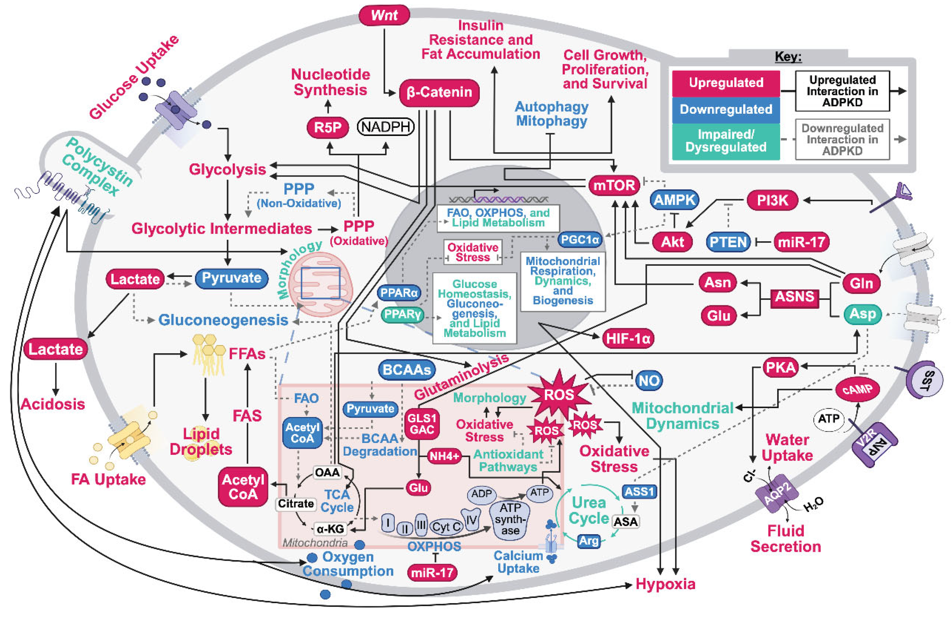
Figure 2.
Overview of complications in autosomal dominant polycystic kidney disease (ADPKD) drug development. ADPKD pathophysiology and external influences combine to complicate therapeutic design, efficacy, and toxicity. This creates obstacles in developing efficacious and safe therapeutic options for ADPKD patients.
Figure 2.
Overview of complications in autosomal dominant polycystic kidney disease (ADPKD) drug development. ADPKD pathophysiology and external influences combine to complicate therapeutic design, efficacy, and toxicity. This creates obstacles in developing efficacious and safe therapeutic options for ADPKD patients.
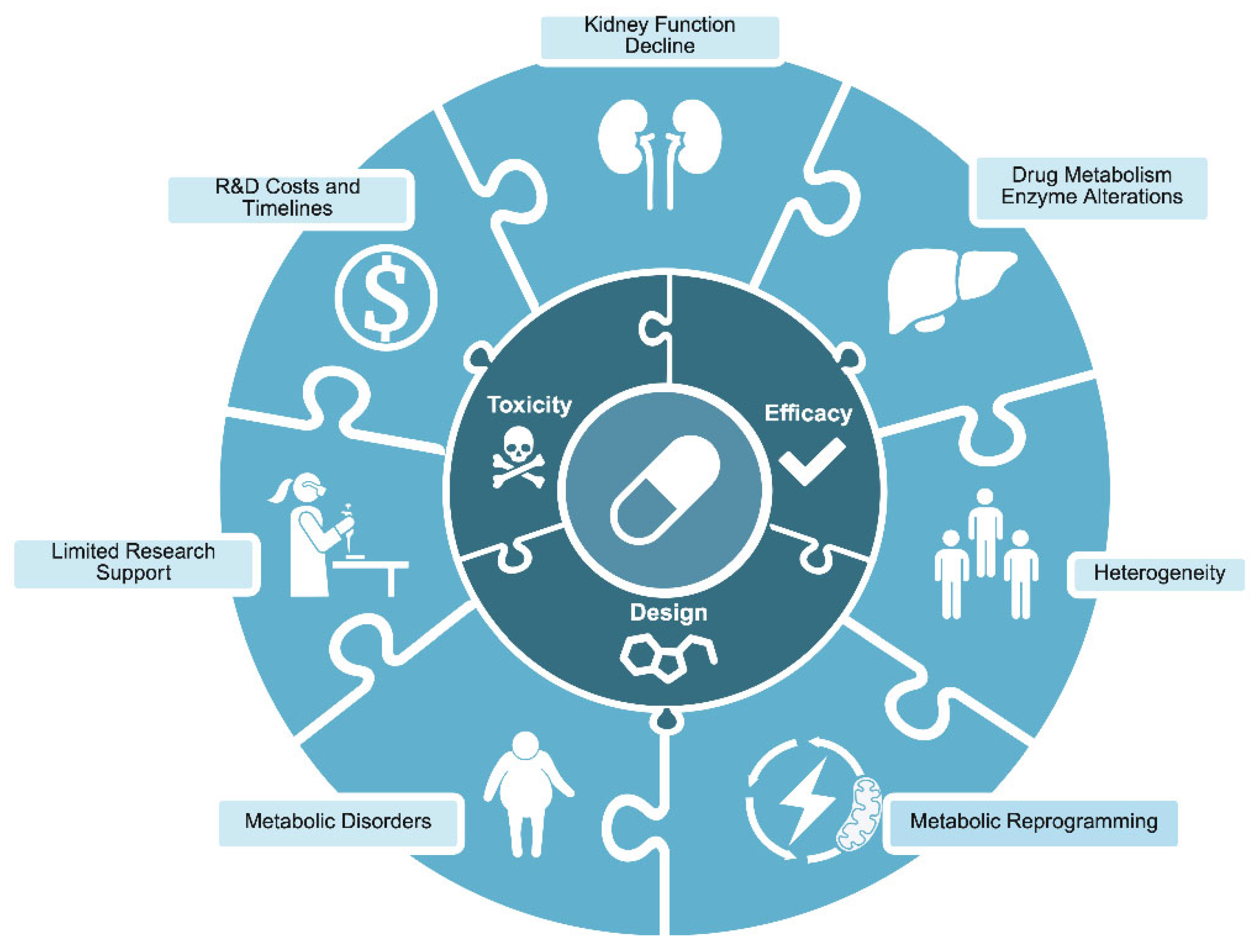
Figure 3.
Traditional drug development compared to drug repurposing. Drug repurposing leverages prior studies and computational tools to reduce the time and costs associated with de novo development.
Figure 3.
Traditional drug development compared to drug repurposing. Drug repurposing leverages prior studies and computational tools to reduce the time and costs associated with de novo development.
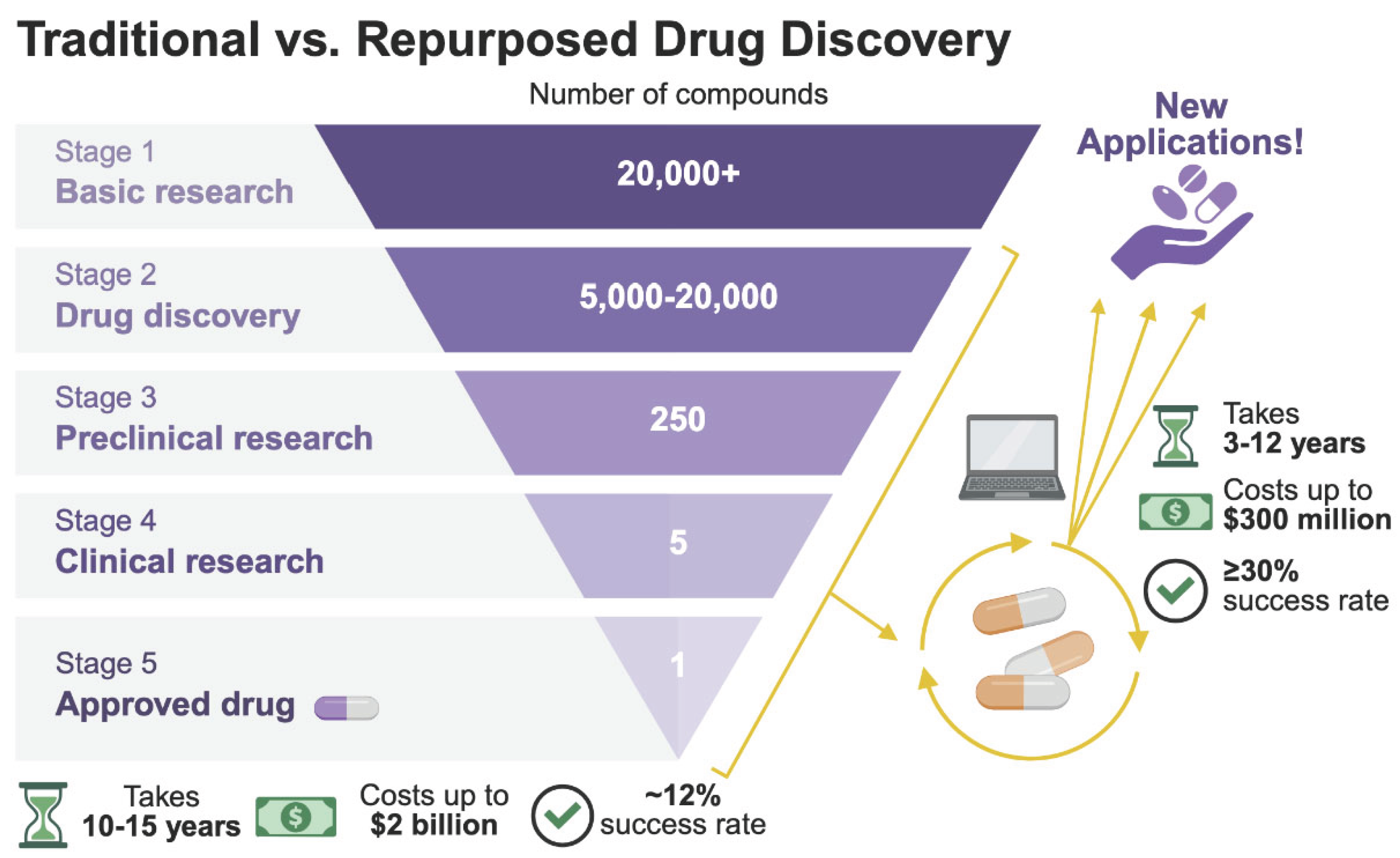

Table 1.
Repurposed autosomal dominant polycystic kidney disease (ADPKD) drugs with their targets and clinical trial outcomes. Drugs in this table were repurposed for molecular targets discussed in this review. Original indications were found using the U.S. Food and Drug Administration (FDA) Drugs@FDA search tool. As shown, metabolic reprogramming is being extensively studied as a therapeutic target with promising results. Conclusive evidence of a successful repurposed drug targeting ADPKD metabolic reprogramming is still lacking.
Table 1.
Repurposed autosomal dominant polycystic kidney disease (ADPKD) drugs with their targets and clinical trial outcomes. Drugs in this table were repurposed for molecular targets discussed in this review. Original indications were found using the U.S. Food and Drug Administration (FDA) Drugs@FDA search tool. As shown, metabolic reprogramming is being extensively studied as a therapeutic target with promising results. Conclusive evidence of a successful repurposed drug targeting ADPKD metabolic reprogramming is still lacking.
| Drug Class | Molecular Target / Mechanism of Action | Drug Compound | Original Indication | Results | Clinical Trials / Relevant PMIDs |
|---|---|---|---|---|---|
| Vaptans | |||||
| V2Ra / Antagonist | Tolvaptan | Hypervolemic or euvolemic hyponatremia | Tolvaptan significantly reduced the rate of TKVb growth and eGFRc decline (P<0.001) compared to the placebo. Patients in the tolvaptan group had a higher frequency of aquaresis- and hepatic-related adverse events. | TEMPO (NCT00428948); REPRISE (NCT02160145); PMID: 23121377; PMID: 29105594 |
|
| Lixivaptan | Euvolemic hyponatremia | Terminated early by sponsor due to concerns about commercial potential and hepatic safety. | ACTION (NCT04064346); ALERT (NCT04152837) | ||
| Rapalogs | |||||
| mTORC1d / Antagonist | Everolimus | Advanced renal cell carcinoma after failure of sunitinib or sorafenib treatment | Everolimus significantly slowed TKV increase (P=0.02) in the first year but was associated with a significantly greater decline in eGFR (P=0.004) compared with placebo. | NCT00414440; PMID: 20581392 |
|
| Sirolimus | Prophylaxis of organ rejection in renal transplant patients | Sirolimus treatment increased TKV and TCVe and did not significantly affect eGFR. Incidence of gastrointestinal adverse events in treated patients was high (94%). SIRENA-II was terminated early due to increased adverse events and renal failure progression in treated participants. A later study found that low-dose oral sirolimus is associated with ovarian toxicity, menstrual cycle disturbances, and ovarian cysts. | SIRENA-II (NCT01223755); NCT00346918; PMID: 26912555; PMID: 20581391; PMID: 19525519; PMID: 23071528 |
||
| Polyunsaturated Fatty Acids | |||||
| PPARf family / Agonist | Gamolenic acid | Atopic dermatitis/eczema; Asthma; Rheumatoid arthritis | Identified as a potential ADPKDg treatment option in a drug repurposing study. Reduced cyst size in renal epithelial cells grown in a 3D-gel matrix. | PMID: 31879244 | |
| Icosapent ethyl / Eicosapentaenoic acid | Hypertriglycidemia | Part of a larger clinical trial investigating the use of calcium channel blockers for the treatment of hypertension in ADPKD patients. Eicosapentaenoic acid administration over 2 years did not have a significant effect on renal function decline or kidney enlargement. | NCT00541853; PMID: 18372389 |
||
| Fibrates | |||||
| PPAR-α / Agonist | Fenofibrate | Hypertriglyceridemia; Primary hypercholesterolemia; Mixed dyslipidemia | In a preclinical trial on Pkd1^RC/RC mice, treatment with fenofibrate reduced TKV by 26.9%, TCV by 60.9%, liver fibrosis by 40%, and cyst proliferation by 71.6% compared to the control group. | PMID: 28903946 | |
| Thiazolidinediones | |||||
| PPAR-γ / Agonist | Pioglitazone | T2DMh | Low-dose pioglitazone was associated with reduced TKV growth rate compared to the placebo; however, the study was underpowered. Due to concerns over side effects with higher doses and preclinical data showing maximal efficacy at a low dose, the study did not examine higher dosages. | NCT02697617; PMID: 34221381 |
|
| Rosiglitazone | In human cystic epithelial cell culture models, rosiglitazone reduced the expression of fibrotic markers and cell proliferation. Male heterozygous Han:SPRD rats treated with rosiglitazone had improved kidney-to-body weight ratios and delayed ADPKD progression. | PMID: 26911848 | |||
| Biguanides | |||||
| AMPKi / Agonist | Metformin | Malaria; T2DM | Metformin treatment was well tolerated in ADPKD patients. Changes in htTKVj (p=0.9) and eGFR (p=0.2) were not statistically significant, but eGFR decline was reduced in the metformin group compared to placebo (-0.41±1.81 vs -3.35±1.70 mL min-1 1.73m-2). Starting kidney size, hypertension, and disease stage may have influenced this difference. | TAME-PKD (NCT02656017); IMPEDE-PKD (NCT04939935); NCT02903511; PMID: 33768205; PMID: 34391872 |
|
| Glucose Analogs | |||||
| Glycolysis / Antagonist | 2-deoxy-d-glucose | Cancer | In a preclinical Pkd1^(ΔC/flox)TmCre murine model, 2-deoxy-d-glucose administration restored AMPK activation, reduced kidney-to-body weight ratio, and was associated with improved kidney function preservation. Treated mice additionally had reduced cyst expansion in both distal tubules and collecting ducts. | PMID: 26534924 | |
| Somatostatin Analogs | |||||
| SSTR1-5k / Agonist | Octreotide | Acromegaly; Thyrotrophinomas | Octreotide-LARl significantly decreased TKV growth in both studies after one year (P=0.002, P=0.04) and at three years in ALADIN (P=0.04). ALADIN found the octreotide-LAR group had reduced eGFR decline, while ALADIN 2 found a higher rate of eGFR decline compared to the placebo (11.3% vs 7.0%). Octreotide treatment was associated with cholelithiasis and cholecystitis adverse events. | ALADIN (NCT00309283); ALADIN 2 (NCT01377246); PMID: 23972263; PMID: 30951521 |
|
| Lanreotide | Acromegaly; Gastroenteropancreatic neuroendocrine tumors | Lanreotide treatment significantly lowered the percent change in htTKV (P=0.02), with a 24% reduction in htTKV growth rate compared to the placebo. There was a high incidence of adverse events in the treatment group, leading 10% of patients to withdraw from the study. 28% of treated patients experienced serious adverse events. An interim analysis of DIPAK-1 concluded somatostatin analog use is associated with an increased risk of hepatic cyst infection. | DIPAK-1 (NCT01616927); PMID: 30422235; PMID: 27995519 |
||
| Pasireotide | Acromegaly; Cushing’s disease | In patients with ADPKD/ADPLDm with severe liver involvement, pasireotide-LAR treatment was associated with a significant decrease in TLVn (P<0.001) and TKV (P=0.02). Adverse events were most commonly related to hyperglycemia, and 19/32 (59%) of treated patients developed diabetes compared to 1/15 (7%) of patients in the placebo group (P<0.001). | NCT01670110; PMID: 32843370 |
||
| Statins | |||||
| HMGCRo/ Antagonist | Pravastatin | Primary hypercholesterolemia; hyperlipidemia; mixed dyslipidemia | In pediatric ADPKD patients, pravastatin treatment significantly slowed htTKV percent change over 3 years (P=0.02). Additionally, significantly fewer participants receiving pravastatin achieved a ≥20% change in htTKV compared to the placebo group (46% vs 68%, P=0.03). In a later study on adult ADPKD patients, pravastatin did not slow htTKV increase, and no significant differences between treatment groups were found. | NCT03273413; NCT00456365; ADPKD-SAT (NCT04284657); PMID: 41077128; PMID: 24721893 |
|
| Incretin Mimics | |||||
| GLP-1Rp / Agonist | Semaglutide | T2DM; Chronic weight management | In a preclinical slowly progressive (Pkd1RC?RC) mouse model, Semaglutide significantly delayed cyst growth (p<0.001), decreased cyst-lining epithelial cell proliferation (p<0.005), and induced apoptosis (p<0.005) compared to age-matched controls. Results indicated Semaglutide can regulate multiple ADPKD signaling pathways, reduce macrophage recruitment, and alleviate renal fibrosis. Semaglutide additionally normalized mitochondrial function and morphology, along with glucose metabolism, in Pkd1RC/RC mouse kidneys. | PMID: 40815122 | |
| Tirzepatide | T2DM; Chronic weight management | Current ongoing Phase 2 clinical trial with completion expected in June 2029. | NCT06582875 | ||
a vasopressin 2 receptor. b total kidney volume. c estimated glomerular filtration rate. d mammalian target of rapamycin complex 1. e total cyst volume. f peroxisome proliferator–activated receptor. g autosomal dominant polycystic kidney disease. h type-2 diabetes mellitus. i adenosine monophosphate-activated protein kinase. j height-adjusted total kidney volume. k somatostatin receptor. l long-acting release. m autosomal dominant polycystic liver disease. n total liver volume. o 3-hydroxy-3- methylglutaryl-coenzyme A reductase. p glucagon-like peptide-1 receptor.
Disclaimer/Publisher’s Note: The statements, opinions and data contained in all publications are solely those of the individual author(s) and contributor(s) and not of MDPI and/or the editor(s). MDPI and/or the editor(s) disclaim responsibility for any injury to people or property resulting from any ideas, methods, instructions or products referred to in the content. |
© 2026 by the authors. Licensee MDPI, Basel, Switzerland. This article is an open access article distributed under the terms and conditions of the Creative Commons Attribution (CC BY) license (http://creativecommons.org/licenses/by/4.0/).
Copyright: This open access article is published under a Creative Commons CC BY 4.0 license, which permit the free download, distribution, and reuse, provided that the author and preprint are cited in any reuse.