Preprint
Article

This version is not peer-reviewed.

Urological Injuries Following Gynecologic and Obstetric Surgery: Incidence, Diagnosis, and Outcomes From a 10-Year Retrospective Cohort

A peer-reviewed article of this preprint also exists.

Submitted:

09 December 2025

Posted:

11 December 2025

You are already at the latest version

Abstract

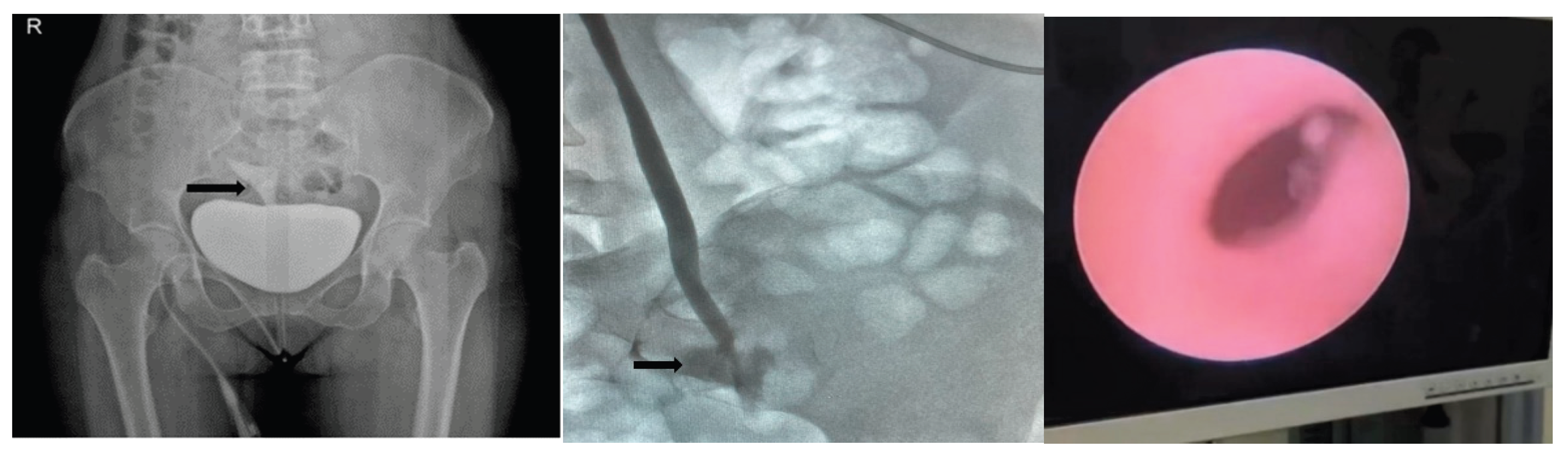
Background/Objectives: Gynecological and obstetric surgeries carry a risk of urinary tract injuries, which remain important causes of surgical morbidity. This study aimed to evaluate the incidence, etiological factors, diagnostic timing, and management outcomes of urological injuries occurring during these procedures over a 10-year period in a high-volume single-center cohort. Methods: This single-center retrospective study reviewed urinary tract injuries that occurred during gynecologic and obstetric procedures performed between January 2014 and December 2024. Among 16,100 surgeries, 223 cases were identified and analyzed regarding injury incidence, etiology, type, diagnostic timing, and management strategies. Results: Bladder injuries constituted 62.3% of cases, ureteral injuries 28.7%, and genitourinary fistulas 9.0%. Most bladder injuries (98.6%) and the majority of ureteral injuries (68.8%) were recognized intraoperatively (p < 0.001). Bladder injuries were most frequently associated with cesarean section, whereas ureteral injuries were more common in malignant gynecologic surgeries (p < 0.05). Conclusions: Early recognition and timely intervention remain critical to reducing morbidity associated with iatrogenic urinary tract injuries. These findings underscore the importance of anatomical knowledge, meticulous surgical technique, and appropriate diagnostic evaluation to optimize perioperative outcomes in gynecologic and obstetric surgery.

Keywords: 
;  ;  

1. Introduction

The shared embryological origin, anatomical proximity, and functional integration of the urogenital system contribute to the relatively high incidence of iatrogenic urinary tract injuries during obstetric and gynecological surgeries [1]. Despite advances in surgical techniques, such injuries remain a significant problem in obstetric and gynecologic procedures. The complex anatomy of the female pelvis and the proximity of the urinary system to the reproductive organs account for approximately 75% of iatrogenic urinary tract injuries in gynecological surgeries [2]. Iatrogenic urinary injuries occur in 0.5–1% of pelvic and abdominal surgeries, with the bladder being the most commonly affected organ [3]. These injuries may present in various forms, including bladder perforation, ureteral ligation or avulsion, and vesicovaginal fistula. In obstetric and gynecological surgeries, such complications can arise due to multiple factors, including the surgical technique employed, adhesions from previous operations, anatomical distortions caused by deep infiltrative pathologies, and the presence of malignancies [4]. Early diagnosis and timely intervention can reduce the severity of complications and contribute to maintaining the patient’s quality of life. In contrast, delays in diagnosis can increase the risk of late-stage complications, such as ureterovaginal or vesicovaginal fistulas, leading to morbidity and a significant decline in quality of life [5]. Therefore, the prevention and early detection of urinary tract injuries in gynecological surgeries are critical for both patient safety and the sustainability of surgical success. In this context, a thorough understanding of pelvic anatomy and careful intraoperative identification of anatomical structures, combined with a systematic and meticulous surgical approach, can significantly reduce the risk of complications. This study provides a comprehensive evaluation, in the context of existing literature, of the management and outcomes of urogenital injuries occurring during obstetric and gynecological surgeries performed at a single center over the past decade.

2. Methods

2.1. Participants

This retrospective study included 223 female patients who were treated at the Department of Urology for urogenital injuries sustained during obstetric or gynecologic surgeries performed at our university hospital between January 2014 and December 2024. The injuries were identified from a wide range of procedures, including cesarean sections, benign hysterectomies, and malignant gynecologic surgeries. The risk of urinary tract injury differed according to the surgical type, being most frequently associated with cesarean section and hysterectomy procedures. Eligible participants were women aged 18 years or older who underwent surgery for either benign or malignant indications and subsequently experienced intraoperative urinary tract injury. Patients who underwent prophylactic or temporary ureteral stent placement were excluded from the analysis. All included cases represent patients directly treated and followed at our institution; injuries diagnosed and managed at outside hospitals were not included in this study.

2.2. Study Design

Patients were divided into three groups based on surgical indications: The first group included malignant gynecologic surgeries, the second comprised obstetric procedures such as cesarean sections, and the third consisted of benign gynecologic surgeries, including myomectomies and hysterectomies. The groups were compared with respect to the occurrence of urological complications and the treatment modalities employed. In addition, cases were categorized as bladder/ureteral injuries or genitourinary fistulas according to the type of injury, and subgroup analyses were performed regarding time of diagnosis, therapeutic approach, and clinical outcomes. Clinical data—including patient age, surgical indication and procedure type, injury site and characteristics, duration of hospitalization, catheterization time, and timing of diagnosis and treatment—were retrieved from medical records. Urological complications were defined as genitourinary tract injuries detected intraoperatively or requiring postoperative intervention. Diagnostic evaluation consisted of physical examination, urogynecological assessment, and imaging studies. Complications and management strategies were analyzed in line with current literature. The study was approved by the Ethics Committee of Hatay Mustafa Kemal University (authorization number: 02/10/2024/03) and conducted in accordance with the principles of the Declaration of Helsinki.

2.3. Statistical Analysis

The analysis was conducted utilising SPSS 22 software, which facilitated the calculation of descriptive statistics, encompassing the mean, minimum, maximum, frequency, and percentage values. Next, the normality of the distribution was evaluated through the utilisation of the Kolmogorov-Smirnov and Shapiro-Wilk tests, while the analysis of non-normally distributed data was conducted through the implementation of the Kruskal-Wallis and post hoc tests. Finally, the statistical significance of the differences between the two groups was determined by applying the Mann-Whitney U, Pearson Chi-square, or Fisher’s Exact tests. The statistical significance was set at p < 0.05.

3. Results

The study included 223 women with a mean age of 48 years (range, 22–93 years) of the cases, 35.4% underwent total abdominal hysterectomy (TAH), 15.2% total laparoscopic hysterectomy (TLH), 26.5% cesarean section (C/S), 18.8% C/S with peripartum hysterectomy, 2.2% C/S with uterine rupture repair, and 1.8% pelvic organ prolapse (POP) surgery. The distribution of injuries was as follows: bladder (62.3%), ureter (28.7%), and genitourinary fistulas, including vesicouterine (VUF), vesicovaginal (VVF), and vesicorectal fistulas (VRF) (9.0%). Bladder injuries measured ≤2 cm in 37.4% of cases and >2 cm in 62.6%. Ureteral injuries were localized distally in 75.0% and to the mid-segment in 25.0%; 25.0% were thermal, 25.0% were full-thickness, and 50.0% were partial injuries. Overall, 81.6% of injuries were diagnosed intraoperatively, predominantly through direct visual inspection (Table 1, Figure 1).
Among patients with fistulas, 55.0% had undergone benign hysterectomy, whereas among those with ureteral injury, 50.0% had undergone malignant hysterectomy (p < 0.001). Across all groups, complication rates were higher following open surgery (p = 0.026) (Table 2).
Bladder injuries were surgically repaired using double-layer sutures in all cases. The most common surgical procedure for ureteral injuries was unilateral ureteroneocystostomy (UNC) (75.0%), followed by bilateral UNC (21.9%) and ureteroureterostomy (3.1%). A percutaneous nephrostomy (PCN) was placed in 26.6% of patients with ureteral injuries. Among fistula cases, VVF repair was the predominant procedure, accounting for 90.0% of all cases. The intraoperative detection rates were 98.6% for bladder injuries, 68.8% for ureteral injuries, and only 5.0% for fistulas (p < 0.001) (Figure 2a–c).
Figure 2. Representative images of urological injuries following gynecologic surgery. Figure 2a, vesicouterine fistula.Figure 2b, ureteral avulsion (nephrourogram). Figure 2c, ureteral avulsion (endoscopic image) Figure 2a,b,c Representative images of urological injuries following gynecologic surgery.
Preprints 188896 g002
The duration of Foley catheterization differed significantly between injury groups (p < 0.001), with the longest mean duration observed in the fistula group (22.3 days) and the shortest in the ureteral injury group (10.1 days). Similarly, the time to surgical repair varied significantly among groups (p < 0.001), being shortest for bladder injuries and longest for fistulas (Table 2).
The mean retention period of double-J stents was significantly longer in Group 1 (47.8 days) compared to Groups 2 (42.1 days) and 3 (41.2 days) (p = 0.039). Foley catheter indwelling time also differed significantly (p < 0.001), with the longest duration recorded in Group 2 (17.3 days). Mortality was observed exclusively in Group 1 (4.8%) (p = 0.035) (Table 3).
Postoperative symptoms showed significant intergroup variation (p < 0.001). The highest rate of asymptomatic patients was in Group 2, while Group 1 demonstrated higher frequencies of flank pain, dysuria, sepsis (27.0%), and fever (39.7%) (Table 3).

4. Discussion

Urinary tract injuries occurring during gynecological and obstetric surgeries are one of the most common urological complications of pelvic surgery, and delays in diagnosis or treatment can increase morbidity and negatively affect quality of life. [6] Reported incidence rates in the literature range from 0.3% to 1.5%, with bladder injuries being the most common and ureteral injuries occurring less frequently. [3] Despite advances in surgical techniques, iatrogenic urinary tract injuries remain a clinically significant concern. In the present study, the incidence was found to be 1.3% among 223 patients operated on in our department over the past decade, a rate approaching the upper limit of values reported in the literature. This finding may be associated with the high proportion of obstetric procedures, previous surgical history, follow-up conditions, as well as surgeon experience and the level of intraoperative vigilance. [7]
The most common procedures in our series were TAH, C/S and TLH. Of the injuries identified, 62.3% involved the bladder, 28.7% the ureter, and 9.0% presented as genitourinary fistulas. This distribution reflects the anatomical vulnerability of the bladder and distal ureter to surgical trauma. Consistent with our findings, previous studies have reported bladder injury as the most common complication, whereas ureteral injuries, although less frequent, are associated with higher morbidity. [8] Fistulas typically represent a secondary outcome of bladder or ureteral injuries that are either missed intraoperatively or diagnosed at a later stage. [9]
Early recognition of iatrogenic urinary tract injuries is critical in reducing morbidity. In our study, 81.6% of lesions were identified intraoperatively, reflecting the importance of surgical awareness and anatomical knowledge. However, 18.4% of injuries were diagnosed postoperatively, indicating that ureteral injuries and fistulas are particularly prone to being overlooked during surgery. Similarly, the literature reports that only one-third of ureteral injuries are recognized intraoperatively, while the majority manifest within 48–72 hours through nonspecific symptoms. [10]
The timing of diagnosis is closely related to the type of injury. In our series, 98.6% of bladder injuries were recognized intraoperatively, compared with 68.8% of ureteral injuries and only 5.0% of fistulas (p<0.001). These data suggest that while bladder injuries are usually evident during surgery, subtle ureteral damage or fistula formation may remain undetected intraoperatively and become apparent later, often due to thermal injury or delayed clinical manifestations. Fistulas are typically diagnosed at a later stage, often presenting with pelvic pain, urinary incontinence, or cyclic hematuria. [11] In high-risk procedures, intraoperative cystoscopy and visualization of ureteral urine jets may help minimize diagnostic delays and improve patient outcomes. These practices are recommended by the AUA, and competency in cystoscopy among gynecologic surgeons may facilitate earlier recognition and management of urinary tract injuries. [12,13]
The type of urinary tract injury varies significantly depending on the surgical indication and technique employed. In our series, 55% of patients who developed fistulas had a history of hysterectomy performed for benign pathology (p<0.001), suggesting that chronic inflammation and adhesions increase dissection difficulty and thereby elevate the risk. [14] In malignant cases, the higher rate of ureteral injuries (50.0%; p<0.001) reflects the impact of more aggressive dissections and parametrial involvement. The significantly greater complication rates observed in open surgeries (p=0.026) highlight the tissue-sparing advantage of minimally invasive approaches and underscore that both surgical indication and approach are critical determinants of complication risk. Therefore, individualized surgical planning remains crucial for minimizing complications. [15] In this cohort, however, the selection of surgical approach was not random but based on the underlying pathology and urgency; most malignant and obstetric cases were performed via open procedures, while minimally invasive techniques were mainly reserved for selected benign indications.
In this study, 62.6% of bladder injuries were larger than 2 cm, indicating that a considerable proportion of cases involved extensive defects associated with an increased risk of complications. The 2 cm threshold is also recognized in the literature as a critical marker for predicting the technical difficulty of repair and the likelihood of postoperative complications.[16] This finding is particularly relevant in procedures requiring bladder dissection, such as abdominal hysterectomy, where surgical expertise and anatomical knowledge are decisive factors. Larger defects not only increase the technical complexity of repair but also prolong catheterization and drainage requirements while elevating the risk of fistula formation. [17] In our cohort, all bladder injuries were repaired using a double-layer suture technique recommended in the literature to ensure watertight closure and optimal healing. Nevertheless, for defects greater than 2 cm, reconstructive methods or the use of interposition grafts may be necessary to further reduce complication rates. Thus, the 2 cm threshold should be regarded as a clinically relevant parameter, both for intraoperative decision-making and for anticipating postoperative outcomes.
The localization and type of ureteral injuries demonstrate that certain segments are particularly vulnerable during pelvic surgery. In this study, 75% of cases involved the distal ureter, likely due to its susceptibility to manipulation during uterine artery ligation or pelvic wall dissection.[10] Thermal injury was observed in 25% of ureteral lesions, a noteworthy finding that underscores the need for cautious use of energy sources, particularly in laparoscopic procedures. Such injuries may go unrecognized intraoperatively and later manifest as strictures or fistulas. [18] For this reason, intraoperative cystoscopy or assessment of ureteral urine jets may be beneficial in suspicious cases.
The most frequently performed treatment for ureteral injuries was UNC with DJ stent placement. In this series, 75% of patients underwent unilateral UNC, 21.9% bilateral UNC, and 3.1% uretero-ureterostomy. Additionally, 26.6% of patients (n=17) required PCN, which was necessary exclusively in cases of ureteral injury. In patients with delayed diagnosis or those at risk for infection, PCN plays a critical role in preserving renal function and providing safe drainage. Consistent with our findings, the literature has also shown that PCN reduces morbidity and improves the success of reconstructive surgery in cases of ureteral injury diagnosed postoperatively. [19]
In the fistula group, primary repair of VVF was performed in 90% of patients, which is consistent with published data. [20] Less common fistulas included VUF and VRF, each observed in 5% of cases. VUF typically develops following cesarean section and is often diagnosed late due to specific symptoms such as cyclic hematuria. [21] VRF, on the other hand, may arise after obstetric complications or extensive pelvic surgery and usually presents as a more complex condition. The success of fistula repair depends on multiple factors, including lesion duration, local inflammation, tissue vascularization, and the presence of infection. Therefore, optimal timing of surgery and individualized treatment planning require a multidisciplinary approach.
The rate of DJ stent placement and the duration of stenting in bladder and ureteral injuries reflect both the severity of the lesion and the therapeutic approach. In this study, DJ stents were placed unilaterally in 76.6% and bilaterally in 21.9% of ureteral injuries, with a mean indwelling time of 44.7 days. This duration exceeds the recommended 4–6 weeks and may be attributed to factors such as patient non-compliance and institutional workload. [22] Significant differences were observed based on surgical etiology; in particular, patients undergoing malignant surgery (Group 1) had a longer mean stent duration of 47.8 days, and both unilateral and bilateral stent placement rates were significantly higher than in other groups (p<0.05).
These findings can be explained by the extensive dissections required in advanced tumor surgeries, distortion of pelvic anatomy, periureteral invasion, and the increased risk of complications. Consistent with this, previous studies have reported that malignant pelvic surgeries are associated with higher rates of ureteral injury and morbidity compared to benign or obstetric procedures, with bilateral involvement particularly common in the presence of parametrial extension. [19] In addition, postoperative inflammation, delayed tissue healing, and adjuvant therapies such as radiotherapy may necessitate prolonged stent indwelling in malignant cases. Notably, the longer hospital stays and the occurrence of mortality exclusively in Group 1 further underscore the greater overall complication burden associated with malignant surgery. Therefore, thorough preoperative risk assessment, intraoperative urological support, and tailored postoperative follow-up strategies are essential for optimizing patient outcomes.
Patients with bladder-ureter injuries showed significant differences in Foley catheterization durations compared to patients in the fistula group (p<0.001). The longest duration was observed in the fistula group, with a mean of 22.3 days, followed by isolated bladder injuries with a mean of 17.4 days (range 1–30) and ureteral injuries with a mean of 10.1 days. The prolonged catheterization in the fistula group is necessary to ensure optimal bladder rest following repair and to promote adequate tissue healing. Indeed, for vesicovaginal and vesicouterine fistulas, 14–21 days of continuous drainage is recommended to improve surgical success. [23] In isolated bladder injuries, the mean catheterization time was 17.4 days, although some studies have reported that as little as 7 days may be sufficient. [24] These findings suggest that longer drainage may be required for complex lesions and that catheterization duration should be individualized according to the type of injury. Also, it should be noted that the duration of catheterization and stent placement in our series largely reflects local institutional practice, which may differ from other centers and should be interpreted within this context.
Postoperative clinical symptoms differed significantly according to surgical etiology (p<0.001). In the obstetric surgery group (Group 2), 90.6% of patients were asymptomatic, whereas this rate declined to 55.6% in the malignant surgery group (Group 1). Flank pain (30.2%) and dysuria (7.9%) were more frequently observed in Group 1, likely reflecting the invasive nature of malignant procedures and the need for more extensive periureteral dissection. [25] Furthermore, the incidence of postoperative sepsis (27.0%) and fever (39.7%) was markedly higher in Group 1, which may be attributable to prolonged operative times, increased blood loss, and immunosuppression in advanced-stage surgeries. These findings highlight the need for closer and more intensive postoperative monitoring in patients undergoing malignant surgery. Consistent with our results, the literature also reports higher rates of infection and complications following pelvic surgeries for malignancy compared to benign or obstetric procedures. [26] The present results reinforce this evidence, further demonstrating the substantially increased risk of complications in malignant surgeries.
In complex gynecological malignancy surgeries, fibrosis, prior radiotherapy, and distorted pelvic anatomy substantially increase the risk of ureteral injury. For this reason, some centers advocate the use of prophylactic DJ stents to mitigate this risk. However, the literature does not provide a clear consensus: while some studies suggest that stenting facilitates intraoperative recognition and earlier detection of injuries , others emphasize that its protective effect is limited and that the surgeon’s experience and dissection skills remain the decisive factors. [27,28] Consequently, routine prophylactic stenting is not recommended, and its use should instead be restricted to selected high-risk cases as a more rational approach. In this study, no patients underwent pre-stenting, and we believe that prospective, large-scale investigations are needed to clarify its potential role and provide meaningful contributions to the literature. Furthermore, preoperative urological consultation was rarely performed, despite evidence indicating that multidisciplinary collaboration and the active involvement of urologists in the surgical team can significantly reduce complications through earlier diagnosis and optimized management. [29] Taken together, these findings suggest that selective use of prophylactic stenting and reinforcement of multidisciplinary strategies represent critical measures for preventing complications in high-risk malignancy surgeries.
Limitations
This study has several limitations. Its retrospective design may lead to information gaps and potential bias, while its single-center nature and reliance on surgeries performed by the same team limit the generalizability of the findings. Although the gynecology and obstetrics surgeons in our department are highly experienced, our institution is one of the busiest tertiary referral centers in the region. The exceptionally high workload, combined with a patient population characterized by low socioeconomic and educational levels, limited health awareness, and high birth rates, may have contributed to delayed intraoperative recognition in a small proportion of cases. In addition, important practices such as preoperative urological consultation were not routinely implemented, which may have increased complication risks in high-risk patients and further limited the interpretation of our results. Nevertheless, despite these limitations, this decade-long analysis provides valuable insights into the diagnosis, management, and outcomes of urological injuries following gynecological and obstetric surgeries.

5. Conclusions

Urinary tract injuries following gynecological and obstetric surgeries exhibit diverse clinical patterns influenced by surgical indication, technique, and pelvic anatomy. In this cohort, bladder injuries and fistula formation were more frequently associated with procedures performed for benign indications, while ureteral injuries occurred more often in malignant cases. Importantly, bladder injuries are generally identifiable intraoperatively and thus potentially avoidable, whereas ureteral and fistulous injuries are more likely to be missed during surgery and diagnosed postoperatively. Timely recognition remains crucial for reducing morbidity and optimizing surgical outcomes. A thorough understanding of pelvic anatomy, meticulous surgical technique, and the judicious use of intraoperative assessment tools are essential to minimize complications. Based on our experience, we believe that closer multidisciplinary collaboration between gynecology and urology teams may provide a positive contribution to improving perioperative management and long-term patient outcomes, particularly in high-risk or complex cases.

Author Contributions

Conceptualization, E.O. and A.B.; methodology, E.O., A.B., F.G., S.G., K.D. and N.T.; software, N.T; validation F.G.; formal analysis, E.O.; investigation, A.B., F.G. and S.G.; resources, E.O. and A.B; data curation, E.O. and A.B; writing—original draft preparation, E.O.; writing—review and editing, E.O. and A.B.; visualization, N.T.; supervision, S.G. and K.D.; project administration, E.O. All authors have read and agreed to the published version of the manuscript.

Funding

This research received no external funding.

Institutional Review Board Statement

This study received approval from the ethics committee of Hatay Mustafa Kemal University (authorization number: 02/10/2024/03, date 4 December 2024) and was conducted in accordance with the Declaration of Helsinki.

Informed Consent Statement

Informed consent was obtained from all subjects involved in the study.

Data Availability Statement

The data are available and can be provided upon request.

Acknowledgments

The authors would like to thank Dr. Hamdi Oğuzman from the Department of Biochemistry at our hospital for his valuable contributions to the statistical analysis of this study. Dr. Hamdi Oğuzman has given permission to be acknowledged by name.

Conflicts of Interest

The authors declare no conflicts of interest.

Abbreviations

IBI=iatrogenic bladder injury, IUI= iatrogenic urinary injuries, TAH= total abdominal hysteroctomy, TLH= total laparoscopic hysteroctomy, C/S=caesarean section, POP=pelvic organ prolapse, VUF=vesicouterine fistula, VVF= vesicovaginal fistula, VRF=vesicorectal fistula, UNC=ureteroneocystostomy, PCN=percutaneous nephrostomy, DJ= double j, AUA=American Urological Association.

References

  1. Chalya, PL; Massinde, AN; Kihunrwa, A; Simbila, S. Iatrogenic ureteric injuries following abdomino-pelvic operations: a 10-year tertiary care hospital experience in Tanzania. World J Emerg Surg 2015, 10, 17. [Google Scholar] [CrossRef]
  2. Wong, JMK; Bortoletto, P; Tolentino, J; Jung, MJ; Milad, MP. Urinary tract injury in gynecologic laparoscopy for benign indication: a systematic review. Obstet Gynecol. 2018, 131, 100–8. [Google Scholar] [CrossRef]
  3. Jensen, AS; Heinemeier, IIK; Schroll, JB; Rudnicki, M. Iatrogenic bladder injury following gynecologic and obstetric surgery: a systematic review and meta-analysis. Acta Obstet Gynecol Scand. 2023, 102, 1608–17. [Google Scholar] [CrossRef] [PubMed]
  4. Jensen, AS; Rudnicki, M. Iatrogenic bladder and ureteral injuries following gynecological and obstetric surgery. Arch Gynecol Obstet. 2023, 307, 511–8. [Google Scholar] [CrossRef]
  5. Esparaz, AM; Pearl, JA; Herts, BR; LeBlanc, J; Kapoor, B. Iatrogenic urinary tract injuries: etiology, diagnosis, and management. Semin Intervent Radiol 2015, 32(2), 195–208. [Google Scholar] [CrossRef] [PubMed]
  6. Aronson, MP; Bose, TM. Urinary tract injury in pelvic surgery. Clin Obstet Gynecol. 2002, 45, 428–438. [Google Scholar] [CrossRef] [PubMed]
  7. Li, X; Yang, K; Ding, G; Zou, X; Ye, L; Wu, J; Zhang, P; Fang, D; Hao, H; Li, Z; Zhu, H; Li, X; Jiang, H; Wang, K; Zhou, L; Li, H. Etiology, characteristics and management of ureteric injury: experience from a nationwide study. Transl Androl Urol 2022, 11(6), 794–802. [Google Scholar] [CrossRef]
  8. Naser, Omar; O’Connor, Eabhann; Greenwell, Tamsin J. Urological complications following gynaecological surgery. Obstetrics, Gynaecology & Reproductive Medicine 2022, 32(Issue 12), 272–281. [Google Scholar] [CrossRef]
  9. Yossepowitch, Ofer; Baniel, Jack; Livne, Pinhas. Re: Urological injuries during cesarean section. Intraoperative diagnosis and management [6]. The Journal of urology 2004, 172, 196–9. [Google Scholar] [CrossRef]
  10. Jacob, GP; Vilos, GA; Al Turki, F; Bhangav, G; Abu-Rafea, B; Vilos, AG; Ternamian, A. Ureteric Injury During Gynaecological Surgery - Lessons from 20 Cases in Canada. Facts Views Vis Obgyn 2020, 12(1), 31–42. [Google Scholar]
  11. Medlen, H; Barbier, H. Vesicovaginal Fistula. In StatPearls [Internet]; Available from; StatPearls Publishing: Treasure Island (FL), 6 Feb 2023. [Google Scholar]
  12. Dandolu, V; Mathai, E; Chatwani, A; Harmanli, O; Pontari, M; Hernandez, E. Accuracy of cystoscopy in the diagnosis of ureteral injury in benign gynecologic surgery. Int Urogynecol J Pelvic Floor Dysfunct 2003, 14(6), 427–31. [Google Scholar] [CrossRef]
  13. Morey, AF; Brandes, S; Dugi, DD, 3rd; et al. Urotrauma: AUA guideline. J Urol 2014, 192(2), 327–35. [Google Scholar] [CrossRef]
  14. Kumar, S; Vatsa, R; Bharti, J; Roy, KK; Sharma, JB; Singh, N; Meena, J; Singhal, S. Urinary fistula-A continuing problem with changing trends. J Turk Ger Gynecol Assoc. 2017, 18(1), 15–19. [Google Scholar] [CrossRef]
  15. El-Achi, V.; Weishaupt, J.; Carter, J.; et al. Robotic versus laparoscopic hysterectomy in morbidly obese women for endometrial cancer. J Robotic Surg 2021, 15, 483–487. [Google Scholar] [CrossRef]
  16. Singh, V; Aggarwal, S; et al. CT and clinical predictors of failure of conservative management in intraperitoneal bladder rupture. Healthcare (Basel) 2025, 13(13), 1594. [Google Scholar] [CrossRef]
  17. Wilson, DJ; Melin, I; Shah, N; O’Connor, RC; Carver, T. Investigating the timing of catheter removal after traumatic bladder injury: a single-institution 12-year experience. Trauma Surg Acute Care Open 2025, 10(1), e001693. [Google Scholar] [CrossRef]
  18. Limbachiya, D; Tiwari, R; Kumari, R. Iatrogenic Thermal Energy-Induced Distal Ureteric Injury and Its Management by Laparoscopy Ureteroureterostomy. JSLS 2023, 27(3), e2023.00030. [Google Scholar] [CrossRef] [PubMed]
  19. De Cicco Nardone, Carlo; Ficarola, Fernando; Feole, Laura; De Luca, Cristiana; Plotti, Francesco; Montera, Roberto; Luvero, Daniela; Larciprete, Giovanni; Marci, Roberto; Angioli, Roberto; Terranova, Corrado. Ureteral injuries management in gynaecologic surgery: the role of the conservative approach. Ital J Gynaecol Obstet. 2024, Vol. 36(No. 2), 215–225. [Google Scholar] [CrossRef]
  20. Chinthakanan, O; Sirisreetreerux, P; Saraluck, A. Vesicovaginal Fistulas: Prevalence, Impact, and Management Challenges. Medicina (Kaunas) 2023, 59(11), 1947. [Google Scholar] [CrossRef]
  21. Ördek, Eser; Pelit, Eyyup; Kati, Bulent. A Rare Complication After Cesarean Section: Vesicouterine Fistula. Mustafa Kemal University Medical Journal 2020, 11. [Google Scholar] [CrossRef]
  22. Birkhäuser, FD; Rütte, TV; Moltzahn, F; Huber, P; Zehnder, P. A Short Double-J Ureteral Stent Indwelling Time Is Safe and Effective Following Minimally Invasive Pyeloplasty: Long-term Results from a Prospective Randomized Controlled Trial. Eur Urol Open Sci. 2025, 74, 28–33. [Google Scholar] [CrossRef]
  23. Sori, DA; Azale, AW; Gemeda, DH. Characteristics and repair outcome of patients with Vesicovaginal fistula managed in Jimma University teaching Hospital, Ethiopia. BMC Urol 2016, 16(1), 41. [Google Scholar] [CrossRef]
  24. Barone, MA; Widmer, M; Arrowsmith, S; Ruminjo, J; Seuc, A; Landry, E; et al. Breakdown of simple female genital fistula repair after 7 day versus 14 day postoperative bladder catheterisation: a randomised, controlled, open-label, non-inferiority trial. Lancet 2015, 386, 56–62. [Google Scholar] [CrossRef]
  25. Iyer, R.; Gentry-Maharaj, A.; Nordin, A.; et al. Predictors of complications in gynaecological oncological surgery: a prospective multicentre study (UKGOSOC—UK gynaecological oncology surgical outcomes and complications). Br J Cancer 2015, 112, 475–484. [Google Scholar] [PubMed]
  26. Wallace, Sumer K., MD; Fazzari, Melissa J., PhD; Chen, Hui; Cliby, MD; Chalas, William A., MD; Eva, MD. Outcomes and Postoperative Complications After Hysterectomies Performed for Benign Compared With Malignant Indications. Obstetrics & Gynecology 2016, 128(3), p 467–475. [Google Scholar] [CrossRef] [PubMed]
  27. Feng, D; Tang, Y; Yang, Y; Wei, X; Han, P; Wei, W. Does prophylactic ureteral catheter placement offer any advantage for laparoscopic gynecological surgery? A urologist’ perspective from a systematic review and meta-analysis. Transl Androl Urol 2020, 9(5), 2262–2269. [Google Scholar] [CrossRef] [PubMed]
  28. Leon, Mateo; Guha, Pree; Lewis, Gregory; Heckman, Michael; Siddiqui, Habeeba; Chen, Anita. Use of prophylactic ureteral stents in gynecologic surgery. Minerva obstetrics and gynecology 2023, 76. [Google Scholar] [CrossRef]
  29. Smith, AL; Weissbart, SJ. Gynecologic Considerations for the Urologic Surgeon. Urology 2021, 150, 116–124. [Google Scholar] [CrossRef]
Figure 1. Intraoperative image showing bladder perforation and its open surgical repair.
Preprints 188896 g001
Table 1. General characteristics of patients.
Variables n=223
Age (years), Mean (Min-Max) 48 (22–93)
Vaginal delivery, Mean (Min-Max) 3.5 (1-13)
Cesarean Section, Mean (Min-Max) 2.6 (1-6)
TAH, n (%) 79 (35.4)
TLH, n (%) 34 (15.2)
C/S, n (%) 59 (26.5)
C/S peripartum hysterectomy, n (%) 42 (18.8)
C/S uterine rupture repair, n (%) 5 (2.2)
Pop surgery, n (%) 4 (1.8)
Bladder injury, n (%) 139 (62.3)
Ureteral injury, n (%) 64 (28.7)
VUF, VVF, VRF, n (%) 20 (9.0)
Bladder Injury Size, n (%)
≤2 cm 52 (37.4)
>2 cm 87 (62.6)
Ureteral Injury Location, n (%)
Distal 48 (75.0)
Middle 16 (25.0)
Ureteral Injury Type, n (%)
Thermal 16 (25.0)
Complete 16 (25.0)
Partial 32 (50.0)
Recognition Time, n (%)
Intraoperative 182 (81.6)
Postoperative 41 (18.4)
Postoperative Repair Time (days),Mean (Min-Max) 68.4 (5–180)
Diagnostic Techniques, n (%)
Direct visual 182 (81.6)
Histogram 1 (0.4)
CT urogram 20 (9.0)
Cystogram + CT urogram 20 (9.0)
Exitus, n (%) 3 (1.3)
Pop surgery: pelvic organ prolapse surgery, CT urogram: computer tomography urogram.
Table 2. Comparison of bladder, ureter, and fistula injuries.
Variables Bladder
Injury
(n=139)
Ureter
Injury
(n=64)
Fistula damage
(n=20)
p value
n (%) n (%) n (%)
Hysterectomy (Etiological)
Benign 27 (19.4) 16 (25.0) 11 (55.0) *0.002
Malignant 27 (19.4) 32 (50.0) 4 (20.0) *<0.001
C/S peripartum hysterectomy 30 (21.6) 9 (14.1) 3 (15.0) *0.400
Hysterectomy (Surgical Technique)
Laparoscopic surgery 17 (12.2) 16 (25.0) 1 (5.0) *0.026
Open surgery 122 (87.8) 48 (75.0) 19 (95.0)
C/S 50 (36.0) 7 (10.9) 2 (10.0) *<0.001
C/S uterine rupture repair 5 (3.6) 0 (0.0) 0 (0.0) *0.454
Pop surgery 4 (2.9) 0 0 *0.528
Bladder Injury Management
Bladder repair (double-layer) 139 (100.0) 0 0 (0.0) -
Ureteral Injury Management
UNC (right/left) 0 (0.0) 48 (75.0) 0 (0.0) -
Bilateral UNC 0 14 (21.9) 0 -
Ureteroureterostomy 0 (0.0) 2 (3.1) 0 -
Fistula Management
VUF repair 0 (0.0) 0 1 (5.0) -
VVF repair 0 0 18 (90.0) -
VRF repair 0 0 (0.0) 1 (5.0) -
Recognition Time
Intraoperative 137 (98.6) 44 (68.8) 1 (5.0) *<0.001
Postoperative 2 (1.4) 20 (31.3) 19 (95.0)
PCN
Present 0 (0.0) 17 (26.6) 0 (0.0) -
Absent 0 47 (73.4) 0 (0.0) -
DJ
Bilateral 0 (0.0) 14 (21.9) 0 (0.0) *<0,001
Unilateral 0 49 (76.6) 0 (0.0)
None 139 (100.0) 1 (1.6) 20 (100.0)
Postoperative Repair Time (days),Mean (Min-Max)
-
n=20
54.2 (10–120)
n=20
90 (60-180)

***0.001
Length of Foley Catheterization (days),
Mean (Min-Max)

17.4 (1–30)

10.1 (1–30)

22.3 (14–45)

**<0,001a,b,c
DJ Stent Retention Period (days),
Mean (Min-Max)

-

44.7 (30–90)

-

-
* Pearson Chi-Square Test or Fisher’s Exact Test, ** Kruskal-Wallis H Test, ***Mann-Whitney-U Test a : Significant difference between bladder and ureter injury, b : Significant difference between bladder and fistula injury, c : Significant difference between ureter and fistula injury.
Table 3. Intergroup comparisons.
Variables Group 1
Malignant (n=63)
Group 2
Obstetric
(n=106)
Group 3 Benign
(n=54)
p value
Postoperative Symptoms, n (%)
None 35 (55.6) 96 (90.6) 35 (64.8) *<0.001
Dysuria 5 (7.9) 2 (1.9) 1 (1.9)
Side pain 19 (30.2) 4 5 (9.3)
Hematuria 0 (0.0) 0 (0.0) 2 (3.7)
Sepsis, n (%) 17 (27.0) 2 (1.9) 3 (5.6) *<0.001
Fever, n (%) 25 (39.7) 10 (9.4) 6 (11.1) *< 0.001
Postoperative bilateral DJ, n (%) 11 (17.5) 2 (1.9) 2 (3.7) *< 0.001
Postoperative unilateral DJ, n (%) 22 (34.9) 14 (13.2) 13 (24.1) *0.004
Postoperative PCN, n (%)
Present 9 (28.1) 3 (18.8) 5 (31.3) *0.811
Absent 23 (71.9) 13 (81.3) 11 (68.8)
DJ stent retention period (days),Mean (Min-Max) 47.8 (30–90) 42.1 (30–90) 41.2 (30–60) **0.039
Length of Foley catheterization (days),Mean (Min-Max) 13.9 (5–28) 17.3 (1–30) 14.7 (1–45) **< 0.001 a, b
Hospital stay (days),Mean (Min-Max) 9.5 (1–35) 7.1 (2–35) 6.1 (1–60) **< 0.001 a,b,c
Exitus, n (%) 3 (4.8) 0 (0.0) 0 (0.0) *0.035
a : Significant difference between Group 1 and Group 2, b : Significant difference between Group 2 and Group 3, c : Significant difference between Group 1 and Group 3, * Pearson Chi-Square Test or Fisher’s Exact Test, ** Kruskal-Wallis H Test.
Disclaimer/Publisher’s Note: The statements, opinions and data contained in all publications are solely those of the individual author(s) and contributor(s) and not of MDPI and/or the editor(s). MDPI and/or the editor(s) disclaim responsibility for any injury to people or property resulting from any ideas, methods, instructions or products referred to in the content.
Copyright: This open access article is published under a Creative Commons CC BY 4.0 license, which permit the free download, distribution, and reuse, provided that the author and preprint are cited in any reuse.
Prerpints.org logo

Preprints.org is a free preprint server supported by MDPI in Basel, Switzerland.

Subscribe

Disclaimer

Terms of Use

Privacy Policy

Privacy Settings

© 2026 MDPI (Basel, Switzerland) unless otherwise stated