Preprint
Article

This version is not peer-reviewed.

Coadministration of Melissa officinalis and Rosmarinus officinalis Alcoholic Extracts Exhibits Neuroprotective and Therapeutic Effects on Kidney Tissue and Apoptosis-Related Gene Expression in a Rat Model of Spinal Cord Injury

Submitted:

07 December 2025

Posted:

12 December 2025

You are already at the latest version

Abstract

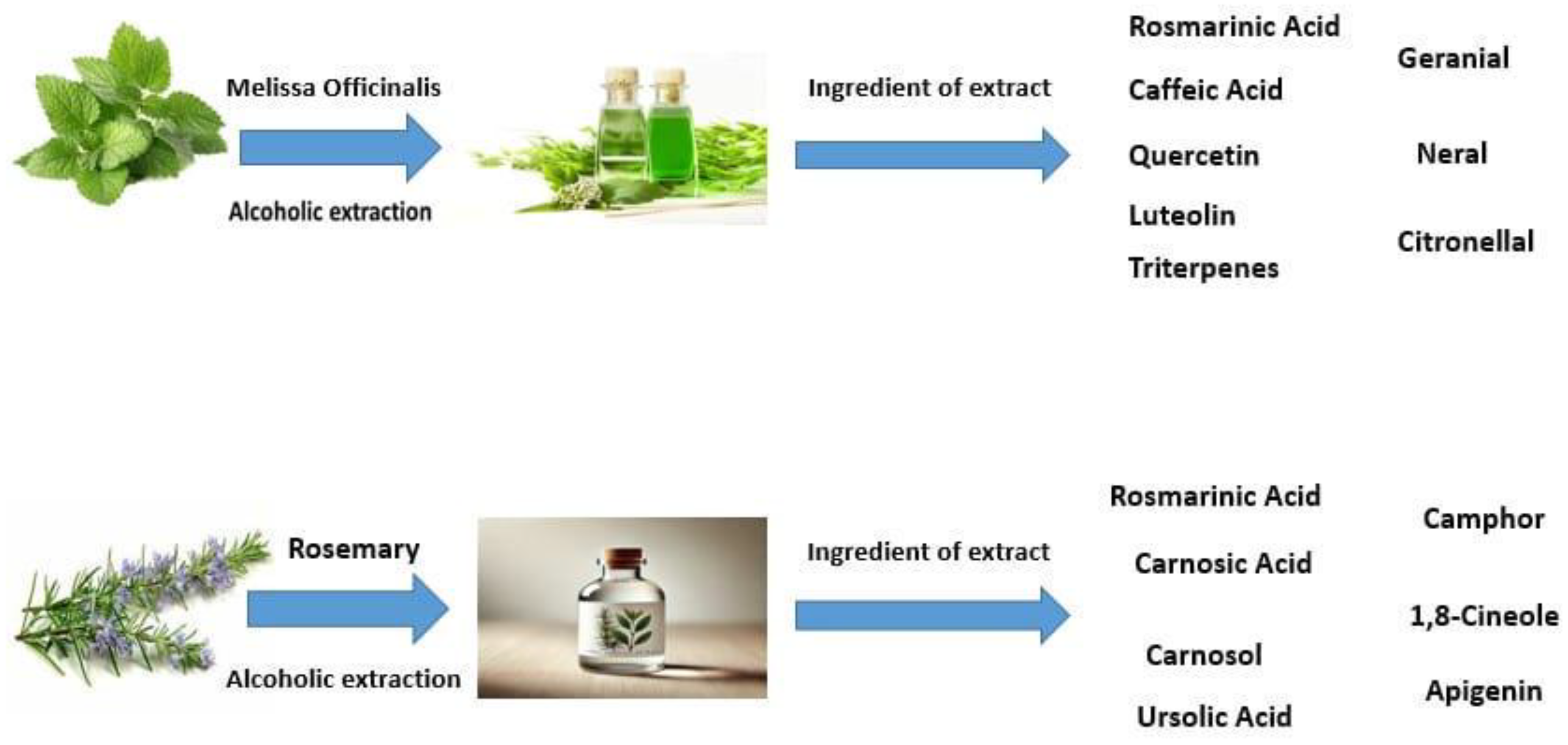
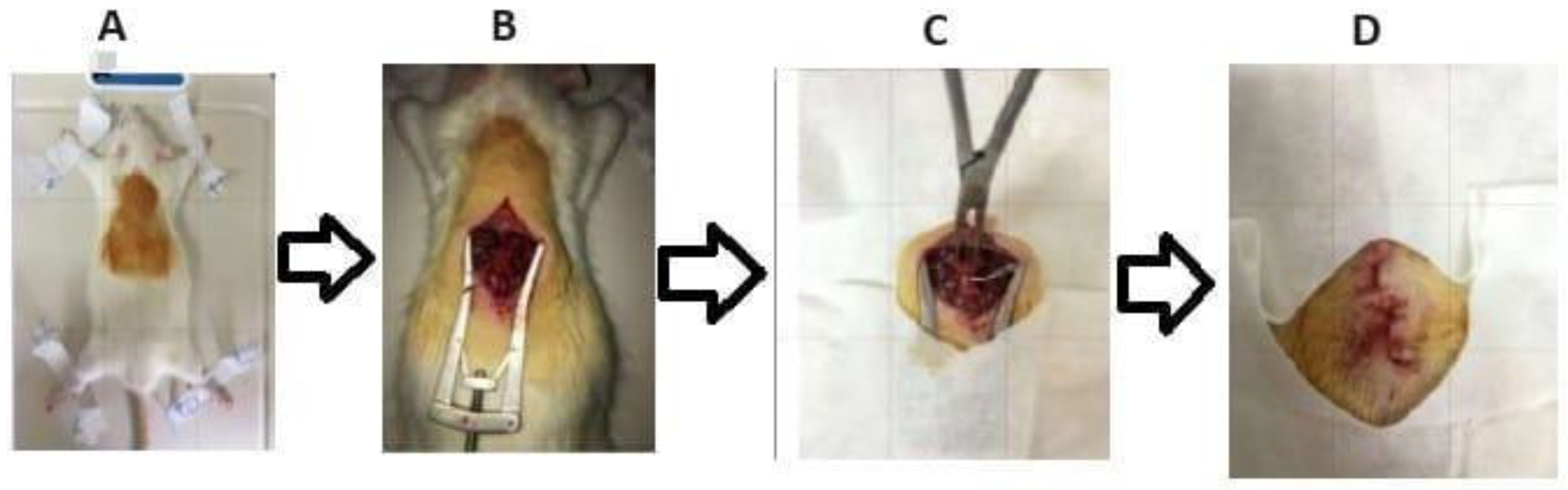
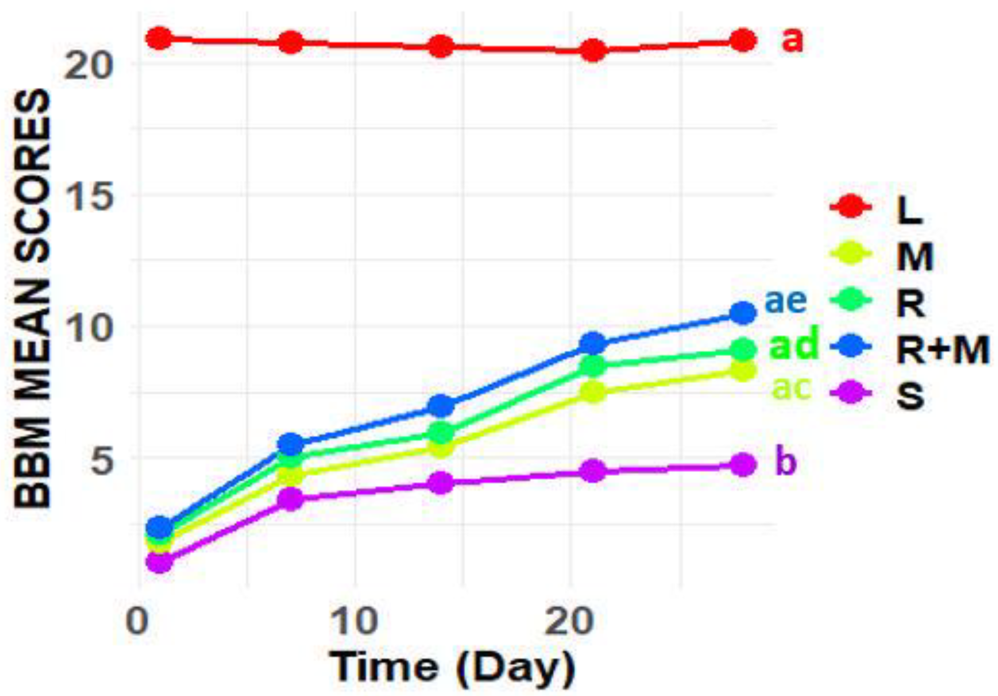
Spinal cord injury (SCI) is linked to a variety of negative outcomes and prognoses that can profoundly affect the lives of individuals, resulting in significant disruptions to multiple facets of their daily activities. A prominent secondary consequence of SCI is the onset of systemic infections, which may disseminate to other organs, including the kidneys, thereby impairing their functionality. Previous studies have demonstrated that the alcoholic extracts of Rosmarinus officinalis and Melissa officinalis possess antioxidant and neuroprotective properties, indicating their potential utility in the treatment and management of SCI and its associated secondary complications. Therefore, this study aimed to examine the combined effects of these extracts on sensory and motor functions, alterations in kidney tissue, and the expression of genes related to inflammation and apoptosis in a rat model of SCI. In this investigation, thirty-five adult male rats were divided into five experimental groups: a control group, a group subjected to spinal cord injury (SCI), a group treated with an alcoholic extract of Melissa officinalis, a group treated with an alcoholic extract of Rosmarinus officinalis, and a group receiving both extracts. The extracts were administered via intraperitoneal injection starting one-day post-SCI and continued for 28 days. Evaluations of sensory and motor functions were performed weekly, while changes in kidney tissue and the expression of genes associated with inflammation and apoptosis were assessed using histomorphometric techniques and quantitative real-time polymerase chain reaction (PCR). The results indicate that the alcoholic Melissa officinalis and Rosmarinus officinalis extracts significantly enhanced sensory and motor functions while reducing the expression levels of genes associated with inflammation (TNF-α) and apoptosis (caspase-3, Bax, and Bcl-2). These findings underscore the potential of these plant extracts in improving the management and treatment of spinal cord injury (SCI) and its secondary effects.

Keywords: 
;  ;  ;  ;  

Introduction:

Spinal cord injury (SCI) represents a severe clinical condition and is among the most complex neurological disorders to manage, often leading to long-term and permanent neurological deficits in affected individuals. Injury to nervous tissue may arise from traumatic events, such as vertebral fractures or dislocations, or may result from various underlying pathological conditions. The impact of a spinal cord injury can result in impairments in the sensory, motor, and autonomic nervous systems in the affected area [1,2,3]. Spinal injuries, characterized by the severance or damage to spinal nerves, have a profound effect on both sensory and motor functions, resulting in a diminished capacity for sensation and movement in the impacted regions. The degree of paralysis, whether partial or complete, is determined by the specific location and severity of the spinal cord injury. Affected individuals may experience a loss of the ability to detect tactile stimuli, temperature variations, or pain, and may also report atypical sensations, such as burning. In more severe instances, the disruption of nerve signals from the brain to the muscles can lead to paralysis, which may present as paraplegia (involving the lower limbs) or quadriplegia (involving all four limbs). Furthermore, individuals with spinal injuries may face additional complications, including difficulties with bladder and bowel control, respiratory challenges, and psychological repercussions such as depression and anxiety [4,5]. At present, a conclusive cure for this disease remains elusive. Nevertheless, substantial progress in foundational research suggests that under certain conditions, nerve cells may possess the capacity for regeneration [6,7]. From a pathophysiological standpoint, spinal cord injuries are categorized into primary and secondary injuries. Primary injuries occur due to an abrupt force exerted on the spinal column at the moment of injury, potentially leading to vertebral fractures or dislocations, which coincide with damage to the spinal cord [6,8].
The severity and extent of spinal cord injury are predominantly determined by the initial level of damage sustained by the spinal cord at the moment of injury, as well as the duration of subsequent compression. Following the primary injury, secondary injuries initiate almost immediately. Within hours of the initial trauma, a series of secondary injury mechanisms are activated, leading to ischemia, inflammation, and the degeneration of neurons and glial cells, all of which play a significant role in advancing secondary spinal cord injury [6,9,10]. Among the key factors contributing to secondary damage are cytokines, various free radicals, and other substances released from damaged cells in the nervous tissue at the injury site. These factors lead to the recruitment of immune cells to the area, including innate immune cells such as neutrophils, macrophages, and microglia, as well as adaptive immune cells like lymphocytes. These immune cells release a range of inflammatory mediators, including pro-inflammatory cytokines (such as TNF-α and interleukin-6), chemokines (like CXCL1 and CXCL12), nitric oxide, and factors related to apoptosis, including members of the caspase protein family. This cascade of events results in cell death and apoptosis of the affected cells, leading to the destruction of nervous tissue and the initiation of a series of inflammatory responses. Collectively, these factors contribute to the disease's progression to other organs and tissues in the body [11]. Clinical manifestations linked to secondary spinal cord injury encompass heightened cellular permeability, the activation of apoptosis-related pathways, ischemia, vascular damage, edema formation, the generation of free radicals, disruptions in ion homeostasis, glial scar formation, and inflammation. The cumulative effects of these injuries exacerbate inflammatory responses, resulting in a pro-inflammatory environment. This induced inflammation has the potential to propagate to adjacent tissues and organs, leading to functional impairments and deficiencies [6,12,13,14,15].
Research has established that spinal cord injury (SCI) triggers substantial systemic inflammation, which represents a significant consequence of the injury. This inflammatory response promotes the spread of the condition to additional organs and tissues, leading to dysfunction within these systems. The inflammatory process is orchestrated by activating various mediators that initiate, propagate, and sustain inflammation. Among these mediators, cytokines such as tumor necrosis factor-alpha (TNF-α) and apoptosis-regulating proteins, including Bax and Bcl-2, are particularly significant. Numerous studies have underscored the pivotal roles of inflammation and apoptosis in the aftermath of SCI and their contributions to disease progression. It is now recognized that apoptosis is governed by an intracellular proteolytic cascade involving members of the caspase family and the Bcl-2 gene family. Specifically, Bax and Bcl-2 are critical in regulating the promotion and inhibition of apoptosis, respectively. Caspase-3, a key member of the caspase family, is integral to the apoptotic and inflammatory processes that ensue following SCI. These proteins participate in several essential cellular events, including chromatin condensation, DNA fragmentation, degradation of vital cellular proteins, clearance of damaged cells, formation of apoptotic bodies, and the terminal phases of apoptosis. Moreover, several cytokines such as interleukin-6 (IL-6), interleukin-1 beta (IL-1β), and tumor necrosis factor-alpha (TNF-α) are essential in the development and spread of inflammation after spinal cord injury (SCI). In particular, TNF-α, primarily produced by macrophages and natural killer cells, is associated with the onset of various inflammatory and autoimmune diseases. Additionally, it promotes the spread of inflammation to other tissues and organs, including the kidneys [16,17,18,19,20,21].
Kidney complications that occur during both the acute and chronic phases subsequent to spinal cord injury (SCI) pose considerable challenges and significant risks to patient health. Spinal cord injury (SCI) disrupts the autonomic nervous system, leading to dysfunction or failure of multiple organs due to the spinal cord's essential role in coordinating bodily functions. The impairment of kidney function and the emergence of urinary disorders following SCI have been thoroughly documented in the literature, underscoring the necessity for meticulous management and ongoing monitoring of kidney status in this patient population [22]. The kidneys are integral to the regulation of blood pressure, the maintenance of fluid balance, the excretion of waste products, the preservation of electrolyte homeostasis, the stabilization of acid-base equilibrium, and the management of endocrine functions. They are responsible for the synthesis of renin, erythropoietin, and the hydroxylation of vitamin D, thereby exhibiting characteristics akin to those of endocrine glands. The kidneys receive approximately 25% of the cardiac output, highlighting their pivotal role in blood pressure regulation and overall cardiovascular health. Kidney dysfunction can significantly disrupt blood pressure regulation and cardiovascular well-being, both directly and indirectly, thereby contributing substantially to mortality rates among individuals with SCI. Given the essential role of the kidneys in maintaining homeostasis and the fact that spinal cord injury (SCI) can lead to severe complications in renal function, it is crucial to assess kidney function as an early indicator for diagnosing complications associated with SCI. Therefore, the early detection and management of kidney complications in this population are essential to prevent further health issues and to improve diagnostic and therapeutic approaches for prevention and treatment. Recent studies have highlighted the importance of monitoring kidney function in patients with spinal cord injuries (SCI) to enhance management strategies, implement more effective treatments, and reduce the risk of serious health complications [23,24,25].
Numerous studies have shown that natural products and herbal medicines possess a variety of active biological properties, making them highly effective in treating different diseases. Research has demonstrated that the alcoholic extracts of two plants—Melissa officinalis and Rosmarinus officinalis—contain a range of substances with potential medicinal effects. Traditional medicine has utilized these extracts to treat a variety of illnesses. For instance, Rosmarinus officinalis has been used to improve memory and focus, reduce headaches and migraines, treat inflammatory diseases like arthritis, and fortify the immune system. Melissa officinalis, on the other hand, is acknowledged as a natural sedative that works well for easing stress, treating sleep difficulties, boosting immunity against viruses, and relieving gastrointestinal problems including bloating and spasms. According to recent research, these plants' alcoholic extracts may be very useful in the management, prevention, and treatment of a variety of neurological conditions, including spinal cord injuries, because of their anti-inflammatory, antioxidant, immunomodulatory, and neuroprotective qualities. This study explores the potential benefits of these extracts in improving kidney function and mitigating kidney complications associated with spinal cord injuries [26,27,28].
Melissa Officinalis, commonly referred to as Lemon balm, is esteemed for its aromatic characteristics and has a longstanding tradition of application in the treatment of diverse health issues. This herb is employed in therapeutic settings as a tonic, antispasmodic, antiseptic, sedative, and analgesic, particularly for mitigating stress-related discomfort and enhancing cognitive performance. Pharmacological investigations have revealed that Melissa Officinalis exhibits a wide array of biological activities, encompassing antioxidant, hypoglycemic, antidepressant, antimicrobial, and analgesic properties. The essential oil and extracts obtained from this plant are abundant in bioactive compounds, including citronellal, geraniol, citral, terpineol, and rosmarinic acid, which play a significant role in its therapeutic effectiveness and the prevention of various conditions, such as cerebral ischemia, spinal injuries, and Alzheimer's disease [29,30]. Rosmarinus officinalis (Rosmary) , belonging to the Lamiaceae family, is acknowledged as an aromatic medicinal plant with a wide range of therapeutic properties. These properties encompass antimicrobial, anticancer, antidiabetic, anti-inflammatory, analgesic, antioxidant, anticoagulant, and diuretic effects. Furthermore, Rosmarinus officinalis's natural essential oils contribute to its extensive application in the perfume industry. The presence of various bioactive compounds in Rosmarinus officinalis extract, including carnosol, rosmarinic acid, and diterpenes, notably amplifies the therapeutic potential of its alcoholic extract in the prevention, management, and treatment of neurological disorders [31,32]. Recent studies indicate that the alcoholic extracts of Melissa officinalis and Rosmarinus officinalis are known as natural compounds with anti-inflammatory and anti-apoptotic properties. These extracts are capable of reducing the expression of apoptotic genes such as Bax, Bcl-2, and Caspase-3, which leads to the reduction of cellular death processes and prevents damage caused by apoptosis. In addition, these extracts can reduce inflammatory factors such as Tumor Necrosis Factor-alpha (TNF-α), helping to prevent the progression of inflammation in damaged tissues. By reducing apoptosis and inflammation, they can protect sensitive tissues such as the spinal cord and kidneys, and may be effective in treating kidney injuries in patients with spinal cord injury. The use of these plants may help reduce kidney tissue destruction and improve kidney function in these patients [33,34].
Research has substantiated the varied therapeutic attributes of these two botanical species. For example, a study conducted in 2012 by Mr. Bayat and his associates indicated that the alcoholic extract of Melissa officinalis demonstrates neuroprotective effects and may be beneficial in both the prevention and treatment of neurological disorders [30]. Furthermore, a 2017 investigation revealed that the alcoholic extract of Rosmarinus officinalis also displays neuroprotective properties and has the potential to alleviate the adverse effects associated with spinal cord injuries in laboratory rats [32]. Additionally, research has demonstrated that the alcoholic extract of Melissa officinalis, due to its anti-inflammatory and antioxidant properties, can aid in improving kidney function and mitigating renal damage caused by oxidative stress [34]. Similarly, the alcoholic extract of Rosmarinus officinalis has shown significant protective effects on kidney tissue by reducing inflammation and oxidative damage, which are crucial in preventing renal complications in various disease models [35]. In light of the bioactive compounds identified in the alcoholic extracts of Melissa officinalis and Rosmarinus officinalis, as well as their significant therapeutic properties, we integrated these extracts into our research framework. The principal aim of this study is to demonstrate the therapeutic effects of the coadministration of alcoholic extracts of Melissa officinalis and Rosmarinus officinalis on kidney tissue and apoptosis-related gene expression in a rat model of spinal cord injury. Although these extracts exhibit significant therapeutic effects individually, our study demonstrated that the combined therapeutic effects of the two plants are superior. This research seeks to propose a novel, innovative, and economically viable therapeutic approach for this condition, while also offering new insights for future research initiatives.

Materials and Methods:
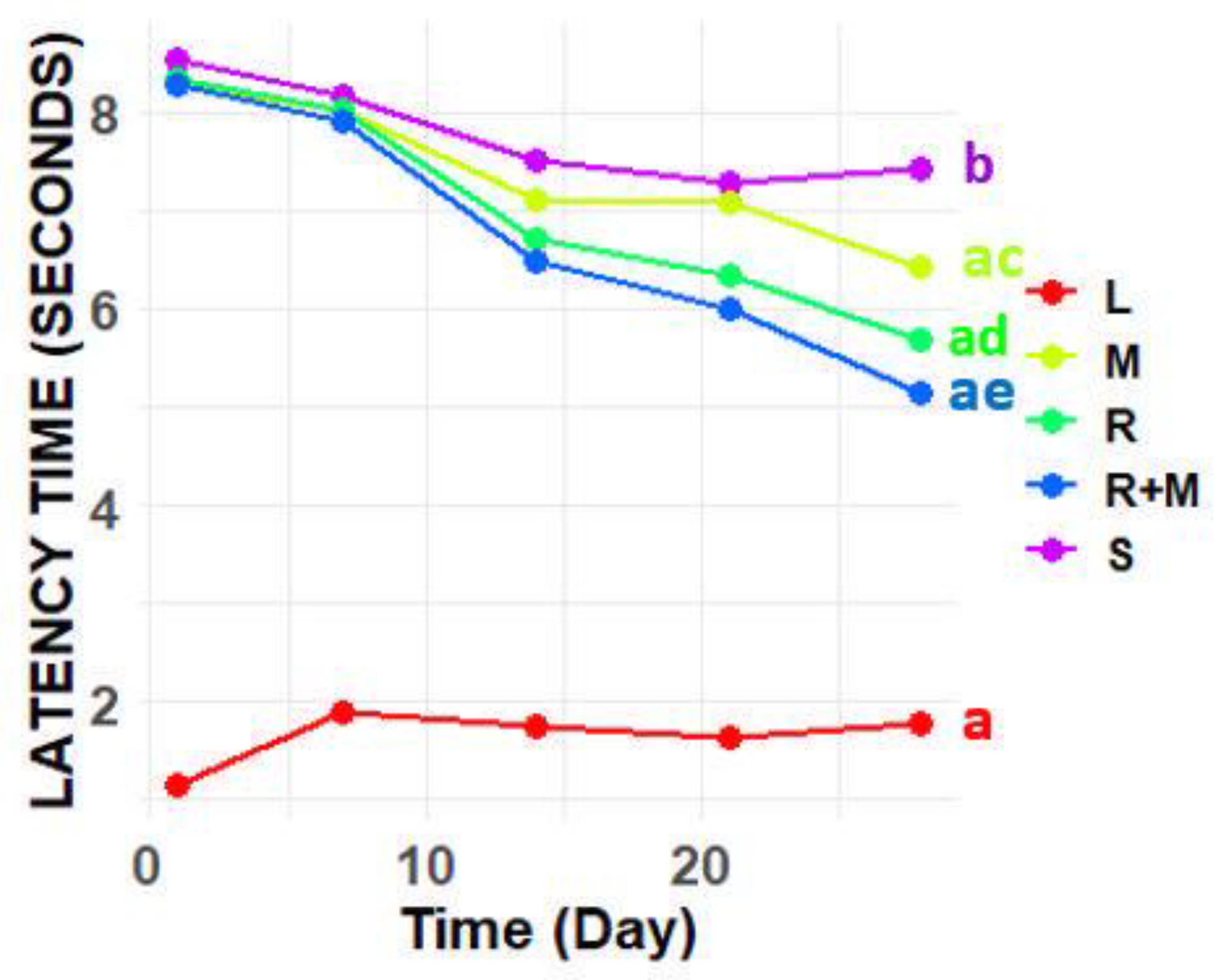
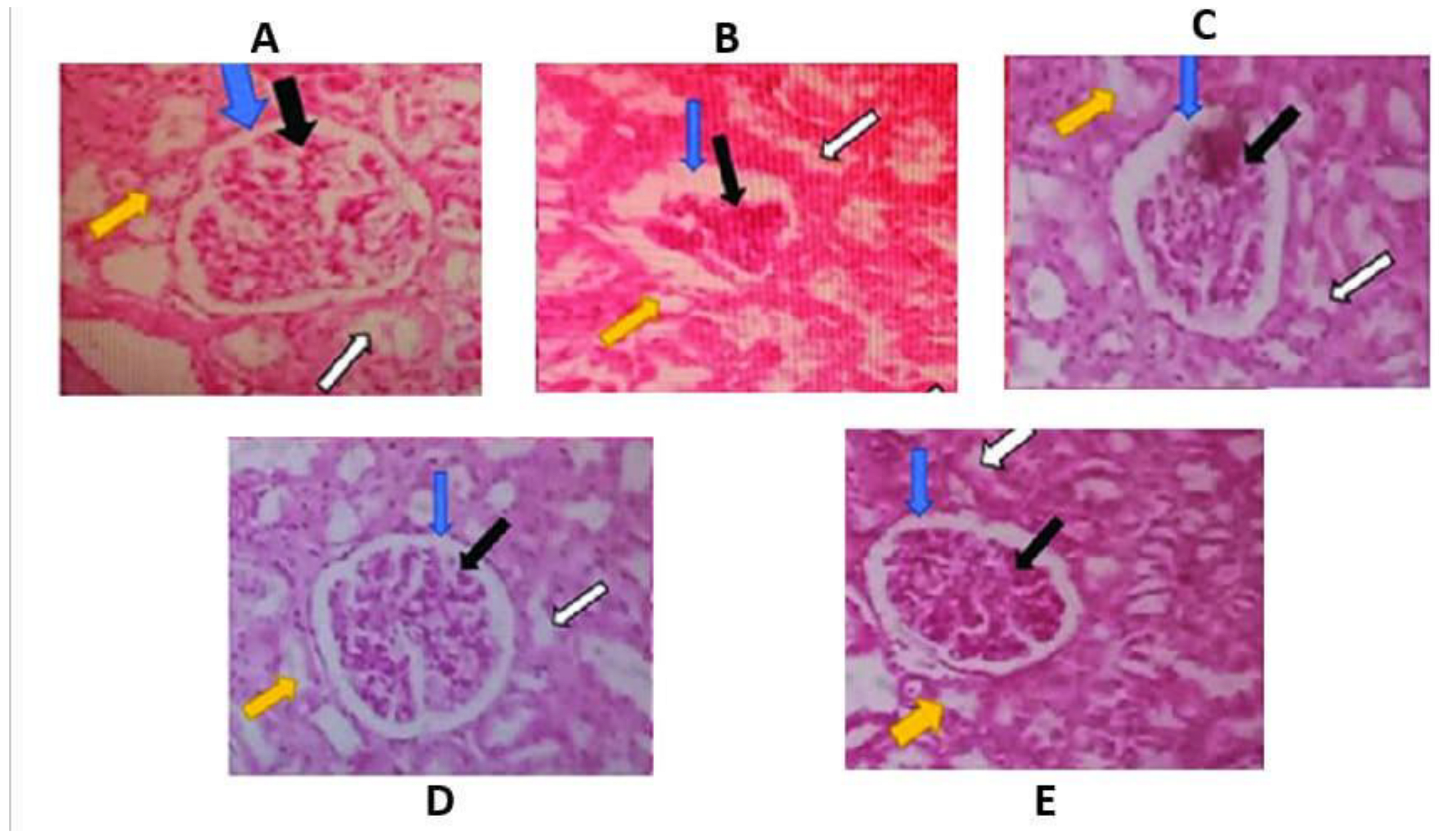
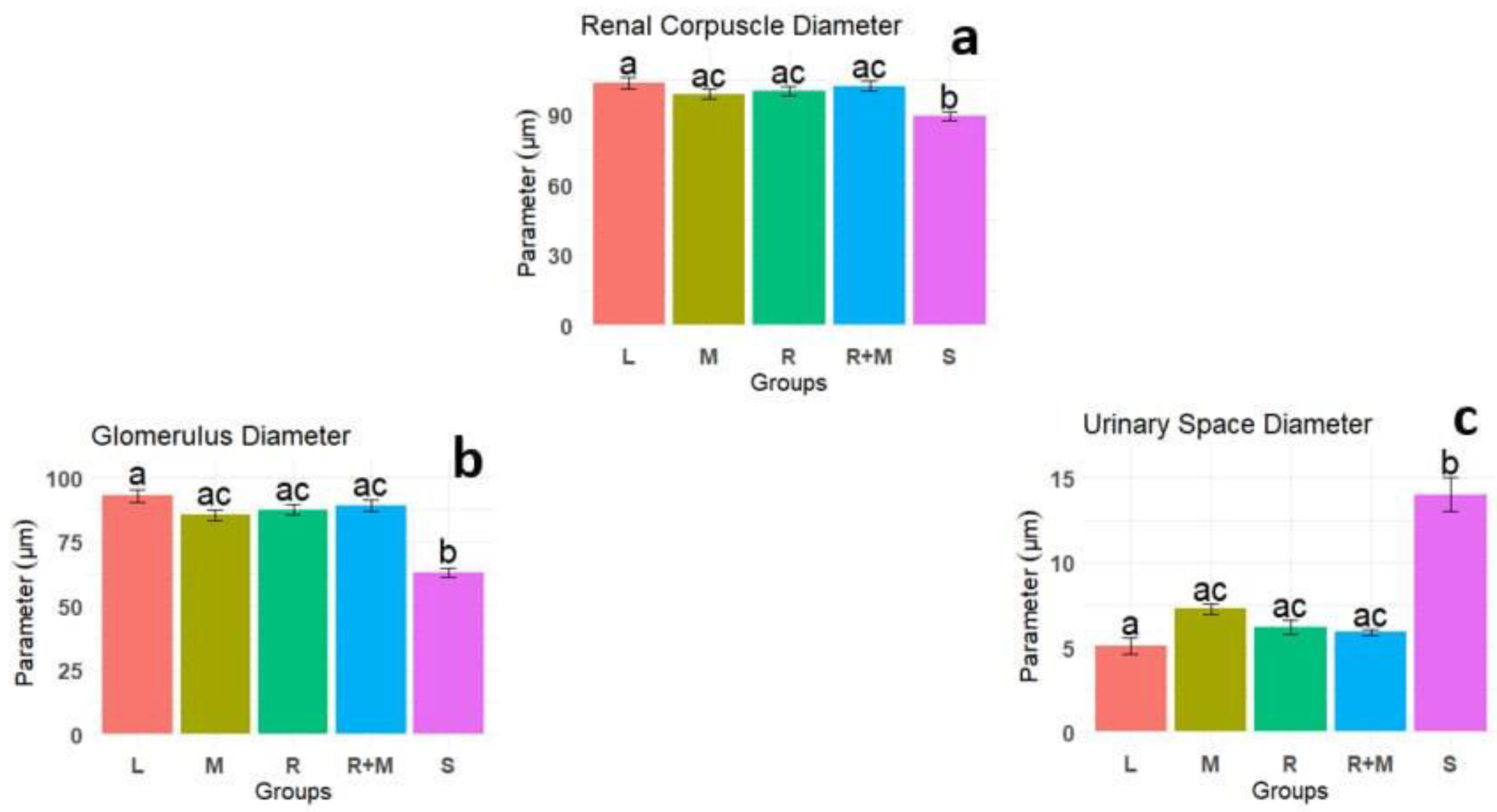
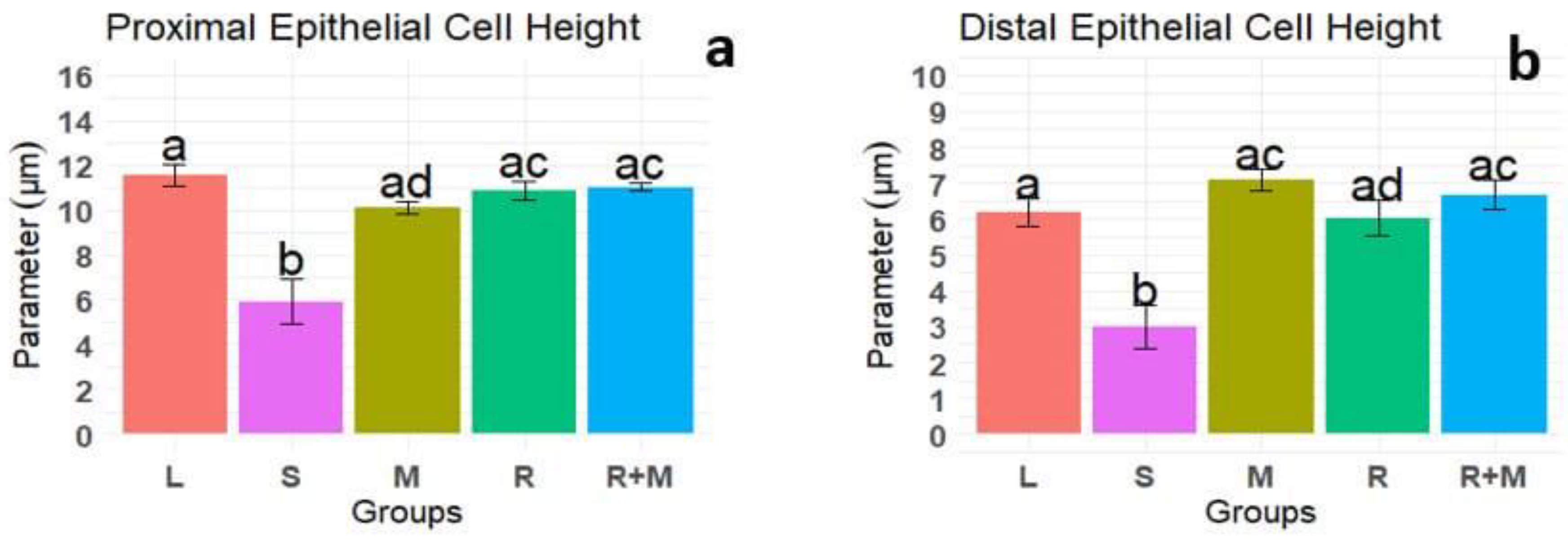
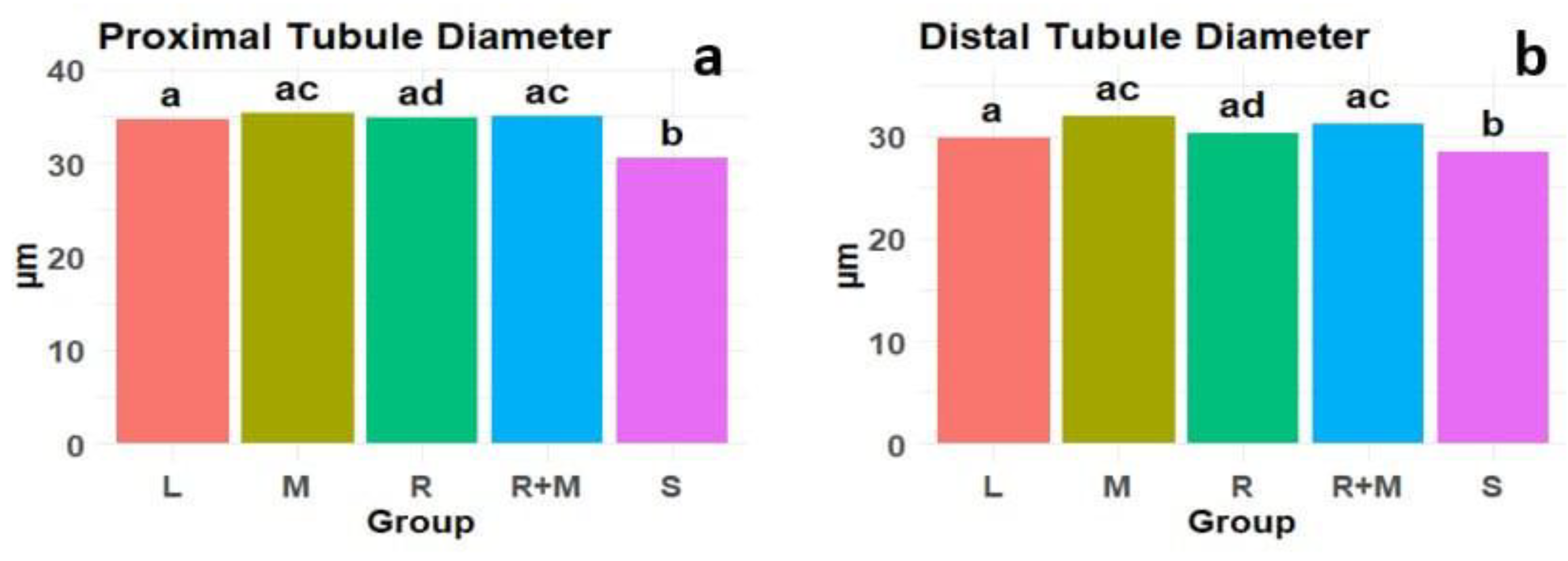
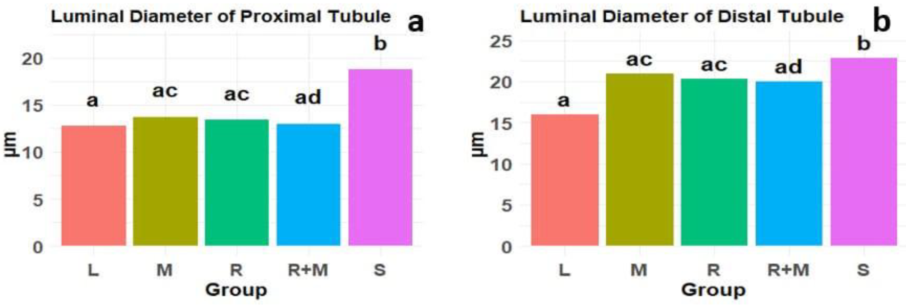
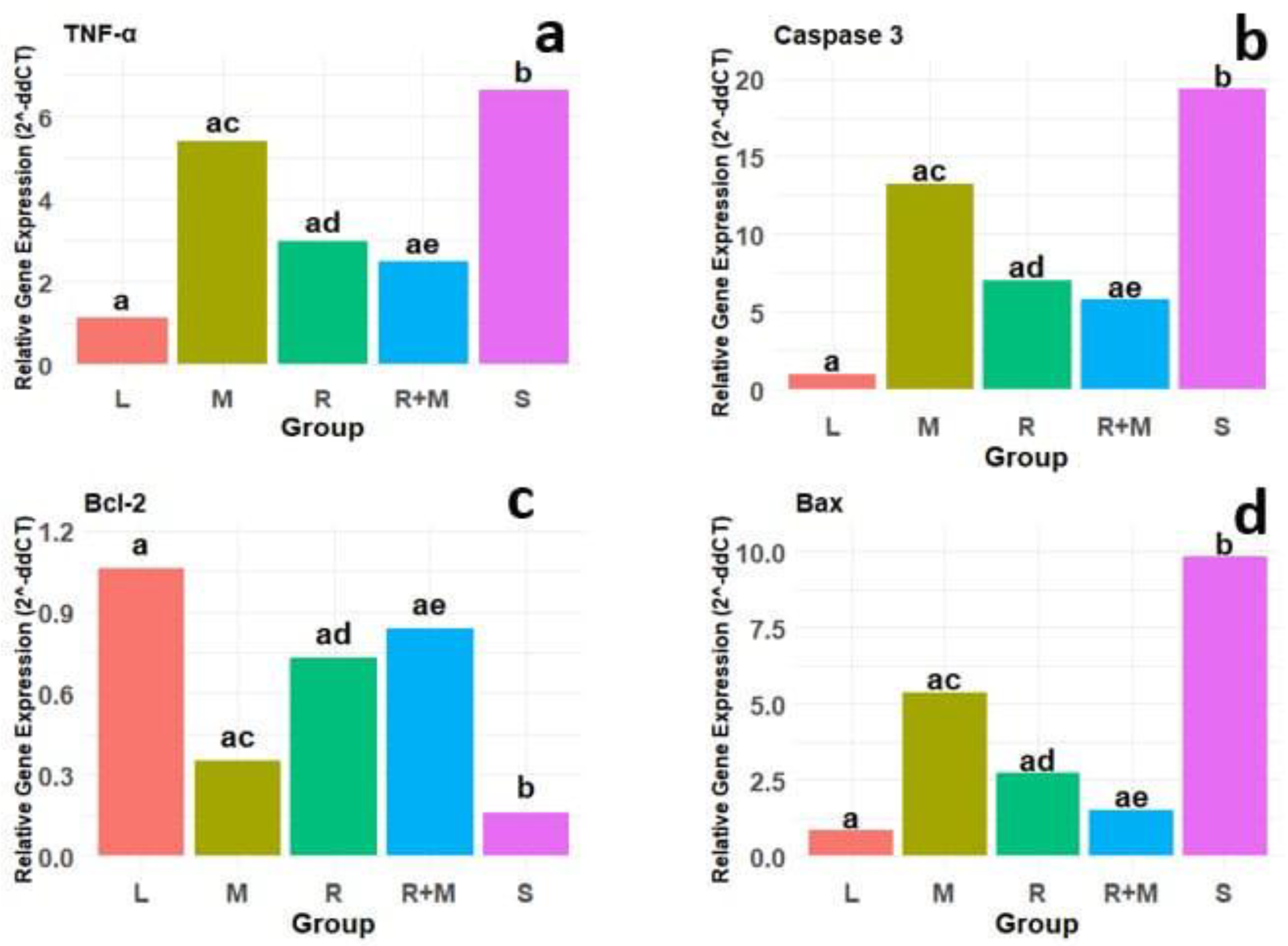
Gathering and extracting plants
The Firouze Medicinal Plant Garden in Tehran provided the botanical specimens, which were then verified by the Islamic Azad University's Tehran Medical Branch's Faculty of Pharmaceutical Sciences. The following protocols were followed to prepare alcoholic extracts from Melissa officinalis and Rosmarinus officinalis:
The freshly harvested leaves of Melissa officinalis and Rosmarinus officinalis were initially subjected to a crushing and drying process at room temperature. Subsequently, 250 mL of 70% ethanol was added to the crushed plant material to prepare the hydroalcoholic extract. The mixture was agitated every 12 hours for a total of 48 hours to ensure thorough extraction. This extraction was conducted using a 500 mL Erlenmeyer flask equipped with a magnetic stirrer (Model IKA C-MAG HS 7) set at 300 RPM to maintain consistent agitation. After the maceration period, the mixture was filtered using a vacuum filtration apparatus (Model Buchi V-700) at a pressure of 20 inHg to separate the liquid extract from the solid residue. The solid residue was then washed with 70% ethanol and filtered again using the same filtration system. The resulting liquid extract was combined with the initial extract to produce the final hydroalcoholic solution [30,36,37,38]. A concentrated plant extract weighing 2.4 grams was obtained through vacuum distillation, which effectively removed the solvent. The extraction yield for Rosmarinus officinalis was 20%; therefore, 12 grams of Rosmarinus officinalis leaves were used to produce 2.4 grams of extract. In contrast, the extraction yield for Melissa officinalis was 15%, necessitating 16 grams of Melissa officinalis leaves to obtain the same 2.4 grams of extract. Subsequently, over the course of one day at room temperature, the extracts from Melissa officinalis and Rosmarinus officinalis were combined with 50 mL of saline solution. The experimental rats received daily doses of these solutions via intraperitoneal injection after the extracts had completely dissolved, for a duration of four weeks.
High-performance liquid chromatography (HPLC)
To assess the concentrations of the primary active constituents in the alcoholic extracts of Rosemary and Melissa officinalis, reverse-phase high-performance liquid chromatography (HPLC) was utilized in conjunction with data analysis techniques. The HPLC apparatus included a K-1001 pump and a K-2800 photodiode array detector, both sourced from Knauer (Berlin, Germany). Chromatographic separation was achieved using a Eurosphere C18 column characterized by a particle size of 5 micrometers and dimensions of 25 mm × 4.6 mm. Data processing was performed using ChromGate software. The mobile phase consisted of 0.01% formic acid and methanol, with a gradient transition from a ratio of 75:25 to 80:20 (v/v) over 30 minutes, a flow rate of 0.5 mL/min, and a column temperature maintained at 25 °C. The detector was calibrated to a wavelength of 330 nm. A detailed composition of each extract (see Figure 1) is provided below:
The Rosemary extract contains 13.4 % caffeic acid, 9.52 % rosmarinic acid, 8.8 % cineole, 5.2 % α-pinene, 3.14 % borneol, 1.52 % camphor, and 1.48 % limonene.
The Melissa Officinalis extract includes 41.3 % estragole, 13.4 % limonene, 6.97 % nerol, 6.03 % geranial, 4.13 % nerallyl acetate, 3.08 % geraniol, less than 0.5% caffeic acid, and 0.75 % rosmarinic acid.
Animals
For this research, a group of 35 adult male Wistar rats, each weighing between 200 and 250 grams, was chosen and used. These rats were sourced from the Faculty of Veterinary Medicine at the University of Tehran and were kept under standard laboratory conditions at the animal facility of Tehran University of Medical Sciences. They had unlimited access to food and water. The laboratory conditions were regulated to maintain a temperature of 22 ± 2°C, relative humidity between 10% and 50%, and a 12-hour light-dark cycle. The study focused specifically on healthy rats within the specified weight range. Rats that were sick did not meet the weight criteria, or did not have spinal cord injuries were excluded from the study.
Creating a Model for Spinal Cord Injuries
The subjects were anesthetized with ketamine (80 mg/kg) and xylazine (15 mg/kg), administered through intraperitoneal injection. The dorsal region of the subjects was sterilized with alcohol and betadine, and the hair in the surgical area was removed. A longitudinal incision was made along the midline, extending from the eighth to the twelfth thoracic vertebrae. The fascia and superficial muscles were retracted to reveal the spinous and transverse processes of the vertebrae. The tenth vertebra was located using a specialized retractor, and a laminectomy was performed under a surgical microscope, ensuring the integrity of the dura mater was maintained. Following the exposure of the spinal cord, a unilateral spinal cord injury was induced through a compression technique, utilizing an aneurysm clip applied to the right side of the spinal cord for one minute. To ensure precision, the aneurysm clip was pre-calibrated by adjusting it to a consistent closing pressure, which ensured uniform application of force. This pre-calibration helped achieve controlled damage to the spinal cord, targeting the lateral side to affect the motor neurons responsible for locomotion. Following the release of the clip, visible signs of localized tissue damage, including slight discoloration and reduced blood flow, were observed. The surrounding tissue was meticulously inspected to ensure that no excessive hemorrhaging occurred [39,40]. To mitigate the risk of dehydration in the animals, 2-3 milliliters of normal saline were administered subcutaneously in the dorsal region of the neck. Following the infliction of the injury, the surgical site was sutured closed. To minimize the potential for infection at the surgical site, as well as within the central nervous system and urinary system, a subcutaneous injection of 15 mg/kg of gentamicin was administered in the dorsal neck area one day before the surgical procedure and was continued for two days postoperatively. The various stages of spinal cord injury are depicted in Figure 2 [41].
The Drug Administration and animal advocacy organizations.
For this study, the animals were randomly divided into five separate groups, each containing seven Wistar rats:
1. Control Group (L): This group of rats underwent laminectomy surgery without any resultant spinal cord damage.
2. Control Group (S): The control group (S) consisted of rats that experienced spinal cord injuries for this research. These rats were given daily subcutaneous injections of 100 mg/kg of a normal saline solution, beginning 24 hours post-injury and lasting for four weeks.
3. Treatment Group 1 (M): consisted of rats with spinal cord injuries that were administered a hydroalcoholic extract of Melissa officinalis via subcutaneous injection at a dosage of 150 mg/kg at the nape of the neck. This therapy started the day after the injury and continued every day for four weeks.
4. Treatment Group 1 (R): consisted of rats with spinal cord injuries that were administered a hydroalcoholic extract of Rosemary via subcutaneous injection at a dosage of 150 mg/kg at the nape of the neck. This therapy started the day after the injury and continued every day for four weeks.
5. Treatment Group 3 (R+M) involved treating spinal cord injured rats concurrently with a combination of hydroalcoholic extracts of Melissa officinalis and Rosemary. Every extract was injected subcutaneously into the nape of the neck at half the recommended dosage. Following the establishment of the spinal cord injury model, this treatment started right away and lasted for four weeks every day.
Assessment of the Spinal Cord Injury Model
A tissue sample was obtained from the injured region to confirm the spinal cord injury model. Microscopic examinations following hematoxylin-eosin staining revealed a compressed region on the right side of the spinal cord in the samples, indicating damage to this area. Furthermore, the analyzed regions showed signs of emptiness, suggesting the presence of axonal loss or degeneration. These findings support the validity of the spinal cord injury model (see Figure 3).
Kidney Tissue Specimen Preparation
After establishing the spinal cord injury model and completing a four-week treatment period, the rats were euthanized to collect kidney tissue samples. Sodium pentobarbital was administered intraperitoneally at a dose of 100 mg/kg to ensure deep and irreversible anesthesia. Following this, 0.9% saline solution and 10% buffered formalin were injected into the heart to perfuse and fix the tissue [42]. A midline incision was then made in the abdominal skin, and the right kidney was removed from four randomly chosen rats in each experimental group. The kidneys were quickly placed in liquid nitrogen for rapid freezing and preservation and later stored at -80 degrees Celsius for future biochemical analyses. The remaining three rats from each group had their kidneys preserved in 10% formalin for histopathological examination and tissue analysis.
The preparation of tissue sections is a meticulous procedure that consists of seven distinct phases. Initially, tissue specimens are fixed in a 10% formalin solution to preserve their structural integrity. Following fixation, the samples undergo a dehydration process, which involves sequential immersion in 70% ethanol, followed by 90% ethanol, and ultimately three immersions in 100% ethanol. In the next phase, the samples are subjected to three one-hour immersions in xylene to remove any residual water, thereby preparing the tissue for paraffin infiltration. The subsequent step involves placing the samples in molten paraffin to ensure thorough permeation of the tissue. After complete infiltration, the samples are carefully positioned in molds filled with molten paraffin, ensuring that any remaining voids are filled, resulting in the complete encapsulation of the tissue specimens. During the sectioning phase, thin sections approximately 5 micrometers thick are cut from the paraffin-embedded blocks using a microtome, facilitating precise examination. Finally, the tissue sections are stained using the Hematoxylin and Eosin (H&E) staining technique, which enhances contrast and highlights cellular components. This comprehensive and precise methodology is essential for the accurate preparation of kidney tissue samples and is critical for enabling subsequent biochemical and histopathological analyses.
Neurological Examination
To assess neurological function in rats, the Basso-Beattie-Bresnahan (BBB) scale was employed, which is specifically designed to evaluate motor function and mobility in an open-field environment. This scale operates on a continuum from 0 to 21 and encompasses various aspects of hind-limb motor performance, including weight support, stepping ability, coordination, and toe spread. Functional assessments were performed and recorded on days 1, 7, 14, 21, and 28 by two researchers who were blinded to the treatment conditions. The final functional score for each rat was determined by calculating the average of the scores obtained from both evaluators [43,44].
Pain sensitivity was evaluated through behavioral tests using the hot water method on the hind limbs of rats after spinal cord injury (SCI). Functional scores were recorded on days 1, 7, 14, 21, and 28. The response to heat was measured by timing how long it took for the rats to withdraw their hind paw from water heated to 60°C. Each rat had its paws individually placed in containers of hot water, and a total of six trials (three for each paw) were conducted for every rat. The average reaction time from these trials was noted. Rats that did not react to the heat were taken out of the hot water after 60 seconds [45,46].
Evaluation of Changes in the Structure of Kidney Tissue
The kidney tissue samples were analyzed using a light microscope. In the field of histology and histological research, each sample was evaluated from three different perspectives utilizing Dino Capture software (Version 2, developed by Dino-Lite Company, Netherlands), with measurements recorded in micrometers (μm). The analysis included the dimensions of various anatomical structures, such as the Bowman's capsule, glomerulus, urinary space, proximal tubule, lumen of the proximal tubule, height of the proximal tubule epithelium, diameter of the distal tubule, lumen of the distal tubule, and height of the distal tubule epithelium. These measurements were performed using the aforementioned software.
Evaluation of Gene Expression RNA Isolation:
RNA was isolated using TRIzol reagent (RNX) (Invitrogen, USA) from the kidney tissue of experimental rats, utilizing four rat kidneys per group, with approximately 100 mg of kidney tissue per rat for RNA extraction. The tissue was homogenized in 1 mL of TRIzol reagent per 100 mg of tissue to ensure complete lysis. Prior to extraction, the tissue samples were stored at -80°C to preserve RNA integrity. For transport, the samples were maintained on dry ice to prevent RNA degradation during transit. After homogenization, 200 μL of chloroform was added to the mixture, which was then thoroughly vortexed for 15 seconds and allowed to equilibrate at room temperature for 5 minutes. The sample was subsequently centrifuged at 4°C for 1 to 5 minutes at a relative centrifugal force (RCF) of 12,000 × g, resulting in phase separation. The RNA, located in the aqueous phase (upper phase), was carefully collected.
For RNA precipitation, an equal volume of isopropanol (typically 1 mL for every 1 mL of TRIzol used) was added to the collected upper phase. The sample was then incubated at -20°C for 1 hour. Following incubation, the sample was centrifuged at 4°C at 12,000 × g for 15 minutes to pellet the RNA. The supernatant was discarded, and the RNA pellet was washed with 75% ethanol prepared using nuclease-free water. One milliliter of 75% ethanol was added to the pellet, and the sample was gently vortexed before being centrifuged at 4°C for 10 minutes at 8,000 × g. The ethanol was carefully removed, and the pellet was air-dried at room temperature for 15 minutes to ensure the complete removal of ethanol.
Finally, the quantification and purity of RNA were assessed using a NanoDrop spectrophotometer.
Synthesis of cDNA from Total RNA:
The initial stage of reverse transcription polymerase chain reaction (RT-PCR) involves synthesizing complementary DNA (cDNA) from RNA using universal primers, such as dT-Oligo or hexamer random primers. Accurate quantification of mRNA levels is essential for analyzing gene expression. Prior to cDNA synthesis, the purity of the RNA was assessed using spectrophotometry with a Nanodrop spectrophotometer, measuring absorbance at 260 nm (A260) and 280 nm (A280) wavelengths to evaluate RNA purity. The A260/A280 ratio was employed to confirm RNA quality, with an optimal value ranging from 1.8 to 2.0 indicating pure RNA. Additionally, RNA integrity was verified through agarose gel electrophoresis to check for intact ribosomal RNA (rRNA) bands, ensuring the absence of RNA degradation. Only RNA samples exhibiting a clear, sharp band pattern were utilized for cDNA synthesis.
For cDNA synthesis, the Premix PCR-2xRT 2-Step Kit (Biofact, Korea) was utilized. This kit contains a reverse transcriptase enzyme along with the necessary reagents for synthesizing cDNA from RNA templates. In this study, 0.5 μg of RNA was used as the template for reverse transcription in a 20 μL reaction volume. The dT-Oligo primer was added at a final concentration of 1 μM, and the reaction mixture was incubated at 42°C for 60 minutes to ensure optimal reverse transcription. Following this incubation, the reaction was terminated by heating at 95°C for 5 minutes to inactivate the reverse transcriptase enzyme. The synthesized cDNA was subsequently employed for gene expression analysis.
Real-time PCR reaction:
In this study, quantitative PCR (qPCR) was utilized to evaluate the expression levels of key target genes, including TNF-α, Caspase-3, Bcl-2, and Bax, which were selected for their essential roles in apoptosis and inflammatory pathways. RNA was extracted from kidney tissue samples, and the expression of these genes was quantified relative to the reference gene β2M, chosen for normalization due to its stable expression across various experimental conditions. A comprehensive list of primers for the genes of interest is presented in Table 1.
For cDNA synthesis, 0.5 μg of RNA was used as the template, and cDNA was synthesized using the Premix PCR-2xRT 2-Step Kit (Biofact, Korea) according to the manufacturer's protocol. Each qPCR reaction was performed in triplicate to ensure the reliability of the data. The reaction mixture consisted of 1 μL of cDNA, 10 μL of SYBR Green Master Mix (Biofact, Korea), 1 μL of each forward and reverse primer (final concentration of 0.5 μM), and 7 μL of nuclease-free water, resulting in a total volume of 20 μL.
The thermocycling conditions were as follows: an initial denaturation at 95°C for 5 minutes, followed by 40 cycles consisting of denaturation at 95°C for 30 seconds, annealing at 55°C for 30 seconds, and extension at 72°C for 30 seconds. Fluorescence was measured at the end of each cycle to monitor the amplification process. The efficiency of the amplification was evaluated using a standard curve, which demonstrated an optimal amplification efficiency range of 90-110% and an R² value exceeding 0.99, thereby confirming the reliability of the amplification process. Melt curve analysis was conducted to verify the specificity of the amplification, with a single peak indicating the absence of non-specific products.
The relative gene expression was calculated using the ΔΔCt method. The ΔCt for each sample was determined by subtracting the Ct value of the reference gene (β2M) from the Ct value of the target gene. The ΔΔCt was then calculated by subtracting the ΔCt of the control group from that of the experimental group. Relative expression levels were determined using the formula: Relative Expression = 2^(-ΔΔCt).
Data analysis was performed using GraphPad Prism 10.0 (GraphPad Software, San Diego, CA). Statistical significance was assessed by comparing relative expression values among experimental groups, with p-values less than 0.05 deemed statistically significant.
Primer Design:
Gene sequences for Caspase-3, Bax, Bcl-2, and TNF-α were retrieved from the NCBI database. Primers for these genes were designed using R software to ensure optimal amplification efficiency and specificity. The design process was conducted with careful attention to key parameters, including primer length, melting temperature (Tm), and GC content, all of which contribute to the reliability and reproducibility of quantitative PCR (qPCR) results.
The primers were designed to ensure the specific amplification of target genes while minimizing non-specific binding and primer-dimer formation. The reference gene β2M was selected for normalization due to its stable expression under experimental conditions. The sequences of the forward and reverse primers, along with their corresponding product sizes and melting temperatures (Tm), are presented in Table 1 below.
Statistical analysis:
Histological changes in kidney tissue were assessed using light microscopy, and image analysis was conducted with Dino Capture Software (Version 2, Dino-Lite Company, Netherlands). The sensory and motor functions of the rats were evaluated using the Basso-Beattie-Bresnahan (BBB) scale to assess locomotor recovery following spinal cord injury. The expression levels of key apoptotic and inflammatory genes were quantified through quantitative real-time PCR (qRT-PCR). The analyzed data included gene expression levels, motor function scores, and histological alterations. Results are presented as means ± standard deviation (SD) for continuous variables, with statistical significance defined as p < 0.05.
Data preparation involved normalizing gene expression data using the ΔΔCt method, with β-actin (ACTB) and β2-microglobulin (B2M) serving as internal controls. The inclusion of B2M as a normalization factor is crucial for correcting variability in RNA quantity or quality between samples, ensuring that observed changes in gene expression reflect biological differences rather than technical artifacts. The normality of the data distribution was assessed using the Shapiro-Wilk test, while the homogeneity of variances was evaluated using Levene's test; both are essential assumptions for conducting ANOVA.
Data analysis was conducted using GraphPad Prism 10.0 (GraphPad Software, San Diego, CA) and R software (version 4.4.2, R Foundation for Statistical Computing, Vienna, Austria). Between-group comparisons were performed using one-way analysis of variance (ANOVA), followed by Tukey’s post-hoc test for multiple comparisons. Significant differences were assessed by comparing relative expression levels between the experimental and control groups. Statistical significance was defined as p < 0.05.

Results

During all phases of the experiments, a substantial disparity was noted between the spinal cord injury (SCI) group and the control group. Nevertheless, this disparity was markedly reduced in the treated groups. Statistical analysis consistently demonstrated a p-value less than 0.05 (p < 0.05), highlighting the significance of the results. This difference constituted the principal criterion for confirming the establishment of the SCI model.
Results of Neurological Performance
The use of alcoholic extracts from Melissa officinalis and Rosmarinus officinalis resulted in enhanced motor function following spinal cord injury, which caused paraplegia (loss of motor function in the hind limbs). Additionally, the spinal cord injury group exhibited significant changes in motor scores compared to the control group (healthy group). Statistical analyses, including the Shapiro-Wilk test for normality, revealed that the data were normally distributed (p > 0.05, Shapiro-Wilk test). The administration of Melissa officinalis alcoholic extract significantly improved motor function in rats compared to the spinal cord injury group. However, when Rosmarinus officinalis alcoholic extract was used for treatment, motor function in rats improved significantly compared to both the spinal cord injury group and the group treated with Melissa officinalis alcoholic extract. One-way ANOVA and Tukey’s post-hoc test revealed significant interaction effects between treatment variables and time (p < 0.01). The Bonferroni post-hoc multiple comparisons test demonstrated a significant improvement in motor function following treatment with Melissa officinalis alcoholic extract at a dose of 150 mg/kg on days 14, 21, and 28 (p < 0.001), as well as treatment with Rosmarinus officinalis alcoholic extract at a dose of 150 mg/kg on days 7, 14, 21, and 28 (p < 0.01). The combined use of alcoholic extracts from Melissa officinalis and Rosmarinus officinalis significantly improved motor function on days 14, 21, and 28 (p < 0.001; see Figure 4).
The concurrent administration of alcoholic extracts derived from Melissa officinalis and Rosmarinus officinalis demonstrated a notable enhancement in sensory function following spinal cord injury. Statistical analyses, including the Shapiro-Wilk test for normality, revealed that the data were normally distributed (p > 0.05, Shapiro-Wilk test). The mean latency to respond to painful stimuli in the group receiving the Melissa officinalis extract was significantly reduced in comparison to the spinal cord injury cohort. Furthermore, the application of Rosmarinus officinalis alcoholic extract as a therapeutic intervention resulted in a marked improvement in sensory function in rats, surpassing the outcomes observed in both the spinal cord injury group and the group treated with Melissa officinalis extract. Additionally, the combined administration of alcoholic extracts from Melissa officinalis and Rosmarinus officinalis led to a significant enhancement in sensory function relative to the other treatment groups and the spinal cord injury group. One-way ANOVA and Tukey’s post-hoc test indicated a significant interaction effect among the variables, including treatment and time (p < 0.001). The Bonferroni post-hoc multiple comparisons test further illustrated that sensory function significantly improved following the administration of Melissa officinalis extract compared to the spinal cord injury group on days 14, 21, and 28 (p < 0.01), post-injury. Similarly, treatment with Rosmarinus officinalis extract also resulted in a reduction of the mean response time to painful stimuli compared to the spinal cord injury group on the same days (p < 0.01). Notably, the simultaneous administration of alcoholic extracts from both Rosmarinus officinalis and Melissa officinalis yielded significant improvements in performance on days 14, 21, and 28 (p < 0.001; see Figure 5).
Outcomes of alterations in texture:.
The current investigation examined the therapeutic effects of alcoholic extracts obtained from two botanical sources, namely Rosmarinus officinalis and Melissa officinalis, in a rat model of spinal cord injury. The primary aim of this study was to evaluate the impact of spinal cord injury on renal tissue and the subsequent functional alterations in the affected rats. Additionally, the research sought to compare these structural and functional changes across various experimental groups in relation to a control group comprising healthy rats. Furthermore, the study aimed to assess the therapeutic efficacy of the alcoholic extracts of Rosmarinus officinalis and Melissa officinalis in ameliorating renal tissue damage and enhancing functional outcomes in rats with spinal cord injuries, as compared to the injury group. To facilitate this investigation, the rats were categorized into five distinct groups: a control group (healthy), a spinal cord injury group, and three spinal cord-injured groups, each receiving different alcoholic extracts. One group was administered the alcoholic extract of Melissa officinalis, another received the alcoholic extract of Rosmarinus officinalis, and the third group underwent a combined treatment with both extracts. The findings showed that alcoholic extracts of both Rosmarinus officinalis and Melissa officinalis, used individually or together, offered substantial therapeutic advantages. These extracts significantly improved changes in renal tissue caused by spinal cord injury in rat models and enhanced renal function. These findings are visually represented in Figure 6, which presents images of kidney tissue samples from all five groups: the control group (A), the spinal cord injury group (B), the group treated with Melissa officinalis extract (C), the group treated with Rosmarinus officinalis extract (D), and the combined treatment group (E). The images effectively depict the observed tissue changes across the different groups.
The subcutaneous administration of alcoholic extracts derived from Rosmarinus officinalis and Melissa officinalis resulted in an increase in the diameters of renal corpuscles and glomeruli, alongside a reduction in the diameter of the urinary space. Histological analysis, complemented by one-way ANOVA and Tukey’s post-hoc test, and normality verified by the Shapiro-Wilk test, indicated that the kidney tissue in the control group, which received physiological saline, maintained a normal structural integrity without any discernible pathological lesions. Conversely, rats with spinal cord injury (SCI) exhibited a significant decrease in the diameters of renal corpuscles and glomeruli when compared to the control group, while the urinary space was found to be enlarged (p < 0.001). In the cohort treated with Melissa officinalis extract, there was a significant enhancement in the diameters of renal corpuscles and glomeruli, coupled with a marked reduction in the diameter of the urinary space, relative to the SCI group (p < 0.01). Treatment with Rosmarinus officinalis extract yielded an even greater improvement in the mean diameters of renal corpuscles, glomeruli, and urinary space compared to both the SCI group and the Melissa officinalis-treated group. Moreover, the concurrent administration of both extracts resulted in a significant decrease in the diameters of renal corpuscles and glomeruli, as well as a substantial increase in urinary space when compared to the other treatment groups and the SCI group (p < 0.01; refer to Figure 7). Thus, the simultaneous application of both alcoholic extracts exhibited more pronounced therapeutic effects than those recorded in the other treatment groups.
The administration of alcoholic extracts derived from Melissa officinalis and Rosmarinus officinalis resulted in a significant enhancement in the height of both proximal and distal epithelial cells following spinal cord injury. Statistical analyses, including the Shapiro-Wilk test for normality, revealed that the data were normally distributed (p > 0.05, Shapiro-Wilk test). The height of these epithelial cells in the spinal cord injury (SCI) group was markedly lower than that observed in the control group (p < 0.03). One-way ANOVA and Tukey's post-hoc test results demonstrated that treatment with the alcoholic extract of Melissa officinalis significantly increased proximal epithelial cells' height compared to the SCI group (p < 0.02). Additionally, the height of distal epithelial cells in this treatment group also exhibited a significant increase relative to other experimental groups (p < 0.02). Furthermore, the application of extracts from Rosmarinus officinalis and Melissa officinalis simultaneously led to a noticeably higher increase in the height of both proximal and distal epithelial cells in comparison to the control group, the group that received only Rosmarinus officinalis extract, and the SCI group (p < 0.001). The height of the proximal and distal epithelial cells was also considerably increased by treatment with Rosmarinus officinalis extract alone as compared to the SCI group (p < 0.01; see Figure 8). When compared to the other treatment groups, this one showed better therapeutic results since the Rosmarinus officinalis-treated group's epithelial cell height was closest to that of the control group (Figure 8).
The administration of an alcoholic extract derived from Rosmarinus officinalis exhibited notable therapeutic effects on the diameters of renal proximal and distal tubules. Statistical analyses, including the Shapiro-Wilk test for normality, revealed that the data were normally distributed (p > 0.05, Shapiro-Wilk test). The diameters of these tubules in the spinal cord injury (SCI) group were significantly reduced compared to those in the control group (p < 0.007). A one-way ANOVA, followed by Tukey’s post-hoc test, indicated that in the group treated with the alcoholic extract of Melissa officinalis, the diameters of both proximal and distal tubules, which had been diminished due to spinal cord injury, significantly increased in comparison to the other experimental groups (p = 0.0073). Moreover, tubule diameters were significantly increased in the group treated with a combination of alcoholic extracts from Rosmarinus officinalis and Melissa officinalis compared to the SCI group, the control group, and the group treated with Rosmarinus officinalis alone (p < 0.01). When compared to the SCI group, the group treated with the Rosmarinus officinalis extract alone showed a substantial increase in proximal tubule diameter, indicating the most marked improvement in both proximal and distal tubule diameters among the different treatment groups. As a result, this therapy demonstrated better therapeutic effectiveness (p = 0.0045; see Figure 9).
The concurrent administration of alcoholic extracts from Melissa officinalis and Rosmarinus officinalis resulted in a significant increase in the lumen diameter of both proximal and distal renal tubules. Statistical analyses, including the Shapiro-Wilk test for normality, revealed that the data were normally distributed (p > 0.05, Shapiro-Wilk test). In the spinal cord injury cohort, the lumen diameter of these renal tubules was markedly greater than that observed in the control group (p < 0.03). The proximal and distal tubule lumen diameters of the Melissa officinalis group showed a significant decrease compared to the spinal cord injury group, according to the one-way ANOVA and Tukey's test results. Additionally, the group administered with the alcoholic extract of Rosmarinus officinalis showed a significant decrease in lumen diameter (p = 0.0065) compared to both the spinal cord injury group and the Melissa officinalis-treated group, despite the lumen diameter having previously increased due to spinal cord injury. Additionally, the combined administration of both extracts led to a significant reduction in the proximal and distal renal tubules' lumen diameter compared to the other treatment groups and the spinal cord injury group, suggesting a notable structural improvement (p = 0.0047; see Figure 10). These findings support the conclusion that the simultaneous application of alcoholic extracts from both plants demonstrates superior therapeutic efficacy compared to the other treatment modalities.
Results of gene expression assessment:
The alcoholic extracts of Melissa officinalis and Rosmarinus officinalis have demonstrated efficacy in the management and treatment of inflammation associated with spinal cord injuries, as evidenced by a significant reduction in the expression of inflammation-related genes, particularly tumor necrosis factor-alpha (TNF-α). Gene expression analysis, including the Shapiro-Wilk test for normality, revealed that the data were normally distributed (p > 0.05, Shapiro-Wilk test). Quantitative reverse transcription polymerase chain reaction (RT-PCR) and subsequent statistical assessments indicated that the expression levels of inflammation-related genes, including TNF-α, were markedly elevated in rats with spinal cord injuries when compared to the control group (p < 0.03). Results from one-way analysis of variance (ANOVA) and Tukey's post hoc test revealed that the group treated with the alcoholic extract of Melissa officinalis exhibited a significant decrease in TNF-α expression relative to the spinal cord injury group (p < 0.0001). Likewise, the group receiving the alcoholic extract of Rosmarinus officinalis showed a significant reduction in TNF-α expression compared to the spinal cord injury group, with this reduction being more pronounced than that observed in the Melissa officinalis treatment group (p = 0.0065). Furthermore, the group that was administered a combination of both plant extracts displayed significantly lower levels of TNF-α expression compared to the other treatment groups as well as the spinal cord injury group (p = 0.0047). These findings suggest that the concurrent application of these extracts exerts a more potent therapeutic effect in modulating and alleviating inflammation resulting from spinal cord injury (refer to Figure 11a, p < 0.02).
The alcoholic extracts of Rosmarinus officinalis and Melissa officinalis exhibit notable therapeutic effects on the expression of genes related to inflammation and apoptosis, both when administered separately and in conjunction.
Gene expression analysis conducted through quantitative RT-PCR, accompanied by statistical assessments, revealed a significant increase in the expression level of caspase 3—a pivotal element in the apoptosis and cell death pathway—in spinal cord-injured rats when compared to the control group (p < 0.03). Statistical analyses, including the Shapiro-Wilk test for normality, revealed that the data were normally distributed (p > 0.05, Shapiro-Wilk test). Results from one-way analysis of variance (ANOVA) and Tukey's test indicated that the expression level of caspase 3 in all three-treatment groups significantly decreased relative to the spinal cord-injured group. This finding implies a beneficial role of the alcoholic extract of Melissa officinalis in modulating and diminishing caspase 3 expression in patients with spinal cord injuries. In the cohort receiving solely the alcoholic extract of Melissa officinalis, there was a significant reduction in caspase 3 expression compared to the injured group (p < 0.01). Notably, the group treated with the alcoholic extract of Rosmarinus officinalis exhibited an even more substantial decrease in expression levels when compared to both the injured group and the group treated with the alcoholic extract of Melissa officinalis (p = 0.0005). Furthermore, the group that received a combination of both plant extracts showed an even more pronounced reduction in caspase 3 expression compared to the injured group and the other treatment groups (p < 0.005, see Figure 11b). These findings suggest that the combined extract of the two plants is more effective in reducing the expression of this gene than the other treatment modalities, underscoring its potential as a superior therapeutic option.
The results of this investigation offer significant insights into the expression levels of the Bcl-2 protein, an essential anti-apoptotic factor integral to the mitochondrial apoptosis pathway. Quantitative RT-PCR analysis, including the Shapiro-Wilk test for normality, revealed that the data were normally distributed (p > 0.05, Shapiro-Wilk test). Subsequent statistical assessments demonstrated a notable reduction in the expression of this gene within the spinal cord injury cohort when compared to the control group (p < 0.01). Furthermore, one-way ANOVA and Tukey’s test indicated that the expression of Bcl-2 in the group administered the alcoholic extract of Melissa officinalis exhibited a significant increase relative to the spinal cord injury group (p < 0.03). This enhancement in gene expression was even more pronounced in the cohort treated with the alcoholic extract of Rosmarinus officinalis (p < 0.01). Additionally, the group receiving both plant extracts concurrently showed a significant elevation in Bcl-2 gene expression compared to the other treatment groups and the spinal cord injury group, underscoring the remarkable therapeutic potential of the alcoholic extracts, which appear to surpass the efficacy of the other treatments (p < 0.001, see Figure 11c).
The results obtained from gene expression evaluation using quantitative RT-PCR and statistical analyses indicated that the expression level of the Bax gene, a crucial apoptotic protein in the intrinsic mitochondrial pathway, significantly increased in the spinal cord injury group compared to the control group (p < 0.04). Statistical analyses, including the Shapiro-Wilk test for normality, revealed that the data were normally distributed (p > 0.05, Shapiro-Wilk test). Additionally, the results from one-way ANOVA and Tukey’s test revealed that the expression level of this gene in the group treated with the alcoholic extract of Melissa officinalis significantly decreased compared to the spinal cord injury group (p < 0.01). Nonetheless, compared to the group treated with Melissa officinalis, the group treated with the alcoholic extract of Rosmarinus officinalis showed a more marked decrease in Bax gene expression, suggesting a more successful therapeutic result. The reduction in gene expression was higher (p < 0.001) in the group that received the combination extract of Melissa officinalis and Rosmarinus officinalis compared to the other treatment groups and the spinal cord injury group (p < 0.005). Based on these findings, it is recommended to use the alcoholic extracts of these two plants to treat and prevent inflammation and apoptosis that arise from spinal cord damage. The group treated with the combined extract showed a greater degree of recovery.

Discussion:

The primary objective of this study is to evaluate the therapeutic effects of the combined alcoholic extracts from two botanical sources, Rosmarinus officinalis and Melissa officinalis. Additionally, this research aims to compare the individual therapeutic effects of each alcoholic extract separately within the study framework. Moreover, the investigation explores the impact of these extracts on renal tissue modifications resulting from inflammatory and apoptotic processes associated with the secondary consequences of spinal cord injury. Additionally, the study assesses the expression levels of genes related to apoptosis and inflammation. Male Wistar rat models were employed for this investigation, with spinal cord injuries induced through a compression technique. Previous studies have demonstrated that this method effectively establishes a spinal cord injury model characterized by gliosis, demyelination, connective tissue loosening, and the development of cavities within both the white and gray matter of the spinal cord. [45,47]. The findings of this study indicate that the presence of vacuoles in the samples, as illustrated in Figure 2, signifies degeneration or loss of spinal axons. This observation is consistent with prior research and corroborates the effective induction of spinal cord injury in the rat models utilized. Furthermore, the investigation demonstrated that post-injury, the rats exhibited an inability to utilize their hind limbs for locomotion and displayed insufficient responses to sensory stimuli.
The current investigation revealed that subjects with induced spinal cord injury exhibited paralysis, which rendered them incapable of ambulation and unresponsive to sensory stimuli. Notably, the neuroprotective effects of alcoholic extracts from Rosmarinus officinalis and Melissa officinalis were highlighted in this study. The results indicated that treatment with these extracts, particularly when utilized in conjunction, led to marked improvements in both sensory and motor functions, as well as enhanced responsiveness to sensory stimuli, in contrast to the spinal cord injury control group. This functional enhancement may be attributed to the presence of anti-inflammatory and antioxidant compounds within the alcoholic extracts of these plant sources. Moreover, these extracts may exhibit acetylcholinesterase inhibitory properties, which could facilitate the reconnection of sensory transmitter cells with nicotinic receptors or modulate cholinergic receptors, thereby enhancing nerve signal transmission. Additionally, the neuroprotective effects of these extracts may mitigate neuronal cell loss, cavity formation, and astrogliosis in the ventral horn of the spinal cord, while also promoting increased myelination in the dorsal white matter. As a result, these effects contribute to the enhancement of sensory and motor functions. The observed improvement in functional recovery following spinal cord injuries can be attributed to the suppression of pro-inflammatory cytokines, inflammatory mediators, and apoptosis-related factors, including tumor necrosis factor-alpha, interleukins 1 and 6, Bax, and caspase 3. These factors are critical to the pathophysiology of spinal cord injuries, particularly regarding the secondary damage that occurs. Furthermore, spinal cord injuries can negatively impact the functionality of other organs and systems within the body, such as the kidneys. An increased expression of inflammatory mediators may exacerbate and perpetuate disease processes, while the upregulation of apoptosis-related genes can lead to cellular death and damage to nerve tissue. The current study demonstrates that the application of alcoholic extracts from Rosmarinus officinalis and Melissa officinalis, particularly in combination, can significantly modulate the expression of factors associated with inflammation and apoptosis. This finding indicates that these extracts may possess therapeutic potential in the treatment and management of spinal cord injuries (p < 0.05), thereby improving disease management and resulting in substantial enhancements in both sensory and motor functions [39,48,49,50].
Empirical research has established that alcoholic extracts derived from Rosmarinus officinalis and Melissa officinalis exhibit neuroprotective effects through a variety of mechanisms, which can be linked to the presence of specific bioactive compounds within these extracts. A key compound identified in both extracts is rosmarinic acid, which serves as a potent antioxidant and anti-inflammatory agent. This compound alleviates oxidative stress and inflammation in neural tissues by inhibiting the generation of free radicals and the production of inflammatory cytokines. Furthermore, rosmarinic acid promotes the expression of antioxidant genes through the activation of the Nrf2/ARE signaling pathway and aids in the attenuation of inflammation by reducing the expression and activity of nuclear factor kappa-light-chain-enhancer of activated B cells (NF-kB), a pivotal regulator of inflammatory responses [26,51]. Another compound found in both alcoholic extracts shows notable antioxidant and anti-inflammatory effects. This compound reduces the production of inflammatory prostaglandins by blocking the activity of the enzymes cyclooxygenase-2 (COX-2) and lipoxygenase (LOX), which helps to lessen inflammation [48]. Moreover, the alcoholic extract of Rosmarinus officinalis is particularly rich in carnosic acid, a strong antioxidant and anti-inflammatory substance. Carnosic acid boosts the expression of antioxidant genes by activating the Nrf2/ARE signaling pathway, similar to the effects of rosmarinic acid. It has also been shown to enhance cognitive function and memory by inhibiting acetylcholinesterase, thus affecting the cholinergic pathway [52,53].
Linalool, an important element found in the alcoholic extract of Melissa officinalis, has demonstrated a significant ability to lessen neural damage. This is due to its anti-inflammatory and antioxidant characteristics. In particular, linalool alleviates oxidative stress by lowering free radical production and protects against cell death and neural tissue injury by modulating Bcl-2 and Bax proteins, in addition to inhibiting caspase activity [34,54,55]. Recent studies show that alcoholic extracts from Rosmarinus officinalis and Melissa officinalis can greatly reduce inflammation and tissue damage after spinal cord injuries. This is accomplished by lowering the levels of inflammatory mediators, including tumor necrosis factor-alpha (TNF-α), and apoptotic factors such as caspases and Bax. Additionally, these extracts are linked to enhancements in sensory and motor functions. Several studies back these results; for example, one study found that Rosmarinus officinalis extract lessens motor impairments in mouse models of spinal cord injury [56]. Furthermore, research conducted by Dr. Hosseini and colleagues showed that a combination of Melissa officinalis extract and dexamethasone significantly enhances sensory and motor functions in rats with neural damage [47]. According to the current study, extracts from Melissa officinalis and Rosmarinus officinalis are both useful for managing and treating secondary problems related to spinal cord injury. Moreover, by optimizing the availability of every active ingredient in each extract and therefore raising the overall efficacy of treatment, the combination of extracts from these two plants produces greater therapeutic outcomes. This enhancement arises from the possibility that every extract contains distinct active ingredients with particular medicinal advantages. Together, these extracts have the potential to enhance each other's beneficial effects and produce better therapeutic results. Furthermore, as was already said, distinct extracts may use different processes to treat wounds and inflammation, and this diversity can aid in improving overall functionality and minimizing harm.
The current investigation examines the effects of extracts derived from Melissa officinalis and Rosmarinus officinalis on pro-inflammatory cytokines, particularly tumor necrosis factor-alpha (TNF-α), as well as their influence on the expression of apoptosis-related genes, namely caspase-3, Bax, and Bcl-2. A considerable amount of research suggests that spinal cord injury (SCI) triggers significant systemic inflammation, which can detrimentally impact various organs and tissues, including the kidneys, thereby compromising their functionality [33,34]. Following SCI, there is an observed upregulation in the production and secretion of inflammatory mediators such as TNF-α, which contributes to both inflammation and secondary damage to the spinal cord. This inflammatory response may subsequently result in increased activity and expression of pro-apoptotic genes, such as Bax and caspase-3, while simultaneously decreasing the expression of the anti-apoptotic gene Bcl-2, potentially intensifying both apoptosis and inflammation [57]. Moreover, the present study revealed a notable increase in TNF-α gene expression within the SCI cohort in comparison to the control group, highlighting the significance of TNF-α as a pivotal inflammatory mediator in the initiation and progression of spinal cord injury and its ramifications for other organs, including the kidneys.
Following a spinal cord injury (SCI), there is a notable increase in the expression of tumor necrosis factor-alpha (TNF-α), which reaches its peak within the initial hours post-injury. This heightened expression persists for several hours before gradually diminishing. TNF-α is synthesized by a variety of neural cell types, including glial cells, astrocytes, neurons, and compromised endothelial cells. Researchers posit that TNF-α is closely associated with the inflammatory response that ensues after the injury, suggesting that the inflammatory processes triggered by the injury may contribute to the elevated levels of this cytokine. Moreover, TNF-α is integral to the mechanisms underlying the secondary effects that occur following spinal cord injury and is linked to the acceleration of disease progression [58]. A 2021 study demonstrated that microRNA-221 (miR-221) targets genes involved in the production of tumor necrosis factor-alpha (TNF-α) in individuals with spinal cord injuries. This targeting is associated with reduced oxidative stress and inflammation, leading to improved functional outcomes [59]. Additionally, another investigation found that the level of TNF-α expression following spinal cord injury is correlated with the degree of post-injury inflammation. It was noted that decreasing the expression of this inflammatory factor can result in a reduction of inflammatory response markers and pain-related behaviors [60]. Furthermore, a research team in a 2020 study reported that spinal cord injuries are associated with increased TNF-α expression in affected patients, a finding that aligns with the results of the current study [61].
Recent investigations have clarified that apoptosis and programmed cell death are essential mechanisms that are activated in response to spinal cord injury. Evidence suggests that multiple factors influence these processes, with Bax and Bcl-2, two key proteins from the Bcl-2 protein family, being particularly noteworthy. Bax is recognized for its role in initiating and facilitating apoptosis, serving as the primary pro-apoptotic member of this protein family. Typically found in the cytosol of mammalian cells, Bax translocates to the mitochondrial membrane at the commencement of apoptosis. In contrast, Bcl-2 acts as a regulator of gene expression, possessing the ability to either promote or inhibit apoptosis, thus functioning as a critical modulator of cell death. Furthermore, additional cellular and molecular pathways are involved in the induction of apoptosis, encompassing both caspase-3-dependent and caspase-3-independent mechanisms. Caspase-3, a crucial protease within the apoptosis pathway, remains inactive until it is activated. This activation occurs when activated DNase cleaves the caspase activator, ultimately culminating in cell death. Caspase-3 plays a pivotal role in the terminal phases of apoptosis [62,63].
The results of this study demonstrate that the expression levels of the caspase-3 and Bax genes in the spinal cord injury cohort were significantly higher than those observed in the control group. Importantly, the expression of these genes in the spinal cord injury group surpassed that of the Bcl-2 gene, which encodes an anti-apoptotic protein. Additionally, a notable decrease in Bcl-2 gene expression was recorded in the spinal cord injury group, indicating the onset of inflammatory processes and the advancement of apoptosis. This phenomenon contributes to cellular and tissue death, ultimately leading to damage across various organs and systems within the body, which are secondary effects of spinal cord injury. These consequences were rapidly evident in rat models following the injury. The levels of inflammatory and apoptosis-related factors exhibited a swift increase post-injury, suggesting the potential for further progression and dissemination to other organs and tissues. A 2017 study highlighted that spinal cord injury can activate mitochondrial apoptosis, a critical process initiated by intrinsic biochemical alterations that culminate in cell death, regulated by the p53 protein. The findings indicated that this condition occurs when Bcl-2 expression diminishes while the expression of caspase-3 and Bax genes escalates [64]. Recent investigations have underscored the role of Triad1 in modulating the neuronal apoptosis process mediated through the p53-caspase-3 pathway following spinal cord injury [65]. In a 2023 study conducted by Dr. Akbari and colleagues, it was revealed that targeting the Bax/Bcl-2 pathway and modulating TNF-α/IL-10 using platelet-rich plasma-derived exosomes loaded with dexamethasone led to significant improvements in symptoms associated with spinal cord injury [66]. Alterations in the activity of genes linked to cell death and immune response, particularly the increased expression of the TNF-α gene—a pivotal inflammatory factor—contribute to the onset and progression of spinal cord injury to other regions of the body, such as the kidneys.
This study examines the therapeutic efficacy of alcoholic extracts derived from Rosmarinus officinalis and Melissa officinalis in the context of spinal cord injuries and their related complications. These complications, which encompass inflammation and cellular apoptosis that may result in dysfunction of other organs, such as the kidneys, are partially linked to the bioactive compounds present in these plant extracts. The results revealed a significant elevation in the expression levels of the caspase-3, Bax, and Bcl-2 genes within the spinal cord injury cohort, while a marked reduction in the expression of the anti-apoptotic Bcl-2 gene was observed. Conversely, the treatment groups exhibited a substantial increase in gene expression levels. Notably, the group receiving a combination of alcoholic extracts from Rosmarinus officinalis and Melissa officinalis demonstrated superior therapeutic efficacy compared to the other treatment groups. Furthermore, the group treated solely with the alcoholic extract of Rosmarinus officinalis exhibited a more significant rate of improvement in comparison to the group receiving the alcoholic extract of Melissa officinalis. A research team has identified that the alcoholic extract of Rosmarinus officinalis can reduce cell death by lowering the levels of pro-inflammatory cytokines, such as interleukin-7 and TNF-α, while also inhibiting the activation of caspase-3 [67]. Additionally, another study has indicated that the alcoholic extract of Melissa officinalis possesses anti-inflammatory properties, which contribute to the reduction of inflammation and cell death by downregulating the expression of inflammation-related genes, including interleukin-6 and TNF-α [68]. Moreover, research has suggested that certain compounds may decrease caspase-3 activity in the spinal cords of injured rats, indicating potential therapeutic advantages [57].
A multitude of studies has established that the alcoholic extract of Rosmarinus officinalis encompasses a wide range of bioactive compounds that possess notable therapeutic properties. This botanical is recognized for its analgesic, anti-inflammatory, headache-relieving, and muscle-relaxing effects, as well as its effectiveness in addressing ailments such as arthritis and rheumatism. Moreover, constituents such as carnosic acid and rosmarinic acid present in rosemary extract demonstrate antidepressant and neuroprotective properties, thereby rendering it a promising candidate for the treatment and management of neurological disorders. Additionally, the alcoholic extract of Rosmarinus officinalis exhibits significant antimicrobial activity, which can be attributed to compounds including pinene, eugenol, and carnosic acid. Other active components within this plant, such as methyl carnosate, myrcene, cimicifuga, genkwanin, and rosmarinic acid, further augment its antimicrobial efficacy. Conversely, Melissa officinalis is celebrated for its antioxidant and anticancer properties, containing compounds such as camphor and alpha-pinene. The presence of rosmarinic acid in the alcoholic extract of lemon balm enhances its anti-inflammatory, anti-allergic, antibacterial, and antiviral effects, suggesting its potential utility in cancer prevention and treatment [69]. Research has also indicated that the alcoholic extract of Melissa officinalis possesses analgesic, anti-inflammatory, and antioxidant properties, which are beneficial in the management of neurological disorders. The neuroprotective effects are primarily attributed to phenolic compounds, including hydroxycinnamic acid, flavonoids, rosmarinic acid, and caffeic acid, as well as luteolin and sialonic acid. Furthermore, the extract exhibits antimicrobial, antispasmodic, antiseptic, anti-inflammatory, and therapeutic effects on the digestive system [70,71]. A research team conducted a study in 2024 that showed Rosmarinus officinalis has neuroprotective qualities because of its antioxidant effects. These qualities could potentially help lessen and enhance memory deterioration linked to aging [72]. Another investigation underscored the anti-inflammatory properties of Rosmarinus officinalis, which are ascribed to its distinctive diterpenoid compounds [73]. Additionally, a separate study found that the alcoholic extract of Melissa officinalis exhibits neuromodulating properties that may enhance emotional well-being and alleviate associated conditions [74]. The alcoholic extracts of these plants have strong neuroprotective qualities and a variety of chemicals, which make them potentially useful for managing and treating spinal cord injuries and their associated secondary problems.
The examination of stained samples employing the Hematoxylin and Eosin (H&E) staining method across the studied groups indicated that spinal cord injury leads to modifications in the structural integrity and tissue composition of the kidneys. Notable disparities were identified in the sizes of renal corpuscles, glomeruli, and urinary spaces, in addition to variations in the height of distal and proximal epithelial cells, the diameters of distal and proximal tubules, and the lumen diameters of both distal and proximal tubules.
The current investigation revealed a significant reduction in the diameter of the renal corpuscle and glomerulus in the cohort of rats subjected to spinal cord injury. In relation to this finding, a study conducted by researchers in 2023 suggested that Rosemary oil extract may exert a positive influence on the structural alterations in kidney tissue linked to diabetes, leading to an increase in the diameter of the renal corpuscle [75]. Additionally, in a previous investigation I undertook, the alcoholic extract of Melissa officinalis was shown to influence the tissue and structural changes resulting from spinal cord injury, thereby affecting the architecture of the glomeruli, the urinary space, and the urinary tubules, which culminated in a marked improvement [76]. The current study further elucidated that the combined administration of the alcoholic extracts of Melissa officinalis and Rosmarinus officinalis significantly augmented the sizes of renal corpuscles, glomeruli, and urinary spaces in rats with spinal cord injury.
In the spinal cord injury group, there was a notable decrease in the height of epithelial cells in both the proximal and distal convoluted tubules when compared to the control group. This decrease was especially significant in the proximal tubule cells near the glomerulus, likely due to their closer location to the glomerulus than the distal tubule cells. While the reduction in height of the distal tubule cells was less severe than that of the proximal cells, these changes in epithelial cell height—crucial for receiving and processing filtrate from the glomerulus and renal corpuscle—could have serious negative clinical consequences for the patient. Spinal cord injury is a complicated condition that can lead to the destruction, necrosis, and death of cells in the urinary system. The therapeutic effects of alcoholic extracts from Rosmarinus officinalis and Melissa officinalis on cells in the proximal area near the glomerulus were assessed in the studied groups. The findings showed that the group treated with the combined extract had significant improvements compared to both the spinal cord injury group and the other two treatment groups. Although all treatment groups showed better results than the spinal cord injury group, the group receiving the combined extract of Rosmarinus officinalis and Melissa officinalis had the most positive therapeutic outcomes. Histomorphometric analysis indicated that the size of epithelial cells in this group was nearly the same as that of the control group. Regarding the impact of the alcoholic extracts on the distal tubule cells, the most notable therapeutic effect was seen in the group treated with the alcoholic extract of Rosmarinus officinalis, where the height of these cells was nearly equivalent to that of the control group. Importantly, improved epithelial cell height was noted in all treated groups, but further research in this area is needed.
The kidney and spinal cord tissues in this investigation showed no signs of toxicity from the alcohol extracts. While some small structural changes were seen in kidney tissues, the effects on tissue function were not greatly affected by these modifications. For instance, the height of the epithelial cells was marginally higher in the group treated with the alcoholic extract of Melissa officinalis than in the control group. These modifications, however, were so little as to cause no functional disruptions and are therefore deemed insignificant.
A study conducted in 2019 revealed that the combination of alcoholic extracts from Rosmarinus officinalis and two other plant sources can induce changes in the renal tubules and the epithelial cells that line them. This combination demonstrated significant therapeutic and protective effects against toxicities [77]. Further research backs up these findings. For example, one study showed that dexmedetomidine can successfully reduce the structural alterations in the kidneys caused by spinal cord injury [78]. The results of these studies align with a more recent investigation, which observed alterations in the diameter of both the distal and proximal tubular lumen following spinal cord injury. These changes were found to improve significantly in the treatment groups, particularly in the group receiving the combined alcoholic extract. Given the kidney's essential role in blood purification and waste elimination, any modifications in its tissue structure and size can negatively impact its functionality and pose a risk to patient health. Therefore, this study seeks to examine the therapeutic effects of the alcoholic extracts of Melissa officinalis and Rosmarinus officinalis, both individually and in combination, in murine models with spinal cord injuries. However, further research in this area is necessary. A 2018 study conducted by the European Union indicated that substances such as aspartame can affect the diameter of the renal corpuscle. The height of the epithelium in various kidney structures within the experimental groups significantly decreased compared to the control group, which is consistent with the findings of the current research. In a previous study I conducted, the alcoholic extract of Melissa officinalis exhibited notable therapeutic effects on changes in kidney tissue and the expression of genes associated with inflammation and apoptotic processes following spinal cord injury in rat models [76]. A 2012 study by Tae Wujang and colleagues investigated the combined effects of metformin, Melissa officinalis, and dandelion on diabetic rats, revealing that this mixture could influence kidney histopathology and alter the vacuoles in urinary tubular cells [79]. Additionally, a separate study conducted in 2023 by Dad and collaborators demonstrated that the natural antioxidants found in cinnamon and Rosmarinus officinalis could offer protective benefits for the kidneys against the harmful effects of acrylamide. Consequently, these antioxidants may represent a promising therapeutic option for the enhancement of kidney health [80].

Conclusion:

The results of this study indicate that spinal cord damage leads to impairments in motor and sensory functions, axon degeneration, the development of astrogliosis, and cell death. Genes associated with inflammation and cell death exhibit differential expression in response to these effects. Furthermore, the aftermath of a spinal cord injury triggers widespread inflammation in various parts of the body, including the kidneys and other tissues and organs. According to the study, patients with spinal cord injuries may benefit from the combined use of alcoholic extracts of Rosmarinus officinalis and Melissa officinalis, which have been shown to enhance sensory and motor functions. Additionally, the individual application of each of these extracts for treatment has also demonstrated significant therapeutic effects. By reducing inflammation and cell death, these extracts contribute to the improvement of kidney function to some extent. Consequently, the use of these extracts may represent a promising treatment strategy for addressing spinal cord injuries and their impact on other organs, such as the kidneys. Based on these findings, it is recommended that future research explore both the combined and separate applications of these extracts to identify the most effective treatment strategy. Moreover, it is crucial to investigate the effects of these extracts on other organs to ensure they do not induce adverse effects, thereby facilitating the identification of the optimal therapeutic approach with minimal side effects. However, further investigation in this area is warranted.

Abbreviation:

SCI = Spinal cord injury
TNF α = Tumor necros factor alpha
Bcl-2 = B-cell lymphoma 2
Bax = Bcl-2-associated X protein

Author contributions

All authors contributed to the study's conception and design. Ali Salehi performed material preparation, data collection, and analysis.

Funding

The authors declare that no funds.

Data availability

The datasets produced and/or examined during the present study can be obtained from the corresponding author upon a reasonable request.

Competing interests

The authors declare that they have no known competing financial interests or personal relationships that could have appeared to influence the work reported in this paper.

Ethical approval

The botanical specimens used in this study were obtained from the Firouze Medicinal Plant Garden in Tehran, with herbarium designations TARI-12345 for Melissa officinalis and TARI-67890 for Rosmarinus officinalis. The species were authenticated by the Faculty of Pharmaceutical Sciences at the Islamic Azad University, Tehran Medical Branch. All procedures related to the collection and preparation of plant materials complied with local and national regulations, and no additional permissions were required. The preparation of alcoholic extracts from Melissa officinalis and Rosmarinus officinalis was carried out following the methods outlined in the methodology section of this paper. This study was conducted in accordance with ethical standards. In this experimental research, ethical approval was obtained from the Ethics Committee of the Islamic Azad University, Tehran Medical Branch. All procedures were conducted in compliance with the guidelines established by the Animal Protection Ethics Committee of the Islamic Azad University. Additionally, the study received IACUC (Institutional Animal Care and Use Committee) approval, ensuring adherence to ethical standards for the use of animals in research.

Consent to participate

Not applicable.

Acknowledgements

Not applicable.

Consent to publish

Not applicable.

References

  1. Chen K, Yu W, Zheng G, Xu Z, Yang C, Wang Y, Yue Z, Yuan W, Hu B, Chen H: Biomaterial-based regenerative therapeutic strategies for spinal cord injury. NPG Asia Materials 2024, 16(1):5. [CrossRef]
  2. Xu J, Ding Y, Shi C, Yuan F, Sheng X, Liu Y, Xie Y, Lu H, Duan C, Hu J et al: Identification of Cathepsin B as a Therapeutic Target for Ferroptosis of Macrophage after Spinal Cord Injury. Aging Dis 2023, 15(1):421-443. [CrossRef]
  3. Mutepfa AR, Hardy JG, Adams CF: Electroactive Scaffolds to Improve Neural Stem Cell Therapy for Spinal Cord Injury. Frontiers in Medical Technology 2022, 4. [CrossRef]
  4. Ahuja CS, Nori S, Tetreault L, Wilson J, Kwon B, Harrop J, Choi D, Fehlings MG: Traumatic Spinal Cord Injury-Repair and Regeneration. Neurosurgery 2017, 80(3s):S9-s22. [CrossRef]
  5. Krause JS, Saunders LL: Health, secondary conditions, and life expectancy after spinal cord injury. Arch Phys Med Rehabil 2011, 92(11):1770-1775. [CrossRef]
  6. Anjum A, Yazid MDi, Fauzi Daud M, Idris J, Ng AMH, Selvi Naicker A, Ismail OHR, Athi Kumar RK, Lokanathan Y: Spinal Cord Injury: Pathophysiology, Multimolecular Interactions, and Underlying Recovery Mechanisms. International Journal of Molecular Sciences 2020, 21(20):7533. [CrossRef]
  7. Giverso C, Loy N, Lucci G, Preziosi L: Cell orientation under stretch: A review of experimental findings and mathematical modelling. J Theor Biol 2023, 572:111564. [CrossRef]
  8. Venkatesh K, Ghosh SK, Mullick M, Manivasagam G, Sen D: Spinal cord injury: pathophysiology, treatment strategies, associated challenges, and future implications. Cell Tissue Res 2019, 377(2):125-151. [CrossRef]
  9. Schaefer SD, Davies BM, Newcombe VFJ, Sutcliffe MPF: Could spinal cord oscillation contribute to spinal cord injury in degenerative cervical myelopathy? Brain Spine 2023, 3:101743. [CrossRef]
  10. Tran AP, Warren PM, Silver J: The Biology of Regeneration Failure and Success After Spinal Cord Injury. Physiol Rev 2018, 98(2):881-917. [CrossRef]
  11. Anwar MA, Al Shehabi TS, Eid AH: Inflammogenesis of Secondary Spinal Cord Injury. Front Cell Neurosci 2016, 10:98. [CrossRef]
  12. He X, Li Y, Deng B, Lin A, Zhang G, Ma M, Wang Y, Yang Y, Kang X: The PI3K/AKT signalling pathway in inflammation, cell death and glial scar formation after traumatic spinal cord injury: Mechanisms and therapeutic opportunities. Cell Prolif 2022, 55(9):e13275. [CrossRef]
  13. Clifford T, Finkel Z, Rodriguez B, Jose ph A, Cai L: Current Advancements in Spinal Cord Injury Research-Glial Scar Formation and Neural Regeneration. Cells 2023, 12(6). [CrossRef]
  14. Gong M, Qi S, Wu Z, Huang Y, Wu L, Wang X, He L, Lin L, Lin D: A novel therapeutic approach to modulate the inflammatory cascade: A timely exogenous local inflammatory response attenuates the sepsis-induced cytokine storm. Cytokine 2024, 176:156533. [CrossRef]
  15. Rönnbäck C, Hansson E: The Importance and Control of Low-Grade Inflammation Due to Damage of Cellular Barrier Systems That May Lead to Systemic Inflammation. Front Neurol 2019, 10:533. [CrossRef]
  16. Liu L, Pei FX, Tang KL, Xu JZ, Li QH: Expression and effect of Caspase-3 in neurons after tractive spinal cord injury in rats. Chin J Traumatol 2005, 8(4):220-224.
  17. Kwiecien JM, Dabrowski W, Dąbrowska-Bouta B, Sulkowski G, Oakden W, Kwiecien-Delaney CJ, Yaron JR, Zhang L, Schutz L, Marzec-Kotarska B et al: Prolonged inflammation leads to ongoing damage after spinal cord injury. PLoS One 2020, 15(3):e0226584. [CrossRef]
  18. Li R, Shang J, Zhou W, Jiang L, Xie D, Tu G: Overexpression of HIPK2 attenuates spinal cord injury in rats by modulating apoptosis, oxidative stress, and inflammation. Biomed Pharmacother 2018, 103:127-134. [CrossRef]
  19. Takagi T, Takayasu M, Mizuno M, Yoshimoto M, Yoshida J: Caspase activation in neuronal and glial apoptosis following spinal cord injury in mice. Neurol Med Chir (Tokyo) 2003, 43(1):20-29; discussion 29-30. [CrossRef]
  20. Brewer KL, Nolan TA: Spinal and supraspinal changes in tumor necrosis factor-alpha expression following excitotoxic spinal cord injury. J Mol Neurosci 2007, 31(1):13-21. [CrossRef]
  21. Wolf P, Schoeniger A, Edlich F: Pro-apoptotic complexes of BAX and BAK on the outer mitochondrial membrane. Biochim Biophys Acta Mol Cell Res 2022, 1869(10):119317. [CrossRef]
  22. Parvin S, Williams CR, Jarrett SA, Garraway SM: Spinal Cord Injury Increases Pro-inflammatory Cytokine Expression in Kidney at Acute and Sub-chronic Stages. Inflammation 2021, 44(6):2346-2361. [CrossRef]
  23. Rodríguez-Romero V, Cruz-Antonio L, Franco-Bourland RE, Guízar-Sahagún G, Castañeda-Hernández G: Changes in renal function during acute spinal cord injury: implications for pharmacotherapy. Spinal Cord 2013, 51(7):528-531. [CrossRef]
  24. Welk B, Fuller A, Razvi H, Denstedt J: Renal stone disease in spinal-cord-injured patients. J Endourol 2012, 26(8):954-959. [CrossRef]
  25. Gomez RA, Sequeira-Lopez MLS: Renin cells in homeostasis, regeneration and immune defence mechanisms. Nat Rev Nephrol 2018, 14(4):231-245. [CrossRef]
  26. Ramanauskiene K, Raudonis R, Majiene D: Rosmarinic Acid and Melissa officinalis Extracts Differently Affect Glioblastoma Cells. Oxid Med Cell Longev 2016, 2016:1564257. [CrossRef]
  27. Atanasova A, Petrova A, Teneva D, Ognyanov M, Georgiev Y, Nenov N, Denev P: Subcritical Water Extraction of Rosmarinic Acid from Lemon Balm (Melissa officinalis L.) and Its Effect on Plant Cell Wall Constituents. Antioxidants 2023, 12(4):888. [CrossRef]
  28. Ulbricht C, Abrams TR, Brigham A, Ceurvels J, Clubb J, Curtiss W, Kirkwood CD, Giese N, Hoehn K, Iovin R et al: An evidence-based systematic review of rosemary (Rosmarinus officinalis) by the Natural Standard Research Collaboration. J Diet Suppl 2010, 7(4):351-413. [CrossRef]
  29. Petrisor G, Motelica L, Craciun LN, Oprea OC, Ficai D, Ficai A: Melissa officinalis: Composition, Pharmacological Effects and Derived Release Systems-A Review. Int J Mol Sci 2022, 23(7). [CrossRef]
  30. Bayat M, Azami Tameh A, Hossein Ghahremani M, Akbari M, Mehr SE, Khanavi M, Hassanzadeh G: Neuroprotective properties of Melissa officinalis after hypoxic-ischemic injury both in vitro and in vivo. Daru 2012, 20(1):42. [CrossRef]
  31. Lešnik S, Bren U: Mechanistic Insights into Biological Activities of Polyphenolic Compounds from Rosemary Obtained by Inverse Molecular Docking. Foods 2022, 11(1):67. [CrossRef]
  32. Shang AJ, Yang Y, Wang HY, Tao BZ, Wang J, Wang ZF, Zhou DB: Spinal cord injury effectively ameliorated by neuroprotective effects of rosmarinic acid. Nutr Neurosci 2017, 20(3):172-179. [CrossRef]
  33. Ghasemzadeh MR, Amin B, Mehri S, Mirnajafi-Zadeh SJ, Hosseinzadeh H: Effect of alcoholic extract of aerial parts of Rosmarinus officinalis L. on pain, inflammation and apoptosis induced by chronic constriction injury (CCI) model of neuropathic pain in rats. J Ethnopharmacol 2016, 194:117-130. [CrossRef]
  34. Ghazizadeh J, Hamedeyazdan S, Torbati M, Farajdokht F, Fakhari A, Mahmoudi J, Araj-Khodaei M, Sadigh-Eteghad S: Melissa officinalis L. hydro-alcoholic extract inhibits anxiety and depression through prevention of central oxidative stress and apoptosis. Exp Physiol 2020, 105(4):707-720. [CrossRef]
  35. Mohamed WAM, Abd-Elhakim YM, Farouk SM: Protective effects of ethanolic extract of rosemary against lead-induced hepato-renal damage in rabbits. Experimental and Toxicologic Pathology 2016, 68(8):451-461. [CrossRef]
  36. Sheikhian L, Jamalifard Y: Multi-walled carbon nanotube-based dispersive solid phase extraction with following back-extraction for HPLC/UV determination of Rosmarinic acid in lemon balm and Rosemary plant samples. Journal of the Indian Chemical Society 2022, 99(8):100595. [CrossRef]
  37. Manolescu D, Uță G-a, Șuțan A, Ducu C, Din A, Moga S, Negrea D, Biță A, Bejena-Ru L, Bejenaru C-n: Biogenic synthesis of noble metal nanoparticles using Melissa officinalis L. and Salvia officinalis L. extracts and evaluation of their biosafety potential. Caryologia 2022, 75(3):65-83. [CrossRef]
  38. Appiah KS, Mardani HK, Omari RA, Eziah VY, Ofosu-Anim J, Onwona-Agyeman S, Amoatey CA, Kawada K, Katsura K, Oikawa Y et al: Involvement of Carnosic Acid in the Phytotoxicity of Rosmarinus officinalis Leaves. Toxins 2018, 10(12):498. [CrossRef]
  39. Hosseini SR, Kaka G, Joghataei MT, Hooshmandi M, Sadraie SH, Yaghoobi K, Mohammadi A: Assessment of Neuroprotective Properties of Melissa officinalis in Combination With Human Umbilical Cord Blood Stem Cells After Spinal Cord Injury. ASN Neuro 2016, 8(6). [CrossRef]
  40. Santos DRD, Teixeira RKC, Araújo NP, Calvo FC, Duarte TB, Ataíde LAP, Chaves RHF, Barros RSM: A new anesthetic protocol to medullary nerve roots access in rats. Acta Cir Bras 2021, 36(9):e360908. [CrossRef]
  41. Dasari VR, Spomar DG, Gondi CS, Sloffer CA, Saving KL, Gujrati M, Rao JS, Dinh DH: Axonal remyelination by cord blood stem cells after spinal cord injury. J Neurotrauma 2007, 24(2):391-410. [CrossRef]
  42. Byrnes KR, Fricke ST, Faden AI: Neuropathological differences between rats and mice after spinal cord injury. J Magn Reson Imaging 2010, 32(4):836-846. [CrossRef]
  43. Lin CY, Androjna C, Rozic R, Nguyen B, Parsons B, Midura RJ, Lee YS: Differential Adaptations of the Musculoskeletal System after Spinal Cord Contusion and Transection in Rats. J Neurotrauma 2018, 35(15):1737-1744. [CrossRef]
  44. Dinh P, Hazel A, Palispis W, Suryadevara S, Gupta R: Functional assessment after sciatic nerve injury in a rat model. Microsurgery: Official Journal of the International Microsurgical Society and the European Federation of Societies for Microsurgery 2009, 29(8):644-649. [CrossRef]
  45. Hayashibe M, Homma T, Fujimoto K, Oi T, Yagi N, Kashihara M, Nishikawa N, Ishizumi Y, Abe S, Hashimoto H et al: Locomotor improvement of spinal cord-injured rats through treadmill training by forced plantar placement of hind paws. Spinal Cord 2016, 54(7):521-529. [CrossRef]
  46. Huot-Lavoie M, Ting WK-C, Demers M, Mercier C, Ethier C: Impaired Motor Learning Following a Pain Episode in Intact Rats. Frontiers in Neurology 2019, 10. [CrossRef]
  47. Hosseini SR, Kaka G, Joghataei MT, Hooshmandi M, Sadraie SH, Yaghoobi K, Mansoori K, Mohammadi A: Coadministration of Dexamethasone and Melissa officinalis Has Neuroprotective Effects in Rat Animal Model with Spinal Cord Injury. Cell J 2017, 19(1):102-116. [CrossRef]
  48. Studzińska-Sroka E, Majchrzak-Celińska A, Bańdurska M, Rosiak N, Szwajgier D, Baranowska-Wójcik E, Szymański M, Gruszka W, Cielecka-Piontek J: Is Caperatic Acid the Only Compound Responsible for Activity of Lichen Platismatia glauca within the Nervous System? Antioxidants (Basel) 2022, 11(10). [CrossRef]
  49. Su X, Jing X, Jiang W, Li M, Liu K, Teng M, Wang D, Meng L, Zhang Y, Ji W: Curcumin-Containing polyphosphazene nanodrug for Anti-Inflammation and nerve regeneration to improve functional recovery after spinal cord injury. Int J Pharm 2023, 642:123197. [CrossRef]
  50. Wang J, Li H, Ren Y, Yao Y, Hu J, Zheng M, Ding Y, Chen YY, Shen Y, Wang LL et al: Local Delivery of β-Elemene Improves Locomotor Functional Recovery by Alleviating Endoplasmic Reticulum Stress and Reducing Neuronal Apoptosis in Rats with Spinal Cord Injury. Cell Physiol Biochem 2018, 49(2):595-609. [CrossRef]
  51. Caleja C, Barros L, Barreira JCM, Ciric A, Sokovic M, Calhelha RC, Beatriz M, Oliveira PP, Ferreira I: Suitability of lemon balm (Melissa officinalis L.) extract rich in rosmarinic acid as a potential enhancer of functional properties in cupcakes. Food Chem 2018, 250:67-74. [CrossRef]
  52. Moore J, Yousef M, Tsiani E: Anticancer Effects of Rosemary (Rosmarinus officinalis L.) Extract and Rosemary Extract Polyphenols. Nutrients 2016, 8(11). [CrossRef]
  53. Jacotet-Navarro M, Laguerre M, Fabiano-Tixier AS, Tenon M, Feuillère N, Bily A, Chemat F: What is the best ethanol-water ratio for the extraction of antioxidants from rosemary? Impact of the solvent on yield, composition, and activity of the extracts. Electrophoresis 2018. [CrossRef]
  54. Sabogal-Guáqueta AM, Hobbie F, Keerthi A, Oun A, Kortholt A, Boddeke E, Dolga A: Linalool attenuates oxidative stress and mitochondrial dysfunction mediated by glutamate and NMDA toxicity. Biomedicine & Pharmacotherapy 2019, 118:109295. [CrossRef]
  55. Bahtiyarca R: THE ESSENTIAL OIL OF LEMON BALM (Melissa officinalis L.), ITS COMPONENTS AND USING FIELDS. Jornal of Faculty of Agriculture, Omu 2006, 21:116-121.
  56. Shimojo Y, Kosaka K, Noda Y, Shimizu T, Shirasawa T: Effect of rosmarinic acid in motor dysfunction and life span in a mouse model of familial amyotrophic lateral sclerosis. J Neurosci Res 2010, 88(4):896-904. [CrossRef]
  57. Luo Y, Fu C, Wang Z, Zhang Z, Wang H, Liu Y: Mangiferin attenuates contusive spinal cord injury in rats through the regulation of oxidative stress, inflammation and the Bcl-2 and Bax pathway. Mol Med Rep 2015, 12(5):7132-7138. [CrossRef]
  58. Cheng YY, Zhao HK, Chen LW, Yao XY, Wang YL, Huang ZW, Li GP, Wang Z, Chen BY: Reactive astrocytes increase expression of proNGF in the mouse model of contused spinal cord injury. Neurosci Res 2020, 157:34-43. [CrossRef]
  59. Sun F, Zhang H, Huang T, Shi J, Wei T, Wang Y: miRNA-221 Regulates Spinal Cord Injury-Induced Inflammatory Response through Targeting TNF-α Expression. Biomed Res Int 2021, 2021:6687963. [CrossRef]
  60. Peng XM, Zhou ZG, Glorioso JC, Fink DJ, Mata M: Tumor necrosis factor-alpha contributes to below-level neuropathic pain after spinal cord injury. Ann Neurol 2006, 59(5):843-851. [CrossRef]
  61. Liu J, Peng L, Li J: The Lipoxin A4 Receptor Agonist BML-111 Alleviates Inflammatory Injury and Oxidative Stress in Spinal Cord Injury. Med Sci Monit 2020, 26:e919883. [CrossRef]
  62. Serapio-Palacios A, Navarro-Garcia F: EspC, an Autotransporter Protein Secreted by Enteropathogenic Escherichia coli, Causes Apoptosis and Necrosis through Caspase and Calpain Activation, Including Direct Procaspase-3 Cleavage. mBio 2016, 7(3). [CrossRef]
  63. Verma S, Singh A, Mishra A: Complex disruption effect of natural polyphenols on Bcl-2-Bax: molecular dynamics simulation and essential dynamics study. J Biomol Struct Dyn 2015, 33(5):1094-1106. [CrossRef]
  64. Kotipatruni RR, Dasari VR, Veeravalli KK, Dinh DH, Fassett D, Rao JS: p53- and Bax-mediated apoptosis in injured rat spinal cord. Neurochem Res 2011, 36(11):2063-2074. [CrossRef]
  65. Wu C, Zhang H, Hong H, Chen C, Chen J, Zhang J, Xue P, Jiang J, Cui Z: E3 ubiquitin ligase Triad1 promotes neuronal apoptosis by regulating the p53-caspase3 pathway after spinal cord injury. Somatosens Mot Res 2022, 39(1):21-28. [CrossRef]
  66. Akbari-Gharalari N, Ghahremani-Nasab M, Naderi R, Aliyari-Serej Z, Karimipour M, Shahabi P, Ebrahimi-Kalan A: Improvement of spinal cord injury symptoms by targeting the Bax/Bcl2 pathway and modulating TNF-α/IL-10 using Platelet-Rich Plasma exosomes loaded with dexamethasone. AIMS Neurosci 2023, 10(4):332-353. [CrossRef]
  67. Coelho VR, Viau CM, Staub RB, De Souza MS, Pflüger P, Regner GG, Pereira P, Saffi J: Rosmarinic Acid Attenuates the Activation of Murine Microglial N9 Cells through the Downregulation of Inflammatory Cytokines and Cleaved Caspase-3. Neuroimmunomodulation 2017, 24(3):171-181. [CrossRef]
  68. Bounihi A, Hajjaj G, Alnamer R, Cherrah Y, Zellou A: In Vivo Potential Anti-Inflammatory Activity of Melissa officinalis L. Essential Oil. Adv Pharmacol Sci 2013, 2013:101759. [CrossRef]
  69. Christopoulou SD, Androutsopoulou C, Hahalis P, Kotsalou C, Vantarakis A, Lamari FN: Rosemary Extract and Essential Oil as Drink Ingredients: An Evaluation of Their Chemical Composition, Genotoxicity, Antimicrobial, Antiviral, and Antioxidant Properties. Foods 2021, 10(12). [CrossRef]
  70. Miraj S, Rafieian K, Kiani S: Melissa officinalis L: A Review Study With an Antioxidant Prospective. J Evid Based Complementary Altern Med 2017, 22(3):385-394. [CrossRef]
  71. Ferreira A, Proença C, Serralheiro ML, Araújo ME: The in vitro screening for acetylcholinesterase inhibition and antioxidant activity of medicinal plants from Portugal. J Ethnopharmacol 2006, 108(1):31-37. [CrossRef]
  72. Eslami Farsani M, Razavi S, Rasoolijazi H, Esfandiari E, Seyedebrahimi R, Ababzadeh S: Neuroprotective effects of rosemary extract on white matter of prefrontal cortex in old rats. Iran J Basic Med Sci 2024, 27(4):518-523. [CrossRef]
  73. Zhou T, Wang J, Lin Z, Zhu H, Hu W, Zhang R, Chen X: Abietane diterpenoids with anti-neuroinflammation activity from Rosmarinus officinalis. Fitoterapia 2024, 174:105866. [CrossRef]
  74. Kara M, Sahin S, Rabbani F, Oztas E, Hasbal-Celikok G, Kanımdan E, Kocyigit A, Kanwal A, Wade U, Yakunina A et al: An in vitro analysis of an innovative standardized phospholipid carrier-based Melissa officinalis L. extract as a potential neuromodulator for emotional distress and related conditions. Front Mol Biosci 2024, 11:1359177. [CrossRef]
  75. Fareed SA, Yousef EM, Abd El-Moneam SM: Assessment of Effects of Rosemary Essential Oil on the Kidney Pathology of Diabetic Adult Male Albino Rats. Cureus 2023, 15(3):e35736. [CrossRef]
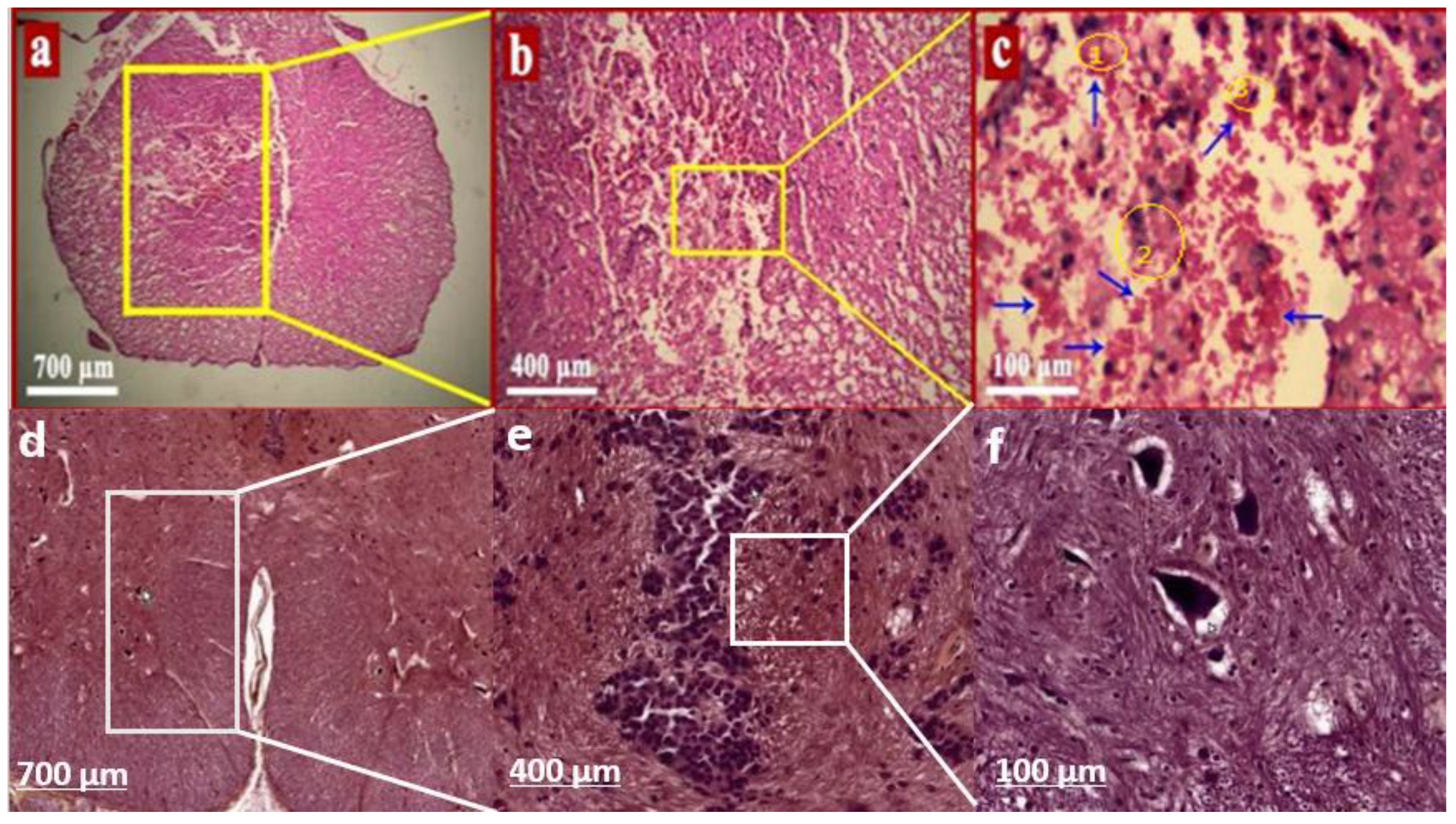
  76. salehi A: Effects of Melissa officinalis Alcoholic Extract on Kidney Tissue and Apoptosis Gene Expression in a Wistar Rat Model With Spinal Cord Injury. 1403.
  77. El-Desouky MA, Mahmoud MH, Riad BY, Taha YM: Nephroprotective effect of green tea, rosmarinic acid and rosemary on N-diethylnitrosamine initiated and ferric nitrilotriacetate promoted acute renal toxicity in Wistar rats. Interdiscip Toxicol 2019, 12(2):98-110. [CrossRef]
  78. Şengel N, Köksal Z, Dursun AD, Kurtipek Ö, Sezen Ş C, Arslan M, Kavutçu M: Effects of Dexmedetomidine Administered Through Different Routes on Kidney Tissue in Rats with Spinal Cord Ischaemia-Reperfusion Injury. Drug Des Devel Ther 2022, 16:2229-2239. [CrossRef]
  79. Choi JY, Jang TW, Song PH, Choi SH, Ku SK, Song CH: Combination Effects of Metformin and a Mixture of Lemon Balm and Dandelion on High-Fat Diet-Induced Metabolic Alterations in Mice. Antioxidants (Basel) 2022, 11(3). [CrossRef]
  80. Elsayed H, komy A, El-Shewy E, Elsayed F: Ameliorative Effect of Cinnamon and Rosemary Oils in Acrylamide–Induced Hepatic Injury in Rats. Bionatura 2024, 9:1-12. [CrossRef]
Figure 1. The alcoholic extracts from Rosmarinus officinalis and Melissa officinalis are shown in the image along with a schematic depiction of the main ingredients in each extract.
Preprints 188559 g001
Figure 2. The figure presents a detailed overview of the induction and establishment of spinal cord injury in rat models. Panel A illustrates the dorsal region of the rat, which has been prepared for injury induction through shaving with a scalpel. Panel B highlights the identification of the tenth vertebra utilizing a specialized retractor. Panel C describes the compression technique applied to induce spinal cord injury, wherein the spinal cord is subjected to pressure for a duration of one minute using an aneurysm clip. Lastly, Panel D signifies the conclusion of the procedure, encompassing the suturing of the surgical site and the verification of spinal cord injury establishment.
Preprints 188559 g002
Figure 3. The micrograph illustrates a segment of the spinal cord that has sustained an injury. Panel a highlights the affected region, indicated by a yellow box, where compression was applied using an aneurysm clip. This panel presents a longitudinal section that encompasses all injured sites. Panel b displays neuronal cells observed at a magnification of 400x. Panel c reveals voids within the tissue, suggesting axonal loss or damage, which confirms the successful establishment of the spinal cord injury model. Panels d, e, and f present images of spinal cord tissue from healthy rats for comparative analysis with the injured specimens. Panel d was captured at 700x magnification, Panel e at 400x, and Panel f at 100x magnification. The absence of voids in these panels, particularly in Panel f, confirms the structural integrity of the spinal cord in healthy rats, indicating no axonal damage or degeneration, and validating that the spinal cord remains intact and healthy.
Preprints 188559 g003
Figure 4. In a controlled study utilizing a rat model of spinal cord injury, alcoholic extracts of Rosmarinus officinalis and Melissa officinalis were administered subcutaneously, both individually and in combination, daily for four weeks. The therapeutic outcomes were subsequently assessed. Evaluations of motor function across the various experimental groups were performed employing Bonferroni multiple comparison tests, with data analysis conducted via one-way ANOVA and Tukey's test, establishing a significance threshold at p < 0.05. The findings indicated a notable enhancement in motor performance among the treated rats across the three treatment groups when compared to the spinal cord injury group.The graphical representations include letters that denote significant differences among the groups. Specifically, as illustrated in Figure 4, the letter a indicates a significant difference between the control group (L) and the other experimental groups. The letter b signifies a significant difference between the spinal cord injury group (S) and the remaining groups. The designation ac denotes a significant difference between the group receiving Melissa officinalis extract (M) and the other groups, while ad indicates a significant difference for the group treated with Rosmarinus officinalis extract (R) in comparison to the other groups. Finally, ae represents a significant difference between the group treated with the combined extract of Rosmarinus officinalis and Melissa officinalis (R+M) and the other groups.
Preprints 188559 g004
Figure 5. Alcoholic extracts from Rosmarinus officinalis and Melissa officinalis, two botanical sources, were injected subcutaneously every day for four weeks, both singly and in combination, into a rat model of spinal cord damage. After then, the therapies' therapeutic effects were evaluated. The sensory function assessments were conducted using Bonferroni multiple comparison tests among the different experimental groups. The obtained data were then analyzed using one-way ANOVA and Tukey's test, with a significance level of p < 0.05. Comparing the treated rats from all three therapy groups to the spinal cord injury group, the results showed significant improvements in their functional abilities.The letters depicted in the graphs signify statistically significant differences among the various groups. As demonstrated in Figure 5, the letter a denotes a significant difference between the control group (L) and the other experimental groups. The letter b indicates a significant difference between the spinal cord injury group (S) and the remaining groups. The letter 'ac' signifies a significant difference between the group receiving Melissa officinalis (M) and the other groups, while 'ad' denotes a significant difference for the group treated with Rosmarinus officinalis (R) in comparison to the other groups. Lastly, ae indicates a significant difference for the group treated with the combination of Rosmarinus officinalis and Melissa officinalis (R+M) relative to the other groups.
Preprints 188559 g005
Figure 6. The histological sections of kidney tissue from various experimental groups are presented. Panel A illustrates the control group, while Panel B corresponds to the spinal cord injury group. Panel C depicts the group that received rosemary plant extract, Panel D represents the group treated with Melissa officinalis extract, and Panel E shows the group administered both rosemary and Melissa officinalis extracts. In the sections from the experimental groups, a black arrow indicates the glomerulus, a blue arrow indicates the urinary space width, a white arrow indicates the distal ureter and a yellow arrow highlights the proximal ureter. When comparing the spinal cord injury group to the control group, the figure shows that there have been substantial changes in the glomerular tissue structure, the urine space width, and the proximal and distal convoluted tubules. These changes imply significant advancements in the treatment groups, pointing to significant therapeutic benefits.
Preprints 188559 g006
Figure 7. The therapeutic efficacy of alcoholic extracts derived from Rosmarinus officinalis and Melissa officinalis, both administered separately and in combination, was assessed through daily subcutaneous injections over a four-week duration in a murine model following the induction of spinal cord injury. Histomorphometric analyses were conducted to evaluate alterations in the diameters of the renal corpuscle (a), glomerulus (b), and urinary space (c) across various experimental groups. The data were subjected to statistical analysis using one-way ANOVA and Tukey's test, with a significance threshold established at p < 0.05. The findings suggest that the application of these extracts, whether individually or in combination, has the potential to mitigate the changes in the diameters of the renal corpuscle (a), glomerulus (b), and urinary space (c) induced by spinal cord injury.In the graphs labeled a, b, and c in Figure 7, the letters represent significant differences among the groups. Specifically, one letter indicates a significant difference between the control group and the other experimental groups, while another letter denotes a significant difference between the spinal cord injury group and the remaining groups. Furthermore, a letter signifies a significant difference in the results among the three groups treated with the alcoholic extracts of Melissa officinalis (M), Rosmarinus officinalis (R), and the combined extract of both plants (R+M) in comparison to the other groups. Significant group differences are represented by the letters in the graphs with the labels a, b, and c in Figure 7. To be more precise, one letter represents a significant difference between the experimental groups and the control group, while another letter shows a significant difference between the groups who were subjected to spinal cord damage and others. Furthermore, a letter denotes a statistically significant variation in the outcomes between the three groups that were administered the alcoholic extracts of Rosmarinus officinalis (R), Melissa officinalis (M), and the combined extract of both plants (R+M) in contrast to the other groups.
Preprints 188559 g007
Figure 8. Through daily subcutaneous injections for four weeks after spinal cord injury induction, the therapeutic benefits of alcoholic extracts from Rosmarinus officinalis and Melissa officinalis, both independently and in combination, were assessed in a rat model. Using histomorphometric analyses, changes in the height of proximal (a) and distal (b) epithelial cells resulting from spinal cord damage were evaluated in several experimental groups. Tukey's test and one-way ANOVA were used to examine the data, and a significance threshold of p<0.05 was taken into account. The findings suggested that using these extracts individually or in combination might lessen the changes in height that spinal cord injuries cause in the proximal (a) and distal (b) epithelial cells. Statistically significant differences between the groups are indicated by the letters shown in the graphs of panels a and b in Figure 8. The letter b denotes the importance of the findings in the spinal cord injury group (S) to the other groups, whereas the letter a denotes the relevance of the results in the control group (L) about the other experimental groups. The designation ad in panel a of Figure 8 highlights the significant results observed in the group administered Melissa officinalis alcoholic extract (M) relative to the other groups. Additionally, ac indicates the notable outcomes for the groups that received the combined extracts of Melissa officinalis and Rosmarinus officinalis (R+M), as well as the group treated with the alcoholic extract (R) of Rosmarinus officinalis. These distinctions illustrate a significant disparity in responses between the treated groups in the study and the other groups. In panel b of Figure 8, the designation "ad" indicates statistical significance for the group administered Rosmarinus officinalis extract (R), whereas ac signifies significance for both the group receiving Melissa officinalis extract (M) and the group treated with the combined extract (R+M) in comparison to the other experimental groups. These results underscore the significant differences observed between the treated and untreated groups.
Preprints 188559 g008
Figure 9. Using daily subcutaneous injections after spinal cord injury induction, the therapeutic benefits of alcoholic extracts from Rosmarinus officinalis and Melissa officinalis, both singly and in combination, were assessed in a rat model over four weeks. Histomorphometric studies were used to evaluate the differences in the diameters of the proximal and distal renal tubules (a and b) as a result of spinal cord damage across the various experimental groups. One-way ANOVA and Tukey's test were used to examine the data, and a significance threshold of p < 0.05 was taken into account. The findings showed that administering these extracts, either singly or in combination, may increase the diameters of the spinal cord injury-affected proximal renal tubules (a) and distal renal tubules (b). In Figure 9a and Figure 9b, the letters annotated on the graphs denote statistically significant differences among the groups. The letter a signifies a significant difference observed in the control group (L) when compared to the other experimental groups. The letter b indicates a significant difference in the spinal cord injury group (S) relative to the other groups. The designation ac denotes a significant difference in both the groups treated with Melissa officinalis alcoholic extract (M) and the group receiving the combined extracts (R+M) in comparison to the other groups. Furthermore, the label ad indicates a significant difference in the group treated with Rosmarinus officinalis alcoholic extract (R) when compared to the other experimental groups.
Preprints 188559 g009
Figure 10. The therapeutic effects of alcoholic extracts derived from Rosmarinus officinalis and Melissa officinalis were assessed in a mouse model of spinal cord injury, with administration occurring both separately and in combination via daily subcutaneous injections over a four-week period. Histomorphometric analyses were performed to evaluate alterations in the lumen diameter of renal proximal tubules (a) and distal tubules (b) resulting from spinal cord injury across various experimental groups. Statistical analysis was conducted using one-way ANOVA followed by Tukey's post hoc test, with a significance threshold set at p<0.05. The findings indicated that the application of these extracts, whether administered individually or in combination, positively influenced the alterations in the lumen diameter of both renal proximal tubules (a) and distal tubules (b) associated with spinal cord injury. In Figure 10a and Figure 10b, the letters denoting statistically significant differences are clearly indicated. The letter a signifies a significant difference observed in the control group (L) when compared to the other experimental groups. The letter b denotes a significant difference in the spinal cord injury group (S) relative to the other groups. The designation ac indicates a significant difference in the groups receiving simultaneous treatment with alcoholic extracts of Melissa officinalis (M) and Rosmarinus officinalis (R) in comparison to the other groups. Furthermore, the label ad emphasizes the significant difference found in the group treated with the combined extracts of Melissa officinalis and Rosmarinus officinalis (R+M) as compared to the other experimental groups.
Preprints 188559 g010
Figure 11. In a rat model of induced spinal cord injury, alcoholic extracts of Rosmarinus officinalis and Melissa officinalis were administered subcutaneously on a daily basis for a duration of four weeks, both individually and in combination, to assess their therapeutic effects. The study evaluated alterations in the expression levels of TNF-α (a), a critical gene associated with the inflammatory response, as well as genes related to apoptosis, including Caspase-3 (b), Bcl-2 (c), and Bax (d). These changes were analyzed across various experimental groups utilizing Bonferroni post hoc multiple comparisons. The data were subjected to one-way ANOVA and Tukey's test, with a significance threshold set at p < 0.05. The findings revealed a notable enhancement in gene expression levels in the treated rats when compared to the spinal cord injury group. As illustrated in panels a, b, c, and d of Figure 11, the designation a on the graphs denotes a statistically significant difference in the expression level of the gene in the control group (L) when compared to the other experimental groups. The letter b indicates a significant difference in gene expression levels within the spinal cord injury group (S) in relation to the other groups. The label ac signifies a significant difference in gene expression levels in the group treated with Melissa officinalis alcoholic extract (M) compared to the other groups. The designation ad reflects a significant difference in gene expression levels in the group treated with Rosmarinus officinalis alcoholic extract (R) in comparison to the other groups. Lastly, the label ae denotes a significant difference in gene expression levels in the group receiving the combined extracts of Rosmarinus officinalis and Melissa officinalis (R+M) relative to the other groups under investigation.
Preprints 188559 g011
Table 1. Primers were made specifically for this study to measure gene expression.
Genes Primer Sequence Forward Primer Sequence Reverse product size(bp) Tm melt Forward (°C) Tm melt Reverse (°C)
B2m (reference) CTTTCTACATCCTGGCTCACA GTCCAGATGATTCAGAGCTC
91
52.40
51.78
TNFa CCCTCACACTCAGATCATCTTCT
TCAGCCACTCCAGCTGCTCCTC
94
55.27
60.43
bcl2
GAGTGGGATACTGGAGATGAA
TGGTAGCGACGAGAGAAGT
90
52.40
51.09
casp3
AAGTGATGGAGATGAAGGAGT
CAGGCGTGAATGATGAAGAG
90
51.00
51.78
Bax
GGAGACACCTGAGCTGAC
CAGCAATCATCCTCTGCAGCT
89
52.60
54.36
Disclaimer/Publisher’s Note: The statements, opinions and data contained in all publications are solely those of the individual author(s) and contributor(s) and not of MDPI and/or the editor(s). MDPI and/or the editor(s) disclaim responsibility for any injury to people or property resulting from any ideas, methods, instructions or products referred to in the content.
Copyright: This open access article is published under a Creative Commons CC BY 4.0 license, which permit the free download, distribution, and reuse, provided that the author and preprint are cited in any reuse.
Prerpints.org logo

Preprints.org is a free preprint server supported by MDPI in Basel, Switzerland.

Subscribe

Disclaimer

Terms of Use

Privacy Policy

Privacy Settings

© 2026 MDPI (Basel, Switzerland) unless otherwise stated