Preprint
Review

This version is not peer-reviewed.

Lactococcus lactis as a New Strategy for Oral Vaccination: Current Insights and Future Perspectives

A peer-reviewed article of this preprint also exists.

Submitted:

26 November 2025

Posted:

27 November 2025

You are already at the latest version

Abstract

Lactococcus lactis, a safe food-grade lactic acid bacterium, has attracted increasing attention as a live biotherapeutic platform for mucosal vaccine development. Its genetic simplicity, absence of endotoxins, and availability of well-characterized inducible systems have enabled controlled expression and delivery of heterologous antigens and therapeutic molecules. This review highlights recent advances in the use of genetically modified L. lactis for mucosal immunization, focusing on expression technologies, routes of administration, and immune mechanisms relevant to protection or tolerance. Preclinical studies demonstrate its capacity to induce both mucosal and systemic immune responses against diverse pathogens, underscoring its potential as a safe and versatile vaccine chassis. Remaining challenges include regulatory harmonization, biosafety concerns, and the need for standardized manufacturing and evaluation frameworks. Together, these developments position L. lactis as a promising candidate for next-generation mucosal vaccines and live biotherapeutic products.

Keywords: 
;  ;  

1. Introduction

The term "probiotic" was first introduced in the 1960s and later formalized by the World Health Organization (WHO) and Food and Agriculture Organization (FAO) in 2001. Probiotics are defined as ‘Live microorganisms which, when administered in adequate amounts, confer a health benefit on the host” [1,2]. A fundamental requirement for probiotic efficacy is their ability to survive the harsh conditions of the gastrointestinal tract [3,4], particularly the acidic environment of the stomach and the presence of bile salts [5,6]. Additional characteristics include adhesion to mucosal and epithelial surfaces [6,7], antimicrobial resistance in specific contexts, as anti-mutagenic properties, and potential anticancer activities. Notably, probiotics have been associated with immunomodulatory effects, such as the regulation of inflammatory cytokines, stimulation of phagocytic activity, and modulation of both innate and adaptive immune responses [8].
The clinical interest in probiotics has expanded significantly in recent years, driven by the increasing global concern over antibiotic resistance, the demand for more targeted and host-friendly therapies, and the growing understanding of the gut microbiota's role in human health [9]. One of the most promising applications of probiotics lies in their use as delivery platforms for therapeutic molecules. Through genetic engineering, probiotic strains can be modified to express heterologous genes encoding therapeutic peptides or proteins [10], opening new possibilities in the treatment and prevention of infectious diseases, inflammation, and even cancer [11].
However, several limitations hinder their broader therapeutic application. A significant obstacle is the lack of specific regulatory frameworks for genetically modified probiotics, which prevents their official registration as genetically engineered organisms in many countries [12]. Unlike conventional probiotics used in foods or supplements, Live Biotherapeutic Products (LBPs) are defined by the U.S. Food and Drug Administration (FDA) as biological products that contain live organisms applicable to the prevention, treatment, or cure of a disease, but which are not vaccines and do not fall under traditional dietary categories. This distinction creates regulatory uncertainty that will be discussed further in subsequent sections [13].
Moreover, challenges persist in the early development stages of LBPs, including ensuring safety, efficacy, and manufacturing quality. In contrast to conventional pharmaceutical products, LBPs currently lack standardized preclinical and clinical guidelines, as well as robust post-marketing surveillance systems [13,14,15]. The absence of established quality controls and biomarkers for efficacy remains a significant hurdle for their widespread clinical adoption. Additionally, safety concerns must be carefully evaluated, including the potential for horizontal gene transfer, unintentional immune activation, or disruption of host microbiota composition.
Physiological barriers also limit the effectiveness of LBPs, as these microorganisms must resist degradation by digestive enzymes and bile fluids to reach their target site [16]. Oral vaccines are considered one of the most promising applications of live biotherapeutic products (LBPs). However, they encounter intrinsic limitations associated with the tolerogenic environment of the gastrointestinal tract, which can attenuate antigen-specific immune activation and reduce overall vaccine efficacy. [17]. To generate a protective immune response, these systems must cross the mucosal barrier, activate antigen-presenting cells (APCs), and ensure effective delivery of the antigen to the appropriate intestinal immune compartments [18].
Among the LBPs, Lactococcus lactis, a member of the lactic acid bacteria group, has emerged as a leading candidate. This bacterium has demonstrated considerable potential in oral vaccine development, particularly through modifications that enable the production of immunogenic proteins capable of eliciting measurable immune responses [15]. This review aims to summarize the most recent advancements in the use of genetically modified microorganisms, with a particular focus on L. lactis as a platform for vaccine delivery. We will examine both experimental and clinical research, highlighting opportunities, technological innovations, and regulatory limitations that define the current landscape and future potential of these live biotherapeutic systems.

2. Advantages of Lactococcus lactis for Its Application in LBPs

Lactococcus lactis is a non-pathogenic, Gram-positive bacterium that has been classified as “Generally Recognized As Safe” (GRAS) by the United States Food and Drug Administration (FDA). Traditionally used in the food industry to produce dairy products, fermented vegetables, and other fermented foods, L. lactis has long been valued for its metabolic simplicity, lack of endotoxins, and extensive history of safe human consumption [19].
The complete genome sequencing of various L. lactis strains [20,21] has facilitated the development of advanced genetic engineering tools, enabling the use of this bacterium as a vehicle for mucosal delivery of bioactive proteins [22]. Three major subspecies have been identified: L. lactis subsp. lactis, subsp. cremoris, and subsp. hordniae. Among these, L. lactis subsp. cremoris MG1363 has become the reference strain for genetic manipulation. This derivative of the ancestral NCDO712 strain (formerly classified as Streptococcus lactis) is plasmid-free, lacks phages, and is devoid of extracellular proteases, making it particularly suitable for laboratory applications [23,24]. In this line, MG1363 has been modified to remove the pLP712 plasmid, which encodes the lac operon and proteases required for casein degradation. As a result, this strain is incapable of growing in milk, limiting its survival and propagation outside of controlled laboratory environments [24]. This biosafety feature, along with its genetic tractability, makes L. lactis an attractive chassis for synthetic biology applications.
Although Lactococcus lactis does not naturally colonize the gastrointestinal tract of humans or animals, it can transiently survive after oral administration, allowing short-term interaction with intestinal mucosal surfaces. In murine models (BALB/c), viable L. lactis cells have been detected in the small intestine up to 24 hours after a single oral dose of 5 × 10¹⁰ CFU, with gradual clearance thereafter [25]. Similarly, in human volunteers, the strain MG1363 was detected in ileal effluents up to four hours after ingestion of 3 × 10⁷ CFU, confirming its transient persistence and low colonization potential [26]. This limited residence time is advantageous for biosafety, as it minimizes long-term colonization while permitting sufficient antigen release and interaction with gut-associated lymphoid tissues (GALT).
Beyond its favorable safety profile, L. lactis displays intrinsic immunomodulatory properties relevant to its use as a live vaccine vector. In vitro studies using human intestinal epithelial cell lines (HT-29 and Caco-2) have demonstrated that L. lactis stimulates the production of chemokines, such as IL-8, thereby promoting the recruitment of immune cells to mucosal sites [27]. In murine models, recombinant L. lactis strains expressing immunoregulatory molecules (e.g., IL-10) [28] or pathogen-derived antigens from Helicobacter pylori [29], HPV-16 [30], and Listeria monocytogenes [31] elicited antigen-specific immune responses, cytokine polarization, and protection against infection or tumor challenge. Together, these findings support the potential of L. lactis not only as a safe delivery vehicle but also as a biologically active adjuvant capable of shaping mucosal and systemic immune responses.
In addition, the favorable safety profile of L. lactis exhibits immunomodulatory properties such as an adjuvant by inducing chemokine expression both in vivo and in vitro. Besides, it can promote the maturation of bone marrow-derived dendritic cells [32,33]. These characteristics have positioned L. lactis as a promising platform for the development of mucosal vaccines, particularly for delivering antigens from viral, bacterial, and parasitic pathogens.

3. Conventional Method Used to Express Antigen Expression in L. lactis

The unique combination of safety, transient persistence, and immunomodulatory potential has established L. lactis as one of the most versatile bacterial chassis for live biotherapeutic and vaccine applications [19]. However, the success of this platform largely depends on the efficiency and regulation of heterologous gene expression. A range of expression systems has been developed to optimize antigen synthesis, secretion, and surface display in L. lactis, each offering distinct levels of control and applicability for mucosal vaccine design. The following section summarizes the main inducible and physiological systems currently employed for antigen expression in L. lactis, highlighting their mechanisms of regulation, strengths, and limitations (Table 1).

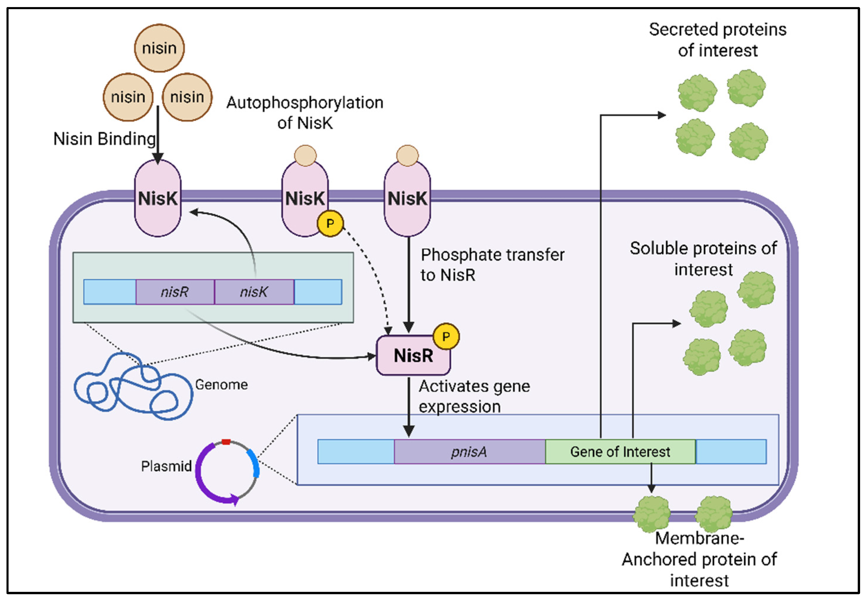
3.1. Nisin-Controlled Gene Expression (NICE)

One of the most widely adopted platforms for heterologous gene expression in L. lactis is the Nisin-Controlled Gene Expression (NICE) system [34]. Initially developed by Ryuter and collaborators [35]. This system allows for tightly regulated and dose-dependent induction of gene expression in response to nisin, an antibiotic approved by the U.S. Food and Drug Administration (FDA) in 1988 and commonly used as a food preservative [36].
The NICE system operates through a two-component signal transduction mechanism, comprising the membrane-bound histidine kinase NisK and the cytoplasmic response regulator NisR (Figure 1) [34] When nisin is added to the culture medium, it binds to NisK, initiating autophosphorylation. NisK then transfers the phosphate group to NisR, activating it. Phosphorylated NisR subsequently induces transcription from nisin-responsive promoters, primarily PnisA. Notably, the promoter driving the expression of the nisK and nisR genes remains constitutively active and is not regulated by nisin, thereby ensuring basal levels of the regulatory proteins regardless of external stimuli [37].
Initially, the NICE system was implemented in L. lactis NZ9700; this strain is a transconjugant from a mating between the nisin-A-producing strain NIZO R5 and the plasmid-free strain MG1614 and contains a single copy of the nisin-sucrose transposon TnS276 at the same chromosomal site as in L. lactis T165.1 and T165.5 [38]. The system was further optimized in L. lactis subsp. cremoris MG1363 by Kuipers et al., who engineered a strain (NZ9000) by inserting nisK, nisR, the 3' end of nisP, and the 5' end of nisF into the chromosome, replacing parts of the pepN and napC genes [39]. This enhanced strain became the standard for NICE-based expression. In NZ9000, gene induction is achieved by adding subinhibitory concentrations of nisin, typically ranging from 0.1 to 10 ng/mL [40].

3.2. Xylose Inducible Expression System (XIES)

In addition to the widely used NICE system, L. lactis has also been engineered to utilize the Xylose-Inducible Expression System (XIES), which offers a tightly regulated alternative for heterologous gene expression. This system is based on the PxylT promoter, identified in the xylose utilization operon of L. lactis subsp. lactis NCDO2118, which controls transcription of the xylT gene encoding a xylose permease [42]
Functional characterization of PxylT demonstrated that it contains a conserved catabolite-responsive element (cre) [43]. In the presence of xylose, the transcriptional activator XylR binds to PxylT and strongly induces transcription—up to 10 000-fold during mid-exponential growth (OD₆₀₀ ≈ 0.4) This promoter can be repeatedly switched on by the addition of xylose and off by washing and resuspending cells in glucose-containing medium, providing precise, reversible, and food-grade control over gene expression.[42]
With these findings, Miyoshi and collaborators developed a complete xylose-inducible expression system combining PxylT with the ribosome-binding site and secretion signal peptide (SP) of the Usp45 protein [44], fused to the Staphylococcus aureus nuclease gene (nuc) as a reporter [45]. This configuration allowed targeted expression of heterologous proteins either in the cytoplasm or secreted into the extracellular medium, demonstrating efficient and controllable protein production in the vegetable isolate L. lactis NCDO2118.
Compared with the nisin-inducible NICE system, XIES presents several advantages: it does not require antibiotic or peptide inducers, making it safer and more cost-effective; and its regulatory logic directly couples gene expression to the carbon source, which is advantageous for food-grade applications [46]. Nonetheless, its practical use is restricted to strains that can metabolize xylose L. lactis NZ9000 lack the xylose utilization pathway [21]. Comparative analyses showed that cytoplasmic and secreted production of the reporter nuclease (Nuc) in L. lactis NCDO2118 was approximately tenfold higher under nisin induction than under xylose induction during exponential growth, although both systems reached similar expression levels in stationary phase [42]. which can affect the timing of protein synthesis Despite these limitations, the XIES system provides a reliable, tunable, and biosafe alternative for regulated protein production in L. lactis, expanding its applicability in mucosal vaccine and biotherapeutic development.

3.3. Zinc Induced Systems

The PZn–zitR system is a zinc-repressible expression system described by Llull and collaborators [47] derived from the zit operon of L. lactis, which encodes the high-affinity zinc uptake ABC transporter ZitSPQ and its regulator ZitR. The regulatory mechanism is based on the ZitR repressor, a MarR-family metalloregulator that binds to the PZn promoter in the presence of excess zinc, thereby blocking transcription of downstream genes. When extracellular zinc becomes limited, ZitR dissociates from the promoter, allowing transcription to proceed. Consequently, the system is activated under conditions of zinc depletion, either naturally during bacterial growth or through chelation with agents such as EDTA. Thus, the system is activated by zinc depletion and can be induced through natural metal starvation or by adding chelating agents such as EDTA. This system provides tight regulation, gradual induction during bacterial growth, and is compatible with food-grade applications, as it does not rely on synthetic inducers like nisin. However, its limitations include the need for precise control of metal concentrations, possible interference with cell metabolism due to chelation, and lower maximal expression levels compared to the nisin-controlled expression (NICE) system. Despite these constraints, PZn–zitR remains a valuable tool for the controlled production of heterologous proteins in L. lactis, especially in mucosal or physiological environments where environmental cues naturally modulate gene expression [47]
In contrast, Zirex, designed by Mu and collaborators [48], introduced the SczA–PczcD regulatory module from Streptococcus pneumoniae into L. lactis, creating the first zinc-inducible expression system for this species. In Zirex, the transcriptional activator SczA binds to two specific motifs within the PczcD promoter to repress transcription in the absence of zinc and activate it in response to Zn²⁺ supplementation [48]. Experimental analyses in L. lactis NZ9000 showed that induction with 0.3–0.7 mM ZnSO₄ during the exponential phase produced strong green fluorescent protein (GFP) expression, reaching approximately 80% of the levels achieved with the nisin-inducible PnisA promoter, while maintaining negligible basal expression under uninduced conditions. Moreover, Zirex exhibited low toxicity even at 0.5 mM Zn²⁺ and allowed precise, dose-dependent induction between 0 and 0.3 mM Zn²⁺. A key advantage of Zirex is its compatibility with other inducible systems: the designers demonstrated a dual-promoter configuration, combining PczcD and PnisA to independently express GFP and mCherry, achieving simultaneous expression with only minor signal interference (~10–20%). These properties make Zirex particularly attractive for the controlled expression of metalloenzymes, zinc-binding antigens, and lantibiotic biosynthetic enzymes, where zinc serves both as inducer and cofactor. Although careful optimization of Zn²⁺ concentrations is required to prevent metal stress, Zirex offers a robust, tunable, and food-grade alternative for controlled heterologous protein production in L. lactis, expanding the available molecular toolbox for mucosal vaccine and biotherapeutic development [48].
More recently, Xu and collaborators reported the ZICE (Zn²⁺-Controlled Expression) system [49], derived from Streptococcus thermophilus, which was successfully applied in L. lactis NZ9000 [49]. The system is based on the sczAst–PczcDst regulatory module, homologous to that of S. pneumoniae, and provides precise zinc-dependent activation. In NZ9000, induction with 0.8 mM ZnSO₄ at mid-exponential growth (OD₆₀₀ ≈ 0.4) resulted in strong expression of GFP and secreted IL-10, reaching approximately 60% of the levels obtained with the NICE system. Notably, the ZICE platform showed no background expression, tight on/off control, and improved expression when yeast extract was removed from the medium, suggesting an interaction between nitrogen availability and metal ion regulation. The stability, low cost, and physiological relevance of zinc as an inducer make ZICE a promising tool for the development of food-grade and probiotic-based expression systems.
Each of the zinc-responsive expression systems developed for Lactococcus spp presents distinctive strengths and constraints depending on the regulatory mechanism and intended application. The native PZn–zitR system offers exceptional regulatory tightness and food-grade compatibility, as it relies exclusively on intrinsic metabolic control without requiring exogenous inducers. However, its activation depends on zinc depletion, which can be difficult to maintain under standard culture conditions and may alter cell physiology when strong chelating agents are used. The Zirex system, in contrast, provides high-level, zinc-inducible expression that reaches up to 80% of the activity of the nisin-controlled promoter while preserving minimal basal leakage. Its dose-dependent and reversible behavior makes it ideal for fine-tuned laboratory applications, although the use of regulatory elements derived from Streptococcus pneumoniae may limit its classification as fully food-grade. The recently developed ZICE system represents a safe alternative, originating from Streptococcus thermophilus, a species with GRAS status. Although ZICE achieves slightly lower maximal expression (45–60% of NICE levels), it combines food-grade safety, low background activity, and simple induction through ZnSO₄ supplementation.

3.4. Stress Induces Controlled Expression System (SICE)

The study by Benbouziane and collaborators designed the Stress-Inducible Controlled Expression (SICE) system in L. lactis, aimed at enhancing the safety and physiological relevance of mucosal vaccine delivery [50]. This system relies on the groESL promoter, which is activated by host-related stress conditions such as heat, acidity, or bile salts [51,52], allowing in situ production and secretion of the antigen or therapeutic protein only during bacterial transit through the host. Using this approach, L. lactis strains expressing IL-10 or the HPV-16 E7 antigen successfully induced localized immune responses and tumor protection in murine models [50].
The main advantages of the SICE system lie in its auto-regulated expression, which eliminates the need for external inducers like nisin, and its localized activation within mucosal environments, thereby improving biosafety and simplifying vaccine administration. However, limitations include potential variability in expression levels due to fluctuating stress conditions and the episomal nature of the plasmid, which may lead to plasmid instability in the absence of selective pressure.
Overall, the SICE platform represents a promising strategy for mucosal immunization, combining controlled antigen release, host-responsive regulation, and the intrinsic safety of L. lactis as a non-pathogenic live vector for delivery [50,53].

4. Routes of Administration

The mucosal immune system is organized into functionally interconnected compartments called mucosa-associated lymphoid tissue (MALT), which includes the gut-associated lymphoid tissue (GALT), nasal-associated lymphoid tissue (NALT), bronchial-associated lymphoid tissue (BALT), skin-associated lymphoid tissues (SALT), and genital organ-associated lymphoid tissues (GOALTs), among others [55]. Antigen exposure at mucosal sites triggers distinct immune pathways, influencing both the magnitude and quality of the immune response. Selecting the administration route for vaccines based on L. lactis is essential to induce the desired immune response, toward immune tolerance for the treatment of autoimmune and allergic diseases or toward robust protective immunity against pathogens [56]. Typically, the strongest immune response is elicited at the site of immunization and in anatomically adjacent mucosal sites [57].

4.1. Oral Administration

Oral administration is the most common and natural route for delivering L. lactis, especially in vaccine and immunomodulatory therapies [58]. This route is convenient, non-invasive, and advantageous for stimulating mucosal immunity within the gastrointestinal tract, primarily through the gut-associated lymphoid tissue (GALT) [59]. It can also induce a substantial local secretory IgA (SIgA) antibody response in the proximal part of the small intestine, the ascending colon, the stomach, and in the mammary and salivary glands [60]. Moreover, oral delivery enhances patient compliance and is suitable for both preventive and therapeutic applications [17,18]. However, a critical limitation of oral administration is the induction of oral tolerance, an immunological mechanism that prevents excessive inflammatory responses against dietary antigens and the resident microbiota [61]. Oral tolerance is characterized by local and systemic immune unresponsiveness following antigen exposure via the oral route [62]. This oral tolerance is mainly mediated by CD4+ regulatory T cells (Tregs) expressing the transcription factor FoxP3 [63]. These cells suppress immune responses through multiple mechanisms, including the production of inhibitory cytokines such as transforming growth factor-beta (TGF-β), interleukin IL-10, and IL-35. Also, they can release cytotoxic enzymes like granzyme and perforin, consume and degrade of ATP and IL-2, and express inhibitory surface molecules such as LAG-3 and CTLA-4 [64].
In the mucosal immunity, IL-10, TGF-β, and CTLA-4 play fundamental roles in maintaining homeostasis, preventing excessive inflammation, and preserving epithelial barrier integrity [65,66,67]. Mucosal FoxP3+ Tregs frequently express IL-10 [68], which suppresses activation of myeloid cells [69], gamma delta T cells [70], and conventional CD4+ T cells [71,72]. STAT3 regulates IL-10 expression, crucial for autocrine activation of Tregs and suppression of Th17 responses [73,74]. TGF-β is essential for Treg suppressive function by enhancing effector activity through the SMAD signaling pathway [75,76]. CTLA-4, an inhibitory receptor constitutively expressed under FoxP3 control [77,78], competes with CD28 for CD80/CD86 ligands on dendritic cells (DCs), modulating immune activation [79,80]. Moreover, CTLA-4 expression in Tregs is induced by the microbiota, reinforcing its role in mucosal immune regulation [81]. Tregs also suppress immune responses via competitive and passive mechanisms, including aggregation around DCs mediated by high LFA-1 expression [82], and efficient sequestration of IL-2 from activated T cells due to high IL-2 receptor expression (CD25 and CD122), without producing IL-2 themselves, reinforced by FoxP3-mediated transcriptional repression of IL-2 [83,84,85,86].
In addition to immunological challenges, oral administration exposes L. lactis to harsh physical and chemical barriers, including gastric acidity, digestive enzymes, and competition from resident microbiota, all of which can reduce viability and antigen delivery efficiency [16]. Variability in effective dose delivery also presents challenges to consistent therapeutic outcomes.

4.2. Nasal Administration

Nasal administration offers an alternative route for delivering L. lactis to stimulate mucosal immunity in the respiratory tract. This route bypasses gastrointestinal barriers and can induce both local mucosal and systemic immune responses [87]. The nasal-associated lymphoid tissue (NALT) acts as the primary inductive site for immune activation in this route, containing microfold (M) cells, dendritic cells (DCs), and organized lymphoid follicles that facilitate efficient antigen sampling and presentation to naive T and B lymphocytes [55]. Following antigen uptake through microfold (M) cells and presentation by mucosal dendritic cells, B cell activation and class switching to IgA are promoted through cytokines such as IL-5 and IL-6, leading to robust secretory IgA (SIgA) production in the upper respiratory tract [88,89]. In addition, activated lymphocytes from NALT migrate to distant effector sites, including the oral, pulmonary, and genital mucosa, through the common mucosal immune system, generating coordinated humoral and cellular protection [90,91]
Nasal immunization can induce Th1/Th2-balanced or Th17-type immune responses depending on the antigen, adjuvant, or bacterial vector employed, resulting in effective cytotoxic T lymphocyte (CTL) responses and long-term memory formation [92,93,94]. Compared with oral administration, the nasal route avoids enzymatic degradation and first-pass tolerance in the gastrointestinal tract, providing a more efficient and rapid activation of mucosal and systemic immunity [95]. However, limitations include mucociliary clearance, restricted dosing volume, and antigen instability within the nasal environment, which may compromise bacterial viability and immunogenicity. Strategies such as mucoadhesive formulations, nanoparticle encapsulation, or co-delivery with mucosal adjuvants are under development to improve vaccine retention and efficacy via this route [89].

4.3. Other Routes od Administration

Rectal immunization induces a pronounced local SIgA response in the rectum and sigmoid colon, a moderate response in the descending colon, and minimal or no response in the proximal colon or small intestine. In contrast, nasal or tonsillar immunization in humans primarily triggers antibody production in the upper respiratory mucosa and its associated secretions, without activating immune responses in the intestinal tract [96]. Interestingly, nasal administration has also been shown to generate strong IgA and IgG responses in the human cervicovaginal mucosa, achieving levels comparable to those seen with direct vaginal immunization. Although the capacity of nasal immunization to prime CD4⁺ and CD8⁺ T cells in the genital tract remains incompletely characterized, evidence from murine models suggests that vaginal vaccination is more effective in inducing strong genital tract T cell–mediated immunity [97]. Sublingual and topical applications have been explored in preclinical settings for L. lactis-based therapies. These methods aim to utilize mucosal immunity in different compartments or to support specific clinical applications; however, evidence remains limited, and additional research is needed to evaluate their feasibility and effectiveness [56].

5. Oral Vaccine Prototypes on L. lactis

Oral vaccines based on genetically modified L. lactis have shown promising results in preclinical models against a variety of infectious agents [98]. These models typically involve the expression of selected antigens chosen for their immunogenicity and role in pathogen virulence or protection, aiming to elicit both mucosal and systemic immune responses. Different vaccination schemes have been explored, often combining mucosal priming with systemic boosting to enhance immune memory and facilitate the effective homing of immune cells to mucosal tissues.

5.1. Viral Vaccines

Human Papilloma Virus (HPV) is a significant cause of cervical cancer worldwide [56]. Existing prophylactic vaccines, such as Gardasil and Cervarix, use virus-like particles (VLPs) to induce immunity. In contrast, Mohseni et al. employed L. lactis to deliver the E7 oncoprotein of HPV16, a key antigen involved in oncogenesis. This choice targets the therapeutic elimination of infected cells expressing E7. Their studies demonstrated increased E7-specific antibody titers and CD4+ T cell responses, showing promise as a therapeutic vaccine platform [99]. These outcomes were further validated in early-phase clinical trials [100], supporting the translational potential of this approach. An interest review was made by the same author with more specific details and data [30]. These results contribute to reducing cervical cancer.
The absence of a protective prophylactic HIV vaccine has allowed for the exploration of different platforms to control the spread of this virus [101]. L. lactis-based oral vaccines have been developed that express HIV antigens, such as the Gag protein fused to the T3 pilus protein of Streptococcus pyogenes, thereby enhancing mucosal delivery [102]. This strategy elicited increases in Gag-specific IgG and IgA in serum, feces, and vaginal secretions, and activated dendritic cells in Peyer's patches, although CD8+ T cell responses remained low. Another approach targeted the V2-V4 “loop” of the HIV envelope protein (Env), achieving systemic and mucosal IFN-γ responses with repeated oral dosing every two weeks [103]. Considering the importance of HIV in public health, a mucosal vaccine against this virus represents a valuable contribution to the development of a vaccine against this pathogen.
Influenza A virus subtypes H1N1 and H3N2 cause seasonal epidemics [104]. Oral vaccines using L. lactis have targeted hemaglutinin (HA), the major surface antigen. Lei et al. demonstrated that L. lactis expressing HA of the H5N1 subtype elicited significant IgG and mucosal IgA responses, conferring complete protection in mice against lethal viral challenge after multiple immunizations [105,106]. More recently, a recombinant strain expressing the conserved HA stalk domain fused to a bacterial anchoring protein demonstrated cross-protection against H5N1, H3N2, and H1N1 strains, highlighting the potential for developing of a universal influenza vaccine [106].
Hepatitis B Virus (HBV) causes chronic liver disease, with existing vaccines available, but a continuous need for novel strategies. Early studies expressed the PreS region of the HBV surface antigen (HBsAg) in L. lactis, inducing intestinal IgA and serum IgG in mice [107]. Co-expression of IFN-γ as an adjuvant further enhanced the IgG response. More recent efforts include the expression of central HBsAg regions from different HBV genotypes; however, preclinical results remain pending [108].
The first L. lactis-based vaccine against the Severe Acute Respiratory Syndrome (SARS) utilized the nucleocapsid (N) protein fused to human GST, inducing specific serum IgG responses in mice [109]. Following the COVID-19 pandemic, L. lactis models expressing the receptor-binding domain (RBD) of the spike (S) protein or conserved S protein regions have been developed, aiming to stimulate protective mucosal and systemic immunity [110]. Another study utilized the highly conserved region (HCR) of the Spike S2 subunit, driven by the nisin-inducible pNZ8149 vector to express the antigen. Mice immunized through oral or intranasal routes exhibited significantly increased levels of anti-SARS-CoV-2 IgG and IgA, along with elevated CD4⁺ and CD8⁺ T-cell responses in lymphoid and intestinal tissues. Notably, the intranasal route elicited stronger humoral and cellular responses, consistent with the activation of nasal-associated lymphoid tissue (NALT) and enhanced mucosal immunity [111]. Importantly, the construct was generated under food-grade conditions, employing a lactose-based selection system rather than antibiotics, reinforcing its biosafety profile. These findings demonstrate that L. lactis can serve as an effective mucosal delivery platform for conserved spike protein antigens, offering a non-invasive and broadly protective vaccination strategy against SARS-CoV-2 and potentially other coronaviruses [111]. The further study of the first vaccine could contribute to the pandemic during 2020.

5.2. Bacteria Vaccines

Clostridium difficile infection is a leading cause of antibiotic-associated diarrhea. Vaccines targeting the toxins TcdA and TcdB have been evaluated using L. lactis expressing these antigens. Guo et al. compared oral L. lactis vaccines expressing single or combined toxins with purified recombinant proteins, finding significantly improved survival and reduced pathology in vaccinated animals. All vaccine groups elicited strong IgG and IgA responses with toxin-neutralizing activity, supporting L. lactis as a cost-effective oral vaccine platform against C. difficile infection [112].
Helicobacter pylori is one of the main causes of gastritis, ulcers, and gastric cancer [113,114]. Initial L. lactis-based vaccines expressed the urease B subunit (UreB), eliciting systemic antibody responses with multi-dose oral regimens [114]. Later studies focused on Th1/Th17 immune induction, using antigens like the neutrophil-activating protein (NapA) and the CagL protein, demonstrating antigen-specific antibodies and cytokine responses linked to protective immunity [29,115].
Streptococcus pneumoniae is one of the leading pathogens responsible for respiratory diseases worldwide [116]. Although polysaccharide-based vaccines are available, they have the limitation of not providing immunity against all serotypes of this bacteria [117,118,119]. On the other hand, conjugate vaccines offer broader protection but are costly, which can limit their accessibility in low-income countries [120,121]. In this context, the use of L. lactis provides a promising alternative to overcome this economic barrier. A study developed a vaccine model using L. lactis engineered to express pneumococcal protective protein A (PppA) on its surface, a protein conserved in serotypes 3, 5, 9, 14, 19, and 23. Different respiratory mucosal immunization protocols were evaluated, including nasal administration of live or inactivated L. lactis expressing PppA, with or without co-administration of a probiotic (L. casei). The mice that received the L. casei both orally and nasally showed the highest levels of anti-PppA IgA and IgG antibodies in bronchoalveolar lavage (BAL) fluid and IgG in serum, respectively, which contributed to protection against infection. However, only the groups that received the live or inactivated vaccine together with oral probiotic administration were able to prevent lung colonization by S. pneumoniae serotypes 3 and 14 in a respiratory infection model. This protection was associated with a preferential stimulation of local and systemic T helper type 1 (Th1) responses, accompanied by moderate Th2 and Th17 activity, as indicated by cytokine profiles in BAL and the IgG1/IgG2a ratio both locally and systemically. Overall, nasal immunization with the inactivated recombinant strain combined with oral probiotic administration effectively stimulated specific cellular and humoral immune responses, protecting against challenge with the two S. pneumoniae serotypes [122].
Enterotoxigenic Escherichia coli can cause diarrhea in children, and suitable vaccines are therefore desired [123]. A study evaluated the immune response after the oral and subcutaneous administration of L. lactis capable of expressing a heat-labile toxin (LTB), a virulence factor, and compared it with the recombinant protein in a rabbit animal model. They demonstrated an increase in IgA levels in the intestine. In vitro neutralization assay showed that the effect of the toxin could be neutralized with 500 µg/ml of IgG isolated from the oral vaccine group. Furthermore, the dose of enterotoxigenic E. coli causing fluid accumulation in the ileal loop test showed a tenfold increase in rabbits immunized with either recombinant L. lactis or LTB protein compared to other groups. [124]
Brucella abortus is a facultative intracellular, Gram-negative bacterial pathogen that primarily infects humans and animals through the digestive tract [125]. B. abortus causes abortion in pregnant cattle and undulant fever in humans [126,127]. B. abortus ribosomal antigen L7/L12, a well-characterized immunogenic protein, was expressed under the nisin-inducible PnisA promoter in L. lactis NZ9000. Through a series of genetic constructs, the antigen was successfully targeted to three cellular locations (the cytoplasmic, secreted, and cell wall-anchored) using combinations of secretion signals, fusion partners, and anchoring domains. The fusion of L7/L12 with the Usp45 signal peptide enabled secretion with a sixfold higher yield than cytoplasmic production. At the same time, the addition of the LEISSTCDA synthetic propeptide or staphylococcal nuclease (Nuc) further increased yield and secretion efficiency up to 50%. Additionally, anchoring L7/L12 to the M6 protein cell-wall domain from Streptococcus pyogenes resulted in stable surface localization, making it particularly suitable for mucosal delivery. This work demonstrated that L. lactis can be engineered to produce and export a protective Brucella antigen in different cellular compartments, establishing the conceptual foundation for safe, food-grade, non-pathogenic mucosal vaccines against brucellosis and other intracellular bacterial infections [128]. The same authors observed significant levels of anti-L7/L12-specific IgA in feces, revealing an induced local humoral immune response. However, serum analysis did not reveal any anti-L7/L12 antibodies, suggesting the absence of a systemic response [129].
Streptococcus agalactiae, also known as Group B Streptococcus (GBS), is a leading cause of neonatal sepsis and meningitis, with no licensed vaccine available. The surface immunogenic protein (SIP), conserved across serotypes, is a promising target [130,131,132]., Oral immunization of mice with this recombinant strain elicited strong systemic and mucosal anti-SIP IgG and IgA responses, enhanced CD4⁺ and CD8⁺ T-cell activation, and reduced Treg (CD4⁺CD25⁺FoxP3⁺) frequencies, consistent with a Th1/Th17-oriented immune profile. Notably, vaccinated animals showed a significant reduction in vaginal GBS colonization, and passive transfer of serum or T cells conferred protection to naive mice, confirming the involvement of both humoral and cellular mechanisms. Collectively, these findings demonstrate that L. lactis can serve as a safe and effective mucosal delivery platform for the SIP antigen, providing a foundation for non-invasive and broadly protective vaccine strategies against GBS [133]. The same methods were used in a tilapia model with the SIP protein and a truncated SIP (tSIP). Fish immunized with the tSIP vaccine also showed the highest level of protection compared to other test groups, and the mortality rate was significantly reduced compared to both control groups. The relative percentage of survival (RPS) against S. agalactiae for both SIP and tSIP-vaccinated groups was 50 % and 89 %, respectively, at 14 days post-challenge. Significant up-regulation of IgM, IL-1β, IL-10, TNF-α, and IFN-γ was observed at day 34 between the vaccinated and control groups. These results indicated that the recombinant lactococcal tSIP vaccine can elicit both cell-mediated and humoral responses and is recommended as a potential oral vaccine against S. agalactiae infection [134].
In summary, L. lactis has been widely used for the development of vaccine prototypes against various viral and bacterial pathogens of public health importance. All key aspects related to antigens, administration routes, and main findings are described in Table 2.

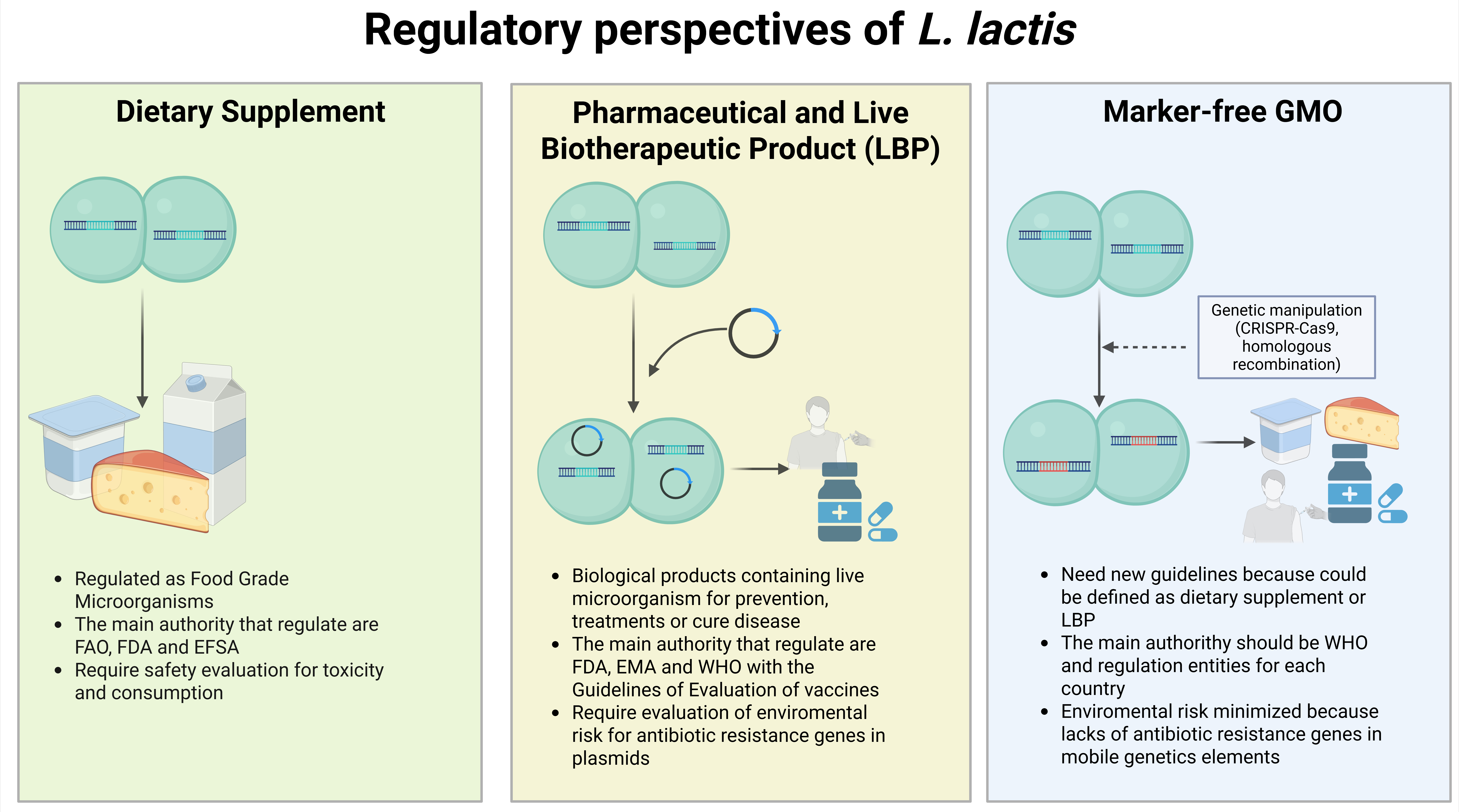
6. Probiotic, Vaccine, or Drug? Regulations, New Technologies, and Solutions

To date, clinical studies using L. lactis as a recombinant bacterial vector have been limited to trials targeting human papillomavirus (HPV), specifically employing the E7. [100] and E6 [135] oncoproteins. Both studies demonstrated the safety profile of the vaccine candidates and their capacity to induce a humoral immune response against the target antigens. These clinical outcomes (IRCT20190504043464N1 and IRCT20190504043464N1, respectively) were supported by prior preclinical evidence from murine models. [135,136]. However, both clinical trials utilized L. lactis strains transformed with the plasmid pNZ8123, which harbors a chloramphenicol resistance gene as a selection marker [40]. This raises significant biosafety concerns, as plasmids carrying antibiotic resistance genes pose the risk of horizontal gene transfer to commensal or pathogenic bacteria in the host microbiota or environment. Studies have documented gene transfer events from Lactobacillus spp. to E. coli. [138] and other in vivo models [139], underscoring the importance of avoiding antibiotic resistance markers in genetically modified probiotics [140].
The World Health Organization (WHO), in its "Guidelines on Nonclinical Evaluation of Vaccines" (2005), defines vaccines as a heterogeneous class of medicinal products containing immunogenic substances capable of inducing specific, active, and protective host immunity against infectious diseases. When live attenuated vaccines are based on genetically modified organisms (GMOs), the guidelines recommend environmental risk assessments during preclinical development. This includes investigating the potential shedding of vaccine strains after administration and evaluating the risk of genetic exchange with non-vaccine strains, a crucial aspect considering evidence of plasmid transfer among related bacteria. [141]
In addition, to the general safety concerns associated with the use of antibiotic resistance markers in GMOs, international regulations impose strict guidelines for the clinical and environmental evaluation of recombinant bacteria in the European Union, L. lactis expressing heterologous genes is considered a GMO and is regulated under Regulation (EC) No. 1829/2003, requiring a comprehensive environmental risk assessment, traceability, and labeling before approval. [142]. The U.S. regulatory framework is divided among the FDA, USDA, and EPA, depending on the nature of the product and its potential environmental or therapeutic impact.
Furthermore, the regulatory classification of L. lactis-based products as probiotics, vaccines, or drugs remains ambiguous. Venugopalan et al. underscore that probiotics may be regulated either as dietary supplements or as drugs, depending on their intended use. [143]. The FDA defines a drug as "an article intended for use in the diagnosis, cure, mitigation, treatment, or prevention of disease."[13] If a product is marketed as a dietary supplement, it falls under food regulations, which are generally less stringent than those for pharmaceuticals or vaccines. Conversely, suppose the probiotic or recombinant microorganism is intended as a drug or vaccine. In that case, it must comply with rigorous regulatory pathways specific to each country, ensuring evidence of safety, efficacy, and quality through preclinical and clinical studies.
Given the evolving landscape of live biotherapeutic products (LBPs) such as genetically engineered L. lactis, there is an urgent need for harmonized regulatory frameworks that address their unique characteristics. This includes establishing standardized guidelines for genetic modification techniques (e.g., marker-free systems), environmental risk assessment, manufacturing quality control, and post-market surveillance. Such regulatory clarity will facilitate the translation of L. lactis-based vaccines and therapeutics from research to clinical use while safeguarding public health and environmental safety.
In recent years, emerging technologies have significantly advanced the development of genetically modified L. lactis-based vaccines. The CRISPR-Cas system enables precise genomic modifications, allowing gene insertion without the use of antibiotic resistance markers or mobile genetic elements, thereby facilitating compliance with biosafety requirements established by national and international regulatory agencies such as the WHO, FDA, and EFSA. Moreover, several studies have already reported the successful application of this technology in L. lactis [144,145,146] In addition, bacterial microencapsulation has been employed as a complementary strategy not only to enhance immune responses and improve cell viability throughout the gastrointestinal tract [147,148] but also as a biocontainment measure to prevent the environmental dissemination of the microorganism [149,150].

7. Learnings and New Perspectives

Over the past decade, numerous reports have highlighted the potential of oral vaccines based on L. lactis, demonstrating both protective efficacy and the induction of humoral and cellular immune responses. However, much of this evidence remains confined to plasmid-based platforms and preclinical stages, limiting scalability and clinical translation despite promising results. We consider it a priority to promote the development of genetically modified organisms (GMOs) with stable genomic integration as a necessary step to meet regulatory requirements and facilitate the transition to clinical studies. In parallel, we urge regulatory authorities to establish harmonized, step-by-step guidelines for evaluating genetically modified probiotics, encompassing biosafety, environmental risk assessment, traceability, mitigation plans, and requirements for release and monitoring. Given the potential of these platforms not only for vaccine development but also for therapeutic applications in cancer, allergies, and other conditions, the availability of clear and up-to-date regulatory frameworks is essential to accelerate their responsible and safe advancement.

8. Conclusions

Genetically engineered L. lactis has evolved from a safe food-grade bacterium to a versatile live biotherapeutic platform capable of delivering antigens, cytokines, and therapeutic molecules at mucosal surfaces. Its well-characterized genetics, absence of endotoxins, and the availability of finely tunable expression systems such as NICE, XIES, zinc-responsive, and stress-inducible promoters have positioned it as one of the most promising candidates for next-generation mucosal vaccines. Preclinical studies have demonstrated its ability to elicit both mucosal and systemic immune responses. However, the clinical translation of L. lactis-based vaccines remains limited by regulatory uncertainty, concerns regarding biosafety and antibiotic resistance markers, and the need for standardized manufacturing and evaluation guidelines. Advances in marker-free genome editing, such as CRISPR-Cas, and biocontainment technologies, including bacterial microencapsulation, offer practical solutions to these challenges, paving the way for the safe and effective deployment of L. lactis as a live vaccine vector. Ultimately, integrating these innovations within harmonized international regulatory frameworks will be essential to fully realize the potential of L. lactis as a new strategy for mucosal immunization and the design of live biotherapeutics.

Author Contributions

Conceptualization, JG-G, MI and AEV, writing—original draft preparation, JG-G . and; writing—review and editing, JG-G, MI, JAS and AEV; visualization, JG-G and MI. and project administration AEV ; funding adquisition AEV. All authors have read and agreed to the published version of the manuscript.

Funding

This research was funded by FONDECYT 1244107, which supported MI's study, and FONDECYT 1231866, which supported JAS's study. Fellowship was awarded to JG (ANID N°21230817).

Institutional Review Board Statement

Not applicable.

Informed Consent Statement

Not applicable.

Data Availability Statement

Not applicable.

Acknowledgments

we thank Catalina Garrido of LIVA COMPANY SPA (Chile) for technical support.

Conflicts of Interest

The authors declare no conflicts of interest.

Abbreviations

The following abbreviations are used in this manuscript:
APCs Antigen-Presenting Cells
BALT Bronchus-Associated Lymphoid Tissue
BAL Bronchoalveolar Lavage
CD Cluster of Differentiation (CD4, CD8, etc.)
CFU Colony-Forming Units
CTL Cytotoxic T Lymphocyte
CTLA-4 Cytotoxic T-Lymphocyte Antigen 4
DCs Dendritic Cells
EPA Environmental Protection Agency
FDA Food and Drug Administration
GALT Gut-Associated Lymphoid Tissue
GFP Green Fluorescent Protein
GMOs Genetically Modified Organisms
GOALTs Genital Organ-Associated Lymphoid Tissues
GRAS Generally Recognized As Safe
GST Glutathione-S-Transferase
HA Hemagglutinin
HBV Hepatitis B Virus
HBsAg Hepatitis B Surface Antigen
HCR Highly Conserved Region
HIV Human Immunodeficiency Virus
HPV Human Papillomavirus
IL Interleukin
IRCT Iranian Registry of Clinical Trials
LBPs Live Biotherapeutic Products
LPS Lipopolysaccharide
LTB Heat-Labile Toxin Subunit B
MALT Mucosa-Associated Lymphoid Tissue
MHC Major Histocompatibility Complex
NALT Nasal-Associated Lymphoid Tissue
NICE Nisin-Controlled Gene Expression system
OD Optical Density
PnisA Nisin-Inducible Promoter A
PppA Pneumococcal Protective Protein A
PxylT Xylose-Inducible Promoter
BD Receptor-Binding Domain
RPS Relative Percent Survival
SALT Skin-Associated Lymphoid Tissue
SARS Severe Acute Respiratory Syndrome
SICE Stress-Inducible Controlled Expression system
SIgA Secretory Immunoglobulin A
SIP Surface Immunogenic Protein
TcdA Clostridioides difficile Toxin A
TcdB Clostridioides difficile Toxin B
Tregs Regulatory T Cells
Usp45 Universal Stress Protein 45 (signal peptide)
VLPs Virus-Like Particles
WHO World Health Organization
XIES Xylose-Inducible Expression System
ZICE Zinc-Controlled Expression system
Zirex Zinc-Inducible Regulatory Expression system

References

  1. C. Hill et al., “The International Scientific Association for Probiotics and Prebiotics consensus statement on the scope and appropriate use of the term probiotic,” Nat Rev Gastroenterol Hepatol, vol. 11, no. 8, pp. 506–514, Aug. 2014. [CrossRef]
  2. Food and Agriculture Organization of the United Nations and World Health Organization, “Probiotics in food: Health and nutritional properties and guidelines for evaluation,” Rome, 2006. Accessed: Nov. 11, 2025. [Online]. Available: https://openknowledge.fao.org/items/1c836817-9be2-4256-83ae-0b5055039b50.
  3. M. Naissinger da Silva, B. L. Tagliapietra, V. do A. Flores, and N. S. Pereira dos Santos Richards, “In vitro test to evaluate survival in the gastrointestinal tract of commercial probiotics,” Curr Res Food Sci, vol. 4, pp. 320–325, 2021. [CrossRef]
  4. M. L. Marco et al., “Health benefits of fermented foods: microbiota and beyond,” Curr Opin Biotechnol, vol. 44, pp. 94–102, Apr. 2017. [CrossRef]
  5. K. G. de C. Lima, M. F. Kruger, J. Behrens, M. T. Destro, M. Landgraf, and B. D. Gombossy de Melo Franco, “Evaluation of culture media for enumeration of Lactobacillus acidophilus, Lactobacillus casei and Bifidobacterium animalis in the presence of Lactobacillus delbrueckii subsp bulgaricus and Streptococcus thermophilus,” LWT - Food Science and Technology, vol. 42, no. 2, pp. 491–495, Mar. 2009. [CrossRef]
  6. U. Hynönen and A. Palva, “Lactobacillus surface layer proteins: structure, function and applications,” Appl Microbiol Biotechnol, vol. 97, no. 12, pp. 5225–5243, Jun. 2013. [CrossRef]
  7. M. Saxelin, S. Tynkkynen, T. Mattila-Sandholm, and W. M. de Vos, “Probiotic and other functional microbes: from markets to mechanisms,” Curr Opin Biotechnol, vol. 16, no. 2, pp. 204–211, Apr. 2005. [CrossRef]
  8. J. Plaza-Diaz, F. J. Ruiz-Ojeda, M. Gil-Campos, and A. Gil, “Mechanisms of Action of Probiotics,” Advances in Nutrition, vol. 10, pp. S49–S66, Jan. 2019. [CrossRef]
  9. M. E. Sanders, D. J. Merenstein, G. Reid, G. R. Gibson, and R. A. Rastall, “Probiotics and prebiotics in intestinal health and disease: from biology to the clinic,” Nat Rev Gastroenterol Hepatol, vol. 16, no. 10, pp. 605–616, Oct. 2019. [CrossRef]
  10. J. Zhou et al., “Programmable probiotics modulate inflammation and gut microbiota for inflammatory bowel disease treatment after effective oral delivery,” Nat Commun, vol. 13, no. 1, p. 3432, Jun. 2022. [CrossRef]
  11. K. Jin, Y. Huang, H. Che, and Y. Wu, “Engineered Bacteria for Disease Diagnosis and Treatment Using Synthetic Biology,” Microb Biotechnol, vol. 18, no. 1, Jan. 2025. [CrossRef]
  12. L. G. Bermúdez-Humarán and P. Langella, “Use of Traditional and Genetically Modified Probiotics in Human Health: What Does the Future Hold?,” Microbiol Spectr, vol. 5, no. 5, Sep. 2017. [CrossRef]
  13. U.S. Food and Drug Administration, “Early Clinical Trials with Live Biotherapeutic Products: Chemistry, Manufacturing, and Control Information. Guidance for Industry,” Silver Spring, MD, Sep. 2003. Accessed: Nov. 11, 2025. [Online]. Available: https://www.fda.gov/regulatory-information/search-fda-guidance-documents/early-clinical-trials-live-biotherapeutic-products-chemistry-manufacturing-and-control-information.
  14. D. Barberio, “Navigating regulatory and analytical challenges in live biotherapeutic product development and manufacturing,” Frontiers in Microbiomes, vol. 3, Aug. 2024. [CrossRef]
  15. S. Pan, J.-C. Hsu, K.-T. Hung, and C.-J. Ho, “Regulatory framework and challenges for live biotherapeutic products in Taiwan,” J Food Drug Anal, vol. 33, no. 2, pp. 97–105, Jun. 2025. [CrossRef]
  16. M. R. Charbonneau, V. M. Isabella, N. Li, and C. B. Kurtz, “Developing a new class of engineered live bacterial therapeutics to treat human diseases,” Nat Commun, vol. 11, no. 1, p. 1738, Apr. 2020. [CrossRef]
  17. J. L. Owen, B. Sahay, and M. Mohamadzadeh, “New generation of oral mucosal vaccines targeting dendritic cells,” Curr Opin Chem Biol, vol. 17, no. 6, pp. 918–924, 2013. [CrossRef]
  18. J. E. Vela Ramirez, L. A. Sharpe, and N. A. Peppas, “Current state and challenges in developing oral vaccines,” Adv Drug Deliv Rev, vol. 114, pp. 116–131, 2017. [CrossRef]
  19. A. A. L. Song, L. L. A. In, S. H. E. Lim, and R. A. Rahim, “A review on Lactococcus lactis: From food to factory,” Microb Cell Fact, vol. 16, no. 1, pp. 1–15, 2017. [CrossRef]
  20. A. Bolotin et al., “The Complete Genome Sequence of the Lactic Acid Bacterium Lactococcus lactis ssp. lactis IL1403,” Genome Res, vol. 11, no. 5, pp. 731–753, May 2001. [CrossRef]
  21. D. M. Linares, J. Kok, and B. Poolman, “Genome Sequences of Lactococcus lactis MG1363 (Revised) and NZ9000 and Comparative Physiological Studies,” J Bacteriol, vol. 192, no. 21, pp. 5806–5812, Nov. 2010. [CrossRef]
  22. L. G. Bermúdez-Humarán, “Lactococcus lactis as a live vector for mucosal delivery of therapeutic proteins,” Hum Vaccin, vol. 5, no. 4, pp. 264–267, 2009. [CrossRef]
  23. K. H. Schleifer, J. Kraus, C. Dvorak, R. Kilpper-Bälz, M. D. Collins, and W. Fischer, “Transfer of Streptococcus lactis and Related Streptococci to the Genus Lactococcus gen. nov.,” Syst Appl Microbiol, vol. 6, no. 2, pp. 183–195, Sep. 1985. [CrossRef]
  24. M. J. Gasson, “Plasmid complements of Streptococcus lactis NCDO 712 and other lactic streptococci after protoplast-induced curing,” J Bacteriol, vol. 154, no. 1, pp. 1–9, Apr. 1983. [CrossRef]
  25. B. Daniel, S. Poiret, V. Dennin, D. Boutillier, and B. Pot, “Bioluminescence imaging study of spatial and temporal persistence of Lactobacillus plantarum and Lactococcus lactis in living mice,” Appl Environ Microbiol, vol. 79, no. 4, pp. 1086–1094, 2013. [CrossRef]
  26. T. Vesa, P. Pochart, and P. Marteau, “Pharmacokinetics of Lactobacillus plantarum NCIMB 8826, Lactobacillus fermentum KLD, and Lactococcus lactis MG 1363 in the human gastrointestinal tract,” 2000. [CrossRef]
  27. T. D. Wallace, S. Bradley, N. D. Buckley, and J. M. Green-Johnson, “Interactions of Lactic Acid Bacteria with Human Intestinal Epithelial Cells: Effects on Cytokine Production,” J Food Prot, vol. 66, no. 3, pp. 466–472, Mar. 2003. [CrossRef]
  28. B. P. Frossard, L. Steidler, and P. A. Eigenmann, “Oral administration of an IL-10–secreting Lactococcus lactis strain prevents food-induced IgE sensitization,” Journal of Allergy and Clinical Immunology, vol. 119, no. 4, pp. 952–959, Apr. 2007. [CrossRef]
  29. X. Peng et al., “Production and delivery of Helicobacter pylori NapA in Lactococcus lactis and its protective efficacy and immune modulatory activity,” Sci Rep, vol. 8, no. 1, p. 6435, Apr. 2018. [CrossRef]
  30. S. Taghinezhad-S, H. Keyvani, L. G. Bermúdez-Humarán, G. G. G. Donders, X. Fu, and A. H. Mohseni, “Twenty years of research on HPV vaccines based on genetically modified lactic acid bacteria: an overview on the gut-vagina axis,” Cellular and Molecular Life Sciences, vol. 78, no. 4, pp. 1191–1206, 2021. [CrossRef]
  31. M. Bahey-El-Din, P. G. Casey, B. T. Griffin, and C. G. M. Gahan, “Lactococcus lactis-expressing listeriolysin O (LLO) provides protection and specific CD8+ T cells against Listeria monocytogenes in the murine infection model,” Vaccine, vol. 26, no. 41, pp. 5304–5314, Sep. 2008. [CrossRef]
  32. K. K. Yam, P. Pouliot, M. M. N’diaye, S. Fournier, M. Olivier, and B. Cousineau, “Innate inflammatory responses to the Gram-positive bacterium Lactococcus lactis,” Vaccine, vol. 26, no. 22, pp. 2689–2699, May 2008. [CrossRef]
  33. M. Medina, E. Vintiñi, J. Villena, R. Raya, and S. Alvarez, “Lactococcus lactis as an adjuvant and delivery vehicle of antigens against pneumococcal respiratory infections,” Bioeng Bugs, vol. 1, no. 5, pp. 313–325, 2010. [CrossRef]
  34. I. Mierau and M. Kleerebezem, “10 years of the nisin-controlled gene expression system (NICE) in Lactococcus lactis,” Appl Microbiol Biotechnol, vol. 68, no. 6, pp. 705–717, Oct. 2005. [CrossRef]
  35. P. G. de Ruyter, O. P. Kuipers, M. M. Beerthuyzen, I. van Alen-Boerrigter, and W. M. de Vos, “Functional analysis of promoters in the nisin gene cluster of Lactococcus lactis,” J Bacteriol, vol. 178, no. 12, pp. 3434–3439, Jun. 1996. [CrossRef]
  36. Food and Drug Administration, “Nisin Preparation; Affirmation of GRAS Status as a Direct Human Food Ingredient,” Washington, DC, Apr. 1988. Accessed: Nov. 11, 2025. [Online]. Available: https://hfpappexternal.fda.gov/scripts/fdcc/index.cfm?set=FinalRules&id=53FR11247.
  37. I. Mierau, K. Olieman, J. Mond, and E. J. Smid, “Optimization of the Lactococcus lactis nisin-controlled gene expression system NICE for industrial applications,” Microb Cell Fact, vol. 4, no. 1, p. 16, Dec. 2005. [CrossRef]
  38. A. P. KUIPERS, M. M. BEERTHUYZEN, R. J. SIEZEN, and W. M. DE VOS, “Characterization of the nisin gene cluster nisABTCIPR of Lactococcus lactis,” Eur J Biochem, vol. 216, no. 1, pp. 281–291, Aug. 1993. [CrossRef]
  39. A. P. Kuipers, P. G. G. A. de Ruyter, M. Kleerebezem, and W. M. de Vos, “Quorum sensing-controlled gene expression in lactic acid bacteria,” J Biotechnol, vol. 64, no. 1, pp. 15–21, Sep. 1998. [CrossRef]
  40. MoBiTec GmbH, “NICE expression system: Handbook for Lactococcus lactis expression system,” Goettingen, Mar. 2015. Accessed: Nov. 11, 2025. [Online]. Available: https://www.mobitec.com/media/mobitec/old_content/NICE_Expression_System-Handbook.pdf.
  41. S. Bakari, F. André, D. Seigneurin-Berny, M. Delaforge, N. Rolland, and A. Frelet-Barrand, “Lactococcus lactis: Recent Developments in Functional Expression of Membrane Proteins,” in Membrane Proteins Production for Structural Analysis, New York, NY: Springer New York, 2014, pp. 107–132. [CrossRef]
  42. A. Miyoshi, E. Jamet, J. Commissaire, P. Renault, P. Langella, and V. Azevedo, “A xylose-inducible expression system for Lactococcus lactis,” FEMS Microbiol Lett, vol. 239, no. 2, pp. 205–212, Oct. 2004. [CrossRef]
  43. Y. Miwa, “Evaluation and characterization of catabolite-responsive elements (cre) of Bacillus subtilis,” Nucleic Acids Res, vol. 28, no. 5, pp. 1206–1210, Mar. 2000. [CrossRef]
  44. M. van Asseldonk, W. M. de Vos, and G. Simons, “Functional analysis of the Lactococcus lactis usp45 secretion signal in the secretion of a homologous proteinase and a heterologous α-amylase,” Mol Gen Genet, vol. 240, no. 3, pp. 428–434, Sep. 1993. [CrossRef]
  45. I. Poquet, S. D. Ehrlich, and A. Gruss, “An Export-Specific Reporter Designed for Gram-Positive Bacteria: Application to Lactococcus lactis,” J Bacteriol, vol. 180, no. 7, pp. 1904–1912, Apr. 1998. [CrossRef]
  46. B. P. de Castro, M. M. Drumond, V. L. Batista, A. Nunes, P. Mancha-Agresti, and V. Azevedo, “Vector Development Timeline for Mucosal Vaccination and Treatment of Disease Using Lactococcus lactis and Design Approaches of Next Generation Food Grade Plasmids,” Front Microbiol, vol. 9, Aug. 2018. [CrossRef]
  47. B. Llull and I. Poquet, “New Expression System Tightly Controlled by Zinc Availability in Lactococcus lactis,” Appl Environ Microbiol, vol. 70, no. 9, pp. 5398–5406, Sep. 2004. [CrossRef]
  48. D. Mu, M. Montalb�n-L�pez, Y. Masuda, and O. P. Kuipers, “Zirex: a Novel Zinc-Regulated Expression System for Lactococcus lactis,” Appl Environ Microbiol, vol. 79, no. 14, pp. 4503–4508, Jul. 2013. [CrossRef]
  49. X. Xu, L. Zhang, Y. Cui, J. Kong, and T. Guo, “Development of Zn2+-controlled expression system for lactic acid bacteria and its application in engineered probiotics,” Synth Syst Biotechnol, vol. 9, no. 1, pp. 152–158, Mar. 2024. [CrossRef]
  50. B. Benbouziane et al., “Development of a Stress-Inducible Controlled Expression (SICE) system in Lactococcus lactis for the production and delivery of therapeutic molecules at mucosal surfaces,” J Biotechnol, vol. 168, no. 2, pp. 120–129, Oct. 2013. [CrossRef]
  51. H. M. Fischer, M. Babst, T. Kaspar, G. Acuña, F. Arigoni, and H. Hennecke, “One member of a gro-ESL-like chaperonin multigene family in Bradyrhizobium japonicum is co-regulated with symbiotic nitrogen fixation genes.,” EMBO J, vol. 12, no. 7, pp. 2901–2912, Jul. 1993. [CrossRef]
  52. S. M. Kristoffersen, S. Ravnum, N. J. Tourasse, O. A. Økstad, A.-B. Kolstø, and W. Davies, “Low Concentrations of Bile Salts Induce Stress Responses and Reduce Motility in Bacillus cereus ATCC 14570,” J Bacteriol, vol. 189, no. 14, pp. 5302–5313, Jul. 2007. [CrossRef]
  53. B. Jacouton et al., “Anti-tumoral Effects of Recombinant Lactococcus lactis Strain Secreting IL-17A Cytokine,” Front Microbiol, vol. 9, Jan. 2019. [CrossRef]
  54. U. Müh, A. G. Pannullo, D. S. Weiss, and C. D. Ellermeier, “A Xylose-Inducible Expression System and a CRISPR Interference Plasmid for Targeted Knockdown of Gene Expression in Clostridioides difficile,” J Bacteriol, vol. 201, no. 14, Jul. 2019. [CrossRef]
  55. C. Kiyono and S. Fukuyama, “NALT- versus PEYER’S-patch-mediated mucosal immunity,” Nat Rev Immunol, vol. 4, no. 9, pp. 699–710, Sep. 2004. [CrossRef]
  56. J. Holmgren and C. Czerkinsky, “Mucosal immunity and vaccines,” Nat Med, vol. 11, no. S4, pp. S45–S53, Apr. 2005. [CrossRef]
  57. R. Anggraeni, I. D. Ana, and H. Wihadmadyatami, “Development of mucosal vaccine delivery: an overview on the mucosal vaccines and their adjuvants,” Clin Exp Vaccine Res, vol. 11, no. 3, p. 235, 2022. [CrossRef]
  58. N. Qiao, G. Du, X. Zhong, and X. Sun, “Recombinant lactic acid bacteria as promising vectors for mucosal vaccination,” Exploration, vol. 1, no. 2, Oct. 2021. [CrossRef]
  59. K. W.-Y. Kwong et al., “Oral Vaccines: A Better Future of Immunization,” Vaccines (Basel), vol. 11, no. 7, p. 1232, Jul. 2023. [CrossRef]
  60. S. J. Challacombe, D. Rahman, and D. T. O’Hagan, “Salivary, gut, vaginal and nasal antibody responses after oral immunization with biodegradable microparticles,” Vaccine, vol. 15, no. 2, pp. 169–175, Feb. 1997. [CrossRef]
  61. K. Szatraj, A. K. Szczepankowska, and M. Chmielewska-Jeznach, “Lactic acid bacteria — promising vaccine vectors: possibilities, limitations, doubts,” J Appl Microbiol, vol. 123, no. 2, pp. 325–339, Aug. 2017. [CrossRef]
  62. E. Wambre and D. Jeong, “Oral Tolerance Development and Maintenance,” Immunol Allergy Clin North Am, vol. 38, no. 1, pp. 27–37, Feb. 2018. [CrossRef]
  63. Z. Chen et al., “FOXP3 and RORγt: Transcriptional regulation of Treg and Th17,” Int Immunopharmacol, vol. 11, no. 5, pp. 536–542, May 2011. [CrossRef]
  64. D. A. A. Vignali, L. W. Collison, and C. J. Workman, “How regulatory T cells work,” Nat Rev Immunol, vol. 8, no. 7, pp. 523–532, Jul. 2008. [CrossRef]
  65. L. Qin et al., “Unraveling the role of Ctla-4 in intestinal immune homeostasis through a novel Zebrafish model of inflammatory bowel disease,” Elife, vol. 13, May 2025. [CrossRef]
  66. M. Jutel et al., “IL-10 and TGF-β cooperate in the regulatory T cell response to mucosal allergens in normal immunity and specific immunotherapy,” Eur J Immunol, vol. 33, no. 5, pp. 1205–1214, May 2003. [CrossRef]
  67. A. Taylor, J. Verhagen, K. Blaser, M. Akdis, and C. A. Akdis, “Mechanisms of immune suppression by interleukin-10 and transforming growth factor-β: the role of T regulatory cells,” Immunology, vol. 117, no. 4, pp. 433–442, Apr. 2006. [CrossRef]
  68. C. H. Uhlig et al., “Characterization of Foxp3+CD4+CD25+ and IL-10-Secreting CD4+CD25+ T Cells during Cure of Colitis,” The Journal of Immunology, vol. 177, no. 9, pp. 5852–5860, Nov. 2006. [CrossRef]
  69. L. M. Williams, G. Ricchetti, U. Sarma, T. Smallie, and B. M. J. Foxwell, “Interleukin-10 suppression of myeloid cell activation — a continuing puzzle,” Immunology, vol. 113, no. 3, pp. 281–292, Nov. 2004. [CrossRef]
  70. S.-G. Park et al., “T Regulatory Cells Maintain Intestinal Homeostasis by Suppressing γδ T Cells,” Immunity, vol. 33, no. 5, pp. 791–803, Nov. 2010. [CrossRef]
  71. Y.-J. Heo et al., “IL-10 suppresses Th17 cells and promotes regulatory T cells in the CD4+ T cell population of rheumatoid arthritis patients,” Immunol Lett, vol. 127, no. 2, pp. 150–156, Jan. 2010. [CrossRef]
  72. D. Jankovic, D. G. Kugler, and A. Sher, “IL-10 production by CD4+ effector T cells: a mechanism for self-regulation,” Mucosal Immunol, vol. 3, no. 3, pp. 239–246, May 2010. [CrossRef]
  73. K. G. Schmetterer and W. F. Pickl, “The IL–10/STAT3 axis: Contributions to immune tolerance by thymus and peripherally derived regulatory T-cells,” Eur J Immunol, vol. 47, no. 8, pp. 1256–1265, Aug. 2017. [CrossRef]
  74. A. Chaudhry et al., “CD4 + Regulatory T Cells Control T H 17 Responses in a Stat3-Dependent Manner,” Science (1979), vol. 326, no. 5955, pp. 986–991, Nov. 2009. [CrossRef]
  75. C. E. Konkel and W. Chen, “Balancing acts: the role of TGF-β in the mucosal immune system,” Trends Mol Med, vol. 17, no. 11, pp. 668–676, Nov. 2011. [CrossRef]
  76. C. Massagué, “TGFβ signalling in context,” Nat Rev Mol Cell Biol, vol. 13, no. 10, pp. 616–630, Oct. 2012. [CrossRef]
  77. M. J. Barnes, T. Griseri, A. M. F. Johnson, W. Young, F. Powrie, and A. Izcue, “CTLA-4 promotes Foxp3 induction and regulatory T cell accumulation in the intestinal lamina propria,” Mucosal Immunol, vol. 6, no. 2, pp. 324–334, Mar. 2013. [CrossRef]
  78. C. S. K. Walker, “Treg and CTLA-4: Two intertwining pathways to immune tolerance,” J Autoimmun, vol. 45, pp. 49–57, Sep. 2013. [CrossRef]
  79. C. E. Rudd, A. Taylor, and H. Schneider, “CD28 and CTLA-4 coreceptor expression and signal transduction,” Immunol Rev, vol. 229, no. 1, pp. 12–26, May 2009. [CrossRef]
  80. S. Van Coillie, B. Wiernicki, and J. Xu, “Molecular and Cellular Functions of CTLA-4,” 2020, pp. 7–32. [CrossRef]
  81. B. C. Lo et al., “Microbiota-dependent activation of CD4 + T cells induces CTLA-4 blockade–associated colitis via Fcγ receptors,” Science (1979), vol. 383, no. 6678, pp. 62–70, Jan. 2024. [CrossRef]
  82. Y. Onishi, Z. Fehervari, T. Yamaguchi, and S. Sakaguchi, “Foxp3 + natural regulatory T cells preferentially form aggregates on dendritic cells in vitro and actively inhibit their maturation,” Proceedings of the National Academy of Sciences, vol. 105, no. 29, pp. 10113–10118, Jul. 2008. [CrossRef]
  83. J. D. Fontenot, M. A. Gavin, and A. Y. Rudensky, “Foxp3 programs the development and function of CD4+CD25+ regulatory T cells,” Nat Immunol, vol. 4, no. 4, pp. 330–336, Apr. 2003. [CrossRef]
  84. V. K. Hench and L. Su, “Regulation of IL-2 gene expression by Siva and FOXP3 in human T cells,” BMC Immunol, vol. 12, no. 1, p. 54, Dec. 2011. [CrossRef]
  85. Y. Wang, M. Nag, J. L. Tuohy, K. De Paris, and J. E. Fogle, “T Regulatory Cell Induced Foxp3 Binds the IL2, IFN γ, and TNF α Promoters in Virus-Specific CD8 + T Cells from Feline Immunodeficiency Virus Infected Cats,” AIDS Res Hum Retroviruses, vol. 34, no. 3, pp. 269–276, Mar. 2018. [CrossRef]
  86. Y. Wu et al., “FOXP3 Controls Regulatory T Cell Function through Cooperation with NFAT,” Cell, vol. 126, no. 2, pp. 375–387, Jul. 2006. [CrossRef]
  87. N. Lycke, “Recent progress in mucosal vaccine development: potential and limitations,” Nat Rev Immunol, vol. 12, no. 8, pp. 592–605, Aug. 2012. [CrossRef]
  88. E. Ramvikas, M. Arumugam, S. R. Chakrabarti, and K. S. Jaganathan, “Nasal Vaccine Delivery,” in Micro and Nanotechnology in Vaccine Development, Elsevier, 2017, pp. 279–301. [CrossRef]
  89. C. R. Neutra and P. A. Kozlowski, “Mucosal vaccines: the promise and the challenge,” Nat Rev Immunol, vol. 6, no. 2, pp. 148–158, Feb. 2006. [CrossRef]
  90. J. Persson et al., “Nasal Immunization Confers High Avidity Neutralizing Antibody Response and Immunity to Primary and Recurrent Genital Herpes in Guinea Pigs,” Front Immunol, vol. 7, Dec. 2016. [CrossRef]
  91. E.-L. Johansson, L. Wassén, J. Holmgren, M. Jertborn, and A. Rudin, “Nasal and Vaginal Vaccinations Have Differential Effects on Antibody Responses in Vaginal and Cervical Secretions in Humans,” Infect Immun, vol. 69, no. 12, pp. 7481–7486, Dec. 2001. [CrossRef]
  92. Y. LI, X. LI, H. LIU, S. ZHUANG, J. YANG, and F. ZHANG, “Intranasal immunization with recombinant Lactococci carrying human papillomavirus E7 protein and mouse interleukin-12 DNA induces E7-specific antitumor effects in C57BL/6 mice,” Oncol Lett, vol. 7, no. 2, pp. 576–582, Feb. 2014. [CrossRef]
  93. E. F. Pacini et al., “Nasal immunization with a L. lactis-derived trans-sialidase antigen plus c-di-AMP protects against acute oral T. cruzi infection,” Vaccine, vol. 40, no. 15, pp. 2311–2323, Apr. 2022. [CrossRef]
  94. T. Ebensen, R. Libanova, K. Schulze, T. Yevsa, M. Morr, and C. A. Guzmán, “Bis-(3′,5′)-cyclic dimeric adenosine monophosphate: Strong Th1/Th2/Th17 promoting mucosal adjuvant,” Vaccine, vol. 29, no. 32, pp. 5210–5220, Jul. 2011. [CrossRef]
  95. J. Tai, M. Han, D. Lee, I.-H. Park, S. H. Lee, and T. H. Kim, “Different Methods and Formulations of Drugs and Vaccines for Nasal Administration,” Pharmaceutics, vol. 14, no. 5, p. 1073, May 2022. [CrossRef]
  96. H. Kleanthous et al., “Rectal and Intranasal Immunizations with Recombinant Urease Induce Distinct Local and Serum Immune Responses in Mice and Protect against Helicobacter pylori Infection,” Infect Immun, vol. 66, no. 6, pp. 2879–2886, Jun. 1998. [CrossRef]
  97. H. M. VanBenschoten and K. A. Woodrow, “Vaginal delivery of vaccines,” Adv Drug Deliv Rev, vol. 178, p. 113956, Nov. 2021. [CrossRef]
  98. R. Levit et al., “Use of genetically modified lactic acid bacteria and bifidobacteria as live delivery vectors for human and animal health,” Gut Microbes, vol. 14, no. 1, Dec. 2022. [CrossRef]
  99. A. H. Mohseni, V. Razavilar, H. Keyvani, M. R. Razavi, and R. A. Khavari-Nejad, “Oral immunization with recombinant Lactococcus lactis NZ9000 expressing human papillomavirus type 16 E7 antigen and evaluation of its immune effects in female C57BL/6 mice,” J Med Virol, vol. 91, no. 2, pp. 296–307, Feb. 2019. [CrossRef]
  100. A. H. Mohseni, S. Sedigheh Taghinezhad, and H. Keyvani, “The First Clinical Use of a Recombinant Lactococcus lactis Expressing Human Papillomavirus Type 16 E7 Oncogene Oral Vaccine: A phase I safety and immunogenicity trial in healthy women volunteers,” Mol Cancer Ther, vol. 19, no. 2, pp. 717–727, 2020. [CrossRef]
  101. H. Prudden et al., “Experimental Medicine for HIV Vaccine Research and Development,” Vaccines (Basel), vol. 11, no. 5, p. 970, May 2023. [CrossRef]
  102. V. Chamcha, A. Jones, B. R. Quigley, J. R. Scott, and R. R. Amara, “Oral Immunization with a Recombinant Lactococcus lactis –Expressing HIV-1 Antigen on Group A Streptococcus Pilus Induces Strong Mucosal Immunity in the Gut,” The Journal of Immunology, vol. 195, no. 10, pp. 5025–5034, Nov. 2015. [CrossRef]
  103. K. Q. Xin et al., “Immunogenicity and protective efficacy of orally administered recombinant Lactococcus lactis expressing surface-bound HIV Env,” Blood, vol. 102, no. 1, pp. 223–228, Jul. 2003. [CrossRef]
  104. World Health Organization, “Influenza update n° 509,” World Health Organization. Accessed: Nov. 11, 2025. [Online]. Available: https://www.who.int/publications/m/item/influenza-update-n--509.
  105. H. Lei, Y. Xu, J. Chen, X. Wei, and D. Man-Kit Lam, “Immunoprotection against influenza H5N1 virus by oral administration of enteric-coated recombinant Lactococcus lactis mini-capsules,” 2010. [CrossRef]
  106. H. Lei, T. Gao, and Q. Cen, “Cross-protective immunity of the haemagglutinin stalk domain presented on the surface of Lactococcus lactis against divergent influenza viruses in mice,” 2020. [CrossRef]
  107. Q. Zhang, J. Zhong, and L. Huan, “Expression of hepatitis B virus surface antigen determinants in Lactococcus lactis for oral vaccination,” Microbiol Res, vol. 166, no. 2, pp. 111–120, Feb. 2010. [CrossRef]
  108. R. N. Umami, R. I. Anwar, H. Murtiyaningsih, and A. Z. Mustopa, “Cloning and Expression of HBcAg Using Food Grade Vector pNZ8149 in Lactococcus Lactis for the Development of HBV Therapeutic Vaccine,” in Proceedings of the 1st International Conference for Health Research – BRIN (ICHR 2022), Dordrecht: Atlantis Press International BV, 2023, pp. 833–846. [CrossRef]
  109. H. Pei et al., “Expression of SARS-coronavirus nucleocapsid protein in Escherichia coli and Lactococcus lactis for serodiagnosis and mucosal vaccination,” Appl Microbiol Biotechnol, vol. 68, no. 2, pp. 220–227, Jul. 2005. [CrossRef]
  110. A. Xuan, J. Park, J. H. Yoo, and E. B. Kim, “Oral Immunization of Mice with Cell Extracts from Recombinant Lactococcus lactis Expressing SARS-CoV-2 Spike Protein,” Curr Microbiol, vol. 79, no. 6, Jun. 2022. [CrossRef]
  111. V. Yurina, O. Rahayu Adianingsih, and N. Widodo, “Oral and intranasal immunization with food-grade recombinant Lactococcus lactis expressing high conserved region of SARS-CoV-2 spike protein triggers mice’s immunity responses,” Vaccine X, vol. 13, p. 100265, Apr. 2023. [CrossRef]
  112. S. Guo et al., “The recombinant Lactococcus lactis oral vaccine induces protection against C. difficile spore challenge in a mouse model,” Vaccine, vol. 33, no. 13, pp. 1586–1595, 2015. [CrossRef]
  113. Y.-C. Lee, M. P. Dore, and D. Y. Graham, “Diagnosis and Treatment of Helicobacter pylori Infection,” Annu Rev Med, vol. 73, no. 1, pp. 183–195, Jan. 2022. [CrossRef]
  114. M. H. Lee, Y. Roussel, M. Wilks, and S. Tabaqchali, “Expression of Helicobacter pylori urease subunit B gene in Lactococcus lactis MG1363 and its use as a vaccine delivery system against H. pylori infection in mice,” Vaccine, vol. 19, no. 28–29, pp. 3927–3935, Jul. 2001. [CrossRef]
  115. M. R. Aliramaei, M. R. Khorasgani, M. R. Rahmani, S. H. Zarkesh Esfahani, and R. Emamzadeh, “Expression of Helicobacter pylori CagL gene in Lactococcus lactis MG1363 and evaluation of its immunogenicity as an oral vaccine in mice,” Microb Pathog, vol. 142, p. 103926, May 2020. [CrossRef]
  116. A. R. Narciso, R. Dookie, P. Nannapaneni, S. Normark, and B. Henriques-Normark, “Streptococcus pneumoniae epidemiology, pathogenesis and control,” Nat Rev Microbiol, vol. 23, no. 4, pp. 256–271, Apr. 2025. [CrossRef]
  117. A. Y.-H. Thong, R. Pawankar, H.-S. Park, and A. H. Abdul Latiff, “Evaluating immune responses to pneumococcal vaccines,” Asia Pac Allergy, vol. 13, no. 3, pp. 127–131, Sep. 2023. [CrossRef]
  118. M. T. El Hussein and H. Saleh, “Pneumonia Vaccines in Primary Care: Evidence-Based Recommendations for Clinical Practice,” The Journal for Nurse Practitioners, vol. 21, no. 9, p. 105510, Oct. 2025. [CrossRef]
  119. T. Chandler, S. Furmanek, R. Carrico, D. Balcom, F. Arnold, and J. Ramirez, “23-Valent Pneumococcal Polysaccharide Vaccination Does Not Prevent Community-Acquired Pneumonia Hospitalizations Due to Vaccine-Type Streptococcus pneumoniae,” Microorganisms, vol. 10, no. 3, p. 560, Mar. 2022. [CrossRef]
  120. A. Chen et al., “Effect and cost-effectiveness of pneumococcal conjugate vaccination: a global modelling analysis,” Lancet Glob Health, vol. 7, no. 1, pp. e58–e67, Jan. 2019. [CrossRef]
  121. Centers for Disease Control and Prevention, “Current CDC Vaccine Price List,” U.S. Department of Health and Human Services.
  122. A. Vintiñi, J. Villena, S. Alvarez, and M. Medina, “Administration of a probiotic associated with nasal vaccination with inactivated Lactococcus lactis -PppA induces effective protection against pneumoccocal infection in young mice,” Clin Exp Immunol, vol. 159, no. 3, pp. 351–362, Jan. 2010. [CrossRef]
  123. World Health Organization, “WHO preferred product characteristics for vaccines against enterotoxigenic Escherichia coli (ETEC),” Geneva, Jun. 2021. Accessed: Nov. 11, 2025. [Online]. Available: https://www.who.int/publications/i/item/who-preferred-product-characteristics-for-vaccines-against-enterotoxigenic-escherichia-coli.
  124. H. Ahmadi Rouzbahani, S. L. Mousavi Gargari, S. Nazarian, and S. Abdollahi, “Protective Immunity Against Enterotoxigenic Escherichia coli by Oral Vaccination of Engineered Lactococcus lactis,” Curr Microbiol, vol. 78, no. 9, pp. 3464–3473, Sep. 2021. [CrossRef]
  125. K. A. Qureshi et al., “Brucellosis: epidemiology, pathogenesis, diagnosis and treatment–a comprehensive review,” Ann Med, vol. 55, no. 2, Dec. 2023. [CrossRef]
  126. L. E. Samartino and F. M. Enright, “Pathogenesis of abortion of bovine brucellosis,” Comp Immunol Microbiol Infect Dis, vol. 16, no. 2, pp. 95–101, Apr. 1993. [CrossRef]
  127. T. Jamil, F. Melzer, J. Njeru, H. El-Adawy, H. Neubauer, and G. Wareth, “Brucella abortus: Current Research and Future Trends,” Curr Clin Microbiol Rep, vol. 4, no. 1, pp. 1–10, Mar. 2017. [CrossRef]
  128. L. A. Ribeiro et al., “Production and Targeting of the Brucella abortus Antigen L7/L12 in Lactococcus lactis : a First Step towards Food-Grade Live Vaccines against Brucellosis,” Appl Environ Microbiol, vol. 68, no. 2, pp. 910–916, Feb. 2002. [CrossRef]
  129. A. S. Pontes et al., “Induction of Partial Protection in Mice after Oral Administration of Lactococcus lactis Producing Brucella abortus L7/L12 Antigen,” J Drug Target, vol. 11, no. 8–10, pp. 489–493, Jan. 2003. [CrossRef]
  130. S. Rioux, D. Martin, H. W. Ackermann, J. Dumont, J. Hamel, and B. R. Brodeur, “Localization of surface immunogenic protein on group B streptococcus,” Infect Immun, vol. 69, no. 8, pp. 5162–5165, 2001. [CrossRef]
  131. D. Martin et al., “Protection from group B streptococcal infection in neonatal mice by maternal immunization with recombinant Sip protein,” Infect Immun, vol. 70, no. 9, pp. 4897–4901, 2002. [CrossRef]
  132. B. R. Brodeur et al., “Identification of group B streptococcal sip protein, which elicits cross-protective immunity,” Infect Immun, vol. 68, no. 10, pp. 5610–5618, 2000. [CrossRef]
  133. D. A. Diaz-Dinamarca et al., “Mucosal vaccination with lactococcus lactis-secreting surface immunological protein induces humoral and cellular immune protection against group b streptococcus in a murine model,” Vaccines (Basel), vol. 8, no. 2, Jun. 2020. [CrossRef]
  134. K. Y. Wong et al., “Recombinant lactococcal-based oral vaccine for protection against Streptococcus agalactiae infections in tilapia (Oreochromis niloticus),” Fish Shellfish Immunol, vol. 149, p. 109572, Jun. 2024. [CrossRef]
  135. S. Taghinezhad-S, A. H. Mohseni, H. Keyvani, and V. Razavilar, “Protection against human papillomavirus type 16-induced tumors in C57BL/6 mice by mucosal vaccination with Lactococcus lactis NZ9000 expressing E6 oncoprotein,” Microb Pathog, vol. 126, pp. 149–156, Jan. 2019. [CrossRef]
  136. S. Taghinezhad-S, A. H. Mohseni, H. Keyvani, and M. R. Razavi, “Phase 1 Safety and Immunogenicity Trial of Recombinant Lactococcus lactis Expressing Human Papillomavirus Type 16 E6 Oncoprotein Vaccine,” Mol Ther Methods Clin Dev, vol. 15, pp. 40–51, Dec. 2019. [CrossRef]
  137. Md. Rokon-Uz-Zaman et al., “Detection of antimicrobial resistance genes in Lactobacillus spp. from poultry probiotic products and their horizontal transfer among Escherichia coli,” Vet Anim Sci, vol. 20, p. 100292, Jun. 2023. [CrossRef]
  138. S. Tao, H. Chen, N. Li, T. Wang, and W. Liang, “The Spread of Antibiotic Resistance Genes In Vivo Model,” Canadian Journal of Infectious Diseases and Medical Microbiology, vol. 2022, pp. 1–11, Jul. 2022. [CrossRef]
  139. M. Bahey-El-Din, “Lactococcus lactis-based vaccines from laboratory bench to human use: An overview,” Vaccine, vol. 30, no. 4, pp. 685–690, 2012. [CrossRef]
  140. World Health Organization, “Annex 1 WHO guidelines on nonclinical,” no. 927, 2005, Accessed: Nov. 11, 2025. [Online]. Available: https://www.who.int/publications/m/item/annex1-nonclinical.p31-63.
  141. European Parliament; Council of the European Union, Regulation (EC) No 1829/2003 of the European Parliament and of the Council of 22 September 2003 on genetically modified food and feed. European Union: https://eur-lex.europa.eu/eli/reg/2003/1829/oj/eng, 2003, pp. 1–23.
  142. V. Venugopalan, K. A. Shriner, and A. Wong-Beringer, “Regulatory Oversight and Safety of Probiotic Use,” Emerg Infect Dis, vol. 16, no. 11, pp. 1661–1665, Nov. 2010. [CrossRef]
  143. T. Guo, Y. Xin, Y. Zhang, X. Gu, and J. Kong, “A rapid and versatile tool for genomic engineering in Lactococcus lactis,” Microb Cell Fact, vol. 18, no. 1, p. 22, Dec. 2019. [CrossRef]
  144. X. Song et al., “Single-plasmid systems based on CRISPR-Cas9 for gene editing in Lactococcus lactis,” J Dairy Sci, vol. 104, no. 10, pp. 10576–10585, Oct. 2021. [CrossRef]
  145. Y. Zhou et al., “Construction of a food-grade gene editing system based on CRISPR-Cas9 and its application in Lactococcus lactis NZ9000,” Biotechnol Lett, vol. 45, no. 8, pp. 955–966, Aug. 2023. [CrossRef]
  146. J. B. Divya and K. M. Nampoothiri, “Encapsulated Lactococcus lactis with enhanced gastrointestinal survival for the development of folate enriched functional foods,” Bioresour Technol, vol. 188, pp. 226–230, Jul. 2015. [CrossRef]
  147. D. Dusso and C. J. Salomon, “Solving the delivery of Lactococcus lactis : Improved survival and storage stability through the bioencapsulation with different carriers,” J Food Sci, vol. 88, no. 4, pp. 1495–1505, Apr. 2023. [CrossRef]
  148. S. Dey and S. Sankaran, “Engineered bacterial therapeutics with material solutions,” Trends Biotechnol, vol. 42, no. 12, pp. 1663–1676, Dec. 2024. [CrossRef]
  149. C. Lee, “Biocontainment strategies for live lactic acid bacteria vaccine vectors,” Bioeng Bugs, vol. 1, no. 1, pp. 75–77, Jan. 2010. [CrossRef]
Figure 1. Upon external addition of nisin, it binds to the membrane-bound histidine kinase NisK, triggering its autophosphorylation. NisK then transfers the phosphate group to the response regulator NisR. Phosphorylated NisR activates transcription from the PnisA promoter, leading to the expression of the gene of interest. The expressed protein can then be directed for secretion, remain soluble in the cytoplasm, or be anchored to the bacterial membrane, depending on its signal peptide or fusion tags. Adapted from [34,41]. Created in BioRender.
Preprints 186956 g001
Table 1. Advantages and disadvantages of the main antigen expression methods for L. lactis.
System Inductor Advantages Disadvantages Ref.
NICE Nisin -Tight, dose-dependent control
-High expression levels
-Well-characterized and widely used
-Suitable for secretion or surface display
-Requires addition of nisin (non-food-grade inducer in some contexts)
-Possible background expression
-Limited in complex media containing nisin inhibitors
[34,35,37,38]
XIES Xylose - Food-grade, no antibiotics or peptides
- Cost-effective inducer
- Direct link between metabolism and expression
- Requires strains able to metabolize xylose
- Slower induction during exponential growth
- Lower maximal expression than NICE
[42,54]
Zinc Repressible System Zinc depletion or chelation (EDTA) - Tight repression and gradual induction
- Food-grade and environmentally responsive
- Ideal for metal-dependent regulation studies
- Requires precise control of Zn²⁺ concentration
- Chelation can disturb cell physiology
- Lower expression yield than NICE
[47]
Zirex Zn2+ - Strong induction (~80% of NICE)
- Very low basal expression
- Dual-promoter compatibility (can combine with NICE)
- Suitable for metalloenzyme production
- Requires fine-tuning of zinc concentration
- May vary between strains
- Still less standardized than NICE
[48]
ZICE Zn2+ Completely food grade (GRAS)
- No basal expression
Lower expression than NICE (45 – 60%) [49]
SICE host-related stress (heat, acidity, bile salts) through the groESL promoter Auto-regulated and self-limiting
- Activation during host transit (in situ expression)
- No external inducer required
- High biosafety potential
- Variable induction due to stress conditions
- Episomal plasmid may be unstable
[50]
Table 2. Mucosal Vaccines based on L. lactis expressing heterologous antigens.
Pathogen Antigen Route of administration Response Reference
HPV E7 Oncoprotein Oral Induced E7-specific IgG antibodies and activation of CD4⁺ T cells; demonstrated therapeutic potential confirmed in early-phase clinical trials [99,100,135]
HIV Gag–T3 pilus fusion; V2–V4 Env loop Oral Increased Gag-specific IgG and IgA in serum, feces, and vaginal secretions; activation of dendritic cells in Peyer’s patches; limited CD8⁺ T-cell response [101,102,103]
Influenza A Hemagglutinin (HA) and HA-stalk fusion Oral Induced strong serum IgG and mucosal IgA responses; conferred complete protection in mice and cross-protection among influenza subtypes [105,106]
HBV PreS region or central HBsAg fragments ± IFN-γ Oral Elicited serum IgG and intestinal IgA; co-expression of IFN-γ enhanced the humoral response. [107,108]
SARS Nucleocapsid (N) protein; RBD; HCR (S2 subunit) Oral / Intranasal Induced anti-SARS-CoV-2 IgG and IgA responses; activated CD4⁺ and CD8⁺ T cells; stronger NALT response via intranasal route; developed under food-grade conditions [109,110,111]
C. difficile Toxin fragments TcdA, TcdB Oral Elicited high IgG and IgA levels with toxin-neutralizing activity; improved animal survival and reduced intestinal pathology [112]
H. pylori UreB; NapA; CagL Oral Induced antigen-specific IgG and mucosal IgA; promoted a Th1/Th17 cytokine profile associated with protection [29,114,115]
S. pneumoniae Pneumococcal protective protein A (PppA) Oral / intranasal Increased IgA and IgG in bronchoalveolar lavage and serum; conferred protection against serotypes 3 and 14; promoted Th1-dominant response with moderate Th2/Th17 activity [122]
E. coli Heat-labile toxin subunit B (LTB) Oral / subcutaneous Induced intestinal IgA and serum IgG; demonstrated in vitro toxin neutralization; provided protection in the rabbit ileal loop assay. [124]
Brucella abortus Ribosomal protein L7/L12 Oral Induced fecal IgA indicating local response; no systemic IgG detected; secretion improved with Usp45 and LEISS pro-peptide; anchored form enhanced mucosal delivery [128]
C. difficile Toxin fragments TcdA, TcdB Oral Elicited high IgG and IgA levels with toxin-neutralizing activity; improved animal survival and reduced intestinal pathology [112]
Disclaimer/Publisher’s Note: The statements, opinions and data contained in all publications are solely those of the individual author(s) and contributor(s) and not of MDPI and/or the editor(s). MDPI and/or the editor(s) disclaim responsibility for any injury to people or property resulting from any ideas, methods, instructions or products referred to in the content.
Copyright: This open access article is published under a Creative Commons CC BY 4.0 license, which permit the free download, distribution, and reuse, provided that the author and preprint are cited in any reuse.
Prerpints.org logo

Preprints.org is a free preprint server supported by MDPI in Basel, Switzerland.

Subscribe

Disclaimer

Terms of Use

Privacy Policy

Privacy Settings

© 2026 MDPI (Basel, Switzerland) unless otherwise stated