Preprint
Article

This version is not peer-reviewed.

Modulator Therapy: Rates and Stages of Respiratory Microbiome Restoration in Cystic Fibrosis Patients Chronically Infected with Pseudomonadota

Submitted:

18 November 2025

Posted:

20 November 2025

You are already at the latest version

Abstract

Chronic lung infection with Pseudomonadota (PCH) in patients with cystic fibrosis (pwCF) is difficult to eradicate. CFTR modulators have a potential role in the prevention of airway infections, but their ability to eradicate chronic infection remains to be investigated. The aim of our study was to evaluate the impact of combination (antibacterial (AT) and modulator (MT)) therapy on the lung microbiome composition (LMC) in the pwCF cohort. The microbiome of sputum samples longitudinally collected from Russian adult pwCF chronically infected with Pseudomonadota CF pathogens (PCH) was analyzed. MT resulted in a trend of bacterial load reduction. LMC did not undergo significant changes in PCH pwCF receiving MT for less than three years. Two-component MT resulted in a temporary decrease in the proportion of the CF pathogen only when combined with a course of AT. Three-component MT has been successful in inducing favorable microbiome changes (with abundance and diversity of anaerobic taxa) over a period of more than 3 years, but not for all cases of Burkholderiales infection. Respiratory system damaged by bronchiectasis is susceptible to new infections, so patient management requires constant monitoring of the LMC and replenishment of the therapeutic landscape with both new modulators and new antibacterial drugs.

Keywords: 
;  ;  ;  ;  ;  ;  ;  

1. Introduction

The healthy lung microbiota is characterized by a low bacterial load and a predominance of representatives from the Bacillota and Bacteroidota phyla, with smaller proportions of Pseudomonadota and Actynomicetota [1]. Normally, the lung microbiome is in an ecologically dynamic state, inherently shaped by the balance of three processes: microaspiration from the upper respiratory tract, clearance, and replication of the local microbes [2]. Cystic fibrosis (CF) is a multisystem autosomal recessive genetic disease in which this balance is disrupted. Mutations in the cftr (cystic fibrosis transmembrane conductance regulator) gene result in abnormal CFTR chloride channel protein, which is normally needed to regulate the flow of chloride, sodium, and bicarbonate ions across epithelial cell membranes [3]. Dysfunction in the chloride channel causes mucus dehydration and impaired mucociliary clearance. This environment provides a breeding ground for bacteria, increases stimulation of the immune system, and results in chronic lung inflammation. The hyperactive inflammatory responses contribute to a decline in lung function and eventually lung failure. CF pathology is therefore characterized by the progressive loss of lung function through a cycle of infection, inflammation, and tissue damage [4].
The typical bacteria infecting the lungs of people with CF (pwCF), the so-called common CF pathogens, are Pseudomonas aeruginosa, Haemophilus influenzae, Staphylococcus aureus, Achromobacter spp., Stenotrophomonas maltophilia, Burkholderia spp. (primarily Burkholderia cepacia complex, Bcc) [5], and nontuberculous mycobacteria (NTM) [6].
At the same time, mucus hyperviscosity and oxygen consumption by neutrophil afflux and bacterial proliferation create anoxic conditions that stimulate anaerobic growth [7]. It has been shown that anaerobic bacteria may be migrating from the oral cavity to the lower airways, and then can colonize the airways of patients at densities comparable to canonical pathogens [8]. Prevotella, Veillonella, Fusobacterium, Atopobium, Peptostreptococcus and Porphyromonas are the most frequently detected in CF lungs [9]. The role of anaerobes in the lungs is still controversial. Some researchers associate more diverse anaerobes with less lung inflammation and better lung function [7]. Other authors point out the virulence and resistance factors of anaerobes and their ability to enhance the virulence and resistance potential of CF pathogens [10].
Changes in the composition of the lung microbiota become more pronounced with age. The "healthy" microbiome, with the prevalence of oral commensal bacteria found in infants, is disrupted by the dominance of CF pathogens by 3-5 years of age in pwCF [11]. In adolescents and adults, the dominance of one typical CF pathogen is usually associated with decreased microbial diversity and positively correlates with worsened lung function, higher inflammation, and increased antibiotic exposure [12].
Although the therapeutic landscape for CF includes applying mucolytics, respiratory physiotherapy, and antibiotics, eradication of CF pathogens remains a very complex task. CF pathogens use the following tactic to evade host defenses: switching between extracellular and intracellular lifestyles. They form aggregates in the lumens of the respiratory tract, control autophagy and shelter inside the macrophages, neutrophils, and pulmonary epithelium, which reduces the effectiveness of standard antibiotic therapy in CF [13].
The development of CFTR modulators, which have been approved for use since 2012, has opened up new possibilities for improving airway clearance in pwCF [14]. Ivacaftor (IVA), a potentiator, was the first modulator approved as monotherapy. IVA improves the function of the CFTR protein at the cell surface [15]. Then, correctors were synthesized that stabilize the conformation, trafficking, and embedding of the CFTR protein to the cell surface: lumacaftor (LUM), tezacaftor (TEZ), elexacaftor (ELX), and vanzacaftor; and a new potentiator, deutivacaftor, was approved [16]. Currently, CFTR modulators are available for use, which are combinations of one (IVA), two (LUM/IVA and TEZ/IVA), and three (ELX/TEZ/IVA and vanzacaftor/TEZ/deutivacaftor) active components. The spectrum of CFTR mutations for which modulator therapy is possible has gradually expanded. Currently, the Food and Drug Administration (FDA) has approved an ELX/TEZ/IVA for pwCF with at least one F508del mutation or any of the 271 predefined rare mutations [17]. In a comparative network meta-analysis of 13 studies of CFTR modulators of different combinations, Iftikhar et al. showed that vanzacaftor/TEZ/deutivacaftor and ELX/TEZ/IVA improved more significantly in predicted forced expiratory volume in one second (FEV1), CF questionnaire-revised respiratory domain scores, and sweat chloride levels, than LUM/IVA, TEZ/IVA, IVA in pwCF carrying at least one allele of F508del mutation [18].
Studies of the effects of modulator therapy on the most common CF pathogen, P. aeruginosa, have demonstrated that despite the success of modulators in restoring CFTR-dependent mucociliary clearance and decreasing P. aeruginosa sputum load, CFTR modulators were not sufficient to eradicate P. aeruginosa in adults [19,20]. A subset of people continues to experience two or more CF pulmonary exacerbations, requiring courses of intravenous antibiotics or hospitalization per year in spite of modulator use [21]. Thus, CFTR modulators have a potential role in the prevention of airway infections, but not in the eradication of chronic infection [15].
In Russia, the Cystic Fibrosis Patient Registry includes 4,018 living patients with 275 genetic variants of the CFTR gene, among which the most common variant is F508del (51.40%) [22]. Four modulator therapy drugs (IVA, LUM/IVA, TEZ/IVA, ELX/TEZ/IVA) are used by Russian patients, two of which (LUM/IVA, ELX/TEZ/IVA) are currently registered in the Russian Federation [22]. Our study cohort included 55 adult pwCF, primarily chronically infected with CF pathogens, representing 20% of adult pwCF registered in the Moscow region. The aim of our study was to evaluate the impact of combination (antibacterial and modulator) therapy on the lung microbiome composition in the pwCF cohort.

2. Materials and Methods

2.1. Materials

171 expectorated sputum samples from 55 adult pwCF, whose characteristics are presented in Table 1, were collected from 2023 to July 2025. For 10 long-term pwCF, data from previously conducted microbiome studies, starting from 2013, were used (BioProjects PRJNA544655, PRJNA544933, PRJNA545010, PRJNA590873, PRJNA717158).
The sputum samples had been collected during medical care at the Pulmonology Research Institute under FMBA of Russia. Samples were stored at -20˚C until they were delivered to the research laboratory. Sputum sample collection and medical record review were approved by the Ethics Committee of the Research Institute of Pulmonology under FMBA of Russia (protocol No. 04-23, April 24, 2023, and protocol No. 07-24, December 18, 2024) and by the Biomedical Ethics Committee of the N.F. Gamaleya National Research Center for Epidemiology and Microbiology (protocol No. 59, September 8, 2023).
Medical records helped to determine the clinical state at the time of sample collection: B, baseline (out of exacerbation); E, pulmonary exacerbation before antibiotic administration; AT, antibiotic treatment of pulmonary exacerbation (course antibiotic therapy), and disease stage based on percent predicted forced expiratory volume in one second (FEV1) values on the day of sample collection: early (FEV1 > 70%), intermediate (70% < FEV1 > 40%), or advanced (FEV1 < 40%) [23]. Disease aggressiveness phenotype (mild, moderate, or severe) was determined for 7 pwCF followed for 5-11 years based on the rate of change in FEV1 [24].

2.2. Methods

DNA Extraction

After a freeze-thaw cycle, total DNA from the sputum samples was extracted using the MagAttract HMW DNA Kit (QIAGEN, Valencia, CA, USA). This Kit enables purification of high-molecular-weight (100–200 kb) DNA using chemical and enzymatic lysis of samples and ensures the removal of contaminants and inhibitors.

Quantitative PCR

Total bacterial DNA levels were determined by quantitative PCR (qPCR) on the LightCycler® 480 II Instrument (F. Hoffmann-La Roche Ltd., Basel, Switzerland). The reaction was carried out using 1× Taqman PCR master mix with 1× LightCycler® 480 ResoLight Dye (F. Hoffmann-La Roche Ltd., Basel, Switzerland), primers for amplification of the V1-V5 fragment of 16S rDNA [25], and 2 μL of 2-10-fold diluted sputum DNA. A 10-fold dilution series of Pseudomonas aeruginosa GIMC5002:PAT-169 (CP043549) genomic DNA was used to construct a calibration curve for calculating 16S rDNA copy number. To convert DNA concentration to 16S rDNA copy number, the presence of four rDNA operon copies per P. aeruginosa genome was taken into account. All samples were assayed in duplicate.

Identification of Some Typical CF Pathogens

To control the chronicity of the infection, alleles of genes characteristic of some typical CF pathogens were determined: trpEP. aeruginosa, gltB, gyrBBurkholderia, nrdA, nusAAchromobacter, gapA - Stenotrophomonas maltophilia (the PubMLST database collection, URL: https://pubmlst.org/, accessed on 5 September 2025), adkAE. coli [26], rpoB, tonBK. pneumonia (the website of Klebsiella pneumoniae species complex, URL: https://bigsdb.pasteur.fr/klebsiella/, accessed on 5 September 2025), groEL - nontuberculous mycobacteria (NTM) [27]. Sputum DNA was amplified and amplicons were sequenced as described in [28].

Microbiome Sequencing

Libraries of 16S rDNA fragments including the V1-V5 regions of the 16S rDNA gene were prepared using Nextera DNA Flex Library Prep Kit (Illumina, San Diego, CA, USA) for sequencing on a NextSeq 500/550 (Illumina, San Diego, CA, USA) or Native Barcoding Kit 24 V14 (Oxford Nanopore Technologies, the Oxford Science Park, UK) with the NEBNext Companion Module v2 for Oxford Nanopore Technologies Ligation Sequencing (New England Biolabs, Ipswich, MA, USA) for sequencing on a GridIon in a Flow Cell R10.4.1 (Oxford Nanopore Technologies, the Oxford Science Park, UK). Sequencing data were deposited in GenBank, BioProject ID: PRJNA717158.

Microbiome Data Analysis

Microbiome data were analyzed using the Microbial Genomics Module of the CLC Genomic Workbench v.21.0.1 (QIAGEN, Germantown, MD, USA). The Greengenes v13_8 database with a similarity level of 97% was used to define Operational Taxonomic Units (OTUs). 19 OUTs were categorized as typical CF pathogens: Mycobacterium (Actinomycetota), Staphylococcus aureus (Bacillota), Pseudomonas, Pseudomonadaceae, Burkholderia, Lautropia, Burkholderiaceae, Burkholderiales, Achromobacter, Alcaligenaceae, Enterobacteriaceae, Klebsiella, Stenotrophomonas, Lysobacteraceae, Haemophilus, Pasteurellaceae, Campylobacter, Moraxellaceae, and Alphaproteobacteria (Pseudomonadota). 30 OUTs were categorized as obligate and facultative anaerobes and named “anaerobes”: Rothia, Atopobium, Actinomyces (Actinomycetota), Capnocytophaga, Prevotella, Bacteroidales, Paludibacter, Porphyromonas, Pararevotella, Weeksellaceae (Bacteroidota), Oribacterium, Granulicatella-2, Lactobacillus, Gemellaceae, Bulleidia, Aerococcaceae, Catonella, Veillonella, Parvimonas, Planococcaceae, Abiotrophia, Enterococcus, Clostridiaceae, Paenibacillus, Gemella, Alloiococcus, Coprococcus, Peptostreptococcus (Bacillota), and Leptotrichia (Fusobacteriota). The facultative anaerobic genus Streptococcus was analyzed separately.
Shannon’s entropy coefficient was used to assess alpha diversity. The significance of differences in alpha diversity among sample groups was calculated using the Kruskal-Wallis H test and the Mann-Whitney U test, and confirmed by PERMANOVA analysis (p-Value < 0.05). Beta diversity was assessed using UniFrac metrics. Principal Coordinate Analysis (PCoA) and MS Excel statistical functions were used for a detailed analysis of similarity between samples. The LEfSe (Linear discriminant analysis Effect Size) algorithm was used to detect taxa that are markers of differences between the analyzed groups [29]. Only reliable values of LDA Score (log 10) >4-5 were taken into account.
XLSTAT 2025.1.3 was used for statistical analysis of the data, performing the Kruskal-Wallis H test and Correlation test by Pearson correlation coefficient (XLSTAT by Lumivero, URL: https://www.xlstat.com/, accessed on 15 September 2025).
Margalef's Richness Index (Dmg) was used to measure and compare the anaerobes OTUs richness across CF microbiomes. Dmg was performed by dividing the number of the anaerobes OTUs (S) by the natural logarithm of the total relative abundance of the anaerobes OTUs (N): Dmg=S/lnN.

3. Results

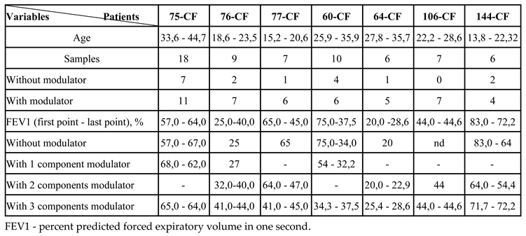
3.1. Patients in the Analysis

The cohort of 55 adult pwCF included 56% of female and 44% of male aged 18.1 - 56.7 years (mean age 30.4, median 29.8). The patients were grouped by the parameters presented in Table 1. Of the three age groups, the oldest age group was the largest. PwCF in this group had a high proportion of the non-F508del / non-F508del mutations, while the youngest age group was dominated by the F508del / F508del mutation. The proportion of the F508del / non-F508del mutations was high in all age groups. In pwCF in the oldest age group, advanced disease stage was prevalent, while younger pwCF had a higher proportion of intermediate disease stage. The majority of pwCF in the younger age groups were receiving modulator therapy (MT), while in the older age group, pwCF with and without MT were represented in approximately equal proportions. PwCF chronically infected with Pseudomonadota (PCH) were predominant in all age groups. The greatest diversity of Pseudomonadota representatives was found in sputum samples from the oldest age group pwCF: P. aeruginosa, Bcc, Achromobacter spp, Enterobacteriacae, H. influenzae. Duration of MT was mainly two-three years in younger age groups and up to one year in the older age group. However, the leader in terms of duration of MT (9 years) was the patient from the older age group. PwCF taking three-component modulators were predominant in the middle and older age groups, while in the younger age group the proportions of two- and three-component modulators were almost equal. The change of the CFTR modulator occurred in all age groups. Only in the oldest age group, three pwCF were noted to refuse to take the two-component modulator due to intolerance. In the younger age group, the triple modulator was discontinued in one CF patient (F508del/non-F508del) due to lack of efficacy.

3.2. Primary Characteristics of Sputum Samples

Sputum samples were primarily assessed for 16S rDNA concentration and the presence of major CF pathogens.

3.2.1. 16S rDNA Gene Concentration

To assess the bacterial load in sputum samples the concentration of the 16S rDNA gene was measured in DNA isolated from sputum, and expressed as 16S rRNA gene copies per ml of sputum. The samples had the following bacterial load: 6% of samples - an average of 2.9E+09 (2.0E+09 - 5.0E+09) copies/ml, 64% of samples - an average of 7.0E+08 (2.0E+08 - 1.8E+09) copies/ml, and 30% of samples - an average 1.0E+08 (2.0E+07 - 1.9E+08) copies/ml.
Analysis of the duration of MT in subgroups based on 16S rDNA concentrations showed that the average duration of MT was 0.5 years in the subgroup with the highest DNA concentration, 1.1 years in the average subgroup, and 1.5 years in the subgroup with the lowest DNA concentration. Thus, the bacterial load tended to decrease with increasing duration of MT in this pwCF cohort.

3.2.2. Typical CF Pathogens in Sputum Samples

Monitoring of typical CF pathogens, especially bacteria of the phylum Pseudomonadota, is important for assessing the effectiveness of antibiotic therapy. We used an express method of amplification and sequencing of taxon-specific targets to identify some relevant CF pathogens and verify the chronicity of infection. Pseudomonadota CF pathogens were revealed in 80% of samples: P. aeruginosa (64%), Achromobacter spp. (11%), Bcc (10%), E. coli (9%), S. maltophilia (7%), H. influenzae (4%), and K. pneumoniae (1%). A representative of the phylum Actinomycetota, the NTM Mycobacterium abscessus, was detected in one patient's sample.
P. aeruginosa isolates from chronically infected pwCF in the study had different genotypes, with the exception of those from siblings. A more detailed analysis of isolates from three new pwCF confirmed the genotype differences. The pwCF were chronically infected with P. aeruginosa ST9 (123-CF), ST198 (137-CF), and ST859 (132-CF).
Most of the samples with BCC contained B. cenocepacia ST709, which is an epidemic clone for Russia [28]. One pwCF was chronically infected with B. multivorans ST835. B. multivorans ST195 and B. cepacia ST9 were detected once. Achromobacters were also diverse and represented by two species and five genotypes: A. ruhlandii ST262 and ST36; A. xylosoxidans ST207, ST257, and ST346.
The following alleles of the adk gene were identified in samples containing E. coli: 92, 14, 21. Two pwCF were infected with E. coli ST648, but the genomes of the isolates had differences that excluded the presence of a single source of infection [26]. A mixed infection of E. coli and S. maltophilia (gap gene alleles: 22 and 11) was revealed in two pwCF. Another CF patient had a coinfection with K. pneumoniae ST716 and S. maltophilia (gap: 183).
Five pwCF were long-term infected with H. influenzae (relative abundance 15 – 62%). It should be noted that in samples with the H. influenzae load of 50% or more, amplification and Sanger sequencing of 16S rDNA fragments made it possible to identify this microorganism.
S. aureus, a CF pathogen of the phylum Bacillota, had a high relative abundance (30 – 94%) in 12 samples from 10 pwCF, primarily from the P-NP (with emerging Pseudomonadota and without Pseudomonadota) group, in clinical states of exacerbation (E) or course of antibiotic therapy (AT).

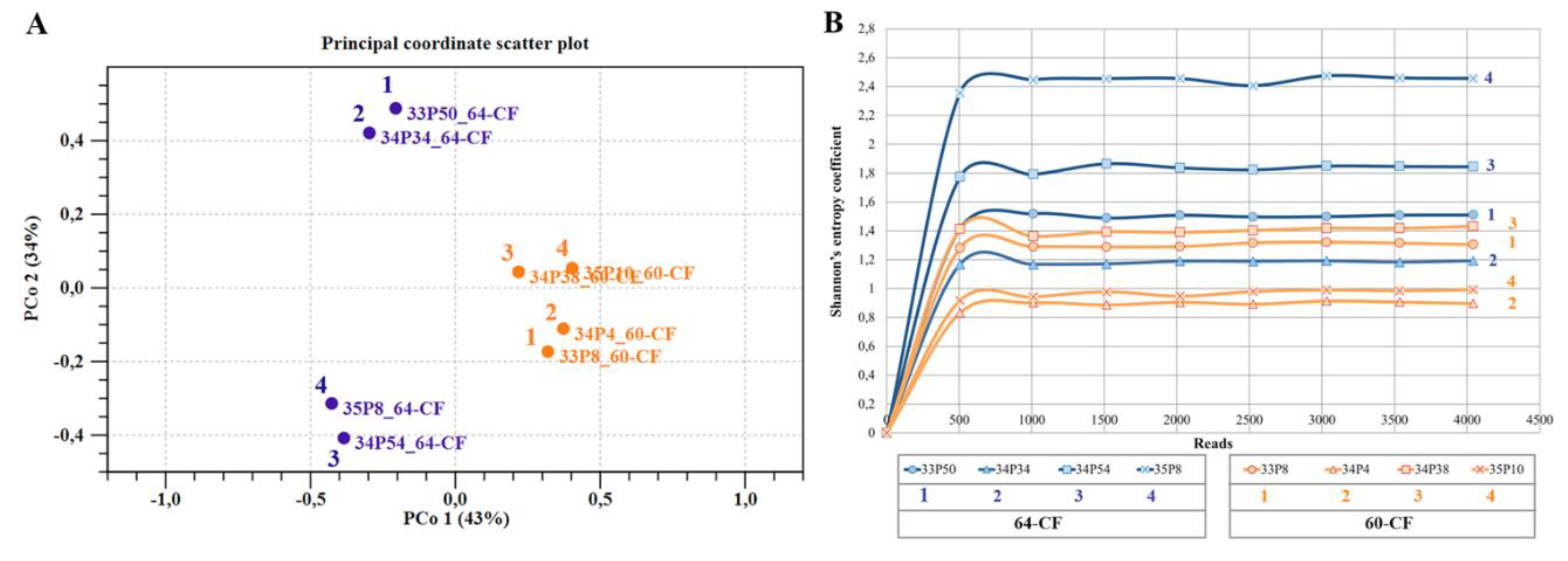
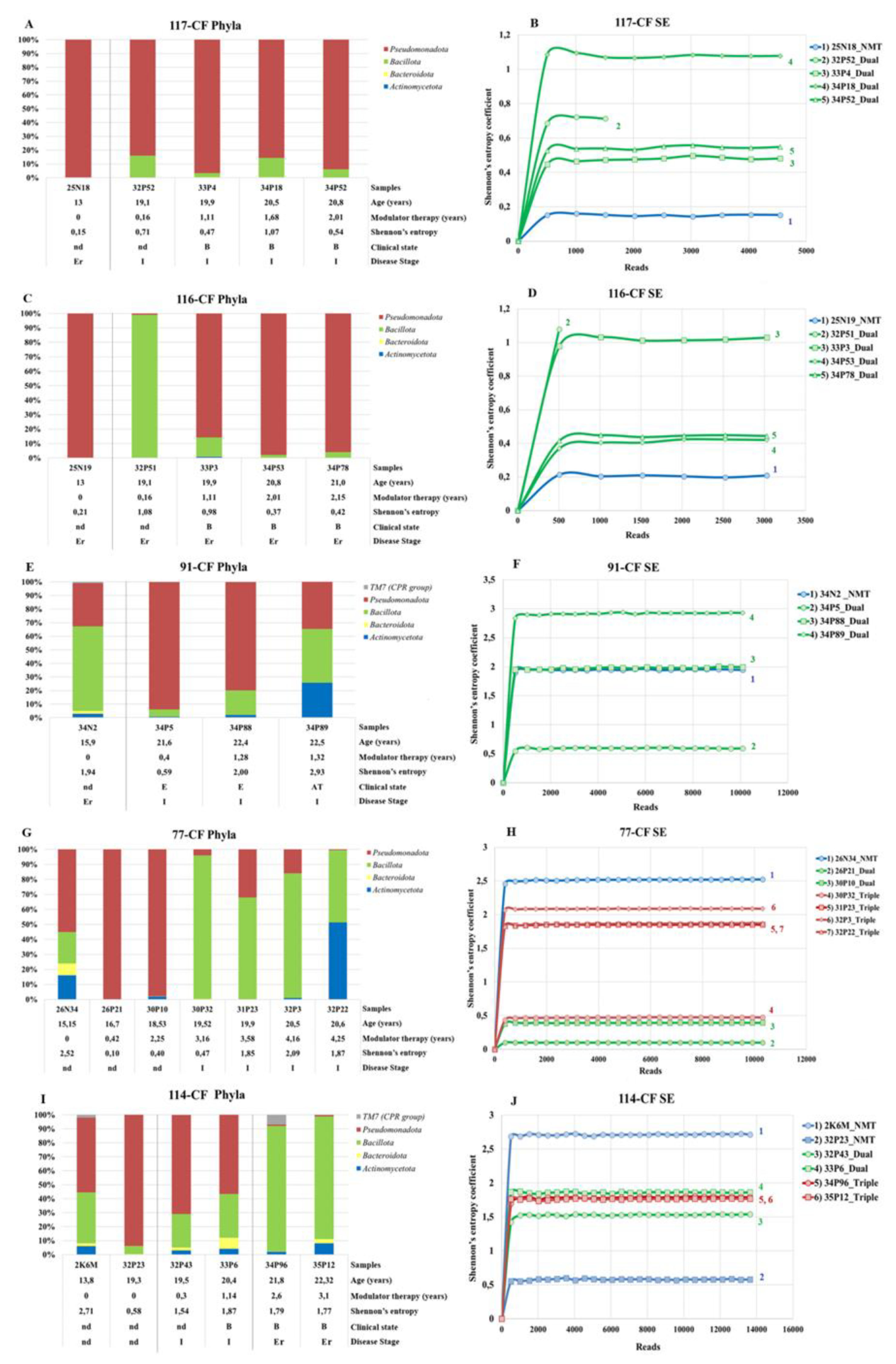
3.3. Microbiome Analysis

Microbiome analysis provides information on the representation of different bacterial taxa in sputum samples, as well as the dynamics of changes in the ratio of taxa in different clinical conditions. To give a general characterization of the microbiome of the samples, we assessed alpha diversity via Shannon’s entropy coefficient, the relative abundance of CF pathogens, and anaerobes (taxa of obligate and facultative anaerobic bacteria) (Table 2). The facultative anaerobe Streptococcus was assessed separately, as the abnormally high abundance for this taxon (26–77%) in some samples deserved special attention.
The analysis showed that the relative abundance of CF pathogens was inversely proportional to Shannon entropy, the relative abundance of anaerobes, and the relative abundance of Sreptococcus with high values of the correlation coefficients. The effect of MT duration on the relative abundance of CF pathogens, anaerobes and Streptococcus was weak, as confirmed by the low values of the correlation coefficient.
The Shannon coefficient, as an indicator of alpha diversity, varied in sputum samples within the range of 0.12 to 3.73, reaching its maximum value in a 29-year-old patient in the early disease stage, in the baseline clinical state, uninfected with Pseudomonadota pathogens, and receiving MT for approximately one year. The minimum Shannon coefficient value was observed in the oldest patient (56.7 years old), in the early disease stage, in the baseline clinical state, chronically infected with P. aeruginosa, and receiving MT for less than one year.
We divided the samples into subgroups based on the Shannon coefficient in increments of one and estimated the marker taxa of each subgroup using the LEfSe (Linear discriminant analysis Effect Size) algorithm (Figure 1). In the subgroup with the highest scores, the marker taxa were anaerobes and TM7-3. TM7-3 is a taxon of the kingdom Bacillati, phylum Minisyncoccota [30], previously known as Candidatus Saccharimonadota and candidate phylum radiation (CPR). TM7-3 bacteria are known for their small genome size and limited synthetic capability, and are usually obligate symbionts or parasites of other organisms [31]. The highest values of TM7-3 relative abundances (11–23%) were observed in samples from pwCF with early disease stages, chronically infected with P. aeruginosa, and in the first six months of receiving MT.
The subgroup with a Shannon coefficient of 2.0 - 2.9 was characterized by the anaerobes Streptococcus, Granulicatella, and Capnocytophaga.
In the subgroup with lower diversity values, the marker taxa were Staphylococcus aureus and Haemophilus. Finally, the subgroup with the lowest Shannon coefficient was distinguished by Pseudomonas and Enterobacteriaceae.
Beta diversity analysis of the samples demonstrates a disproportion in the microbiome composition not only of CF pathogens but also of anaerobic taxa. Principal coordinate analysis (Figure 2) shows a centripetal movement of samples with increasing proportions of the CF pathogens. The trajectory of movement is determined by the pathogen taxon. Unprecedentedly high relative abundances of CF pathogens were observed in samples from pwCF infected with Bcc, P. aeruginosa, Achromobacter spp, and Enterobacteriaceae, reaching 98-100%; for S. aureus, the relative abundance was somewhat lower, reaching 84%. The maximum relative abundance for Haemophilus was 62%. Increasing relative abundance of Streptococcus, Prevotella, and Rotia also causes samples to move away from the center.

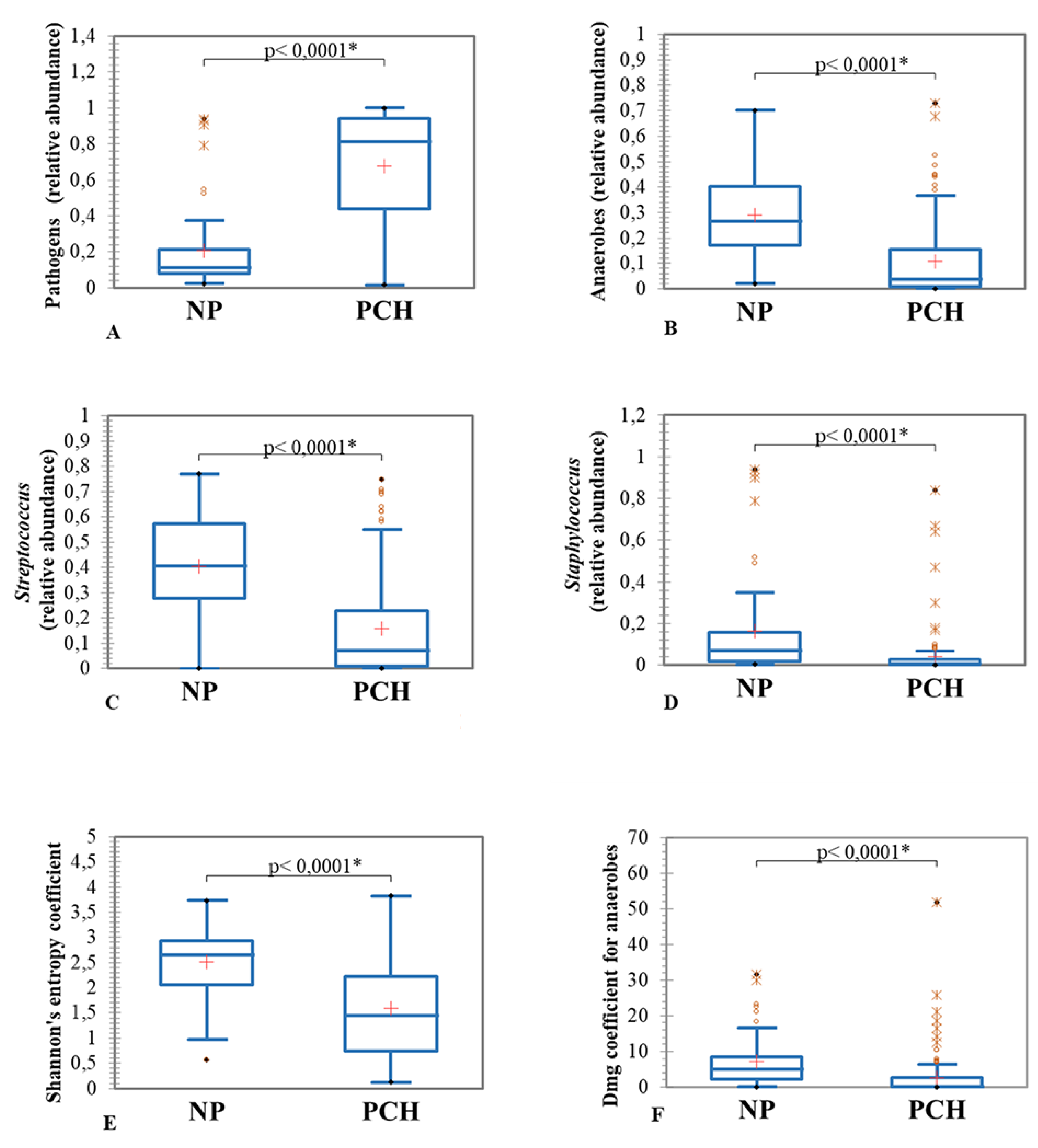
3.3.1. Clinical State and Sputum Microbiome Composition

The composition of the sputum microbiome responds to the patient's clinical state. The relative abundance of CF pathogens is significantly higher in samples obtained during exacerbations (Figure 3A), while the relative abundance of Streptococcus increases in the baseline clinical state (Figure 3B). The duration of MT was higher in samples obtained in the baseline clinical state compared with samples in the exacerbation and with samples in the AT clinical state (Figure 3C).

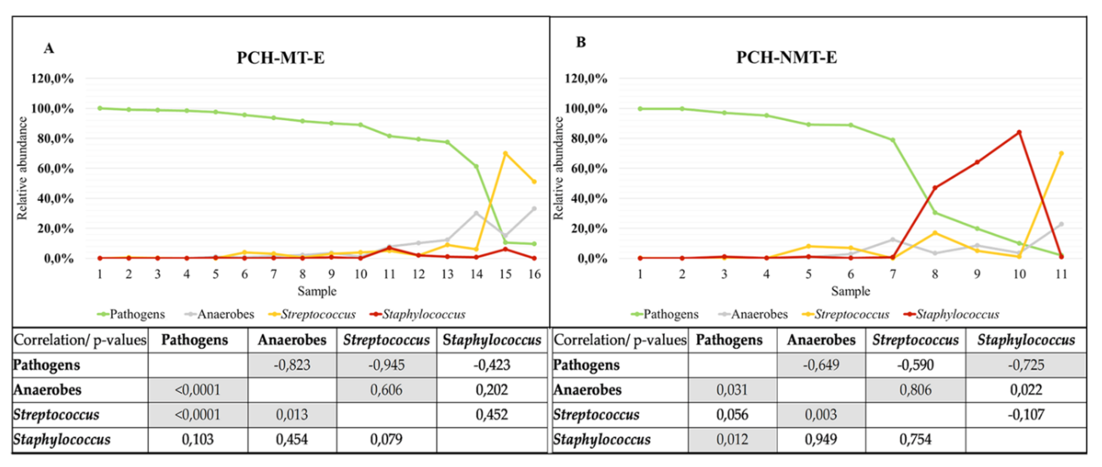
3.3.2. CFTR Modulators and Sputum Microbiome Composition

The composition of the CFTR modulators influenced the microbiome of sputum samples. Samples obtained from pwCF receiving the three-component modulator showed better reductions in the proportion of CF pathogens (Figure 4A) and increase in the relative abundance of Streptococcus (Figure 4B). The alpha diversity of the microbiome in these samples was also significantly higher (Figure 4C).
Modulator therapy treatment influenced microbiome marker taxa (Figure 5). Marker taxa in samples from pwCF not receiving MT included Pseudomonadota CF pathogens: Achromobacter, Enterobacteriaceae, and Stenotrophomonas (left part of the Figure 5). Microbiome markers in samples from pwCF receiving MT included Streptococcus (right part of the Figure 5).

3.3.3. Samples Subgroups Based on the Presence/Absence of the Pseudomonadota Pathogens

Since Pseudomonadota pathogens were predominant among CF pathogens, we divided the pwCF cohort samples into two subgroups based on the presence/absence of the Pseudomonadota pathogen in the microbiome of the samples: PCH and NP. (Table 3).
The PCH subgroup had a higher proportion of older pwCF, more often in advanced disease stage, while the NP subgroup had a higher proportion of pwCF in early disease stage. The proportion of samples obtained during MT was higher in the PCH subgroup, while the average duration of MT did not differ significantly between the subgroups. Most samples in the NP subgroup were obtained in the B clinical state. The proportion of samples obtained during the E and AT clinical states was slightly higher in the PCH subgroup.
Comparison of the microbiomes of the samples from the PCH and NP subgroups (Figure 6) showed that the PCH subgroup had a higher relative abundance of CF pathogens in general (Figure 6A), but a lower proportion of Staphylococcus aureus (Figure 6D). Meanwhile, the relative abundance of anaerobes (Figure 6B) and Streptococcus (Figure 6C), as well as alpha diversity (Figure 6E) and species richness of anaerobes (Figure 6F), were significantly higher for samples from the NP subgroup.
An exacerbation is the time of manifestation of CF pathogens. We compared samples from PCH pwCF in exacerbation clinical state from two subgroups: receiving (MT) and not receiving (NMT) modulator therapy. The samples were ranked in order of descending relative abundance of Pseudomonadota CF pathogens (Figure 7). In the MT subgroup, a decrease in the relative abundance of CF pathogens led to an increase in the proportion of anaerobes and Streptococcus (Figure 7A). Moreover, the increase in the relative abundance of Streptococcus was abnormally high, reaching 70% in a pwCF with an intermediate disease stage. In the NMT subgroup, with a decrease in the proportion of Pseudomonadota CF pathogens, the proportion of Staphylococcus primarily increased, reaching 84% in a pwCF with an intermediate disease stage (Figure 7B). The proportion of anaerobes and Streptococcus remained low until the relative abundance of Pseudomonadota CF pathogens decreased to 2%. Only then did the proportion of Streptococcus rise to an extremely high 70% in a pwCF also with an intermediate disease stage. The values of the correlation coefficients (Figure 7) confirm the inverse proportionality between the relative abundance of Pseudomonadota CF pathogens and anaerobes and Streptococcus in the MT subgroup and between the relative abundance of Pseudomonadota CF pathogens and anaerobes and Staphylococcus in the NMT subgroup. The high reliability of the values is confirmed by p-values <0.05.

3.3.4. Microbiome of Samples from Patients Receiving Long-Term Modulator Therapy

Seven pwCF with a duration of modulator therapy of 3.1 to 9.1 years were allocated to a separate group for analysis (Table 4). For five pwCF from the observed cohort, earlier samples registered in our previous projects were added. Samples from pwCF 76-CF and 77-CF, obtained earlier, supplemented this group in accordance with the duration of observation, but these pwCF were not included in the observed cohort with 55 individuals. PwCF 75-CF, 76-CF, and 77-CF were previously infected with NTM and received prolonged, aggressive antibiotic therapy before initiating modulator therapy, which resulted in the absence of NTM growth on special media. During the observation period, we detected only traces of NTM DNA (0.1 - 0.2%) in 75-CF samples after three years of treatment with the single-component CFTR modulator (two samples), and in 76-CF samples after switching from a single-component modulator to a two-component modulator and then after switching to a three-component modulator (one sample each). In 77-CF samples, NTM was detected once, after 2.3 years of receiving the two-component modulator, with a relative abundance of 2%, which allowed the amplification of groEL and identification of Mycobacterium abscessus. These data confirm improved pulmonary drainage with modulator therapy.
We previously described the eradication of B. cenocepacia ST710 in pwCF 75-CF [28], and a significant amount of this bacterium in the microbiome was observed long before the start of MT (Figure 8A). During MT, an increase in the relative abundance of CF pathogens was noted in association with a Haemophilus infection, leading to an exacerbation. In some 75-CF samples, an increase in the proportion of Mycoplasma, reaching 12%, was observed. In the remaining 75-CF samples, the proportion of anaerobes and Streptococcus was high, and the species diversity of anaerobes (Dmg) reached a maximum (51.8) after 9 years of MT. Long-term observation of pwCF allowed us to determine the disease aggressiveness (or phenotype), according to Schluchter et al., who developed a “topographical” depiction of CF lung disease aggressiveness as a function of FEV1% predicted and age [24,32]. Since the FEV1 of the 75-CF patient was 57% at 33.6 years of age, and by 44.7 years of age it had increased to 65-64% (Table 4), due to the action of CFTR modulators, the disease phenotype can be assessed as mild lung disease.
PwCF 75-CF and 76-CF had a severe lung disease phenotype as the decline in FEV1 occurred dramatically rapidly. A triple-modulator allowed patient 76-CF to increase FEV1 from a critically low 25% to 44% (Table 4) and maintain it at 45% for patient 77-CF. Both pwCF were infected with P. aeruginosa (Figure 8B, C). A decrease in the relative abundance of P. aeruginosa led to a classic increase in the proportion of S. aureus in patient 76-CF, and to an increase in the proportion of Streptococcus in patient 77-CF. Anaerobes were absent in samples of patient 76-CF before the start of MT. After six months of receiving a single-component modulator, Dmg rose to 7.0, but then remained low or equal to zero. Only after 4.5 years of MT, during the 1.5 years of which the patient received a three-component modulator, the diversity of anaerobes increased and Dmg was 11.9. Patient 77-CF had anaerobes in one sample before MT, then anaerobes were absent and appeared only after 4.3 years of treatment with modulators (the last 1.5 years - with a three-component modulator).
Patient 114-CF with moderate lung disease was also infected with P. aeruginosa (Figure 8D). As the relative abundance of P. aeruginosa decreased, it was outcompeted by S. aureus, which mirrored the microbiome composition dynamics of patient 76-CF's samples. A 13-year-old patient's sample had a Dmg of 7.0. Later, even with MT, anaerobe species diversity remained low, and only after 3.1 years of treatment with modulators (the last 1.3 years - with a three-component modulator) did it rise to 4.7 Dmg.
Thus, in three pwCF, P. aeruginosa was present in trace amounts in the last analyzed samples, but a conclusion about the eradication of this CF pathogen can only be made after a series of subsequent samples negative for P. aeruginosa.
The next three pwCF (60-CF, 64-CF, and 106-CF) were infected with Burkholderiales (Figure 9). Patient 60-CF, who had long-standing high lung function and a mild lung disease phenotype, had episodes of low B. multivorans ST835 relative abundance (Figure 9A) and a diversity of anaerobes with Dmg 6.1. However, in simultaneously obtained sinus lavage samples, the relative abundance of B. multivorans ST835 was very high [33], which subsequently led to an increase in the proportion of B. multivorans ST835 in the lung microbiome, a decrease in FEV1, even in the presence of MT (Table 4), and a change in the lung disease phenotype in the moderate. Switching to a three-component modulator did not lead to improvement in lung drainage; the proportion of B. multivorans ST835 continued to remain high, and anaerobes were extremely low.
Patient 64-CF with severe lung disease, infected with B. cenocepceae ST709, maintained a high relative abundance of B. cenocepceae in the lung microbiome (Figure 9B), supported by sinus-adapted B. cenocepceae [33] during 2.6 years of treatment with a two-component modulator. Switching to a three-component modulator significantly reduced the proportion of B. cenocepceae and increased anaerobic diversity in the lung microbiome to Dmg 5.0.
Analysis of beta diversity in samples from pwCF 60-CF and 64-CF clearly demonstrated an improvement in the microbiome status of patient 64-CF compared to samples from 60-CF (Figure 10A). The Shannon entropy of samples from 64-CF increased progressively with increasing duration of the three-component MT, while the Shannon coefficient decreased for samples from 60-CF (Figure 10B). So, the lung microbiome of CF patients is influenced by multiple factors, and therapy for normalizing microbiome composition is personalized.
Patient 106-CF with moderate lung disease had been infected with A. ruhlandii ST262 since childhood (Figure 9C). Two-component MT, which maintained lung function, did not reduce the relative abundance of A. ruhlandii. Switching to the three-component modulator had a beneficial effect on the microbiome composition: the proportion of A. ruhlandii decreased and a diverse range of anaerobes appeared (peak Dmg was 19). However, the decrease in the proportion of A. ruhlandii to trace amounts after 5.8 years of MT led to an increase in P. aeruginosa and S. aureus. In the next sputum sample (after 6 years of MT), a sharp increase in the relative abundance of A. ruhlandii and a decrease in the diversity of anaerobes to a minimum were observed. Thus, even with long-term three-component MT, achieving stabilization of the lung microbiome composition with a low load of CF pathogens is extremely difficult.

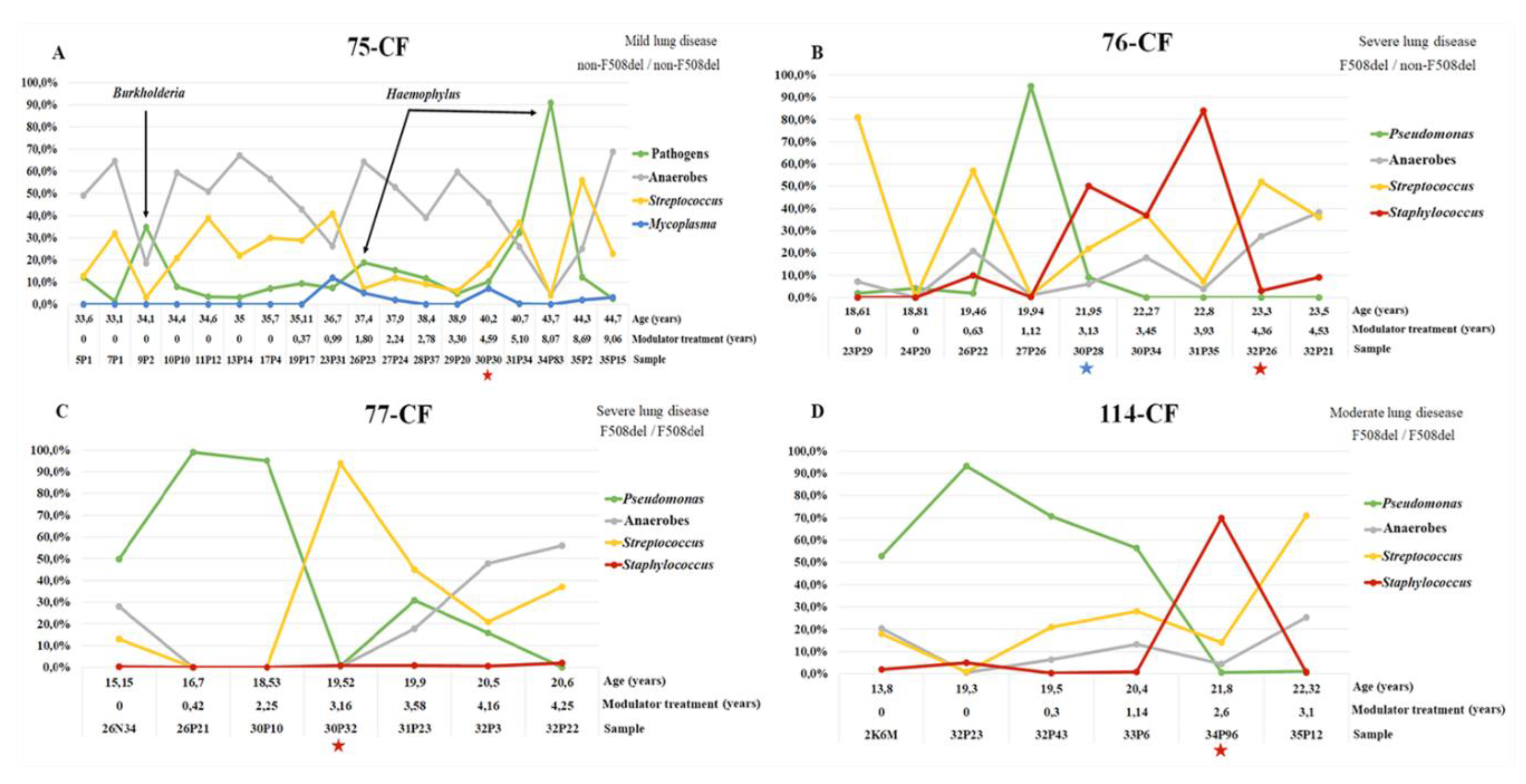
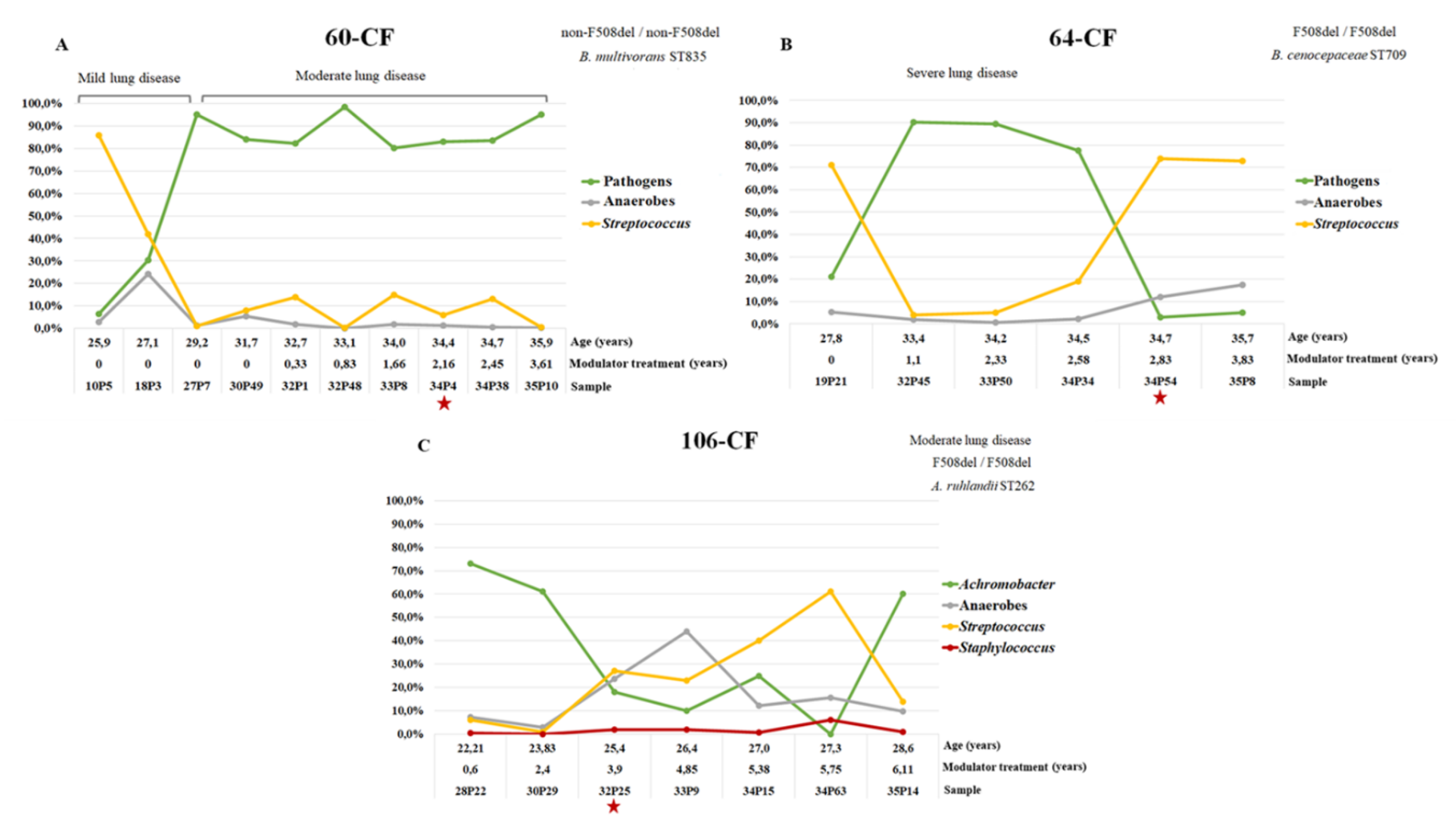
3.3.5. Dynamics of the Microbiome Composition of Patients Chronically Infected with Pseudomonadota since Adolescence

For five young PCH patients who were observed from 13-15 to 20-22 years of age, we tracked the dynamics of changes in the microbiome (Figure 11). All pwCF had the F508del/F508del CFTR mutation and were infected with P. aeruginosa at the beginning of the follow-up. The concomitant CF pathogen for all but one patient (77-CF) was S. aureus. All patients but one (77-CF) had a moderate disease aggressiveness phenotype. Three pwCF (91-CF, 116-CF, and 117-CF) received only a dual modulator for 1.3 to 2.1 years. A dual modulator therapy for patients 77-CF and 114-CF was changed to a triple modulator after 2.7 and 1.8 years of treatment, respectively.
Only two patients (77-CF and 114-CF) at the first observation point had the highest Shannon entropy index for their microbiome during the study, about 2.5, (Figure 11H, J). The lowest Shannon entropy values (around 0.2) at age 13 were in twins 116-CF and 117-CF (Figure 11B, D), when the microbiome contained the highest proportion of the phylum Pseudomonadota (Figure 11A, C). Only once, at the beginning of treatment with a two-component modulator, did the proportion of Pseudomonas in patient 116-CF decrease to 1%; at the remaining observation points, Pseudomonadota predominated. Note that a decrease in the relative abundance of P. aeruginosa led to an increase in the proportion of S. aureus.
The Shannon entropy index for the microbiome of the third patient (91-CF), who received dual modulator therapy, increased only after several courses of aggressive antibiotic therapy prescribed due to a series of exacerbations (Figure 11F). At the fourth observation point, the relative abundance of P. aeruginosa decreased, S. aureus was absent, but the proportion of Rothia and Granulicatella increased to an abnormal 22-27%. An increase in the microbiome diversity of patients 77-CF and 114-CF was observed after receiving the triple modulator, but the Shannon entropy index only reached a value of approximately 2 (Figure 11H, J), which is slightly lower than that of the older PCH patient 64-CF (Figure 10B).
Thus, the bacterial load of CF pathogens in all patients in the young group was already high in early adolescence. Two-component modulator therapy did not reduce the relative abundance of the CF pathogen complex, but could, in conjunction with antibiotic therapy, influence the dominant pathogen. Triple modulator therapy was the most effective in improving microbiome diversity and reducing the proportion of CF pathogens.

4. Discussion

CFTR modulators of various compositions, approved since 2012, have expanded the therapeutic landscape for CF and improved quality of life for pwCF affected by modulator therapy (MT). The developers and researchers of CFTR modulators were awarded the Lasker Clinical Research Prize in 2025. However, a complete understanding of the modulator's impact on lung infections, particularly in chronically infected adult pwCF, remains unclear. Previous studies of the effects of modulator treatment on the lung microbiome have mostly involved F508del CFTR homozygous participants and have lasted no more than a year [18,34,35].
Our three-year study included 55 adult pwCF chronically infected with CF pathogens, primarily of the phylum Pseudomonadota. Participants had not only F508del CFTR-homozygous, but also F508del CFTR-heterozygous and non-F508del CFTR mutations. The study showed that with increasing duration of modulator therapy, there was a trend toward a decrease in bacterial load in sputum samples. A trend toward decreased total bacterial abundance was also shown by Hilliam et al., who studied the bacterial community of the middle meatus of pwCF after 42 months of therapy with the ELX/TEZ/IVA modulator [36]. However, a number of studies characterize the results of modulator therapy only based on the average sputum densities of traditional CF pathogens, noting a 10- to 100-fold reduction in this indicator [34,37].
In our cohort of 55 adult pwCF, most of whom were chronically infected with Pseudomonadota (PCH), the effect of MT duration on the relative abundance of CF pathogens, anaerobes and Streptococcus was weak, as confirmed by the low values of the correlation coefficient (Table 2). Some pwCF showed significant fluctuations in typical CF pathogens in sputum samples, while others pwCF showed a consistently high proportion of pathogens in the microbiome of sputum samples. Unprecedentedly high relative abundances of CF pathogens were observed in samples from patients infected with Bcc, P. aeruginosa, Achromobacter spp, and Enterobacteriaceae, reaching 98-100%. Neerincx et al. also observed only a temporary reduction in P. aeruginosa abundance in sputum under the influence of LUM/IVA modulator therapy [38].
A decrease in the relative abundance of CF pathogens correlated with an increase in the Shannon coefficient, as an indicator of alpha diversity. Anaerobes were marker taxa for the subgroups of samples with the highest Shannon coefficient values: 3.0-3.9 and 2.0-2.9 (Figure 1). In the 3.0-3.9 group, anaerobes were complemented by the TM7-3 taxon of the phylum Minisyncoccota, and in the 2.0-2.9 group, by Streptococcus, a facultative anaerobe considered separately. Samples in which the relative abundance of anaerobes was more than 10% were found in 81% of pwCF in the NP group and in 33% of pwCF in the PCH group, both with and without modulator therapy. In the PCH group, such samples were more often associated with antibiotic therapy, and in the NP group, they were approximately equally common in the baseline and exacerbation states. Overall, across the entire cohort, samples with a higher relative abundance of anaerobes were observed in the baseline state (p = 0.015). Only four patients with early or intermediate lung disease in the NP group had a higher relative abundance of anaerobes in the exacerbation state compared to the baseline state. The proportion of S. aureus as a CF pathogen in these samples was 2.0 - 52.0%. According to observations by Zhao et al. and Carmody et al. the relative abundance of anaerobic genera increased significantly during exacerbations before episodic antibiotic treatment compared with baseline [23,39]. A detailed analysis of the patient P2 samples presented by Zhao et al. [39] showed that the observation is true for samples with a relative abundance of CF pathogens of 40-50% in the baseline state; with an increase in the relative abundance of CF pathogens, the relative abundance of anaerobes during exacerbation becomes minimal. Most patients in our cohort had a high relative abundance of CF pathogens in their sputum samples.
The role of anaerobes in the lung microbiome of pwCF remains highly controversial. Some researchers showed that the relative abundance of anaerobes in sputum samples was associated with less inflammation [40] and improved lung function [41]. Other researchers have identified potential mechanisms by which anaerobes can lead to disease progression in the lungs of pwCF: a) the production of short-chain fatty acids by anaerobes stimulates the release of proinflammatory cytokines from human bronchial epithelial cells [42], b) anaerobes degrade mucin and synthesize nutrients for CF pathogens [43], c) anaerobes produce β-lactamases, which mediate antimicrobial resistance [44,45].
Particular attention should be paid to the excessive increase in the proportion of individual taxa of anaerobes with a decrease in the relative abundance of CF pathogens. In our pwCF cohort's samples, the proportion of anaerobes reached 40% for Rothia, Prevotella, Capnocytophaga, and Oribacterium, 27-28% for Athopobium and Granulicatella, 23% for TM7-3, 18% for Lactobacillus, and 15% for Bulleidia. Meanwhile, in healthy individuals, the proportion of the most abundant anaerobe, Prevotella, was 15% in the right upper lobe of the lungs and 19% in the supraglottic space [46]. The proportion of Streptococcus increased even more significantly with a decrease in the proportion of CF pathogens, reaching 23 - 77% in the cohort samples, compared to 5% in the right upper lobe of the lungs and 13% in the supraglottic space in healthy individuals [46].
In our study, on the one hand, Streptococcus was the marker taxon in the MT group (Figure 5) and competed with the Pseudomonadata CF pathogens in the PCH group and with Staphylococcus in the NP group. On the other hand, during exacerbations, in some patients' samples with a low proportion of CF pathogens, the relative abundance of streptococcus increased to 32-70%. It should be noted that several studies have shown that the presence of Streptococcus as the predominant species in the lungs correlated with exacerbations [47,48].
Long-term modulator therapy is essential for improving the airway microbiota in pwCF. A Danish national cohort study of airway pathogens showed that even three years after ELX/TEZ/IVA initiation in 201 pwCF over 12 years of age, airway secretions in two-thirds of pwCF remained culture-positive with the same airway pathogens as before they started ELX/TEZ/IVA [49]. In our cohort of 55 adult patients, the average duration of modulator therapy was 2.1 years (range, 0.23–9.0). Therefore, we supplemented the study with data from seven pwCF receiving modulators for more than three years (3.1 – 9.0).
For patients chronically infected with P. aeruginosa, it was important to switch to the three-component modulator ELX/TEZ/IVA, which reduced the proportion of P. aeruginosa, as well as S. aureus and Streptococcus, competitors that replace the main CF pathogen (Figure 8A, B, C). As a result, the microbiome with a relative abundance of P. aeruginosa less than 1%, S. aureus - 0.5-9% and with a diversity of anaerobes corresponded to the most favorable pulmonary type 4 as defined by Widder et al. [50]. We demonstrated an even clearer effect of the modulator's composition in a group of pwCF with F508del/F508del CFTR mutation infected with P. aeruginosa and observed from early adolescence. Of the three pwCF receiving the dual-modulator LUM/IVA, two twins with moderate lung disease (Figure 11A, C) had a consistently high relative abundance of P. aeruginosa but an extremely low relative abundance of anaerobes, including Streptococcus, corresponding to pulmonary type 5, according to Widder et al. [50]. However, over the last three years of observation, the patients showed only a slight decrease in predicted FEV1%, and the absence of exacerbations. In the third CF patient receiving LUM/IVA, a decrease in the relative abundance of P. aeruginosa occurred only with courses of antibiotic treatment for exacerbation (Figure 11E).
In pwCF with the F508del/F508del CFTR mutation infected with Burkholderiales (B. cenocepacia ST709 - 64CF and A. ruhlandii ST262 - 106CF), when switching to a three-component modulator ELX/TEZ/IVA, a temporary decrease in the relative abundance of the main CF pathogen was observed, but eradication was not achieved even after six years of modulator therapy in the case of 106-CF (Figure 9C). Thus, at the last observation point, the microbiome of patient 64-CF temporarily had pulmotype 4, and in patient 106 CF could be classified as pulmotype 6. In our opinion, such an interpretation of pulmotypes is possible by replacing the position of P. aeruginosa with any Pseudomonadota CF pathogen in the model of pulmotypes by Widder et al. [50].
In the patient with the non-F508del/non-F508del CFTR mutation infected with B. multivorans ST835, a year and a half after switching to ELX/TEZ/IVA did not lead to a decrease in the relative abundance of the main CF pathogen, but predicted FEV1% indicators somewhat stabilized at a low value of 37.5, despite episodes of exacerbation (Figure 9A, Table 4).
Overall, in a cohort of 55 adult patients, the relative abundance of CF pathogens was significantly lower, while the relative abundance of Streptococcus and the Shannon entropy coefficient were significantly higher in the group of patients receiving the three-component modulator CFTR (Figure 4). Thus, the composition of the modulator is important for clearing the lower respiratory tract from the chronic CF pathogen.
At the same time, observations of the microbiome of a pwCF who received modulator therapy for 9 years, 4.5 of which were spent on ELX/TEZ/IVA, showed that morphological abnormalities in the respiratory system facilitate infection with new CF pathogens (Haemophilus, Mycoplasma, and others), which provokes exacerbations. Thus, in this patient, we continue to observe changes in the microbiome from pulmotype 4 to pulmotype 5 and back to pulmotype 4 at the last observation point, which showed the highest diversity of anaerobes in the sample.

5. Conclusions

The long-term study of adult patients predominantly chronically infected with Pseudomonadota CF pathogens with a wide range of mutations describes the structure and dynamics of airway bacterial communities in patients with CF, linking these indicators to changes in clinical status, disease stage, and severity. A key observation is that modulator therapy results in a reduction in the bacterial load without eradication of typical CF pathogens. Only long-term three-component modulator therapy brings the lung microbiome closer to a relatively favorable state, but does not ensure complete restoration of a healthy microbiome. Respiratory system damaged by bronchiectasis is susceptible to new infections, so patient management requires constant monitoring of the microbiome composition and replenishment of the therapeutic landscape with both new modulators and new antibacterial drugs.

6. Patents

Sputum samples from 55 adult CF patients were included in the study. The participants signed informed consent to take part in the study, and the research protocol was approved by the Biomedical Ethics Committee of the N.F. Gamaleya National Research Center for Epidemiology and Microbiology (protocol No. 59, September 8, 2023) and the Ethics Committee of the Research Institute of Pulmonology under FMBA of Russia (protocol No. 04-23, April 24, 2023, and protocol No. 07-24, December 18, 2024).

Author Contributions

Conceptualization, O.L.V. and E.L.A.; methodology, O.L.V., N.N.R., M.S.K., E.L.A.; software, O.L.V., N.N.R., M.S.K, E.I.E.; validation, N.N.R., M.S.K, E.I.E and O.L.V.; formal analysis, N.N.R.; investigation, N.N.R., M.S.K, E.I.E and O.L.V.; resources, O.L.V., E.L.A. and R.U.K..; data curation, O.L.V., N.N.R. and E.L.A.; writing—original draft preparation, O.L.V., N.N.R., M.S.K; writing—review and editing, O.L.V., N.N.R. and E.L.A.; visualization, O.L.V., N.N.R. and M.S.K; supervision, O.L.V. and E.L.A.; project administration, O.L.V., E.L.A. and A.L.G.; funding acquisition, A.L.G. All authors have read and agreed to the published version of the manuscript.

Funding

The study is supported by the State assignment of the N.F. Gamaleya National ResearchCenter for Epidemiology and Microbiology (No. 056-00066-23-00).

Institutional Review Board Statement

In this section, please add the Institutional Review Board Statement and approval number for studies involving humans or animals. You might choose to exclude this statement if the study did not require ethical approval. Please note that the Editorial Office might ask you for further information. Please add “The study was conducted in accordance with the Declaration of Helsinki, and approved by the Institutional Review Board (or Ethics Committee) of NAME OF INSTITUTE (protocol code XXX and date of approval).” for studies involving humans. OR “The animal study protocol was approved by the Institutional Review Board (or Ethics Committee) of NAME OF INSTITUTE (protocol code XXX and date of approval).” for studies involving animals. OR “Ethical review and approval were waived for this study due to REASON (please provide a detailed justification).” OR “Not applicable” for studies not involving humans or animals.

Informed Consent Statement

The study was carried out with the informed consent of the patients.

Data Availability Statement

The reported results can be found in the GenBank NCBI. BioProject ID: PRJNA717158.

Conflicts of Interest

The authors declare no conflicts of interest.

Abbreviations

The following abbreviations are used in this manuscript:
CF Cystic fibrosis
CFTR Cystic fibrosis transmembrane conductance regulator
pwCF Patients or people with CF
IVA Ivacaftor
LUM Lumacaftor
TEZ Tezacaftor
ELX Elexacaftor
MT Modulator treatment
NMT Non-modulator treatment
PCH Chronically infected with Pseudomonadota
NP With emerging Pseudomonadota and without Pseudomonadota
Bcc Burkholderia cepacia complex
FEV1, %FEV1 Percent predicted forced expiratory volume in one second
B Baseline
E Exacerbation
AT Course of antibiotic therapy
Dmg Margalef's Richness Index
OTU Operational Taxonomic Units
PCoA Principal Coordinate Analysis
LEfSe Linear discriminant analysis Effect Size
early disease stage FEV1 > 70%
intermediate 70% < FEV1 > 40%
advanced FEV1 < 40%

References

  1. Marsland, B.J.; Gollwitzer, E.S. Host-microorganism interactions in lung diseases. Nat. Rev. Immunol. 2014, 14, 827–835. [Google Scholar] [CrossRef] [PubMed]
  2. Lipinksi, J.H.; Ranjan, P.; Dickson, R.P.; O'Dwyer, D.N. The Lung Microbiome. J. Immunol. 2024, 212, 1269–1275. [Google Scholar] [CrossRef] [PubMed]
  3. Bell, S.C.; Mall, M.A.; Gutierrez, H.; Macek, M.; Madge, S.; Davies, J.C.; Burgel, P.R.; Tullis, E.; Castaños, C.; Castellani, C.; Byrnes, C.A.; Cathcart, F.; Chotirmall, S.H.; Cosgriff, R.; Eichler, I.; Fajac, I.; Goss, C.H.; Drevinek, P.; Farrell, P.M.; Gravelle, A.M.; Havermans, T.; Mayer-Hamblett, N.; Kashirskaya, N.; Kerem, E.; Mathew, J.L.; McKone, E.F.; Naehrlich, L.; Nasr, S.Z.; Oates, G.R.; O'Neill, C.; Pypops, U.; Raraigh, K.S.; Rowe, S.M.; Southern, K.W.; Sivam, S.; Stephenson, A.L.; Zampoli, M.; Ratjen, F. The future of cystic fibrosis care: a global perspective. Lancet Respir. Med. 2020, 8, 65–124. [Google Scholar] [CrossRef]
  4. Elborn, J.S. Cystic fibrosis. Lancet. 2016, 388, 2519–2531. [Google Scholar] [CrossRef]
  5. Blanchard, A.C.; Waters, V.J. Microbiology of cystic fibrosis airway disease. Semin. Respir. Crit. Care Med. 2019, 40, 727–736. [Google Scholar] [CrossRef]
  6. Richards, C.J.; Olivier, K.N. Nontuberculous Mycobacteria in Cystic Fibrosis. Semin. Respir. Crit. Care Med. 2019, 40, 737–750. [Google Scholar] [CrossRef]
  7. O'Neill, K.; Bradley, J.M.; Johnston, E.; McGrath, S.; McIlreavey, L.; Rowan, S.; Reid, A.; Bradbury, I.; Einarsson, G.; Elborn, J.S.; Tunney, M.M. Reduced bacterial colony count of anaerobic bacteria is associated with a worsening in lung clearance index and inflammation in cystic fibrosis. PLoS One. 2015, 10, e0126980. [Google Scholar] [CrossRef] [PubMed]
  8. Sibley, C.D.; Grinwis, M.E.; Field, T.R.; Eshaghurshan, C.S.; Faria, M.M.; Dowd, S.E.; Parkins, M.D.; Rabin, H.R.; Surette, M.G. Culture enriched molecular profiling of the cystic fibrosis airway microbiome. PLoS One. 2011, 6, e22702. [Google Scholar] [CrossRef]
  9. Muhlebach, M.S.; Hatch, J.E.; Einarsson, G.G.; McGrath, S.J.; Gilipin, D.F.; Lavelle, G.; Mirkovic, B.; Murray, M.A.; McNally, P.; Gotman, N.; Davis Thomas, S.; Wolfgang, M.C.; Gilligan, P.H.; McElvaney, N.G.; Elborn, J.S.; Boucher, R.C.; Tunney, M.M. Anaerobic bacteria cultured from cystic fibrosis airways correlate to milder disease: a multisite study. Eur. Respir. J. 2018, 52, 1800242. [Google Scholar] [CrossRef]
  10. Lamoureux, C.; Guilloux, C.A.; Beauruelle, C.; Gouriou, S.; Ramel, S.; Dirou, A.; Le Bihan, J.; Revert, K.; Ropars, T.; Lagrafeuille, R.; Vallet, S.; Le Berre, R.; Nowak, E.; Héry-Arnaud, G. An observational study of anaerobic bacteria in cystic fibrosis lung using culture dependent and independent approaches. Sci Rep. 2021, 11, 6845. [Google Scholar] [CrossRef]
  11. Muhlebach, M.S.; Zorn, B.T.; Esther, C.R.; Hatch, J.E.; Murray, C.P.; Turkovic, L.; Ranganathan, S.C.; Boucher, R.C.; Stick, S.M.; Wolfgang, M.C. Initial acquisition and succession of the cystic fibrosis lung microbiome is associated with disease progression in infants and preschool children. PLoS Pathog. 2018, 14, e1006798. [Google Scholar] [CrossRef] [PubMed]
  12. Zemanick, E.T.; Wagner, B.D.; Robertson, C.E.; Ahrens, R.C.; Chmiel, J.F.; Clancy, J.P.; Gibson, R.L.; Harris, W.T.; Kurland, G.; Laguna, T.A.; McColley, S.A.; McCoy, K.; Retsch-Bogart, G.; Sobush, K.T.; Zeitlin, P.L.; Stevens, M.J.; Accurso, F.J.; Sagel, S.D.; Harris, J.K. Airway microbiota across age and disease spectrum in cystic fibrosis. Eur. Respir. J. 2017, 50, 1700832. [Google Scholar] [CrossRef] [PubMed]
  13. Kember, M.; Grandy, S.; Raudonis, R.; Cheng, Z. Non-Canonical Host Intracellular Niche Links to New Antimicrobial Resistance Mechanism. Pathogens. 2022, 11, 220. [Google Scholar] [CrossRef]
  14. Cigana, C.; Giannella, R.; Colavolpe, A.; Alcalá-Franco, B.; Mancini, G.; Colombi, F.; Bigogno, C.; Bastrup, U.; Bertoni, G.; Bragonzi, A. Mutual Effects of Single and Combined CFTR Modulators and Bacterial Infection in Cystic Fibrosis. Microbiol. Spectr. 2023, 11, e0408322. [Google Scholar] [CrossRef]
  15. Harvey C, Weldon S, Elborn S, Downey DG, Taggart C. The Effect of CFTR Modulators on Airway Infection in Cystic Fibrosis. Int J Mol Sci. 2022;23(7):3513. [CrossRef]
  16. Keating, C.; Yonker, L.M.; Vermeulen, F.; Prais, D.; Linnemann, R.W.; Trimble, A.; Kotsimbos, T.; Mermis, J.; Braun, A.T.; O'Carroll, M.; Sutharsan, S.; Ramsey, B.; Mall, M.A.; Taylor-Cousar, J.L.; McKone, E.F.; Tullis, E.; Floreth, T.; Michelson, P.; Sosnay, P.R.; Nair, N.; Zahigian, R.; Martin, H.; Ahluwalia, N.; Lam, A.; Horsley, A.; VX20-121-102 Study Group; VX20-121-103 Study Group. Vanzacaftor-tezacaftor-deutivacaftor versus elexacaftor-tezacaftor-ivacaftor in individuals with cystic fibrosis aged 12 years and older (SKYLINE Trials VX20-121-102 and VX20-121-103): results from two randomised, active-controlled, phase 3 trials. Lancet Respir. Med. 2025, 13, 256–271. [Google Scholar] [CrossRef]
  17. Vertex Announces U.S. FDA Approval for TRIKAFTA (elexacaftor/tezacaftor/ivacaftor and ivacaftor) to Include Additional Non-F508del TRIKAFTA-Responsive Variants. Accessed January 6, 2025. https://investors.vrtx.com/news-releases/news-release-details/vertex-announcesus-fda-approval-trikafta. 6 January.
  18. Iftikhar, I.H.; Rao, S.T.; Nadama, R.; Janahi, I.; BaHammam, A.S. Comparative Efficacy of CFTR Modulators: A Network Meta-analysis. Lung. 2025, 203, 49. [Google Scholar] [CrossRef]
  19. Malet, K.; Faure, E.; Adam, D.; Donner, J.; Liu, L.; Pilon, S.J.; Fraser, R.; Jorth, P.; Newman, D.K.; Brochiero, E.; Rousseau, S.; Nguyen, D. Intracellular Pseudomonas aeruginosa within the Airway Epithelium of Cystic Fibrosis Lung Tissues. Am. J. Respir. Crit. Care Med. 2024, 209, 1453–1462. [Google Scholar] [CrossRef] [PubMed]
  20. Ledger, E.L.; Smith, D.J.; The, J.J.; Wood, M.E.; Whibley, P.E.; Morrison, M.; Goldberg, J.B.; Reid, D.W.; Wells, T.J. Impact of CFTR Modulation on Pseudomonas aeruginosa Infection in People With Cystic Fibrosis. J. Infect. Dis. 2024, 230, e536–e547. [Google Scholar] [CrossRef]
  21. Somerville, L.; Borish, L.; Noth, I.; Albon, D. Modulator-refractory cystic fibrosis: Defining the scope and challenges of an emerging at-risk population. Ther. Adv. Respir. Dis. 2024, 18, 17534666241297877. [Google Scholar] [CrossRef]
  22. Amelina, E.L.; Kashirskaya, N.Y.; Kondratyeva, E.I.; Krasovskiy, S.A.; Starinova, M.A.; Voronkova, A.Yu.; Ginter, E.K. (Eds.) Registry of Patients with Cystic Fibrosis in the Russian Federation 2023; Publishing House “MEDPRACTIKA-M”: Moscow, Russia, 2025. [Google Scholar]
  23. Carmody, L.A.; Caverly, L.J.; Foster, B.K.; Rogers, M.A.M.; Kalikin, L.M.; Simon, R.H.; VanDevanter, D.R.; LiPuma, J.J. Fluctuations in airway bacterial communities associated with clinical states and disease stages in cystic fibrosis. PLoS One. 2018, 13, e0194060. [Google Scholar] [CrossRef]
  24. Konstan, M.W.; Wagener, J.S.; VanDevanter, D.R. Characterizing aggressiveness and predicting future progression of CF lung disease. J Cyst Fibros. 2009, 8, S15–9. [Google Scholar] [CrossRef]
  25. Voronina, O.L.; Kunda, M.S.; Ryzhova, N.N.; Aksenova, E.I.; Semenov, A.N.; Lasareva, A.V.; Amelina, E.L.; Chuchalin, A.G.; Lunin, V.G.; Gintsburg, A.L. The Variability of the Order Burkholderiales Representatives in the Healthcare Units. Biomed. Res. Int. 2015, 2015, 680210. [Google Scholar] [CrossRef] [PubMed]
  26. Voronina, O.L.; Kunda, M.S.; Ryzhova, N.N.; Ermolova, E.I.; Goncharova, E.R.; Koroleva, E.A.; Kapotina, L.N.; Morgunova, E.Y.; Amelina, E.L.; Zigangirova, N.A. Chronic Escherichia coli ST648 Infections in Patients with Cystic Fibrosis: The In Vitro Effects of an Antivirulence. Agent. Int. J. Mol. Sci. 2025, 26, 8650. [Google Scholar] [CrossRef]
  27. Velayati, A.A.; Farnia, P.; Mozafari, M.; Malekshahian, D.; Seif, S.; Rahideh, S.; Mirsaeidi, M. Molecular epidemiology of nontuberculous mycobacteria isolates from clinical and environmental sources of a metropolitan city. PLoS One. 2014, 9, e114428. [Google Scholar] [CrossRef]
  28. Voronina, O.L.; Kunda, M.S.; Ryzhova, N.N.; Aksenova, E.I.; Sharapova, N.E.; Semenov, A.N.; Amelina, E.L.; Chuchalin, A.G.; Gintsburg, A.L. On Burkholderiales order microorganisms and cystic fibrosis in Russia. BMC Genomics. 2018, 19, 74. [Google Scholar] [CrossRef] [PubMed]
  29. Segata, N.; Izard, J.; Waldron, L.; Gevers, D.; Miropolsky, L.; Garrett, W.S.; Huttenhower, C. Metagenomic biomarker discovery and explanation. Genome Biol. 2011, 12, R60. [Google Scholar] [CrossRef]
  30. Nakajima, M.; Nakai, R.; Hirakata, Y.; Kubota, K.; Satoh, H.; Nobu, M.K.; Narihiro, T.; Kuroda, K. Minisyncoccus archaeiphilus gen. nov., sp. nov., a mesophilic, obligate parasitic bacterium and proposal of Minisyncoccaceae fam. nov., Minisyncoccales ord. nov., Minisyncoccia class. nov. and Minisyncoccota phyl. nov. formerly referred to as Candidatus Patescibacteria or candidate phyla radiation. Int. J. Syst. Evol. Microbiol. 2025, 75, 006668. [Google Scholar] [CrossRef] [PubMed]
  31. Murugkar, P.P.; Collins, A.J.; Chen, T.; Dewhirst, F.E. Isolation and cultivation of candidate phyla radiation Saccharibacteria (TM7) bacteria in coculture with bacterial hosts. J. Oral. Microbiol. 2020, 12, 1814666. [Google Scholar] [CrossRef]
  32. Schluchter, M.D.; Konstan, M.W.; Drumm, M.L.; Yankaskas, J.R.; Knowles, M.R. Classifying severity of cystic fibrosis lung disease using longitudinal pulmonary function data. Am. J. Respir. Crit. Care Med. 2006, 174, 780–6. [Google Scholar] [CrossRef]
  33. Voronina, O.L.; Ryzhova, N.N.; Kunda, M.S.; Loseva, E.V.; Aksenova, E.I.; Amelina, E.L.; Shumkova, G.L.; Simonova, O.I.; Gintsburg, A.L. Characteristics of the Airway Microbiome of Cystic Fibrosis Patients. Biochemistry (Mosc). 2020, 85, 1–10. [Google Scholar] [CrossRef]
  34. Mianowski, L.; Doléans-Jordheim, A.; Barraud, L.; Rabilloud, M.; Richard, M.; Josserand, R.N.; Durieu, I.; Reynaud, Q. One year of ETI reduces lung bacterial colonisation in adults with cystic fibrosis. Sci. Rep. 2024, 14, 29298. [Google Scholar] [CrossRef] [PubMed]
  35. Schaupp, L.; Addante, A.; Völler, M.; Fentker, K.; Kuppe, A.; Bardua, M.; Duerr, J.; Piehler, L.; Röhmel, J.; Thee, S.; Kirchner, M.; Ziehm, M.; Lauster, D.; Haag, R.; Gradzielski, M.; Stahl, M.; Mertins, P.; Boutin, S.; Graeber, S.Y.; Mall, M.A. Longitudinal effects of elexacaftor/tezacaftor/ivacaftor on sputum viscoelastic properties, airway infection and inflammation in patients with cystic fibrosis. Eur. Respir. J. 2023, 62, 2202153. [Google Scholar] [CrossRef] [PubMed]
  36. Hilliam, Y.; Armbruster, C.R.; Rapsinski, G.J.; Marshall, C.W.; Moore, J.; Koirala, J.; Krainz, L.; Gaston, J.R.; Cooper, V.S.; Lee, S.E.; Bomberger, J.M. Cystic fibrosis pathogens persist in the upper respiratory tract following initiation of elexacaftor/tezacaftor/ivacaftor therapy. Microbiol. Spectr. 2024, 12, e0078724. [Google Scholar] [CrossRef]
  37. Nichols, D.P.; Morgan, S.J.; Skalland, M.; Vo, A.T.; Van Dalfsen, J.M.; Singh, S.B.; Ni, W.; Hoffman, L.R.; McGeer, K.; Heltshe, S.L.; Clancy, J.P.; Rowe, S.M.; Jorth, P.; Singh, P.K. PROMISE-Micro Study Group. Pharmacologic improvement of CFTR function rapidly decreases sputum pathogen density, but lung infections generally persist. J. Clin. Invest. 2023, 133, e167957. [Google Scholar] [CrossRef] [PubMed]
  38. Neerincx, A.H.; Whiteson, K.; Phan, J.L.; Brinkman, P.; Abdel-Aziz, M.I.; Weersink, E.J.M.; Altenburg, J.; Majoor, C.J.; Maitland-van der Zee, A.H.; Bos, L.D.J. Lumacaftor/Ivacaftor Changes the Lung Microbiome and Metabolome in Cystic Fibrosis Patients. ERJ Open Res. 2021, 7, 00731–2020. [Google Scholar] [CrossRef]
  39. Zhao, J.; Schloss, P.D.; Kalikin, L.M.; Carmody, L.A.; Foster, B.K.; Petrosino, J.F.; Cavalcoli, J.D.; VanDevanter, D.R.; Murray, S.; Li, J.Z.; Young, V.B.; LiPuma, J.J. Decade-long bacterial community dynamics in cystic fibrosis airways. Proc. Natl. Acad. Sci. USA. 2012, 109, 5809–14. [Google Scholar] [CrossRef]
  40. Zemanick, E.T.; Wagner, B.D.; Robertson, C.E.; Ahrens, R.C.; Chmiel, J.F.; Clancy, J.P.; Gibson, R.L.; Harris, W.T.; Kurland, G.; Laguna, T.A.; McColley, S.A.; McCoy, K.; Retsch-Bogart, G.; Sobush, K.T.; Zeitlin, P.L.; Stevens, M.J.; Accurso, F.J.; Sagel, S.D.; Harris, J.K. Airway microbiota across age and disease spectrum in cystic fibrosis. Eur. Respir. J. 2017, 50, 1700832. [Google Scholar] [CrossRef]
  41. Lamoureux, C.; Guilloux, C.A.; Beauruelle, C.; Gouriou, S.; Ramel, S.; Dirou, A.; Le Bihan, J.; Revert, K.; Ropars, T.; Lagrafeuille, R.; Vallet, S.; Le Berre, R.; Nowak, E.; Héry-Arnaud, G. An observational study of anaerobic bacteria in cystic fibrosis lung using culture dependent and independent approaches. Sci. Rep. 2021, 11, 6845. [Google Scholar] [CrossRef]
  42. Mirković, B.; Murray, M.A.; Lavelle, G.M.; Molloy, K.; Azim, A.A.; Gunaratnam, C.; Healy, F.; Slattery, D.; McNally, P.; Hatch, J.; Wolfgang, M.; Tunney, M.M.; Muhlebach, M.S.; Devery, R.; Greene, C.M.; McElvaney, N.G. The Role of Short-Chain Fatty Acids, Produced by Anaerobic Bacteria, in the Cystic Fibrosis Airway. Am. J. Respir. Crit. Care Med. 2015, 192, 1314–24. [Google Scholar] [CrossRef]
  43. Flynn, J.M.; Niccum, D.; Dunitz, J.M.; Hunter, R.C. Evidence and Role for Bacterial Mucin Degradation in Cystic Fibrosis Airway Disease. PLoS Pathog. 2016, 12, e1005846. [Google Scholar] [CrossRef]
  44. Sherrard, L.J.; McGrath, S.J.; McIlreavey, L.; Hatch, J.; Wolfgang, M.C.; Muhlebach, M.S.; Gilpin, D.F.; Elborn, J.S.; Tunney, M.M. Production of extended-spectrum β-lactamases and the potential indirect pathogenic role of Prevotella isolates from the cystic fibrosis respiratory microbiota. Int. J. Antimicrob. Agents. 2016, 47, 140–5. [Google Scholar] [CrossRef]
  45. Caverly, L.J.; LiPuma, J.J. Good cop, bad cop: anaerobes in cystic fibrosis airways. Eur Respir J. 2018, 52, 1801146. [Google Scholar] [CrossRef]
  46. Dickson, R.P.; Erb-Downward, J.R.; Freeman, C.M.; McCloskey, L.; Beck, J.M.; Huffnagle, G.B.; Curtis, J.L. Spatial Variation in the Healthy Human Lung Microbiome and the Adapted Island Model of Lung Biogeography. Ann. Am. Thorac. Soc. 2015, 12, 821–30. [Google Scholar] [CrossRef]
  47. Parkins, M.D.; Sibley, C.D.; Surette, M.G.; Rabin, H.R. The Streptococcus milleri group--an unrecognized cause of disease in cystic fibrosis: a case series and literature review. Pediatr. Pulmonol. 2008, 43, 490–7. [Google Scholar] [CrossRef] [PubMed]
  48. Scott, J.E.; O'Toole, G.A. The Yin and Yang of Streptococcus Lung Infections in Cystic Fibrosis: a Model for Studying Polymicrobial Interactions. J. Bacteriol. 2019, 201, e00115–19. [Google Scholar] [CrossRef] [PubMed]
  49. Bendixen, M.P.; Jeppesen, M.; Irazoki, O.; Jensen, C.B.; Wang, M.; Petersen, J., Katzenstein, T.L.; Pressler, T.; Skov, M.; Olesen, H.V.; Jensen-Fangel, S.; Qvist, T.; Johansen, H.K.; TransformCF Study Group. Change in pulmonary pathogens 3 years after elexacaftor/tezacaftor/ivacaftor: an observational study in the Danish cystic fibrosis cohort. Clin. Microbiol. Infect. 2025, S1198-743X(25)00408-2. [CrossRef]
  50. Widder, S.; Zhao, J.; Carmody, L.A.; Zhang, Q.; Kalikin, L.M.; Schloss, P.D.; LiPuma, J.J. Association of bacterial community types, functional microbial processes and lung disease in cystic fibrosis airways. ISME J. 2022, 16, 905–914. [Google Scholar] [CrossRef] [PubMed]
Figure 1. LEfSe results on sputum microbiome in groups with different biodiversity expressed via Shannon’s entropy coefficient (SE).
Preprints 185622 g001
Figure 2. Principal coordinate analysis showing beta diversity in the microbiome of sputum samples from patients chronically infected with Pseudomonadota. Pink circles - Enterobacteriaceae, yellow circles - Haemophylus, green circles – Pseudomonas, blue circles -Achromobacter , red circles - Burkholseria; arrows indicate samples with the highest proportion of the designated taxon.
Preprints 185622 g002
Figure 3. Comparison of microbiomes between groups of samples during exacerbation (E), course antibiotic therapy (AT) and outside of exacerbation (baseline, B): A - Pathogens, B – Streptococcus, C – duration of modulator therapy. * - significant at level alpha ≤ 0.05.
Preprints 185622 g003
Figure 4. Comparison of microbiomes between groups not receiving modulator therapy and receiving mono-, dual-, and triple-component CFTR modulators: A - Pathogens, B – Streptococcus, C – Shannon’s entropy coefficient. CFTR modulators are categorized by the number of components in the therapy: mono (one-component, e.g., ivacaftor), dual (two-component, e.g., tezacaftor/ivacaftor or lumacaftor/ivacaftor), and triple (three-component, e.g., elexacaftor/tezacaftor/ivacaftor). * - significant at level alpha ≤ 0.05.
Preprints 185622 g004
Figure 5. LEfSe results on sputum microbiome in groups of patients receiving (MT) and not receiving (NMT) therapy with CFTR modulators.
Preprints 185622 g005
Figure 6. Comparison of microbiomes between groups of patients chronically infected with CF pathogens of the phylum Pseudomonadota (PCH) and patients with emerging or absent CF pathogens of the phylum Pseudomonadota (NP). A – Pathogens, B – Anaerobes, C – Streptococcus, D-Staphylococcus, E - Shannon’s entropy coefficient, F - Margalef's index (Dmg) for anaerobes. * - significant at level alpha ≤ 0.05.
Preprints 185622 g006
Figure 7. Change in the proportion of anaerobes, Streptococcus and Staphylococcus with a decrease in the proportion of Pseudomonadota CF pathogens in sputum samples of patients chronically infected with Pseudomonadota (PCH) during exacerbation (E): A - receiving CFTR modulators (MT), B – not receiving modulator therapy (NM). Values in cells highlighted in gray are reliable, as confirmed by a p-value of less than 0.05.
Preprints 185622 g007
Figure 8. Change in the proportion of actual bacteria in the microbiome of sputum samples of patients with long-term use of CFTR modulators. Blue star - transition to a two-component modulator; red star - transition to a three-component modulator: A – patient 75-CF, B - patient 76-CF, C - patient 77-CF, D - patient 114-CF.
Preprints 185622 g008
Figure 9. Change in the proportion of actual bacteria in the microbiome of sputum samples of patients with long-term use of CFTR modulators. Red star - transition to a three-component modulator: A – patient 60-CF, B - patient 64-CF, C - patient 106-CF.
Preprints 185622 g009
Figure 10. Characterization of the microbiome of sputum samples from two patients chronically infected with Burkholderia. A - principal coordinate analysis showing beta diversity in the microbiome; B - Shannon entropy coefficient characterizing the alpha diversity of the microbiome in sputum samples.
Preprints 185622 g010
Figure 11. Phyla and alpha diversity in samples of patients chronically infected with Pseudomonadota since adolescence.
Preprints 185622 g011
Table 1. Characteristics of CF patients in different age groups.
Age groups 18-23 years old 24-30 years old 31-56 years old
Patients (portion) in age groups 15 (27.27%) 15 (27.27%) 25 (45.45%)
CFTR Mutation patients/portion
F508del / F508del 8 (53.3%) 5 (33.3%) 3 (12%)
F508del / non-F508del 6 (40%) 9 (60%) 12 (48%)
non-F508del / non-F508del 1 (6.7%) 1 (6.7%) 10 (40%)
Disease Stage patients/portion
Early, FEV1 ≥70% 4 (26.7%) 4 (26.7%) 2 (8%)
Intermediate, 40%≤FEV1<70% 9 (60%) 7 (46.7%) 10 (40%)
Advanced, FEV1<40% 2 (13.3%) 4 (26.7%) 13 (52%)
Modulator therapy patients/portion
modulator treatment - MT 11 (73.3%) 13 (86.7%) 13 (52%)
non-modulator treatment -NMT 4 (26.6%) 2 (13.3%) 12 (48%)
Prevalent groups of microorganisms patients/portion
chronically infected with Pseudomonadota - PCH 8 (53.3%) 9 (60%) 22 (88%)
with emerging Pseudomonadota - P 3 (20%) 1 (6.7%) 2 (8%)
without Pseudomonadota - NP 4 (26.7%) 5 (33.3%) 1 (4%)
Prevalent Pseudomonadota microorganisms patients/portion
Pseudomonas aeruginosa 5 (33.3%) 4 (26.7%) 13 (52%)
Burkholderia cepacia complex 1 (6.7%) 1 (6.7%) 3 (12%)
Achromobacter spp 0 1 (6.7%) 4 (16%)
Enterobacteriacae 1 (6.7%) 3 (20%) 1 (4%)
Haemophilus influenzae 1 (6.7%) 0 1 (4%)
Duration of modulator treatment (years) patients
Y_0 (Y < 1) 2 2 6
Y_1 (1 ≤ Y < 2) 4 5 1
Y_2 (2 ≤ Y < 3) 4 4 3
Y_3 (3 ≤ Y < 4) 1 1 2
Y_6 (6 ≤ Y < 7) 1
Y_9 (9 ≤ Y < 10) 1
Type of modulator patients
single-component 0 1 3
two-component 5 3 1
three-component 4 8 6
medicine was changed 2 1 3
medicine was canseled 1 3
FEV1 - percent predicted forced expiratory volume in one second.
Table 2. Correlation matrix between the main parameters in the sputum sample.
Variables Pathogenes Anaerobes Streptococcus Modulator therapy Shennon’s entropy coefficient
Pathogens 1 -0,765 -0,836 -0,317 -0,780
Anaerobes -0,765 1 0,414 0,267 0,760
Streptococcus -0,836 0,414 1 0,329 0,500
Modulator therapy -0,317 0,267 0,329 1 0,264
Shannon’s entropy coefficient -0,780 0,760 0,500 0,264 1
Pearson Correlation Coefficient Values calculated using XLSTAT 2025.1.2 are different from 0 with a significance level alpha=0,05.
Table 3. Сomparison of two groups of patients differing in the Pseudomonadot CF pathogen detection consistency.
Preprints 185622 i001
Table 4. Characteristics of patients treated with long-term CFTR modulators.
Preprints 185622 i002
Disclaimer/Publisher’s Note: The statements, opinions and data contained in all publications are solely those of the individual author(s) and contributor(s) and not of MDPI and/or the editor(s). MDPI and/or the editor(s) disclaim responsibility for any injury to people or property resulting from any ideas, methods, instructions or products referred to in the content.
Copyright: This open access article is published under a Creative Commons CC BY 4.0 license, which permit the free download, distribution, and reuse, provided that the author and preprint are cited in any reuse.
Prerpints.org logo

Preprints.org is a free preprint server supported by MDPI in Basel, Switzerland.

Subscribe

Disclaimer

Terms of Use

Privacy Policy

Privacy Settings

© 2025 MDPI (Basel, Switzerland) unless otherwise stated