Submitted:
28 September 2025
Posted:
30 September 2025
You are already at the latest version
Abstract
Early diagnosis of pancreatic cancer, particularly in intraductal papillary mucinous neoplasm (IPMN), remains challenging despite advances in imaging and biomarkers. Pancreatic adenocarcinoma has a high mortality rate, therefore its early detection and adequate interventions are necessary to improve the disease outcome. Most IPMNs are asymptomatic and discovered incidentally. MRI is a preferred tool for diagnosing malignant IPMN, with sensitivity 90.7–94.1% and specificity 84.7–87.2% in detecting mural nodules >5 mm, a strong predictor of high-risk lesions. Radiomics further enhances diagnostic accuracy (sensitivity 91–96%, specificity 78–81%), especially when combined with CA 19-9, which has lower sensitivity (73–90%) but higher specificity (79–95%). CT, though less effective for small mural nodules, remains widely used; its accuracy improves with radiomics and clinical variables (sensitivity 90.4%, specificity 74%). Conventional EUS shows lower performance (sensitivity 60%, specificity 80%), but its advanced variations have improved outcomes. Contrast-enhanced EUS visualizes mural nodules with more than 90% sensitivity and involvement of main pancreatic duct with sensitivity 83.5% and specificity 87%. EUS-fine needle aspiration allows cyst fluid analysis; however, CEA, glucose, and KRAS/GNAS mutations show poor value for malignancy risk. Cytology has low sensitivity (28.7–64.8%) but high specificity (84–94%) in diagnostic malignant changes and strongly affects further management. EUS-through the needle biopsy yields high diagnostic accuracy (sensitivity 90%, specificity 95%) but carries a range 2–23% adverse events, which limits its wide use. EUS-confocal laser endomicroscopy provides real-time microscopic evaluation, detecting malignant IPMN with sensitivity 90% and specificity 73%, though its availability is limited. New emerging biomarkers available in cyst fluid or blood include mucins, miRNA panels (sensitivity 66.7–89%, specificity 89.7–100%), lipidomics, and cancer metabolite profiling, with diagnostic accuracy approaching 89–91%. Pancreatoscopy enables direct MPD visualization and biopsy with sensitivity 64–100% and specificity 75–100%, though adverse events occur in around 12% cases. Combining advanced imaging, EUS-based tissue acquisition, and novel biomarkers holds promise for earlier and more accurate detection of malignant IPMN, potentially improving PDAC outcomes.
Keywords:
IPMN
; pancreatic cancer
; pancreatic cyst
; pancreatic cystic neoplasm
; radiomics
; MRI
; CT
; EUS
1. Introduction
Pancreatic ductal adenocarcinoma (PDAC) is one of the most fatal cancers. In 2022, 510566 cases of PDAC were diagnosed worldwide, among which 467005 died from this cause [1]. The incidence of PDAC is systematically increasing and it mostly occurs in developed countries: USA, Europe, Australia and Argentina. PDAC is already the third-leading cause of cancer death in both sexes combined in the United States [2,3]. Unfortunately, PDAC remains asymptomatic for a long time and most patients are diagnosed at a late stage when the disease is advanced and tumour is unresectable [4]. The 5-year survival rate at that stage is only about 12% [5].
Intraductal papillary mucinous neoplasm (IPMN) is one of the PDAC precursors [6]. IPMN arises from the epithelial lining of the main pancreatic duct or/and its branches [7]. Pancreatic cysts, the most common feature of IPMNs [8,9], are increasingly detected due to the widespread use of high quality imaging techniques [6,8,9,10]. It is estimated that 4-14% of the general population may have pancreatic cysts of a different nature [11]. IPMNs are estimated to be the most common type of cyst among all pancreatic cysts [8]. Morphologically, IPMN is divided into three subtypes: main duct IPMN (MD-IPMN), branch duct IPMN (BD-IPMN) and mixed type IPMN (MT-IPMN) [7,10]. Each of the IPMN subtypes is characterised by the different risk of progression to malignancy, ranging from 1-38% in BD-IPMN up to 33-85% for MD-IPMN [12].
MD-IPMN is associated with an over 5 mm dilation of the main pancreatic duct (MPD) with no detectable causes for obstruction. In BD-IPMN over 5 mm dilatation of side ducts with no main duct changes is seen. Finally, MT-IPMN is characterised by the dilation of both main and side branches of the pancreatic ducts [13]. PDAC is most often located in the pancreatic head (71%) and less frequently in the pancreatic body (13%) and tail (16%) [14]. Similarly, the main localization of IPMN is the head of the pancreas [15]. Histologically, there are four IPMN subtypes: gastric, intestinal, pancreatobiliary and oncocytic. The names are derived from the organs whose native tumour features are mimicked by the respective IPMN subtypes [16,17]. Among them, gastric subtype, usually found in BD-IPMN, presents the lowest lower malignancy risk, while the intestinal and pancreatobiliary subtypes have poorer prognosis and are more often associated with the risk of high-grade dysplasia (HGD) and PDAC [7,10]. Regarding the neoplasia status, IPMN may contain the full spectrum from low-grade dysplasia (LG-IPMN), high-grade dysplasia (HG-IPMN) to invasive carcinoma (IC-IPMN) [18]. HG-IPMN is defined by pronounced architectural and cytological abnormalities, including atypia and the presence of irregular, branching papillae. In contrast, LG-IPMN displays these features to a much lesser extent [19]. Invasive adenocarcinoma may be the outcome of the IPMN progression or occur separately in any part of the pancreas, and is then called concomitant PDAC [20,21].
The nomenclature of the grade of malignancy of IPMN has not been officially established and many publications use different terms and definitions of malignant lesion. Nevertheless, all grades of IPMN are considered neoplastic, as they represent precursor lesions to PDAC. Grading IPMN is crucial for clinical management, as it directly impacts prognosis and guides treatment decisions – observation vs surgery. Therefore, despite some terminological inconsistency, this review adopts the following convention: whenever, in relation to IPMN, a benign change is mentioned, it means LG-IPMN, while a malignant change refers to HG-IPMN or IC-IPMN. Similarly, the low-risk lesion applies to LG-IPMN, while the high-risk change applies to HG-IPMN or IC-IPMN.
Although the early diagnosis of IPMN, assessment of IPMN-related malignancy as well as a choice of an adequate management method and timing are essential, all of them constitute a significant clinical challenge.
2. Diagnosis and Management
Most pancreatic cystic lesions (PCLs) are diagnosed incidentally during imaging studies such as endoscopic ultrasound (EUS), computed tomography (CT), or magnetic resonance imaging (MRI) performed for unrelated indications. Cysts may cause no symptoms or noncharacteristic ones. Those include abdominal distension, acute abdominal pain suggestive of acute pancreatitis, nausea, steatorrhea, weight loss, jaundice or recent diabetes [22,23,24,25]. MRI, CT, and EUS are generally considered equivalent for diagnosing IPMN, with a slight preference for MRI due to its higher sensitivity in detecting mural nodules - one of the key predictors of malignancy [22,26]. It is important to note that MRI and CT tend to overestimate nodule size, while EUS tends to underestimate it [27]. Despite recent advances, differentiating IPMN from other cystic lesions and accurately assessing malignancy risk through imaging remains challenging (Table 1).
To aid in this process, a set of clinical and radiological predictors of malignancy has been established to guide risk assessment and management of IPMN. The International Association of Pancreatology Guidelines (IAP), revised in 2024 in Kyoto, highlight the high risk stigmata (HRS) and worrisome features (WF) in imaging techniques, as the malignancy risk features (Table 2). The HRS are as follows: (1) obstructive jaundice in a patient with cystic lesion of the head of pancreas, (2) enhancing mural nodule ≥ 5 mm or solid component, (3) main pancreatic duct ≥ 10 mm, (4) suspicious or positive results of cytology, if performed [22]. HRS are strong predictive factors of HG-IPMN or IC-IPMN, but do not have satisfactory specificity. The WF are as follows: (1) acute pancreatitis, (2) increased serum level of CA19-9, (3) new onset or acute exacerbation of diabetes mellitus (DM) within the past year, (4) cyst ≥ 30 mm, (5) enhancing mural nodule < 5 mm, (6) thickened/enhancing cyst walls, (7) MPD ≥ 5 mm and < 10 mm, (8) abrupt change in caliber of pancreatic duct with distal pancreatic atrophy, (9) lymphadenopathy, and (10) cystic growth rate ≥ 2.5 mm/year [22].
Jaundice is recognized as one of the HRS by all major guidelines except AGA. As a symptom of HG/IC-IPMN it is characterised by a low pooled sensitivity of 26% but a high specificity of 97% [42]. Various studies suggest different thresholds for mural nodule size, though the most commonly adopted cutoff for HRS is ≥ 5 mm [43]. The diagnostic performance of mural nodules ≥ 5 mm in predicting HG/IC-IPMN shows sensitivity 64.6-79.7%, specificity 79.8-84.5%, PPV 73.4-87.4%, NPV 56.1-86.1%, and overall accuracy 72.6-80.7% [43,44,45,46]. MPD dilatation is included in all guidelines, but with varying cutoff thresholds. The IAP Guidelines classify MPD ≥ 10 mm as HRS and 5–9 mm as WF [22], which is followed by European Guidelines [26]. In contrast, the ACR defines MPD 7–9 mm as WF [47]. In summary, the presence of jaundice, a contrast-enhancing mural nodule or solid component, or MPD dilatation ≥10 mm is associated with PPV for malignancy ranging from 56% to 89% [26].
Other features are considered of lower predictive value and are categorized as WF. The interpretation of WFs, however, varies significantly. The strongest consensus exists around the presence of an enhancing mural nodule/solid component, as well as enlarged cyst size, dilated MPD and cystic growth rate, however with different cutoffs, as described in Table 2. Some features, such as thickened cyst wall, abrupt change of MPD diameter with distal atrophy, lymphadenopathy, abdominal pain, and non-enhancing mural nodules, are mentioned in single guidelines, indicating limited agreement in these areas.
Specific risk factors differ across various guidelines, as summarized in Table 2. The distinction between HRS and WF is uniquely specified in the IAP Guidelines and other guidelines do not classify risk factors in this way, although this distinction is widely accepted in clinical practice. Notably, the HRS defined by IAP are consistently recognized across all major guidelines.
The high-risk features described above constitute the basis for further management of the patient. In general, patients with a lesion most likely to be malignant (HGD or IC) should undergo pancreatic resection. The others are considered for CT, MRI or EUS surveillance [10]. According to the IAP Guidelines, surgery should be proposed when any of the HRS is present or when additional conditions are met: multiple WF, repeated acute pancreatitis likely to aggravate patient’s life quality and if patient is fit for surgery. It has been shown that the more WF are present, the higher is the risk of malignancy in IPMN. The presence of one WF increases the malignancy risk by 22%, two WFs by 34%, three by 59%, and four or more by 100% [54]. Decisions about surgery should also consider the patient's general condition, comorbidity, patient’s preferences and life expectancy. An optimal surgical strategy for IPMN includes radical pancreatectomy with lymphadenectomy or organ-preserving pancreatectomy, depending on whether the lesion is suspected to be invasive or not [22].
A major challenge is accurately selecting BD-IPMN cases for surgery. This subtype accounts for about 80% of IPMNs and carries the lowest risk of progression to cancer (1-48%) [12,22]. However, this risk may be overestimated because many benign BD-IPMNs were managed conservatively through observation rather than surgery [22].
In the absence of any HRS or WF, the patient should be placed under surveillance. The frequency of follow-up examinations (preferred MRI, CT and/or EUS) depends on the size of the largest initial lesion. For cysts smaller than 20 mm: once after 6 months, then every 18 months. For cysts measuring 20-30 mm: twice at 6-month intervals, then annually. For cysts larger than 30 mm: every 6 months [22]. If there is no progression, surveillance can be stopped after 5 years. However, if the size of the cyst increases, the HRS and WF assessment should be repeated and surgery should be reconsidered [22].
Surveillance is also recommended after surgery: if the excised lesion is invasive, the same surveillance as in PDAC is necessary. If the excised lesion is not invasive, regular imaging tests are performed as part of the surveillance: every 6 months, if HGD is present or family history of pancreatic cancer, otherwise, every 12 months. If the surveillance proceeds correctly, which means not changing over time small cyst (<2 cm) without HRS and WF, it can be discontinued after 5 years. Also, surveillance should be stopped when a patient is unfit for surgery or has life expectancy <10 years [22]. The optimal methods for surveillance are MRI, along with physical examination, monitoring of tumor markers: carcinoembryonic antigen (CEA), CA 19-9, and diabetes - hemoglobin A1c. CT and EUS should be considered as a secondary option for surveillance when changes are observed in the MRI [22]. European Guidelines state that MRI is the preferred method for surveillance, however CT and MRI have similar accuracy for the cyst assessment [26]. Also, the AGA Guidelines place MRI first, highlighting the drawbacks of other methods: radiation in CT and the relatively high invasiveness of EUS [48].
3. MRI Imaging in Detecting High Risk Malignancy in IPMN
Some studies showed that new MRI techniques and MRCP can improve the detection of contrast-enhanced mural nodules. The value of 1.5T MRI with diffusion weighted imaging (DWI) was evaluated in patients who underwent the pancreatic surgery for IPMN. MRI was performed prior to surgery and showed the correlation between the presence of contrast-enhanced mural nodules≥5mm and pathologically confirmed high grade dysplasia with sensitivity of 90.77% and specificity of 84.62% [28]. Another retrospective study performed with the 3T MRCP. Imaging was done before surgical treatment in 73 patients with pathologically proven IPMN. MRCP showed sensitivity of 94.1% and specificity of 87.2% for detecting enhancing mural nodule ≥ 5 mm in patients with malignant IPMN [33].
On the other hand MRI has poorer results in detecting MPD dilation. 3T MRCP was performed in 151 patients prior to IPMN resection. Among the patients studied, 68.9% had LGD, 31.1% - HGD or invasive carcinoma. In this study, 51.7% patients were classified preoperatively as MD/MT-IPMN and 48.3% - as BD-IPMN. The cut-off value of the main pancreatic duct diameter considered malignant in the mentioned study was 1.0 cm. Results compared to the histopathological findings showed sensitivity of 38.3% and specificity of 93.1% for MPD dilation in all types of IPMN. Relatively low MRCP efficacy was due to BD-IPMN results, in particular, which showed no difference in MPD diameter between low risk and high risk disease. Univariate analysis of MRI results like MPD, mural nodules, cyst size and localization showed that mural nodules were the only significant predictor for high risk IPMN in this study. The best results were obtained with MPD diameter of 0.77cm with sensitivity 61.7% and specificity 87.5% [34].
MRI could also include detecting proton density fat fraction (PDFF). PDFF is an imaging biomarker that allows the quantitative assessment of adipose tissue, based on MR signal intensity [55]. Pancreatic fat induced chronic inflammation leading to development of pancreatic cancer, suggesting that pancreatic fatty infiltration is a risk factor for PDAC [56]. 24 patients with PDAC underwent 3T MRI prior to pancreatectomy. Histologic pancreatic fat friction was defined as the percentage of intraparenchymal fat of the total pancreatic parenchyma and it was measured by the image analysis software. The MRI-PDFF was measured manually by the radiologist from the fat friction images. Images were obtained from the ROI and estimated resection line. Histological fat friction was correlated with MRI-PDFF (p<0.01). The median MRI-PDFF in PDAC was 9.09% and in the control group – 3.42%. The PDFF was significantly higher in the PDAC group ( p<0.01) showing that higher PDFF is correlated with PDAC [56]. 3T MRI study was performed in IPMN-invasive carcinoma, IPMN and subjects with no pancreatic lesions (controls). It was demonstrated that PDFF is significantly higher in IPMN-invasive carcinoma than in IPMN and controls (p<0.001). There was no PDFF difference between the IPMN group and normal pancreas group (p=0.916) [57]. PDFF in pancreas was also elevated in obesity with BMI >85% and in the pancreatic exocrine insufficiency or chronic pancreatitis and in the pancreatic endocrine insufficiency [58].
In another new technique, radiomics, a number of critical quantitative features are extracted from digital images. The detailed, high-dimensional databases created in this way serve as a starting point to mathematical analysis, enabling prediction of a certain clinical output, e.g. the risk of malignancy in IPMN based on the difference in radiological pictures between healthy patients and group with pathologically proven IPMN [35,59]. In the multicenter study in 202 patients, the radiomic MRI model with pathologically diagnosed BD-IPMN was evaluated. For better validation, patients were divided into training cohort groups (103 patients) and two external validation cohorts (first validation with 48 patients) and (second validation group with 51 patients). The radiomic model showed good results in predicting IPMN histological grade (LGD, HGD and IC). AUC in the training group was 0.836, in the first validation group AUC was 0.811 and in the second validation group was 0.822. The combined nomogram of radiomic, MPD diameter together with the CA 19-9 level showed even better results - AUC of 0.903, sensitivity of 73.4% and specificity of 94.8% has been shown for the training group, AUC of 0.884 sensitivity of 90% and specificity of 79% -for the first validation group and AUC of 0.876 sensitivity of 85.7% and specificity of 83.7% for second validation group. These data confirm that combined nomograms may be a very valuable diagnostic tool for predicting the histological grade of IPMN [29]. In another retrospective study with 60 patients with pathological proven IPMN underwent CT and 1.5T MRI with MRCP prior to surgical intervention.The MRI pictures were analysed by 2 radiomics systems. The diagnostic performance of radiomics in predicting malignant potential of IPMN was based on international consensus Fukuoka guidelines for the management of IPMN from 2017. The MRI radiomics with LR (logistic regression) showed sensitivity 91.3%, specificity 78.4% and AUC 0.879 for predicting malignant IPMN. The MRI radiomics with SVM (support vector machine) showed sensitivity 95.7%, specificity 81.1% and AUC 0.940 [60].
4. Computed Tomography in Detecting Malignancy in IPMN
g Contrast-enhanced CT and MRI presented similar diagnostic performance for differentiation of malignant and benign IPMNs according to the 2017 International Consensus Fukuoka Guidelines [30,31]. In a retrospective study on 175 patients who underwent preoperative CT and MRI, and were assessed for presence of HRS, CT predicted malignant IPMN with sensitivity of 79.5% and specificity 67.8%, while MRI with sensitivity of 86.4% and specificity of 64.4% [30]. In a similar study done in 86 patients, which defined malignant IPMN as the presence of four or more WF or at least one HRS, CT predicted malignancy with sensitivity of 86% and specificity of 74%, while MRI with sensitivity of 89% and specificity of 83%, which was not significantly different [31].
Several studies confirmed high diagnostic performance of preoperative CT-based radiomics analysis to stratify the risk of malignant IPMN. In a large study on 408 patients by Tobaly et al. [35] a multivariate model of radiomics-only data based on CT features, previously determined in univariate analysis as significant, differentiated malignant from benign tumours in two cohorts with an AUC of 0.71-0.84; sensitivity 69%-82%; specificity 57%-74%. These radiomic features included differences in shape, matrix gray saturation, neighbouring gray tone difference matrix and others. Adding variables representing indications for surgery in the form of HRS and WF, according to 2017 ICG Fukuoka and 2018 European Recommendations, gave comparable values: AUC 0.75-0.83; sensitivity 69%-80%; specificity 65%-72%. In addition, Chakraborty et al. [36] combined various radiologic features (for example high- or low-intensity pixels representing, respectively, solid components in cysts or hypoattenuated dilations in parenchyma, and image texture features based on spatial pattern and contrast) with 5 clinical variables (age at resection, cyst size, presence of solid component, presence of symptoms, gender). As a result, high AUC of 0.81, sensitivity 84%; specificity 70% were obtained. Since BD-IPMN is often benign, correct assessment of BD-IPMN malignancy before potential resection is particularly important [35]. In a group of patients with postsurgical diagnosis of BD-IPMN, the model based only on specific quantitative imaging features - extracted from CT images and designed to provide an in-depth analysis of mural nodules - turned out to be more accurate than the clinical model alone (age, cyst size, presence of solid component, symptoms, gender) (AUC 0.76 vs 0.67). The combination of both imaging and clinical features achieved slightly better performance: AUC of 0.79 with maximal sensitivity 71% and specificity 82% [61]. Lou et al. [37] extracted radiomics features (first, second and high-order) independently in the arterial and venous phase. Interestingly, the venous phase radiomics features appeared to be more accurate than arterial phase in prediction of the malignancy of IPMN. An integrated model of clinical and venous phase radiological features achieved high predictive performance: sensitivity 90%; specificity 74%; AUC 0.904. The authors noted that, although the arterial phase model included unique high-dimensional radiomics features that were individually less effective than those from the venous phase, these features may still contribute value in a multiphase radiomics assessment [37].
Kim et al. [62] demonstrated that CT may be useful in predicting HG-PanIN in patients with IPMN. The study included 251 patients who underwent partial pancreatectomy for IPMN and had a pathologically confirmed PanIN grade. Although PanIN is not visible in imaging, it has been shown that common CT features, such as mural nodule size (with a cutoff value of ≥ 15 mm) and abrupt MPD change with distal pancreatic atrophy, were significant predictors of HG-PanIN in multivariate analysis. Furthermore, HG-PanIN was significantly associated with tumor recurrence in pancreatic remnants. Besides that, an abrupt MPD caliber change and lymphadenopathy were found to be highly predictive for high-grade dysplasia or invasive cancer in IPMN [63].
Noteworthy, CT imaging can be valuable in preoperative differentiation of already existing IPMN-based cancers: colloid carcinoma and tubular adenocarcinoma. This differentiation is particularly important due to different prognosis, which is better for colloid carcinoma. The authors identified CT findings that correlated with this cancer type, confirmed at surgery. The most specific were calcification of cystic lesion (specificity 97.9%) and fistulas (specificity 100%). In addition, the features strongly associated with colloid carcinoma were: cyst location in pancreatic head, uncinate process or neck of the pancreas, septation of cystic lesions, largest cystic lesion diameter ≥ 28 mm and mural nodules in cystic lesion or MPD. In contrast, tubular adenocarcinoma was more strongly associated with extracystic or extracellular solid mass and an abrupt change in MPD calibre due to an adjacent extracystic or extraductal solid mass pressure. This latter feature was the most effective for differentiating between those two cancer types with sensitivity 81.3% and specificity 92.3% [64].
Finally, fish mouth ampulla sign is a well-known IPMN sign in duodenoscopy (Figure 1), but has been recently visualized in CT, as an uninterrupted column of water attenuation material running from the PD to the duodenal lumen. This sign in CT demonstrates extremely high specificity (up to 100%) in the diagnosis of MD-IPMN or MT-IPMN. Nevertheless, its presence is not correlated with the malignancy risk [65].
The efficacy of CT in detecting malignancy in IPMN based on the abovementioned studies have been summarized in Table 3.
MRI and CT should be viewed as complementary, as they capture HRS features in different ways. PDFF, a marker available through MRI, has recently emerged as an indicator of inflammation and pancreatic steatosis, both of which are risk factors for cancer development in IPMN. Nevertheless, the most significant potential for advancement lies in the integration of information technology, mathematics, and artificial intelligence. Radiomics enables the extraction of quantitative features from MRI and CT images that are not discernible through conventional radiological analysis, yet may hold substantial predictive value in statistical models. It also may help to avoid human error, due to lack of time or different expertise. Combining radiomic image features with clinical data into a unified model has proven even more effective. This integrative approach may serve as a foundation for developing robust predictive tools, such as nomograms, to improve clinical risk stratification in patients with IPMN.
5. Endoscopic Ultrasound in Detecting Malignancy in IPMN
The European Guidelines recommend EUS as an adjunct to other imaging methods when the clinical or radiological suspicion is not cleared of malignancy [26]. A similar recommendation appears in the latest IAP Kyoto guidelines from 2024, which introduced an important change in the approach to the EUS. In particular, EUS, including EUS-FNA and CH-EUS, was endorsed for evaluating the HRS and WF, due to its ability to confirm the presence and size of mural nodules and to enable cyst fluid sampling or biopsy of solid components [22]. In the latest literature EUS is equivalent to MRI and CT in detecting HGD and IC in IPMN [66].
Several drawbacks of EUS are acknowledged, including its high dependence on the experience of the operators, differences in the amount and quality of fluid collected during EUS-FNA, and lower replicability of the results. A 2021 meta-analysis of 28 studies involving 1812 patients found that EUS represented the poorest accuracy in distinguishing benign from malignant IPMN among all studied imaging techniques: CT, DWI, EUS, MRI/MRCP, PET/CT with pooled sensitivity 60%; pooled specificity 80%; AUC 0.79. This meta-analysis took into account studies in which the malignancy was confirmed with postsurgical specimen or biopsy pathology or cytology [32]. However, there has been significant progress in EUS technology recently, with new EUS-assisted techniques, like CH-EUS and EUS-guided needle-based laser confocal endomicroscopy (EUS-nCLE). Recent studies have shown that the sensitivity of CH-EUS in the diagnosis of mural nodules is above 90%, which is higher than conventional EUS and CT [67,68]. Conventional EUS and CH-EUS present valuable advantages in the assessment of mural nodules in IPMN: high spatial resolution imaging, better differentiation between intracystic mural nodules and mucous clots, due to mural lesion vascularity visualisation [43,68]. Moreover, CH-EUS is able to evaluate MPD involvement in BD-IPMN (Figure 2), defined as the presence of papillary projections extending into the MPD, or evidence of mural nodules or solid components infiltrating MPD, which is helpful in predicting malignancy in BD-IPMN. In a study on 166 patients who underwent resection for BD-IPMN, CH-EUS achieved sensitivity of 83.5% and specificity of 87% in detecting MPD involvement [69].
5.1. EUS-Guided Fine Needle Aspiration
EUS-FNA improves diagnostic accuracy in distinction between mucinous and nonmucinous PCN, as well as between benign and malignant lesions [26]. However, EUS-FNA is an invasive procedure, albeit relatively safe, and should be therefore reserved for cases where imaging results are inconclusive, further diagnostic clarification is necessary and its result (e.g. neoplasia in cytology) is expected to influence the patient’s management. In cases where imaging strongly suggests malignancy, such as the presence of HRS, surgical intervention is typically undertaken without prior EUS-FNA [22,26].
EUS-FNA is considered as a relatively safe procedure, with a 3.4% risk of complications [26]. In a meta-analysis of 5124 patients with PCL comprising the EUS-FNA-associated adverse effects, morbidity and mortality were 2.66% and 0.19%, respectively. Complications included mainly pancreatitis, pain and infection [70]. There is also a small risk of peritoneal cancer cells dissemination due to EUS-FNA [22].
5.1.1. Cyst Fluid Analysis
EUS-FNA enables an acquisition of cyst fluid for: cytology, presence of mucin, CEA, amylase and glucose, and KRAS/GNAS mutations. A cyst fluid CEA (cutoff value > 192 ng/ml) has a sensitivity of 56-58% and specificity of 87-96% for identifying mucinous cysts [71,72], while glucose (cutoff value < 50 mg/dl) - 91% and 86%, respectively [72]. Amylase presence in cyst fluid shows the communication with MPD [73]. However, amylase levels are not specific to any particular type of pancreatic cyst. For instance, it can vary widely in IPMN, ranging from 1119 to 38290 IU/L (median 5090 IU/L) [74]. Amylase is useful primarily for excluding a pseudocyst, with high specificity of 98% when the level is below 250 IU/l [75].
The most common genetic mutations detected in IPMN are KRAS (found in 40-70% of cases) and GNAS (40-65%) [76,77]. GNAS mutations are mainly found in intestinal types of IPMN and in 50% of gastric types [77]. Metaanalysis of KRAS mutations in pancreatic cystic fluid for diagnosing HGD or IPMN driven adenocarcinoma showed sensitivity 54%, specificity 51% and AUC 0,51. Methanalysis of GNAS mutation showed sensitivity 29%, specificity 46% and AUC 0.29 in detecting HGD or IPMN-IC [41].
5.1.2. Cytology
Cytology obtained via EUS-FNA is characterised by a low diagnostic sensitivity of 28.7% (ranging from 4.8% to 61.5%) in detecting malignant PCL [38], but a positive result has high specificity and a significant impact on management [22]. In a study by Wesali et al. [78] in 58 patients who underwent surgery for suspected PCL, the combination of EUS, cytology and CEA level (cutoff value >1000 ng/ml) was able to detect PCL with HGD or IC, yielding a sensitivity of 33% and specificity of about 75%.
To improve cytology sensitivity, the Papanicolaou Society of Cytopathology developed standardised guidelines, which included unified indications for EUS-FNA, detailed pathology descriptions, images, ancillary testing, post-biopsy treatment and management, alongside six-level terminology classification: (1) nondiagnostic, (2) negative, (3) atypical, (4) neoplastic: benign and other, (5) suspicious, (6) positive/malignant) [79,80]. This multidisciplinary approach resulted in a reduction in the number of atypical and nondiagnostic results, as well as enabled more definitive diagnoses of neoplasia [80,81,82,83]. The use of these guidelines has presented varied diagnostic sensitivity, specificity, PPV and NPV of EUS-FNA in detecting malignant lesions, respectively 48.3-100%, 66.7-100%, 88-100%, 33.3-100% [82,84,85,86,87]. In a study of 41 patients after resection due to IPMN, preceding cytology results strongly correlated with post-surgical pathology (sensitivity 67%, specificity 94%) [88]. The WHO Reporting System for Pancreatobiliary Cytopathology recently revised these guidelines, introducing a seven-tiered system with categories based on cytological features and ancillary tests, each representing a different malignancy risk: (1) insufficient/nondiagnostic 5-25%, (2) benign 0-15%, (3) atypical 30-40%, (4) pancreatic neoplasm of low risk/low grade (PaN-low) 5-20%, (5) PaN of high risk/high grade (PaN-high) 60-95%, (6) suspicious for malignancy 80-100%, (7) positive for malignancy 99-100% [83]. The WHO system retained the previous multidisciplinary approach, but changed the classification of neoplasia. The ‘neoplastic’ category is now divided into PaN-low and PaN-high, with the majority comprising IPMNs and other mucinous cystic neoplasms. This enables a more accurate assessment of the malignancy risk of LG-IPMN and HG-IPMN [83]. These revised guidelines standardise cytological interpretation, risk assessment, additional biochemical and molecular tests, and further management, aiming to improve communication between clinicians and cytopathologists.
According to the 2024 Kyoto Guidelines, suspicious or positive results of cytology are classified as HRS and should be considered a reason to pursue surgery [22].
5.2. EUS-Guided Through the Needle Biopsy
EUS-TTNB, also known as EUS-microforceps biopsy, is a diagnostic tool that provides histological specimens from the cystic lesion such as: cyst wall, septa or mural nodules [89]. Biopsy forceps are passed through the EUS-FNA needle under EUS guidance and histological samples are collected from the different parts of the cyst or suspected intracystic masses. EUS-FNA is disturbed due to low cellularity of samples. CEA level available from EUS-FNA does not correlate with level of dysplasia. EUS-TTNB allows the evaluation of cyst type and dysplasia grade. EUS-TNNB gives macroscopically visible, abundant samples [90,91]. The meta-analysis of 3641 patients with PCLs showed EUS-TTNB method has sensitivity and specificity in differentiating mucinous from non-mucinous lesions of 87% and 83%, respectively and in malignant cysts diagnostics-the sensitivity of 90% and specificity of 95% [43,92].
Single center study EUS-TTNB and EUS-FNA was performed in 57 patients. Different stages of IPMN were detected in EUS-TTNB. Cytological material from EUS-FNA showed IPMN cells only in 18 of 57 patients [91]. In another multicenter study with 111 patients with PCL EUS-FNA and EUS-TTNB was performed. Mucinous cyst was diagnosed with EUS-TTBN in 61 of them and only 11 cases were detected with EUS-FNA (p<0.001). Surgical pathology was consistent with EUS-TTNB in all IPMN cases operated with 100%. Accuracy, while EUS-FNA showed IPMN only in 1 of 9 cases. EUS-FNA did not show any dysplasia grade in later detected HGD in or invasive carcinoma . EUS-TTNB dysplasia grade correlated in 4 of 5 cases with postsurgical results [93].
The advantages of EUS-TTNB is the possibility to show specific cyst type and grade of dysplasia. EUS-FNB is a diagnostic tool to increase tissue accusation by developing needles. Tissue sampling available from EUS-FNB is easier for pathological evaluation and more accurate molecular diagnostics, and immunohistochemical stains [94]. EUS-FNB gives adequate specimens for histological evaluation of PCLs only in 46.1% [95]. If the HGD or invasive carcinoma is detected in EUS-TTNB, surgical treatment should be performed [22]. The disadvantage of EUS-TTNB is special training and necessary experience. Some adverse effects may occur with a range 2-23% and seem higher than EUS-FNA. The most common are intracystic bleeding mostly without clinical implication. The pooled rate for acute pancreatitis was around 3%. Mostly acute pancreatitis had a mild clinical course, but some severe ones are possible. Infections are rarely seen and antibiotic prophylaxis does not increase inflation rate. The severe adverse events pool rate is around 1.1% mostly due to severe acute pancreatitis [96].
5.3. EUS-Guided Needle-Based Confocal Laser Endomicroscopy
EUS-nCLE is a novel technique that shows real time pancreatic cyst epithelial lining pattern in a way close to microscopic. It is performed under classic EUS but requires intravenous photosensitizing contrast prior to imaging. A laser probe is introduced into the target cyst through the needle under the control of EUS. The laser probe touches the cystic wall and it is gently moved around to visualize different areas of internal lining, visualizing in vivo patterns corresponding to particular histopathology images. Different types of pancreatic cysts show various endomicroscopy imaging patterns, which may indicate a specific type of cyst and even grade of dysplasia in IPMN [97,98]. In endomicroscopy IPMN shows finger-like papillary projections. The grade of dysplasia can be determined by the epithelium thickness. LGD in EUS-nCLE is characterized by a thin layer of the epithelium. HGD has a significantly thicker lining and it is much darker which seems to suggest many layers and nuclear abnormalities [97]. Some studies were performed to determine sensitivity and specificity of EUS-nCLE. In 26 patients it showed sensitivity, specificity and accuracy for detection of HGD or IC in IPMN of 90%, 73% and 83%, respectively [99]. In addition, with this method, HGD or IC was detected with 87% sensitivity and 100% specificity and AUC of 0.95 [99]. The final diagnosis was based on resected specimen pathology in 24 patients and in 2-on metastatic liver lesion biopsy [99]. Another study used a multicenter database with 76 most common pancreatic cyst EUS-nCLE images, among them- 37 IPMN. It showed sensitivity, specificity and accuracy for mucinous from non mucinous cyst differentiation of 95.2%, 94.2% and 94.8, respectively. The sensitivity, specificity and accuracy of differentiating IPMN from other PCL was 84.4%, 88.0% and 86.2% [100]. EUS-nCLE is promising in determination of cyst type, but currently not widely available. It requires special endoscopic training. Experienced operators have high diagnostic accuracy in determining mucus from non mucinous lesions, specific cysts type and even grade of dysplasia. EUS-nCLE may be a useful adjunct in visually targeted biopsy. Some studies already show that combining EUS-nCLE and EUS-TTNB in PCLs is safe [101]. The EUS-nCLE equipment is expensive and requires special maintenance. EUS-nCLE procedures may cause complications, such as acute pancreatitis (2.28%), intracystic bleeding, abdominal pain and infection [97,102].
6. New Emerging Markers for Malignancy in IPMN
This section will describe potential new biomarkers obtained from cyst fluid or tissue specimens during surgery or EUS procedure, which may be useful in the future.
Mucins (MUC) are highly glycosylated proteins released into the cyst from epithelium [73] and the positive correlation between MUC protein expression and the degree of dysplasia has been shown [103]. Stiles et al. [104] found that membranous expression of MUC13 in IPMN tissue was significantly higher in malignant than in benign IPMN, which was detected in immunohistochemistry of surgical specimens. In a logistic regression model, the mRNA expression of MUC4 in cyst fluid, assessed using quantitative polymerase chain reaction, showed a strong association with malignant IPMN in samples with a confirmed post-surgical diagnosis of IPMN from institutional databases and repositories [105].
A panel of nine miRNAs (miR-18a, miR-24, miR-30a-3p, miR-92a, miR-99b, miR-106b, miR-142-3p, miR-342-3p, miR-532-3p) present in cyst fluid, proposed by Matthaei et al. [106], showed high efficacy in assessing malignancy of pancreatic lesions with sensitivity 89%, specificity 100% and AUC of 1. Analysing the same panel of nine miRNAs, Utomo et al. [107] confirmed its high specificity in estimating the risk of malignancy (HG-IPMN, IC-IPMN, mucinous cystic neoplasm). In a cohort with histological confirmation, a logistic regression model calculated risk of malignancy, which, at an optimal cut-off point of 25%, has sensitivity of 66.7% and specificity of 89.7%. Shirakami et al. [108] evaluated another panel of six miRNAs (miR-711, miR-3679-5p, miR-6126, miR-6780b-5p, miR-6798-5p, miR-6879-5p), using RT-PCR, whose levels were significantly elevated in cyst fluid obtained from intraductal papillary mucinous carcinoma compared to intraductal papillary mucinous adenoma. The final diagnoses were obtained with the histopathology of the postsurgical specimen. Importantly, miRNA molecules may prove unstable in contact with pancreatic juice or bile, which can be a major drawback in routine miRNA evaluation.
Another line of research evaluates the metabolomic and lipidomic profiles for detecting malignancy. Metabolic reprogramming is a key characteristic of cancer. In simplified terms, it involves changing metabolic processes to increase the production of macromolecules, such as lipids, proteins and nucleic acids, which is supposed to support cell growth and proliferation [109]. In particular, development and advancement of pancreatic cancer are linked to the modifications in blood metabolic profiles assessed in targeted mass spectrometry [110,111,112]. Gaiser et al. [113] performed an analysis of 100 metabolites and over 1000 lipids in cyst fluid and blood of patients undergoing resection. Patients with IPMN had significant lipid pathway alterations compared to those with serous cysts. Integrated metabolome and lipidome data were extremely effective in distinguishing between mucous and serous cyst (balanced accuracy 100%, sensitivity 100%, specificity 100%) and slightly less effective in diagnosing LG-IPMN or HG-IPMN/IC-IPMN (balanced accuracy 89-91%, sensitivity 88-89%, specificity 91-92%). In cyst fluid, choline, spermidine, phosphoethanolamine, betaine, 2-aminoisobutyrate, and 4-L-hydroxyproline demonstrated the most statistically significant differences between mucinous and serous cyst groups, while in plasma, the most discriminative compounds were glycine, serine, 2-aminoisobutyrate, dimethylglycine, and glyceraldehyde. Although these analytes were identified based on their strong discriminatory power (the highest variable importance in projection scores), the authors did not report specific cut-off values for their concentrations [113]. In most of those studies, the final diagnosis was based on postsurgical pathologic evaluation.
Finally, a murine monoclonal antibody Das-1, developed to interact with colon epithelial protein (CEP) expressed in normal human epithelial cells of the colon, has been revealed to interact with antigens present in high-grade IPMN and other mucinous lesions. Reactivity with Das-1 was assessed by the sandwich ELISA assay. The ELISA plate was coated with Das-1 antibodies and cyst fluid samples, normalized by protein amount, or enriched CEP protein for positive control [114]. Positive reactivity of Das-1 with a cut-off value of optical density (OD) ≥ 0,104, successfully diagnosed mucinous lesions with sensitivity of 88.2% and specificity of 98.5%. Also, Das-1 presented notable value in predicting the high risk IPMN with sensitivity of 88.3% and specificity of 92.7% [115].
7. Pancreatoscopy
Pancreatoscopy (POP) is a diagnostic tool that allows direct visualisation of the MPD. At duodenoscopy, through the working channel of the apparatus, the smaller scope is being passed into the MPD via the major papilla. The smaller endoscope allows probe movement, has the washing canal, and allows even for therapeutic lithotripsy. Another system used for POP is using only one ultra slim endoscope with an inflatable balloon which dilates and stabilises the MPD and allows an ultrathin gastroscope entering the MPD [116]. In both cases, the targeted biopsy may also be performed. Pancreatoscopy allows for the targeted biopsies and pancreatic fluid evaluation after lavage. POP may be used for the IPMN visualisation and targeted biopsy. Meta-analysis of 25 studies on different POP systems was performed with the final evaluation based on surgical specimen evaluation, biopsy or long term follow-up observation. POP sensitivity yielded 64-100% and specificity: 75-100% [117]. In a more recent multicenter cohort study, all patients underwent CT, MRI or both prior to pancreatoscopy. Pancreatoscopy was performed due to MPD dilation and suspicion of MD-IPMN or MT-IPMN. At POP different types of biopsies were taken, like: brushing cytology of pancreatic MD, fluid after irrigation of MD or forceps biopsy taken from the tumour. In 85% cases, POP may detect HDG, LGD, pancreatic stones and mucus in MD. Based on the POP and biopsy the decision was made on surgery, MRI follow up or pancreatic stenting. Among 29 operated patients 90% (26 patients) had correctly recognized pancreatic intraductal pathology.The most often MD/MT-IPMN with LGD, MD/MT-IPMN with HGD, MD/MT-IPMN with no dysplasia or no MD-IPMN but HDG in irrigation fluid were detected [118]. In another single centre in 36 patients POP was performed due to main papilla and MPD dilation. Imaging studies like CT, MRCP or USG were performed prior to POP. The macroscopic view of the pancreatic duct was obtained, showing different IPMN morphology. In the MPD protrusions like sessile, semipedunculated, villous and vegetative lesions were determined. Similar lesions were detected in 2010 by Miura et. al. [119]. During POP cytology, biopsy or both were performed from those protrusions. The specificity and sensitivity of biopsy/cytology obtained at POP was 85% and 87.5%. In operated patients, different IPMN lesions showed various dysplasia grades. In 7 sessile lesions no dysplasia was determined. In 10 Semipedunculated changes showed 3 adenocarcinomas and 3 atypic cells, 3 had no dysplasia and one patient refused surgery. In villous lesions 8 adenocarcinoma and 2 atypia cells were found. In vegetative lesions 7 patients had adenocarcinoma detected and 2 were followed up with increased tendency to malignant transformation. Result shows that different IPMN morphology is characterised by different dysplasia grades. Villous and vegetative lesions had the highest malignant potential and should be directed into surgical treatment. Semipedunculated lesion should be qualified for surgery based on cytological results. Cytology/biopsy under POP showed higher sensitivity and specificity 85%, 87.5% than EUS-FNA biopsy sensitivity of 28.7% [38] making POP more accurate in diagnostic IPMN [120]. POP was performed in 27 IPMN patients with positive mural nodules. 20 patients had WF, 7 had HRS. Under POP mural nodule biopsy and fluid sampling lavage was performed. With POP in 13 patients definitive malignancy was diagnosed just after POP. 8 of them had malignancy determined in POP, 4 had mural nodules >10mm, which was the indication for immediate surgery. The remaining patient preferred surgical treatment despite a benign lesion detected in POP. In 9 of 13 patients operated just after POP malignancy was confirmed with POP biopsy and pathology. Sensitivity and specificity of biopsy specimens obtained at POP were 63% and 100% and fluid lavage in POP had sensitivity 89% and 100%. 12 of 13 specimens were confirmed with the final pathology making POP guided fluid ductal lavash more accurate than POP biopsy in detecting malignancy. Final pathology showed LG-IPMN in 4 patients, HG-IPMN in 5 patients, invasive carcinoma in 2, one of them had PDAC and LG-IPMN and once patient had mainly LG-IPMN with HG-IPMN [121]. The disadvantages of POP are the high cost and special training is necessary. The pooled adverse events from meta-analyses of 17 studies was 12%. The most common one was post-ERCP pancreatitis with a pooled rate of 10%. 70.6% had mild severity of post-ERCP pancreatitis, 20.6 moderate and severe in 5.9%. One death due to post-ERCP pancreatitis also occurred. Other adverse events were post sphincterotomy bleeding 1.3%, cholangitis 8.3% The availability is also low. POP accuracy depends on the MPD anatomy, ductal stenosis or possible pancreatic stones could be challenging, making this procedure not possible in some patients [117].
8. Conclusions
Current pancreatic cancer diagnosis in IPMN is based on a combination of imaging techniques, biomarker analysis and histopathological evaluation. The 2024 International evidence-based Kyoto guidelines for the management of IPMN primarily recommend MRI/MRCP and MDCT for detecting IPMN. For further evaluation in more complicated cases EUS and EUS-FNA and molecular markers in cystic fluid like CEA and glucose level are recommended. 2018 European evidence-based guidelines on pancreatic cystic neoplasms advise MRI as a primary diagnostic tool. 2015 AGA Guidelines recommends MRI and CT as a prior diagnostic tool. EUS-FNA should be performed in patients with 2 high risk features. All recommendations mentioned above recommend MRI, CT as the first diagnostic tool and EUS with additional techniques for further evaluations. Nevertheless, the early and accurate differentiation of low and high malignancy risk IPMN remains a clinical challenge. Emerging technologies show great promise in this area. AI-driven radiomics which applies deep-learning processes to imaging data analysis improves ability to detect the high risk IPMN in CT and MRI with greater precision.
Cystic fluid analysis is another important tool in IPMN diagnosis. Glucose and CEA helps determinate whenever the cyst is mucinous. Standard cytology shows correlation with post surgical pathology with limited sensitivity but with good specificity. Among advanced endoscopic procedures: EUS-nCLE offers in vivo histopathology advancing immediate and more accurate differentiation of malignant lesions. EUS-nCLE has high potential for routine use thanks to examination results allowing reducing the need for unnecessary surgery by determining high grade dysplasia and invasive carcinoma from other lesions. Actual disadvantagement remains the high cost of the examination, limited availability, necessary training and expertise. EUS-TTNB improves collecting histopathology tissue samples. New emerging markers like mucin, miRNA, unique proteins and metabolic changes distinguish malignant lesions with high specificity and sensitivity is another rapidly emerging technique and might be groundbreaking in the future. Pancreatoscopy remains valuable especially in targeted biopsy of different IPMN lesions of high dysplasia and cancer risk. In conclusion, traditional imaging and biomarker analysis remain the foundation of IPMN diagnosis. Future prospective may be integrations of genetic diagnosis, AI-driven radiomic and advanced endoscopic techniques may improve the early malignancy detection and risk stratification in IPMN. Further clinical studies are necessary to widen the use of these techniques in routine clinical practice.
Author Contributions
Conceptualization, W.P., M.S. and E.M.-W.; methodology, W.P., M.S. and E.M.-W.; investigation, W.P., M.S., E.M.-W.; resources, B.W., Ł.D.; writing—original draft preparation, W.P., M.S.; writing—review and editing, E.M.-W.; supervision, E.M.-W. All authors have read and agreed to the published version of the manuscript.
Funding
The study was supported by grant No. 503/1-002-01/503-11-001 from the Medical University of Lodz, Poland.
Conflicts of Interest
The authors declare no conflicts of interest.
References
- Bray, F.; Laversanne, M.; Sung, H.; Ferlay, J.; Siegel, R.L.; Soerjomataram, I.; Jemal, A. Global Cancer Statistics 2022: GLOBOCAN Estimates of Incidence and Mortality Worldwide for 36 Cancers in 185 Countries. CA. Cancer J. Clin. 2024, 74, 229–263. [Google Scholar] [CrossRef] [PubMed]
- Winter, K.; Talar-Wojnarowska, R.; Dąbrowski, A.; Degowska, M.; Durlik, M.; Gąsiorowska, A.; Głuszek, S.; Jurkowska, G.; Kaczka, A.; Lampe, P.; et al. Diagnostic and Therapeutic Recommendations in Pancreatic Ductal Adenocarcinoma. Recommendations of the Working Group of the Polish Pancreatic Club. Gastroenterol. Rev. 2019, 14, 1–18. [Google Scholar] [CrossRef] [PubMed]
- Erratum to “Cancer Statistics, 2024. ” CA. Cancer J. Clin. 2024, 74, 203–203. [CrossRef]
- Kolbeinsson, H.M.; Chandana, S.; Wright, G.P.; Chung, M. Pancreatic Cancer: A Review of Current Treatment and Novel Therapies. J. Invest. Surg. 2023, 36, 2129884. [Google Scholar] [CrossRef]
- Halbrook, C.J.; Lyssiotis, C.A.; Pasca Di Magliano, M.; Maitra, A. Pancreatic Cancer: Advances and Challenges. Cell 2023, 186, 1729–1754. [Google Scholar] [CrossRef]
- Kaiser, J.; Scheifele, C.; Hinz, U.; Leonhardt, C.-S.; Hank, T.; Koenig, A.-K.; Tjaden, C.; Hackert, T.; Bergmann, F.; Büchler, M.W.; et al. IPMN-Associated Pancreatic Cancer: Survival, Prognostic Staging and Impact of Adjuvant Chemotherapy. Eur. J. Surg. Oncol. 2022, 48, 1309–1320. [Google Scholar] [CrossRef]
- Kim, J.Y.; Hong, S.-M. Precursor Lesions of Pancreatic Cancer. Oncol. Res. Treat. 2018, 41, 603–610. [Google Scholar] [CrossRef]
- Elta, G.H.; Enestvedt, B.K.; Sauer, B.G.; Lennon, A.M. ACG Clinical Guideline: Diagnosis and Management of Pancreatic Cysts. Am. J. Gastroenterol. 2018, 113, 464–479. [Google Scholar] [CrossRef]
- Chang, X.Y.; Wu, Y.; Jiang, Y.; Wang, P.Y.; Chen, J. RNF43 Mutations in IPMN Cases: A Potential Prognostic Factor. Gastroenterol. Res. Pract. 2020, 2020, 1–10. [Google Scholar] [CrossRef]
- Gardner, T.B.; Park, W.G.; Allen, P.J. Diagnosis and Management of Pancreatic Cysts. Gastroenterology 2024, 167, 454–468. [Google Scholar] [CrossRef]
- Zerboni, G.; Signoretti, M.; Crippa, S.; Falconi, M.; Arcidiacono, P.G.; Capurso, G. Systematic Review and Meta-Analysis: Prevalence of Incidentally Detected Pancreatic Cystic Lesions in Asymptomatic Individuals. Pancreatology 2019, 19, 2–9. [Google Scholar] [CrossRef]
- Gonda, T.A.; Cahen, D.L.; Farrell, J.J. Pancreatic Cysts. N. Engl. J. Med. 2024, 391, 832–843. [Google Scholar] [CrossRef]
- Oo, J.; Brown, L.; Loveday, B.P. Intraductal Papillary Mucinous Neoplasm: Overview of Management. Aust. J. Gen. Pract. 2024, 53, 835–839. [Google Scholar] [CrossRef]
- Van Erning, F.N.; Mackay, T.M.; Van Der Geest, L.G.M.; Groot Koerkamp, B.; Van Laarhoven, H.W.M.; Bonsing, B.A.; Wilmink, J.W.; Van Santvoort, H.C.; De Vos-Geelen, J.; Van Eijck, C.H.J.; et al. Association of the Location of Pancreatic Ductal Adenocarcinoma (Head, Body, Tail) with Tumor Stage, Treatment, and Survival: A Population-Based Analysis. Acta Oncol. 2018, 57, 1655–1662. [Google Scholar] [CrossRef]
- Jabłońska, B.; Szmigiel, P.; Mrowiec, S. Pancreatic Intraductal Papillary Mucinous Neoplasms: Current Diagnosis and Management. World J. Gastrointest. Oncol. 2021, 13, 1880–1895. [Google Scholar] [CrossRef]
- Furukawa, T.; Klöppel, G.; Volkan Adsay, N.; Albores-Saavedra, J.; Fukushima, N.; Horii, A.; Hruban, R.H.; Kato, Y.; Klimstra, D.S.; Longnecker, D.S.; et al. Classification of Types of Intraductal Papillary-Mucinous Neoplasm of the Pancreas: A Consensus Study. Virchows Arch. 2005, 447, 794–799. [Google Scholar] [CrossRef]
- Ban, S.; Naitoh, Y.; Mino-Kenudson, M.; Sakurai, T.; Kuroda, M.; Koyama, I.; Lauwers, G.Y.; Shimizu, M. Intraductal Papillary Mucinous Neoplasm (IPMN) of the Pancreas: Its Histopathologic Difference Between 2 Major Types. Am. J. Surg. Pathol. 2006, 30, 1561–1569. [Google Scholar] [CrossRef] [PubMed]
- Basturk, O.; Hong, S.-M.; Wood, L.D.; Adsay, N.V.; Albores-Saavedra, J.; Biankin, A.V.; Brosens, L.A.A.; Fukushima, N.; Goggins, M.; Hruban, R.H.; et al. A Revised Classification System and Recommendations From the Baltimore Consensus Meeting for Neoplastic Precursor Lesions in the Pancreas. Am. J. Surg. Pathol. 2015, 39, 1730–1741. [Google Scholar] [CrossRef] [PubMed]
- Furukawa, T.; Fukushima, N.; Itoi, T.; Ohike, N.; Mitsuhashi, T.; Nakagohri, T.; Notohara, K.; Shimizu, M.; Tajiri, T.; Tanaka, M.; et al. A Consensus Study of the Grading and Typing of Intraductal Papillary Mucinous Neoplasms of the Pancreas. Pancreas 2019, 48, 480–487. [Google Scholar] [CrossRef] [PubMed]
- Yamaguchi, K.; Kanemitsu, S.; Hatori, T.; Maguchi, H.; Shimizu, Y.; Tada, M.; Nakagohri, T.; Hanada, K.; Osanai, M.; Noda, Y.; et al. Pancreatic Ductal Adenocarcinoma Derived From IPMN and Pancreatic Ductal Adenocarcinoma Concomitant With IPMN. Pancreas 2011, 40, 571–580. [Google Scholar] [CrossRef]
- Hecht, E.M.; Khatri, G.; Morgan, D.; Kang, S.; Bhosale, P.R.; Francis, I.R.; Gandhi, N.S.; Hough, D.M.; Huang, C.; Luk, L.; et al. Intraductal Papillary Mucinous Neoplasm (IPMN) of the Pancreas: Recommendations for Standardized Imaging and Reporting from the Society of Abdominal Radiology IPMN Disease Focused Panel. Abdom. Radiol. 2021, 46, 1586–1606. [Google Scholar] [CrossRef] [PubMed]
- Ohtsuka, T.; Fernandez-del Castillo, C.; Furukawa, T.; Hijioka, S.; Jang, J.-Y.; Lennon, A.M.; Miyasaka, Y.; Ohno, E.; Salvia, R.; Wolfgang, C.L.; et al. International Evidence-Based Kyoto Guidelines for the Management of Intraductal Papillary Mucinous Neoplasm of the Pancreas. Pancreatology 2024, 24, 255–270. [Google Scholar] [CrossRef]
- Tjaden, C.; Sandini, M.; Mihaljevic, A.L.; Kaiser, J.; Khristenko, E.; Mayer, P.; Hinz, U.; Gaida, M.M.; Berchtold, C.; Diener, M.K.; et al. Risk of the Watch-and-Wait Concept in Surgical Treatment of Intraductal Papillary Mucinous Neoplasm. JAMA Surg. 2021, 156, 818. [Google Scholar] [CrossRef]
- Salvia, R.; Burelli, A.; Perri, G.; Marchegiani, G. State-of-the-Art Surgical Treatment of IPMNs. Langenbecks Arch. Surg. 2021, 406, 2633–2642. [Google Scholar] [CrossRef]
- De Pretis, N.; Martinelli, L.; Amodio, A.; Caldart, F.; Crucillà, S.; Battan, M.S.; Zorzi, A.; Crinò, S.F.; Conti Bellocchi, M.C.; Bernardoni, L.; et al. Branch Duct IPMN-Associated Acute Pancreatitis in a Large Single-Center Cohort Study. Diagnostics 2025, 15, 1676. [Google Scholar] [CrossRef]
- The European Study Group on Cystic Tumours of the Pancreas European Evidence-Based Guidelines on Pancreatic Cystic Neoplasms. Gut 2018, 67, 789–804. [CrossRef]
- Huynh, T.; Ali, K.; Vyas, S.; Dezsi, K.; Strickland, D.; Basinski, T.; Chen, D.-T.; Jiang, K.; Centeno, B.; Malafa, M.; et al. Comparison of Imaging Modalities for Measuring the Diameter of Intraductal Papillary Mucinous Neoplasms of the Pancreas. Pancreatology 2020, 20, 448–453. [Google Scholar] [CrossRef]
- D’Onofrio, M.; Tedesco, G.; Cardobi, N.; De Robertis, R.; Sarno, A.; Capelli, P.; Martini, P.T.; Giannotti, G.; Beleù, A.; Marchegiani, G.; et al. Magnetic Resonance (MR) for Mural Nodule Detection Studying Intraductal Papillary Mucinous Neoplasms (IPMN) of Pancreas: Imaging-Pathologic Correlation. Pancreatology 2021, 21, 180–187. [Google Scholar] [CrossRef] [PubMed]
- Cui, S.; Tang, T.; Su, Q.; Wang, Y.; Shu, Z.; Yang, W.; Gong, X. Radiomic Nomogram Based on MRI to Predict Grade of Branching Type Intraductal Papillary Mucinous Neoplasms of the Pancreas: A Multicenter Study. Cancer Imaging 2021, 21, 26. [Google Scholar] [CrossRef] [PubMed]
- Min, J.H.; Kim, Y.K.; Kim, S.K.; Kim, H.; Ahn, S. Intraductal Papillary Mucinous Neoplasm of the Pancreas: Diagnostic Performance of the 2017 International Consensus Guidelines Using CT and MRI. Eur. Radiol. 2021, 31, 4774–4784. [Google Scholar] [CrossRef]
- Lee, J.E.; Choi, S.-Y.; Min, J.H.; Yi, B.H.; Lee, M.H.; Kim, S.S.; Hwang, J.A.; Kim, J.H. Determining Malignant Potential of Intraductal Papillary Mucinous Neoplasm of the Pancreas: CT versus MRI by Using Revised 2017 International Consensus Guidelines. Radiology 2019, 293, 134–143. [Google Scholar] [CrossRef]
- Liu, H.; Cui, Y.; Shao, J.; Shao, Z.; Su, F.; Li, Y. The Diagnostic Role of CT, MRI/MRCP, PET/CT, EUS and DWI in the Differentiation of Benign and Malignant IPMN: A Meta-Analysis. Clin. Imaging 2021, 72, 183–193. [Google Scholar] [CrossRef]
- Hong, S.B.; Lee, N.K.; Kim, S.; Seo, H.-I.; Park, Y.M.; Noh, B.G.; Kim, D.U.; Han, S.Y.; Kim, T.U. Diagnostic Performance of Magnetic Resonance Image for Malignant Intraductal Papillary Mucinous Neoplasms: The Importance of Size of Enhancing Mural Nodule within Cyst. Jpn. J. Radiol. 2022, 40, 1282–1289. [Google Scholar] [CrossRef] [PubMed]
- Zhang, H.; Cao, Y.; Ren, S.; Guo, K.; Zhang, Y.; Lin, T.; Wang, Y.; Chen, X.; Wang, Z. Threshold of Main Pancreatic Duct Diameter in Identifying Malignant Intraductal Papillary Mucinous Neoplasm by Magnetic Resonance Imaging. Technol. Cancer Res. Treat. 2023, 22, 15330338231170942. [Google Scholar] [CrossRef]
- Tobaly, D.; Santinha, J.; Sartoris, R.; Dioguardi Burgio, M.; Matos, C.; Cros, J.; Couvelard, A.; Rebours, V.; Sauvanet, A.; Ronot, M.; et al. CT-Based Radiomics Analysis to Predict Malignancy in Patients with Intraductal Papillary Mucinous Neoplasm (IPMN) of the Pancreas. Cancers 2020, 12, 3089. [Google Scholar] [CrossRef]
- Chakraborty, J.; Midya, A.; Gazit, L.; Attiyeh, M.; Langdon-Embry, L.; Allen, P.J.; Do, R.K.G.; Simpson, A.L. CT Radiomics to Predict High-risk Intraductal Papillary Mucinous Neoplasms of the Pancreas. Med. Phys. 2018, 45, 5019–5029. [Google Scholar] [CrossRef] [PubMed]
- Lou, F.; Li, M.; Chu, T.; Duan, H.; Liu, H.; Zhang, J.; Duan, K.; Liu, H.; Wei, F. Comprehensive Analysis of Clinical Data and Radiomic Features from Contrast Enhanced CT for Differentiating Benign and Malignant Pancreatic Intraductal Papillary Mucinous Neoplasms. Sci. Rep. 2024, 14, 17218. [Google Scholar] [CrossRef]
- Tacelli, M.; Celsa, C.; Magro, B.; Barchiesi, M.; Barresi, L.; Capurso, G.; Arcidiacono, P.G.; Cammà, C.; Crinò, S.F. Diagnostic Performance of Endoscopic Ultrasound Through-the-needle Microforceps Biopsy of Pancreatic Cystic Lesions: Systematic Review with Meta-analysis. Dig. Endosc. 2020, 32, 1018–1030. [Google Scholar] [CrossRef] [PubMed]
- Suzuki, R.; Thosani, N.; Annangi, S.; Guha, S.; Bhutani, M.S. Diagnostic Yield of EUS-FNA-Based Cytology Distinguishing Malignant and Benign IPMNs: A Systematic Review and Meta-Analysis. Pancreatology 2014, 14, 380–384. [Google Scholar] [CrossRef]
- Wang, Q.-X.; Xiao, J.; Orange, M.; Zhang, H.; Zhu, Y.-Q. EUS-Guided FNA for Diagnosis of Pancreatic Cystic Lesions: A Meta-Analysis. Cell. Physiol. Biochem. 2015, 36, 1197–1209. [Google Scholar] [CrossRef]
- Tanaka, M.; Heckler, M.; Liu, B.; Heger, U.; Hackert, T.; Michalski, C.W. Cytologic Analysis of Pancreatic Juice Increases Specificity of Detection of Malignant IPMN-A Systematic Review. Clin. Gastroenterol. Hepatol. Off. Clin. Pract. J. Am. Gastroenterol. Assoc. 2019, 17, 2199–2211.e21. [Google Scholar] [CrossRef]
- Heckler, M.; Brieger, L.; Heger, U.; Pausch, T.; Tjaden, C.; Kaiser, J.; Tanaka, M.; Hackert, T.; Michalski, C.W. Predictive Performance of Factors Associated with Malignancy in Intraductal Papillary Mucinous Neoplasia of the Pancreas: Factors Associated with Malignancy in Intraductal Papillary Mucinous Neoplasia of the Pancreas. BJS Open 2018, 2, 13–24. [Google Scholar] [CrossRef]
- Ohno, E.; Balduzzi, A.; Hijioka, S.; De Pastena, M.; Marchegiani, G.; Kato, H.; Takenaka, M.; Haba, S.; Salvia, R. Association of High-Risk Stigmata and Worrisome Features with Advanced Neoplasia in Intraductal Papillary Mucinous Neoplasms (IPMN): A Systematic Review. Pancreatology 2024, 24, 48–61. [Google Scholar] [CrossRef]
- Tsumura, A.; Hirono, S.; Kawai, M.; Okada, K.; Miyazawa, M.; Kitahata, Y.; Kobayashi, R.; Hayami, S.; Ueno, M.; Yanagisawa, A.; et al. Surgical Indication for Intraductal Papillary Mucinous Neoplasm without Mural Nodule ≥5 Mm. Surgery 2021, 169, 388–395. [Google Scholar] [CrossRef]
- Fang, X.; Liu, F.; Li, J.; Cao, K.; Wang, T.; Zhang, H.; Li, Q.; Meng, Y.; Yu, J.; Feng, X.; et al. Computed Tomography Nomogram to Predict a High-Risk Intraductal Papillary Mucinous Neoplasm of the Pancreas. Abdom. Radiol. 2021, 46, 5218–5228. [Google Scholar] [CrossRef]
- Jeon, S.K.; Kim, J.H.; Yoo, J.; Kim, J.-E.; Park, S.J.; Han, J.K. Assessment of Malignant Potential in Intraductal Papillary Mucinous Neoplasms of the Pancreas Using MR Findings and Texture Analysis. Eur. Radiol. 2021, 31, 3394–3404. [Google Scholar] [CrossRef]
- Megibow, A.J.; Baker, M.E.; Morgan, D.E.; Kamel, I.R.; Sahani, D.V.; Newman, E.; Brugge, W.R.; Berland, L.L.; Pandharipande, P.V. Management of Incidental Pancreatic Cysts: A White Paper of the ACR Incidental Findings Committee. J. Am. Coll. Radiol. 2017, 14, 911–923. [Google Scholar] [CrossRef]
- Vege, S.S.; Ziring, B.; Jain, R.; Moayyedi, P.; Adams, M.A.; Dorn, S.D.; Dudley-Brown, S.L.; Flamm, S.L.; Gellad, Z.F.; Gruss, C.B.; et al. American Gastroenterological Association Institute Guideline on the Diagnosis and Management of Asymptomatic Neoplastic Pancreatic Cysts. Gastroenterology 2015, 148, 819–822. [Google Scholar] [CrossRef]
- Hwang, J.A.; Choi, S.-Y.; Lee, J.E.; Kim, S.S.; Lee, S.; Moon, J.Y.; Heo, N.H. Pre-Operative Nomogram Predicting Malignant Potential in the Patients with Intraductal Papillary Mucinous Neoplasm of the Pancreas: Focused on Imaging Features Based on Revised International Guideline. Eur. Radiol. 2020, 30, 3711–3722. [Google Scholar] [CrossRef]
- Wu, Y.H.A.; Oba, A.; Beaty, L.; Colborn, K.L.; Rodriguez Franco, S.; Harnke, B.; Meguid, C.; Negrini, D.; Valente, R.; Ahrendt, S.; et al. Ductal Dilatation of ≥5 Mm in Intraductal Papillary Mucinous Neoplasm Should Trigger the Consideration for Pancreatectomy: A Meta-Analysis and Systematic Review of Resected Cases. Cancers 2021, 13, 2031. [Google Scholar] [CrossRef]
- Kang, M.J.; Jang, J.; Lee, S.; Park, T.; Lee, S.Y.; Kim, S. Clinicopathological Meaning of Size of Main-Duct Dilatation in Intraductal Papillary Mucinous Neoplasm of Pancreas: Proposal of a Simplified Morphological Classification Based on the Investigation on the Size of Main Pancreatic Duct. World J. Surg. 2015, 39, 2006–2013. [Google Scholar] [CrossRef]
- Kang, J.S.; Park, T.; Han, Y.; Lee, S.; Lim, H.; Kim, H.; Kim, S.H.; Kwon, W.; Kim, S.-W.; Jang, J.-Y. Clinical Validation of the 2017 International Consensus Guidelines on Intraductal Papillary Mucinous Neoplasm of the Pancreas. Ann. Surg. Treat. Res. 2019, 97, 58. [Google Scholar] [CrossRef]
- Kwong, W.T.; Lawson, R.D.; Hunt, G.; Fehmi, S.M.; Proudfoot, J.A.; Xu, R.; Giap, A.; Tang, R.S.; Gonzalez, I.; Krinsky, M.L.; et al. Rapid Growth Rates of Suspected Pancreatic Cyst Branch Duct Intraductal Papillary Mucinous Neoplasms Predict Malignancy. Dig. Dis. Sci. 2015, 60, 2800–2806. [Google Scholar] [CrossRef]
- Zelga, P.; Hernandez-Barco, Y.G.; Qadan, M.; Ferrone, C.R.; Kambadakone, A.; Horick, N.; Jah, A.; Warshaw, A.L.; Lillemoe, K.D.; Balakrishnan, A.; et al. Number of Worrisome Features and Risk of Malignancy in Intraductal Papillary Mucinous Neoplasm. J. Am. Coll. Surg. 2022, 234, 1021–1030. [Google Scholar] [CrossRef]
- Department of Radiology, Hacettepe University School of Medicine, Ankara, Turkey; Idilman, I. S. Proton Density Fat Fraction: Magnetic Resonance Imaging Applications beyond the Liver. Diagn. Interv. Radiol. 2022, 28, 83–91. [Google Scholar] [CrossRef]
- Fukui, H.; Hori, M.; Fukuda, Y.; Onishi, H.; Nakamoto, A.; Ota, T.; Ogawa, K.; Ninomiya, K.; Tatsumi, M.; Osuga, K.; et al. Evaluation of Fatty Pancreas by Proton Density Fat Fraction Using 3-T Magnetic Resonance Imaging and Its Association with Pancreatic Cancer. Eur. J. Radiol. 2019, 118, 25–31. [Google Scholar] [CrossRef]
- Sotozono, H.; Kanki, A.; Yasokawa, K.; Yamamoto, A.; Sanai, H.; Moriya, K.; Tamada, T. Value of 3-T MR Imaging in Intraductal Papillary Mucinous Neoplasm with a Concomitant Invasive Carcinoma. Eur. Radiol. 2022, 32, 8276–8284. [Google Scholar] [CrossRef]
- Swauger, S.E.; Fashho, K.; Hornung, L.N.; Elder, D.A.; Thapaliya, S.; Anton, C.G.; Trout, A.T.; Abu-El-Haija, M. Association of Pancreatic Fat on Imaging with Pediatric Metabolic Co-Morbidities. Pediatr. Radiol. 2023, 53, 2030–2039. [Google Scholar] [CrossRef]
- Gillies, R.J.; Kinahan, P.E.; Hricak, H. Radiomics: Images Are More than Pictures, They Are Data. Radiology 2016, 278, 563–577. [Google Scholar] [CrossRef]
- Cheng, S.; Shi, H.; Lu, M.; Wang, C.; Duan, S.; Xu, Q.; Shi, H. Radiomics Analysis for Predicting Malignant Potential of Intraductal Papillary Mucinous Neoplasms of the Pancreas: Comparison of CT and MRI. Acad. Radiol. 2022, 29, 367–375. [Google Scholar] [CrossRef]
- Attiyeh, M.A.; Chakraborty, J.; Gazit, L.; Langdon-Embry, L.; Gonen, M.; Balachandran, V.P.; D’Angelica, M.I.; DeMatteo, R.P.; Jarnagin, W.R.; Kingham, T.P.; et al. Preoperative Risk Prediction for Intraductal Papillary Mucinous Neoplasms by Quantitative CT Image Analysis. HPB 2019, 21, 212–218. [Google Scholar] [CrossRef]
- Kim, M.C.; Kim, J.H.; Jeon, S.K.; Kang, H.-J. CT Findings and Clinical Effects of High Grade Pancreatic Intraepithelial Neoplasia in Patients with Intraductal Papillary Mucinous Neoplasms. PLOS ONE 2024, 19, e0298278. [Google Scholar] [CrossRef]
- Kwon, W.; Han, Y.; Byun, Y.; Kang, J.S.; Choi, Y.J.; Kim, H.; Jang, J.-Y. Predictive Features of Malignancy in Branch Duct Type Intraductal Papillary Mucinous Neoplasm of the Pancreas: A Meta-Analysis. Cancers 2020, 12, 2618. [Google Scholar] [CrossRef]
- Fouladi, D.F.; Raman, S.P.; Hruban, R.H.; Fishman, E.K.; Kawamoto, S. Invasive Intraductal Papillary Mucinous Neoplasms: CT Features of Colloid Carcinoma Versus Tubular Adenocarcinoma of the Pancreas. Am. J. Roentgenol. 2020, 214, 1092–1100. [Google Scholar] [CrossRef]
- Zhang, T.T.; Sadler, T.J.; Whitley, S.; Brais, R.; Godfrey, E. The CT Fish Mouth Ampulla Sign: A Highly Specific Finding in Main Duct and Mixed Intraductal Papillary Mucinous Neoplasms. Br. J. Radiol. 2019, 92, 20190461. [Google Scholar] [CrossRef]
- Correa-Gallego, C.; Miyasaka, Y.; Hozaka, Y.; Nishino, H.; Kawamoto, M.; Vieira, D.L.; Ohtsuka, T.; Wolfgang, C. Surveillance after Resection of Non-Invasive Intraductal Papillary Mucinous Neoplasms (IPMN). A Systematic Review. Pancreatology 2023, 23, 258–265. [Google Scholar] [CrossRef]
- Kin, T.; Shimizu, Y.; Hijioka, S.; Hara, K.; Katanuma, A.; Nakamura, M.; Yamada, R.; Itoi, T.; Ueki, T.; Masamune, A.; et al. A Comparative Study between Computed Tomography and Endoscopic Ultrasound in the Detection of a Mural Nodule in Intraductal Papillary Mucinous Neoplasm –Multicenter Observational Study in Japan. Pancreatology 2023, 23, 550–555. [Google Scholar] [CrossRef]
- Yamashita, Y.; Kawaji, Y.; Shimokawa, T.; Yamazaki, H.; Tamura, T.; Hatamaru, K.; Itonaga, M.; Ashida, R.; Kawai, M.; Kitano, M. Usefulness of Contrast-Enhanced Harmonic Endoscopic Ultrasonography for Diagnosis of Malignancy in Intraductal Papillary Mucinous Neoplasm. Diagnostics 2022, 12, 2141. [Google Scholar] [CrossRef]
- Ohno, E.; Kawashima, H.; Ishikawa, T.; Iida, T.; Suzuki, H.; Uetsuki, K.; Yashika, J.; Yamada, K.; Yoshikawa, M.; Gibo, N.; et al. Can Contrast-Enhanced Harmonic Endoscopic Ultrasonography Accurately Diagnose Main Pancreatic Duct Involvement in Intraductal Papillary Mucinous Neoplasms? Pancreatology 2020, 20, 887–894. [Google Scholar] [CrossRef] [PubMed]
- Zhu, H.; Jiang, F.; Zhu, J.; Du, Y.; Jin, Z.; Li, Z. Assessment of Morbidity and Mortality Associated with Endoscopic Ultrasound-guided Fine-needle Aspiration for Pancreatic Cystic Lesions: A Systematic Review and Meta-analysis. Dig. Endosc. 2017, 29, 667–675. [Google Scholar] [CrossRef]
- Pflüger, M.J.; Jamouss, K.T.; Afghani, E.; Lim, S.J.; Rodriguez Franco, S.; Mayo, H.; Spann, M.; Wang, H.; Singhi, A.; Lennon, A.M.; et al. Predictive Ability of Pancreatic Cyst Fluid Biomarkers: A Systematic Review and Meta-Analysis. Pancreatology 2023, 23, 868–877. [Google Scholar] [CrossRef]
- McCarty, T.R.; Garg, R.; Rustagi, T. Pancreatic Cyst Fluid Glucose in Differentiating Mucinous from Nonmucinous Pancreatic Cysts: A Systematic Review and Meta-Analysis. Gastrointest. Endosc. 2021, 94, 698–712.e6. [Google Scholar] [CrossRef]
- Hao, S.; Takahashi, C.; Snyder, R.A.; Parikh, A.A. Stratifying Intraductal Papillary Mucinous Neoplasms by Cyst Fluid Analysis: Present and Future. Int. J. Mol. Sci. 2020, 21, 1147. [Google Scholar] [CrossRef]
- Park, W.G.-U.; Mascarenhas, R.; Palaez-Luna, M.; Smyrk, T.C.; O’Kane, D.; Clain, J.E.; Levy, M.J.; Pearson, R.K.; Petersen, B.T.; Topazian, M.D.; et al. Diagnostic Performance of Cyst Fluid Carcinoembryonic Antigen and Amylase in Histologically Confirmed Pancreatic Cysts. Pancreas 2011, 40, 42–45. [Google Scholar] [CrossRef]
- Van Der Waaij, L.A.; Van Dullemen, H.M.; Porte, R.J. Cyst Fluid Analysis in the Differential Diagnosis of Pancreatic Cystic Lesions: A Pooled Analysis. Gastrointest. Endosc. 2005, 62, 383–389. [Google Scholar] [CrossRef]
- Fischer, C.G.; Wood, L.D. From Somatic Mutation to Early Detection: Insights from Molecular Characterization of Pancreatic Cancer Precursor Lesions. J. Pathol. 2018, 246, 395–404. [Google Scholar] [CrossRef]
- Takahashi, K.; Takeda, Y.; Ono, Y.; Isomoto, H.; Mizukami, Y. Current Status of Molecular Diagnostic Approaches Using Liquid Biopsy. J. Gastroenterol. 2023, 58, 834–847. [Google Scholar] [CrossRef]
- Wesali, S.; Demir, M.A.; Verbeke, C.S.; Andersson, M.; Bratlie, S.O.; Sadik, R. EUS Is Accurate in Characterizing Pancreatic Cystic Lesions; a Prospective Comparison with Cross-Sectional Imaging in Resected Cases. Surg. Endosc. 2021, 35, 6650–6659. [Google Scholar] [CrossRef] [PubMed]
- Pitman, M.B.; Centeno, B.A.; Ali, S.Z.; Genevay, M.; Stelow, E.; Mino-Kenudson, M.; Fernandez-del Castillo, C.; Max Schmidt, C.; Brugge, W.; Layfield, L. Standardized Terminology and Nomenclature for Pancreatobiliary Cytology: The Papanicolaou Society of Cytopathology Guidelines. Diagn. Cytopathol. 2014, 42, 338–350. [Google Scholar] [CrossRef]
- Pitman, M.B.; Layfield, L.J. The Papanicolaou Society of Cytopathology System for Reporting Pancreaticobiliary Cytology: Definitions, Criteria and Explanatory Notes; Springer International Publishing: Cham, 2015; ISBN 978-3-319-16588-2. [Google Scholar]
- Sung, S.; Del Portillo, A.; Gonda, T.A.; Kluger, M.D.; Tiscornia-Wasserman, P.G. Update on Risk Stratification in the Papanicolaou Society of Cytopathology System for Reporting Pancreaticobiliary Cytology Categories: 3-Year, Prospective, Single-institution Experience. Cancer Cytopathol. 2020, 128, 29–35. [Google Scholar] [CrossRef] [PubMed]
- Wright, P.K.; Shelton, D.A.; Holbrook, M.R.; Thiryayi, S.A.; Narine, N.; Slater, D.; Rana, D.N. Outcomes of Endoscopic Ultrasound-guided Pancreatic FNAC Diagnosis for Solid and Cystic Lesions at Manchester Royal Infirmary Based upon the Papanicolaou Society of Cytopathology Pancreaticobiliary Terminology Classification Scheme. Cytopathology 2018, 29, 71–79. [Google Scholar] [CrossRef]
- Pitman, M.B.; Centeno, B.A.; Reid, M.D.; Siddiqui, M.T.; Layfield, L.J.; Perez-Machado, M.; Weynand, B.; Stelow, E.B.; Lozano, M.D.; Fukushima, N.; et al. The World Health Organization Reporting System for Pancreaticobiliary Cytopathology. Acta Cytol. 2023, 67, 304–320. [Google Scholar] [CrossRef]
- Ilyas, A.M.; Bohra, M.; More, N.M.; Naik, L.P. Papanicolaou Society of Cytopathology System for Reporting Pancreaticobiliary Cytology: Risk Stratification and Cytology Scope - 2.5-Year Study. Cytojournal 2022, 19, 33. [Google Scholar] [CrossRef] [PubMed]
- López-Ramírez, A.N.; Villegas-González, L.F.; Serrano-Arévalo, M.L.; Flores-Hernández, L.; Lino-Silva, L.S.; González-Mena, L.E. Reclassification of Lesions in Biopsies by Fine-Needle Aspiration of Pancreas and Biliary Tree Using Papanicolaou Classification. J. Gastrointest. Oncol. 2018, 9, 847–852. [Google Scholar] [CrossRef] [PubMed]
- Saieg, M.A.; Munson, V.; Colletti, S.; Nassar, A. The Impact of the New Proposed Papanicolaou Society of Cytopathology Terminology for Pancreaticobiliary Cytology in Endoscopic US-FNA : A single-Institutional Experience. Cancer Cytopathol. 2015, 123, 488–494. [Google Scholar] [CrossRef] [PubMed]
- Smith, A.L.; Abdul-Karim, F.W.; Goyal, A. Cytologic Categorization of Pancreatic Neoplastic Mucinous Cysts with an Assessment of the Risk of Malignancy: A Retrospective Study Based on the P Apanicolaou S Ociety of C Ytopathology Guidelines. Cancer Cytopathol. 2016, 124, 285–293. [Google Scholar] [CrossRef]
- Serinelli, S.; Khurana, K.K. Intraductal Papillary Mucinous Neoplasms of the Pancreas: Cytologic-Histologic Correlation Study and Evaluation of the Cytologic Accuracy in Identifying High-Grade Dysplasia/Invasive Adenocarcinoma. Cytojournal 2024, 21, 6. [Google Scholar] [CrossRef]
- Dhar, J.; Samanta, J.; Nabi, Z.; Aggarwal, M.; Conti Bellocchi, M.C.; Facciorusso, A.; Frulloni, L.; Crinò, S.F. Endoscopic Ultrasound-Guided Pancreatic Tissue Sampling: Lesion Assessment, Needles, and Techniques. Medicina (Mex.) 2024, 60, 2021. [Google Scholar] [CrossRef]
- Vilas-Boas, F.; Ribeiro, T.; Macedo, G.; Dhar, J.; Samanta, J.; Sina, S.; Manfrin, E.; Facciorusso, A.; Conti Bellocchi, M.C.; De Pretis, N.; et al. Endoscopic Ultrasound-Guided Through-the-Needle Biopsy: A Narrative Review of the Technique and Its Emerging Role in Pancreatic Cyst Diagnosis. Diagnostics 2024, 14, 1587. [Google Scholar] [CrossRef]
- Kovacevic, B.; Klausen, P.; Rift, C.V.; Toxværd, A.; Grossjohann, H.; Karstensen, J.G.; Brink, L.; Hassan, H.; Kalaitzakis, E.; Storkholm, J.; et al. Clinical Impact of Endoscopic Ultrasound-Guided through-the-Needle Microbiopsy in Patients with Pancreatic Cysts. Endoscopy 2021, 53, 44–52. [Google Scholar] [CrossRef]
- Li, S.; Wang, Z.; Pan, C.; Wu, C.; Li, Z.; Jin, Z.; Wang, K. Comparative Performance of Endoscopic Ultrasound-Based Techniques in Patients With Pancreatic Cystic Lesions: A Network Meta-Analysis. Am. J. Gastroenterol. 2023, 118, 243–255. [Google Scholar] [CrossRef] [PubMed]
- Yang, D.; Trindade, A.J.; Yachimski, P.; Benias, P.; Nieto, J.; Manvar, A.; Ho, S.; Esnakula, A.; Gamboa, A.; Sethi, A.; et al. Histologic Analysis of Endoscopic Ultrasound-Guided Through the Needle Microforceps Biopsies Accurately Identifies Mucinous Pancreas Cysts. Clin. Gastroenterol. Hepatol. 2019, 17, 1587–1596. [Google Scholar] [CrossRef] [PubMed]
- De Lusong, M.A.A.; Pajes, A.N.N.I. Evaluation of Fine Needle Biopsy (FNB) for Endoscopic Ultrasound (EUS)-Guided Tissue Acquisition of Pancreatic Masses to Negate the Need for Rapid On-Site Evaluation: A Randomized Control Trial. Acta Med. Philipp. 2024, 58, 51–56. [Google Scholar] [CrossRef]
- Barresi, L.; Tarantino, I.; Traina, M.; Granata, A.; Curcio, G.; Azzopardi, N.; Baccarini, P.; Liotta, R.; Fornelli, A.; Maimone, A.; et al. Endoscopic Ultrasound-Guided Fine Needle Aspiration and Biopsy Using a 22-Gauge Needle with Side Fenestration in Pancreatic Cystic Lesions. Dig. Liver Dis. 2014, 46, 45–50. [Google Scholar] [CrossRef]
- Facciorusso, A.; Arvanitakis, M.; Crinò, S.F.; Fabbri, C.; Fornelli, A.; Leeds, J.; Archibugi, L.; Carrara, S.; Dhar, J.; Gkolfakis, P.; et al. Endoscopic Ultrasound-Guided Tissue Sampling: European Society of Gastrointestinal Endoscopy (ESGE) Technical and Technology Review. Endoscopy 2025, 57, 390–418. [Google Scholar] [CrossRef] [PubMed]
- Krishna, S.; Abdelbaki, A.; Hart, P.A.; Machicado, J.D. Endoscopic Ultrasound-Guided Needle-Based Confocal Endomicroscopy as a Diagnostic Imaging Biomarker for Intraductal Papillary Mucinous Neoplasms. Cancers 2024, 16, 1238. [Google Scholar] [CrossRef]
- Jiang, J.; Chao, W.-L.; Cao, T.; Culp, S.; Napoléon, B.; El-Dika, S.; Machicado, J.D.; Pannala, R.; Mok, S.; Luthra, A.K.; et al. Improving Pancreatic Cyst Management: Artificial Intelligence-Powered Prediction of Advanced Neoplasms through Endoscopic Ultrasound-Guided Confocal Endomicroscopy. Biomimetics 2023, 8, 496. [Google Scholar] [CrossRef]
- Krishna, S.G.; Hart, P.A.; DeWitt, J.M.; DiMaio, C.J.; Kongkam, P.; Napoleon, B.; Othman, M.O.; Yew Tan, D.M.; Strobel, S.G.; Stanich, P.P.; et al. EUS-Guided Confocal Laser Endomicroscopy: Prediction of Dysplasia in Intraductal Papillary Mucinous Neoplasms (with Video). Gastrointest. Endosc. 2020, 91, 551–563.e5. [Google Scholar] [CrossRef]
- Machicado, J.D.; Napoleon, B.; Lennon, A.M.; El-Dika, S.; Pereira, S.P.; Tan, D.; Pannala, R.; Girotra, M.; Kongkam, P.; Bertani, H.; et al. Accuracy and Agreement of a Large Panel of Endosonographers for Endomicroscopy-Guided Virtual Biopsy of Pancreatic Cystic Lesions. Pancreatology 2022, 22, 994–1002. [Google Scholar] [CrossRef]
- Cheesman, A.R.; Zhu, H.; Liao, X.; Szporn, A.H.; Kumta, N.A.; Nagula, S.; DiMaio, C.J. Impact of EUS-Guided Microforceps Biopsy Sampling and Needle-Based Confocal Laser Endomicroscopy on the Diagnostic Yield and Clinical Management of Pancreatic Cystic Lesions. Gastrointest. Endosc. 2020, 91, 1095–1104. [Google Scholar] [CrossRef]
- Saghir, S.M.; Dhindsa, B.S.; Daid, S.G.S.; Mashiana, H.S.; Dhaliwal, A.; Cross, C.; Singh, S.; Bhat, I.; Ohning, G.V.; Adler, D.G. Efficacy of EUS-Guided Needle-Based Confocal Laser Endomicroscopy in the Diagnosis of Pancreatic Lesions: A Systematic Review and Meta-Analysis. Endosc. Ultrasound 2022, 11, 275–282. [Google Scholar] [CrossRef]
- Maker, A.V.; Katabi, N.; Gonen, M.; DeMatteo, R.P.; D’Angelica, M.I.; Fong, Y.; Jarnagin, W.R.; Brennan, M.F.; Allen, P.J. Pancreatic Cyst Fluid and Serum Mucin Levels Predict Dysplasia in Intraductal Papillary Mucinous Neoplasms of the Pancreas. Ann. Surg. Oncol. 2011, 18, 199–206. [Google Scholar] [CrossRef] [PubMed]
- Stiles, Z.E.; Khan, S.; Patton, K.T.; Jaggi, M.; Behrman, S.W.; Chauhan, S.C. Transmembrane Mucin MUC13 Distinguishes Intraductal Papillary Mucinous Neoplasms from Non-Mucinous Cysts and Is Associated with High-Risk Lesions. HPB 2019, 21, 87–95. [Google Scholar] [CrossRef] [PubMed]
- Maker, A.V.; Hu, V.; Kadkol, S.S.; Hong, L.; Brugge, W.; Winter, J.; Yeo, C.J.; Hackert, T.; Büchler, M.; Lawlor, R.T.; et al. Cyst Fluid Biosignature to Predict Intraductal Papillary Mucinous Neoplasms of the Pancreas with High Malignant Potential. J. Am. Coll. Surg. 2019, 228, 721–729. [Google Scholar] [CrossRef]
- Matthaei, H.; Wylie, D.; Lloyd, M.B.; Dal Molin, M.; Kemppainen, J.; Mayo, S.C.; Wolfgang, C.L.; Schulick, R.D.; Langfield, L.; Andruss, B.F.; et al. miRNA Biomarkers in Cyst Fluid Augment the Diagnosis and Management of Pancreatic Cysts. Clin. Cancer Res. 2012, 18, 4713–4724. [Google Scholar] [CrossRef]
- Utomo, W.K.; Looijenga, L.H.; Bruno, M.J.; Hansen, B.E.; Gillis, A.; Biermann, K.; Peppelenbosch, M.P.; Fuhler, G.M.; Braat, H. A MicroRNA Panel in Pancreatic Cyst Fluid for the Risk Stratification of Pancreatic Cysts in a Prospective Cohort. Mol. Ther. Nucleic Acids 2016, 5, e350. [Google Scholar] [CrossRef]
- Shirakami, Y.; Iwashita, T.; Uemura, S.; Imai, H.; Murase, K.; Shimizu, M. Micro-RNA Analysis of Pancreatic Cyst Fluid for Diagnosing Malignant Transformation of Intraductal Papillary Mucinous Neoplasm by Comparing Intraductal Papillary Mucinous Adenoma and Carcinoma. J. Clin. Med. 2021, 10, 2249. [Google Scholar] [CrossRef]
- Boroughs, L.K.; DeBerardinis, R.J. Metabolic Pathways Promoting Cancer Cell Survival and Growth. Nat. Cell Biol. 2015, 17, 351–359. [Google Scholar] [CrossRef] [PubMed]
- Di Gangi, I.M.; Mazza, T.; Fontana, A.; Copetti, M.; Fusilli, C.; Ippolito, A.; Mattivi, F.; Latiano, A.; Andriulli, A.; Vrhovsek, U.; et al. Metabolomic Profile in Pancreatic Cancer Patients: A Consensus-Based Approach to Identify Highly Discriminating Metabolites. Oncotarget 2016, 7, 5815–5829. [Google Scholar] [CrossRef]
- Mayers, J.R.; Wu, C.; Clish, C.B.; Kraft, P.; Torrence, M.E.; Fiske, B.P.; Yuan, C.; Bao, Y.; Townsend, M.K.; Tworoger, S.S.; et al. Elevation of Circulating Branched-Chain Amino Acids Is an Early Event in Human Pancreatic Adenocarcinoma Development. Nat. Med. 2014, 20, 1193–1198. [Google Scholar] [CrossRef]
- Mehta, K.Y.; Wu, H.-J.; Menon, S.S.; Fallah, Y.; Zhong, X.; Rizk, N.; Unger, K.; Mapstone, M.; Fiandaca, M.S.; Federoff, H.J.; et al. Metabolomic Biomarkers of Pancreatic Cancer: A Meta-Analysis Study. Oncotarget 2017, 8, 68899–68915. [Google Scholar] [CrossRef]
- Gaiser, R.A.; Pessia, A.; Ateeb, Z.; Davanian, H.; Fernández Moro, C.; Alkharaan, H.; Healy, K.; Ghazi, S.; Arnelo, U.; Valente, R.; et al. Integrated Targeted Metabolomic and Lipidomic Analysis: A Novel Approach to Classifying Early Cystic Precursors to Invasive Pancreatic Cancer. Sci. Rep. 2019, 9, 10208. [Google Scholar] [CrossRef]
- Das, K.K.; Xiao, H.; Geng, X.; Fernandez-del-Castillo, C.; Morales-Oyarvide, V.; Daglilar, E.; Forcione, D.G.; Bounds, B.C.; Brugge, W.R.; Pitman, M.B.; et al. mAb Das-1 Is Specific for High-Risk and Malignant Intraductal Papillary Mucinous Neoplasm (IPMN). Gut 2014, 63, 1626.1–1634. [Google Scholar] [CrossRef]
- Das, K.K.; Geng, X.; Brown, J.W.; Morales-Oyarvide, V.; Huynh, T.; Pergolini, I.; Pitman, M.B.; Ferrone, C.; Al Efishat, M.; Haviland, D.; et al. Cross Validation of the Monoclonal Antibody Das-1 in Identification of High-Risk Mucinous Pancreatic Cystic Lesions. Gastroenterology 2019, 157, 720–730.e2. [Google Scholar] [CrossRef] [PubMed]
- Luca, L.D.; Repici, A.; Koçollari, A.; Auriemma, F.; Bianchetti, M.; Mangiavillano, B. Pancreatoscopy: An Update. World J. Gastrointest. Endosc. 2019, 11, 22–30. [Google Scholar] [CrossRef]
- De Jong, D.M.; Stassen, P.M.C.; Groot Koerkamp, B.; Ellrichmann, M.; Karagyozov, P.I.; Anderloni, A.; Kylänpää, L.; Webster, G.J.M.; Van Driel, L.M.J.W.; Bruno, M.J.; et al. The Role of Pancreatoscopy in the Diagnostic Work-up of Intraductal Papillary Mucinous Neoplasms: A Systematic Review and Meta-Analysis. Endoscopy 2023, 55, 25–35. [Google Scholar] [CrossRef]
- Vehviläinen, S.; Fagerström, N.; Valente, R.; Seppänen, H.; Udd, M.; Lindström, O.; Mustonen, H.; Swahn, F.; Arnelo, U.; Kylänpää, L. Single-Operator Peroral Pancreatoscopy in the Preoperative Diagnostics of Suspected Main Duct Intraductal Papillary Mucinous Neoplasms: Efficacy and Novel Insights on Complications. Surg. Endosc. 2022, 36, 7431–7443. [Google Scholar] [CrossRef] [PubMed]
- Miura, T.; Igarashi, Y.; Okano, N.; Miki, K.; Okubo, Y. Endoscopic Diagnosis of Intraductal Papillary-Mucinous Neoplasm of the Pancreas by Means of Peroral Pancreatoscopy Using a Small-Diameter Videoscope and Narrow-Band Imaging. Dig. Endosc. Off. J. Jpn. Gastroenterol. Endosc. Soc. 2010, 22, 119–123. [Google Scholar] [CrossRef]
- Kishimoto, Y.; Okano, N.; Ito, K.; Takuma, K.; Hara, S.; Iwasaki, S.; Yoshimoto, K.; Yamada, Y.; Watanabe, K.; Kimura, Y.; et al. Peroral Pancreatoscopy with Videoscopy and Narrow-Band Imaging in Intraductal Papillary Mucinous Neoplasms with Dilatation of the Main Pancreatic Duct. Clin. Endosc. 2022, 55, 270–278. [Google Scholar] [CrossRef] [PubMed]
- Koshita, S.; Noda, Y.; Kanno, Y.; Ogawa, T.; Kusunose, H.; Sakai, T.; Yonamine, K.; Miyamoto, K.; Kozakai, F.; Okano, H.; et al. Digital Peroral Pancreatoscopy to Determine Surgery for Patients Who Have Intraductal Papillary Mucinous Neoplasms of the Pancreas with Mural Nodules. Endosc. Int. Open 2024, 12, E1401–E1410. [Google Scholar] [CrossRef]
Figure 1.
Endoscopic view of the major duodenal papilla with the fish mouth sign. Source – B. Włodarczyk, Dept of Digestive Tract Diseases, Medical University of Lodz, Poland.
Figure 1.
Endoscopic view of the major duodenal papilla with the fish mouth sign. Source – B. Włodarczyk, Dept of Digestive Tract Diseases, Medical University of Lodz, Poland.
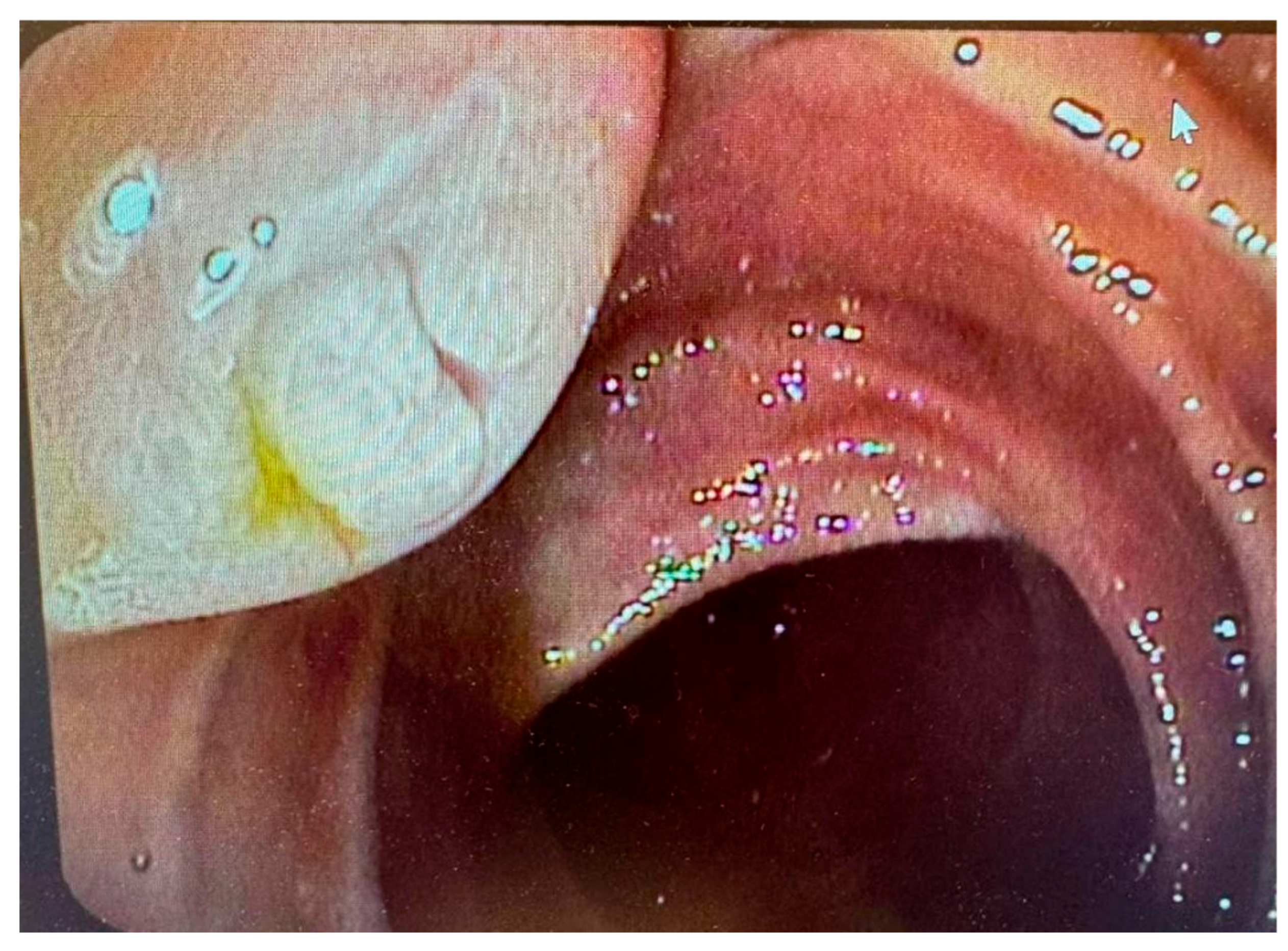
Figure 2.
IPMN in the EUS. (a) MPD dilated to 14 mm; (b) Pancreatic tumor. Source – B. Włodarczyk, Dept of Digestive Tract Diseases, Medical University of Lodz, Poland.
Figure 2.
IPMN in the EUS. (a) MPD dilated to 14 mm; (b) Pancreatic tumor. Source – B. Włodarczyk, Dept of Digestive Tract Diseases, Medical University of Lodz, Poland.
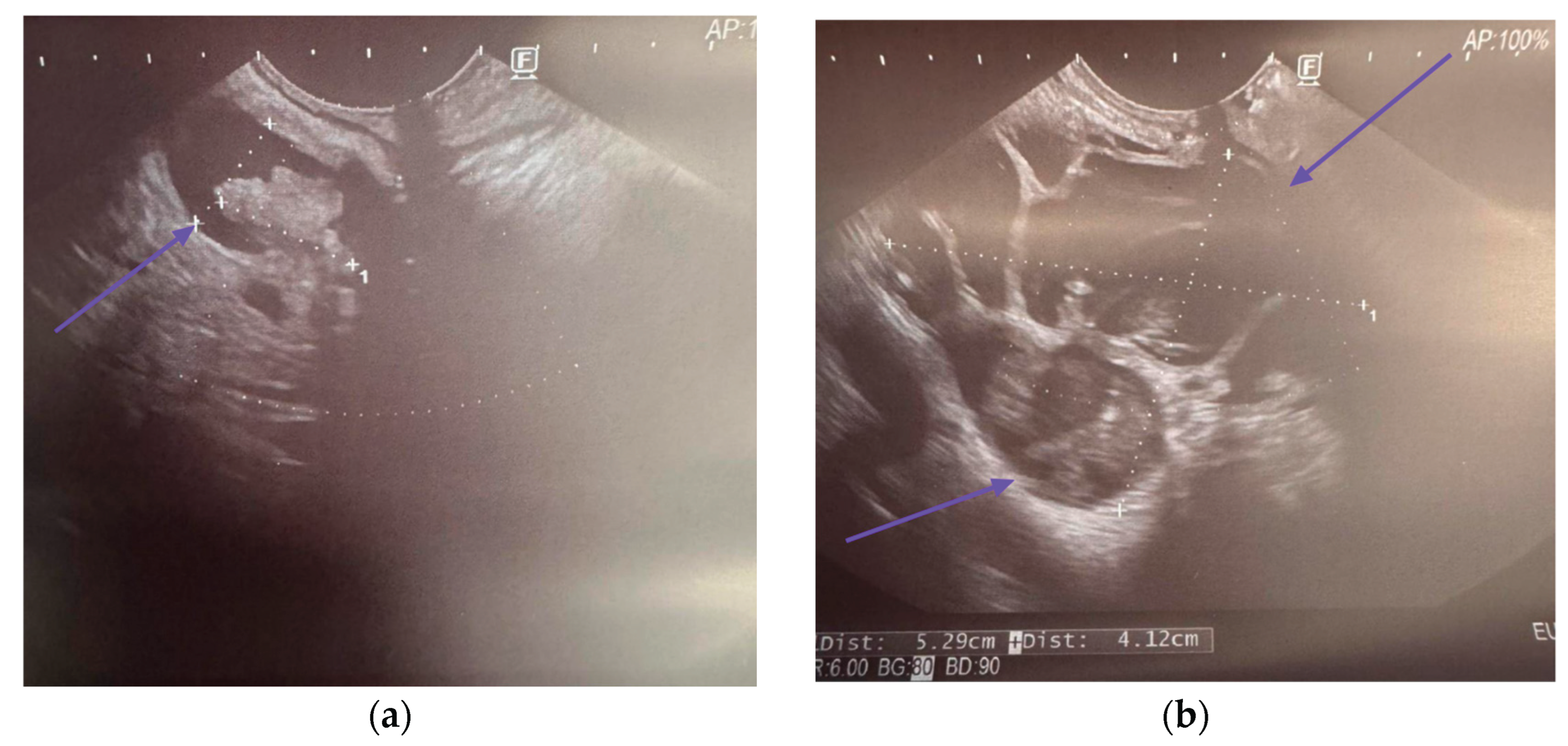

Table 1.
Comparison of commonly used techniques efficacy in IPMN diagnosis.
| Technique | Sensitivity | Specificity | AUC | PPV | NPV | References |
| MRI | 73.4-90.8% | 64.4-94.8% | 0.811-0.903 | 71% | 82.4% | [28,29,30,31,32] |
| MRCP | 38.3-94.1% | 62.5-93.1% | N/A | N/A | N/A | [28,32,33,34] |
| CT | 62-90.4% | 57-86% | 0.71-0.904 | 48-80% | 58-90% | [30,31,32,35,36,37] |
| EUS | 60% | 80% | 0.79 | N/A | N/A | [32] |
| EUS-FNA cytology | 28.7-64.8% | 84-94% | 0.84-0.94 | N/A | N/A | [38,39,40,41] |
| EUS-TTNB | 69.5% | N/A | N/A | N/A | N/A | [38] |
Table 2.
High-risk factors of malignancy of pancreatic cyst in the different guidelines with their sensitivity and specificity values in predicting malignancy of IPMN [8,22,26,47,48].
| Risk factors | Sensitivity | Specificity | Guidelines | References | |
| jaundice | 26-83% | 61-97% | IAP European ACG ACR |
[22,42] | |
| enhancing mural nodule or solid component | ≥ 5 mm | 64.6-100% | 73-87.5% | IAP European ACG* ACR* AGA* |
[22,43,49] |
| < 5 mm | N/A | N/A | IAP European ACG* ACR* AGA* |
- | |
| main pancreatic duct dilation | ≥ 10 mm | 28.2-51.7% | 78.7-87.5% | IAP European ACR AGA* |
[43,49,50] |
| ≥ 7 mm | 53.8% | 80.7% | ACR AGA* |
[51] | |
| > 5 mm | 54.7-74.8% | 58.6-78% | ACG AGA* |
[50,52] | |
| ≥ 5 mm and < 10 mm | N/A | N/A | IAP European AGA* |
- | |
| positive cytology | 28.7-64.8% | 84-94% | IAP European ACG AGA |
[38,39,40,41] | |
| acute pancreatitis | 32-42.6% | 86-86.1% | IAP European ACG |
[42,49] | |
| new-onset or worsening diabetes | 46% | 83% | IAP European ACG |
[42] | |
| increased serum level of CA19-9 | > 37 U/mL | 41-74% | 85-96% | IAP European ACG |
[22,42,49] |
| cyst diameter | ≥ 40 mm | N/A | N/A | European | - |
| ≥ 30 mm | 56.1-64% | 53.7-69% | IAP ACG ACR AGA |
[42,52] | |
| thickened/enhancing cyst walls | 23-38.5% | 89.7-95% | IAP ACR |
[42,52] | |
| abrupt change in caliber of pancreatic duct with distal pancreatic atrophy (IAP) focal dilation of pancreatic duct concerning for MD-IPMN or an obstructing lesion (ACG) |
19.3% | 95.9% | IAP ACG |
[52] | |
| lymphadenopathy | 5.2-20% | 93-99.6% | IAP | [42,52[ | |
| cystic growth rate | ≥ 5 mm/year | 56% | 97% | European | [53] |
| > 3 mm/year | N/A | N/A | ACG | - | |
| ≥ 2.5 mm/year | 60.9% | 70.3% | IAP | [52] | |
| abdominal pain | N/A | N/A | European | - | |
| non-enhancing mural nodule | N/A | N/A | ACR | - | |
* They do not specify a cut-off point for size. Abbreviations: IAP, International Association of Pancreatology; ACG, American College of Gastroenterology; ACR, American College of Radiology; AGA, American Gastroenterological Association.
Table 3.
CT role in malignancy detection in IPMN.
| Technique | Additional features assessed | Sensitivity | Specificity | Accuracy | PPV | NPV | AUC | References |
| CT | HRS, WF (2017 ICG Fukuoka) | 79.5-86% | 67.8-74% | 73.7-78% | 71.4% | 76.6% | - | [30,31] |
| CT + radiomics | - | 68-82% | 57-84% | 64-78% | 48-80% | 58-90% | 0.71-0.84 | [35,36,37] |
| HRS, WF (2017 ICG Fukuoka, 2018 European) | 69-80% | 65-72% | 67-76% | 72-78% | 61-75% | 0.75-0.83 | [35] | |
| age, cyst size, presence of solid component, symptoms, gender | 19-93% | 35-100% | 50-80% | 36-97% | 78-95% | 0.74-0.81 | [36,61] | |
| Type of IPMN, cyst size, cystic solid, CA199, CA125, bilirubin, alkaline phosphatase, gamma-ggtt, diabetes | 90.4% | 74% | 80% | - | - | 0.904 | [37] |
Disclaimer/Publisher’s Note: The statements, opinions and data contained in all publications are solely those of the individual author(s) and contributor(s) and not of MDPI and/or the editor(s). MDPI and/or the editor(s) disclaim responsibility for any injury to people or property resulting from any ideas, methods, instructions or products referred to in the content. |
© 2025 by the authors. Licensee MDPI, Basel, Switzerland. This article is an open access article distributed under the terms and conditions of the Creative Commons Attribution (CC BY) license (http://creativecommons.org/licenses/by/4.0/).
Copyright: This open access article is published under a Creative Commons CC BY 4.0 license, which permit the free download, distribution, and reuse, provided that the author and preprint are cited in any reuse.


