Preprint
Review

This version is not peer-reviewed.

Acute Promyelocytic Leukemia: Pathophysiology, Diagnosis and Clinical Management

A peer-reviewed article of this preprint also exists.

Submitted:

19 September 2025

Posted:

22 September 2025

You are already at the latest version

Abstract

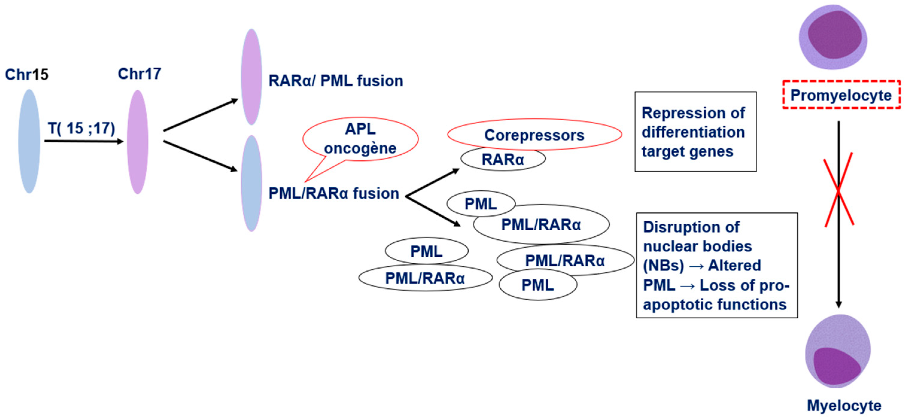
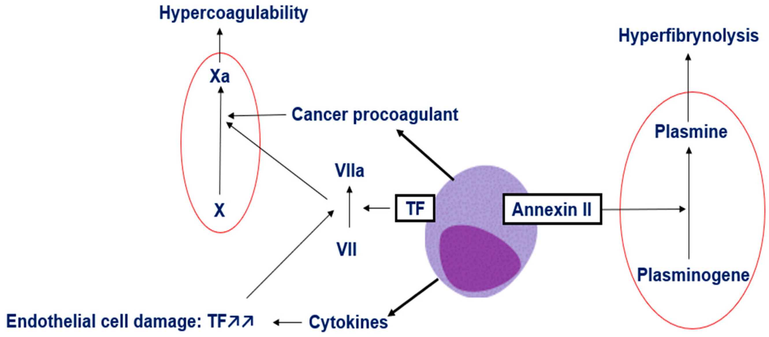
Background/Objectives: Acute promyelocytic leukemia (APL) is a distinct subtype of acute myeloid leukemia characterized by the t(15;17)(q24;q21) translocation, generating the PML-RARα fusion gene that blocks myeloid differentiation and drives leukemogenesis. Despite advances in therapy, early mortality remains a major challenge due to severe coagulopathy. This review aims to summarize recent insights into APL pathophysiology, diagnostic approaches, and management strategies. Methods: We performed a comprehensive review of the literature addressing the molecular mechanisms of APL, its associated coagulopathy, and current diagnostic and therapeutic standards, with a focus on evidence-based recommendations for clinical practice. Results: The hallmark PML-RARα oncoprotein disrupts nuclear body function and retinoic acid signaling, resulting in differentiation arrest and apoptosis resistance. APL-associated coagulopathy arises from overexpression of tissue factor, release of cancer procoagulant, inflammatory cytokines, and annexin II-mediated hyperfibrinolysis. Diagnosis requires integration of cytomorphology, immunophenotyping, coagulation studies, and molecular confirmation. Immediate initiation of all-trans-retinoic acid (ATRA) upon clinical suspicion, combined with aggressive supportive care, is critical to control bleeding risk. Conclusions: APL is now a highly curable leukemia when recognized early and treated with targeted therapy. Rapid diagnosis, prompt ATRA administration, and meticulous hemostatic support are essential to reduce early mortality. Further refinements in minimal residual disease monitoring are expected to improve patient outcomes.

Keywords: 
;  ;  ;  ;  ;  

1. Introduction

Acute promyelocytic leukemia (APL) represents between 5 and 20% of adult acute myeloid leukemia cases, with an estimated annual incidence of 1 to 7.4 cases per 1,000,000 person-years. It is characterized by a reciprocal and balanced translocation between the promyelocytic leukemia protein (PML) gene on chromosome 15 and the retinoic acid receptor α (RARα) gene on chromosome 17 [1,2]. The reciprocal t(15;17) translocation, found in more than 95% of APL cases, leads to the formation of the PML-RARα fusion protein, an oncogenic chimeric protein that plays a central role in leukemogenesis by interfering with multiple cellular pathways, resulting in increased proliferation of myeloid progenitors and a differentiation block at the promyelocytic stage [3,4].The onset of APL is usually associated with a severe thrombohemorrhagic diathesis, mainly driven by disseminated intravascular coagulation (DIC), which remains the leading cause of early mortality [5,6]. A systematic screening panel, that includes prothrombin time (PT), activated partial thromboplastin time (aPTT), D-dimers, and fibrinogen levels should be promptly implemented to aid in early diagnosis and guide the immediate initiation of supportive care aimed at preventing hemorrhagic complications [7,8,9]. Classical APL can be recognized by a distinct morphology of abnormal promyelocytes with a heavy granulation pattern and characteristic cells containing single Auer rods or bundles of Auer rods in the cytoplasm (faggot cells). Cytomorphology plays a crucial role in the early diagnostic orientation, as it often raises immediate suspicion of APL, and allows for prompt intervention. However, in certain cases, morphologic overlap with other subtypes of acute myeloid leukemia (AML) with reactive conditions may pose a diagnostic challenge, thereby necessitating additional investigations. Flow cytometry is a key complementary tool that confirms the myeloid origin of the blasts detected in peripheral blood or bone marrow aspirate. Most APL cases share a similar immunophenotype, negative for CD34 and HLA-DR, and positive for CD13, CD33, and CD117. MPO is strongly positive [10]. Of note, the immunophenotype described above is not specific to APL and can be seen in other types of AML. Ultimately, molecular confirmation through detection of the t(15;17)(q24;q21) translocation or the PML- RARα fusion transcript remains the definitive diagnostic criterion, enabling classification and initiation of targeted therapy [11]. The introduction of all-trans retinoic acid (ATRA), as well as of arsenic trioxide (ATO) in the treatment of APL, was crucial to achieving the current remarkable cure rates [12]. As opposed to the traditional cytotoxic chemotherapeutic agents conventionally used in the treatment of various cancers. This review aims to summarize recent insights into APL pathophysiology, particularly the mechanisms of coagulopathy, and to provide an updated overview of current diagnostic approaches, with a focus on APL cases harboring the PML-RARα fusion, as most clinical literature and data pertain to patients with this variant.

2. Pathophysiology of Acute Promyelocytic Leukemia

Under physiological conditions, PML, also known as TRIM19, is a nuclear protein encoded by the PML gene on chromosome 15q24. It represents the structural core of promyelocytic leukemia nuclear bodies (PML-NBs), which are dynamic, membrane-less nuclear domains involved in the regulation of essential cellular processes. PML-NBs act as multifunctional platforms that recruit and concentrate various regulatory proteins, thereby modulating apoptosis, senescence, DNA damage response and repair, telomere homeostasis, and p53-mediated stress signaling [13,14,15]. RARα acts as a ligand-dependent transcription factor that heterodimerizes with retinoid X receptors (RXR) to regulate genes involved in self-renewal and differentiation. Alongside the tumor-suppressive functions of PML, this coordinated activity preserves genomic integrity and ensures proper hematopoietic differentiation [16]. However, PML-RARα disrupts PML nuclear bodies (PML-NBs), impairing the tumor-suppressive functions of PML and promoting cell survival. Mechanistically, the fusion protein prevents proper sumoylation-dependent assembly and phase separation of PML, thereby abolishing the structural integrity of PML-NBs [13]. In parallel, the RARα moiety of the fusion protein alters normal retinoic acid signaling. Unlike the wild-type RARα, which activates the transcription of differentiation-associated genes upon ligand binding. PML-RARα aberrantly recruits transcriptional co-repressors such as nuclear receptor corepressor (N-CoR), silencing mediator of retinoid and thyroid receptors (SMRT), and histone deacetylases (HDACs) to retinoic acid response elements (RAREs), resulting in sustained transcriptional repression [16]. This silencing of myeloid differentiation programs causes a maturation arrest at the promyelocyte stage, leading to the pathological accumulation of immature blasts in the bone marrow (Figure 1).

3. Mechanisms of Coagulopathy in APL

Following vascular injury, exposure of tissue factor (TF) initiates the extrinsic pathway by binding factor VIIa, leading to activation of factor X. Together with factor V, it forms the prothrombinase complex that generates thrombin. Thrombin acts as the key effector of coagulation: it converts fibrinogen into fibrin, activates factors V, VIII, and XI, and stimulates platelet activation and aggregation, creating a procoagulant surface that stabilizes clot formation. To avoid excessive thrombosis, several anticoagulant pathways maintain balance. Thrombomodulin–thrombin complexes activate protein C, which with protein S inactivates factors Va and VIIIa, while antithrombin III neutralizes thrombin and factor Xa. In parallel, the fibrinolytic system, mediated by tissue plasminogen activator (tPA) and urokinase (uPA), produces plasmin to degrade fibrin, a process tightly regulated by inhibitors such as α2-antiplasmin and Thrombin-Activatable Fibrinolysis Inhibitor (TAFI) [17,18].
In APL, leukemic promyeloblasts exhibit markedly elevated expression of TF, primarily driven by aberrant activation of the TF promoter via the dysregulated RARα signaling pathway [19]. This overexpression of TF initiates the extrinsic coagulation cascade promoting coagulation abnormalities in APL. In addition to this TF-driven mechanism, another significant contributor to the hypercoagulable state in APL is cancer procoagulant, a cysteine protease released by tumor cells that can directly activate factor X independently of factor VII [6]. Furthermore, TF-containing microparticles (TFMPs), which are membrane vesicles shed from leukemic promyeloblasts, have been identified in APL. These microparticles express functionally active TF and are thought to amplify thrombin generation and contribute to the systemic coagulopathy observed in APL, positioning them as additional key mediators of the prothrombotic phenotype [20]. A final etiology contributing to the prothrombotic state of APL is the cancer-driven release of inflammatory cytokines that cause vascular damage and upregulation of multiple components of the coagulation cascade. The cytokines IL-1B, IL-6, and TNFα are overexpressed in patients with AML and APL. These proinflammatory cytokines further drive excess endothelial cell TF expression, and TNFα also decreases thrombomodulin transcription, leading to hypercoagulability and impaired regulation of coagulation. Unrestricted malignant promyelocyte production of TF, cancer procoagulant (contributes to Xa production), and proinflammatory cytokines (IL1β, TNFα) lead to abnormal levels of IIa, driving the development of a consumptive coagulopathy and leading to thrombosis and subsequent hemorrhage (Figure 2) [6].
Fibrinolysis play a significant role in the coagulopathy associated with APL [6]. Increased expression of annexin II, a tPA and uPA receptor and activator that is present on the surface of APL promyeloblasts, contributes to hyperfibrinolysis and drives hemorrhage [6,21]. A significantly diminished level of alpha2-antiplasmin is seen in APL, leading to an inability to counteract the APL-driven elevation in plasmin levels/activity [22]. Furthermore, multiple studies have shown that plasminogen Activator Inhibitor-1(PAI-1) activity and the formation of tPA/PAI-1 complexes were significantly reduced in APL patients via a proposed mechanism involving proteolytic degradation of PAI-1, leading to an inability to counteract APL-driven tPA hyperactivity [22]. A final potential mechanism promoting hemorrhage in APL is a deficit in primary hemostasis [21,22]. Most patients with APL are thrombocytopenic at the time of diagnosis, which may significantly contribute to early bleeding events.

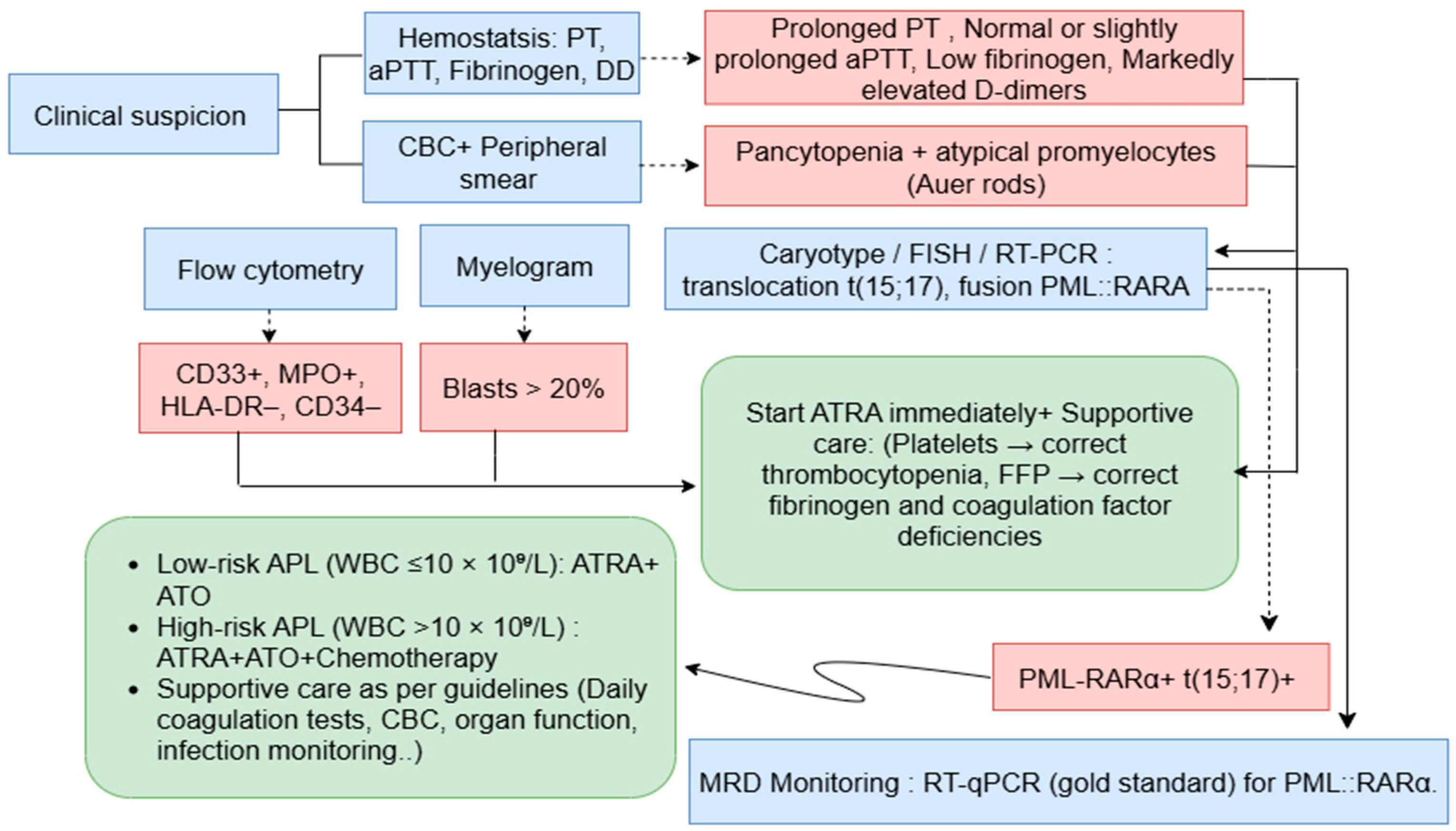
4. Acute Promyelocytic Leukemia Diagnosis

Clinical and Morphological Assessment

APL is both a diagnostic and therapeutic emergency, as patients consistently present with a clinical picture that includes coagulopathy leading to a significant risk of bleeding. A complete blood count (CBC) is usually the first step, typically revealing pancytopenia [23]. Anemia and thrombocytopenia are common, and if hemoglobin or platelet levels are dangerously low, immediate transfusions are indicated. Some cases instead present with leukocytosis, a factor that plays a critical role in risk-adapted treatment decisions. According to current recommendations, risk stratification is primarily based on the presenting white blood cell (WBC) and platelet counts: patients with WBC ≤10 ×10⁹/L are considered low- to intermediate-risk, whereas those with WBC >10 ×10⁹/L are classified as high-risk, requiring treatment intensification [24]. Bone marrow examination remains central to the morphological diagnosis. Abnormal promyelocytes usually account for >20% of marrow cells, often displaying eccentric and bilobed nuclei, folded contours, and prominent nucleoli [25]. Their cytoplasm contains irregular azurophilic granules and characteristic Auer rods, which may be seen in bundles, forming the so-called faggot cells, a hallmark of the hypergranular variant of APL [26]. In contrast, the hypogranular or microgranular variant shows a relative paucity or absence of visible cytoplasmic granules, along with bilobed or reniform nuclei [27]. This variant is frequently associated with higher leukocyte counts and may pose diagnostic challenges [28]. Rare morphological subtypes, including basophilic or atypical forms, have also been described and require careful recognition to avoid misclassification.

Immunophenotyping by Flow Cytometry

While the morphology of APL is highly characteristic and can facilitate diagnosis in many cases, reliance on morphology alone may not be sufficient in cases with atypical features. Flow cytometry immunophenotypic analysis is a routinely used tool and an indispensable component in the diagnosis, classification, prognostication, and monitoring AML subtypes, including APL. It identifies marker profiles with high diagnostic value for APL by combining positive and negative marker expression to support diagnosis. This analysis is performed on fresh bone marrow samples, which typically exhibit an immunophenotype characterized by high side scatter (SSC) and positivity for CD13, CD33, CD117, CD64, and bright MPO, along with double negative of CD34, HLA-DR [10,29]. The microgranular variant of APL usually shares a similar immunophenotype with the hypergranular form, except for the expression of CD34 [30]. Expert evaluation of this immunophenotypic pattern has been shown to effectively support the diagnosis of APL at the molecular level. Thus, adding minimal residual disease (MRD) evaluation by integrating the classic Leukemia-Associated Immunophenotype (LAIP) approach with the Different-from-Normal (DfN) approach allows for comprehensive tracing of all aberrancies at and beyond diagnosis, including newly formed postdiagnosis aberrancies. LAIPs are cells exhibiting abnormal antigen expression patterns. Examples include cross-lineage antigen expression, such as the presence of lymphoid markers CD56 and CD2 on promyelocyte blasts; asynchronous antigen expression, for instance, the coexpression of markers not usually found together during normal differentiation, like CD117 (a precursor marker) alongside CD11b/CD15 without progressive maturation; and over or underexpression of antigens compared to normal levels, such as the markedly high expression of CD33 (+++). A standard fixed monoclonal antibody panel is used to identify DfN patterns at all stages of disease and treatment using multiparametric flow cytometry (MFC). This panel typically includes CD45, CD34, HLA-DR, CD117, CD13, CD33, MPO, CD15, CD11b, and CD11c [31,32]. It is designed to comprehensively cover normal myeloid maturation, detect aberrant immunophenotypes, and monitor phenotypic shifts that may occur after treatment.

Coagulation Studies

In addition, evaluation of coagulation parameters is essential in the diagnosis and management of APL, which is frequently complicated by severe, early-onset coagulopathy. This hemostatic disturbance is primarily driven by aberrant expression of TF and annexin II on promyelocytic blasts, leading to activation of the extrinsic coagulation pathway and excessive plasmin generation. Together, these mechanisms create a highly unstable balance between bleeding and thrombosis. Routine laboratory evaluation should therefore include a full coagulation profile at baseline and serially during induction therapy. Prolongation of PT is common, reflecting depletion of extrinsic and common pathway factors (VII, X, V, II, and fibrinogen). Simultaneous activation and consumption of intrinsic pathway factors (VIII, IX, XI) result in prolonged aPTT. Low fibrinogen levels (<150 mg/dL) and markedly elevated D-dimer concentrations are characteristic of the disseminated intravascular coagulation (DIC)-like state seen in APL [33]. Recent guidelines emphasize the D-dimer/fibrinogen ratio as an additional prognostic tool, with higher ratios correlating with increased bleeding risk [6]. Thrombocytopenia further exacerbates the hemorrhagic diathesis, underscoring the importance of early and aggressive supportive care.

Molecular and Cytogenetic Analyses

Molecular genetic confirmation of the PML-RARα translocation should be obtained as quickly as possible to warrant initiation of full induction therapy. In this regard, the identification of the APL-specific genetic lesion can first be achieved by conventional karyotyping, which frequently reveals the balanced translocation t(15;17)(q24.1;q21.2), corresponding to the molecular fusion of the PML and RARα genes. However, fluorescence in situ hybridization (FISH) may be required when cytogenetic analysis is suboptimal, for example due to poor chromosome morphology or insufficient cell growth, which may result in a cryptic translocation. In addition, real-time quantitative polymerase chain reaction (RT-qPCR) allows rapid amplification of a specific cDNA segment and determination of the isoform type based on the breakpoint location (bcr1, bcr2, or bcr3). This information not only has potential prognostic value but also guides the design of primers for follow-up. Moreover, next-generation sequencing (NGS) can provide complementary insights at diagnosis by disclosing additional gene mutations beyond PML-RARα. It may also reveal an increased mutational burden, including point mutations affecting the RARα and/or PML moieties of the hybrid oncoprotein in relapsed samples. Finally, it is essential to highlight that reverse transcription polymerase chain reaction (RT-PCR) remains the gold standard [34]. This method enables the precise identification of specific RARα rearrangements and is indispensable for diagnosis, treatment monitoring, and minimal residual disease (MRD) assessment.

5. Clinical Management

A provisional diagnosis of APL based on clinical presentation and hematologic assessment is routinely available before genetic confirmation, to enable prompt initiation of supportive care. Immediate ATRA treatment which targets the Retinoic acid (RA) receptor and induces terminal differentiation of APL blasts, is critical as it is known to rapidly reduce the biologic drivers of APL-associated coagulopathy and does not have a deleterious effect in treating other AML subtypes. Supportive transfusion therapy is central to the management of APL, particularly in the early phase when life-threatening coagulopathy predominates. According to the 2022 European LeukemiaNet (ELN) guidelines, cryoprecipitate or plasma transfusions are recommended to correct hypofibrinogenemia, while platelet transfusions are indicated to counteract severe thrombocytopenia [34,35]. Both fibrinogen and platelets are essential for the initiation and propagation of clot formation; their deficiency significantly amplifies the risk of catastrophic hemorrhage, which remains the major cause of early mortality in APL. The recommended transfusion thresholds are to maintain fibrinogen concentrations above 100–150 mg/dL and platelet counts above 30,000–50,000/µL [6,36]. Close laboratory monitoring every six hours, including platelet counts, fibrinogen, PT/INR, and aPTT, is recommended to promptly detect and manage consumptive coagulopathy [6]. Pharmacologic approaches such as heparin and recombinant thrombomodulin (rTM) have been investigated for their potential to modulate coagulation, heparin through antithrombin III mediated inhibition of factors II and X, and rTM through protein C activation and annexin II downregulation. However, their routine use is not recommended due to the risk of hemorrhage and limited clinical benefit, and antifibrinolytic therapy is also discouraged outside clinical trials in the absence of robust randomized evidence. Beyond pharmacologic interventions, invasive procedures during induction should ideally be deferred until coagulation parameters have stabilized.
ATO has emerged as a cornerstone of therapy in both induction and consolidation for APL, with its use tailored according to initial risk stratification. For patients with low- or intermediate-risk disease (WBC ≤10 × 10⁹/L), the combination of ATRA and ATO is now considered the preferred frontline regimen, enabling chemotherapy-free induction and consolidation with excellent long-term outcomes, reduced treatment-related toxicity, and lower risk of secondary malignancies compared with anthracycline-based approaches. In contrast, patients with high-risk disease (WBC >10 × 10⁹/L) typically require the addition of anthracycline-based chemotherapy to ATRA and ATO, to effectively control hyperleukocytosis and reduce the risk of early death from hemorrhage or differentiation syndrome [9,37,38].
During consolidation, repeated cycles of ATRA and ATO have demonstrated sustained molecular remission rates in low- and intermediate-risk patients, while high-risk patients generally receive ATRA combined with chemotherapy, with or without ATO, depending on institutional preference and patient tolerance. Maintenance therapy, once a standard component of treatment, is no longer universally recommended in the ATO era, particularly for patients who achieve durable molecular remission with ATRA-ATO regimens. However, some centers still incorporate maintenance in high-risk settings or when ATO is not available [12,34].
Although APL is associated with high cure rates, relapse can still occur in a fraction of adult patients after achieving complete remission (CR). MRD monitoring has therefore become a cornerstone of APL management, particularly in high-risk patients, as it enables early relapse prediction and guides preemptive therapeutic interventions. Molecular relapse, indicated by rising or persistent PML-RARα transcript levels, usually precedes overt hematological relapse, which is defined by the reappearance of more than 20% blasts in a single bone marrow sample and should be confirmed through molecular testing. RT-qPCR targeting the PML-RARα transcript is widely used for MRD detection because of its high sensitivity, reproducibility, and standardization. Current guidelines recommend performing the first MRD assessment at the end of consolidation, as persistent positivity at this time strongly correlates with relapse risk. In high-risk patients, ongoing molecular monitoring every three months for 2-3 years is advised, whereas extended surveillance is generally unnecessary in low- and intermediate-risk patients who achieve molecular negativity, except in specific clinical situations [19,39].
Figure 3. Diagnostic Workflow and Early Management of APL.
Preprints 177434 g003

6. Conclusions

Acute promyelocytic leukemia represents a distinct and highly curable subtype of acute myeloid leukemia when recognized promptly and treated appropriately. Advances in understanding its unique pathophysiology, driven by the PML-RARA fusion and its downstream disruption of myeloid differentiation, have led to targeted therapies that have transformed patient outcomes. Nonetheless, early mortality remains a challenge, due to life-threatening coagulopathy, underscoring the critical importance of rapid diagnosis identify markers and/or scoring parameters to predict hemorrhagic and/or thrombotic risk, and immediate initiation of all-trans retinoic acid, based therapy. The integration of modern hematologic, cytogenetic, and molecular tools enables precise and timely diagnosis, while ongoing research into minimal residual disease monitoring holds promise for further optimizing patient management. Continued efforts to refine diagnostic algorithms, improve early detection, and expand access to specialized care are essential to sustain and extend the remarkable success achieved in APL treatment.

Author Contributions

Conceptualization: M.A. and Y.A.; Methodology: M.A. and Y.A.; Formal Analysis: M.A.; Investigation: M.A., Y.A., and I.T.; Resources: Y.A. and I.T.; Writing and original draft preparation: M.A.; Review and Editing: I.T., A.H.M., and M.A.; Supervision: A.H.M.; Project Administration: A.H.M.

Funding

This research received no external funding.

Acknowledgments

The authors would like to sincerely thank the staff of the Laboratory of Hematology at the University Hospital Hassan II of Fez for their valuable technical support and assistance.

Conflicts of Interest

The authors declare no conflicts of interest.

References

  1. Bidikian, A.; Bewersdorf, J.P.; Kewan, T.; Stahl, M.; Zeidan, A.M. Acute Promyelocytic Leukemia in the Real World: Understanding Outcome Differences and How We Can Improve Them. Cancers 2024, 16, 4092. [Google Scholar] [CrossRef]
  2. Santana-Hernández, J.; Corona-Rivera, A.; Mendoza-Maldonado, L.; Santana-Bejarano, U.F.; Cuero-Quezada, I.; Marquez-Mora, A.; Serafín-Saucedo, G.; Brukman-Jiménez, S.A.; Corona-Rivera, R.; Ortuño-Sahagún, D.; et al. Acute Promyelocytic Leukemia with <em>PML/RARA</Em> (Bcr1, Bcr2 and Bcr3) Transcripts in a Pediatric Patient. Oncol. Lett. 2024, 27, 1–8. [Google Scholar] [CrossRef]
  3. Eckardt, J.-N.; Schmittmann, T.; Riechert, S.; Kramer, M.; Sulaiman, A.S.; Sockel, K.; Kroschinsky, F.; Schetelig, J.; Wagenführ, L.; Schuler, U.; et al. Deep Learning Identifies Acute Promyelocytic Leukemia in Bone Marrow Smears. BMC Cancer 2022, 22, 201. [Google Scholar] [CrossRef]
  4. Zhang, X.; Sun, J.; Yu, W.; Jin, J. Current Views on the Genetic Landscape and Management of Variant Acute Promyelocytic Leukemia. Biomark. Res. 2021, 9, 33. [Google Scholar] [CrossRef]
  5. Xi, X.-D.; Mao, J.-H.; Wang, K.-K.; Caen, J.; Chen, S.-J. Hémorragie dans la leucémie aiguë promyélocytaire et au-delà: les rôles du facteur tissulaire et des mécanismes de régulation sous-jacents. Bull. Académie Natl. Médecine 2023, 207, 408–415. [Google Scholar] [CrossRef]
  6. Hermsen, J.; Hambley, B. The Coagulopathy of Acute Promyelocytic Leukemia: An Updated Review of Pathophysiology, Risk Stratification, and Clinical Management. Cancers 2023, 15, 3477. [Google Scholar] [CrossRef] [PubMed]
  7. Choudhry, A.; DeLoughery, T.G. Bleeding and Thrombosis in Acute Promyelocytic Leukemia. Am. J. Hematol. 2012, 87, 596–603. [Google Scholar] [CrossRef]
  8. Sabljic, N.; Thachil, J.; Pantic, N.; Mitrovic, M. Hemorrhage in Acute Promyelocytic Leukemia—Fibrinolysis in Focus. Res. Pract. Thromb. Haemost. 2024, 8, 102499. [Google Scholar] [CrossRef]
  9. Odetola, O.; Tallman, M.S. How to Avoid Early Mortality in Acute Promyelocytic Leukemia. Hematology 2023, 2023, 248–253. [Google Scholar] [CrossRef]
  10. Kankhaw, S.; Owattanapanich, W.; Promsuwicha, O.; Thong-ou, T.; Ruchutrakool, T.; Khuhapinant, A.; Paisooksantivatana, K.; Kungwankiattichai, S. Immunophenotypic Profiling of Acute Promyelocytic Leukemia: Insights From a Large Cohort. Cancer Rep. 2025, 8, e70198. [Google Scholar] [CrossRef]
  11. Klausner, M.; Stinnett, V.; Ghabrial, J.; Morsberger, L.; DeMetrick, N.; Long, P.; Zhu, J.; Smith, K.; James, T.; Adams, E.; et al. Optical Genome Mapping Reveals Complex and Cryptic Rearrangement Involving PML::RARA Fusion in Acute Promyelocytic Leukemia. Genes 2024, 15, 1402. [Google Scholar] [CrossRef]
  12. Iyer, S.G.; Elias, L.; Stanchina, M.; Watts, J. The Treatment of Acute Promyelocytic Leukemia in 2023: Paradigm, Advances, and Future Directions. Front. Oncol. 2023, 12, 1062524. [Google Scholar] [CrossRef]
  13. Voisset, E.; Moravcsik, E.; Stratford, E.W.; Jaye, A.; Palgrave, C.J.; Hills, R.K.; Salomoni, P.; Kogan, S.C.; Solomon, E.; Grimwade, D. Pml Nuclear Body Disruption Cooperates in APL Pathogenesis and Impairs DNA Damage Repair Pathways in Mice. Blood 2018, 131, 636–648. [Google Scholar] [CrossRef]
  14. Shima, Y.; Honma, Y.; Kitabayashi, I. PML-RARα and Its Phosphorylation Regulate PML Oligomerization and HIPK2 Stability. Cancer Res. 2013, 73, 4278–4288. [Google Scholar] [CrossRef] [PubMed]
  15. Tan, Y.; Li, J.; Zhang, S.; Zhang, Y.; Zhuo, Z.; Ma, X.; Yin, Y.; Jiang, Y.; Cong, Y.; Meng, G. Cryo-EM Structure of PML RBCC Dimer Reveals CC-Mediated Octopus-like Nuclear Body Assembly Mechanism. Cell Discov. 2024, 10, 118. [Google Scholar] [CrossRef]
  16. Jimenez, J.J.; Chale, R.S.; Abad, A.C.; Schally, A.V. Acute Promyelocytic Leukemia (APL): A Review of the Literature. Oncotarget 2020, 11, 992–1003. [Google Scholar] [CrossRef] [PubMed]
  17. Chaudhry, R.; Killeen, R.B.; Babiker, H.M. Physiology, Coagulation Pathways. In StatPearls; StatPearls Publishing: Treasure Island (FL), 2025. [Google Scholar]
  18. Park, S.; Park, J.K. Back to Basics: The Coagulation Pathway. Blood Res. 2024, 59. [Google Scholar] [CrossRef]
  19. Liquori, A.; Ibañez, M.; Sargas, C.; Sanz, M.Á.; Barragán, E.; Cervera, J. Acute Promyelocytic Leukemia: A Constellation of Molecular Events around a Single PML-RARA Fusion Gene. Cancers 2020, 12, 624. [Google Scholar] [CrossRef]
  20. Zhao, H.; Sun, J.; Yan, L.; Jin, B.; Hou, W.; Cao, F.; Li, H.; Zhou, J.; Zhang, Y. Tissue Factor–Bearing Microparticles Are a Link between Acute Promyelocytic Leukemia Cells and Coagulation Activation: A Human Subject Study. Ann. Hematol. 2021, 100, 1473–1483. [Google Scholar] [CrossRef]
  21. Hambley, B.C.; Tomuleasa, C.; Ghiaur, G. Coagulopathy in Acute Promyelocytic Leukemia: Can We Go Beyond Supportive Care? Front. Med. 2021, 8, 722614. [Google Scholar] [CrossRef]
  22. Sabljic, N.; Thachil, J.; Pantic, N.; Mitrovic, M. Hemorrhage in Acute Promyelocytic Leukemia—Fibrinolysis in Focus. Res. Pract. Thromb. Haemost. 2024, 8. [Google Scholar] [CrossRef]
  23. Taylor, J.; Nonas, S. A DIFFERENT KILLER: EARLY DEATH IN ACUTE PROMYELOCYTIC LEUKEMIA PRESENTING WITH SEPTIC SHOCK. CHEST 2020, 158, A903. [Google Scholar] [CrossRef]
  24. Iyer, S.G.; Elias, L.; Stanchina, M.; Watts, J. The Treatment of Acute Promyelocytic Leukemia in 2023: Paradigm, Advances, and Future Directions. Front. Oncol. 2023, 12. [Google Scholar] [CrossRef]
  25. Arber, D.A.; Orazi, A.; Hasserjian, R.P.; Borowitz, M.J.; Calvo, K.R.; Kvasnicka, H.-M.; Wang, S.A.; Bagg, A.; Barbui, T.; Branford, S.; et al. International Consensus Classification of Myeloid Neoplasms and Acute Leukemias: Integrating Morphologic, Clinical, and Genomic Data. Blood 2022, 140, 1200–1228. [Google Scholar] [CrossRef]
  26. Sehgal, T.; Sharma, P. Auer Rods and Faggot Cells: A Review of the History, Significance and Mimics of Two Morphological Curiosities of Enduring Relevance. Eur. J. Haematol. 2023, 110, 14–23. [Google Scholar] [CrossRef] [PubMed]
  27. Ladak, N.; Liu, Y.; Burke, A.; Lin, O.; Chan, A. Acute Promyelocytic Leukemia: A Rare Presentation without Systemic Disease. Hum. Pathol. Rep. 2024, 37, 300753. [Google Scholar] [CrossRef]
  28. Cheli, E.; Chevalier, S.; Kosmider, O.; Eveillard, M.; Chapuis, N.; Plesa, A.; Heiblig, M.; Andre, L.; Pouget, J.; Mossuz, P.; et al. Diagnosis of Acute Promyelocytic Leukemia Based on Routine Biological Parameters Using Machine Learning. Haematologica 2022, 107, 1466–1469. [Google Scholar] [CrossRef]
  29. Cai, Q.; Lan, H.; Yi, D.; Xian, B.; Zidan, L.; Li, J.; Liao, Z. Flow Cytometry in Acute Myeloid Leukemia and Detection of Minimal Residual Disease. Clin. Chim. Acta 2025, 564, 119945. [Google Scholar] [CrossRef]
  30. Fang, H.; Wang, S.A.; Hu, S.; Konoplev, S.N.; Mo, H.; Liu, W.; Zuo, Z.; Xu, J.; Jorgensen, J.L.; Yin, C.C.; et al. Acute Promyelocytic Leukemia: Immunophenotype and Differential Diagnosis by Flow Cytometry. Cytometry B Clin. Cytom. 2022, 102, 283–291. [Google Scholar] [CrossRef]
  31. Di Noto, R.; Mirabelli, P.; Del Vecchio, L. Flow Cytometry Analysis of Acute Promyelocytic Leukemia: The Power of ‘Surface Hematology. ’ Leukemia 2007, 21, 4–8. [Google Scholar] [CrossRef]
  32. Röhnert, M.A.; Kramer, M.; Schadt, J.; Ensel, P.; Thiede, C.; Krause, S.W.; Bücklein, V.; Hoffmann, J.; Jaramillo, S.; Schlenk, R.F.; et al. Reproducible Measurable Residual Disease Detection by Multiparametric Flow Cytometry in Acute Myeloid Leukemia. Leukemia 2022, 36, 2208–2217. [Google Scholar] [CrossRef] [PubMed]
  33. Shahmarvand, N.; Oak, J.S.; Cascio, M.J.; Alcasid, M.; Goodman, E.; Medeiros, B.C.; Arber, D.A.; Zehnder, J.L.; Ohgami, R.S. A Study of Disseminated Intravascular Coagulation in Acute Leukemia Reveals Markedly Elevated D-Dimer Levels Are a Sensitive Indicator of Acute Promyelocytic Leukemia. Int. J. Lab. Hematol. 2017, 39, 375–383. [Google Scholar] [CrossRef]
  34. De Figueiredo-Pontes, L.L.; Catto, L.F.B.; Chauffaille, M.D.L.L.F.; Pagnano, K.B.B.; Madeira, M.I.A.; Nunes, E.C.; Hamerschlak, N.; De Andrade Silva, M.C.; Carneiro, T.X.; Bortolheiro, T.C.; et al. Diagnosis and Management of Acute Promyelocytic Leukemia: Brazilian Consensus Guidelines 2024 on Behalf of the Brazilian Association of Hematology, Hemotherapy and Cellular Therapy. Hematol. Transfus. Cell Ther. 2024, 46, 553–569. [Google Scholar] [CrossRef] [PubMed]
  35. Döhner, H.; Wei, A.H.; Appelbaum, F.R.; Craddock, C.; DiNardo, C.D.; Dombret, H.; Ebert, B.L.; Fenaux, P.; Godley, L.A.; Hasserjian, R.P.; et al. Diagnosis and Management of AML in Adults: 2022 Recommendations from an International Expert Panel on Behalf of the ELN. Blood 2022, 140, 1345–1377. [Google Scholar] [CrossRef]
  36. Sanz, M.A.; Fenaux, P.; Tallman, M.S.; Estey, E.H.; Löwenberg, B.; Naoe, T.; Lengfelder, E.; Döhner, H.; Burnett, A.K.; Chen, S.-J.; et al. Management of Acute Promyelocytic Leukemia: Updated Recommendations from an Expert Panel of the European LeukemiaNet. Blood 2019, 133, 1630–1643. [Google Scholar] [CrossRef]
  37. Sanz, M.A.; Fenaux, P.; Tallman, M.S.; Estey, E.H.; Löwenberg, B.; Naoe, T.; Lengfelder, E.; Döhner, H.; Burnett, A.K.; Chen, S.-J.; et al. Management of Acute Promyelocytic Leukemia: Updated Recommendations from an Expert Panel of the European LeukemiaNet. Blood 2019, 133, 1630–1643. [Google Scholar] [CrossRef]
  38. Kantarjian, H.M.; DiNardo, C.D.; Kadia, T.M.; Daver, N.G.; Altman, J.K.; Stein, E.M.; Jabbour, E.; Schiffer, C.A.; Lang, A.; Ravandi, F. Acute Myeloid Leukemia Management and Research in 2025. CA. Cancer J. Clin. 2025, 75, 46–67. [Google Scholar] [CrossRef]
  39. Chen, L.; Zhu, H.; Dong, F.; Li, Y.; Liu, Q.; Hu, Y.; Zhang, Y.; Jin, J.; Hu, J.; Liu, T.; et al. Recommendations on Minimal Residual Disease Monitoring in Acute Promyelocytic Leukemia Treated with Combination of All- Trans Retinoic Acid and Arsenic Trioxide. Blood 2023, 142, 2946. [Google Scholar] [CrossRef]
Figure 1. Schematic representation of chromosomal translocation t (15;17) that occurs at APL: The chromosomal translocation t(15;17) generates the PML/RARα fusion protein, which disrupts regulation of differentiation target genes and alters PML nuclear bodies, leading to a block in promyelocyte maturation.
Preprints 177434 g001
Figure 2. Pathophysiology of APL-Associated Coagulopathy.
Preprints 177434 g002
Disclaimer/Publisher’s Note: The statements, opinions and data contained in all publications are solely those of the individual author(s) and contributor(s) and not of MDPI and/or the editor(s). MDPI and/or the editor(s) disclaim responsibility for any injury to people or property resulting from any ideas, methods, instructions or products referred to in the content.
Copyright: This open access article is published under a Creative Commons CC BY 4.0 license, which permit the free download, distribution, and reuse, provided that the author and preprint are cited in any reuse.
Prerpints.org logo

Preprints.org is a free preprint server supported by MDPI in Basel, Switzerland.

Subscribe

Disclaimer

Terms of Use

Privacy Policy

Privacy Settings

© 2026 MDPI (Basel, Switzerland) unless otherwise stated