Submitted:
14 September 2025
Posted:
17 September 2025
You are already at the latest version
Abstract
Hepatitis E virus (HEV) is an under-recognized cause of acute viral hepatitis and remains a significant public health issue in Africa. Globally, over 20 million HEV infections and nearly 3 million symptomatic cases happen each year, but the true burden in Africa is poorly understood because of limited surveillance, inconsistent diagnostics, and underreporting. Outbreaks have been reported in several African countries, including Sudan, Uganda, Chad, Ghana, and Nigeria, often linked to floods, displacement, and poor sanitation. Prevalence studies show significant variation, from less than 1% in Tanzania to over 80% in Egypt, indicating different epidemiological situations across the continent. In Nigeria, two major outbreaks have been documented, while seroprevalence studies suggest widespread exposure in humans and high infection rates in pigs. Vulnerable groups include pregnant women, HIV-positive individuals, and livestock handlers; these findings reflect broader trends across Africa where waterborne transmission and zoonotic reservoirs help sustain HEV circulation. The challenges in controlling HEV across Africa include weak surveillance systems, inconsistent diagnostics, limited genetic characterization of circulating strains, inadequate knowledge of risk factors, gaps in blood bank safety, uncertain zoonotic pathways, and low public awareness. Addressing these issues requires a One Health approach that integrates human, veterinary, and environmental health systems, with priority interventions including expanding affordable diagnostics, strengthening WASH infrastructure, routine animal and blood donor screening, targeted education for high-risk groups, and investment in molecular epidemiology. Therefore, this review consolidates current knowledge of HEV in Africa, with insights from Nigeria, and provides recommendations for control through a coordinated One Health approach, essential to reduce transmission, protect vulnerable groups, and build resilient surveillance systems capable of preventing future outbreaks.
Keywords:
Hepatitis E virus
; Nigeria
; one health
; zoonosis
; epidemiology
; surveillance
; prevention
Background
Hepatitis E virus (HEV) is a globally significant cause of acute viral hepatitis, accounting for an estimated 20 million infections and over 3 million symptomatic cases annually [1]. HEV-induced hepatitis manifests as either acute or chronic viral hepatitis, transmitted through the ingestion of contaminated food or water via the enteric-fecal-oral route [2]. Although previously regarded as a self-limiting disease, HEV has garnered increasing attention owing to its severe clinical implications within vulnerable populations, including pregnant women and immunocompromised individuals, as well as its capacity to induce both sporadic cases and extensive waterborne outbreaks [3]. Mortality rates generally range from 1% to 2% among the general population but can surpass 40% during third-trimester pregnancies [2,3], with numerous reports documenting vertical transmission and adverse maternal–fetal outcomes [4]. HEV belongs to the family Hepeviridae and the genus Orthohepevirus [5], and is a small, icosahedral, non-enveloped virus with a single-stranded, positive-sense RNA genome measuring approximately 27–34 nm in diameter [6,7]. Five major genotypes infect mammals and birds: genotypes 1 and 2 are restricted to humans and cause large outbreaks in regions with poor sanitation [8], while genotypes 3 and 4 circulate in various animal reservoirs—including pigs, wild boars, and deer—and occasionally spill over into humans through zoonotic transmission [9,10,11]. Genotype 5 has been detected in birds and is not considered infectious to humans [9]. This diversity highlights the dual nature of HEV as both a waterborne and zoonotic pathogen.
The prevalence of HEV across Africa varies widely with 85.1% being documented in Egypt [10], 17.05% in Algeria [11], 7.7% in Nigeria [12], 10.7% in South Africa, 2.2 - 6.8% in Morocco [13] and <1% in Tanzania [14]. HEV outbreaks has been experienced in over half of sub-Saharan countries, with most outbreaks often linked to seasonal flooding, displacement, and inadequate sanitation [15] and inaccessibility to clean water [16]. Other risk factors associated with HEV in Nigeria includes, unhygienic environment, poverty and limited healthcare access [17]. These disparities collectively emphasizes the severe public health challenge faced in several African countries [16]. In Nigeria, studies on HEV remains limited due to patient’s choice of traditional medications and self-medication [17]. Nigeria therefore exemplifies a useful case study for understanding the African HEV burden and for developing targeted One Health interventions [15]. Additionally, tackling this issue necessitates a One Health approach that integrates human, animal, and environmental health considerations. This review consolidates existing evidence on HEV epidemiology in Nigeria, highlights major challenges in surveillance, diagnostics, and prevention, and proposes strategies for adopting a One Health framework to reduce the impact of HEV-induced hepatitis.
Methods
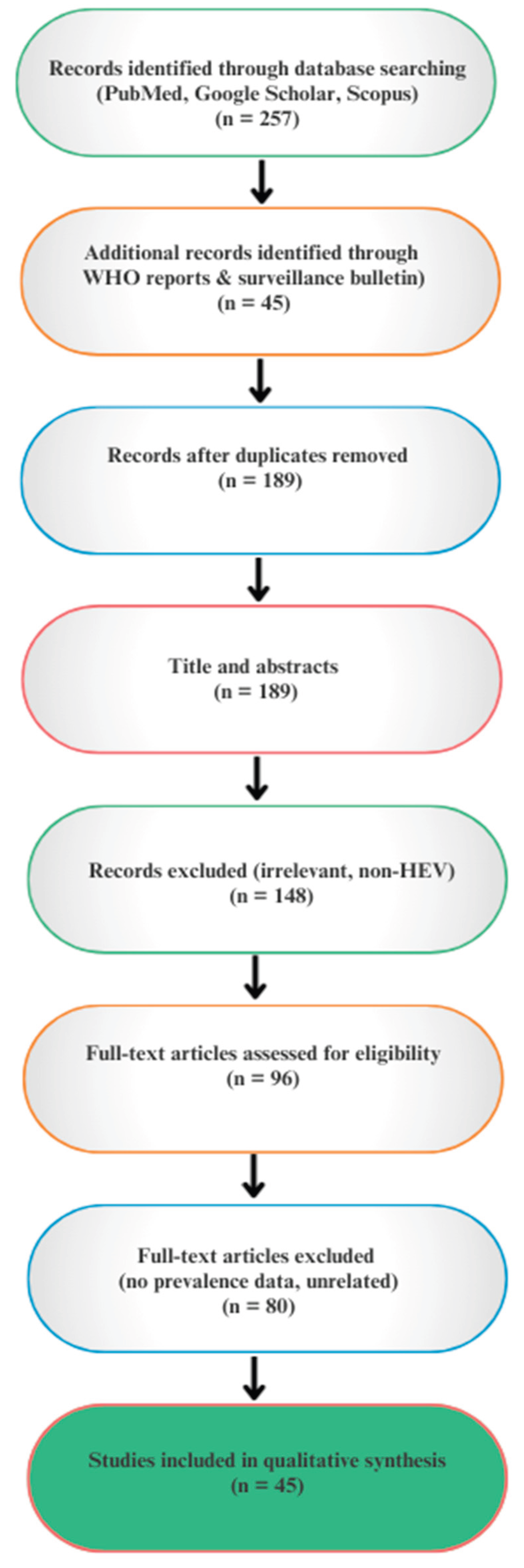
This review was developed through a systematic search of peer-reviewed literature on hepatitis E virus (HEV), with a special emphasis on Nigeria and the broader African context. Scholarly databases, including PubMed, Google Scholar, and Scopus, were queried between January and March 2025. Search terms combined medical subject headings (MeSH) and free text keywords such as: “Hepatitis E Virus,” “HEV,” “Nigeria,” “Africa,” “epidemiology,” “outbreak,” “seroprevalence,” “zoonosis,” “One Health”, and “prevalence.” Boolean operators (“AND,” “OR”) were applied to refine results.
Inclusion Criteria
- Studies published in English between 1990 and 2025.
- Articles reporting human or animal HEV prevalence, outbreak investigations, or case series from Nigeria and Africa.
- Reviews, surveillance reports, and policy papers relevant to HEV epidemiology, prevention, or One Health strategies.
Exclusion Criteria
- Studies focusing on unrelated viral hepatitis (e.g., hepatitis A, B, C, D).
- Case reports or studies lacking primary epidemiological or diagnostic data.
- Duplicated reports already included in systematic reviews unless they contributed additional local context.
Data Extraction and Synthesis
Titles and abstracts were screened for relevance, and eligible full texts were reviewed in detail. Data extracted included study year, location, sample population, diagnostic method, prevalence, and key findings. To contextualize Nigerian findings, comparative African data were included, particularly from West African countries with similar ecological and socioeconomic conditions. Where available, results were cross-referenced with WHO fact sheets, national outbreak reports, and systematic reviews
Figure 1.
PRISMA-style workflow for study selection.
Results And Discussions
- Burden of HEV in Africa
Hepatitis E virus (HEV) is widely distributed across Africa, with evidence of circulation in at least half of the continent’s countries. Prevalence rates, however, show striking variation, reflecting differences in sanitation, water quality, population health, and diagnostic methods (Table 1a & 1b). In Burkina Faso, seroprevalence rates of 10–19% have been documented among blood donors and pregnant women [13]. In adults suffering from chronic liver diseases, a seroprevalence of 14% has also been reported [18]. Similarly in Cameroon, HEV RNA has been identified in 14% of HIV-infected adults and 2% of HIV-infected children [19], whereas studies from the Central African Republic indicated a prevalence of 24% among patients attending sexually transmitted disease clinics [20]. In Djibouti, a prevalence of 13% was recorded among male peacekeepers [21]. North Africa consistently exhibits a high seroprevalence, with Egypt reporting IgG positivity rates exceeding 60–80% across various groups, including pregnant women, patients with chronic liver diseases, and residents of rural areas [22,23,24,25,26,27,28,29]. The Southern and Eastern Africa regions generally report lower prevalence trends; in Gabon, pregnant women have recorded approximately 14% prevalence, while young villagers have documented 0% prevalence [30,31]. This may suggest an enduring endemicity with continuous exposure among the general population. West Africa exhibits moderate to high prevalence levels. In Ghana, the prevalence of HEV detected by RT-PCR in HIV-positive patients was approximately 45% [19]. Seroprevalence varies from 4% to 38%, depending on the subgroup within the Ghanaian human sample population [33,34]. Serological studies conducted on pigs in Ghana revealed a prevalence rate exceeding 60% [35]. In Jordan, a prevalence ranging from approximately 8% to 14% was observed across different sampled ruminant species [36]. In Madagascar, among slaughterhouse workers, a prevalence of 14% was documented, with an even higher percentage recorded within the sampled animal population [37]. Among blood donors in Morocco, a seroprevalence of 8% was recorded [38]. For the adult population in Morocco, a prevalence of approximately 2% was reported utilizing ELISA and Western blotting techniques [39]. In Nigeria, Osundare et al. [2021] further elaborated on this data by recognizing notably high seroprevalence rates among HIV-positive individuals (11.4%), animal handlers (7.9%), and pregnant women (6.3%), thereby confirming these cohorts as high-risk categories within the nation [40]. Research involving animals confirms the significance of pigs as a reservoir. In Plateau State, Nigeria, widespread detection of HEV antibodies in pigs has been reported; however, the prevalence in goats and cattle was low or absent, indicating that swine serve as the primary zoonotic reservoir [41]. In South Africa, investigations have indicated the presence of rural-urban gradients, with a higher prevalence of HEV observed among rural populations [42,43] These findings substantiate the predominant correlation between environmental conditions, sanitation, and the prevalence of this virus. In Tanzania, a 6% prevalence rate was estimated in women aged 15-45 years [44] which is one of the lowest among university students in Africa, at 0.2% [45], indicating heterogeneity of exposure within and between regions. Studies from Tunisia, on the other hand, provide granularity by subgroup, suggesting background seropositivity in the general population and indicating endemic transmission from broader-margin surveys [45]. Poly-transfused adults [46] and Pregnant women [47] showed prevalences of approximately 28% and 12% respectively, reflecting both biological vulnerability and transfusion-related exposure risks in contexts lacking systematic screening. Central Africa data indicate moderate endemicity, whereas Zambia yielded prevalence rates of 16% in children and 42% in urban adults [48,49].
Animal studies (Table 1b) underscore the zoonotic dimension of HEV transmission in Africa. High infection rates have been observed in pigs from Madagascar (71%) [37], Ghana (62%) [35], and Nigeria (33–65%), [17,41,56] while goats, sheep, and cattle show variable or negligible infection. Detection of HEV antibodies in nonhuman primates in Cameroon further supports the existence of diverse animal reservoirs [57]. The consistently high prevalence in pigs and other animals indicates that zoonotic transmission is a widespread concern across the continent.
- 2.
- HEV Epidemiology in Nigeria
Despite repeated evidence, hepatitis E virus (HEV) remains underrecognized in Nigeria. Historically, it was overshadowed by hepatitis B and C in clinical settings, and early cases were frequently misdiagnosed or underreported. To date, only two outbreaks have been formally documented. The first occurred in Port Harcourt (1997–1998), resulting in a small cluster of 10 confirmed cases [59]. The second, a large epidemic in Bornu State (2017), affected 1,815 people with a case fatality rate of 0.6% [52]. In response, local authorities launched WASH sensitization and HEV awareness campaigns [52]. These outbreaks underscore the urgent need for preventive strategies to mitigate future epidemics, even in periods when morbidity rates appear low. The historical trajectory of HEV in Nigeria reflects broader challenges in infectious disease surveillance. Weak reporting systems are further exacerbated by climatic disruptions, especially floods and other environmental disturbances that contaminate water supplies and increase the risk of HEV transmission [52]. Improved surveillance and diagnostics, alongside recognition of HEV as a significant public health threat, have more recently led to an increase in prevalence data from Nigeria and other African countries [40].
Recent epidemiological investigations present a detailed overview of HEV prevalence across diverse Nigerian populations. Serological assessments often target IgM and IgG antibodies, which provide critical epidemiological insight. Elevated IgM levels indicate recent or active infection, while IgG positivity reflects prior exposure and immunity [56].
- Okagbue et al. (2019): Reviewing 1,178 human and 210 pig samples across a 10-year dataset, reported 10.8% prevalence in humans and a striking 65.7% in pigs [17]. Although a weak, non-significant association was found between human and animal infections (r = 0.327, p = 0.474), regression analysis (p = 0.376) explained only 9.3% of variability. The odds ratio (OR = 1.03, 95% CI: 0.95–1.20) suggested slightly higher prevalence in animals, reinforcing pigs as a key reservoir. Food and animal handlers, as well as those lacking access to clean water, were identified as primary risk groups.
- Antia et al. (2018): Reported 57.5% prevalence in pigs, but no evidence of infection in goats and cattle [60].
- Olayinka et al. (2020): Detected 13.2% prevalence in pigs using fecal samples tested by ELISA [61].
- Oluremi et al. (2023): Reported 14.9% IgG and 1.3% IgM positivity in communities in southwestern Nigeria, indicating both past exposure and ongoing active transmission [58].
- Ekiti State study: Documented an overall 13.4% antibody prevalence, consistent with findings from Oluremi et al. [62].
Together, these findings confirm that HEV is established in Nigeria, with humans exhibiting moderate exposure (6–15%) and animals, particularly pigs, demonstrating a high prevalence (>50%). The data accentuate both waterborne and zoonotic transmission pathways, indicating the highest risk among pregnant women, HIV-positive individuals, and occupationally exposed groups.
Demographic, Occupational, and Regional Patterns of HEV in Nigeria
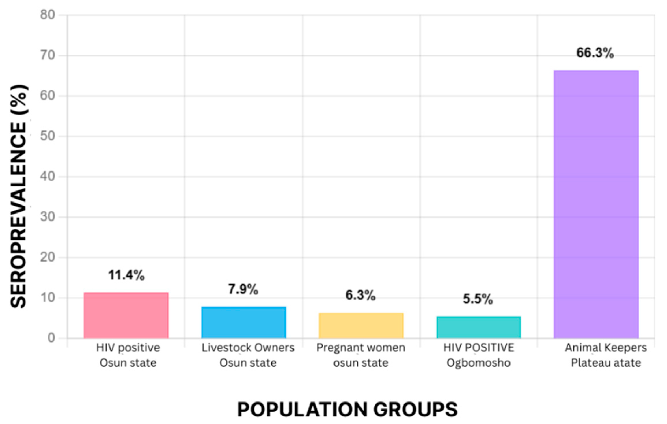
Hepatitis E virus (HEV) affects all demographic groups in Nigeria, but certain populations are disproportionately vulnerable. Residents of rural areas, HIV-positive individuals, veterinary workers, women of reproductive age, and particularly pregnant women face the greatest risk [63]. In Osun State, seroprevalence was reported at 11.4% among HIV-positive persons, 7.9% among livestock owners, and 6.3% among pregnant women [40]. Similar variability is seen elsewhere: a study in Ogbomoso found 5.5% prevalence among HIV-positive individuals [40], while Osundare et al. (2021) confirmed higher rates of 11.4% in Osun State [63]. Gender-specific differences have also been documented. In Osundare’s study of HIV-positive participants, no significant difference was observed in the overall prevalence of anti-HEV antibodies between men (13.9%) and women (10.6%). However, anti-HEV IgM prevalence was markedly higher in men (6.5%) than in women (0.7%), suggesting recent or ongoing infection may be more frequent among males [62]. Occupational risk factors are particularly evident among livestock handlers. While Osundare’s study reported a relatively modest 7.9% seroprevalence among animal keepers in Osun State [63], much higher rates were recorded in Plateau State, where animal handlers showed prevalence levels exceeding 66% [41]. Junaid et al. (2014) also reported high HEV prevalence among pig farmers in Plateau State [41], linking this to occupational exposure and regional hazards such as intensive livestock rearing, population density, and environmental conditions.
Geographic disparities across Nigeria are striking. Southern states such as Ekiti, Osun, and Ibadan show moderate prevalence, while northern and northeastern regions, including Plateau and Borno, are recognized hotspots for transmission [64]. Outbreak-prone areas in these regions often suffer from inadequate water infrastructure and poor sanitation, conditions exacerbated by seasonal flooding and displacement events that favor HEV spread [65]. The most vulnerable Nigerians remain those in rural and peri-urban communities with limited access to clean water and healthcare [63,65,66]. However, current epidemiological surveys have largely focused on the livestock industry, particularly pigs, while data on other potential animal reservoirs remain sparse. This highlights the need for broader surveillance to clarify the zoonotic landscape of HEV in Nigeria.
Figure 2.
HEV Seroprevalence Among At-Risk Populations in Nigeria.
- 3.
- Comparative Insights: Nigeria vs Africa
When positioned against broader African data:
- Nigeria’s human prevalence (10–15%) is moderate compared with Egypt (50–80%), but higher than Tanzania (0.2–6%).
- Nigeria’s pig prevalence (30–65%) is among the highest on the continent, comparable to Ghana and Madagascar.
- Occupational exposure patterns (butchers, pig handlers) [38] mirror those reported in Ghana and Cameroon, suggesting a shared West/Central African risk profile.
These comparisons reinforce the argument that Nigeria’s HEV burden is both representative of African trends and a key case study for One Health interventions.
Challenges Affecting HEV-Induced Hepatitis
Despite growing recognition of the hepatitis E virus (HEV) as a public health threat, several challenges hinder effective surveillance, prevention, and control in Nigeria and across Africa. These barriers span epidemiological, diagnostic, genetic, and socio-environmental domains.
- Underestimated and Unquantified Burden
The health impact of HEV infection remains largely underestimated, primarily due to inadequate surveillance and reporting systems. Although statistical studies suggest that more than 20 million HEV infections occur globally each year [1,9,66], this estimate may be incomplete, as there are no comprehensive data repositories for many regions [66]. Underreporting and under-documentation reflect limited awareness of HEV among healthcare providers and the absence of routine screening in healthcare facilities [9]. This oversight narrows the perception of the actual burden of HEV, reduces opportunities for preventive interventions, and impedes the development of effective control measures. Ultimately, the paucity of reliable epidemiological data makes it difficult to define the true impact of HEV in the population and complicates efforts to allocate and manage public health resources effectively [66].
- 2.
- Inconsistencies in Diagnostic Practices
Inconsistencies in diagnostic techniques for HEV remain a major challenge, often leading to misdiagnosis or delayed identification of infections. These gaps are particularly acute in developing regions, where access to sophisticated laboratory equipment is limited. Although five genotypes of HEV have been identified worldwide [8,9], the virus’s complex epidemiology [67] has hindered the establishment of universal diagnostic standards. Consequently, healthcare workers frequently struggle to diagnose and confirm HEV infections, especially in areas without well-equipped laboratories [67,68,69]. This diagnostic uncertainty contributes to delayed treatment and worsens the clinical impact of HEV. Addressing the problem requires expanding access to reliable and affordable diagnostic kits, particularly in low-resource settings. Potential strategies include the development of low-cost, efficient diagnostic procedures, training and educational programs for healthcare professionals, and the formation of international diagnostic partnerships to promote standardization and knowledge sharing [67].
- 3.
- Limited Genetic Characterization of Circulating Strains
The absence of regularly described HEV strains with well-defined genetic profiles limits the ability to assess the virus’s epidemiology. Without adequate genetic characterization, it becomes difficult to classify circulating strains accurately, which in turn hampers understanding of transmission routes, the effectiveness of control measures, outbreak management, and the evolutionary dynamics of the virus [70].
- 4.
- Insufficient knowledge of Predisposing Risk Factors
Information on the predisposing factors for HEV-induced hepatitis remains limited [66,70,69]. Significant gaps persist in understanding the drivers of HEV infection across different age groups and populations. Identifying these risk factors is critical for designing effective prevention strategies and tailoring public health interventions to protect high-risk groups [69].
- 5.
- Pathophysiology in Immunocompromised Hosts
The etiological factors and physiological mechanisms that drive chronic liver injury in HEV-infected immunocompromised patients remain poorly understood. This knowledge gap limits understanding of the natural history of HEV in this population and constrains the development of effective therapeutic interventions [71].
- 6.
- Unclear Genetic Risk Factors
The genetic determinants that predispose individuals to HEV infection and facilitate viral entry into host cells are not well understood [72]. While evidence suggests that certain host genes may mediate viral attachment and replication, the mechanisms remain unclear. This lack of knowledge represents a barrier to understanding HEV transmission dynamics and developing effective containment strategies [72].
- 7.
- Zoonotic Uncertainty
Screening blood donations for HEV is critical to reducing the risk of transfusion-transmissible infections. Effective testing of blood banks can significantly lower the public health threat associated with HEV transmission through transfusion [64,65,73]. At the same time, although HEV has been isolated from several animal species, the extent to which these animals contribute to human infection is not well defined. Further research is needed to clarify zoonotic transmission pathways, which are essential for designing effective prevention and control strategies [74].
- 8.
- Environmental Contamination
HEV in wastewater has been reported in some Nigerian communities, but the relevance of HEV wastewater pollution to humans has not been established. Knowledge of factors affecting HEV transmission is crucial for developing a comprehensive strategy in the healthcare system [75].
- 9.
- Gaps in Public Awareness and Education
Current efforts to inform the Nigerian population about HEV-induced hepatitis remain limited, inconsistent, and often ineffective. Strengthening communication systems is essential to improve public understanding and to assess community responses during outbreaks [17]. The absence of sustained educational interventions leaves both the public and healthcare workers with inadequate knowledge of HEV transmission and prevention. Well-designed awareness programs are crucial, as they can significantly reduce infection rates by improving understanding of the virus and promoting protective practices [76].
Prevention and Control of HEV-Induced Hepatitis: The One Health Approach.
The One Health idea recognizes the interdependence of human, animal, and environmental health in combating zoonotic diseases such as HEV [77,78]. Collaboration between disciplines is crucial for implementing prevention and control measures that benefit public health, veterinary health, and the physical environment [78]. A One Health approach is necessary to understand and contain HEV transmission, as HEV can be transmitted zoonotically [79] - via substances of human origin, animals, food, and the environment. They should be considered part of the One Health concept to prevent the spread of HEV from animals to humans.
- Human-Animal-Environment Interface
HEV transmission involves interactions between people, domestic animals (particularly pigs), and the environment. A thorough investigation of this interface by conceptualizing factors that enhance viral infection of human cells through exploration of the JAK-STAT signaling pathways and other signaling pathways that mediate HEV entry into hepatocytes will help to clarify the interface and mode of action [80]. A better understanding of therapeutics and antivirals can be created for effective disease management. Various high-risk practices, knowledge gaps, and misconceptions about HEV have been shown to exist among occupationally exposed groups in slaughterhouses, including slaughterhouse workers, butchers, veterinarians, and livestock handlers [46,81]. This group of people needs to be examined qualitatively and quantitatively for socio-cultural and workplace factors that influence the adoption of preventive measures, such as using PPE in their respective populations [82]. Further analysis of the main epidemiological factors triggering HEV-induced hepatitis should be conducted to understand how the season and climatic conditions of the environment strengthen or reduce the factors contributing to the transmission of viral pathogens in the most susceptible species, including humans [66]. Understanding this interface is crucial for effective disease management since previous studies by Lu and Wu (2020) confirmed the complexity of reservoirs and pollution that sustain the transmission cycle [83].
- 2.
- Integration of Epidemiological Data
Combining human health data and case reports of suspected and confirmed cases of HEV-induced hepatitis with veterinary reports from local slaughterhouses and abattoirs, as well as environmental data on weather forecasts, climatic changes, and other competing environmental hazards, contributes to a comprehensive analysis of HEV transmission dynamics [84]; Further, supporting the identification and integration of sources of infection and the assessment of risk factors. Veterinary and genomic sciences have played a critical role in disease epidemiology, with veterinary sciences focusing on screening animals while genomics elucidates genetic factors that influence susceptibility and transmission [85]. To integrate the epidemiological trend line, the veterinary and genomic approaches must be combined to monitor the trend line of disease prevalence and incidence. A holistic approach that would lead to more effective control measures.
- 3.
- Animal Screening as a Key Component of the One Health Framework
Monitor and test all livestock and domestic animals, particularly those previously reported to transmit HEV, as well as all other animals in the vicinity. Additionally, test the range of putative viral vectors that can acquire viral deposits through the fecal-oral route, as they are in susceptible proximity to humans and could potentially spread HEV infection to prevent zoonotic transmission [86]. Surveillance and screening techniques include Routine clinical examination for signs of HEV infection in animals, which can help identify symptomatic cases. Laboratory tests: Diagnostic methods such as polymerase chain reaction (PCR), enzyme-linked immunosorbent assays (ELISA), and serological tests are used to detect HEV in animal samples [87]; Risk-based assessment and targeted screening in high-risk areas or farms with known outbreaks can effectively control the spread of HEV [88]; Evidence-based case studies, particularly in countries with extensive swine industries, regions with high swine populations, such as Nigeria, to prevent transmission of infected pigs or offal to humans through undercooked pork [89].
- 4.
- Community and Veterinary Rural and Urban Extension Services
A Veterinary Medicine and Agriculture Advisory Committee should be established and adequately utilized by the government and research institutions [9,90] Research has shown that average farmers need to be more educated, especially in developing countries like Nigeria, and it is essential to ensure that they are adequately informed about the research findings [78,91]. Rural extension services that understand, interpret, translate, and disseminate research findings to rural and local farmers in diverse communities are essential in combating hepatitis caused by HEV and preventing future increases in viral pathogenesis affecting the population [92]. It is crucial to involve farmers and veterinarians in HEV surveillance and control measures. Education about proper animal care and hygiene practices can reduce the risk of HEV transmission.
Recommendations and Perspectives: A Need for a Holistic Treatment Approach
The persistence and spread of HEV in Nigeria result from a combination of environmental, socioeconomic, and infrastructural challenges. Climatic conditions, seasonal flooding, and inadequate water management frequently contaminate drinking sources, while poor sanitation infrastructure in overcrowded urban centers further drives transmission [29,67]. Compounding these issues are poverty, low educational attainment, and limited access to healthcare, all of which sustain the high prevalence of HEV [67]. Addressing these drivers requires a holistic and multi-layered response. A national strategy should prioritize the development of robust surveillance systems capable of integrating clinical, environmental, and veterinary data [90]. Such systems would enable timely detection of outbreaks and support evidence-based interventions. Parallel to this, hygiene and preventive practices—including boiling water, hand washing, and proper cooking of meat—must be promoted through sustained public health campaigns. Community education and engagement are critical to ensure these practices become part of everyday routines [93].
Policy and regulatory actions should target food safety and environmental hygiene, with governments and health authorities ensuring routine inspection of animals, as well as systematic pre- and post-slaughter data collection in abattoirs [94]. These measures, combined with stronger food safety enforcement, will reduce zoonotic transmission risks. Community-based initiatives that empower local populations to understand HEV risks and adopt preventive measures can further enhance compliance and long-term control [94]. Future directions should include advancing diagnostic capacity [85], making tools more accessible for both human and animal screening. Genomic research offers an additional frontier, with the potential to uncover genetic determinants of susceptibility and transmission that could inform targeted interventions [94]. Finally, combating HEV effectively will require strong international collaboration, partnerships among governments, global health authorities, and research institutions are vital for sharing best practices, harmonizing control measures, and strengthening collective resilience against HEV [11,94].
Limitations
We acknowledge the limitations inherent in this review methodology. Firstly, dependence on published studies excludes unpublished surveillance data, potentially leading to an underestimation of the actual HEV burden. Secondly, inconsistencies in diagnostic methods (ELISA, PCR, rapid immunoassays) across studies hinder comparability. Thirdly, Nigeria-specific data remain limited, with most studies concentrated in particular states, thereby restricting the ability to generalize findings to the entire nation. Notwithstanding these limitations, triangulating evidence from multiple sources has enabled us to delineate epidemiological trends and principal challenges pertinent to policy development and One Health interventions.
Conclusions
Hepatitis E virus (HEV) remains a neglected yet significant cause of acute viral hepatitis in Nigeria and across Africa [1,9]. Although sporadic outbreaks have highlighted the public health risk, the true burden is underestimated due to poor surveillance, inconsistent diagnostics, and widespread underreporting [11]. Seroprevalence studies show that HEV is maintained not only through contaminated water sources but also amplified by zoonotic reservoirs, particularly pigs [63,64,65]. 62=65 Pregnant women, HIV-infected individuals, and livestock handlers are the most vulnerable populations [66,68,70], emphasizing both clinical and occupational health implications. Overcoming these challenges requires moving beyond isolated interventions. A One Health approach that integrates human, veterinary, and environmental health is essential. Nigeria offers a clear case study of these issues, but its lessons resonate across Africa. The continent’s diverse yet interconnected experiences underline both the scale of the problem and the urgency for coordinated action. Strengthening HEV surveillance, diagnostics, and control within a continent-wide One Health framework will be crucial to reducing the burden of this neglected pathogen and preventing future epidemics.
Author Contribution
All Authors Contributed to the Manuscript equally
Data Availability Statement
Data is provided within the manuscript or supplementary information files.
Conflict of Interest
The authors declare no conflict of interest.
References
- WHO Health Fact Sheet, 2023. https://www.who.int/news-room/fact-sheets/detail/hepatitis-e.
- Panda, S. K., Thakral, D., & Rehman, S. (2007). Hepatitis E virus. Reviews in medical virology, 17(3), 151–180. [CrossRef]
- Purcell, R. H., & Emerson, S. U. (2008). Hepatitis E: an emerging awareness of an old disease. Journal of hepatology, 48(3), 494–503. [CrossRef]
- Patra S, Kumar A, Trivedi SS, Puri M, Sarin SK. Maternal and fetal outcomes in pregnant women with acute hepatitis E virus infection. Ann Intern Med. 2007 Jul 3;147(1):28-33. PMID: 17606958. [CrossRef]
- Waqar S, Sharma B, Koirala J. Hepatitis E. [Updated 2023 Jun 26]. In: StatPearls [Internet]. Treasure Island (FL): StatPearls Publishing; 2024 Jan-. Available from: https://www.ncbi.nlm.nih.gov/books/NBK532278/.
- Wang, B., & Meng, X. J. (2021). Structural and molecular biology of hepatitis E virus. Computational and structural biotechnology journal, 19, 1907–1916. [CrossRef]
- Yamashita T, Mori Y, Miyazaki N, Cheng RH, Yoshimura M, Unno H, Shima R, Moriishi K, Tsukihara T, Li TC, Takeda N, Miyamura T, Matsuura Y. Biological and immunological characteristics of hepatitis E virus-like particles based on the crystal structure. Proc Natl Acad Sci U S A. 2009 Aug 04;106(31):12986-91.
- Dong, C., Meng, J., Dai, X., Liang, J. H., Feagins, A. R., Meng, X. J., Belfiore, N. M., Bradford, C., Corn, J. L., Cray, C., Glass, G. E., Gordon, M. L., Hesse, R. A., Montgomery, D. L., Nicholson, W. L., Pilny, A. A., Ramamoorthy, S., Shaver, D. D., Drobeniuc, J., Purdy, M. A., … Teo, C. G. (2011). Restricted enzooticity of hepatitis E virus genotypes 1 to 4 in the United States. Journal of clinical microbiology, 49(12), 4164–4172. [CrossRef]
- Boadella, M., Casas, M., Martín, M., Vicente, J., Segalés, J., de la Fuente, J., & Gortázar, C. (2010). Increasing contact with hepatitis E virus in red deer, Spain. Emerging infectious diseases, 16(12), 1994–1996. [CrossRef]
- Saade MC, Haddad G, el Hayek M, Shaib Y. The burden of Hepatitis E virus in the Middle East and North Africa region: a systematic review. The Journal of Infection in Developing Countries [Internet]. 2022 May 30 [cited 2025 Sep 9];16(05):737–44. Available from: https://jidc.org/index.php/journal/article/view/15701.
- Boukhrissa H, Mechakra S, Mahnane A, Boussouf N, Gasmi A, Lacheheb A. Seroprevalence of hepatitis E virus among blood donors in eastern Algeria. Tropical Doctor [Internet]. 2022 Oct 1 [cited 2025 Sep 9];52(4):479–83. Available from: https://pubmed.ncbi.nlm.nih.gov/35791644/.
- Ekanem E, Ikobah J, Okpara H, Udo J. Seroprevalence and predictors of hepatitis E infection in Nigerian children. The Journal of Infection in Developing Countries [Internet]. 2015 Nov 30 [cited 2025 Sep 9];9(11):1220–5. Available from: https://jidc.org/index.php/journal/article/view/6736.
- Tucker TJ, Kirsch RE, Louw SJ, Isaacs S, Kannemeyer J, Robson SC. Hepatitis E in South Africa: Evidence for sporadic spread and increased seroprevalence in rural areas. Journal of Medical Virology [Internet]. 1996 Oct [cited 2025 Sep 9];50(2):117–9. Available from: https://pubmed.ncbi.nlm.nih.gov/8915876/.
- Kim JH, Nelson KE, Panzner U, Kasture Y, Labrique AB, Wierzba TF. A systematic review of the epidemiology of hepatitis E virus in Africa. BMC Infectious Diseases [Internet]. 2014 Jun 5 [cited 2025 Sep 9];14(1):1–13. Available from: https://bmcinfectdis.biomedcentral.com/articles/10.1186/1471-2334-14-308.
- Bagulo, H., Majekodunmi, A. O., & Welburn, S. C. (2020). Hepatitis E in Sub Saharan Africa - A significant emerging disease. One health (Amsterdam, Netherlands), 11, 100186. [CrossRef]
- Elduma AH, Zein MMA, Karlsson M, Elkhidir IME, Norder H. A Single Lineage of Hepatitis E Virus Causes Both OutbrA, Opanuga AA. Hepatitis E infection in Nigeria: a systematic review. Open Access Maced J Med Sci. 2019;7(10):1719–1722. [CrossRef]
- Okagbue HI, Adamu MO, Bishop SA, Oguntunde PE, Odetunmibi OA, Opanuga AA. Hepatitis E infection in Nigeria: a systematic review. Open Access Maced J Med Sci. 2019;7(10):1719–1722. [CrossRef]
- Aubry, P., et al. (1997) Seroprevalence of Hepatitis E Virus in an Adult Urban Popund Hygiene, 57, 272-273. [CrossRef]
- Feldt T, Sarfo FS, Zoufaly A, Phillips RO, Burchard G, van Lunzen J, Jochum J, Chadwick D, Awasom C, Claussen L, Drosten C, Drexler JF, Eis-Hubinger AM. Hepatitis E virus infections in HIV-infected patients in Ghana and Cameroon. J Clin Virol. 2013;14:18–23. [CrossRef]
- Pawlotsky JM, Belec L, Gresenguet G, Deforges L, Bouvier M, Duval J, Dhumeaux D. High prevalence of hepatitis B, C, and E markers in young sexually active adults from the Central African Republic. J Med Virol. 1995;14:269–272. [CrossRef]
- Gambel, J. M., Drabick, J. J., Seriwatana, J., & Innis, B. L. (1998). Seroprevalence of hepatitis E virus among United Nations Mission in Haiti (UNMIH) peacekeepers, 1995. The American journal of tropical medicine and hygiene, 58(6), 731-736.
- Abe K, Li TC, Ding X, Win KM, Shrestha PK, Quang VX, Ngoc TT, Taltavull TC, Smirnov AV, Uchaikin VF, Luengrojanakul P, Gu H, El-Zayadi AR, Prince AM, Kikuchi K, Masaki N, Inui A. International collaborative survey on epidemiology of hepatitis E virus in 11 countries. Southeast Asian J Trop Med Public Health. 2006;14:90–95. [PubMed] [Google Scholar].
- Fix AD, Abdel-Hamid M, Purcell RH, Shehata MH, Abdel-Aziz F, Mikhail N, el Sebai H, Nafeh M, Habib M, Arthur RR, Emerson SU, Strickland GT. Prevalence of antibodies to hepatitis E in two rural Egyptian communities. Am J Trop Med Hyg. 2000;14:519–523.
- Gad YZ, Mousa N, Shams M, Elewa A. Seroprevalence of subclinical HEV infection in asymptomatic, apparently healthy, pregnant women in Dakahlya Governorate, Egypt. Asian J Transfus Sci. 2011;14:136–139.
- Darwish MA, Faris R, Clemens JD, Rao MR, Edelman R. High seroprevalence of hepatitis A, B, C, and E viruses in residents in an Egyptian village in The Nile Delta: a pilot study. Am J Trop Med Hyg. 1996;14:554–558.
- Abdel Hady SI, El-Din MS, El-Din ME. A high hepatitis E virus (HEV) seroprevalence among unpaid blood donors and haemodialysis patients in Egypt. J Egypt Public Health Assoc. 1998;14:165–179.
- Amer AF, Zaki SA, Nagati AM, Darwish MA. Hepatitis E antibodies in Egyptian adolescent females: their prevalence and possible relevance. J Egypt Public Health Assoc. 1996;14:273–284.
- El-Esnawy NA. Examination for hepatitis E virus in wastewater treatment plants and workers by nested RT-PCR and ELISA. J Egypt Public Health Assoc. 2000;14:219–231.
- Stoszek SK, Abdel-Hamid M, Saleh DA, El Kafrawy S, Narooz S, Hawash Y, Shebl FM, El Daly M, Said A, Kassem E, Mikhail N, Engle RE, Sayed M, Sharaf S, Fix AD, Emerson SU, Purcell RH, Strickland GT. High prevalence of hepatitis E antibodies in pregnant Egyptian women. Trans R Soc Trop Med Hyg. 2006;14:95–101. [CrossRef]
- Caron M, Kazanji M. Hepatitis E virus is highly prevalent among pregnant women in Gabon, central Africa, with different patterns between rural and urban areas. Virol J. 2008;14:158. [CrossRef]
- Richard-Lenoble D, Traore O, Kombila M, Roingeard P, Dubois F, Goudeau A. Hepatitis B, C, D, and E markers in rural equatorial African villages (Gabon) Am J Trop Med Hyg. 1995;14:338–341.
- Feldt T, Sarfo FS, Zoufaly A, Phillips RO, Burchard G, van Lunzen J, Jochum J, Chadwick D, Awasom C, Claussen L, Drosten C, Drexler JF, Eis-Hubinger AM. Hepatitis E virus infections in HIV-infected patients in Ghana and Cameroon. J Clin Virol. 2013;14:18–23. [CrossRef]
- Adjei AA, Aviyase JT, Tettey Y, Adu-Gyamfi C, Mingle JA, Ayeh-Kumi PF, Adiku TK, Gyasi RK. Hepatitis E virus infection among pig handlers in Accra, Ghana. East Afr Med J. 2009 Aug;86(8):359-63. [CrossRef]
- Martinson FE, Marfo VY, Degraaf J. Hepatitis E virus seroprevalence in children living in rural Ghana. West Afr J Med. 1999;14:76–79.
- Bagulo, H., Majekodunmi, A.O., Welburn, S.C. et al. Hepatitis E seroprevalence and risk factors in humans and pig in Ghana. BMC Infect Dis 22, 132 (2022). [CrossRef]
- Obaidat, Mohammad M., and Amira A. Roess. “Individual Animal and Herd Level Seroprevalence and Risk Factors of Hepatitis E in Ruminants in Jordan.” Infection Genetics and Evolution, vol. 81, July 2020, p. 104276. [CrossRef]
- Temmam, S., Besnard, L., Andriamandimby, S. F., Foray, C., Rasamoelina-Andriamanivo, H., Héraud, J. M., Cardinale, E., Dellagi, K., Pavio, N., Pascalis, H., & Porphyre, V. (2013). High prevalence of hepatitis E in humans and pigs and evidence of genotype-3 virus in swine, Madagascar. The American journal of tropical medicine and hygiene, 88(2), 329–338. [CrossRef]
- Aamoum, A., Baghad, N., Boutayeb, H., & Benchemsi, N. (2004). Séroprévalence de l’hépatite E a Casablanca [Seroprevalence of hepatitis E virus in Casablanca]. Medecine et maladies infectieuses, 34(10), 491–492.
- Bernal, M.C., Leyva, A., Garcia, F. et al. Seroepidemiological study of hepatitis E virus in different population groups. Eur. J. Clin. Microbiol. Infect. Dis. 14, 954–958 (1995). [CrossRef]
- Osundare FA, Klink P, Akanbi OA, Wang B, Harms D, Ojurongbe O, et al. Hepatitis E virus infection in high-risk populations in Osun State, Nigeria. One Health. 2021;13:100256. [CrossRef]
- Junaid S., Agina S. E., Jaiye K., “Seroprevalence of Hepatitis E Virus Among Domestic Animals in Plateau State–Nigeria.” British Microbiology Research Journal, vol. 4, no. 8, Jan. 2014, pp. 924–34. [CrossRef]
- Tucker TJ, Kirsch RE, Louw SJ, Isaacs S, Kannemeyer J, Robson SC. Hepatitis E in South Africa: evidence for sporadic spread and increased seroprevalence in rural areas. J Med Virol. 1996;14:117–119.
- Grabow WO, Favorov MO, Khudyakova NS, Taylor MB, Fields HA. Hepatitis E seroprevalence in selected individuals in South Africa. J Med Virol. 1994;14:384–388. [CrossRef]
- Stark K, Poggensee G, Hohne M, Bienzle U, Kiwelu I, Schreier E. Seroepidemiology of TT virus, GBC-C/HGV, and hepatitis viruses B, C, and E among women in a rural area of Tanzania. J Med Virol. 2000;14:524–530.
- Ben Halima M, Arrouji Z, Slim A, Lakhoua R, Ben Redjeb S. [Epidemiology of hepatitis E in Tunisia] Tunis Med. 1998;14:129–131.
- Hannachi N, Boughammoura L, Marzouk M, Tfifha M, Khlif A, Soussi S, Skouri H, Boukadida J. [Viral infection risk in polytransfused adults: seroprevalence of seven viruses in central Tunisia] Bull Soc Pathol Exot. 2011;14:220–225. [CrossRef]
- Hannachi N, Hidar S, Harrabi I, Mhalla S, Marzouk M, Ghzel H, Ghannem H, Khairi H, Boukadida J. [Seroprevalence and risk factors of hepatitis E among pregnant women in central Tunisia] Pathol Biol (Paris) 2011;14:e115–e118. [CrossRef]
- Rezig D, Ouneissa R, Mhiri L, Mejri S, Haddad-Boubaker S, Ben Alaya N, Triki H. [Seroprevalences of hepatitis A and E infections in Tunisia] Pathol Biol (Paris) 2008;14:148–153. [CrossRef]
- Jacobs C, Chiluba C, Phiri C, Lisulo MM, Chomba M, Hill PC, Ijaz S, Kelly P. Seroepidemiology of Hepatitis E Virus Infection in an Urban Population in Zambia: Strong Association With HIV and Environmental Enteropathy. J Infect Dis. 2014;14:652–657. [CrossRef]
- Miller WC, Shao JF, Weaver DJ, Shimokura GH, Paul DA, Lallinger GJ. Seroprevalence of viral hepatitis in Tanzanian adults. Trop Med Int Health. 1998;14:757–763. [CrossRef]
- Pawlotsky JM, Belec L, Gresenguet G, Deforges L, Bouvier M, Duval J, Dhumeaux D. High prevalence of hepatitis B, C, and E markers in young sexually active adults from the Central African Republic. J Med Virol. 1995;14:269–272. [CrossRef]
- Borno State Government, Nigeria Health Sector. Northeast Nigeria Response Health Sector Bulletin #36 (November 2017). Available from: http://origin.who.int/health-cluster/countries/nigeria/Borno-Health-Sector-Bulletin-Issue36.pdf. Published 2017.
- Meldal BH, Sarkodie F, Owusu-Ofori S, Allain JP. Hepatitis E virus infection in Ghanaian blood donors – the importance of immunoassay selection and confirmation. Vox Sanguinis. 2012;14:30–36.
- Olayinka, A., Ifeorah, I. M., Omotosho, O., Faleye, T. O. C., Odukaye, O., Bolaji, O., Ibitoye, I., Ope-Ewe, O., Adewumi, M. O., & Adeniji, J. A. (2020). A possible risk of environmental exposure to HEV in Ibadan, Oyo State, Nigeria. Journal of Immunoassay & Immunochemistry, 41(5), 875–884. [CrossRef]
- Traoré, Kuan Abdoulaye, et al. “Seroprevalence of Fecal-Oral Transmitted Hepatitis a and E Virus Antibodies in Burkina Faso.” PloS ONE, vol. 7, no. 10, Oct. 2012, p. E48125. [CrossRef]
- Capai L, Masse S, Gallian P, Souty C, Isnard C, Blanchon T, et al. Seroprevalence Study of Anti-HEV IgG among Different Adult Populations in Corsica, France, 2019. Microorganisms. 2019;7(10):460. [CrossRef]
- Modiyinji AF, Amougou Atsama M, Monamele Chavely G, Nola M, Njouom R. Detection of hepatitis E virus antibodies among Cercopithecidae and Hominidae monkeys in Cameroon. J Med Primatol. 2019;48(6):364–6.
- Oluremi AS, Casares-Jimenez M, Opaleye OO, Caballero-Gomez J, Ogbolu DO, Lopez-Lopez P, et al. Butchering activity is the main risk factor for hepatitis E virus (Paslahepevirus balayani) infection in southwestern Nigeria: a prospective cohort study. Front Microbiol. 2023;14. [CrossRef]
- Buisson Y, Grandadam M, Coursaget P, Cheval P, Rehel P, Nicand E, et al. Identification of a novel hepatitis E virus in Nigeria. J Gen Virol. 2000;81(4):903–909. [CrossRef]
- Antia RE, Adekola AA, Jubril AJ, Ohore OG, Emikpe BO. Hepatitis E virus infection seroprevalence and the associated risk factors in animals raised in Ibadan, Nigeria. J Immunoassay Immunochem. 2018;39(5):509–520. [CrossRef]
- Olayinka, A., Ifeorah, I. M., Omotosho, O., Faleye, T. O. C., Odukaye, O., Bolaji, O., Ibitoye, I., Ope-Ewe, O., Adewumi, M. O., & Adeniji, J. A. (2020). A possible risk of environmental exposure to HEV in Ibadan, Oyo State, Nigeria. Journal of Immunoassay & Immunochemistry, 41(5), 875–884. [CrossRef]
- Adesina O.A., Japhet M.O., Donbraye E., Kumapayi T.E., Kudoro A. Anti hepatitis E virus antibodies in sick and healthy Individuals in Ekiti State, Nigeria. Afr. J. Microbiol. Res. 2009;3:533–536.
- Osundare FA, Klink P, Majer C, Akanbi OA, Wang B, Faber M, et al. Hepatitis E Virus Seroprevalence and Associated Risk Factors in Apparently Healthy Individuals from Osun State, Nigeria. Pathogens. 2020;9(5):392. [CrossRef]
- Abioye JOK, Anarado KS, Babatunde S. Seroprevalence of Helicobacter pylori infection among students of Bingham University, Karu in North-Central Nigeria. Int J Pathog Res. 2021;7(4):38–47. [CrossRef]
- Ashipala DO, Tomas N, Joel MH. Hepatitis E. In: Advances in human services and public health (AHSPH) book series. 2021. p. 144–156. [CrossRef]
- Webb, G. W., & Dalton, H. R. (2019). Hepatitis E: an underestimated emerging threat. Therapeutic advances in infectious disease, 6, 2049936119837162. [CrossRef]
- Raji, Y. E., Toung, O. P., Taib, N. M., & Sekawi, Z. B. (2022). Hepatitis E Virus: An emerging enigmatic and underestimated pathogen. Saudi journal of biological sciences, 29(1), 499–512. [CrossRef]
- Maehira, Y., & Spencer, R. C. (2019). Harmonization of Biosafety and Biosecurity Standards for High-Containment Facilities in Low- and Middle-Income Countries: An Approach From the Perspective of Occupational Safety and Health. Frontiers in public health, 7, 249. [CrossRef]
- Balaban, H. Y., Aslan, A. T., Akdoğan-Kittana, F. N., Alp, A., Dağ, O., Ayar, Ş. N., Vahabov, C., Şimşek, C., Yıldırım, T., Göker, H., Ergünay, K., Erdem, Y., Büyükaşık, Y., & Şimşek, H. (2022). Hepatitis E Virus Prevalence and Associated Risk Factors in High-Risk Groups: A Cross-Sectional Study. The Turkish journal of gastroenterology : the official journal of Turkish Society of Gastroenterology, 33(7), 615–624. [CrossRef]
- Pérez-Gracia, M. T., García, M., Suay, B., & Mateos-Lindemann, M. L. (2015). Current Knowledge on Hepatitis E. Journal of clinical and translational hepatology, 3(2), 117–126. [CrossRef]
- Takakusagi, S., Kakizaki, S., & Takagi, H. (2023). The Diagnosis, Pathophysiology, and Treatment of Chronic Hepatitis E Virus Infection-A Condition Affecting Immunocompromised Patients. Microorganisms, 11(5), 1303. [CrossRef]
- Realpe-Quintero, M., Montalvo, M. C., Mirazo, S., Panduro, A., Roman, S., Johne, R., & Fierro, N. A. (2018). Challenges in research and management of hepatitis E virus infection in Cuba, Mexico, and Uruguay. Revista panamericana de salud publica = Pan American journal of public health, 42, e41. [CrossRef]
- Bi, H., Yang, R., Wu, C., & Xia, J. (2020). Hepatitis E virus and blood transfusion safety. Epidemiology and infection, 148, e158. [CrossRef]
- Yugo, D. M., Cossaboom, C. M., & Meng, X. J. (2014). Naturally occurring animal models of human hepatitis E virus infection. ILAR journal, 55(1), 187–199. [CrossRef]
- Nemes, K., Persson, S., & Simonsson, M. (2023). Hepatitis A Virus and Hepatitis E Virus as Food- and Waterborne Pathogens-Transmission Routes and Methods for Detection in Food. Viruses, 15(8), 1725. [CrossRef]
- Mbachu, C. N. P., Ebenebe, J. C., Okpara, H. C., Chukwuka, J. O., Mbachu, I. I., Elo-Ilo, J. C., Ndukwu, C. I., & Egbuonu, I. (2021). Hepatitis e prevalence, knowledge, and practice of preventive measures among secondary school adolescents in rural Nigeria: a cross-sectional study. BMC public health, 21(1), 1655. [CrossRef]
- Horefti E. (2023). The Importance of the One Health Concept in Combating Zoonoses. Pathogens (Basel, Switzerland), 12(8), 977. [CrossRef]
- Eussen, B.G., Schaveling, J., Dragt, M.J. et al. Stimulating collaboration between human and veterinary health care professionals. BMC Vet Res 13, 174 (2017). [CrossRef]
- Velavan, T. P., Pallerla, S. R., Johne, R., Todt, D., Steinmann, E., Schemmerer, M., Wenzel, J. J., Hofmann, J., Shih, J. W. K., Wedemeyer, H., & Bock, C. T. (2021). Hepatitis E: An update on One Health and clinical medicine. Liver international : official journal of the International Association for the Study of the Liver, 41(7), 1462–1473. [CrossRef]
- Ezeonwumelu, I. J., Garcia-Vidal, E., & Ballana, E. (2021). JAK-STAT Pathway: A Novel Target to Tackle Viral Infections. Viruses, 13(12), 2379. [CrossRef]
- Rajendiran, S., Li Ping, W., Veloo, Y., & Syed Abu Thahir, S. (2024). Awareness, knowledge, disease prevention practices, and immunization attitude of hepatitis E virus among food handlers in Klang Valley, Malaysia. Human vaccines & immunotherapeutics, 20(1), 2318133. [CrossRef]
- George, J., Shafqat, N., Verma, R., & Patidar, A. B. (2023). Factors Influencing Compliance With Personal Protective Equipment (PPE) Use Among Healthcare Workers. Cureus, 15(2), e35269. [CrossRef]
- Lu, L., & Wu, J. (2020). "Hepatitis E Virus Transmission and Its Interactions with the Host." In Microorganisms, 8(6), 960. [CrossRef]
- Paltiel, A. D., Zheng, A., & Zheng, A. (2022). “Investing in Community Health: The Impact of Continuous Funding on Disease Management.” American Journal of Public Health, 112(3), 431-439. [CrossRef]
- Raimondo, M., et al., (2021). "Molecular Detection of Hepatitis E Virus in Pigs: A Review of Current Techniques." Veterinary Microbiology, 252, 108965.
- Kenney S. P. (2019). The Current Host Range of Hepatitis E Viruses. Viruses, 11(5), 452. [CrossRef]
- Talapko, J., Meštrović, T., Pustijanac, E., & Škrlec, I. (2021). Towards the Improved Accuracy of Hepatitis E Diagnosis in Vulnerable and Target Groups: A Global Perspective on the Current State of Knowledge and the Implications for Practice. Healthcare (Basel, Switzerland), 9(2), 133. [CrossRef]
- Dubbert, T., Meester, M., Smith, R. P., Tobias, T. J., Di Bartolo, I., Johne, R., Pavoni, E., Krumova-Valcheva, G., Sassu, E. L., Prigge, C., Aprea, G., May, H., Althof, N., Ianiro, G., Żmudzki, J., Dimitrova, A., Alborali, G. L., D’Angelantonio, D., Scattolini, S., Battistelli, N., … Burow, E. (2024). Biosecurity measures to control hepatitis E virus on European pig farms. Frontiers in veterinary science, 11, 1328284. [CrossRef]
- Augustyniak A, Pomorska-Mól M. An Update in Knowledge of Pigs as the Source of Zoonotic Pathogens. Animals. 2023; 13(20):3281. [CrossRef]
- Mohr BJ, Souilem O, Fahmy SR, et al. Guidelines for the establishment and functioning of Animal Ethics Commitees (Institutional Animal Care and Use Committees) in Africa. Laboratory Animals. 2024;58(1):82-92. [CrossRef]
- Pawlak K, Kołodziejczak M. The Role of Agriculture in Ensuring Food Security in Developing Countries: Considerations in the Context of the Problem of Sustainable Food Production. Sustainability. 2020; 12(13):5488. [CrossRef]
- Danso-Abbeam, G., Ehiakpor, D.S. & Aidoo, R. Agricultural extension and its effects on farm productivity and income: insight from Northern Ghana. Agric & Food Secur 7, 74 (2018). [CrossRef]
- Njoga, E. O., Ilo, S. U., Nwobi, O. C., Onwumere-Idolor, O. S., Ajibo, F. E., Okoli, C. E., Jaja, I. F., & Oguttu, J. W. (2023). Pre-slaughter, slaughter and post-slaughter practices of slaughterhouse workers in Southeast, Nigeria: Animal welfare, meat quality, food safety and public health implications. PloS one, 18(3), e0282418. [CrossRef]
- Zhang, XX., Jin, YZ., Lu, YH. et al. Infectious disease control: from health security strengthening to health systems improvement at global level. glob health res policy 8, 38 (2023). [CrossRef]

Table 1a.
Human HEV prevalence in African countries.
| Country | Population Studied | Year | Diagnostic Method | Prevalence | Reference |
|---|---|---|---|---|---|
| South Africa | Canoeists high-risk, medical students low-risk. | 1994 | ELISA, Western blot | 2.6%, 1.80% | [43] |
| Djibouti | Male peacekeepers in Haiti | 1995 | ELISA | 13.00% | [21] |
| Gabon | Young villagers | 1995 | ELISA | 0.00% | [31] |
| Morocco | Adults | 1995 | ELISA & Western blot | 2.20% | [39] |
| Egypt | Residents of a semiurban village in the Nile River Delta | 1996 | ELISA | 57% | [25] |
| Egypt | Healthy Females (16 - 25 years) | 1996 | ELISA | 38.90% | [27] |
| South Africa | Blacks in rural and urban area | 1996 | ELISA | 15.3%, 6.6% | [42] |
| Burundi | Adults with chronic liver disease | 1997 | ELISA | 14% | [18] |
| Central African Republic | STD clinic patients | 1997 | EIA | 24.2% | [51] |
| Egypt | Blood donors, Haemodialysis patients | 2000 | EIA | 45.2%, 39.6% | [24] |
| Tanzania | Healthy students | 1998 | ELISA | 0.20% | [49] |
| Tunisia | Elderly, children with blood disorders, donors | 1998 | - | 46%, 29.5%, 22% | [44] |
| Ghana | Teenagers | 1999 | ELISA | 4.40% | [33] |
| Egypt | Rural residents of selected countries (16-30 years) | 2000 | ELISA | >20% | [24] |
| Egypt | Rural residents of two communities 1st decade, 2nd to 8th decade | 2000 | ELISA | 60%, 76%- >60% | [27] |
| Egypt | Sewage treatment plant workers | 2000 | Serology, Nested RT-PCR | 20–40 years: 50.9% – 50.4% 41–50 years: 43.2% 51–60 years: 46.2% |
[30] |
| Tanzania | Women between 15 and 45 years | 2000 | ELISA | 6.60% | [44] |
| Morocco | Blood donors | 2004 | ELISA | 8.50% | [38] |
| Egypt | Pregnant women | 2006 | EIA | 84.3% | [29] |
| Nigeria | Internally displaced persons | 2007 | ELISA | 64% | [52] |
| Gabon | Pregnant women | 2008 | ELISA | 14.2% | [30] |
| Tunisia | Healthy persons between 16 and 25 years | 2008 | - | 4.30% | [48] |
| Ghana | Pig handlers, Pregnant women | 2009 | ELISA | 38.1%, 28.7% | [33] |
| Ghana | Blood donors | 2009 | ELISA, Western blot and Transcriptase PCR | 4.06% | [53] |
| Nigeria | Sick and healthy individuals IgG, IgM | 2009 | 14.9%, 1.3% | [54] | |
| Egypt | Asymptomatic pregnant women (HCV)+ve and -ve | 2011 | ELISA | 71.42%, 46.7% | [22] |
| Tunisia | Polytransfused patients | 2011 | - | 28.9% | [46] |
| Tunisia | Pregnant women | 2011 | - | 12.10% | [47] |
| Burkina Faso | Blood donors | 2012 | ELISA and Immunochromatography | 19.1% | [55] |
| Burkina Faso | Pregnant women | 2012 | ELISA | 11.6% | [55] |
| Cameroon | HIV-infected adults | 2013 | RT-PCR | 14.2% | [19] |
| Ghana | HIV patients | 2013 | RT-qPCR | 45.3% | [19] |
| Madagascar | Slaughterhouse workers | 2013 | ELISA | 14.10% | [37] |
| Nigeria | Butchers, farmers (Plateau State) | 2014 | ELISA | High prevalence | [41] |
| Zambia | Urban adults, children | 2014 | ELISA | 42.0%, 16.0% | [49] |
| Cameroon | HIV-infected children | 2019 | RT-PCR | 2.0% | [19] |
| Nigeria | Hospitalized, healthcare workers, children, community, food handlers. | 2019 | ELISA, PCR | 10.8% | [17] |
| Nigeria | HIV+, animal handlers, pregnant women | 2021 | ELISA, PCR | 11.4%, 7.9%, 6.3% | [40] |
| Nigeria | Animal handlers, villagers, and students (IgG, IgM) | 2023 | ELISA | 14.9%, 1.3% | [54] |
Table 1b.
Animal HEV prevalence in African countries.
| Country | Animal Species | Year | Diagnostic Method | Prevalence | Reference |
|---|---|---|---|---|---|
| Madagascar | Pigs | 2013 | ELISA, RT-PCR | 71.20% | [37] |
| Nigeria | Pigs, Goats, Sheep, Cattle | 2014 | ELISA | 32.8%, 37.2%, 10.5%, 0% | [41] |
| Nigeria | Pigs | 2018 | ELISA | 57.50% | [40] |
| Cameroon | Mandrill, Gorilla, Chimpanzee, Baboon | 2019 | ELISA | 11.1%, 14.3%, 5.9%, 8.7% | [58] |
| Nigeria | pigs | 2019 | RT-PCR | 65.70% | [17] |
| Jordan | Cows, Sheep, Goats | 2020 | ELISA | 14.5%, 12.7%, 8.3% | [36] |
| Nigeria | Pigs | 2020 | ELISA, RT-PCR | 13.20% | [56] |
| Ghana | Pigs | 2022 | ELISA | 62.40% | [35] |
Disclaimer/Publisher’s Note: The statements, opinions and data contained in all publications are solely those of the individual author(s) and contributor(s) and not of MDPI and/or the editor(s). MDPI and/or the editor(s) disclaim responsibility for any injury to people or property resulting from any ideas, methods, instructions or products referred to in the content. |
© 2025 by the authors. Licensee MDPI, Basel, Switzerland. This article is an open access article distributed under the terms and conditions of the Creative Commons Attribution (CC BY) license (http://creativecommons.org/licenses/by/4.0/).
Copyright: This open access article is published under a Creative Commons CC BY 4.0 license, which permit the free download, distribution, and reuse, provided that the author and preprint are cited in any reuse.


