Preprint
Article

This version is not peer-reviewed.

Hog1 MAP Kinase Controls Early Riboflavin Overproduction Under Combined Acidic pH and Salinity in the Yeast Debaryomyces hansenii

Submitted:

11 September 2025

Posted:

11 September 2025

You are already at the latest version

Abstract

Riboflavin (vitamin B2) is an essential precursor of flavin cofactors involved in redox metabolism, and its industrial production increasingly relies on microbial fermentation. Debaryomyces hansenii (previously as syn. Candida famata) is a halotolerant flavinogenic yeast previously exploited for riboflavin biosynthesis; however, its biotechnological application has been limited by genetic instability and poor understanding of its regulatory networks. Here, we uncover a novel role for the High Osmolarity Glycerol (HOG) pathway in riboflavin metabolism of D. hansenii. Using the first stable knockout mutant (Dhhog1Δ), we demonstrate that loss of DhHog1 triggers early, premature, and enhanced secretion of riboflavin under acidic and saline conditions, visible as a yellow fluorescent pigment in the culture medium. Accelerated riboflavin accumulation in the mutant was accompanied by altered assimilation of phosphorus, sulfur, and magnesium, but not iron, suggesting that regulation extends beyond classical iron limitation. Gene expression analyses showed consistent up-regulation of RIB1, RIB4, and RIB6 genes and derepression of the iron regulator SEF1 in Dhhog1Δ, supporting a model where DhHog1 negatively controls riboflavin biosynthesis through stress-responsive transcription factors and pseudo iron-starvation signaling. Our findings broaden the functional scope of the HOG pathway by linking osmotic stress adaptation with secondary metabolism and establish DhHog1 as a key negative regulator of early riboflavin overproduction and secretion. This work provides new insights into yeast stress-metabolism crosstalk and highlights D. hansenii as a promising platform for metabolic engineering of industrial riboflavin production.

Keywords: 
;  ;  ;  ;  

1. Introduction

Debaryomyces hansenii (previously as syn. Candida famata) is an ascomycetous yeast of the subphylum Saccharomycotina, belonging to the CTG(Ser1) clade, a group of yeasts that predominantly translate the CTG codon as serine instead of leucine (Krassoswki, et al., 2018; Ochoa-Gutiérrez et al., 2022; Prista et al., 2016). One of its most studied traits is halotolerance, as it can grow in media containing up to 4 M NaCl (Onishi, 1963), and is considered an extremotolerant yeast (González et al., 2025; Segal-Kischinevzky et al., 2022). This ability is largely mediated by the High Osmolarity Glycerol (HOG) pathway, a phosphorylation cascade that activates Hog1 MAP kinase (Bansal and Mondal, 2000; Sánchez et al., 2020). Phosphorylated Hog1 orchestrates adaptive responses, promoting glycerol accumulation and regulating osmoprotective genes (de la Fuente-Colmenares et al., 2024; Hohmann, 2002, 2009; Posas et al., 1996; Sánchez et al., 2020).
Compared with its close relative, Candida albicans, the production of stable mutants in D. hansenii has been hindered by a low occurrence of homology-directed repair of its DNA, as well as the scarcity of optimized genetic tools geared towards the modification of this specific yeast, due in part to the ambiguous translation of the CUG codon characteristic of the yeasts belonging to the CTG clade (Gomes et al., 2007; Minhas et al., 2009; Ochoa-Gutierrez et al., 2022; Richard et al., 2005). The first stable null mutant, Dhhog1Δ, was developed for the further study of D. hansenii’s HOG pathway (Sánchez et al., 2020).
In addition to halotolerance, D. hansenii is a flavinogenic yeast, capable of synthesizing and secreting riboflavin (RF; vitamin B2) (Gadd and Edwards, 1986). RF is a precursor of the flavin coenzymes FAD and FMN, which are essential for multiple physiological processes, including redox homeostasis, protein folding, DNA repair, fatty acid β-oxidation, amino acid oxidation, and choline metabolism (Baron and Haylemon, 1995; Bray et al., 1964; Frerman, 1988; Mansoorabadi et al., 2007; Macheroux et al., 2011; Walsh & Wencewicz, 2013). Unlike plants, fungi, and most prokaryotes, animals cannot synthesize riboflavin and must obtain it from their diet or, to a lesser extent, gut microbiota (LeBlanc et al., 2013; Nysten & Van Dijck, 2023). Therefore, industrial-scale production of riboflavin is of great importance. Biological synthesis has replaced chemical synthesis due to higher efficiency, reduced waste, lower energy requirements, and use of renewable substrates such as sugars or vegetable oils (Vandamme, 1992; Stahmann et al., 2000; Lim et al., 2001; Abbas & Sibirny, 2011; Schwechheimer et al., 2016). The global riboflavin market is projected to be valued at USD 508.6 million in 2025, with steady growth expected to reach USD 872.24 million by 2033, representing a compound annual growth rate (CAGR) of 6.98% during this period (Sharma, 2025).
The first report of D. hansenii’s flavinogenic capacity was made by Gadd and Edwards (1986), when they noticed a yellow pigment in the supernatant of iron-depleted media. Riboflavin (vitamin B2) is a yellow, water-soluble compound whose green fluorescence can be detected within 440/535 nm excitation/emission wavelengths. Since then, iron limitation has become one of the most commonly used conditions in flavinogenic media, among others such as exposure to copper, cobalt, zinc, chromium, and industrial protein-rich wastewaters (Fedorovich et al., 1999; Fedorovych et al., 2001; Seda-Miró et al., 2007).
The biosynthesis pathway of riboflavin has been elucidated in the D. hansenii synonym species C. famata. The pathway, which includes six RIB genes, involves sequential dephosphorylation and reduction reactions that convert GTP and ribulose-5-phosphate into riboflavin through several intermediates (Figure 1). Key enzymes include GTP cyclohydrolase II (Rib1), DArPP deaminase (Rib2), 6,7-dimethyl-8-ribityllumazine synthase (Rib4), riboflavin synthase (Rib5), 4-dihydroxy-2-butanone-4-phosphate synthase (Rib6), and 5-amino-6-(5-phosphoribosylamino) uracil reductase (Rib7) (Bacher et al., 2001; Voronovsky et al., 2004). In C. famata, the overexpression of RIB1 and RIB6 has proven effective for the overproduction of riboflavin (Petrovska et al., 2022).
Unlike S. cerevisiae, yeasts belonging to the CTG clade possess a negative feedback loop involved in both iron and riboflavin metabolism. Sef1 is a transcriptional activator of iron acquisition and riboflavin biosynthesis genes that remains repressed by Sfu1 in iron-replete media (Andreieva et al., 2020a, 2020b; Chen et al., 2011; Demuyser et al., 2020).
Iron limitation has been framed as a necessary condition to induce the excretion of riboflavin in most yeasts (Ruchala et al., 2025). In Ashbya gossypii, the induction of flavinogenesis has been linked to oxidative stress, UV-light exposure, sporulation, and endoplasmic reticulum stress (Kurabayashi et al., 2025; Nieland and Stahmann, 2013; Ruchala et al., 2025; Silva et al., 2018; Stahmann et al., 2001). In other yeasts, including Candida albicans, Cryptococcus neoformans, and Saccharomyces cerevisiae, Hog1 has been identified as a negative regulator of iron assimilation (Kaba et al., 2013; Lee et al., 2014; Martins et al., 2018). However, a direct connection between Hog1 signaling and riboflavin biosynthesis has not yet been established.
This study aims to elucidate the role of DhHog1 in riboflavin metabolism under saline stress, integrating genetic, biochemical, and physiological approaches to uncover the interplay between osmotic stress signaling and flavinogenesis in D. hansenii.

2. Materials and Methods

2.1. Strains and growth conditions

Precultures of D. hansenii wild-type strain CBS767 (WT) and the isogenic mutant Dhhog1Δ (generated by Sánchez et al., 2020) were grown and maintained on Yeast Peptone Dextrose agar (YPD agar; 1% yeast extract, 2% peptone, 2% glucose, and 2% agar). Liquid precultures were prepared in YPD medium using a flask-to-medium ratio of 2:5, incubated at 28 °C with shaking at 180 rpm overnight, representing the seed condition (time 0 h). Cells were recovered by centrifugation at 1,100 × g for 5 min, washed twice, and resuspended in sterile distilled water. Subsequently, cultures were grown in a minimal medium based on Yeast Nitrogen Base (YNB) supplemented with 2% glucose, 0.5% ammonium sulfate as the sole nitrogen source, and 0.6 M NaCl (from now on referred to as MM + 0.6 M NaCl).

2.2. Growth curves and pH measurements

Washed preculture cells were used as an inoculum for the minimum medium at an initial optical density of 0.05 (measured at 600 nm, OD600nm). Successive OD600nm measurements were taken using a Beckman Coulter DU® 640 spectrophotometer. For the flavinogenic condition (MM + 0.6 M NaCl, initial pH 4.3), the exponential growth phase (Log) was defined at OD600nm = 0.5 (18–20 h), whereas the stationary growth phase (Stat) was defined at OD600nm = 3.8–4.0 (48 h).
For pH experiments, the basal pH of the minimal medium YNB (initially 4.3) was adjusted to 6.8–7.0 using 2 N NaOH. pH measurements were carried out at defined time intervals with a potentiometer (pH 210, Microprocessor pH Meter, Hanna Instruments), previously calibrated according to the manufacturer’s instructions.

2.3. Riboflavin measurements

Extracellular riboflavin was quantified by measuring fluorescence intensity at emission/excitation wavelengths of 440/535 nm. A riboflavin stock solution (20.1 mg/L) was prepared by dissolving riboflavin powder in distilled water under mild heating (< 30 °C). Culture aliquots (15 mL) were centrifuged, and supernatant was filtered through 0.22 μm pore-size filters. Subsequently, 200 μL of the filtered supernatants were transferred to black 96-well Costar® assay plates with clear flat bottoms, and fluorescence was measured with a Synergy H1 Microplate Reader (Biotek), gain = 50. Riboflavin concentrations were determined by interpolation against a standard curve generated from 1:2 serial dilutions of an initial 10.05 mg/L riboflavin solution.

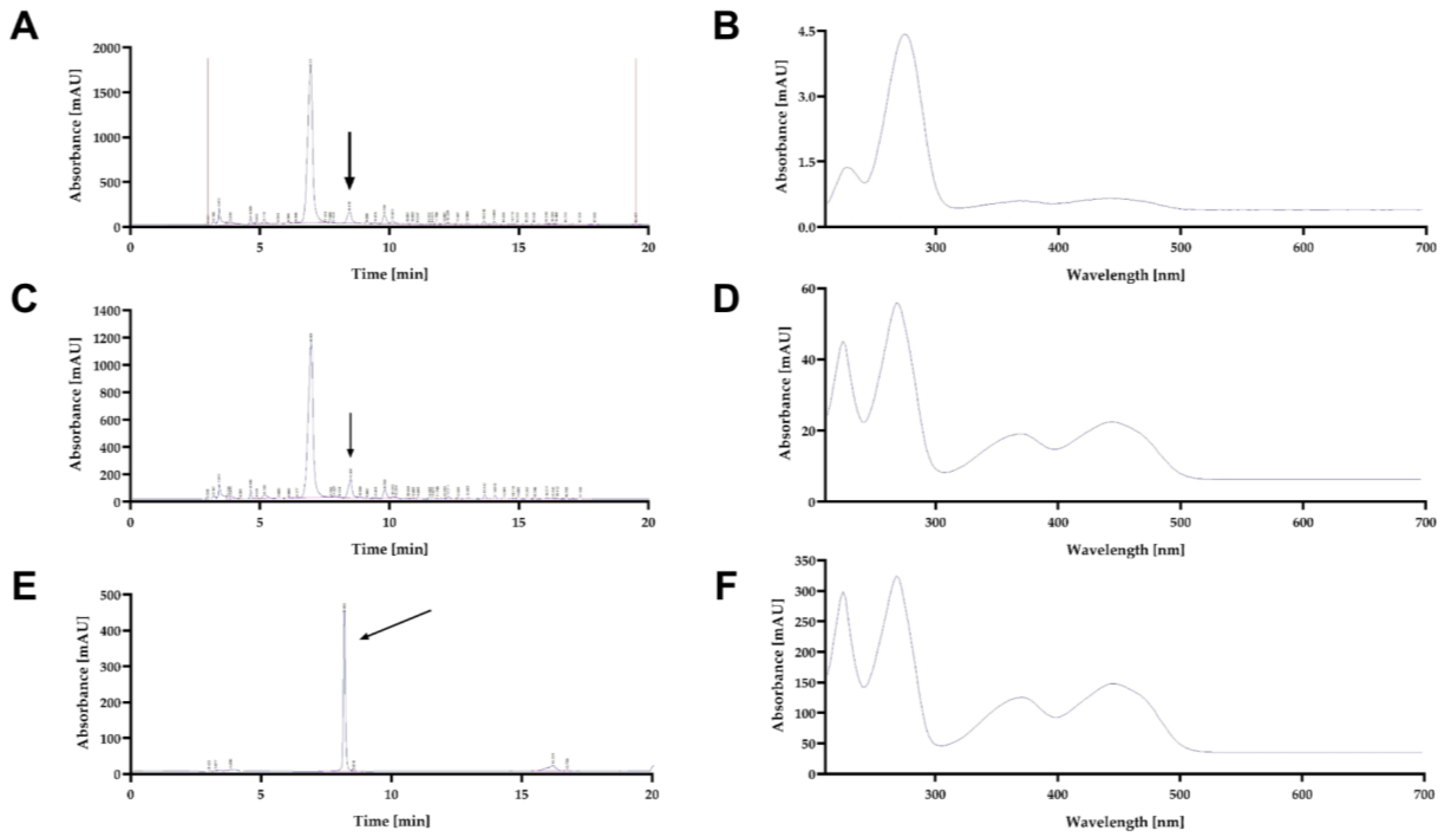
2.4. Riboflavin extraction and RP-HPLC-DAD analysis

Sterile culture supernatants were lyophilized to concentrate the fluorescent compound and remove water. The resulting dried powder was extracted with 50 mL HPLC-grade methanol and vortexed for 5 min at room temperature (25 ± 1.0 °C). Extracts were centrifuged at 3500 rpm for 10 min, and the methanolic phase was recovered and dried at 35 °C for 72 h. Reversed-phase high-performance liquid chromatography (RP-HPLC) coupled with a Diode Array Detector (DAD), was then carried out, following the method described by Jiménez-Nava et al. (2023). Methanolic residues were resuspended in 10 mL HPLC-grade water, and 2 mL aliquots were loaded in triplicate onto C18 solid-phase extraction (SPE) cartridges (Alltech® Maxi-Clean™, Thermo Fisher Scientific, Waltham, MA, USA). Cartridges were eluted under vacuum with 1.2 mL of HCl-acidified methanol (0.002% v/v). Eluates were analyzed using an Agilent 1260 Infinity Series system equipped with a DAD, monitoring 280 and 440 nm, with a Zorbax SB-Phenyl column (150 mm × 4.6 mm, 5 μm, Agilent Technologies, Santa Clara, CA, USA). The mobile phase was acetonitrile and 0.05% phosphoric acid in water at 0.5 mL/min, applying a linear gradient of 10-100% acetonitrile over 15 min.
Absorption spectra of the HPLC-separated fractions were obtained directly from DAD data and visualized with Agilent ChemStation software, using a riboflavin standard (Sigma-Aldrich, Cat. No. 47861) for reference.

2.5. Element analysis

Supernatants were obtained by centrifuging cultured cells at 18 and 48 h, and 15 mL were collected for analysis. A total of 43 elements and 1 compound were determined by Inductively Coupled Plasma-Optical Emission Spectroscopy (ICP-OES), including Ag, Al, Ar, B, Ba, Be, Bi, Br, Ca, Cd, Cl, Co, Cr, Cs, Cu, F, Fe, Ga, Hg, I, In, K, La, Li, Mg, Mn, Mo, Na, Ni, P, Pb, PO4, S, Sb, Sc, Se, Si, Sn, Sr, Te, Ti, V, W, and Zn. Quantification was performed with the proprietary Triton ICP-OES Test, a validated and patented protocol of TRITON Applied Reef Bioscience (https://www.triton-lab.de/en/icp-oes).

2.6. RNA extraction

Total RNA was extracted following the protocol described by Schmitt et al. (1990). Cultures were first grown overnight in rich medium (YPD) to obtain seed cultures (time 0 h), and subsequently inoculated into minimal medium with 0.6 M NaCl. Cells were harvested at exponential phase and stationary phase.
Briefly, cells were washed twice, centrifuged at 1,100 × g, and mechanically disrupted using a vortex mixer with sterile glass microbeads (425–600 μm) previously incubated in phenol (pH 4.5). Samples were incubated at 65 °C for 5 min and vortexed twice for 30 s. The mixture was chilled and centrifuged to separate the aqueous and phenolic phases. The aqueous phase was extracted twice with phenol:chloroform:isoamyl alcohol (25:24:1) and chloroform:isoamyl alcohol (24:1). RNA was precipitated by adding 1/10 volumes of sodium acetate (3 M, pH 5.2) and 2.5 volumes of absolute ethanol, followed by incubation at −20 °C for 30 min and centrifugation at 16,000 × g. The resulting pellet was washed, dried, and resuspended in RNase-free water. RNA integrity was assessed by electrophoresis on a 1% denaturing agarose gel.

2.7. Identification of riboflavin biosynthesis genes

The D. hansenii genome remains only partially annotated, with most gene assignments based on in silico predictions of orthologous genes. To validate these annotations, multiple sequence alignments of selected riboflavin biosynthesis proteins and the transcription factor Sef1 were carried out against their orthologous in Saccharomyces cerevisiae and Candida albicans (Supplementary Figures S1-S14). Amino acid sequences were further compared to confirm that the predicted ORFs corresponded to the expected protein functions (RIB1, locus DEHA2A12870g; RIB2, locus DEHA2E11374g, RIB4, locus DEHA2D04180g; RIB5, locus DEHA2D13926g; RIB6, locus DEHA2G09504g; RIB7, locus DEHA2G10010g; SEF1, locus DEHA2C16676g). Protein identifiers (IDs) were retrieved from the NCBI Protein Database. Both analyses were performed within the NCBI BlastP Tool.

2.8. In silico predictions of transcription factor binding sites

The intergenic region spanning from −625 bp upstream to the start codon of each gene was scanned with position-specific scoring matrices (PSSMs) using the Regulatory Sequence Analysis Tools (RSAT) platform (https://rsat.france-bioinformatique.fr/fungi/), employing the full matrix-scan tool within the Fungi Server (Turatsinze et al., 2008). Binding motifs for Hog1-regulated transcription factors were identified, including Sef1 (ID: 2900038, locus DEHA2C16676g), Sko1 (ID: 2901375, locus DEHA2D09196g), Skn7 (ID: 2913769, locus DEHA2B08052g), Msn2/4 (ID: 2899670, locus DEHA2A08382g), and Yap1 (ID: 2904514, locus DEHA2G02420g).
Except for Sef1, motif matrices were retrieved from the JASPAR database, specifically from the 2024 core fungi collection: Sko1 (ID: MA0382.1), Skn7 (ID: MA0381.1), Msn2/4 (ID: MA0341.1/MA0342.1), and Yap1 (ID: MA0415.1). Putative Sef1 binding sites were searched manually in each intergenic region based on the DNA recognition motifs identified by Chen et al. (2011) for C. albicans Sef1. A D. hansenii-specific background model estimation method with a Markov order of 1 was applied, and a significance threshold of p-values < 0.0006 was used for significant determination.

2.9. Analysis of gene expression

Total RNA was treated with DNaseI (RQ1 RNase-Free DNase, Promega) to eliminate contaminating genomic DNA. cDNA synthesis reactions were performed using the RevertAid H Minus First Strand cDNA Synthesis Kit (Thermo Scientific, Waltham, MA, United States) with oligo(dt) primers, following the manufacturer’s recommendations.
Quantitative PCR (RT-qPCR) was performed using the standard curve method with gene-specific deoxyoligonucleotides designed for the genes encoding Hog1 MAPK (DhHOG1), sugar transporter-like protein (DhSTL1), the transcriptional activator Sef1 (DhSEF1), GTP cyclohydrolase II (DhRIB1), DArPP deaminase (DhRIB2), 6,7-dimethyl-8-ribityllumazine synthase (DhRIB4), riboflavin synthase (DhRIB5), 4-dihydroxy-2-butanone-4-phosphate synthase (DhRIB6), 5-amino-6-(5-phosphoribosyl-amino) uracil reductase (DhRIB7). The actin gene (DhACT1) was used as the housekeeping control.
Deoxyoligonucleotides were evaluated to ensure the absence of dimer formation and cross-hybridization, and only deoxyoligonucleotides pairs with amplification efficiencies >90% were used (Table 1). RT-qPCR was performed with a Rotor-Gene Q thermocycler (Qiagen) using SYBR Green as the detection dye (KAPA SYBR Fast Kit, Roche). Cycling conditions were: 94 °C for 5 min (1 cycle), followed by 35 cycles of 94 °C for 15 s, 60 °C for 20 s, and 72 °C for 20 s.
Relative transcript levels were normalized to the WT mean Ct for each gene using the 2^−ΔΔCt method, and data were expressed as fold change values. Results represent mean ± SD from three biological replicates, each analyzed in duplicate (technical replicates).

2.10. Statistical analysis

For all in vitro experiments, statistical significance was evaluated using Student’s t-test in GraphPad Prism 10.2.3 (GraphPad Software Inc.). A p-value < 0.05 was considered statistically significant. For in silico predictions of transcription factor binding sites (TFBS), a significance threshold of p-values ≤ 0.0006 was considered for the selection of riboflavin-related genes.

3. Results

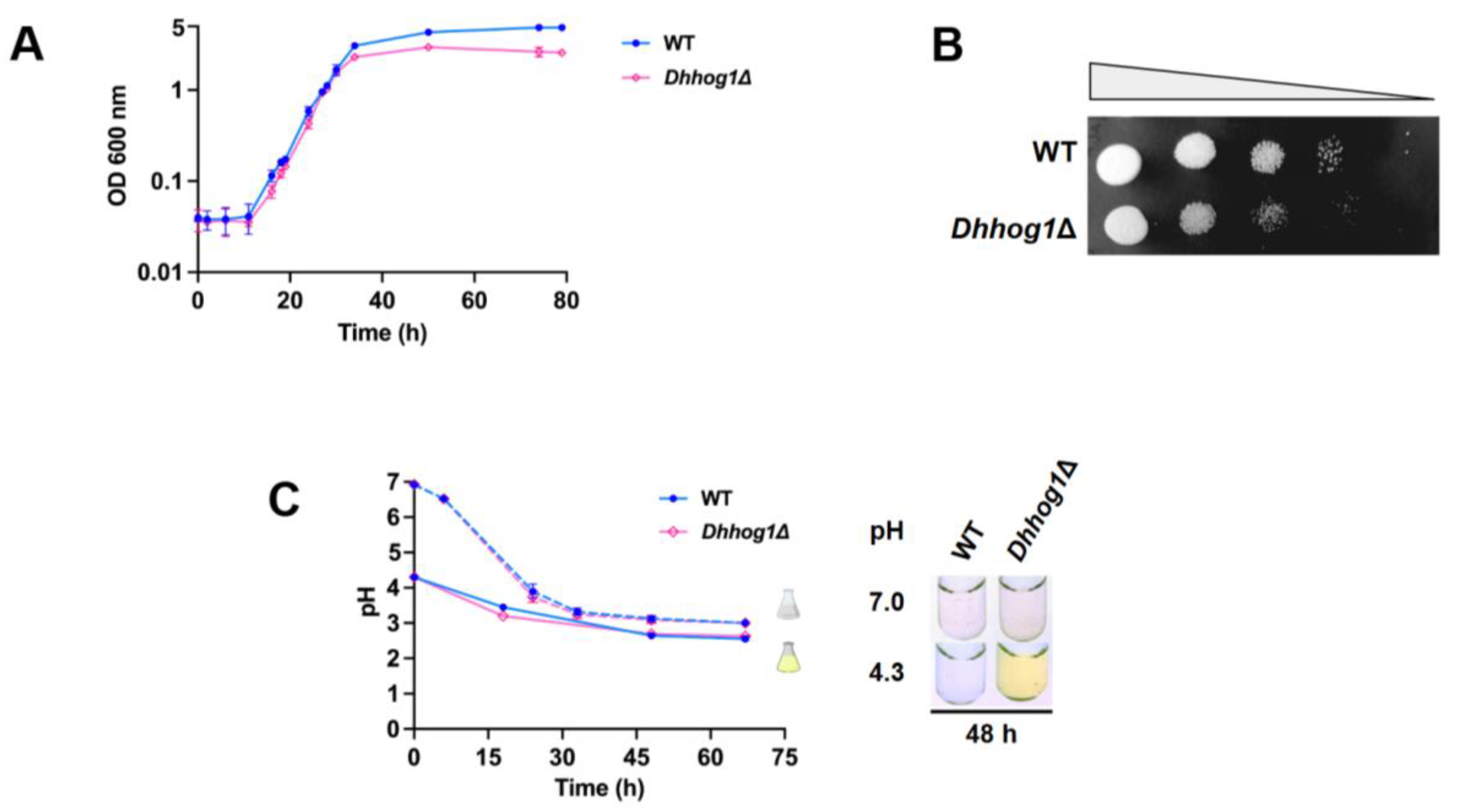
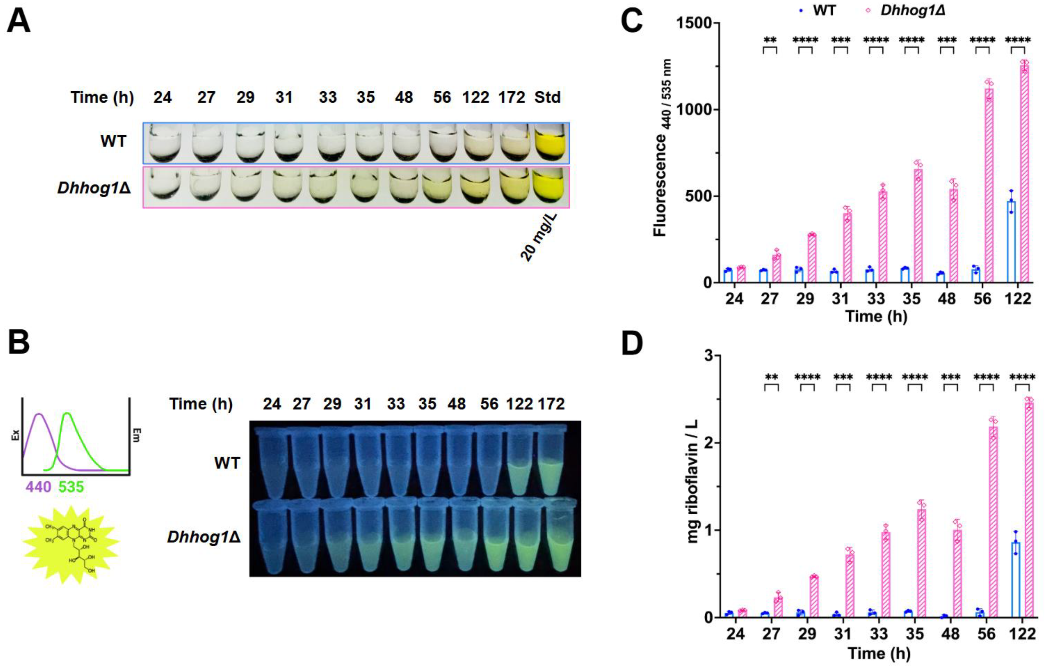
3.1. Initial low pH induces the excretion and accumulation of riboflavin in D. hansenii

The high-osmolarity glycerol (HOG) pathway, with the MAP kinase Hog1 as its terminal effector under salt stress, is recognized as the most conserved signaling cascade for stress adaptation in fungi (de Nadal et al., 2002; Hohmann, 2002, 2009; Hohmann et al., 2007; Saito and Posas, 2012). Given previous observations of riboflavin efflux in acidic media (Perl et al., 1976; Vanetti and Aquarone, 1992), we investigated whether the initial medium pH modulates growth and riboflavin excretion in D. hansenii under saline stress, raising the possibility that DhHog1 signaling may also influence riboflavin metabolism. Comparative assays were conducted between the WT strain and the Dhhog1Δ mutant in minimal medium (YNB) with glucose (2%) supplemented with 0.6 M NaCl under either neutral or acidic initial pH conditions. Significant differences in biomass and cell viability were observed during the stationary phase (Figure 2A–B), highlighting the role of DhHog1 in maintaining cellular fitness under osmotic stress.
Remarkably, cultures initiated at low pH exhibited secretion of a yellow pigment as early as the stationary phase in the Dhhog1Δ mutant (48 h) (Figure 2C), whereas in the WT strain, pigment accumulation became evident only at the late stationary phase (122–172 h) (Figure 4A). By contrast, no pigment secretion was detected in either strain when cultures were initiated at neutral pH (Figure 2C), even after 5 days of growth. These findings indicate that initial acidic conditions are a key determinant for pigment production and excretion in D. hansenii, with an accelerated onset in the absence of HOG1.
Previous studies have reported that D. hansenii is capable of producing riboflavin, a metabolite responsible for the characteristic yellow coloration (Gadd and Edwards, 1986, Seda-Miró et al., 2007). Based on this evidence, we hypothesized that the yellow pigment observed under our culture conditions could correspond to riboflavin. To test this, we identify the pigment by RP-HPLC-DAD analyses in the culture supernatants from the Dhhog1Δ mutant, revealing the presence of a major fluorescent compound. Retention times closely matched those of a riboflavin standard (8.450 ± 0.003 min). The Dhhog1Δ mutant exhibited clearer chromatographic separation and UV-Vis absorption peaks characteristic of flavins (λmax = 223, 268, 370, 445 nm), consistent with riboflavin identity. In contrast, WT supernatants displayed weaker signals with partially overlapping absorption peaks. Representative chromatograms and UV-Vis spectra are shown in Figure 3A–F.
Furthermore, we examined the fluorescence properties of the riboflavin by recording excitation and emission spectra at the characteristic wavelengths (excitation: 440 nm; emission: 520–535 nm). Supernatants were collected at multiple time points (24–122 h, Figure 4A), and riboflavin concentrations were quantified by fluorimetry using a standard calibration curve (Figure 4B–D). These experiments confirm that the accumulation of riboflavin in the supernatant of cultures initiated at low pH, accumulates more rapidly over time in the Dhhog1Δ mutant. This observation indicates, for the first time, that DhHog1 likely plays a role in the riboflavin biosynthesis in D. hansenii.

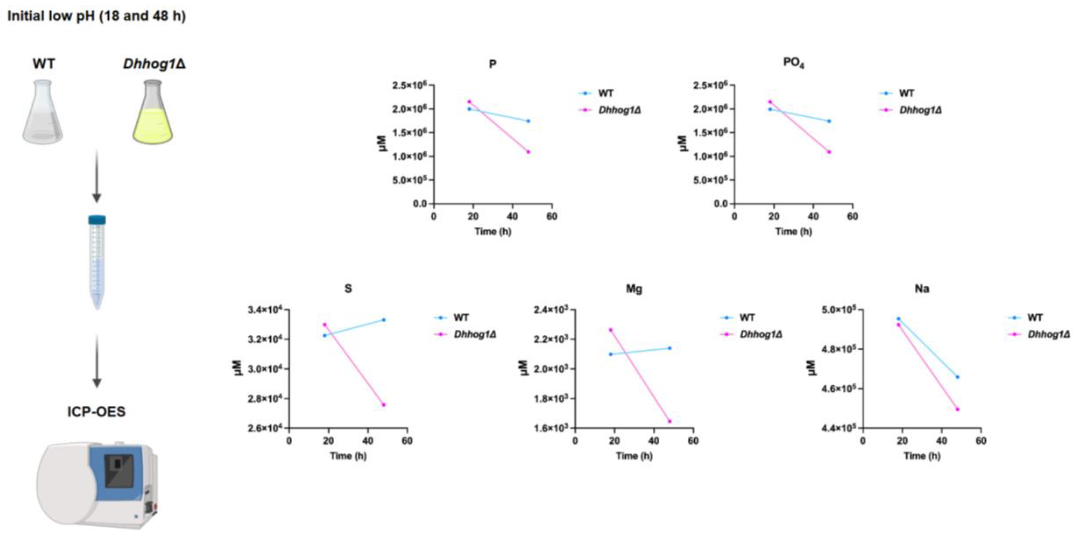
3.2. Accelerated uptake of essential elements in Dhhog1Δ cells

Iron limitation and the availability of other essential elements have been reported as key triggers for riboflavin excretion in riboflavin-producing yeasts (Averianova et al., 2020; Ruchala et al., 2025). To determine whether differences in elemental uptake contribute to the enhanced riboflavin accumulation observed in the Dhhog1Δ mutant, culture supernatants from 18 and 48 h under saline conditions and initial low pH were analyzed by Inductively Coupled Plasma Optical Emission Spectroscopy (ICP-OES) (Figure 5).
By 48 h, the Dhhog1Δ mutant displayed more rapid assimilation of several essential elements, including phosphorus, phosphates, sulfur, and magnesium, compared to the WT strain. In contrast, uptake of iron and other elements was similar in both strains (Table S1), indicating that mechanisms other than iron limitation are likely responsible for stimulating riboflavin excretion in D. hansenii. These results suggest that accelerated assimilation of specific elements (P, S, Mg, and Na) in the Dhhog1Δ mutant may contribute to the earlier and higher riboflavin production observed under acidic and saline conditions.

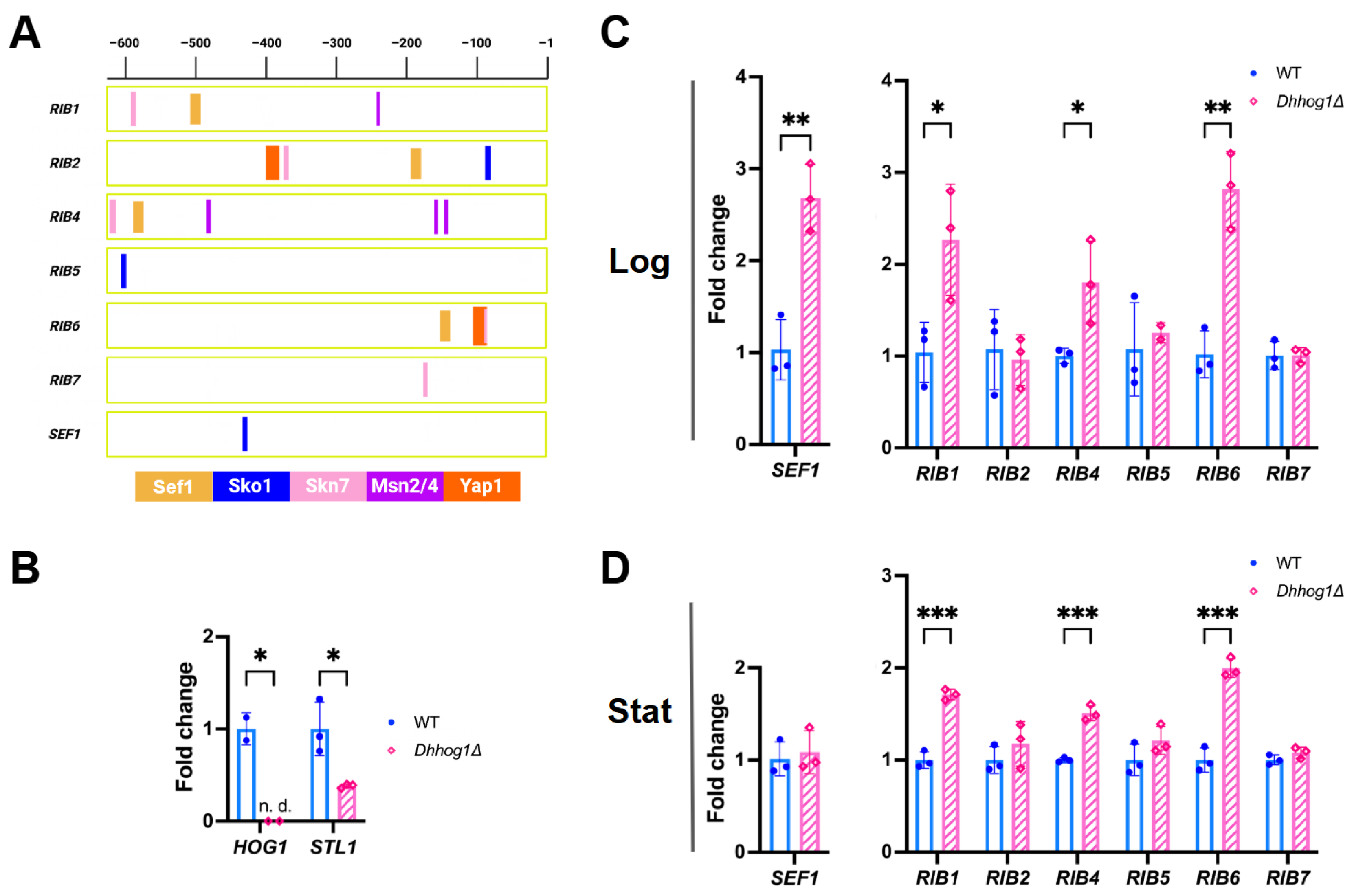
3.3. Riboflavin biosynthesis genes are upregulated in the Dhhog1Δ mutant

The riboflavin biosynthesis genes RIB1, RIB2, RIB5, RIB6, and RIB7 were previously cloned from the synonymous species Candida famata (Dmytruk et al., 1999; Voronovsky et al., 2002, 2004), and their sequences are available in NCBI GenBank as part of the Genolevures Consortium’s annotated reference genome for D. hansenii (ASM644v2). The RIB4 gene sequence was inferred from its homologs in S. cerevisiae and C. albicans and incorporated into the Genolevures Consortium reference genome (Morgunova et al., 2007; Skrzypek et al., 2017). Amino acid sequence alignments confirmed the identity of the main riboflavin biosynthetic enzymes and revealed the degree of sequence conservation among them (Table 2).
To investigate the potential regulation of riboflavin biosynthesis genes by DhHog1 in D. hansenii, we performed an in silico analysis to identify putative stress-related transcription factor (TF) binding sites within the promoters of RIB genes and the transcription factor SEF1 (Figure 6A). Previous work by de la Fuente-Colmenares validated the likely conservation of function in silico of Sko1, Skn7, Msn2/4, and Yap1 proteins in D. hansenii by comparing their sequences with homologs from S. cerevisiae and C. albicans, revealing conserved or semi-conserved residues within critical domains (de la Fuente-Colmenares et al., 2024). This suggests that these TFs perform similar regulatory roles across these yeast species.
Our intergenic region analysis revealed that the RIB gene promoters contain binding motifs for Hog1-controlled transcription factors Sko1, Skn7, Msn2/4, and Yap1, as well as for the iron metabolism regulator Sef1 (Figure 6A). Specifically, SEF1-binding sequences were found in the promoters of RIB1, RIB2, RIB4, and RIB6, while SKO1 motifs were present in RIB2, RIB5, and SEF1 promoters. SKN7-binding sequences were detected in RIB1, RIB2, RIB4, and RIB7. MSN2/4 motifs in RIB1 and RIB4, and YAP1-binding sites in RIB2 and RIB6. It is worth noting that RIB5 and SEF1 each contain a single SKO1-binding site, whereas RIB7 harbors a single SKN7-binding site, suggesting that their transcription may be predominantly regulated by Sko1 or Skn7.
The absence of HOG1 expression was confirmed in the Dhhog1Δ mutant, accompanied by reduced expression of STL1, a downstream HOG pathway gene involved in glycerol symport (Figure 6B). Expression profiles further reveal that DhHog1 acts as a negative regulator of SEF1, RIB1, RIB4, and RIB6 genes during the logarithmic phase (Log), with upregulation of RIB1, RIB4, and RIB6 genes persisting into the stationary phase (Stat) in the Dhhog1Δ mutant (Figure 6C–D). These findings suggest a model in which DhHog1 negatively modulates riboflavin biosynthesis in D. hansenii, likely through the coordinated control of stress-responsive transcription factors such as Sko1, Skn7, Msn2/4, Yap1, and together with the iron metabolism regulator Sef1.

4. Discussion

The results of this study broaden the functional scope of the HOG pathway in Debaryomyces hansenii, revealing that the MAP kinase DhHog1 participates not only in classical osmoadaptation but also in the regulation of riboflavin biosynthesis. Previous work has established DhHog1 as a central effector of the high-osmolarity glycerol response, orchestrating glycerol accumulation and catalase expression in response to saline and oxidative stress (de la Fuente-Colmenares et al., 2024; Sánchez et al., 2020). Our findings add a new layer to this paradigm by showing that DhHog1 acts as a negative regulator of riboflavin secretion, delaying its onset until stationary phase. The premature yellow pigment accumulation observed in the Dhhog1Δ mutant exposed to NaCl under acidic conditions suggests that DhHog1 integrates extracellular initial pH and osmotic cues that prevent riboflavin overproduction during active growth. This role highlights the versatility of the HOG pathway as a global integrator of environmental signals beyond osmostress.

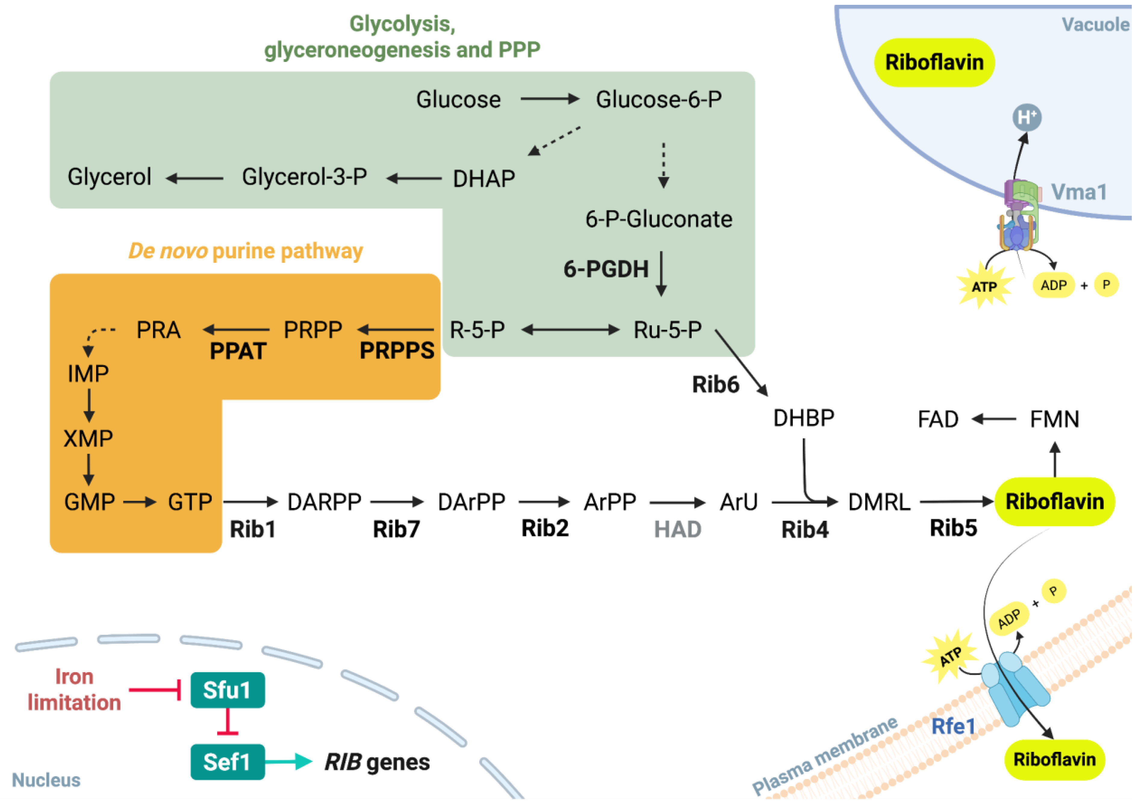
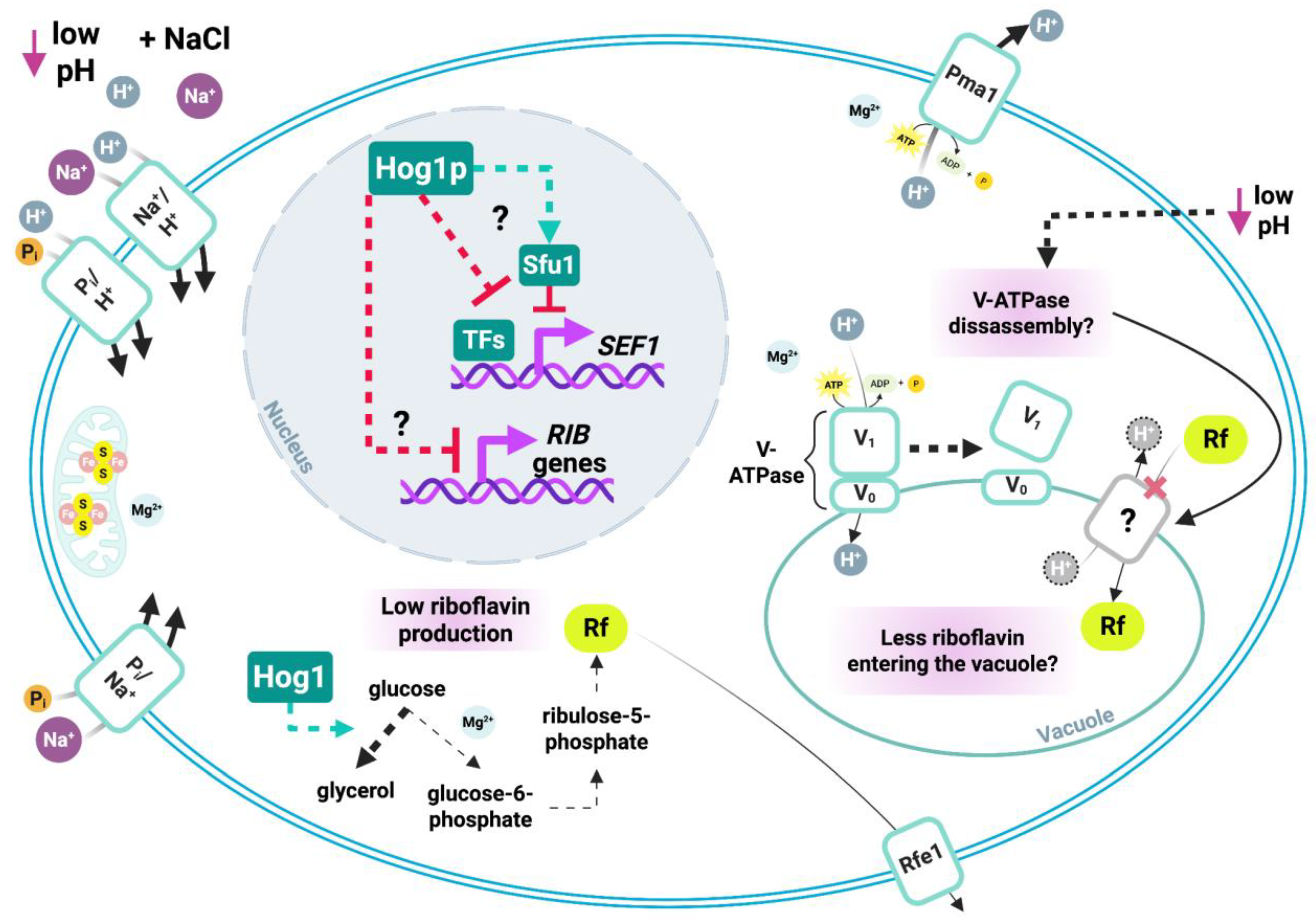
4.1. Conserved riboflavinogenesis in Debaryomyces species and potential repression by DhHog1

The identification of riboflavin as the fluorescent compound is consistent with previous reports of riboflavin production by D. hansenii and closely related species, including D. fabryi, D. subglobosus, and D. prosopidis (Averianova et al., 2020; Nguyen et al., 2009; Ruchala et al., 2025). Historically, taxonomic heterogeneity in the Debaryomyces clade complicated the assignment of riboflavin-producing strains, as exemplified by the reclassification of Candida famata VKM Y-9 (source of industrial riboflavin overproducers) as D. subglobosus (Dmytruk et al., 2011, 2014, 2020; Nguyen et al., 2009).
Beyond Debaryomyces, riboflavin biosynthesis has been reported across Debaryomycetaceae, including Candida albicans, Meyerozyma guilliermondii, C. membranifaciens, C. tropicalis, Schwanniomyces occidentalis, and Hyphopichia wangnamkhiaoensis (Amornrattanapan, 2013; Demuyser et al., 2020; Fedorovych et al., 2001, 2020; Jiménez-Nava et al., 2024; Palma et al., 2022). The higher riboflavin levels observed in the Dhhog1Δ mutant compared to the WT strain indicate that DhHog1 acts as a negative regulator of RIB1, RIB4, RIB6, and SEF1. This observation further implies that similar regulatory mechanisms may be conserved and could operate in a comparable manner across other riboflavin-producing yeast species.

4.2. HOG-dependent effects of pH and NaCl on vacuolar dynamics and riboflavin secretion

The vacuolar H+/ATPase in yeast, whose catalytic subunit is encoded by VMA1, actively pumps protons from the cytosol into the vacuole (Kakinuma et al., 1981; Anraku et al., 1989; Hirata et al., 1990). Vma1 activity relies on the regulated assembly and disassembly of its subunits in response to metabolic cues such as glucose availability and cytosolic pH (Deprez et al., 2018). The assembly of Vma1 is reduced when yeast cells are grown in an acidic medium compared with neutral pH (Diakov and Kane, 2010). Therefore, under our growth conditions, the initially low extracellular pH (4.3) may have impaired Vma1 activity. Disassembly of Vma1 results in cytosolic acidification, thereby altering the protonation state of many biomolecules (Orij et al., 2011).
Yeast flavin-containing monooxygenase (yFMO) catalyzes the oxidation of thiols provided by cysteine, cysteamine, and glutathione (Suh et al., 1996). Oxidized thiols are essential for the proper assembly of functional iron-sulfur clusters (Bandyopadhyay and Outten, 2022). While some flavoproteins contain iron-sulfur clusters, the biogenesis of these cofactors itself depends on a flavin-dependent ferredoxin reductase (Lange et al., 2000; Webert et al., 2014). Under our experimental conditions, the pH range of 4.3–2.5 lies below the pKa of many biological thiols (Ferrer-Sueta, 2022). Such an acidic environment is likely to favor thiol protonation, thereby limiting their reactivity and availability for iron-sulfur cluster assembly. A shortage of iron-sulfur clusters is, in turn, known to trigger iron-deficiency response pathways through the activation and nuclear retention of the transcription factor Sef1 (Ror and Panwar, 2019).
These circumstances may lead to: 1) excess flavin production relative to the number of functional sulfur-iron proteins in which they can be incorporated; moreover, the redox potential of the isoalloxazine ring in flavins may be reduced in a protonic environment, making them less effective for redox homeostasis; 2) increased demand for flavoproteins involved in thiols oxidation and iron-sulfur cluster assembly; or, 3) sustained expression of the Sef1-regulated iron regulon caused by defective biogenesis of mitochondrial iron-sulfur clusters, thereby mimicking an iron-starved state.
Besides the intrinsic effects of low pH, the parallel upregulation of SEF1 and RIB1, RIB4, and RIB6 genes in the Dhhog1Δ mutant suggests that riboflavin overproduction reflects a pseudo iron-starvation state caused by defective iron-sulfur homeostasis rather than extracellular iron limitation.
Another intriguing possibility, not directly tested here, is that exposure to NaCl could enhance vacuolar fission in the Dhhog1Δ mutant, releasing riboflavin stored in vacuoles into the cytosol and facilitating secretion via plasma membrane riboflavin excretase Rfe1 (Tsyrulnyk et al., 2021). In S. cerevisiae, vacuolar fission has been reported in conditions of hyperosmotic, oxidative and endoplasmic reticulum stress (Gokbayrak et al., 2022). These stressors are expected to be exacerbated in a hog1Δ mutant, as suggested by previous reports in D. hansenii, S. cerevisiae, and C. albicans (Bicknell et al., 2010; De la Fuente-Colmenares et al., 2024; Husain et al., 2022; Sánchez et al., 2020; Torres-Quiroz et al., 2010). On the other hand, reduced Vma1 activity due to low extracellular pH (Diakov and Kane, 2010) could impair the vacuolar proton gradient and, consequently, diminish riboflavin influx into the vacuoles. The hypothesis that halted Vma1 activity contributes to riboflavin secretion is further supported by the riboflavin excretion phenotype observed in C. famata null VMA1 mutants (Andreieva et al., 2020a, 2020b).
D. hansenii possesses Na/H+ antiporters that mediate both Na+ efflux from the cytosol and Na+ sequestration into the vacuole (Velkova and Sychrova, 2006). In acidic medium, cytosolic proton accumulation can be accelerated by the presence of NaCl (Sychrová et al., 1999). Furthermore, a more protonic environment could increase the required energy expenditure to pump protons out of the cell. Consequently, maintaining cytosolic pH homeostasis may require further ATP-dependent activity of the plasma membrane H+/ATPase (Pma1) and Vma1, which mediate proton efflux and vacuolar sequestration, respectively. Under such conditions, Na+ sequestration into the vacuole via vacuolar Na/H+ antiporters may be hindered by a reduced Vma1 activity in an initially low extracellular pH, leading to further hyperosmotic stress and promoting vacuolar fragmentation.
Cytosolic acidification may also partially explain the increased demand for phosphorus and oxidative phosphorylation-related elements required for ATP production, as well as Mg2+ for ATP hydrolysis, to sustain proton transport into the vacuole or out of the cell in the Dhhog1Δ mutant. However, unlike S. cerevisiae, a direct regulation of Na/H+ antiporters by phosphorylated Hog1 has not been demonstrated in D. hansenii. Nonetheless, the upregulation of cation transporters expression remains one of the major adaptative responses of D. hansenii to saline stress, together with glycerol accumulation (Almagro et al., 2001; Proft and Struhl, 2004; Velkova and Sychrova, 2006; Carcía-Salcedo et al., 2007; Montiel and Ramos, 2007).
Another possible explanation for the accelerated element uptake observed in the Dhhog1Δ mutant may be the impairment of autophagic processes. In S. cerevisiae, Hog1 is required for the activation of mitophagy (Mao et al., 2011). The absence of Hog1 could lead to the accumulation of dysfunctional mitochondria that keep consuming cellular resources for ATP synthesis. Yet, Hog1-dependent regulation of autophagy has not been established in D. hansenii.

4.3. HOG signaling and multi-element homeostasis in riboflavin secretion

Riboflavin secretion has historically been associated with nutritional stress, particularly iron limitation (Demain, 1972; Gadd & Edwards, 1986; Fedorovich et al., 1999; Knight et al., 2002). Riboflavin and its derivatives can act as siderophore-like molecules, enhancing iron solubility and uptake (David et al., 2025). In C. albicans, C. glabrata, S. cerevisiae, and Cryptococcus neoformans, Hog1 has been implicated in repressing iron acquisition systems under iron sufficiency, while hog1Δ mutants show derepression of iron uptake, increased intracellular iron, and activation of iron-sulfur enzymes (Kaba et al., 2013; Lee et al., 2014; Martins et al., 2018; Thomas et al., 2013; Srivastava et al., 2015; Sahu et al., 2024). Surprisingly, our ICP-OES results revealed no differences in iron assimilation between WT and mutant strains. Instead, we observed accelerated uptake of phosphorus, sulfur, and magnesium. The comparable sodium uptake observed between strains may suggest the involvement of sodium transporters operating independently of Hog1.
These findings indicate that riboflavin accumulation in D. hansenii involves a broader reconfiguration of elemental homeostasis rather than being exclusively dependent on iron availability.

4.4. Sulfur and phosphate metabolism as key contributors to riboflavin overproduction in the Dhhog1Δ mutant

Sulfur metabolism may provide a mechanistic link between DhHog1 function, oxidative stress adaptation, and riboflavin overproduction. Sulfate assimilation fuels the synthesis of methionine and cysteine, which in turn support glutathione biosynthesis, one of the main antioxidant systems in yeasts (Penninckx, 2002). D. hansenii contains lower glutathione pools than S. cerevisiae under both basal and oxidative stress conditions (Navarrete et al., 2009). Yeast glutathione reductase (GR) and flavin-containing monooxygenase (yFMO) are both flavin-dependent enzymes. Additionally, glycine, an indispensable precursor for the synthesis of GTP and riboflavin, is also required for glutathione synthesis (Meister, 1988; Santos et al., 2022). Compared with the non-riboflavin-overproducing yeast S. cerevisiae, the lower glutathione content reported in D. hansenii may reduce the cellular demand for flavins and glycine within this antioxidant system, thereby increasing the availability of these metabolites for riboflavin accumulation.
Methionine biosynthesis is positively regulated by Hog1 in C. albicans (Enjalbert et al., 2006). In S. cerevisiae, Hog1 also activates the histone methyltransferase Dot1, which requires S-adenosylmethionine (SAM) as a methyl donor (Separovich et al., 2024). In the absence of Hog1, a reduced methionine pool combined with impaired Hog1-mediated Dot1 activation could lead to the derepression of multiple genes not directly regulated by the HOG pathway.
We hypothesize that this mechanism is consistent with reports in C. famata, where mutants blocked in methionine biosynthesis (MET2) overproduced riboflavin, and sulfate limitation induced a pseudo iron-starvation phenotype characterized by enhanced intracellular iron accumulation (Dmytruk et al., 2006). Although riboflavin secretion under sulfate limitation has not been systematically studied in this system, the parallels with our findings are striking.
An additional connection between sulfur and phosphate metabolism may arise from tRNA thiolation. The availability of sulfur amino acids determines the amount of tRNA thiolation (Gupta et al., 2019; Laxman et al., 2013). Reduced tRNA thiolation activates methionine de novo and salvage pathways (Laxman et al., 2013); thus, if methionine biosynthesis is impaired in the Dhhog1Δ mutant, this could account for the increased sulfur demand observed. Moreover, tRNA thiolation has been shown to connect sulfur amino acid availability with phosphate assimilation via the PHO regulon (Gupta et al., 2019). In this context, inorganic phosphate is required for sugar phosphorylation in the pentose phosphate pathway, for pyridoxal-5-phosphate-dependent transamination reactions, and purine biosynthesis. This metabolic convergence positions phosphate and amino acids as integral precursors for both purines and flavins. Given that methionine availability promotes the synthesis of nucleotides and multiple amino acids (Walvekar et al., 2018), it remains possible that upregulation of the methionine regulon in the Dhhog1Δ mutant activates PHO-regulated phosphate assimilation pathways, thereby providing the purine precursors required for riboflavin biosynthesis.
The faster uptake of magnesium may be coupled with the accelerated assimilation of phosphorus, as Mg2+ associates with several phosphometabolites and is an essential cofactor for ATP-dependent enzymes, including proton pumps (Walker, 1994).
Regarding the potential role of riboflavin in oxidative stress, studies in A. gossypii have shown that intracellular riboflavin accumulation increases susceptibility to sunlight-induced ROS production and genotoxicity, whereas extracellular riboflavin can exert a protective effect (Stahmann et al., 2001; Silva et al., 2018). Thus, depending on the intracellular redox balance and UV exposure, riboflavin may act either as an antioxidant or as a pro-oxidant, owing to its different oxidation and protonation states.

4.5. DhHog1-mediated negative regulation of riboflavin biosynthesis via stress- and iron-responsive transcription factors

Promoter analysis revealed potential binding sites for Hog1-regulated transcription factors, including Sko1, Skn7, Msn2/4, Yap1, and the iron regulator Sef1, suggesting that riboflavin biosynthesis is under multifactorial control by stress- and iron-responsive transcriptional networks. Our expression analysis demonstrates that RIB1, RIB4, and RIB6 were consistently upregulated in the Dhhog1Δ mutant. These results support the conclusion that DhHog1 represses riboflavin biosynthesis through the modulation of stress-responsive transcription factors. The induction of SEF1 reinforces the hypothesis that riboflavin overproduction is driven by a pseudo iron-starvation response, consistent with previous transcriptomic reports showing enrichment of riboflavin biosynthetic genes under salt stress in D. hansenii and D. subglobosus (Navarrete et al., 2021; Weintraub et al., 2025). Importantly, our study adds the novel observation that initial acidification of the medium is required for riboflavin secretion, highlighting the importance of combined stress factors in controlling this metabolic output.
In S. cerevisiae, Yap1- and Skn7-mediated pathways are specifically involved in responses to oxidative and osmotic stresses, respectively (Auesukaree 2017; Yaakoub 2022). Msn2/4 regulate a broad set of genes implicated in general stress adaptation (Mat Nanyan and Takagi 2020; Hasan et al., 2002). Sko1 functions primarily as a repressor that, once phosphorylated by Hog1, recruits the SAGA histone acetylase and SWI/SNF nucleosome-remodeling complexes to activate the expression of stress-responsive genes (Ikner and Shiozaki 2005; Proft and Struhl 2002). Based on these parallels, we propose that in D. hansenii DhHog1 may modulate riboflavin biosynthesis at least in part through the action of these transcription factors, thereby representing a conserved regulatory mechanism.

4.6. DhHog1 signaling as a metabolic switch linking carbon and phosphate fluxes to riboflavin biosynthesis

In D. hansenii, phosphorylated Hog1 promotes the synthesis of glycerol-3-phosphate from glycolysis-derived dihydroxiacetone-phosphate through the upregulation of GPD1 (Sánchez et al., 2020). In the absence of DhHog1, a larger pool of glucose-6-phosphate may become available for the synthesis of ribulose-5-phosphate through the oxidative branch of the pentose phosphate pathway.
At non-stressful concentrations of NaCl (0.5 M), the glyoxylate shunt is activated in D. hansenii (Ruiz-Pérez et al., 2023). Since glyoxylate is an important precursor for glycine biosynthesis (Schlösser et al., 2004; Takada and Noguchi, 1985), this pathway could serve as a relevant source of intermediates for GTP and riboflavin biosynthesis under our experimental conditions. The NaCl-induced activation of glyoxylate and respiration-related enzymes in D. hansenii is usually accompanied by intracellular phosphate accumulation, probably enabled by the phosphate/Na+ symporter Pho89 (Calahorra et al., 2009; Navarrete et al., 2021; Ruiz-Pérez et al., 2023; Sánchez et al., 2008). In acidic medium, phosphate uptake could be further enhanced by the phosphate/H+ symporter Pho84 (Lau et al., 1998; Eskes et al., 2018).
The faster phosphate assimilation observed in the Dhhog1Δ mutant suggests an increased ATP demand for proton extrusion across the plasma membrane or sequestration into vacuoles, as well as an enhanced activation of the PHO-regulon, possibly triggered by a pseudo-methionine starvation response, as discussed earlier. Furthermore, the biosynthesis of glycine from glyoxylate also requires phosphate, since it depends on pyridoxal-5-phosphate as a cofactor (Schlösser et al., 2004; Takada and Noguchi, 1985).

4.7. Concluding remarks and perspectives

Taken together, our results support a model in which DhHog1 coordinates extracellular initial low pH sensing, elemental assimilation, and riboflavin metabolism (Figure 7). In WT D. hansenii, we found that DhHog1 prevents premature riboflavin production by repressing RIB gene activation and secretion until the stationary phase. Loss of DhHog1 led to derepression, earlier riboflavin secretion, SEF1 upregulation, and accelerated sulfur, phosphorus and magnesium assimilation.
Although this work provides novel evidence linking DhHog1 to riboflavin regulation under acidic and saline stress, the underlying mechanisms remain only partially resolved. The precise contribution of sulfur assimilation, iron–sulfur cluster biogenesis, and vacuolar dynamics could not be directly addressed, as transporter activity, vacuolar morphology, and protein–DNA interactions were not experimentally tested. Likewise, whether DhHog1 represses RIB promoters through direct modulation of transcription factors (e.g., Sef1, Sko1, Msn2/4, Yap1, or Sfu1) or indirectly through metabolic adjustments remains to be clarified. Future studies combining promoter occupancy analyses, phosphoproteomics, and metabolic flux profiling will be essential to disentangle these alternatives. Beyond mechanistic questions, it will also be relevant to determine whether riboflavin secretion represents a mere metabolic overflow or a bona fide adaptive antioxidant strategy. Addressing these points will not only refine our understanding of stress integration in yeasts but may also uncover opportunities to exploit Hog1-dependent pathways for biotechnological riboflavin production.

Funding

The authors declare that financial support was received for the research, authorship, and/or publication of this article. Funding was provided by the Dirección General de Asuntos del Personal Académico (DGAPA), Grants IA204923 and IN217825, and by the Secretaría de Ciencia, Humanidades, Tecnología e Innovación (SECIHTI), Grant CBF-2025-I-14. Additional institutional support was received from the Facultad de Ciencias, UNAM, through the Grupos Interdisciplinarios de Investigación (GII-UNAM) within the framework of the project “Intensificación de los procesos para la obtención de biocompuestos a partir de aguas residuales” (Project GII3307, Instituto de Ingeniería, UNAM).

Acknowledgments

The authors thank Viviana Escobar Sánchez and Yair Romero López from the Departamento de Biología Celular, Facultad de Ciencias, UNAM, for their technical support with the protocols. We are also grateful to Tatiana Fiordelisio Coll, Diana G. Ríos López, and Diego Zamarrón Hernández from the Laboratorio Nacional de Soluciones Biomiméticas para Diagnóstico y Terapia (LaNSBioDyT), Facultad de Ciencias, UNAM, for their technical support and assistance with spectrofluorometer analyses and sample lyophilization. This research was supported by the Posgrado en Ciencias Biológicas, UNAM, and by the Secretaría de Ciencia, Humanidades, Tecnología e Innovación (SECIHTI) through a graduate scholarship awarded to Diana Villarreal-Huerta (CVU: 1185735), Benjamín Mendoza-Tellez (CVU: 1281284), Miguel Rosas-Paz (CVU: 966189). We thank Genaro Vázquez-Victorio for his support in the creation and editing of Figure 1, Figure 7.

References

  1. Abbas, C.A.; Sibirny, A.A. Genetic Control of Biosynthesis and Transport of Riboflavin and Flavin Nucleotides and Construction of Robust Biotechnological Producers. Microbiol. Mol. Biol. Rev. 2011, 75, 321–360. [Google Scholar] [CrossRef] [PubMed]
  2. Almagro, A.; Prista, C.; Benito, B.; Loureiro-Dias, M.C.; Ramos, J. Cloning and Expression of Two Genes Coding for Sodium Pumps in the Salt-Tolerant Yeast Debaryomyces hansenii. J. Bacteriol. 2001, 183, 3251–3255. [Google Scholar] [CrossRef]
  3. Amornrattanapan, P. Riboflavin production by Candida tropicalis isolated from seawater. Sci. Res. Essays 2013, 8, 43–47. [Google Scholar] [CrossRef]
  4. Andreieva, Y.; Petrovska, Y.; Lyzak, O.; Liu, W.; Kang, Y.; Dmytruk, K.; Sibirny, A. Role of the regulatory genes SEF1, VMA1 and SFU1 in riboflavin synthesis in the flavinogenic yeast Candida famata (Candida flareri). Yeast 2020, 37, 497–504. [Google Scholar] [CrossRef]
  5. Andreieva, Y.; Lyzak, O.; Liu, W.; Kang, Y.; Dmytruk, K.; Sibirny, A. SEF1 and VMA1 Genes Regulate Riboflavin Biosynthesis in the Flavinogenic Yeast Candida famata. Cytol. Genet. 2020, 54, 379–385. [Google Scholar] [CrossRef]
  6. Anraku, Y.; Umemoto, N.; Hirata, R.; Wada, Y. Structure and function of the yeast vacuolar membrane proton ATPase. J. Bioenerg. Biomembr. 1989, 21, 589–603. [Google Scholar] [CrossRef]
  7. Auesukaree, C. Molecular mechanisms of the yeast adaptive response and tolerance to stresses encountered during ethanol fermentation. J. Biosci. Bioeng. 2017, 124, 133–142. [Google Scholar] [CrossRef]
  8. Averianova, L.A.; Balabanova, L.A.; Son, O.M.; Podvolotskaya, A.B.; Tekutyeva, L.A. Production of Vitamin B2 (Riboflavin) by Microorganisms: An Overview. Front. Bioeng. Biotechnol. 2020, 8, 570828. [Google Scholar] [CrossRef] [PubMed]
  9. Bacher, A.; Eberhardt, S.; Eisenreich, W.; Fischer, M.; Herz, S.; Illarionov, B.; Kis, K.; Richter, G. (2001). Biosynthesis of riboflavin. In Vitamins & Hormones (Vol. 61, pp. 1–49). Elsevier. [CrossRef]
  10. Bandyopadhyay, T.; Outten, C.E. (2022). The role of thiols in iron–sulfur cluster biogenesis. In Redox Chemistry and Biology of Thiols (pp. 487–506). Elsevier. [CrossRef]
  11. Bansal, P.K.; Mondal, A.K. Isolation and sequence of the HOG1 homologue from Debaryomyces hansenii by complementation of the hog1Δ strain of Saccharomyces cerevisiae. Yeast 2000, 16, 81–88. [Google Scholar] [CrossRef]
  12. Baron, S.F.; Hylemon, P.B. Expression of the bile acid-inducible NADH:flavin oxidoreductase gene of Eubacterium sp. VPI 12708 in Escherichia coli. BBA Protein Struct. Mol. Enzymol. 1995, 1249, 145–154. [Google Scholar] [CrossRef]
  13. Bicknell, A.A.; Tourtellotte, J.; Niwa, M. Late Phase of the Endoplasmic Reticulum Stress Response Pathway Is Regulated by Hog1 MAP Kinase. J. Biol. Chem. 2010, 285, 17545–17555. [Google Scholar] [CrossRef] [PubMed]
  14. Bray, R.C.; Palmer, G.; Beinert, H. Direct Studies on the Electron Transfer Sequence in Xanthine Oxidase by Electron Paramagnetic Resonance Spectroscopy. J. Biol. Chem. 1964, 239, 2667–2676. [Google Scholar] [CrossRef]
  15. Calahorra, M.; Sánchez, N.S.; Peña, A. Activation of fermentation by salts in Debaryomyces hansenii. FEMS Yeast Res. 2009, 9, 1293–1301. [Google Scholar] [CrossRef]
  16. Carcí a-Salcedo, R.; Montiel, V.; Calero, F.; Ramos, J. Characterization of DhKHA1, a gene coding for a putative Na+ transporter from Debaryomyces hansenii. FEMS Yeast Res. 2007, 7, 905–911. [Google Scholar] [CrossRef] [PubMed]
  17. Chen, C.; Pande, K.; French, S.D.; Tuch, B.B.; Noble, S.M. An Iron Homeostasis Regulatory Circuit with Reciprocal Roles in Candida albicans Commensalism and Pathogenesis. Cell Host Microbe 2011, 10, 118–135. [Google Scholar] [CrossRef]
  18. David, M.S.; Zhu, Z.; McDonald, M.R.; Badiee, M.; Mortimer, I.P.; Leung, A.K.L.; Culotta, V.C. Crosstalk between iron and flavins in the opportunistic fungal pathogen Candida albicans. J. Biol. Chem. 2025, 301, 110396. [Google Scholar] [CrossRef]
  19. de la Fuente-Colmenares, I.; González, J.; Sánchez, N.S.; Ochoa-Gutiérrez, D.; Escobar-Sánchez, V.; Segal-Kischinevzky, C. Regulation of Catalase Expression and Activity by DhHog1 in the Halotolerant Yeast Debaryomyces hansenii Under Saline and Oxidative Conditions. J. Fungi 2024, 10, 740. [Google Scholar] [CrossRef]
  20. de Nadal, E.; Alepuz, P.M.; Posas, F. Dealing with osmostress through MAP kinase activation. EMBO Rep. 2002, 3, 735–740. [Google Scholar] [CrossRef]
  21. Demain, A.L. Riboflavin Oversynthesis. Annu. Rev. Microbiol. 1972, 26, 369–388. [Google Scholar] [CrossRef] [PubMed]
  22. Demuyser, L.; Palmans, I.; Vandecruys, P.; Van Dijck, P. Molecular Elucidation of Riboflavin Production and Regulation in Candida albicans, toward a Novel Antifungal Drug Target. mSphere 2020, 5, 00714-20. [Google Scholar] [CrossRef]
  23. Deprez, M.-A.; Eskes, E.; Wilms, T.; Ludovico, P.; Winderickx, J. pH homeostasis links the nutrient sensing PKA/TORC1/Sch9 ménage-à-trois to stress tolerance and longevity. Microb. Cell 2018, 5, 119–136. [Google Scholar] [CrossRef]
  24. Diakov, T.T.; Kane, P.M. Regulation of Vacuolar Proton-translocating ATPase Activity and Assembly by Extracellular pH. J. Biol. Chem. 2010, 285, 23771–23778. [Google Scholar] [CrossRef] [PubMed]
  25. Dmytruk, K.V.; Abbas, C.A.; Voronovsky, A.Y.; Kshanovska, B.V.; Sybirna, K.A.; Sybirny, A.A. Cloning of structural genes involved in riboflavin synthesis of the yeast Candida famata. Ukr. Biokhim. Zh. 1999, 76, 78–87. [Google Scholar] [PubMed]
  26. Dmytruk, K.V.; Voronovsky, A.Y.; Sibirny, A.A. Insertion mutagenesis of the yeast Candida famata (Debaryomyces hansenii) by random integration of linear DNA fragments. Curr. Genet. 2006, 50, 183–191. [Google Scholar] [CrossRef]
  27. Dmytruk, K.V.; Yatsyshyn, V.Y.; Sybirna, N.O.; Fedorovych, D.V.; Sibirny, A.A. Metabolic engineering and classic selection of the yeast Candida famata (Candida flareri) for construction of strains with enhanced riboflavin production. Metab. Eng. 2011, 13, 82–88. [Google Scholar] [CrossRef] [PubMed]
  28. Dmytruk, K.; Lyzak, O.; Yatsyshyn, V.; Kluz, M.; Sibirny, V.; Puchalski, C.; Sibirny, A. Construction and fed-batch cultivation of Candida famata with enhanced riboflavin production. J. Biotechnol. 2014, 172, 11–17. [Google Scholar] [CrossRef]
  29. Dmytruk, K.V.; Ruchala, J.; Fedorovych, D.V.; Ostapiv, R.D.; Sibirny, A.A. Modulation of the Purine Pathway for Riboflavin Production in Flavinogenic Recombinant Strain of the Yeast Candida famata. Biotechnol. J. 2020, 15, 1900468. [Google Scholar] [CrossRef]
  30. Enjalbert, B.; Smith, D.A.; Cornell, M.J.; Alam, I.; Nicholls, S.; Brown, A.J.P.; Quinn, J. Role of the Hog1 Stress-activated Protein Kinase in the Global Transcriptional Response to Stress in the Fungal Pathogen Candida albicans. Mol. Biol. Cell 2006, 17, 1018–1032. [Google Scholar] [CrossRef]
  31. Eskes, E.; Deprez, M.-A.; Wilms, T.; Winderickx, J. pH homeostasis in yeast; the phosphate perspective. Curr. Genet. 2018, 64, 155–161. [Google Scholar] [CrossRef]
  32. Fedorovich, D.; Protchenko, O.; Lesuisse, E. Iron uptake by the yeast Pichia guilliermondii. Flavinogenesis and reductive iron assimilation are co-regulated processes. Biometals 1999, 12, 295–300. [Google Scholar] [CrossRef] [PubMed]
  33. Fedorovych, D.; Kszeminska, H.; Babjak, L.; Kaszycki, P.; Kołoczek, H. Hexavalent chromium stimulation of riboflavin synthesis in flavinogenic yeast. Biometals 2001, 14, 23–31. [Google Scholar] [CrossRef]
  34. Fedorovych, D.; Boretsky, V.; Pynyaha, Y.; Bohovych, I.; Boretsky, Y.; Sibirny, A. Cloning of Genes Sef1 and Tup1 Encoding Transcriptional Activator and Global Repressor in the Flavinogenic Yeast Meyerozyma (Candida, Pichia) guilliermondii. Cytol. Genet. 2020, 54, 413–419. [Google Scholar] [CrossRef]
  35. Ferrer-Sueta, G. (2022). Chemical basis of cysteine reactivity and specificity: Acidity and nucleophilicity. In Redox Chemistry and Biology of Thiols (pp. 19–58). Elsevier. [CrossRef]
  36. Frerman, F.E. Acyl-CoA dehydrogenases, electron transfer flavoprotein and electron transfer flavoprotein dehydrogenase. Biochem. Soc. Trans. 1988, 16, 416–418. [Google Scholar] [CrossRef] [PubMed]
  37. Gadd, G.M.; Edwards, S.W. Heavy-metal-induced flavin production by Debaryomyces hansenii and possible connexions with iron metabolism. Trans. Br. Mycol. Soc. 1986, 87, 533–542. [Google Scholar] [CrossRef]
  38. Gokbayrak, Z.D.; Patel, D.; Brett, C.L. Acetate and hypertonic stress stimulate vacuole membrane fission using distinct mechanisms. PLoS One 2022, 17, e0271199. [Google Scholar] [CrossRef]
  39. Gomes, A.C.; Miranda, I.; Silva, R.M.; Moura, G.R.; Thomas, B.; Akoulitchev, A.; Santos, M.A. A genetic code alteration generates a proteome of high diversity in the human pathogen Candida albicans. Genome Biol. 2007, 8, R206. [Google Scholar] [CrossRef]
  40. González, J.; Villarreal-Huerta, D.; Rosas-Paz, M.; Segal-Kischinevzky, C. (2025). Biotechnological Applications of Yeasts Under Extreme Conditions. In P. Buzzini, B. Turchetti, & A. Yurkov (Eds.), Extremophilic Yeasts (pp. 459–501). Springer Nature Switzerland. [CrossRef]
  41. Gupta, R.; Walvekar, A.S.; Liang, S.; Rashida, Z.; Shah, P.; Laxman, S. A tRNA modification balances carbon and nitrogen metabolism by regulating phosphate homeostasis. eLife 2019, 8, e44795. [Google Scholar] [CrossRef]
  42. Haase, I.; Sarge, S.; Illarionov, B.; Laudert, D.; Hohmann, H.; Bacher, A.; Fischer, M. Enzymes from the Haloacid Dehalogenase (HAD) Superfamily Catalyse the Elusive Dephosphorylation Step of Riboflavin Biosynthesis. ChemBioChem 2013, 14, 2272–2275. [Google Scholar] [CrossRef] [PubMed]
  43. Hasan, R.; Leroy, C.; Isnard, A.; Labarre, J.; Boy-Marcotte, E.; Toledano, M.B. The control of the yeast H2O2 response by the Msn2/4 transcription factors. Mol. Microbiol. 2002, 45, 233–241. [Google Scholar] [CrossRef]
  44. Hirata, R.; Ohsumk, Y.; Nakano, A.; Kawasaki, H.; Suzuki, K.; Anraku, Y. Molecular structure of a gene, VMA1, encoding the catalytic subunit of H(+)-translocating adenosine triphosphatase from vacuolar membranes of Saccharomyces cerevisiae. J. Biol. Chem. 1990, 265, 6726–6733. [Google Scholar] [CrossRef]
  45. Hohmann, S. Osmotic Stress Signaling and Osmoadaptation in Yeasts. Microbiol. Mol. Biol. Rev. 2002, 66, 300–372. [Google Scholar] [CrossRef]
  46. Hohmann, S.; Krantz, M.; Nordlander, B. (2007). Yeast Osmoregulation. In Methods in Enzymology (Vol. 428, pp. 29–45). Elsevier. [CrossRef]
  47. Hohmann, S. Control of high osmolarity signalling in the yeast Saccharomyces cerevisiae. FEBS Lett. 2009, 583, 4025–4029. [Google Scholar] [CrossRef]
  48. Husain, F.; Pathak, P.; Román, E.; Pla, J.; Panwar, S.L. Adaptation to Endoplasmic Reticulum Stress in Candida albicans Relies on the Activity of the Hog1 Mitogen-Activated Protein Kinase. Front. Microbiol. 2022, 12, 794855. [Google Scholar] [CrossRef]
  49. Ikner, A.; Shiozaki, K. Yeast signaling pathways in the oxidative stress response. Mutat. Res./Fund. Mol. Mech. Mutagen. 2005, 569, 13–27. [Google Scholar] [CrossRef]
  50. Jiménez-Nava, R.A.; Zepeda-Vallejo, L.G.; Santoyo-Tepole, F.; Chávez-Camarillo, G. Ma.; Cristiani-Urbina, E. RP-HPLC Separation and 1H NMR Identification of a Yellow Fluorescent Compound—Riboflavin (Vitamin B2)—Produced by the Yeast Hyphopichia wangnamkhiaoensis. Biomolecules 2023, 13, 1423. [Google Scholar] [CrossRef] [PubMed]
  51. Jiménez-Nava, R.A.; Chávez-Camarillo, G. Ma.; Cristiani-Urbina, E. Kinetics of Riboflavin Production by Hyphopichia wangnamkhiaoensis under Varying Nutritional Conditions. Int. J. Mol. Sci. 2024, 25, 9430. [Google Scholar] [CrossRef]
  52. Kaba, H.E.; Nimtz, M.; Müller, P.P.; Bilitewski, U. Involvement of the mitogen activated protein kinase Hog1p in the response of Candida albicans to iron availability. BMC Microbiol. 2013, 13, 16. [Google Scholar] [CrossRef] [PubMed]
  53. Kakinuma, Y.; Ohsumi, Y.; Anraku, Y. Properties of H+-translocating adenosine triphosphatase in vacuolar membranes of Saccharomyces cerevisiae. J. Biol. Chem. 1981, 256, 10859–10863. [Google Scholar] [CrossRef]
  54. Knight, S.A.B.; Lesuisse, E.; Stearman, R.; Klausner, R.D.; Dancis, A. Reductive iron uptake by Candida albicans: Role of copper, iron and the TUP1 regulator. Microbiology 2002, 148, 29–40. [Google Scholar] [CrossRef]
  55. Krassowski, T.; Coughlan, A.Y.; Shen, X.-X.; Zhou, X.; Kominek, J.; Opulente, D.A.; Riley, R.; Grigoriev, I.V.; Maheshwari, N.; Shields, D.C.; Kurtzman, C.P.; Hittinger, C.T.; Rokas, A.; Wolfe, K.H. Evolutionary instability of CUG-Leu in the genetic code of budding yeasts. Nat. Commun. 2018, 9, 1887. [Google Scholar] [CrossRef] [PubMed]
  56. Kurabayashi, K.; Enshasy, H.A.E.; Park, E.Y.; Kato, T. Increased production of riboflavin in Ashbya gossypii by endoplasmic reticulum stresses. Arch. Microbiol. 2025, 207, 167. [Google Scholar] [CrossRef]
  57. Lange, H.; Kaut, A.; Kispal, G.; Lill, R. A mitochondrial ferredoxin is essential for biogenesis of cellular iron-sulfur proteins. PNAS 2000, 97, 1050–1055. [Google Scholar] [CrossRef]
  58. Lau, W.-T. W.; Schneider, K.R.; O’Shea, E.K. A Genetic Study of Signaling Processes for Repression of PHO5 Transcription in Saccharomyces cerevisiae. Genetics 1998, 150, 1349–1359. [Google Scholar] [CrossRef]
  59. Laxman, S.; Sutter, B.M.; Wu, X.; Kumar, S.; Guo, X.; Trudgian, D.C.; Mirzaei, H.; Tu, B.P. Sulfur Amino Acids Regulate Translational Capacity and Metabolic Homeostasis through Modulation of tRNA Thiolation. Cell 2013, 154, 416–429. [Google Scholar] [CrossRef] [PubMed]
  60. LeBlanc, J.G.; Milani, C.; De Giori, G.S.; Sesma, F.; Van Sinderen, D.; Ventura, M. Bacteria as vitamin suppliers to their host: A gut microbiota perspective. Curr. Opin. Biotechnol. 2013, 24, 160–168. [Google Scholar] [CrossRef]
  61. Lee, K.-T.; Lee, J.-W.; Lee, D.; Jung, W.-H.; Bahn, Y.-S. A Ferroxidase, Cfo1, Regulates Diverse Environmental Stress Responses of Cryptococcus neoformans through the HOG Pathway. Mycobiol. 2014, 42, 152–157. [Google Scholar] [CrossRef] [PubMed]
  62. Lim, S.H.; Choi, J.S.; Park, E.Y. Microbial production of riboflavin using riboflavin overproducers, Ashbya gossypii, Bacillus subtilis, and Candida famata: An overview. Biotechnol. Bioprocess Eng. 2001, 6, 75–88. [Google Scholar] [CrossRef]
  63. Macheroux, P.; Kappes, B.; Ealick, S.E. Flavogenomics – a genomic and structural view of flavin-dependent proteins. FEBS J. 2011, 278, 2625–2634. [Google Scholar] [CrossRef]
  64. Mansoorabadi, S.O.; Thibodeaux, C.J.; Liu, H. The Diverse Roles of Flavin Coenzymes – Nature’s Most Versatile Thespians. J. Org. Chem. 2007, 72, 6329–6342. [Google Scholar] [CrossRef]
  65. Mao, K.; Wang, K.; Zhao, M.; Xu, T.; Klionsky, D.J. Two MAPK-signaling pathways are required for mitophagy in Saccharomyces cerevisiae. J. Cell. Biol. 2011, 193, 755–767. [Google Scholar] [CrossRef]
  66. Martins, T.S.; Pereira, C.; Canadell, D.; Vilaça, R.; Teixeira, V.; Moradas-Ferreira, P.; De Nadal, E.; Posas, F.; Costa, V. The Hog1p kinase regulates Aft1p transcription factor to control iron accumulation. BBA Mol. Cell. Biol. Lipids 2018, 1863, 61–70. [Google Scholar] [CrossRef]
  67. Mat Nanyan, N.S.B.; Takagi, H. Proline Homeostasis in Saccharomyces cerevisiae: How Does the Stress-Responsive Transcription Factor Msn2 Play a Role? Front. Genet. 2020, 11, 438. [Google Scholar] [CrossRef]
  68. Meister, A. Glutathione metabolism and its selective modification. J. Biol. Chem. 1988, 263, 17205–17208. [Google Scholar] [CrossRef]
  69. Minhas, A.; Biswas, D.; Mondal, A.K. Development of host and vector for high-efficiency transformation and gene disruption in Debaryomyces hansenii. FEMS Yeast Res. 2009, 9, 95–102. [Google Scholar] [CrossRef] [PubMed]
  70. Montiel, V.; Ramos, J. Intracellular Na+ and K+ distribution in Debaryomyces hansenii. Cloning and expression in Saccharomyces cerevisiae of DhNHX1: Intracellular Na+ and K+ distribution in Debaryomyces hansenii. FEMS Yeast Res. 2007, 7, 102–109. [Google Scholar] [CrossRef]
  71. Morgunova, E.; Saller, S.; Haase, I.; Cushman, M.; Bacher, A.; Fischer, M.; Ladenstein, R. Lumazine Synthase from Candida albicans as an Anti-fungal Target Enzyme. J. Biol. Chem. 2007, 282, 17231–17241. [Google Scholar] [CrossRef] [PubMed]
  72. Navarrete, C.; Siles, A.; Martí nez, J.L.; Calero, F.; Ramos, J. Oxidative stress sensitivity in Debaryomyces hansenii. FEMS Yeast Res. 2009, 9, 582–590. [Google Scholar] [CrossRef] [PubMed]
  73. Navarrete, C.; Frost, A.T.; Ramos-Moreno, L.; Krum, M.R.; Martínez, J.L. A physiological characterization in controlled bioreactors reveals a novel survival strategy for Debaryomyces hansenii at high salinity. Yeast 2021, 38, 302–315. [Google Scholar] [CrossRef]
  74. Nguyen, H.-V.; Gaillardin, C.; Neuvéglise, C. Differentiation of Debaryomyces hansenii and Candida famata by rRNA gene intergenic spacer fingerprinting and reassessment of phylogenetic relationships among D. hansenii, C. famata, D. fabryi, C. flareri (=D. subglobosus) and D. prosopidis: Description of D. vietnamensis sp. nov. closely related to D. nepalensis. FEMS Yeast Res. 2009, 9, 641–662. [Google Scholar] [CrossRef]
  75. Nieland, S.; Stahmann, K.-P. A developmental stage of hyphal cells shows riboflavin overproduction instead of sporulation in Ashbya gossypii. Appl. Microbiol. Biotechnol. 2013, 97, 10143–10153. [Google Scholar] [CrossRef]
  76. Nysten, J.; Van Dijck, P. Can we microbe-manage our vitamin acquisition for better health? PLOS Pathog. 2023, 19, e1011361. [Google Scholar] [CrossRef]
  77. Ochoa-Gutiérrez, D.; Reyes-Torres, A.M.; De La Fuente-Colmenares, I.; Escobar-Sánchez, V.; González, J.; Ortiz-Hernández, R.; Torres-Ramírez, N.; Segal-Kischinevzky, C. Alternative CUG Codon Usage in the Halotolerant Yeast Debaryomyces hansenii: Gene Expression Profiles Provide New Insights into Ambiguous Translation. J. Fungi 2022, 8, 970. [Google Scholar] [CrossRef]
  78. Onishi, H. Osmophilic yeasts. Adv. Food Res. 1963, 53–94. [Google Scholar]
  79. Orij, R.; Brul, S.; Smits, G.J. Intracellular pH is a tightly controlled signal in yeast. BBA Gen. Subjects 2011, 1810, 933–944. [Google Scholar] [CrossRef]
  80. Palma, M.; Mondo, S.; Pereira, M.; Vieira, É.; Grigoriev, I.V.; Sá-Correia, I. Genome Sequence and Analysis of the Flavinogenic Yeast Candida membranifaciens IST 626. J. Fungi 2022, 8, 254. [Google Scholar] [CrossRef] [PubMed]
  81. Penninckx, M. An overview on glutathione in Saccharomyces versus non-conventional yeasts. FEMS Yeast Res. 2002, 2, 295–305. [Google Scholar] [CrossRef]
  82. Perl, M.; Kearney, E.B.; Singer, T.P. Transport of riboflavin into yeast cells. J. Biol. Chem. 1976, 251, 3221–3228. [Google Scholar] [CrossRef] [PubMed]
  83. Petrovska, Y.; Lyzak, O.; Ruchala, J.; Dmytruk, K.; Sibirny, A. Co-Overexpression of RIB1 and RIB6 Increases Riboflavin Production in the Yeast Candida famata. Fermentation 2022, 8, 141. [Google Scholar] [CrossRef]
  84. Posas, F.; Wurgler-Murphy, S.M.; Maeda, T.; Witten, E.A.; Thai, T.C.; Saito, H. Yeast HOG1 MAP Kinase Cascade is Regulated by a Multistep Phosphorelay Mechanism in the SLN1–YPD1–SSK1 “Two-Component” Osmosensor. Cell 1996, 86, 865–875. [Google Scholar] [CrossRef] [PubMed]
  85. Prista, C.; Michán, C.; Miranda, I.M.; Ramos, J. The halotolerant Debaryomyces hansenii, the Cinderella of non-conventional yeasts. Yeast 2016, 33, 523–533. [Google Scholar] [CrossRef]
  86. Proft, M.; Struhl, K. Hog1 Kinase Converts the Sko1-Cyc8-Tup1 Repressor Complex into an Activator that Recruits SAGA and SWI/SNF in Response to Osmotic Stress. Mol. Cell 2002, 9, 1307–1317. [Google Scholar] [CrossRef]
  87. Proft, M.; Struhl, K. MAP Kinase-Mediated Stress Relief that Precedes and Regulates the Timing of Transcriptional Induction. Cell 2004, 118, 351–361. [Google Scholar] [CrossRef]
  88. Richard, G.-F.; Kerrest, A.; Lafontaine, I.; Dujon, B. Comparative Genomics of Hemiascomycete Yeasts: Genes Involved in DNA Replication, Repair, and Recombination. Mol. Biol. Evol. 2005, 22, 1011–1023. [Google Scholar] [CrossRef] [PubMed]
  89. Ror, S.; and Panwar, S.L. Sef1-Regulated Iron Regulon Responds to Mitochondria-Dependent Iron–Sulfur Cluster Biosynthesis in Candida albicans. Front. Microbiol. 2019, 10, 1528. [Google Scholar] [CrossRef]
  90. Ruchala, J.; Andreieva, Y.A.; Tsyrulnyk, A.O.; Sobchuk, S.M.; Najdecka, A.; Wen, L.; Kang, Y.; Dmytruk, O.V.; Dmytruk, K.V.; Fedorovych, D.V.; Sibirny, A.A. Cheese whey supports high riboflavin synthesis by the engineered strains of the flavinogenic yeast Candida famata. Microb. Cell Fact. 2022, 21, 161. [Google Scholar] [CrossRef]
  91. Ruchala, J.; Najdecka, A.; Wojdyla, D.; Liu, W.; Sibirny, A. Regulation of Riboflavin Biosynthesis in Microorganisms and Construction of the Advanced Overproducers of this Vitamin. Int. J. Mol. Sci. 2025, 26, 6243. [Google Scholar] [CrossRef]
  92. Ruiz-Pérez, F.S.; Ruiz-Castilla, F.J.; Leal, C.; Martínez, J.L.; Ramos, J. Sodium and lithium exert differential effects on the central carbon metabolism of Debaryomyces hansenii through the glyoxylate shunt regulation. Yeast 2023, 40, 265–275. [Google Scholar] [CrossRef]
  93. Sa, N.; Rawat, R.; Thornburg, C.; Walker, K.D.; Roje, S. Identification and characterization of the missing phosphatase on the riboflavin biosynthesis pathway in Arabidopsis thaliana. Plant J. 2016, 88, 705–716. [Google Scholar] [CrossRef] [PubMed]
  94. Sahu, M.S.; Purushotham, R.; Kaur, R. The Hog1 MAPK substrate governs Candida glabrata-epithelial cell adhesion via the histone H2A variant. PLOS Genet. 2024, 20, e1011281. [Google Scholar] [CrossRef] [PubMed]
  95. Saito, H.; Posas, F. Response to Hyperosmotic Stress. Genet. 2012, 192, 289–318. [Google Scholar] [CrossRef]
  96. Sánchez, N.S.; Arreguí n, R.; Calahorra, M.; Peña, A. Effects of salts on aerobic metabolism of Debaryomyces hansenii. FEMS Yeast Res. 2008, 8, 1303–1312. [Google Scholar] [CrossRef] [PubMed]
  97. Sánchez, N.S.; Calahorra, M.; González, J.; Defosse, T.; Papon, N.; Peña, A.; Coria, R. Contribution of the mitogen-activated protein kinase Hog1 to the halotolerance of the marine yeast Debaryomyces hansenii. Curr. Genet. 2020, 66, 1135–1153. [Google Scholar] [CrossRef]
  98. Santos, L.O.; Silva, P.G.P.; Lemos Junior, W.J.F.; De Oliveira, V.S.; Anschau, A. Glutathione production by Saccharomyces cerevisiae: Current state and perspectives. Appl. Microbiol. Biotechnol. 2022, 106, 1879–1894. [Google Scholar] [CrossRef] [PubMed]
  99. Sarge, S.; Haase, I.; Illarionov, B.; Laudert, D.; Hohmann, H.; Bacher, A.; Fischer, M. Catalysis of an Essential Step in Vitamin B2 Biosynthesis by a Consortium of Broad Spectrum Hydrolases. ChemBioChem 2015, 16, 2466–2469. [Google Scholar] [CrossRef]
  100. Schlösser, T.; Gätgens, C.; Weber, U.; Stahmann, K. -Peter. Alanine: Glyoxylate aminotransferase of Saccharomyces cerevisiae –encoding gene AGX1 and metabolic significance. Yeast 2004, 21, 63–73. [Google Scholar] [CrossRef]
  101. Schmitt, M.E.; Brown, T.A.; Trumpower, B.L. A rapid and simple method for preparation of RNA from Saccharomyces cerevisiae. Nucleic Acids Res. 1990, 18, 3091–3092. [Google Scholar] [CrossRef]
  102. Schwechheimer, S.K.; Park, E.Y.; Revuelta, J.L.; Becker, J.; Wittmann, C. Biotechnology of riboflavin. Appl. Microbiol. Biotechnol. 2016, 100, 2107–2119. [Google Scholar] [CrossRef]
  103. Seda-Miró, J.M.; Arroyo-González, N.; Pérez-Matos, A.; Govind, N.S. Impairment of cobalt-induced riboflavin biosynthesis in a Debaryomyces hansenii mutant. Can. J. Microbiol. 2007, 53, 1272–1277. [Google Scholar] [CrossRef] [PubMed]
  104. Segal-Kischinevzky, C.; Romero-Aguilar, L.; Alcaraz, L.D.; López-Ortiz, G.; Martínez-Castillo, B.; Torres-Ramírez, N.; Sandoval, G.; González, J. Yeasts Inhabiting Extreme Environments and their Biotechnological Applications. Microorganisms 2022, 10, 794. [Google Scholar] [CrossRef]
  105. Separovich, R.J.; Karakatsanis, N.M.; Gao, K.; Fuh, D.; Hamey, J.J.; Wilkins, M.R. Proline-directed yeast and human MAP kinases phosphorylate the Dot1p/ DOT1L histone H3K79 methyltransferase. The FEBS J. 2024, 291, 2590–2614. [Google Scholar] [CrossRef]
  106. Sharma, A. Riboflavin Vitamin B2 Market Report 2025 (Global Edition). 2025. Available online: https://www.cognitivemarketresearch.com/vitamin-b2-riboflavin-market-report (accessed on 28 August 2025).
  107. Silva, R.; Aguiar, T.Q.; Oliveira, R.; Domingues, L. Light exposure during growth increases riboflavin production, ROS accumulation and DNA damage in Ashbya gossypii riboflavin-overproducing strains. FEMS Yeast Res. 2018, 19. [Google Scholar] [CrossRef]
  108. Skrzypek, M.S.; Binkley, J.; Binkley, G.; Miyasato, S.R.; Simison, M.; Sherlock, G. The Candida Genome Database (CGD): Incorporation of Assembly 22, systematic identifiers and visualization of high throughput sequencing data. Nucleic Acids Res. 2017, 45, D592–D596. [Google Scholar] [CrossRef] [PubMed]
  109. Srivastava, V.K.; Suneetha, K.J.; Kaur, R. The mitogen-activated protein kinase CgHog1 is required for iron homeostasis, adherence and virulence in Candida glabrata. FEBS J. 2015, 282, 2142–2166. [Google Scholar] [CrossRef] [PubMed]
  110. Stahmann, K.-P.; Revuelta, J.L.; Seulberger, H. Three biotechnical processes using Ashbya gossypii, Candida famata, or Bacillus subtilis compete with chemical riboflavin production. Appl. Microbiol. Biotechnol. 2000, 53, 509–516. [Google Scholar] [CrossRef] [PubMed]
  111. Stahmann, K. -Peter, Arst, H.N.; Althöfer, H.; Revuelta, J.L.; Monschau, N.; Schlüpen, C.; Gätgens, C.; Wiesenburg, A.; Schlösser, T. Riboflavin, overproduced during sporulation of Ashbya gossypii, protects its hyaline spores against ultraviolet light. Environ. Microbiol. 2001, 3, 545–550. [Google Scholar] [CrossRef] [PubMed]
  112. Suh, J.-K.; Poulsen, L.L.; Ziegler, D.M.; Robertus, J.D. Molecular Cloning and Kinetic Characterization of a Flavin-Containing Monooxygenase from Saccharomyces cerevisiae. Arch. Biochem. Biophys. 1996, 336, 268–274. [Google Scholar] [CrossRef]
  113. Sychrová, H.; Ramı́rez, J.; Peña, A. Involvement of Nha1 antiporter in regulation of intracellular pH in Saccharomyces cerevisiae. FEMS Microbiol. Lett. 1999, 171, 167–172. [Google Scholar] [CrossRef]
  114. Takada, Y.; Noguchi, T. Characteristics of alanine: Glyoxylate aminotransferase from Saccharomyces cerevisiae, a regulatory enzyme in the glyoxylate pathway of glycine and serine biosynthesis from tricarboxylic acid-cycle intermediates. Biochem. J. 1985, 231, 157–163. [Google Scholar] [CrossRef]
  115. Thomas, E.; Roman, E.; Claypool, S.; Manzoor, N.; Pla, J.; Panwar, S.L. Mitochondria Influence CDR1 Efflux Pump Activity, Hog1-Mediated Oxidative Stress Pathway, Iron Homeostasis, and Ergosterol Levels in Candida albicans. Antimicrob. Agents Chemother. 2013, 57, 5580–5599. [Google Scholar] [CrossRef]
  116. Torres-Quiroz, F.; García-Marqués, S.; Coria, R.; Randez-Gil, F.; Prieto, J.A. The Activity of Yeast Hog1 MAPK Is Required during Endoplasmic Reticulum Stress Induced by Tunicamycin Exposure. J. Biol. Chem. 2010, 285, 20088–20096. [Google Scholar] [CrossRef]
  117. Tsyrulnyk, A.O.; Fedorovych, D.V.; Dmytruk, K.V.; Sibirny, A.A. (2021). Overexpression of Riboflavin Excretase Enhances Riboflavin Production in the Yeast Candida famata. In M. Barile (Ed.), Flavins and Flavoproteins (Vol. 2280, pp. 31–42). Springer US. [CrossRef]
  118. Turatsinze, J.-V.; Thomas-Chollier, M.; Defrance, M.; van Helden, J. Using RSAT to scan genome sequences for transcription factor binding sites and cis-regulatory modules. Nat. Protoc. 2008, 3, 1578–1588. [Google Scholar] [CrossRef] [PubMed]
  119. Vandamme, E.J. Production of vitamins, coenzymes and related biochemicals by biotechnological processes. J. Chem. Technol. Biotechnol. 1992, 53, 313–327. [Google Scholar] [CrossRef]
  120. Vanetti, M.C.D.; Aquarone, E. Riboflavin excretion by Pachysolen tannophilus grown in synthetic medium supplemented with d-xylose. World J. Microbiol. Biotechnol. 1992, 8, 190–191. [Google Scholar] [CrossRef]
  121. Velkova, K.; Sychrova, H. The Debaryomyces hansenii NHA1 gene encodes a plasma membrane alkali-metal-cation antiporter with broad substrate specificity. Gene 2006, 369, 27–34. [Google Scholar] [CrossRef] [PubMed]
  122. Voronovsky, A.; Abbas, C.; Fayura, L.; Kshanovska, B.; Dmytruk, K.; Sybirna, K.; Sibirny, A. Development of a transformation system for the flavinogenic yeast. FEMS Yeast Res. 2002, 2, 381–388. [Google Scholar] [CrossRef]
  123. Voronovsky, A.Y.; Abbas, C.A.; Dmytruk, K.V.; Ishchuk, O.P.; Kshanovska, B.V.; Sybirna, K.A.; Gaillardin, C.; Sibirny, A.A. Candida famata (Debaryomyces hansenii) DNA sequences containing genes involved in riboflavin synthesis. Yeast 2004, 21, 1307–1316. [Google Scholar] [CrossRef] [PubMed]
  124. Walker, G.M. The Roles of Magnesium in Biotechnology. Crit. Rev. Biotechnol. 1994, 14, 311–354. [Google Scholar] [CrossRef]
  125. Walsh, C.T.; Wencewicz, T.A. Flavoenzymes: Versatile catalysts in biosynthetic pathways. Nat. Prod. Rep. 2013, 30, 175–200. [Google Scholar] [CrossRef]
  126. Walvekar, A.S.; Srinivasan, R.; Gupta, R.; Laxman, S. Methionine coordinates a hierarchically organized anabolic program enabling proliferation. Mol. Biol. Cell 2018, 29, 3183–3200. [Google Scholar] [CrossRef]
  127. Webert, H.; Freibert, S.-A.; Gallo, A.; Heidenreich, T.; Linne, U.; Amlacher, S.; Hurt, E.; Mühlenhoff, U.; Banci, L.; Lill, R. Functional reconstitution of mitochondrial Fe/S cluster synthesis on Isu1 reveals the involvement of ferredoxin. Nat. Commun. 2014, 5, 5013. [Google Scholar] [CrossRef]
  128. Weintraub, S.J.; Li, Z.; Nakagawa, C.L.; Collins, J.H.; Young, E.M. Oleaginous Yeast Biology Elucidated With Comparative Transcriptomics. Biotechnol. Bioeng. 2025, 122, 677–697. [Google Scholar] [CrossRef] [PubMed]
  129. Yaakoub, H.; Mina, S.; Calenda, A.; Bouchara, J.-P.; Papon, N. Oxidative stress response pathways in fungi. Cell. Mol. Life Sci. 2022, 79, 333. [Google Scholar] [CrossRef] [PubMed]
Figure 1. Simplified schematic of the riboflavin biosynthetic pathway in flavinogenic yeasts under iron limitation. The pathway is fueled by glycolysis and pentose phosphate pathway precursors (green square: 6-PGDH, 6-phosphogluconate dehydrogenase; R-5-P, ribose-5-phosphate; Ru-5-P, ribulose-5-phosphate). PRS3 encodes 5-phosphoribosyl-1-pyrophosphate synthetase (PRPPS), responsible for PRPP (5-phosphoribosyl-1-pyrophosphate) production. The ADE4 gene encodes 5-phosphoribosyl-1-pyrophosphate amidotransferase (PPAT), a key enzyme in purine biosynthesis that catalyzes the first step of the de novo pathway. Purine-derived precursors (orange square: PRPP, 5-phosphoribosyl-1-pyrophosphate; PRD, 5-phosphoribosylamine; IMP, inosine monophosphate; XMP, xanthosine monophosphate; GMP, guanosine monophosphate; GTP, guanosine triphosphate). Key genes/enzymes of riboflavin pathway include RIB1/Rib1 (GTP cyclohydrolase II), RIB2/Rib2 (DArPP deaminase), a putative haloacid dehalogenase (HAD), RIB4/Rib4 (dimethylribityllumazine synthase), RIB5/Rib5 (riboflavin synthase), RIB6/Rib6 (4-dihydroxy-2-butanone-4-phosphate synthase), and RIB7/Rib7 (5-amino-6-(5-phosphoribosylamino) uracil reductase). The intermediates in the riboflavin pathway include DARPP (2,5-diamino-6-ribosylamino-4(3H)-pyrimidinone 5'-phosphate), DArPP (5-amino-6-(D-ribitylamino)uracil 5'-phosphate), ArPP (5-amino-6-ribityl-amino-2,4(1H,3H)pyrimidinedione 5’-phosphate), ArU (5-amino-6-(D-ribitylamino)uracil), DHBP (3,4-dihydroxy-2-butanone-4-phosphate), DMRL (6,7-dimethyl-8-ribityllumazine), FMN (flavin mononucleotide), and FAD (flavin adenine dinucleotide). In the vacuole, riboflavin can be stored through a process dependent on Vma1, the vacuolar ATPase subunit A. Riboflavin can also be secreted via Rfe1, a riboflavin excretase. Under iron homeostasis conditions, the GATA-type transcription factor Sfu1 prevents iron toxicity in iron-replete medium by inhibiting Sef1, a transcription factor that activates iron acquisition genes, including RIB genes. Blue arrows indicate positive regulation; red blunt arrows indicate negative regulation. Image based on findings reported in the literature (Dmytruk et al., 1999; Voronovsky et al., 2002, 2004; Haase et al., 2013; Sarge et al., 2015; Sa et al., 2016; Ruchala et al., 2022; 2025). Created using BioRender.com, accessed on 29 August 2025.
Preprints 176225 g001
Figure 2. Initial low pH induces the secretion and accumulation of a yellow pigment in the culture supernatant of D. hansenii. (A) Growth curves of wild type (WT, blue) and the Dhhog1Δ mutant (pink) were determined in MM + 0.6 M NaCl at an initial low pH (pH 4.3). (B) Cells of WT and the Dhhog1Δ mutant were grown in MM + 0.6 M NaCl to the stationary phase (74 h), followed by 10-fold serial dilutions (up to 10–4). A 10 μL aliquot of each dilution was spotted onto YPD agar plates containing 0.6 M NaCl and incubated at 28 °C for 3 days. (C) The pH of cultures grown in MM supplemented with 0.6 M NaCl, initiated under neutral (pH 6.8–7.0, dotted lines) and acidic (pH 4.3, solid lines) conditions, was monitored over a 70 h period. Representative images from three independent experiments are shown. Data represent the mean ± standard deviation (SD) of three independent experiments (n = 3).
Preprints 176225 g002
Figure 3. RP-HPLC-DAD analysis and UV-Vis spectra of riboflavin in D. hansenii WT and Dhhog1Δ. Chromatograms at 280 nm obtained by RP-HPLC-DAD for WT (A), Dhhog1Δ (C), and riboflavin standard (E), with arrows indicating the HPLC-separated fraction whose UV-Vis spectra are shown in panels B, D, and F. Samples were collected during stationary phase from strains grown in MM + 0.6 M NaCl at initial pH 4.3. Riboflavin accumulation was higher in the Dhhog1Δ mutant compared with WT. Values represent the mean of at least three independent measurements.
Preprints 176225 g003
Figure 4. Quantification of riboflavin in the culture supernatant of D. hansenii. (A) A yellow pigment was observed in the supernatants from the WT and Dhhog1Δ cultures; the color appeared earlier in the culture time in the Dhhog1Δ strain. (B) Samples were collected at defined time points (24–172 h) from WT and Dhhog1Δ strains grown in MM + 0.6 M NaCl at an initial low pH (4.3) and excited with a UV light transilluminator. (C–D) Quantification was performed by measuring fluorescence (440/535 nm) and comparing with a calibration curve generated from a riboflavin standard in a 96-well spectrofluorometer. Data represent the mean ± standard deviation (SD) of three independent experiments (n = 3). Significant differences: p-value ≤ 0.05 (*), ≤ 0.005 (**), ≤ 0.0005 (***), ≤ 0.00005 (****).
Preprints 176225 g004
Figure 5. Identification and quantification of macro nutrients and trace elements in the culture supernatant of D. hansenii WT and Dhhog1Δ strains. Supernatants were collected from WT and Dhhog1Δ cultures grown in MM + 0.6 M NaCl at an initial low pH (4.3) at 18 and 48 h, and analyzed by Inductively Coupled Plasma-Optical Emission Spectroscopy (ICP-OES). A total of 43 elements (plus phosphate) were measured; the profiles of phosphorus (P), phosphates (PO4), sulfur (S), magnesium (Mg), and sodium (Na), are shown. Quantification was performed using the Triton ICP-OES Test.
Preprints 176225 g005
Figure 6. Predicted stress-related transcription factor binding sites in the promoter regions of riboflavin biosynthesis genes and corresponding gene expression in WT and Dhhog1Δ strains. (A) Schematic representation of intergenic region from −625 bp upstream to the +1 ATG; predicted motifs are highlighted for SEF1 (yellow), SKO1 (blue), SKN7 (pink), MSN2/4 (purple), and YAP1 (orange). (B) As an initial control, HOG1 and STL1 expression were detected in WT (blue) and Dhhog1Δ (pink) strains. As expected, HOG1 expression was not detected (n.d.) in the mutant, whereas partial STL1 expression was observed, consistent with its role as a positive regulator previously described (Sánchez et al., 2020). (C–D) Gene expression analysis of SEF1, RIB1, RIB2, RIB4, RIB6, and RIB7 under MM + 0.6 M NaCl at an initial low pH in WT and Dhhog1Δ strains. Total RNA was extracted from WT and mutant cells during the mid-log phase (Log, 18 h) and stationary phase (Stat, 48 h) and analyzed by RT-qPCR. Bars represent fold changes in gene expression, with transcript levels normalized to ACT1. Values are presented as the mean of at least three independent measurements ± (SD). Significant differences: p-value ≤ 0.05 (*), ≤ 0.005 (**), ≤ 0.0005 (***).
Preprints 176225 g006
Figure 7. Hypothetical model of riboflavin regulation under acidic and saline conditions. Hog1 negatively regulates RIB1, RIB4, and RIB6 by downregulating transcriptional activators such as SEF1, and possibly via SFU1 or other transcription factors (TFs). Low pH and Na⁺ enhance assimilation of essential elements, including inorganic phosphate (Pi) via H⁺- and Na⁺-mediated antiporters, and increases ATP demand for proton extrusion through Pma1 and Vma1. Low pH–induced V-ATPase disassembly may impair the vacuolar proton gradient, limiting riboflavin influx and promoting cytosolic accumulation. In WT, Hog1 channels glucose into glycerol, reducing riboflavin precursors. In Dhhog1Δ, absence of Hog1 may trigger a pseudo iron- or sulfur-starvation state, accelerating the assimilation of Pi, S, and Mg2+, and cytosolic riboflavin is secreted via Rfe1. Direct interactions of Hog1 with transcriptional activators or repressors mediating RIB and SEF1 repression remain unknown. Rf: Riboflavin. Legend for arrows: thick arrows, upregulated in WT; thin arrows, downregulated in WT; dashed arrows, indirect mechanisms; green dashed arrows, positive Hog1 regulation; red blunt arrows, negative Hog1 regulation. Created using BioRender.com, accessed on 7 September 2025.
Preprints 176225 g007
Table 1. Deoxyoligonucleotides used for RT-qPCR relative gene expression analysis.
Gene ID,
ORF
Fw 5’ → 3’ Rv 5’ → 3’
DhACT1 2901278,
DEHA2D05412g
CCCAGAAGAACACCCAGTTT CGGCTTGGATAGAAACGTAGAA
DhRIB1 2899385,
DEHA2A12870g
AAGACACCCTGCTGATGGTC TGTCGGGGTTGTTGGTCAAT
DhRIB2 2902834,
DEHA2E11374g
TGGAACCATGCTCCTTGAGATT CTGGCTCCACAACACCAACA
DhRIB4 2901083,
DEHA2D04180g
TGTTTGACCGATGAGCAAGC ACACATTTCGACAGCAGCAG
DhRIB5 2901307,
DEHA2D13926g
GCCTGGGTGTAACTGACCAT GGAGAAGGGGTTCATTGCCA
DhRIB6 2904849,
DEHA2G09504g
TGGTCTTATGAAGTCTACCGGC TATGCTGATGGCACGACCAC
DhRIB7 2904875,
DEHA2G10010g
ACTTGCACCTCCTTCAACCAT GGTGCATTTGTCAGGCTTCC
DhSEF1 2900038,
DEHA2C16676g
CCGTTTGCTTCGACCCTTTA CTGCCAACAATGCTACCGTG
DhSTL1 2902951,
DEHA2E01364g
TGGGAATGGCTGACACTTATG GCTCTTCTACCCAACCTATCAATC
DhHOG1 2902985,
DEHA2E20944g
AACCGCTCGCTGAATGGAAT TCTCCACCTCCAGACGTGAT
Table 2. Sequence similarity and query coverage of selected D. hansenii riboflavin biosynthesis proteins compared with S. cerevisiae and C. albicans.
Protein name,
Identifier
S. cerevisiae C. albicans
Similarity (%) Query cover (%) Similarity (%) Query cover (%)
DhRib1,
DEHA2A12870p
77 80 85 92
DhRib2,
DEHA2E11374p
73 86 80 100
DhRib4,
DEHA2D04180p
83 99 95 100
DhRib5,
DEHA2D13926p
73 98 82 100
DhRib6,
DEHA2G09504p
73 98 88 100
DhRib7,
DEHA2G10010p
62 100 65 100
DhSef1,
DEHA2C16676p
67 76 70 100
Disclaimer/Publisher’s Note: The statements, opinions and data contained in all publications are solely those of the individual author(s) and contributor(s) and not of MDPI and/or the editor(s). MDPI and/or the editor(s) disclaim responsibility for any injury to people or property resulting from any ideas, methods, instructions or products referred to in the content.
Copyright: This open access article is published under a Creative Commons CC BY 4.0 license, which permit the free download, distribution, and reuse, provided that the author and preprint are cited in any reuse.
Prerpints.org logo

Preprints.org is a free preprint server supported by MDPI in Basel, Switzerland.

Subscribe

Disclaimer

Terms of Use

Privacy Policy

Privacy Settings

© 2025 MDPI (Basel, Switzerland) unless otherwise stated