Submitted:
05 September 2025
Posted:
08 September 2025
You are already at the latest version
Abstract
Venomous snake bites are a global public health issue, causing between 81,000 and 138,000 annual deaths and 400,000 permanent disabilities. This study investigated the oral bacterial diversity and antibiotic resistance profiles in captive Viperidae snakes. Oral swabs from 48 specimens across four species (Porthidium lansbergii, Bothriechis nigroviridis, Cerrophidion sasai, and Bothrops asper) were analyzed using culture-dependent methods. Bacterial isolation and identification via the VITEK 2 automated system revealed 41 strains from 12 genera, predominantly Gram-negative bacteria, including Morganella morganii (11 isolates) and Providencia rettgeri (10 isolates). Antibiotic susceptibility testing demonstrated significant resistance patterns, with 100% resistance to cephalothin and cefazolin in M. morganii, and emerging extended-spectrum β-lactamase (ESBL) production in Enterobacter cloacae and Sphingomonas paucimobilis. Comparative analysis showed P. lansbergii exhibited the highest bacterial diversity. These findings highlight the complex oral microbiota of venomous snakes and underscore the need for evidence-based antibiotic strategies in snakebite management, particularly given the global rise in antimicrobial resistance.
Keywords:
Viperidae family
; oral microbiome
; antibiotic resistance
; snakebite infections
The global burden of snakebites represents a significant public health challenge, particularly in tropical and subtropical regions. Every year, snakebites result in an estimated 1.2 to 5.5 million envenomations, with fatalities ranging from 81,000 to 138,000 individuals [1]. The impact is disproportionately felt in rural areas of developing countries, where agricultural workers and their families face the highest risk due to frequent interactions with snake habitats. The socioeconomic consequences of snakebites extend beyond immediate health effects, often leading to long-term disability and loss of income for victims and their families [1]. In Central America, the situation mirrors global trends. The region faces significant challenges in snakebite management, including inadequate healthcare infrastructure, lack of trained personnel, and insufficient availability of antivenoms. For instance, Panama records over 2,000 snakebite cases, with a 2-3% fatality rate. Between 2007 and 2021, snakebite incidence ranged from 34 to 65 cases per every 100,000 inhabitants per year [8].
The oral microbiome of venomous snakes plays a crucial role in both snake ecology and the clinical implications of snakebites. This complex ecosystem harbors a diverse array of bacteria, including potentially pathogenic species such as Morganella morganii, and members of the Enterobacteriaceae family [2]. These bacteria can lead to severe infections following snakebites, manifesting as conditions like necrotizing fasciitis and abscess formation. The composition of the snake oral microbiome is influenced by various factors, including environmental conditions and dietary habits. This dynamic interplay between snakes and their environment underscores the ecological complexity of snake microbiomes and their potential implications for zoonotic infections in humans [5].
A particularly concerning aspect of snake oral microbiomes is the prevalence of antibiotic-resistant bacteria. Studies have identified multiple bacterial strains resistant to common antibiotics such as amoxicillin, penicillin, and oxacillin. Studies on Taiwanese venomous snakes showed a significant proportion of antibiotic-resistant oral bacteria [2]. The presence of antibiotic resistance genes (ARGs) in snake oral microbiomes reflects a broader trend observed in various animal species, potentially exacerbating the global issue of antimicrobial resistance. The transfer of resistance genes from snake oral bacteria to human pathogens is also a growing concern, as it could further complicate the treatment of snakebite-related infections and contribute to the broader challenge of antibiotic resistance [13].
This study investigated the bacterial diversity present in the oral cavity of captive Viperidae snakes. The research project characterized bacterial cultures obtained from oral cavity swabs, identified aerobic bacteria using biochemical methods, and evaluated their antibiotic resistance and sensitivity profiles. This study's significance lies in its potential to advance our understanding of snake microbiomes and their implications for human health, particularly in the context of snakebite treatment.
The study examined a total of 48 venomous snake specimens maintained in captivity at the serpentarium of the Center for Research and Information on Drugs and Toxins (CIIMET) at the Universidad de Panamá, comprising 21 Porthidium lansbergii, 8 Bothriechis nigroviridis, 15 Cerrophidion sasai, and 4 Bothrops asper individuals. For B. asper, CO2 sedation was used due to their large size, while smaller species were manually restrained. Swab samples were immediately placed in Amies liquid ESwab® transport medium. Samples were cultured on Blood agar, MacConkey agar, and Chocolate agar under aerobic conditions. After incubation, colonies were isolated and subjected to Gram staining. Isolated colonies of bacteria were then identified using the VITEK® 2 COMPACT automated system (bioMérieux). Bacterial suspensions were prepared to a 0.5 McFarland standard and inoculated into appropriate VITEK® 2 cards for both identification and antimicrobial susceptibility testing. GN and GP cards were used for Gram-negative and Gram-positive bacteria identification, respectively, while AST-N401 and AST-P663 cards were used for antibiotic susceptibility testing. The VITEK® 2 system provided biochemical identification of bacteria based on various tests and determined antibiotic resistance profiles following Clinical and Laboratory Standards Institute (CLSI) guidelines.
A total of 41 bacterial strains belonging to 12 different genera were isolated and identified from 48 initial samples. The most frequently isolated species were Morganella morganii (11 isolates), Providencia rettgeri (10 isolates), and Pseudomonas aeruginosa and Serratia fonticola (3 isolates each). Gram-negative bacteria predominated in the oral cavities of the analyzed snakes. The comparison between snake species revealed interesting patterns in oral microbiota. P. lansbergii exhibited the highest bacterial diversity, with 9 different species identified, while B. nigroviridis and B. asper showed lower diversity (Figure 1). M. morganii was found in all snake species, while P. rettgeri was common in B. nigroviridis, C. sasai, and P. lansbergii. Some species-specific bacteria were also identified, such as Proteus vulgaris and Enterobacter cloacae in B. asper, and Klebsiella oxytoca and Staphylococcus lentus in P. lansbergii.
Porthidium lansbergii exhibited significantly higher bacterial diversity (Shannon index = 2.413, species richness = 12) compared to the other three species. Bothrops asper showed the lowest diversity (Shannon index = 0.611, species richness = 2), representing a 295% difference in diversity between the highest and lowest species. The coefficient of variation for Shannon diversity was 49.2%, indicating substantial interspecies variation (Figure 1).
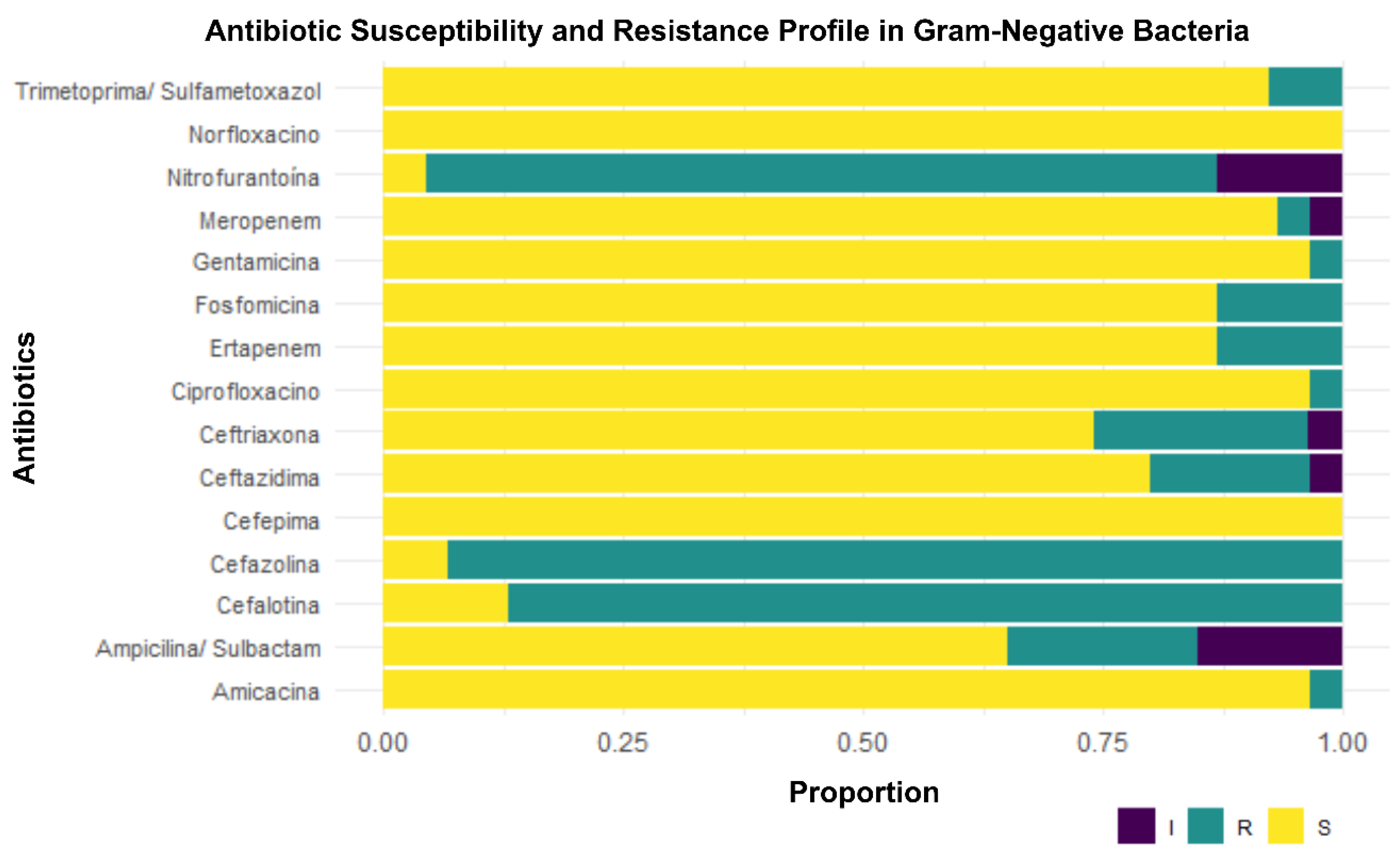
Antibiotic susceptibility testing revealed variable resistance patterns among the isolated bacteria (Figure 2). Notably, high resistance to cefalothin and cefazolin was observed in M. morganii and P. rettgeri isolates. Some Morganella morganii isolates demonstrated resistance to ertapenem, indicating potential carbapenemase production in these strains. Other strains, including a Sphingomonas paucimobilis isolate and Enterobacter cloacae isolate, showed multidrug resistance profiles suggestive of extended-spectrum β-lactamase (ESBL) production. These findings highlight the diverse and potentially antibiotic-resistant bacterial communities present in the oral cavities of captive venomous snakes, which may have implications for snakebite treatment and management.
The selection of culture media, including Blood Agar, MacConkey Agar, and Chocolate Agar, was based on their widespread use in clinical microbiology and their ability to facilitate the isolation and identification of a wide range of medically important bacteria. Blood Agar, an enriched medium, allows for the growth of a variety of microorganisms, including both Gram-positive and Gram-negative bacteria, and facilitates the detection of hemolysis. MacConkey Agar, a selective and differential medium, is used for the isolation of Gram-negative bacteria, especially enterobacteria, and allows for the differentiation of lactose-fermenting species. Chocolate Agar, an enriched medium containing additional growth factors, is used for the isolation of fastidious bacteria such as Haemophilus and Neisseria.
Our results revealed a notable diversity of cultivable bacteria in the oral cavity of the studied venomous snakes. We successfully isolated and identified 41 bacterial strains belonging to 12 different genera. However, it is important to note that not all samples yielded bacterial growth on the utilized media, and some colonies could not be biochemically identified. This finding underscores the inherent limitations of culture-dependent methods, as not all bacteria present in a complex ecosystem like the oral cavity are cultivable or identifiable using these techniques.
Regarding Gram-positive bacteria, the results revealed the identification of Staphylococcus lentus in one sample. This consistency suggests that Gram-negative bacteria might be fundamental components of the oral microbiota of snakes, regardless of geographical region. The observed differences in bacterial composition among the four studied snake species are particularly interesting. Porthidium lansbergii exhibited the highest bacterial diversity, while Bothrops asper and Bothriechis nigroviridis showed more limited diversity. These variations could be attributed to various factors, including differences in species-specific adaptations with respect to habitat and physiology. Additionally, they might reflect variations in intrinsic immunological defense mechanisms of each snake species.
The presence of Morganella morganii in all studied snake species is notable and consistent with its frequent isolation in previous studies. This bacterium has been associated with severe infections in humans following snakebites. In humans, Morganella can cause infections in various systems, including the bloodstream, urinary tract, and respiratory system, underscoring their clinical importance [7]. The genus Morganella comprises Gram-negative bacilli commonly found in water, soil, and the intestinal flora of various animals. This widespread presence underscores the potential for zoonotic transmission and the importance of monitoring antibiotic resistance in environmental isolates [10].
Providencia rettgeri, the second most frequently isolated and identified bacterium in this study is considered an opportunistic pathogen. Bacteria of the genus Providencia can be found in various environments and organisms, with some species being part of the human intestinal microbiota. Among the species most frequently isolated in clinical contexts and responsible for urinary and nosocomial infections in humans are P. alcalifaciens, P. rettgeri, and P. stuartii [15].
Pseudomonas aeruginosa and Serratia fonticola exhibit Gram-negative bacilli morphology and inhabit various niches, such as soil, water, and in associations with plants and animals. P. aeruginosa produces enzymes such as proteases and elastases, capable of degrading immunoregulatory proteins, including surfactants A and D, immunoglobulins, and antibacterial peptides [11]. S. fonticola, on the other hand, has the ability to form biofilms, invade human corneal epithelial cells, cause keratitis, and produce gelatinase, elastase, and alkaline protease [12].
Regarding the antibiotic resistance profiles obtained in this study, we analyzed the antimicrobial resistance and sensitivity patterns of bacteria isolated from the oral cavity of snakes. The antibiogram is a key tool for evaluating the susceptibility or resistance of bacteria to specific antimicrobials. This analysis includes quantitative tests such as the determination of the Minimum Inhibitory Concentration (MIC), which measures the lowest concentration of an antibiotic capable of inhibiting visible bacterial growth. The use of international standards such as those of the CLSI ensures that results are comparable between laboratories and useful for guiding clinical decisions. Furthermore, advances in automated techniques have allowed for faster and more precise results, optimizing the clinical management of patients.
Morganella morganii exhibited high resistance to cephalothin and cefazolin (100% of strains), which is consistent with its known ability to produce extended-spectrum β-lactamases (ESBL) or low-level carbapenemasas. However, this species demonstrated sensitivity to antibiotics such as ceftazidime, ceftriaxone, ertapenem, and meropenem, suggesting that these agents could be effective in treating M. morganii infections. Previous studies have documented the association of this bacterium with nosocomial infections and its ability to develop resistance to multiple classes of antibiotics, underscoring its clinical importance [9].
Providencia rettgeri showed universal resistance to cephalothin, cefazolin, and nitrofurantoin, but high sensitivity to ceftazidime, ceftriaxone, cefepime, and carbapenems. This aligns with previous results highlighting its role as an opportunistic pathogen in urinary and nosocomial infections [6]. Resistance to nitrofurantoin is a commonly observed pattern in P. rettgeri, limiting its therapeutic use. In the case of Serratia fonticola, resistance to cefazolin, ceftazidime, and ceftriaxone was observed, but sensitivity to cefepime, meropenem, and fluoroquinolones was noted. This is consistent with previous research that has reported variations in susceptibility profiles among different Serratia species [14].
Pseudomonas aeruginosa showed intrinsic resistance to cefazolin in all strains, which is a widely documented characteristic trait due to the natural impermeability of its outer membrane. However, variable sensitivity to meropenem (66.7% sensitive) and high sensitivity to ceftazidime, cefepime, and aminoglycosides were observed. This profile is consistent with studies highlighting the relative efficacy of carbapenems and aminoglycosides against P. aeruginosa [4]. Additionally, its ability to form biofilms could influence the observed variability in antimicrobial sensitivity. The isolation of Enterobacter cloacae, with a profile compatible with ESBL production, highlights its relevance as an emerging pathogen in nosocomial infections.
The observed resistance to third-generation cephalosporins underscores the need for continuous surveillance to mitigate the clinical impact of these resistant strains. The detection of ESBL or carbapenemase-producing strains in species such as Morganella morganii, Enterobacter cloacae, and Sphingomonas paucimobilis reinforces the global concern about the dissemination of resistance genes among Gram-negative bacteria [3]. These enzymes not only confer resistance to β-lactams but also limit available therapeutic options.
The obtained results underscore the importance of conducting specific follow-up tests to identify underlying molecular mechanisms, such as ESBL or carbapenemase-encoding genes (e.g., blaCTX-M or blaKPC), to guide more precise therapeutic decisions. Furthermore, continuous monitoring of the antimicrobial profile is crucial to prevent outbreaks associated with multidrug-resistant bacteria. The finding of S. lentus as the only Gram-positive bacteria isolated, represents a relevant contribution to the knowledge about snake-associated microbiota and its possible implications for public health. Recent studies have identified S. lentus in various contexts, such as animal infections and environmental contamination. A previous study reported the ability of some S. lentus strains to cause bacteremia in small animals, suggesting an underestimated pathogenic potential [15]. The absence of an antimicrobial susceptibility profile for this strain limits our understanding of its resistance or sensitivity to specific antimicrobial agents.
It is important to note that, although the Vitek 2 Compact system uses phenotypic methods based on susceptibility patterns to predict the presence of ESBL and carbapenemase producers, it does not genetically confirm these resistance mechanisms. Further molecular confirmation would be necessary to definitively validate these findings and to fully characterize all the resistance mechanisms.
Our findings contribute to the growing body of knowledge on the oral microbiota of venomous snakes and its potential implications for snakebite management. The predominance of Gram-negative bacteria, particularly M. morganii and P. rettgeri, in the oral cavities of the studied snakes aligns with previous research and suggests a consistent pattern across different geographical regions. This information is crucial for developing targeted treatment strategies for snakebite-related infections. The observed differences in bacterial composition among snake species highlight the complexity of snake oral microbiomes and underscore the need for species-specific considerations in snakebite management. The high bacterial diversity found in P. lansbergii, for instance, may have implications for the risk and severity of secondary infections following bites from this species.
It is important to highlight that the culture-dependent approach using only aerobic conditions inherently excluded anaerobic bacteria, which can constitute a significant portion of oral microbiomes, as well as fastidious organisms requiring specialized growth conditions. This may lead to an underestimation of the true pathogenic potential and resistance mechanisms present in snake oral cavities relevant to snakebite management. Therefore, the actual bacterial diversity in these samples may be significantly underestimated due to these methodological constraints.
The antibiotic resistance profiles observed in this study, particularly the high resistance to certain antibiotics in M. morganii and P. rettgeri, raise concerns about the potential challenges in treating snakebite-related infections. The detection of ESBL or carbapenemase-producing strains further emphasizes the importance of careful antibiotic selection and the need for ongoing surveillance of antimicrobial resistance in snake-associated bacteria. These findings have important implications for both veterinary medicine and clinical therapy of venomous snakebites. They suggest that antibiotic treatment following snakebites should consider the potential presence of resistant Gram-negative bacteria. Furthermore, the identification of potentially pathogenic bacteria in snake oral cavities underscores the importance of prompt and appropriate wound care following snakebites to minimize the risk of secondary infections. Future research should focus on expanding our understanding of snake oral microbiomes across different species and geographical regions. The use of advanced molecular techniques, such as full-length 16S rRNA next-generation sequencing could provide a more comprehensive picture of the bacterial diversity in snake oral cavities. Such approaches could help identify uncultivable bacteria and provide insights into the functional roles of different bacterial species within the snake oral microbiome.
References
- Afroz, A.; Siddiquea, B.N.; Chowdhury, H.A.; Jackson, T.N.; Watt, A.D. Snakebite envenoming: A systematic review and meta-analysis of global morbidity and mortality. PLoS Neglected Tropical Diseases. 2024, 18, e0012080. [Google Scholar] [CrossRef] [PubMed]
- Chuang, P.C.; Lin, W.H.; Chen, Y.C.; Chien, C.C.; Chiu, I.M.; Tsai, T.S. Oral bacteria and their antibiotic susceptibilities in Taiwanese venomous snakes. Microorganisms. 2022, 10, 951. [Google Scholar] [CrossRef]
- Egwuatu, T.; Ogunrinde, O.; Osibeluwo, B.; Osuagwu, C. Carbapenemase Genes in Gram-Negative Bacteria: Detection and Implications in Clinical Isolates from Patient Samples. Egyptian Academic Journal of Biological Sciences G, Microbiology. 2023, 15, 13–25. [Google Scholar] [CrossRef]
- Franco-Gonzalez, J.F.; Matamoros-Recio, A.; Torres-Mozas, A.; Rodrigo-Lacave, B.; Martin-Santamaria, S. Lipid-A-dependent and cholesterol-dependent dynamics properties of liposomes from gram-negative bacteria in ESKAPE. Scientific Reports. 2022, 12, 0000. [Google Scholar] [CrossRef] [PubMed]
- Hu, S.; Lou, Z.; Shen, Y.; Tu, M. Bacteriological studies of venomous snakebite wounds in Hangzhou, southeast China. American Journal of Tropical Medicine and Hygiene. 2022, 107, 925–929. [Google Scholar] [CrossRef] [PubMed]
- Kovačević, Z.; Čabarkapa, I.; Šarić, L; Pajić, M.; Tomanić, D.; Kokić, B.; Božić, D.D. Natural Solutions to Antimicrobial Resistance: The Role of Essential Oils in Poultry Meat Preservation with Focus on Gram-Negative Bacteria. Foods. 2024, 13, 3905. [Google Scholar] [CrossRef] [PubMed]
- Manos, J.; Belas, R. The Genera Proteus, Providencia, and Morganella. En: Springer eBooks. 2006:245-269. [CrossRef]
- Ministerio de Salud de Panamá. (2021). Situación Epidemiológica de la Picadura de Alacrán y Mordedura de Ofidio en la República de Panamá. Años: 2020 y 2021.
- Novelli, M.; Bolla, J.M. RND Efflux Pump Induction: A Crucial Network Unveiling Adaptive Antibiotic Resistance Mechanisms of Gram-Negative Bacteria. Antibiotics. 2024, 13, 501. [Google Scholar] [CrossRef] [PubMed]
- Park, S.Y.; Lee, K.; Cho, Y.; Lim, S.R.; Kwon, H.; Han, J.E.; Kim, J.H. Emergence of Third-Generation Cephalosporin-Resistant Morganella morganii in a Captive Breeding Dolphin in South Korea. Animals. 2020, 10, 2052. [Google Scholar] [CrossRef] [PubMed]
- Paz-Zarza, V.M.; Mangwani-Mordani, S.; Martínez-Maldonado, A.; Álvarez-Hernández, D.; Solano-Gálvez, S.G.; Vázquez-López, R. Pseudomonas aeruginosa: patogenicidad y resistencia antimicrobiana en la infección urinaria. Revista Chilena De Infectología. 2019, 36, 180–189. [Google Scholar] [CrossRef] [PubMed]
- Samonis, G.; Vouloumanou, E.K.; Christofaki, M.; Dimopoulou, D.; Maraki, S.; Triantafyllou, E.; Kofteridis, D.P.; Falagas, M.E. Serratia infections in a general hospital: characteristics and outcomes. European Journal of Clinical Microbiology & Infectious Diseases. 2011, 30, 653–660. [Google Scholar] [CrossRef]
- Sarkar, B.; Sultana, A.; Binti, N.N.; Chowdhury, F.T.; Afrin, S.; Fahim, M.; Rahman, T.; Rahman, A. Nature’s Pharmacy under Siege: Investigating Antibiotic Resistance Pattern in Endophytic Bacteria of Medicinal Plants. Advances in Microbiology. 2024, 14, 183–208. [Google Scholar] [CrossRef]
- Seni, J.; Akaro, I.L.; Mkinze, B.; Kashinje, Z.; Benard, M.; Mboowa, G.; Aruhomukama, D.; Sserwadda, I.; Joloba, M.L.; Mshana, S.E.; Kidenya, B.R. Gastrointestinal tract colonization rate of Extended-Spectrum Beta-Lactamase-Producing Gram-Negative bacteria and associated factors among orthopaedic patients in a tertiary hospital in Tanzania: implications for infection prevention. Infection and Drug Resistance. 2021, 14, 1733–1745. [Google Scholar] [CrossRef] [PubMed]
- Yuan, C.; Wei, Y.; Zhang, S.; Cheng, J.; Cheng, X.; Qian, C.; Wang, Y.; Zhang, Y.; Yin, Z.; Chen, H. Comparative Genomic Analysis Reveals Genetic Mechanisms of the Variety of Pathogenicity, Antibiotic Resistance, and Environmental Adaptation of Providencia Genus. Front Microbiol. 2020, 11, 572642. [Google Scholar] [CrossRef] [PubMed]
Figure 1.
Bacterial Diversity by Snake Species Using Culture-Dependent Methods. This bar plot compares the bacterial diversity isolated from the oral cavity and venom of four snake species. Porthidium lansbergii exhibits the highest diversity, followed by Cerrophidion sasai, while Bothrops asper and Bothriechis nigroviridis show lower diversity levels.
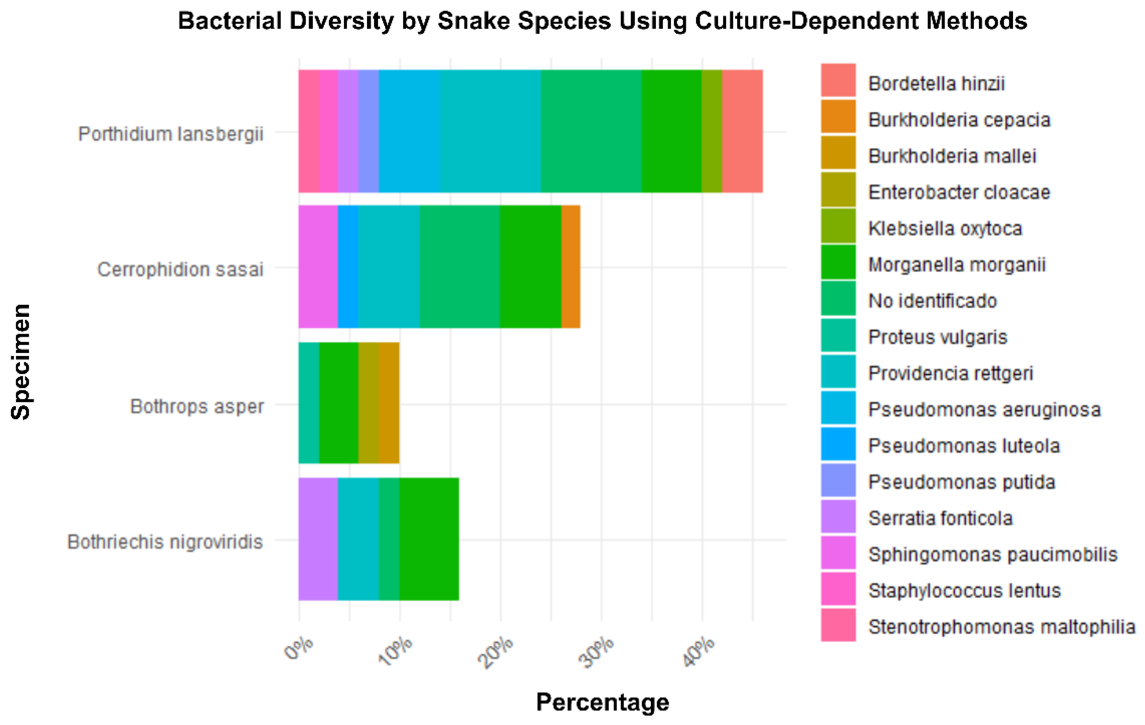
Figure 2.
Antibiotic Susceptibility and Resistance Profile in Gram-Negative Bacteria. This bar plot shows the proportion of Gram-negative bacteria classified as susceptible (S), intermediate (I), or resistant (R) to various antibiotics, highlighting differences in effectiveness.
Figure 2.
Antibiotic Susceptibility and Resistance Profile in Gram-Negative Bacteria. This bar plot shows the proportion of Gram-negative bacteria classified as susceptible (S), intermediate (I), or resistant (R) to various antibiotics, highlighting differences in effectiveness.
Disclaimer/Publisher’s Note: The statements, opinions and data contained in all publications are solely those of the individual author(s) and contributor(s) and not of MDPI and/or the editor(s). MDPI and/or the editor(s) disclaim responsibility for any injury to people or property resulting from any ideas, methods, instructions or products referred to in the content. |
© 2025 by the authors. Licensee MDPI, Basel, Switzerland. This article is an open access article distributed under the terms and conditions of the Creative Commons Attribution (CC BY) license (http://creativecommons.org/licenses/by/4.0/).
Copyright: This open access article is published under a Creative Commons CC BY 4.0 license, which permit the free download, distribution, and reuse, provided that the author and preprint are cited in any reuse.


