
Submitted:
08 September 2025
Posted:
09 September 2025
You are already at the latest version
Abstract
Objectives: To evaluate the safety of a novel patch graft, EverPatch PlusⓇ in glaucoma drainage device (GDD) surgery. Methods: We prospectively studied 18 eyes undergoing GDD implantation with EverPatch PlusⓇ and 105 control eyes with conventional scleral patch grafts at Sensho-kai Eye Institute. Postoperative conjunctiva overlying the EverPatch PlusⓇ was studied using anterior segment optical coherence tomography. Patches were secured to the sclera with at least four nonabsorbable sutures. The prevalence of patch extrusion was compared between the EverPatch and control cohorts. Results: In 16 of 18 cases, wounds healed well, and EverPatch remained covered by intact vascularized conjunctiva for >4 months. Conjunctival thickness at 1 mm and 3 mm from the anterior edge of the EverPatch increased by 74% and 87% at two weeks, then returned to baseline by three months. Although conjunctival retraction occurred, EverPatch position relative to the scleral spur remained stable, with no patch-related inflammation. Conjunctival bleaching occurred in two eyes at two months. One case (5.6%), with four prior ocular surgeries developed anterior edge lift-up and patch extrusion on postoperative day (POD) 80. Fluid accumulation around the EverPatch occurred in 8 eyes, resolving within one month in 7; one case with conjunctival bleaching progressed to bleb leak by POD 71. Conjunctival microcysts appeared in 8 eyes, resolving spontaneously within one month. Conclusion: Despite its different material properties from biological patch grafts, EverPatch PlusⓇ may be a viable alternative for tube coverage when firmly secured to the sclera.
Keywords:
artificial sclera
; patch material
; glaucoma drainage device
; EverPatch
; polycarbonate urethane
; anterior segment optical coherence tomography
; extrusion
Introduction
The silicone tube, known for its high biocompatibility, is a standard component of glaucoma drainage device (GDD). However, when placed on the sclera postoperative degeneration of the overlying conjunctiva may lead to tube exposure. To prevent such complications, various biological materials such as donor sclera, autologous sclera, pericardium, fascia lata, cornea, and amniotic membrane have been employed as patch grafts to cover the tube [1,2,3,4]. Among these, donor sclera and pericardium are commonly used due to their favorable conformity to surrounding tissues and cost-effectiveness.
Recently, the scleral tunnel technique has gained popularity as an alternative approach [5,6]. However, the risk of exposure with this method remained high up to 6.9%, particularly in patients under 65 years of age[7]. Thus, patch graft continues to be the mainstream method of tube coverage.
While biological patch grafts offer advantages such as suitable hardness, elasticity, toughness, affordability, and ease of handling, their typical thickness of 400 to 500μm can create a cosmetically noticeable elevation of the conjunctiva. This protrusion may induce progressive thinning or atrophy of the overlying tissues, eventually leading to patch graft exposure.
Furthermore, biological grafts are biodegradable and tend to thin over time[1,2,3,4], thereby increasing the risk of GDD exposure. They also carry potential risks of viral transmission and prion-related diseases, such as Creutzfeldt-Jakob disease [5,6].
To overcome the limitations associated with biological patch materials, the use of non-degradable synthetic material has been explored. Polytetrafluoroethylene (PTFE; Gore-tex®, Teflon), a chemically inert hydrophobic polymer with low electric polarizability, has been widely used in various medical applications, including artificial blood vessels, dura mater substitutes, sutures, and ligaments. Its use as a patch graft material for GDD has been previously reported[8,9]. However, PTFE may induce a mild fibrotic reaction [10] and has been associated with abscess formation in some case [11]. Styrene-block-isobutylene-block-styrene (SIBS), another synthetic hydrophobic polymer, is used as the tube material in the PreserFlo Microshunt. Although studies have evaluated the biocompatibility of SIBS when embedded in tissue, this material has not been adopted for use as GDD patch grafts [12].
Polycarbonate urethane (PCU), a biocompatible and hydrophobic polymer, has shown promise in a range of biomedical applications. It has been used in artificial blood vessels [13], joint arthroplasty[14], heart valves [15], nerve wraps for peripheral nerve regeneration, gingival tissue engineering, drug delivery systems, cartilage reconstruction and more.
The material of EverPatch PlusⓇ (CorNeat Vision, Ra’anana Israel) is a non-woven aromatic polycarbonate urethane sheet specifically designed for use in GDD surgeries to cover the drainage tube and prevent external protrusion. Its bio-affinity characteristics such as cytotoxicity, ocular irritation, systemic toxicity, and teratogenicity were evaluated according to ISO10993 standards. These assessments were registered under the US Clinical Trials Registry number NCT05469867 and conducted between December 11, 2020, and July 1, 2023. The trials were conducted at several locations including Prism Eye Institute, in Oakville, Ontario, Canada; The University Health Network at Toronto Western Hospital; Kwale Eye Hospital in Mombasa, Kenya; and Kenyata National Hospital and Lions Sight First Hospital in Nairobi, Kenya.
Histopathological studies conducted on animal eyes 13 weeks post-implantation revealed significant fibroblast infiltration and collagen fibril formation within the EverPatch, without evidence of adverse immune responses [16].
Human testing, registered under NCT04037917, was conducted from August 8, 2022, to June 18, 2024, at DaVinci Eye Care in Tbilisi, Georgia. This study evaluated the clinical outcomes of 10 patients aged 18 to 80 years over a 12-month period.
Finaly, this device received FDA 510(k) clearance.
The original EverPatch was a rectangular sheet measuring 0.5 cm x 0.65 cm and 100-150 μm thick, as shown in Figure 1. In the study using rectangular EverPatch conducted in Geogia, there was a 10% incidence of conjunctival penetration at the edges of the device.
Cytotoxicity or abnormal tissue reactions were not reported with this artificial patch material[17]. In later studies, extrusion of the EverPatch was observed in 13 of 29 eyes (approximately 45%) that underwent implantation of rectangular EverPatch at Wilmer Eye Institute[17]. To investigate the underlying causes of this high prevalence of extrusion, we employed anterior segment OCT to evaluate conjunctival thickness and associated clinical features. The CorNeat Co. released a Shield type EverPatch featuring a smooth anterior edge in January 2025 (Figure 1). We used 15 Shield-type patches and 3 rectangular-type patches in our practice and report here on their clinical performance over a follow-up period exceeding four months.
The Shield type (1a) has a rounded anterior edge, smaller surface area, and four fixation holes. In contrast, the rectangular type (1b) has a larger surface area and six fixation holes.
Subject and Methods
This prospective clinical study included eighteen eyes of eighteen patients who received CorNeat EverPatch PlusⓇ implantation. The prevalence of extrusion was compared with that in 105 eyes of 95 patients who received human donor scleral patch (64 eyes) or autologous scleral flap patching (41 eyes) at the Sensho-kai Eye Institute. Patients had severe glaucoma that was refractory to conventional glaucoma surgery. All patients underwent GDD surgery by two surgeons (EC and TC). The EverPatch PlusⓇ cohort underwent GDD surgery between January 2025 and May 2025. Seventeen patients were Japanese, and one was Chinese. The control cohort includes a total of 105 Japanese eyes that underwent Ahmed Glaucoma valve implantation with either a scleral allograft patch (n=64) or an autologous scleral flap (n=41) between January 2020 and January 2025. The patch material was switched from scleral patch to EverPatch in January 2025.
Inclusion criteria were refractory glaucoma requiring GDD surgery, clear cornea, and absence of conjunctival erosion or active conjunctivitis prior to surgery. Patients with poor compliance and those who received Baerveldt glaucoma Implants were excluded from this study. The study adhered to the tenets of the Declaration of Helsinki and was approved by the institutional review board of Sensho-kai (C2025-01R, 2023-03R). Informed consent was obtained from all patients. Baseline characteristics of participants are listed in Table 1.
Anterior Segment Optical Coherence Tomography (AS-OCT) Evaluation.
Patients were instructed to look downward or upward, and the light beam of the AS-OCT was directed perpendicular to the surface of the EverPatch. Conjunctival thickness was measured using the built-in measurement software of the AS-OCT system. Preoperatively, the deep episcleral vascular plexus often manifested as a hypo-reflective layer above the hyper-reflective sclera, allowing measurement of the conjunctiva-Tenon’s capsule complex. However, this hypo-reflective layer was indistinct in most postoperative cases. In such instances, the thickness was measured as the distance between the conjunctival surface and the surface of the EverPatch, which appeared as a white amorphous mass on AS-OCT.
This study focused on assessing the postoperative thickness of the conjunctiva-subconjunctival tissue complex over the EverPatch, the distance between the patch material and the anterior edge of the conjunctival incision, the distance between the scleral spur and anterior edge of the EverPatch, as well as the presence of subconjunctival fluid accumulation, microcysts, vascularity of the overlying conjunctiva, and wound dehiscence. The effects of the EverPatch on conjunctival thickness were assessed at 1 mm and 3 mm from the anterior edge of the EverPatch, however, before surgery, where no patch material existed, measurements were taken at 1 mm and 3 mm from the scleral spur. These evaluations were conducted using AS-OCT (CASIA II, Tomey Co., Aichi, Japan), infrared slit-lamp imaging, and standard slit-lamp examination.
Humphrey Visual Field Analyzer (Carl Zeiss Meditec Co., Tokyo), specular microscopy (CellCheck, Konan Co., Nishinomiya), Optos California (Nikon solutions Co., Tokyo), and Hamada’s infrared light Camera (Hamada-Shokai, Kyoto) were used to assess visual field defects, corneal endothelium, and infrared photographs of patch graft. Intraocular pressure (IOP) was measured using a Goldmann applanation tonometer.
Statistical Analysis
Statistical analyses were performed using Bell Curve for Excel (Social Survey Research Information Co., Tokyo, Japan). Analysis of variance (ANOVA) was used for comparing numerical data. The Kaplan–Meier survival analysis was used to assess prevalence of extrusion over time. The Wilcoxon signed-rank test was employed for paired comparison; Haberman residual analysis was used for categorical data. Reproducibility of data was assessed by calculating intraclass correlation coefficient.
Surgical Techniques:
Under local anesthesia, a radial incision and a fornix-based conjunctival incision was made at the limbus. In eyes with severe conjunctival scarring, a blunt knife was used with care to avoid conjunctival perforation. The tendons of two adjacent rectus muscles were exposed, and 4-0 silk traction sutures were placed. Subconjunctival scar tissue was excised to create adequate space for implantation. The plate of either an Ahmed glaucoma valve (AGV FP7; New world Medical, Rancho Cucamonga CA) or Paul Glaucoma Implant (PGI; Advanced Ophthalmic Innovations, Singapore) was positioned 8 mm posterior to the limbus and secured to the sclera using two 5-0 polyethylene sutures.
Before intraocular insertion, the tube of the AGV or PGI was passed through both the anterior and posterior loops of the EverPatch Plus.
For tube insertion into the ciliary sulcus, viscoelastic material was injected beneath the iris to create space. A 23G or 25 G needle was used to enter the sulcus through the sclera at a point 2.5mm posterior to the limbus. For anterior chamber insertion, no viscoelastic was used; a disposable needle was used to enter 2.0 mm from the limbus, parallel to the iris plane.
For pars plana insertion, the entry point was 3.5 mm posterior to the limbus.
The tube was then inserted into the ciliary sulcus, anterior chamber, or vitreous cavity through the pars plana, depending on the surgical plan.
The Shield type EverPatch Plus had four fixation holes, which were threaded with 9-0 nylon sutures and firmly secured to the sclera. The suture knots were rotated and buried in the sclera. The rectangular-type EverPatch had six fixation holes; all were threaded and similarly secured the scleral surface.
In the donor sclera cohort, a 3×5 mm scleral patch was placed over the tube. After insertion of the tube into the anterior chamber, ciliary sulcus or vitreous cavity, the patch was firmly secured to sclera using four 9-0 nylon sutures. In the autologous scleral flap cohort, a half thickness scleral flap measuring 4×5mm was created, and the tube was inserted into the anterior chamber or vitreous cavity beneath the flap. The flap was then tightly secured with four 9-0 nylon sutures.
The Tenon’s capsule and conjunctiva were carefully pulled forward and sutured at the limbus using a “locking” suture (combined running suture and single knot) with at least ten passes of 9-0 twisted polyglactin sutures.
The surgical field was disinfected with povidone-iodine. Dexamethasone (0.4mg) was injected, subconjunctivally, and the eye was dressed with ofloxacin and betamethasone ointment.
Postoperative care included topical gatifloxacin (GatiFlo) and 0.1% betamethasone (Rinderon), instilled four times daily for one month. Postoperative IOP was closely monitored, and anti-glaucoma medications were prescribed as needed.
Results
Baseline patient characteristics of the EverPatch cohort and the control cohort are summarized in Table 1. In the EverPatch cohort, eighteen eyes from eighteen patients with refractory glaucoma underwent implantation of GDD; five with AGV and thirteen with PGI. The Shield-type EverPatchⓇ was used in fifteen eyes, while the rectangular type was used in three. The tube was inserted into the ciliary sulcus in ten eyes, the anterior chamber in six, and the vitreous cavity in two. Tube placement was performed through the superior temporal quadrant in fifteen cases and through the inferonasal quadrant in three. Six cases followed for more than 6 months, and the mean follow-up period was 4.1±1.0 months.
Intraoperative findings of the Shield type and rectangular type EverPatch PlusⓇ is shown in Figure 1.
The clinical course of 18 eyes who underwent EverPatch implantation and 105 eyes who underwent conventional scleral patch is summarized in Table 2. Differences in preoperative IOP and the postoperative IOP at one month and three months between the EverPatch cohort and control group were not significant (P=0.463, 0.162 and 0.200 by ANOVA , respectively)
Clinical Course and Conjunctival Thickness in a Representative Case
The clinical course and postoperative changes in the dimensions of the conjunctiva-Tenon capsule complex after tube patching with the Shield-type EverPatch Plus® are presented in one representative case (Figure 2).
Thie patient was a 74-year-old man with POAG and an IOP of 40 mmHg despite five topical medications. He had a history of failed deep sclerectomy and underwent PGI implantation into the anterior chamber. The tube was covered with a Shield-type EverPatch® (CorNeat Vision, Ra’anana Israel) and implanted in the superotemporal quadrant.
Preoperatively, the subconjunctival tissue was cicatrized, and the conjunctival thickness measured 0.217 mm and 0.194 mm at 1 mm and 3 mm, respectively, from the scleral spur (Figure 2a). Three days after surgery, the conjunctival thickness at 1 mm and 3 mm from the front edge of the EverPatch increased to 0.379 mm (55% increase) and 0.345 mm (78% increase), respectively (Figure 2b). On POD 16, accumulation of transparent fluid was observed at the posterior part of the EverPatch. On the same day, the conjunctival thickness at 1 and 3mm from the front edge of the EverPatch increased to 0.342 mm (58% increase) and 0.594 mm (206% increase), respectively (Figure 2c). This fluid resolved by POD 30, and the conjunctival thickness decreased to 0.189 mm (18% decrease) at 1 mm and 0.269 mm (38% increase) at 3 mm (Figure 2d).
At 2 and 3 months postoperatively, the conjunctival thickness at 1 mm was 0.169 mm (22% decrease) and 0.124 mm (43% decrease), and at 3 mm, it was 0.137 mm (29% decrease) and 0.166 mm (14% decrease), respectively (Figure 2 e, f). At six months the EverPatch was covered with intact and well vascularized conjunctiva, and the thickness of conjunctiva at 1 mm and 3 mm was 0.125mm and 0.099 mm, respectively. The distance from the front edge of the EverPatch to the conjunctival incision site decreased from 1.635 mm on POD 3 to 1.229 mm three months postoperatively.
In all six cases followed for more than 6 months, no abnormal vascular dilatation, vascular bleaching, or extrusion of the patch material was observed.
In 16 of 18 eyes (89%), the conjunctiva overlying the EverPatch remained intact for 4 months with no extrusion. One representative case is shown.
(2a) Preoperative AS OCT: Subconjunctival tissue in the limbal area was scarred. Conjunctival thickness measured 0.217 mm and 0.194 mm at 1 mm and 3mm from the scleral spur, respectively.
(2b) Postoperative day (POD) 3. Conjunctiva was edematous. Thickness at 1mm and 3 mm from the anterior edge of the EverPatch increased to 0.379 and 0.345 mm, respectively.
(2c). POD 16: Transparent fluid accumulation was observed at posterior portion of the EverPatch. Conjunctival thickness at 1 mm and 3 mm from the anterior edge measured 0.342 mm and 0.594 mm, respectively. Arrow indicates a fenestration of the EverPatch.
(2d) POD30: Accumulated fluid resolved, and conjunctival edema subsided. Conjunctival thickness decreased to 0.189 mm and 0.269 mm at 1 mm and 3 mm, respectively.
(2e, 2f) Conjunctival appearance at 2 months (e) and 3 months (f): Conjunctival thickness continued to decrease, and the distance between the anterior edge of the EverPatch and conjunctival incision site also gradually shortened. Despite these changes, no extrusion of the EverPatch was observed.
(2g 2h) Six months postoperatively: Slit-lamp microscopy (2g) showed no conjunctival inflammation or ischemia. Infrared imaging (2h) revealed no migration or defects of the EverPatch.
Overall trends in conjunctival thickness, as assessed by anterior segment OCT(CASIA II), are shown in Figure 3 and Table 3. Considerable inter-patient variation was observed in the early postoperative period, which gradually diminished over time. The thickness peaked at 3.9 days and progressively decreased thereafter, with a more pronounced reduction at 1mm than at 3 mm (Figure 3, Table 3).
Thickness peaked between 3.9 days and 2 weeks, then gradually decreased to preoperative levels by 2 months. Thinning was more pronounced at 1 mm than at 3 mm from the anterior edge of the EverPatch.
At 1mm from the anterior edge of the EverPatch, conjunctival thickness at 3.9 days and 2weeks was significantly greater than the preoperative value, whereas thickness at 1, 2, and 3 months were not. The thickness at 3.9 days was also significantly greater than that at 1, 2, and 3 months, but not differ significantly from that at 2 weeks.
Similar trends were observed in the conjunctival thickness at 3 mm from the anterior edge of the EverPatch (Figure 3, Table 3).
The distance between the conjunctival incision site and the front edge of the EverPatch was 2.07 ± 0.69 mm within one week postoperatively (mean 3.9 ± 2.2 days) and retracted to 1.75 ± 0.71 mm at 3 months (P=0.117, Wilcoxon signed-rank test). A shortening of > 0.2 mm was observed in 6 of 18 eyes.
The distance between the scleral spur (SS) and the front edge of the EverPatch (EP) is shown in Table 4. Cases in which measurement was not possible due to peripheral anerior synechia or poor-quality angle images were excluded.
The SS-EP distance remained largely stable throughout follow-up, and the small increase observed were not statistically significant (Table 4).
Conjunctival microcysts, which are common findings after filtering surgery, were observed in 8 of 18 patient (Figure 4). These microcysts resolved spontaneously within one month without any sequalae. If the conjunctiva was thick preoperatively, postoperative edema tends to be more pronounced.
(Figure 4a) A 65-year-old patient with neovascular glaucoma, extensive peripheral anterior synechiae, and preoperative IOP of 66 mmHg on maximal medical therapy including oral acetazolamide underwent pars plana implantation of AGV FP7 with rectangular EverPatch coverage following two vitrectomies and one failed endoscopic cyclophotocoagulation. Preoperative conjunctival thickness was 0.313 mm and 0.268 mm at 1 mm and 3 mm from the scleral spur, respectively, both above the cohort mean of 0.262 mm and 0.267 mm. Three days after implantation, the conjunctiva was markedly swollen with numerous microcysts, thickness at 1 mm measured 1.448 mm.
(4b)POD 10: Swelling partially subsided (1.201 mm), but fluid accumulation appeared over the EverPatch.
(4c) POD 17: Both sewelling and fluid accumulation resolved.
(4d) At 1 month, thickness at 1 mm decreased to 0.729 mm (+133% vs. preoperative), and by 3 months to 0.771 mm (+189% vs. preoperative value).
Accumulation of fluid around the patch was observed in 8 of 18 eyes within the first two weeks after surgery. In 7 of these cases the fluid resolved spontaneously within one month (Figure 2c, d, Figure 4b, 4d). In the remaining case, fluid accumulation persisted and eventually developed an ischemic filtering bleb by the second postoperative month.
In 5 of 18 cases, neither fluid accumulation nor conjunctival microcyst were observed around the patch material.
At 3 months, the conjunctival wound healed well in 16 of 18 patients (89%), and the EverPatch was covered by intact conjunctiva. An example of six months postoperative appearance of the EverPatch and overlying conjunctiva observed with a conventional slit lamp is shown in Figure 2g, and corresponding infrared image is shown in Figure 2h.
Conjunctival bleaching was observed in 2 of 18 patients. The ischemic area was localized to the anterior portion of the EverPatch. One of these patients was a 74-year-old woman with neovascular glaucoma who underwent ciliary sulcus implantation of the Paul Glaucoma implant along with patching of the Shield type EverPatch, following two previous trabeculectomies at the superior temporal quadrant.
On POD 50, the bleached area was concave, surrounded by a vascularized zone, and demonstrated fluorescein pooling. AS-OCT demonstrated subconjunctival fluid accumulation adjacent to the EverPatch (Figure 5a), consistent with a localized ischemic filtering bleb. Al this stage, the conjunctival thickness was 0.377 mm at 1 mm and remained intact, as confirmed by a negative Seidel testing (Figure 5b). However, the ischemic bleb subsequently progressed to a leaking bleb by POD 71 (Figure 5c). Another notable finding was the elevation or “folding” of the patch (Figure 5c), suggesting an external force pushing the patch forward. The overlying conjunctiva at this site had become markedly thinned. During surgical repair with conjunctival rotation, we confirmed a presence of weak adhesion between the conjunctiva and the EverPatch, however, this adhesion was quite weak, and the two layers could be easily separated.
(5a): POD 50 slit lamp image: Localized conjunctival bleaching overlying the EverPatch with subconjunctival fluid accumulation (bleb formation; arrow). The ischemic area was surrounded by vessel rich conjunctiva and was Seidel negative.
(5b): POD 50 AS-OCT: Conjunctival thickness at ischemic area remained thickness of 0.377mm.
(5c): POD 71: Disrupted conjunctiva (arrow) at the site of prior conjunctival bleaching. Focal thinning was evident at the site of EverPatch elevation. Folding of the patch (asterisk) suggests an external force pushing the EverPatch forward. The superficial white line indicates a hyperreflective signal from leaked aqueous humor.
In another case, conjunctival bleaching was observed at the anterior portion of the EverPatch (Figure 6a) without fluorescein staining. The conjunctival thickness at 1 mm from the anterior edge of the EverPatch progressively decreased, measuring 0.172 mm at 1 month (Figure 6b), 0.153 mm at 2 months (Figure 6c) and 0.103 mm at 3 months (Figure 6d). Despite this thinning, there was no evidence of subconjunctival fluid, Seidel positivity or conjunctival erosion. The conjunctival bleaching was found exclusively at the anterior portion of the EverPatch.
(6a) POD 65. Slit-lamp image showing conjunctival bleaching (arrow) located to the anterior portion of the EverPatch.
(6b-d): Progressive reduction in conjunctival thickness at 1 mm from the anterior edge of the EverPatch: 0.172mm at 1 month (b), 0.153 mm at 2 months (c), and 0.103 mm at 3 months (d). Despite progressive thinning, no extrusion, subconjunctival fluid or Seidel positivity was observed.
Extrusion of the EverPatch occurred in 1 of 18 patients (5.6%) on POD 80.
The patient was a 67-year-old Chinese man with primary angle closure glaucoma who had undergone four prior ocular surgeries in the right eye: viscocanalostomy, trabeculectomy, Ahmed glaucoma valve implantation in the superior temporal quadrant, and Descemet Stripping Automated Endothelial Keratopathy (DSAEK). He exhibited extensive peripheral anterior synechiae, and his IOP remained above 30 mmHg despite four topical medications. Axial length measured 23.39 mm, and corneal curvature was as small as 7.18mm. As a fifth intervention, a Paul Glaucoma implant was placed in the inferonasal quadrant with tube insertion into the ciliary sulcus reducing IOP to 9 mmHg by 2 months postoperatively.
On POD 20, conjunctival thickness at 1 mm from the anterior edge of the EverPatch was thick enough, measuring 0.593 mm. The distance between the scleral surface and the patch surface was 0.386 mm (Figure 7a). The distance from the conjunctival incision site to the anterior edge of the EverPatch was 3.21mm, which shortened to 1.625 mm on POD 80 . Conjunctival thickness rapidly decreased to 0.266 mm on POD 37 (Figure 7b) and 0.107 mm on POD 51 (Figure 7c). Finally, the EverPatch had extruded on POD 80 (Figure 7d). The distance between the bottom of the tube and the outer surface of the EverPatch increased from 0.642 mm on POD 51 to 0.753 mm on POD 80, indicating a progressive elevation of the EverPatch (Figure 7d).
No abnormal exudate, subconjunctival abscess, or sign of infection was observed, and the patient reported no pain or discomfort. The likely cause of extrusion was compression-induced atrophy of compromised tissue rather than inflammation. The conjunctiva was not ischemic on POD 20 (Figure 7e), and the extruded portion was located away from the conjunctival incision site at the limbus (arrow, Figure 7f)
(7a) : POD 20 in a patient with primary angle closure glaucoma combined with severe anterior synechiae. Conjunctival thickness at 1 mm from the anterior edge of the EverPatch was 0.593 mm, exceeding the cohort average of 0.468 mm (2 weeks) and 0.455 mm (1 month) in 18 patients. The distance between the scleral surface and the EverPatch was 0.386 mm with no intervening space, indicating firm fixation of the EverPatch to the sclera.
(7b): POD 37. Conjunctival thickness decreased rapidly to 0.266 mm.
(7c): POD 51. Thickness at 1 mm further decreased to 0.107 mm. The distance between the scleral surface and EverPatch surface was not measurable in this cross-section; instead, the distance from the inferior end of the tube to the EverPatch surface was measured at 0.642mm, greater than that at POD 20.
(7d): On POD 80, the EverPatch had extruded. The distance from the inferior end of the tube to the EverPatch surface increased to 0.753mm, suggesting anterior edge lift-up.
(7e): Slit-lamp image on POD 20 showing no wound dehiscence or abnormal inflammation.
(7f): Slit-lamp image on POD 80. The extrusion site was separated from the surgical incision site (arrow), indicating that wound dehiscence at the incision site was not the cause.
The prevalence of extrusion in patients who underwent EverPatch implantation was compared with that in 64-eyes with allogenic donor scleral patch and 41 eyes with autologous scleral patch (Figure 8). No significant difference in extrusion prevalence was found between the groups (P=0.798, Log-rank test).
No statistically significant difference was observed in the probability of patch or tube extrusion between the 18 eyes with EverPatch tube coverage and 105 eyes with scleral covering (64 allogenic donor sclera, and 41 autologous sclera) (logrank test P=0.798).
Discussion
The EverPatch is made of hydrophobic polycarbonate urethane, which differs fundamentally from traditional biological patch grafts such as sclera or pericardium, which are hydrophilic. It is very thin measuring only 100 to 150 μm, typically four to five times thinner than sclera, and is non-degradable. The bioaffinity of the EverPatch has been enhanced by its fine fibrous structures. According to the CorNeat company website, animal studies have demonstrated significant fibroblast infiltration and collagen fibril formation within the EverPatch [16].
However, studies by Kanter et al. suggest that the pore size of the woven structure is only 3.7 µm, which is likely too small to permit fibroblast infiltration [17]. Although polycarbonate urethane demonstrates no cytotoxicity or foreign body reaction, which are favorable features for use as a graft material, its hydrophobic nature and structural characteristics may limit its ability to adhere firmly to adjacent conjunctival or scleral tissue. If the patch is not firmly secured to the sclera with sutures, its weak adhesion may lead to patch dislocation and mechanical damage to the surrounding conjunctiva. In the present study, poor adhesion was evident during surgical repair of aqueous leakage case and aligns with the findings of Kanter et al [17].
In our study, the EverPatch was securely anchored to the sclera with at least four nylon sutures. This likely contributed to its stability, as reflected by the minimal change in the distance between the scleral spur and the anterior edge of the EverPatch over the three-month follow-up, and the absence of significant migration (Table 3). In contrast, Kanter et al. reported securing the EverPatch with only two sutures in 21 of 29 eyes (72.4%), and using absorbable Vicryl sutures in 16 of 29 eyes (53.3%) [17]. In two Vicryl cases, the patch displaced and migrated onto the cornea.
In conventional GDD surgery using biological patch graft, the graft is often secured with only two sutures, and the conjunctival wound is closed with two corner stitches. This approach may be insufficient for synthetic materials like the EverPatch, which have weaker adhesion to sclera and conjunctival than biological grafts. When conjunctival closure relies solely on two corner stitches, migrated patch material can lead to wound dehiscence. In our cases, the conjunctiva was closed using a “locking suture” (running suture reinforced with a knot), which may have helped maintaining conjunctival integrity and prevented wound dehiscence. These technical differences, particularly the use of multiple nylon fixation sutures and secure conjunctival closure, may explain the marked difference between the high extrusion rate reported by Kanter (45%) and much lower rate in our study (5.6%) [17].
When using the EverPatch, another important point to note is to eliminate vertical misalignment of the patch. If the distance between fixation sutures is shorter than the patch length, the patch may bend, leading to edge elevation or body folding (Figure 5,7). In the case shown in Figure 5, the distance between the scleral spur and the anterior edge of the EverPatch remained stable, however, elevation of the posterior portion occurred. This suggests that a short distance between anterior and posterior sutures, or a forward-directed force exerted by the plate of GDD, may have caused upward displacement and subsequent atrophy of the overlying conjunctiva (Figure 5c). Due to the weak adhesion between conjunctiva and synthetic material, deflection of the EverPatch may lead to separation, gap formation and subsequent aqueous leakage. Therefore, synthetic graft such as the EverPatch should be secured firmly to the sclera in a matter that prevents vertical deflection.
If the patch is excessively large, achieving stable fixation without distortion becomes more challenging. In this regard, smaller patches, such as the Shield type, may offer advantages by reducing deformation risk and improving conformity.
Bleaching of the overlying conjunctiva, which is a sign of vascular loss and a potential precursor to tissue erosion, was observed in two cases. In addition to mechanical compression atrophy of tissues, local ischemia may also contribute to this finding. Large, oxygen-impermeable patches can induce localized ischemia in the overlying conjunctiva; thus, using a smaller patch may help reduce this risk.
Baseline conjunctival thickness may also influence extrusion risk, with thinner conjunctiva predisposing to higher vulnerability. Conjunctival thickness varies among ethnicities. Reported value at 1 mm and 3 mm from the scleral spur were 223.0 µm and 190.8 µm in Hispanic individuals, and 203.8 µm and 187.4 µm in Caucasians, respectively[18]. In contrast, Chinese individuals have shown thickness of 238.8 µm at 3 to 5 mm from the limbus [19]. Most patients in our study were Japanese, whose conjunctiva may be thicker than that of white or Black individuals.
A history of multiple prior surgeries is a well-established risk factor for patch extrusion.
In our study, the preoperative conjunctival thickness measured 262 µm and 267 µm at 1 mm and 3 mm from the scleral spur, respectively. Notably, 16 of 18 eyes included had a history of previous ocular surgeries, suggesting that the increased baseline thickness reflected subconjunctival scarring and higher risk of extrusion in this study cohort. As shown in Figure 4, conjunctival edema was more pronounced, and postoperative thickness remain elevated, in eyes with preoperative conjunctiva scarring and thickening. Although the underlying mechanism remains unclear, previous studies have reported a higher prevalence of patch extrusion in eyes with a surgical history, despite increased conjunctival thickness [20,21].
In this study, the patient who experienced patch extrusion had undergone multiple ocular surgeries, used many topical anti-glaucoma medications, and received PGI implantation in the infero-nasal quadrant, all well-established risk factors for extrusion. In addition, the presence of severe postoperative exotropia likely promoted drying of the nasal conjunctiva, contributing to the extrusion. Additionally, the patient’s small palpebral fissure may have necessitated excessive force to open the eyelids during examinations, placing mechanical stress on an already vulnerable conjunctiva, particularly when the patch was elevated.
From this perspective, the extrusion in this case was likely the result of multiple interacting factors rather than the use of the EverPatch alone.
The hydrophobicity of the EverPatch may not be an unfavorable property for an implant, as many hydrophobic materials, such as polyethylene, nylon, polypropylene, silicon and SIBS (Styrene-Isobutylene-Styrene), are widely used in sutures and implantable devices. As previously reported, biological materials can deposit on hydrophobic surface and convert them to hydrophilic surfaces. In practice, the contact angle of 94.6°in new EverPatch decreased to 66.6°in an explanted EverPatch[17]. Factors such as design, size, and application techniques can also influence biocompatibility of materials. Enhancing pore size by fabricating a woven sheet from various polyurethane fibers or applying hydrophilic surface coatings may further improve the material’s affinity to surrounding tissues[22].
Fluid accumulation adjacent to the EverPatch occurred in 8 of 18 eyes, resolving within 1 month in all but one case (Figure 5). Such fluid accumulations are typically associated with filtering surgeries [23]. The clear appearance of the accumulated fluid on AS-OCT, along with the absence of vascular engorgement or patient discomfort, rules out the possibility of cytotoxicity or foreign body reaction. If the patch material or any released irritating monomers were cytotoxic, one would expect progressive exudation, vasodilation, and inflammation over time. In contrast, the fluid accumulation and conjunctival microcysts observed between 3 days and 2 weeks postoperatively resolved within 1 month, a pattern resembling the conjunctival response seen after the tube coverage with biological patch materials[1,4]. These findings, together with the progressive thinning of postoperative conjunctiva (Figure 3), suggest a minimal postoperative inflammatory response.
Given that GDD implantation functions as a form of filtering surgery, it is plausible that aqueous humor pooled on the implant plate or leaked from the tube’s insertion site could lead to accumulation of aqueous humor around the EverPatch.
Additionally, the anterior chamber remained clear with no signs of uveitis, patients reported no foreign body sensation, and slit-lamp examinations at 6 months showed no significant vasodilation (Figure 2g), all of which support the absence of an adverse tissue response. Reports by Kanter et al [17], together with information presented on the manufacturer’s website, are consistent with the low cytotoxicity and minimal foreign body reaction associated with polycarbonate urethane.
The rectangular EverPatch measured 0.5cm × 0.65cm with a thickness of 100-150μm. A key concern regarding this design is the potential mechanical stress exerted by its sharp edges on surrounding tissues. Rounding the front edge may help reduce mechanical stress on the overlying conjunctiva. Despite this design refinement, extrusion occurred in 1 of 15 cases using the Sield type EverPatch (6.7%), a rate comparable to previously reported GDD or tube extrusion rates [20,24]. Improvement of surgical technique may mitigate the risk; however, the prevalence of extrusion may not be lower than that observed with biological patch grafts. Further improvement of hydrophobicity and design may be necessary to enhance the safety of the PCU patch graft.
Limitation: The follow-up period in this study was relatively short (4.1±1.0 months), and the sample size was limited to 18 subjects. As ensuring safety is our highest priority, long-term studies with larger cohorts are warranted to future evaluate the clinical performance and safety of the EverPatch Plus.
Conclusion
The EverPatch Plus, a synthetic patch graft fundamentally different from traditional biological materials, was used to cover tubes in 18 glaucoma drainage device surgeries. Postoperative conjunctival thickness increased by 74-87% at 2 weeks and returned to preoperative levels by 3 months. When the patch was firmly secured to the sclera, the 3- month extrusion rate was 5.6%, with no exudation or inflammation observed around the artificial patch graft.
These findings suggest that firm scleral fixation may reduce the risk of extrusion to a level comparable with biological patch grafts.
Author Contributions
Conceptualization, E.C.; methodology, E.C.; software, E.C.; validation, E.C. and T.C.; formal analysis, E.C.; investigation, E.C.; resources, E.C..; data curation, E.C.; writing— original draft preparation, E.C.; writing—review and editing, T.C..; visualization, T.C.; supervision, L.H.; project administration, E.C.; funding acquisition, E.C. All authors have read and agreed to the published version of the manuscript.
Funding
This research received no external funding.
Institutional Review Board Statement
The design of this study was approved by the Institutional Review Board (IRB) of Sensho-kai (Head H Amano); the approval number is C2025-1R on 19 January 2025. The study was conducted in accordance with the Declaration of Helsinki.
Informed Consent Statement
Informed consent was obtained from all subjects involved in the study. Written informed consent for publication was not specifically obtained from the participating patients; however, the possibility of publication was disclosed during the process of obtaining consent for surgical agreement. The content of this paper does not identify individual participants, and the IRB waived the requirement for additional consent for publication.
Data Availability Statement
The research data used in this research are available upon request from the corresponding author.
Acknowledgments
We extend our gratitude to the technicians at Sensho-kai for providing data that contributed to this study. Corresponding author confirms that all individuals included have consented to participate in this study.
Conflicts of Interest
The authors declare no conflicts of interest
References
- Akbas, Y.B.; Alagoz, N.; Sari, C.; Altan, C.; Yasar, T. Evaluation of pericardium patch graft thickness in patients with Ahmed glaucoma valve implantation: an anterior segment OCT study. Jpn J Ophthalmol. 2024, 68, 192–199. [Google Scholar] [CrossRef]
- de Luna, R.A.; Moledina, A.; Wang, J.; Jampel, H.D. Measurement of Gamma-Irradiated Corneal Patch Graft Thickness After Aqueous Drainage Device Surgery. JAMA Ophthalmol. 2017, 135, 941–946. [Google Scholar] [CrossRef] [PubMed]
- Smith, M.F.; Doyle, J.W.; Ticrney, J.W., Jr. A comparison of glaucoma drainage implant tube coverage. J Glaucoma. 2002, 11, 143–147. [Google Scholar] [CrossRef] [PubMed]
- Schipper, P.; Weber, C.; Lu, K.; Fan, S.; Prokosch, V.; Holz, F.G.; Mercieca, K. Anterior segment OCT for imaging PAUL(®) glaucoma implant patch grafts: a useful method for follow-up and risk management. Graefes Arch Clin Exp Ophthalmol. 2024. [Google Scholar] [CrossRef]
- Thakur, S.; Ichhpujani, P.; Kumar, S. Grafts in Glaucoma Surgery: A Review of the Literature. Asia Pac J Ophthalmol (Phila). 2017, 6, 469–476. [Google Scholar] [CrossRef] [PubMed]
- Zalta, A.H. Long-term experience of patch graft failure after Ahmed Glaucoma Valve® surgery using donor dura and sclera allografts. Ophthalmic Surg Lasers Imaging. 2012, 43, 408–415. [Google Scholar] [CrossRef]
- Youn, S.; Yan, D.B. Five-Year Outcomes of Graft-Free Tube Shunts and Risk Factors for Tube Exposures in Glaucoma. J Glaucoma. 2024, 33, 139–147. [Google Scholar] [CrossRef]
- Jacob, T.; LaCour, O.J.; Burgoyne, C.F.; LaFleur, P.K.; Duzman, E. Expanded polytetrafluoroethylene reinforcement material in glaucoma drain surgery. J Glaucoma. 2001, 10, 115–120. [Google Scholar] [CrossRef]
- Yasuoka, K.; Tada, K.; Yasuoka, K. Ahmed Glaucoma Valve (AGV) implantation using Gore-Tex. Jpn J Ophthalmic Surg. 2024, 37, 541–545. [Google Scholar]
- Leszczynski, R.; Gumula, T.; Stodolak-Zych, E.; Pawlicki, K.; Wieczorek, J.; Kajor, M.; Blazewicz, S. Histopathological Evaluation of a Hydrophobic Terpolymer (PTFE-PVD-PP) as an Implant Material for Nonpenetrating Very Deep Sclerectomy. Invest Ophthalmol Vis Sci. 2015, 56, 5203–5209. [Google Scholar] [CrossRef]
- Jawad, S.; Halenda, K. Late-onset Pseudomonas aeruginosa orbital cellulitis following glaucoma drainage device implantation. Am J Ophthalmol Case Rep. 2024, 34, 102054. [Google Scholar] [CrossRef]
- Nakamura, K.; Fujimoto, T.; Okada, M.; Maki, K.; Shimazaki, A.; Kato, M.; Inoue, T. Tissue Reactivity to, and Stability of, Glaucoma Drainage Device Materials Placed Under Rabbit Conjunctiva. Transl Vis Sci Technol. 2022, 11, 9. [Google Scholar] [CrossRef]
- Gu, Y.; Tian, C.; Qin, Y.; Sun, Y.; Liu, S.; Li, H.; Duan, X.; Shu, C.; Ouyang, C. The novel hybrid polycarbonate polyurethane / polyester three-layered large-diameter artificial blood vessel. J Biomater Appl. 2022, 36, 965–975. [Google Scholar] [CrossRef] [PubMed]
- Lazic, S.; Kellett, C.; Afzal, I.; Mohan, R.; Killampalli, V.; Field, R.E. Three-year results of a polycarbonate urethane acetabular bearing in total hip arthroplasty. Hip Int. 2020, 30, 303–308. [Google Scholar] [CrossRef] [PubMed]
- Sun, M.; Elkhodiry, M.; Shi, L.; Xue, Y.; Abyaneh, M.H.; Kossar, A.P.; Giuglaris, C.; Carter, S.L.; Li, R.L.; Bacha, E.; et al. A biomimetic multilayered polymeric material designed for heart valve repair and replacement. Biomaterials. 2022, 288, 121756. [Google Scholar] [CrossRef] [PubMed]
- The CorNeat EverPatch: Superior Alternative to Tissue Grafts. accessed on 2025/1/29. https://www.corneat.com/news/the-corneat-everpatch%3A-superior-alternative-to-tissue-grafts.
- Kanter, J.; Garkal, A.; Cardakli, N.; Pitha, I.; Sabharwal, J.; Schein, O.D.; Ramulu, P.Y.; Parikh, K.S.; Johnson, T.V. Early Postoperative Conjunctival Complications Leading to Exposure of Surgically Implanted CorNeat EverPatch Devices. Ophthalmology. 2025, 132, 799–814. [Google Scholar] [CrossRef]
- Fernández-Vigo, J.I.; Fernández-Aragón, S.; Burgos-Blasco, B.; Ly-Yang, F.; De-Pablo-Gómez-de-Liaño, L.; Almorín-Fernández-Vigo, I.; Martínez-de-la-Casa, J.M.; Fernández-Vigo, J. Comparison in conjunctival-Tenon’s capsule thickness, anterior scleral thickness and ciliary muscle dimensions between Caucasians and Hispanic by optical coherence tomography. Int Ophthalmol. 2023, 43, 3969–3977. [Google Scholar] [CrossRef]
- Zhang, X.; Li, Q.; Liu, B.; Zhou, H.; Wang, H.; Zhang, Z.; Xiang, M.; Han, Z.; Zou, H. In vivo cross-sectional observation and thickness measurement of bulbar conjunctiva using optical coherence tomography. Invest Ophthalmol Vis Sci. 2011, 52, 7787–7791. [Google Scholar] [CrossRef]
- Al-Beishri, A.S.; Malik, R.; Freidi, A.; Ahmad, S. Risk Factors for Glaucoma Drainage Device Exposure in a Middle-Eastern Population. J Glaucoma. 2019, 28, 529–534. [Google Scholar] [CrossRef]
- Byun, Y.S.; Lee, N.Y.; Park, C.K. Risk factors of implant exposure outside the conjunctiva after Ahmed glaucoma valve implantation. Jpn J Ophthalmol. 2009, 53, 114–119. [Google Scholar] [CrossRef]
- Chen, Y.; Guo, Y.; Li, X.; Chen, Y.; Wang, J.; Qian, H.; Wang, J.; Wang, Y.; Hu, X.; Wang, J.; et al. Comparison study of surface-initiated hydrogel coatings with distinct side-chains for improving biocompatibility of polymeric heart valves. Biomater Sci. 2024, 12, 2717–2729. [Google Scholar] [CrossRef]
- Kawana, K.; Kiuchi, T.; Yasuno, Y.; Oshika, T. Evaluation of trabeculectomy blebs using 3-dimensional cornea and anterior segment optical coherence tomography. Ophthalmology. 2009, 116, 848–855. [Google Scholar] [CrossRef]
- Geffen, N.; Buys, Y.M.; Smith, M.; Anraku, A.; Alasbali, T.; Rachmiel, R.; Trope, G.E. Conjunctival complications related to Ahmed glaucoma valve insertion. J Glaucoma. 2014, 23, 109–114. [Google Scholar] [CrossRef]
Figure 1.
a,b: Intraoperative findings of Shield-type (left, 1a) and rectangular-type (right,1b) EverPatch Plus® during Ahmed Glaucoma valve implantation.
Figure 1.
a,b: Intraoperative findings of Shield-type (left, 1a) and rectangular-type (right,1b) EverPatch Plus® during Ahmed Glaucoma valve implantation.
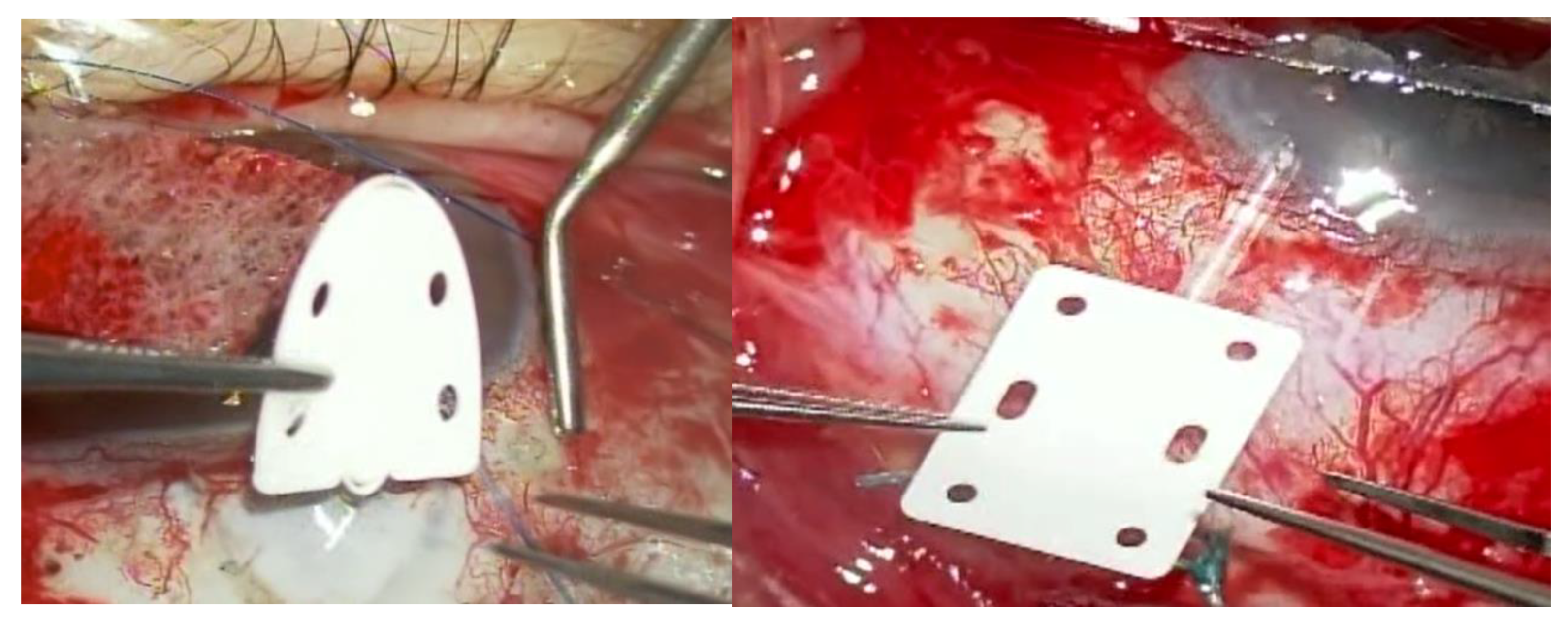
Figure 2.
(a-h): AS-OCT and slit-lamp images of the conjunctiva after EverPatch Plus coverage in a case successfully followed for more than 6 months.
Figure 2.
(a-h): AS-OCT and slit-lamp images of the conjunctiva after EverPatch Plus coverage in a case successfully followed for more than 6 months.
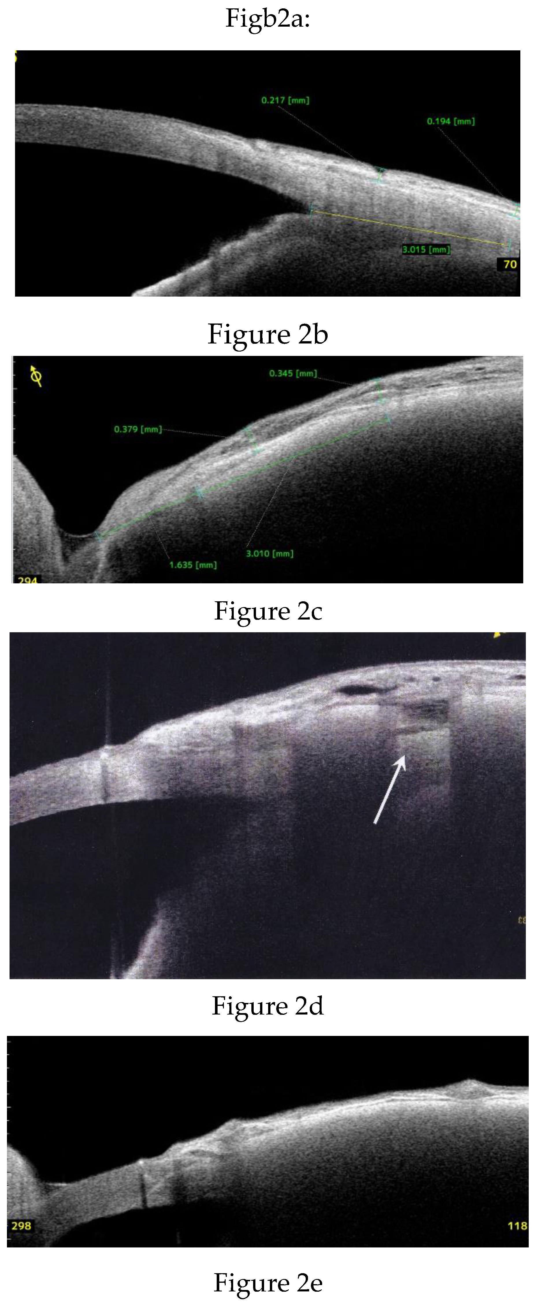
Figure 3.
Postoperative trends in conjunctival thickness following EverPatch Plus coverage.
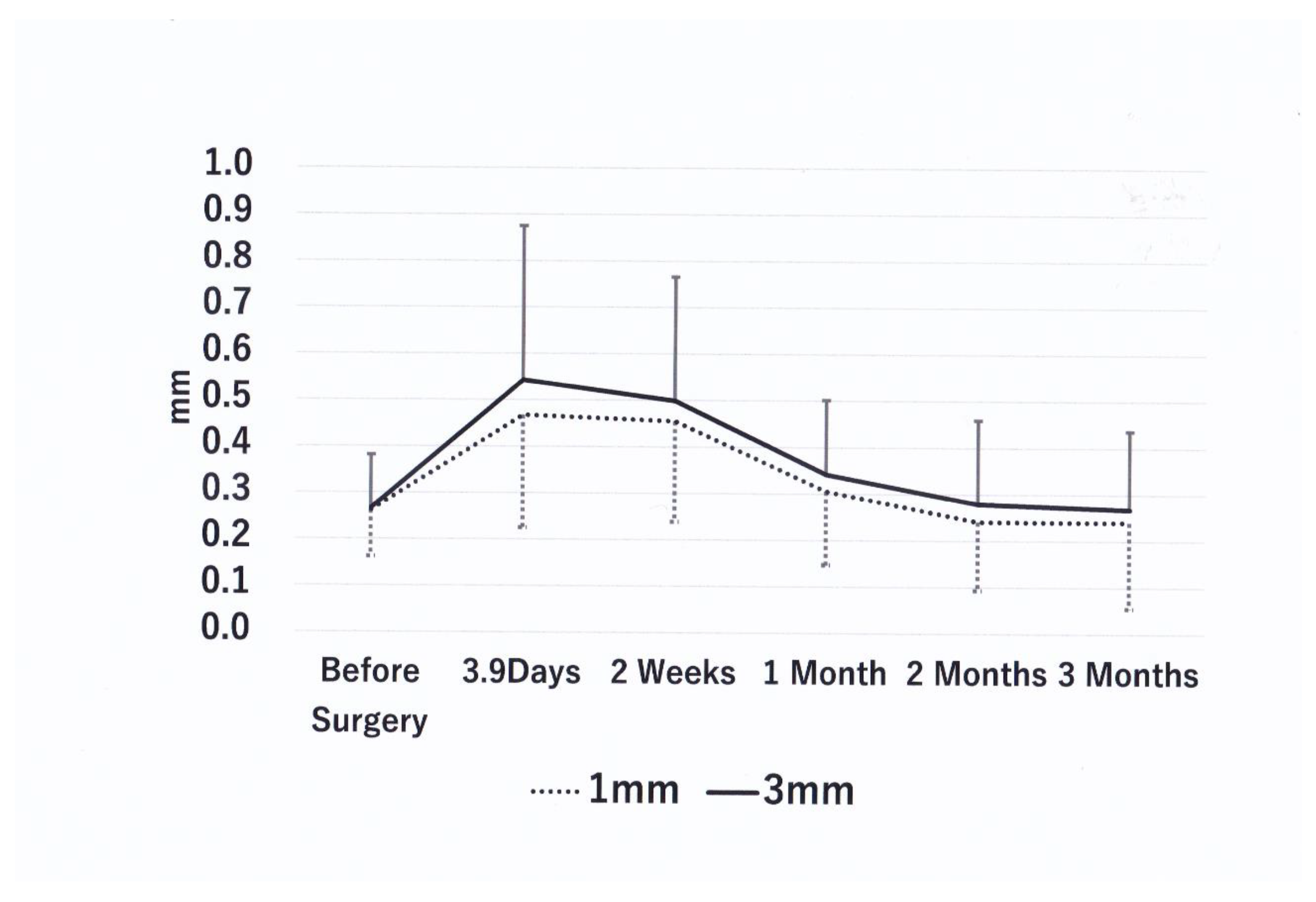
Figure 4.
(a-d) Representative case of conjunctival microcysts and fluid accumulation resolving within one month.
Figure 4.
(a-d) Representative case of conjunctival microcysts and fluid accumulation resolving within one month.
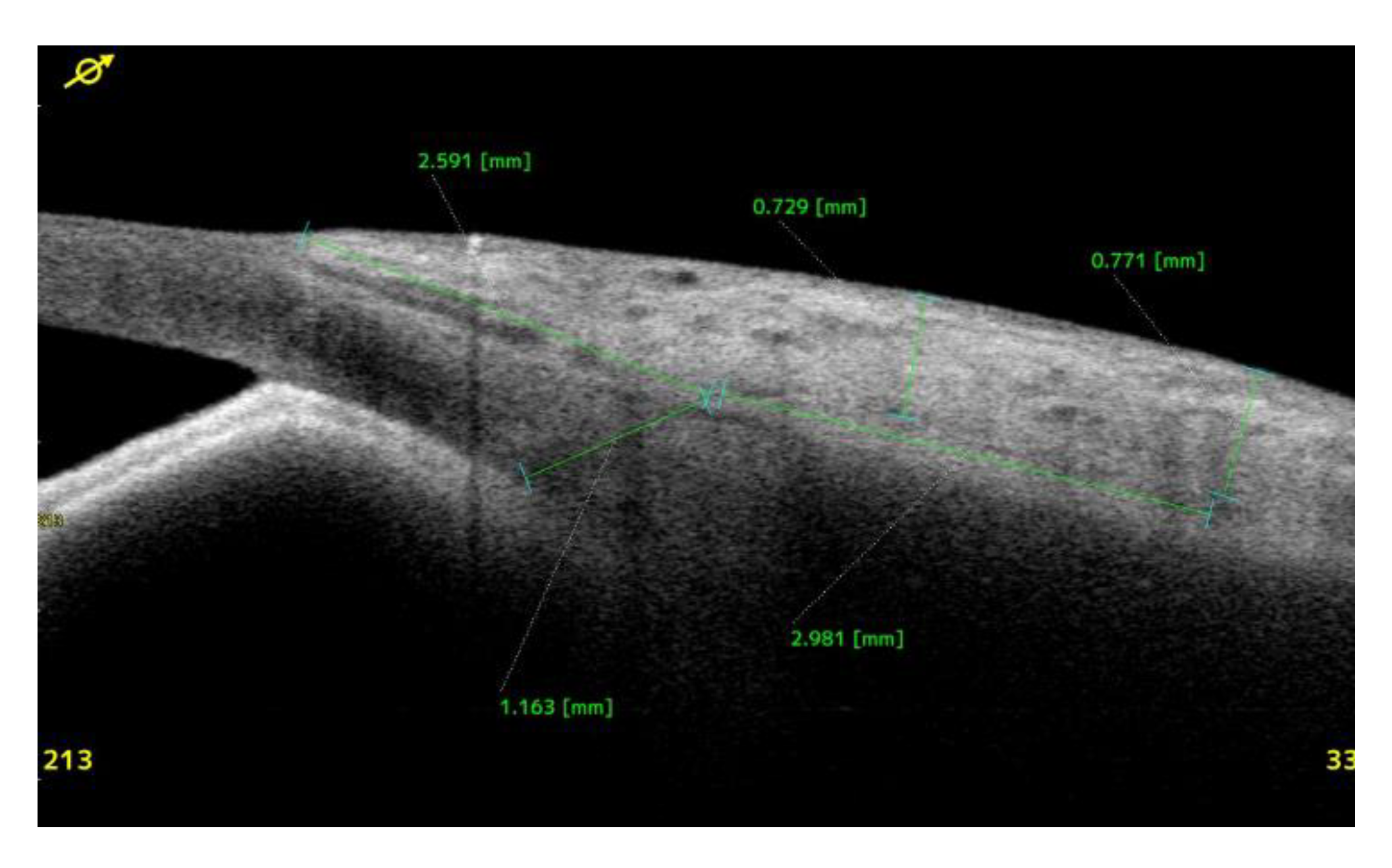
Figure 5.
AS-OCT images of an eye that progressed from conjunctival bleaching to bleb leaks.
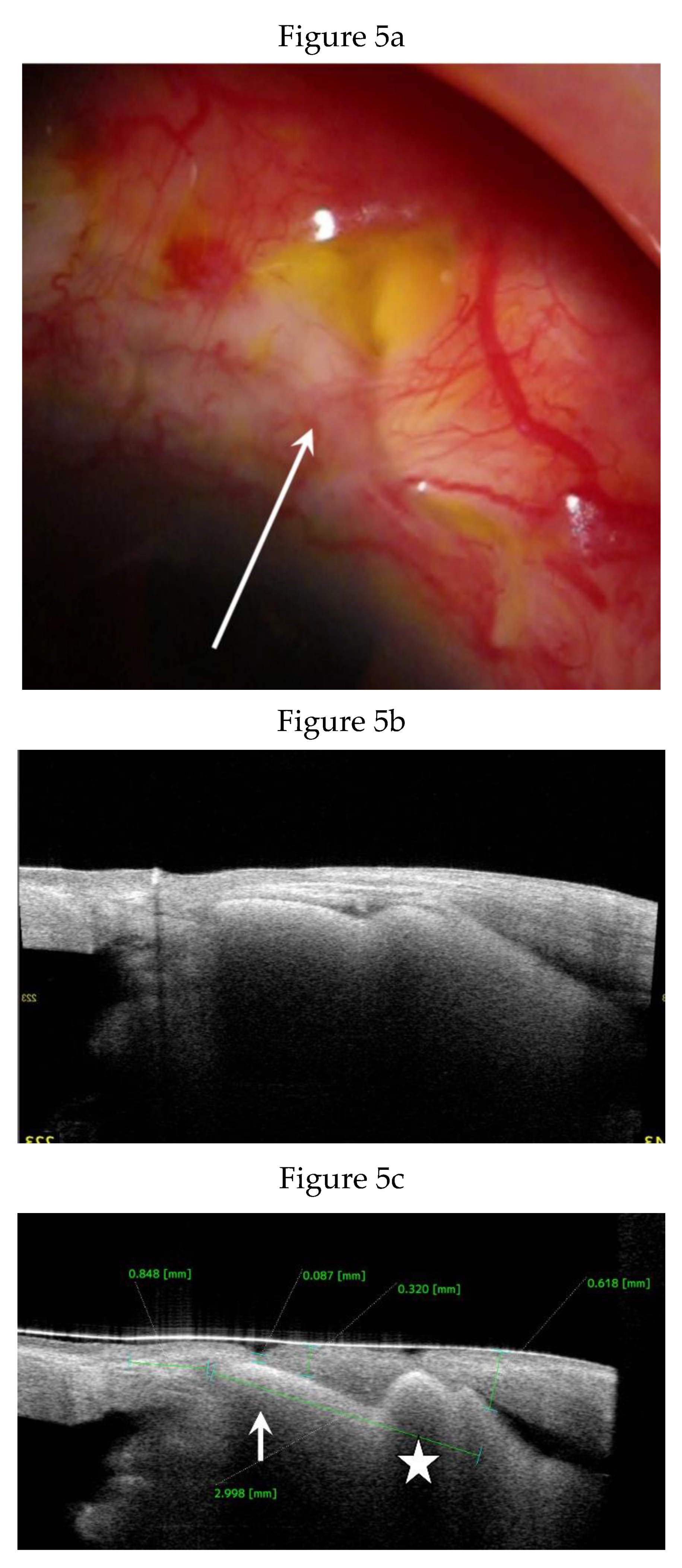
Figure 6.
AS-OCT images of an eye with conjunctival bleaching persisting for 3 months without bleb leaks.
Figure 6.
AS-OCT images of an eye with conjunctival bleaching persisting for 3 months without bleb leaks.
Figure 7.
AS-OCT and slit-lamp images of eyes that developed EverPatch extrusion.
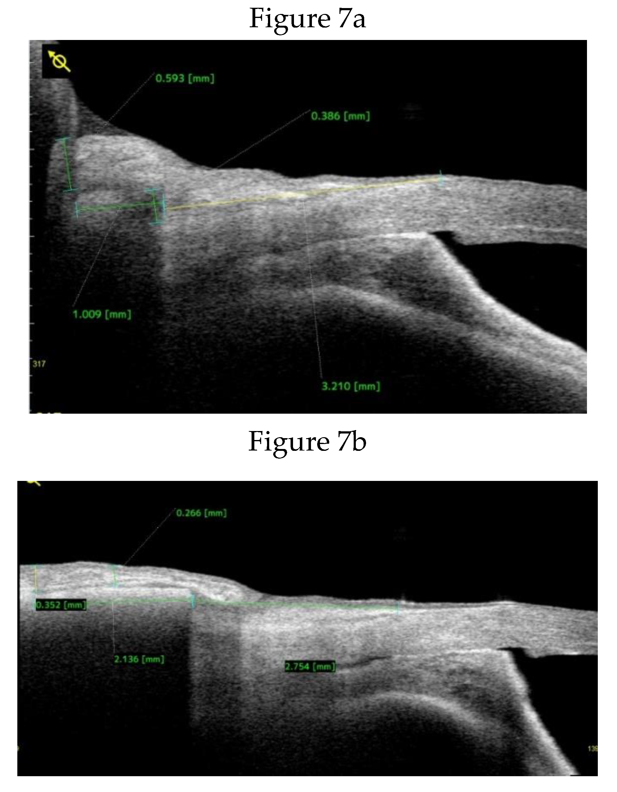
Figure 8.
Difference in prevelence of extrusion among EverPatch cohort, allograft donor sclera cohort, and autologous sclera patch cohort.
Figure 8.
Difference in prevelence of extrusion among EverPatch cohort, allograft donor sclera cohort, and autologous sclera patch cohort.
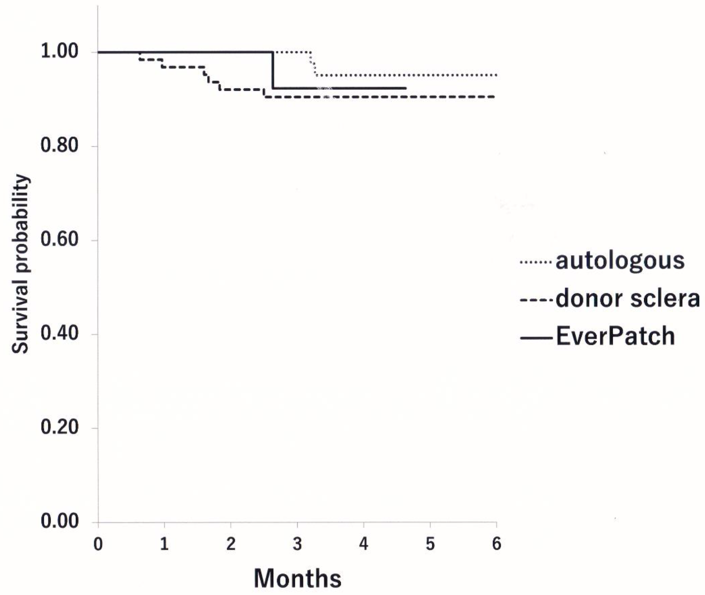

Table 1.
Demographic data of patients treated with EverPatch and scleral patch.
| EverPatch | Scleral Patch | P Value (ANOVA) | |
| Age | 65.8±15.1 | 62.5±17.9 | 0.463 |
| Type of Glaucoma | NVG 5, POAG 7, PEG 2,SG 2,PACG 2 | NVG 27, POAG 25, PEG 10, SG 26, PACG 5 | 0.132 (residual analysis) |
| Sex: Male/Female | 11/7 | 57/48 | 0.619 |
| Side: R/L | 8/10 | 50/55 | 1.00 |
| Pre-op IOP (mmHg) | 29.8±13.7 | 29.4±9.4 | 0.758 |
| Pre-op BCVA (logMAR) | 0.901±0.923 | 0.708±0.857 | 0.383 |
| Pre-op meds | 4.3±1.2 | 4.1±1.2 | 0.491 |
| Number of prior surgeries | 2.0±1.2 | 2.7±1.9 | 0.121 |
| Visual field defects (HFA: MD) | -24.3±9.2* | -18.9±9.3* | 0.107 |
| Type of GDD used | AGV 5, PGI 13 | AGV 105 | NA |
| Endothelial cell density (cells/mm2) | 2149±524 | 1993±636 | 0.504 |
| Type of EverPatch | Shield type 15, Rectangular type 3 | NA | |
| Quadrant of tube insertion | Sup-temporal 15, Infero-nasal 3 | Sup-temporal 52, Sup-nasal 12, Inf-temporal 30, Inf-nasal 11 | 0.008 (residual analysis) |
| Tube tip location | Anterior chamber 6, Ciliary sulcus 10, pars plana 2 | Anterior chamber 26, ciliary sulcus 21, pars plana 58 | P<0.001(residual analysis) |
| Pre-op conjunctival thickness at 1 mm from SS | 0.262±0.100 | NA | |
| Pre-op conjunctival thickness at 3 mm from SS | 0.267±0.114 | NA |
NVG: Neovascular glaucoma, POAG; primary open angle glaucoma, PEG; Exfoliation glaucoma, SG; secondary glaucoma, PACG; primary angle closure glaucoma, *: visual field data of 4 patients in EverPatch cohort and 48 patients in control was not available due to severe visual disturbance. Pre-op: preoperative, BCVA (logMAR): best corrected visual acuity by logarithmic minimal angular resolution. HFA MD: mean deviation assessed by Humphrey visual field analyzer, dB: decibels. GDD: glaucoma drainage device, AGV: Ahmed Glaucoma valve, PGI: Paul Glaucoma Implant. SS: scleral spur
Table 2.
Summary of postoperative IOP and visual acuity.
| Pre-op | 2W | 1M | 2M | 3M | ||
| EverPatch | # of medications | 4.3±1.2 | 1.2±2.0 | 1.8±2.1 | 2.0±2.1 | 2.2±2.0 |
| IOP | 31.1±13.0 | 15.6±6.3 | 16.6±4.3 | 17.5±8.8 | 16.1±4.9 | |
| logMAR BCVA | 0.869±0.823 | 1.067±0.993 | 1.087±0.963 | 1.105±0.972 | 1.018±1.058 | |
| Control | # of medications | 4.1±1.2 | NA | NA | NA | 2.2±1.8 |
| IOP | 29.7±10.9 | NA | 19.7±9.0 | NA | 18.1±5.7 | |
| logMAR BCVA | 0.708±0.857 | NA | NA | NA | NA |
IOP: intraocular pressure, logMAR: logarithms minimal angle resolution, BCVA: best corrected visual acuity.
Table 3.
Trends in conjunctival thickness following patching with EverPatch Plus.
| Time point | 1 mm thickness (mm) | % Change vs Baseline | P vs. Baseline | P vs 3.9 days | 3 mm Thickness (mm) | % Change vs Baseline | P vs Baseline | P vs 3.9 days |
| Baseline | 0.262±0.100 | 0 | - | - | 0.267±0.114 | 0 | - | - |
| 3.9 days N=18 | 0.468±0.217 | 78.4 | 0.004↑ | - | 0.543±0.332 | 103.4 | 0.001↑ | - |
| 2 weeks N=18 | 0.455±0.217 | 73.6 | 0.004↑ | 0.609(ns) | 0.498±0.267 | 86.8 | 0.002↑ | 0.925(ns) |
| 1 month N=18 | 0.305± 0.16 | 16.5 | 0.246(ns) | 0.007↓ | 0.342±0.160 | 28.2 | 0.052(ns) | 0.006↓ |
| 2 months N=18 | 0.241±0.147 | -8.2 | 0.687(ns) | 0.001↓ | 0.279±0.180 | 4.5 | 0.831(ns) | 0.002↓ |
| 3 months N=16 | 0.240±0.185 | -8.3 | 0.501(ns) | 0.005↓ | 0.268±0.169 | 0.4 | 0.877(ns) | 0.006↓ |
↑=significantly greater than comparator; ↓= significantly less than comparator; ns=not significant.
Table 4.
Trends in a distance between scleral spur (SS) and the anterior edge of the EverPatch following glaucoma drainage device surgery.
Table 4.
Trends in a distance between scleral spur (SS) and the anterior edge of the EverPatch following glaucoma drainage device surgery.
| Time point | SS-EP (mm) | P* | Time point | Conjunctival incision-EP (mm) | P* |
| 4.9 days N=9 | 1.01±0.20 | 3.9 days N=12 | 2.07 ± 0.69 | ||
| 2 weeks N=7 | 1.07±0.23 | 0.893 | |||
| 1 month N=14 | 1.13±0.34 | 0.263 | |||
| 2 months N=15 | 1.20±0.39 | 0.953 | |||
| 3 months N=14 | 1.21±0.40 | 0.484 | 3 months N=12 | 1.75 ± 0.71 | 0.117 |
P*: P value by Wilcoxon signed-rank test comparing one-week data with each corresponding time point.
Disclaimer/Publisher’s Note: The statements, opinions and data contained in all publications are solely those of the individual author(s) and contributor(s) and not of MDPI and/or the editor(s). MDPI and/or the editor(s) disclaim responsibility for any injury to people or property resulting from any ideas, methods, instructions or products referred to in the content. |
© 2025 by the authors. Licensee MDPI, Basel, Switzerland. This article is an open access article distributed under the terms and conditions of the Creative Commons Attribution (CC BY) license (http://creativecommons.org/licenses/by/4.0/).
Copyright: This open access article is published under a Creative Commons CC BY 4.0 license, which permit the free download, distribution, and reuse, provided that the author and preprint are cited in any reuse.


