
Submitted:
31 August 2025
Posted:
02 September 2025
You are already at the latest version
Abstract
Enterobiasis is an intestinal parasitic infection that presents with anal itching and can have serious complications. A 44-year-old soldier with no known medical history was found deceased in his vehicle, not far from his father's farm. Autopsy revealed generalized visceral congestion and heavy, congested lungs, suggestive of acute pulmonary edema.Sterile swabs were systematically applied to the natural orifices (mouth, nose, anus, ear canals) of all autopsied bodies. Suprisingly, anal swab revealed the presence of eggs of Enterobius vermicularis, and the anal region was free of eczematization after inspection.Even though no link was found between the parasite and this patient's death. The gastrointestinal tract can be severely affected, potentially leading to a fatal outcome.Clinicians need to be aware that enterobiasis is common in crowded environments, especially among soldiers. It can also be a taboo subject, which may lead to neglect and potentially fatal outcomes in some young adult patients.
Keywords:
Enterobius vermicularis
; enterobiasis
; pruritus
; parasitosis
; post-mortem
; asymptomatic
Introduction
Enterobius vermicularis adults (pinworms) in a nematode that resides and reproduces in the cecum. Gravid females migrate to the perianal region to deposit their eggs, a process often noticed by the host during the night [1].
Enterobiasis (or oxyuriasis), caused by Enterobius vermicularis, is an infection primarily affecting the large bowel and cecum. Common symptoms include anal itching, as well as intestinal or nervous system disorders [2].
Although Enterobius vermicularis is generally considered a non-invasive parasite that cannot penetrate a healthy intestinal mucous membrane, it has been known to migrate to other tracts and organs, including the urinary bladder, peritoneum, kidneys, liver, and lungs [2].
While this agent's role as a causative factor in acute appendicitis is controversial, its contribution to mortality is less recognized [2].
Enterobiasis with abdominal pain and/or acute appendicitis, ranging from acute appendicitis form to suppurative appendicitis variant have been documented [3,4,5,6].
Deaths directly caused by this parasite are rarely reported and are usually diagnosed after an autopsy [1,2].
By inducing mucosal inflammation and ulceration, pinworms compromise the integrity of the bowel mucosa, which can lead to penetration of deeper layers of the bowel wall, intestinal perforation, and hemorrhage [2,7,8,9].
In some cases, severe gastrointestinal complications may lead to death [2].
Clinicians should be alert to Enterobius vermicularis as a potential cause of fatal acute abdominal pain, particularly in adolescent and young adult patients [1].
Case Report
A 44-year-old soldier with no known medical history was found deceased in his vehicle, not far from his father's farm. His body was transferred to the local forensic medicine department for an autopsy requested by the public prosecutor.
External examination revealed no signs of violence or suspicious traumatic injuries (Figure 1). However, signs of decerebration, bloody nasal discharge, and indicators suggesting cerebral distress and cardiorespiratory distress were observed. Cyanosis of the extremities, earlobes, and face was also present.
Internal examination revealed generalized visceral congestion and heavy, congested lungs containing a frothy fluid, suggestive of acute pulmonary edema. The heart was enlarged. The entire organ was excised for a comprehensive histopathological examination. Toxicological analyses were also performed, but they were negative.
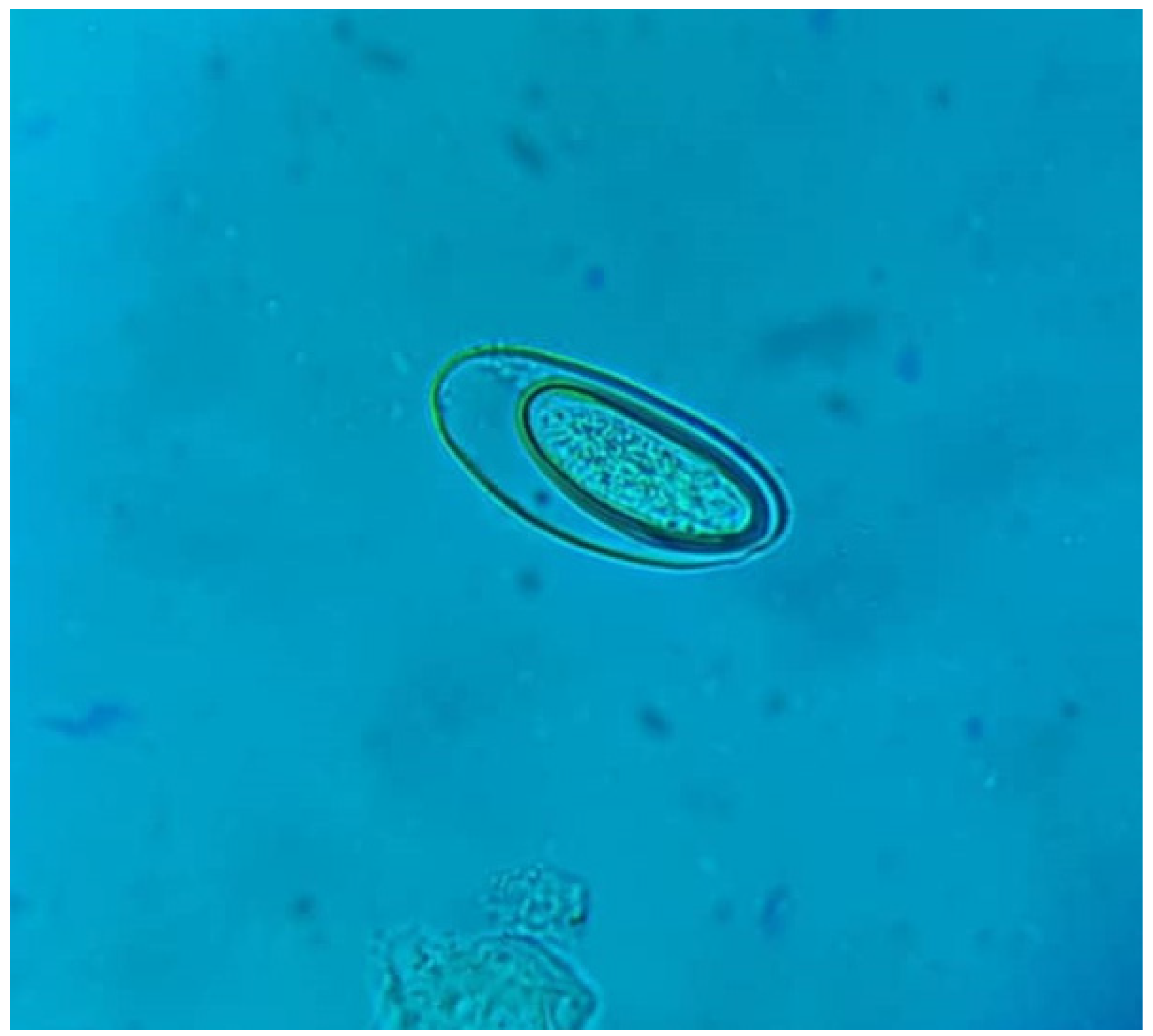
A microbiological examination of the natural orifices unexpectedly revealed asymmetrical and translucent eggs of Enterobius vermicularis on the anal swabs (Figure 2).
The patient had two children. Based on the interview with the deceased's wife, no clinical evidence of enterobiasis was found in his children, and there were no signs of eczematization in the perianal area.
Although mortality from Enterobius vermicularis is rarely reported, the parasite should not be neglected.
Discussion
When a young person dies, forensic investigations are initiated to determine the cause and manner of death, involving a meticulous examination of the body and the scene. This involves a thorough examination of the body and the scene, which might include autopsies, imaging, and biological analyses.
The leading causes of death in young adults are unintentional injuries, followed by homicide and suicide. The other origin of death is cancer and heart disease [10,11].
In countries where enterobiasis is common, it continues to be a public health and economic issue. [2,12]
Enterobiasis exhibits varied pathophysiology and infestation routes, leading to diverse clinical presentations [1,13,14], which can explain this apparently asymptomatic case, or neglected condition. In addition, the close quarters in military barracks pose a significant risk compared to the general population.
Enterobius vermicularis is generally a non-invasive parasite that does not penetrate a healthy intestinal mucous membrane [2].
The characteristic symptoms of enterobiasis include perianal, perineal, or vulvar irritation, which can lead to sleep disturbances, restlessness, and appetite loss. Because Enterobius vermicularis deposits its eggs outside the intestinal lumen, standard stool microscopy is not relevant [1,15].
The Graham's scotch tape method is the standard for detecting these infections. In rare instances, the parasite can infect extra-intestinal sites, such as the urinary tract, kidneys, biliary tree, fallopian tubes, and eyes, especially in patients from rural settings [1,15].
No eczematization was found in the anal region of our deceased patient, which would have indicated a superinfection and proven severe pruritus.
Enterobius vermicularis is not considered a major cause of death. Multiple samples of the intestinal wall should be taken at random in order to find the pinworms inside [2].
Histological examination of the duodenum and proximal ileum can reveal diffuse necrosis accompanied by multiple foci of transmural hemorrhage [2].
It can also show the thick cuticles and internal organs of the worms can be easily recognizable within the intestinal wall [2,16].
Chronic and massive parasitic intestinal infection can have a serious hemodynamic impact.
Reports of death directly caused by this parasite are rare, and diagnoses are usually made following an autopsy [1,2]
This case demonstrates the simplicity of anal swab examination, allowing for quick diagnosis of enterobiasis and potentially prompting further ileal histological assessment.
Enterobiasis can lead to severe complications, significant morbidity, and mortality [1]. Other similar cases may go unnoticed, and the number of fatal undiagnosed cases remains unknown.
Learning Points
- The transmission route of enterbiasis is primarily fecal-oral, and the eggs of the pinworm can survive on surfaces for up to three weeks.
- Enterobiasis can be considered a taboo subject, especially among adults. This can be a significant problem because it often leads to a delay in seeking treatment, which in turn makes the infection harder to manage.
- In a situation like a military community, where the risk of transmission is already high due to close living quarters, this reluctance to seek help can be especially problematic. It can lead to persistent, ongoing outbreaks that affect many people.
- The histological examinations of the colon were not performed in time to prove a possible link between enterobiasis and the patient's death. However, routinely, anal swabing can help to assess the prevalence of disease and establish the possible fatal cases.
Conclusion
Enterobiasis is rarely reported as a direct cause of death; it's typically diagnosed during an autopsy.
Anal swabing should be used in forensic investigation as a simple and epidemiological tool to comprehand the parasitic disease and its complications
References
- Al-Shouli ST, Barry M, Binkhamis K, AlHogail N, Alafaleq NO, Dufailu OA, Aljerian K. Fatal Case of a Child Harboring Enterobius vermicularis. Healthcare (Basel). 2023 Mar 22;11(6):917. [CrossRef]
- Mansueto G, De Simone M, Ciamarra P, Capasso E, Feola A, Campobasso CP. Infections Are a Very Dangerous Affair: Enterobiasis and Death. Healthcare (Basel). 2021 Nov 27;9(12):1641. [CrossRef]
- Hammood, Z.D.; Salih, A.M.; Mohammed, S.H.; Kakamad, F.H.; Salih, K.M.; Omar, D.A.; Hassan, M.N.; Sidiq, S.H.; Mustafa, M.Q.; Habibullah, I.J.; et al. Enterobius vermicularis causing acute appendicitis, a case report with literature review. Int. J. Surg. Case Rep. 2019, 63, 153–156. [CrossRef]
- Taghipour, A.; Olfatifar, M.; Javanmard, E.; Norouzi, M.; Mirjalali, H.; Zali, M.R. The neglected role of Enterobius vermicularis in appendicitis: A systematic review and meta-analysis. PLoS ONE 2020, 15, e0232143. [CrossRef]
- Efared, B.; Atsame-Ebang, G.; Soumana, B.M.; Tahiri, L.; Hammas, N.; El Fatemi, H.; Chbani, L. Acute suppurative appendicitis associated with Enterobius vermicularis: An incidental finding or a causative agent? A case report. BMC Res. Notes 2017, 10, 494. [CrossRef]
- Rajendran, S.; Carmody, E.; Murphy, M.; Barry, B. Enterobius granulomas as a cause of abdominal pain. BMJ Case Rep. 2015, 2015, bcr2015210464.
- Cateau E., Yacoub M., Tavilien C., Becq-Giraudon B., Rodier M.-H. Enterobius vermicularis in the kidney: An unusual location. J. Med Microbiol. 2010;59:860–861. [CrossRef]
- Zahariou A., Karamouti M., Papaioannou P. Enterobius vermicularis in the male urinary tract: A case report. J. Med. Case Rep. 2007;1:137. [CrossRef]
- Sammour Z.M., Gomes C.M., Tome A.L.F., Bruschini H., Srougi M. Prolonged irritative voiding symptoms due to Enterobius vermicularis bladder infestation in an adult patient. Braz. J. Infect. Dis. 2008;12:352. [CrossRef]
- M Noun, AM Boumelik, KB Ayad, ID Bensefia, S Benfarhate, O Hadjazi. Y Merad. Stab wound suicide mimicking homicide: a forensic case report. HAL open science.
- M Noun, MA Boumelik, S Benfarhate, MD Merzoug, KB Ayad, ID Bensefia, Y Merad. Autopsy of a Suicide Note: When Final Words Reveal the Suffering of a Suicidal Crisis–A Forensic Case Study, Preprints, 2025.
- Yabanoğlu H., Aytaç H.Ö., Türk E., Karagülle E., Calışkan K., Belli S., Kayaselçuk F., Tarım M.A. Parasitic infections of the appendix as a cause of appendectomy in adult patients. Turk. Soc. Parasitol. 2014;38:12–16. [CrossRef]
- Merad Y, Merbouh A, Benallal K, Belfodel S, Adjmi-Hamoudi H. Prevalence of enterobiasis among urban school children in Sidi-bel-Abbes, Algeria. International Journal of Innovation and Applied Studies 24 (2), 453-458, 2018.
- Merad Y, Nasrallah A, Rehamnia Y, Ebaidallah K, Rezoug Y, Bouroumi F. A leech in the upper tract of a Shepherd: Case report and review of literature. Journal of Current Medical Research and Opinion 7 (01), 1988-1996, 2024.
- Harumatsu T., Baba T., Orokawa T., Sunagawa H., Ieiri S. A rare case of acute appendicitis with Enterobius vermicularis. Pediatr. Int. 2022;64:e15195. [CrossRef]
- Sinniah B., Leopairut J., Neafie R.C., Connor D.H., Voge M. Enterobiasis: A histopathological study of 259 patients. Ann. Trop. Med. Parasitol. 1991;85:625–635. [CrossRef]
Figure 1.
Autopy of patient without any sign of violence or superficial apparent lesion.
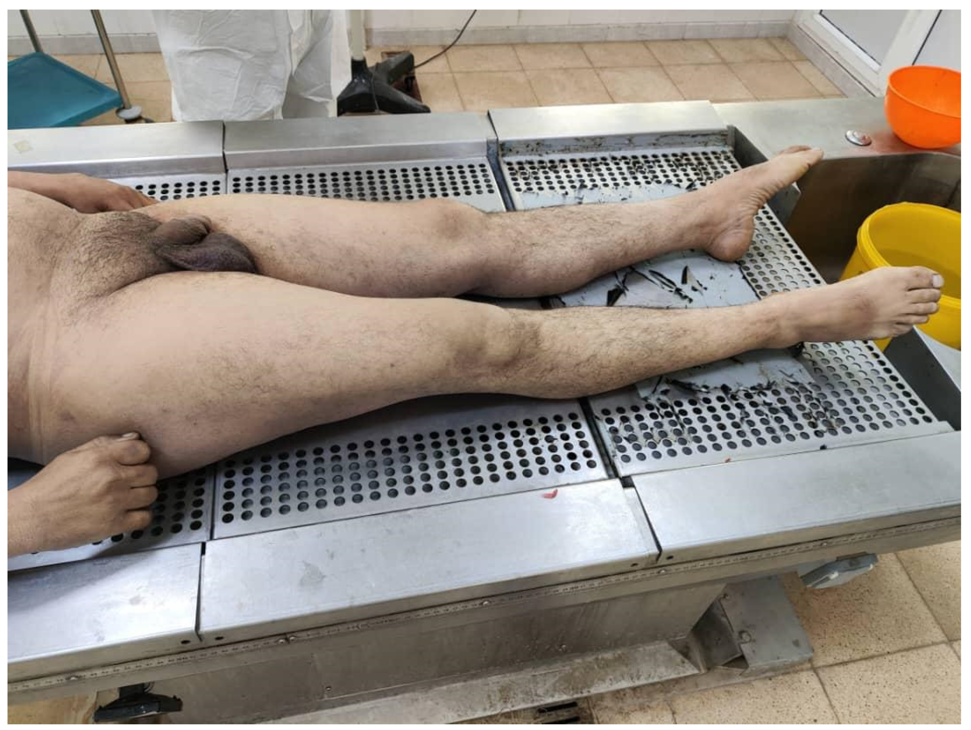
Figure 2.
Postmortem anal swab revealing eggs of Enterobius vermicularis.
Disclaimer/Publisher’s Note: The statements, opinions and data contained in all publications are solely those of the individual author(s) and contributor(s) and not of MDPI and/or the editor(s). MDPI and/or the editor(s) disclaim responsibility for any injury to people or property resulting from any ideas, methods, instructions or products referred to in the content. |
© 2025 by the authors. Licensee MDPI, Basel, Switzerland. This article is an open access article distributed under the terms and conditions of the Creative Commons Attribution (CC BY) license (http://creativecommons.org/licenses/by/4.0/).
Copyright: This open access article is published under a Creative Commons CC BY 4.0 license, which permit the free download, distribution, and reuse, provided that the author and preprint are cited in any reuse.


