Preprint
Review

This version is not peer-reviewed.

Clarifying the Dual Role of Staphylococcus spp. in Cheese Production

A peer-reviewed article of this preprint also exists.

Submitted:

19 August 2025

Posted:

21 August 2025

You are already at the latest version

Abstract

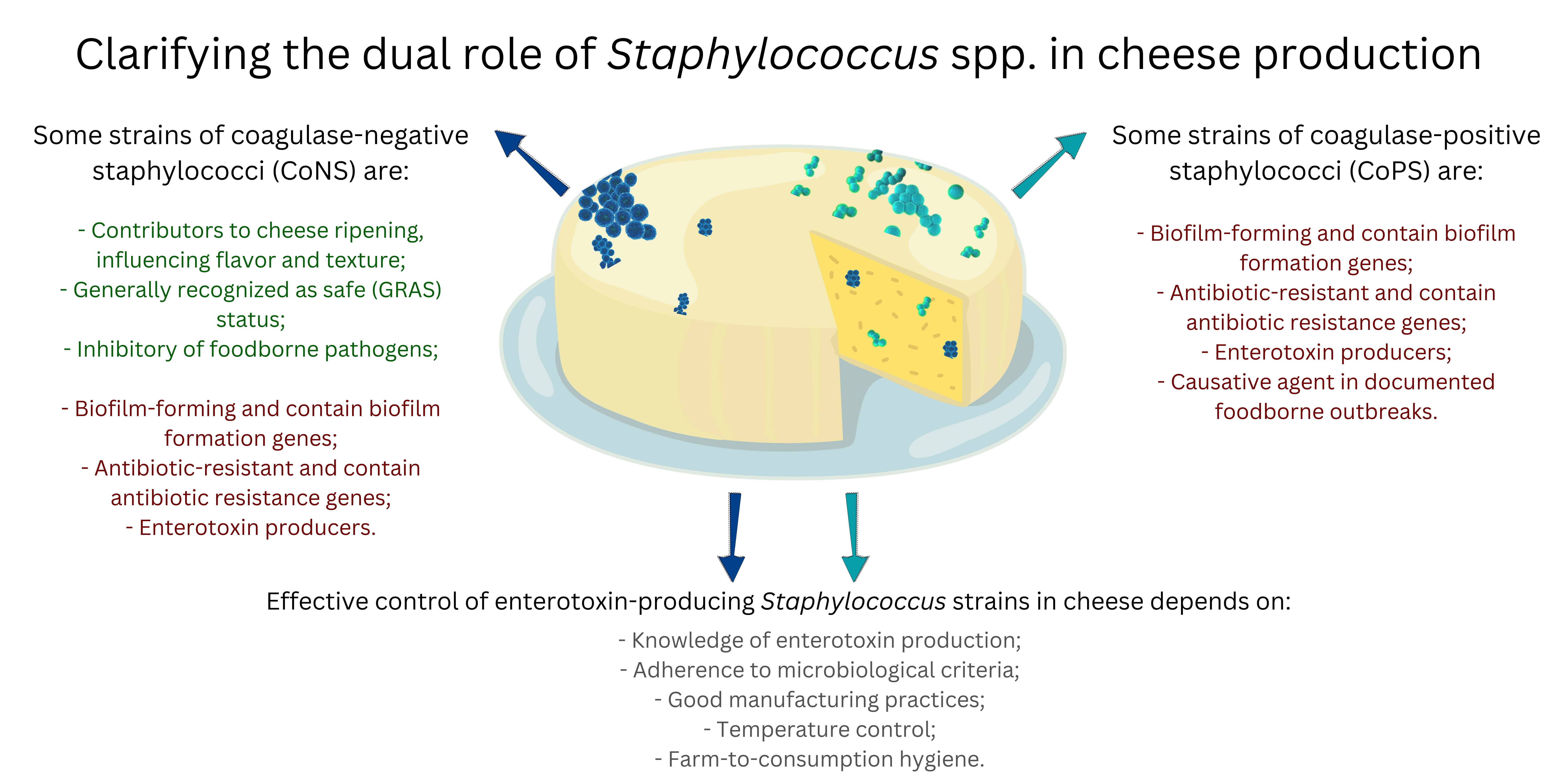
Staphylococcus spp. present a dual role during cheese production as some species may be pathogenic, while others bring beneficial characteristics to the final products. Coagulase-positive staphylococci (CoPS) species, particularly Staphylococcus aureus, are of concern due to their potential to produce enterotoxins commonly linked to foodborne outbreaks. These enterotoxins, encoded by a set of genes, can cause severe gastroenteritis, particularly vomiting. Many other members of the genus can harbor a wide range of genes encoding virulence factors and capability of forming biofilms on various surfaces. The alarming prevalence of antibiotic-resistant strains, including methicillin-resistant S. aureus (MRSA), further complicates their control. In contrast, some strains of coagulase-negative Staphylococcus species (CoNS) positively contribute to cheese ripening, influencing flavor and texture. Some strains are even considered safe for use in food production and have been studied as inhibitors of foodborne pathogens. Conversely, the expression of enterotoxin genes in some Staphylococcus species, particularly S. aureus, is regulated by different mechanisms including quorum sensing. Understanding enterotoxin gene expression in various environmental conditions, including during cheese production and ripening, can aid in developing targeted interventions. The risks posed by enterotoxin producing Staphylococcus in cheese are evident since numerous food poisoning outbreaks have been reported. Also concerning is the fact that several Staphylococcus species pose risks to animal health and livestock production. Effective control measures include adherence to microbiological criteria for CoPS and enterotoxin levels in cheeses with special attention to animal health, good manufacturing practices (GMP), temperature control, and strict hygiene protocols. This review highlights the need to balance the beneficial roles of CoNS in cheese production with the risks associated with virulent and enterotoxigenic strains of CoNS and CoPS.

Keywords: 
;  ;  ;  ;  

1. Introduction

The genus Staphylococcus is composed of Gram-positive, spherical bacteria that characteristically divide in more than one plane, forming arrangements resembling clusters of grapes. These bacteria have a diameter between 0.5 and 1.5 μm, are non-motile and non-sporogenic, catalase-positive, and can be aerobic, facultative an-aerobes or strictly anaerobic, such as Staphylococcus aureus subsp. anaerobius [1,2,3]. This group is susceptible to lysis by lysostaphin and resistant to lysis by lysozyme [4]. Acting either as commensals or opportunistic pathogens, they are taxonomically classified under the Kingdom Bacteria, Phylum Bacillota, Class Bacilli, Order Bacillales, and Family Staphylococcaceae [5].
The genus Staphylococcus is composed of 72 species and 14 subspecies, some of which are common inhabitants of the skin and respiratory tract of humans and warm-blooded animals, with few species that can be found in soil and aquatic environments [6,7]. In general, Staphylococcus spp. are present in most diverse environments and are considered symbionts [8]. According to Foster [9], Staphylococcus spp. are among the most resistant non-sporulating microorganisms: they can withstand high concentrations of salt, desiccation, heat and are more tolerant to common disinfectants than most bacteria.
Species in the Staphylococcus genus are classified as coagulase-positive (CoPS) or coagulase-negative (CoNS) according to their ability to produce coagulase [4]. However, different strains from some species, such as Staphylococcus hyicus, present variation in coagulase production, being considered coagulase-variable staphylococci (CoVS) [10]. Most CoPS species are recognized as pathogenic, although some strains may asymptomatically colonize healthy individuals, while CoNS are primarily saprophytic or associated with opportunistic infections [4]. Coagulase is the main virulence factor of CoPS, functioning as a critical mechanism of defense by inducing fibrin deposition around the cells, protecting them in the infected area. Moreover, there is a correlation between CoPS and enterotoxin production, further enhancing their pathogenicity [11,12].
The first time Staphylococcus was associated with foodborne illness dates back to as early as 1884 when spherical organisms in cheese caused a large food-poisoning outbreak in the United States. Other outbreaks attributed to the consumption of staphylococcal contaminated foods occurred in France in 1894, in the United States in 1907, and in the Philippines in 1914. In 1930, Gail Dack and his colleagues at the University of Chicago were able to demonstrate that the cause of a food poisoning that occurred from the consumption of a contaminated Christmas sponge cake with cream filling was due to a toxin produced by the isolated staphylococci [13].
This review explores the dual role of Staphylococcus spp. in cheese production, highlighting both the beneficial contributions of CoNS to cheese ripening and the risks posed by CoNS and CoPS, particularly S. aureus, due to their potential to produce enterotoxins and to form biofilms. The review examines the regulatory mechanisms of enterotoxin gene expression, including quorum sensing, and discusses effective control measures to minimize the risks associated with enterotoxin-producing strains in cheese. Ultimately, this work shows that not all Staphylococcus spp. are detrimental in the production of dairy products.

2. Coagulase-Positive Staphylococci (CoPS)

The coa gene, responsible for encoding coagulase production, plays a pivotal role in the virulence of CoPS, especially in S. aureus, by facilitating the conversion of fibrinogen into fibrin, which aids in clot formation and immune evasion. Genetic analyses have revealed considerable variability in the coa gene, indicating the adaptability of CoPS in diverse environments [14]. These genetic variations can occur both chromosomally and through horizontal gene transfer via plasmids, contributing to the spread of virulent and antimicrobial-resistant strains. Notably, livestock-associated methicillin-resistant S. aureus (LA-MRSA) strains have been shown to harbor novel recombinant staphylocoagulase types, highlighting the significant role of horizontal gene transfer in the evolution and dissemination of these strains [15]. Phylogenetic analyses of CoPS isolates demonstrate distinct clusters based on coa types, which are often associated with specific infection sites and geographic origins [16].
The CoPS species (n = 9) are shown in Table 1. All other species are CoNS (n = 67), except for a few species that are CoVS (n = 4) [7,10,17,18]. Five species that belonged to the genus Staphylococcus were reclassified to the genus Mammaliicoccus: M. fleurettii, M. lentus, M. sciuri, M. stepanovicii and M. vitulinus [19].

2.1. Staphylococcus aureus

S. aureus is a highly adaptable pathogen with a remarkable ability to thrive in diverse environments, contributing to its broad spectrum of infections. It can grow across a wide range of conditions, including temperatures from 7 to 48.5 °C (optimal 30–37 °C), pH levels from 4.2 to 9.3 (optimal 7.0–7.5), and sodium chloride concentrations up to 15% [20,21]. This adaptability makes S. aureus particularly significant in food safety, especially in foods requiring extensive handling during processing.
The virulence of S. aureus is driven by a variety of mechanisms, including the production of toxins such as alpha-hemolysin and Panton-Valentine leukocidin (PVL), along with superantigens like toxic shock syndrome toxin-1 (TSST-1) and staphylococcal enterotoxins (SEs). These virulence factors can lead to severe conditions such as tissue necrosis, vascular thrombosis, and bacteremia [22]. In the context of foodborne illness, staphylococcal food poisoning (SFP) is specifically attributed to the production of SEs, which are the primary virulence factor responsible for the gastrointestinal symptoms [23].
S. aureus is also noted for its role in hematogenous metastasis, biofilm formation, and the persistence of chronic infections, contributing to its ability to evade the immune system and resist treatment with antibiotics [24]. The combination of toxin production, invasiveness, and antibiotic resistance enables S. aureus to cause a wide range of symptoms, from superficial skin infections to more severe illnesses like toxic shock syndrome (TSS-like).
Strains of S. aureus can be classified into two groups based on their resistance to oxacillin/methicillin: methicillin-susceptible S. aureus (MSSA) and methicillin-resistant S. aureus (MRSA). MRSA strains emerged soon after the introduction of semisynthetic penicillins [25] and these strains are resistant to nearly all beta-lactam antibiotics, with the exception of ceftaroline and ceftobiprole [26]. Its clinical relevance lies in its association with poor prognoses and increased healthcare demands, as patients with MRSA infections often face longer hospitalizations, more extensive diagnostics, and higher mortality rates [27]. While recent data suggests a decline in the proportion of MRSA isolates, it remains a critical pathogen in the European Union/European Economic Area (EU/EEA), especially in Southern and Eastern European countries, where resistance levels remain high [28,29,30].
The presence of S. aureus in many environments is a significant concern due to its potential to cause severe infections and its increasing resistance to antibiotics. Thus, effective monitoring and control strategies are essential to reduce the risk posed by this pathogen in both clinical and food safety contexts.

2.2. Others Coagulase-Positive Staphylococci (CoPS)

Other CoPS, such as S. intermedius and S. coagulans, as well as S. hyicus, a CoVS, also pose significant health risks. These species, alongside S. aureus, can produce enterotoxins and coagulase, contributing to foodborne illnesses and animal infections.
S. aureus subsp. aureus is the most extensively studied subspecies and is a common cause of foodborne diseases through the production of heat-stable enterotoxins, leading to SFP [16,20].
In veterinary medicine, S. hyicus is a significant pathogen in swine, causing exudative epidermitis, also known as “greasy pig disease”. While primarily of concern in veterinary contexts, human infections, though rare, have been documented [31].
S. intermedius is commonly associated with animals such as dogs, and rarely as the cause of SFP in humans. However, it was implicated in an outbreak in 1991, when more than 265 people in the western United States became ill after consuming food contaminated with S. intermedius [32,33]. Its close relatives, S. pseudintermedius and S. delphini, are known to colonize various animal species, with increasing antibiotic resistance adding to their public health concern [34,35]. S. pseudintermedius, primarily associated with canine and feline infections like pyoderma and otitis externa, has gained attention due to the emergence of methicillin-resistant strains (MRSP), presenting a challenge similar to that of MRSA in humans. The rise of MRSP underscores the need for improved infection control measures in veterinary healthcare [35,36].
Other CoPS, such as S. lutrae, have been predominantly isolated from wildlife, like otters [37], with no evidence of human infection. Meanwhile, S. coagulans has been linked to infections in dogs and occasional human cases, particularly in immunocompromised individuals [38]. While more commonly associated with skin infections in companion animals, S. coagulans can also pose a food safety risk due to its presence in animal hosts.
S. delphini and S. argenteus have been isolated from both animals and humans. S. delphini is mainly found in dolphins and horses, while S. argenteus, closely related to S. aureus, has emerged as a significant human pathogen [39]. Finally, S. schweitzeri, primarily isolated from primates, shares genetic similarities with S. aureus [40], raising concerns about its zoonotic potential.

3. Coagulase-Negative Staphylococci (CoNS)

Most research on antibiotic resistance of staphylococci isolated from food has focused on the species S. aureus, while less attention has been paid to the CoNS group [41]. For many years, CoNS were considered non-pathogenic and were usually identified only at the genus level. Their role in food can be considered dual in nature, as some species bring beneficial characteristics to food, while others may be pathogenic.
CoNS belong to the saprophytic microbiota of the skin and mucous membranes of warm-blooded animals and humans, but are also found in foods such as meat, cheese and milk, and have been considered emerging pathogens with high incidence [42]. Their incidence in food is much higher than that of CoPS and, generally, most species are commensals; however, in other circumstances, some can act as pathogens [42,43]. In dairy products, especially on the surface of various types of cheese, CoNS are frequently found, either as contaminating species or as useful species to determine flavor or develop organoleptic properties. Their presence may not be an immediate hazard to public health, but they can become a risk factor [44]. In processed foods, CoNS may be indicative of hygiene failures in handling [45], and in foods derived from raw milk, in particular, they are of great importance, since Staphylococcus spp. are the most common causes of mastitis [46].
The CoNS group has 67 species, some of which have Generally Recognized As Safe (GRAS) status and are considered positive microbiota as they are responsible for the organoleptic characteristics of the final products. Some CoNS can even be used as starter culture in the production of cheeses, sausages, and fermented meats, due to their aromatic and pigmenting capacity [47].
Some CoNS species may play a beneficial role in producing certain fermented foods. However, safety concerns arise due to identified risk factors associated with some strains, as well as reports of nosocomial and urinary tract infections linked to S. epidermidis and S. saprophyticus, which are CoNS species commonly found in fermented foods [48,49]. Risk factors also identified correspond to virulence, in particular the production of enterotoxins, antibiotic resistance, and the ability to adhere and form biofilms [48,50,51,52].
Food poisoning of staphylococcal (SFP) origin is among the most common foodborne diseases and, contrary to what was previously thought, can be associated with both CoPS and CoNS strains [53]. This generates increasing interest in CoNS strains, since they have been associated with infections in humans, and in the induction of SFP, due to their ability to produce enterotoxins [54,55]. Food processing does not eliminate these toxins, which, unlike bacteria, have greater resistance to high temperatures, a wide pH range and proteolytic enzymes [56]. The toxins are also resistant to drying or freezing, and are insensitive to enzymatic digestion in the human gastrointestinal tract [57].
For a long time, the production of cytolytic toxins was attributed exclusively to S. aureus, but toxigenic factors or corresponding genes have been detected also in S. epidermidis and other CoNS species [58]. Exfoliative toxins (ETs), including ExhA, ExhB, ExhC, and ExhD, have been identified in some strains of S. hyicus. These toxins likely cause exudative epidermitis in pigs, a skin lesion that has several features in common with staphylococcal scalded skin syndrome (SSSS) in humans and share sequence similarities with the ETs of S. aureus: ETA, ETB, and ETD. Furthermore, TSST-1-associated enterotoxins and ETs were identified in a CoNS collection, following detection of hemolytic activities during a comprehensive immunoblot analysis, where a significant proportion of the tested strains produced the toxins [59].
SEs are toxins that cause vomiting after reaching the gastrointestinal tract, but other toxins, called staphylococcal enterotoxin-like proteins (SEls) and that lack the ability to induce vomiting, can also be produced [60]. More than 24 different serological types of SE have been identified in strains from different foodborne outbreaks, clinical cases or isolated from animals. The first five SE genes identified, which code for SEA, SEB, SEC, SED and SEE, known as classical enterotoxins, are frequently linked to foodborne outbreaks due to their ability to induce vomiting in humans. SEls are also considered a threat to humans, since they have been identified in cases of SFP outbreaks even without the presence of SEs. In addition, the new SE genes and the TSST-1, which belongs to a family of SE-associated toxins, are capable of stimulating large populations of T cells [61] (Table 2).
Studies using PCR and/or DNA microarrays revealed that the occurrence of SE genes in CoNS isolated from foods is very rare [62]. However, Nunes et al. [63] isolated CoNS from Minas Frescal cheese marketed in southeastern Brazil, and all strains carried multiple enterotoxin genes. The most frequently detected genes using an enzyme-linked immunosorbent assay (ELISA) were sea and seb (90% and 70%, respectively), followed by sec/see, seh/sei, and sed with intermediate incidence (60%, 50%, and 40%, respectively). The lowest incidence was observed for seg/selk/selq/selr and selu (20% and 10%, respectively). Notably, the most frequent species were S. saprophyticus (40%), S. xylosus (30%), M. sciuri (20%, former name S. sciuri), and S. piscifermentans (10%). Additionally, Andrade et al. [64] observed that among the CoNS and CoPS species with enterotoxigenic potential, the seg and seh genes occurred in the species S. cohnii subsp. cohnii, S. chromogenes, S. epidermidis, S. hominis, S. hyicus, S. lugdunensis, S. saprophyticus, S. ureilyticus, and S. xylosus, with seg gene being the most predominant.
Chajecka-Wierzchowska et al. [65] evaluated 118 CoNS isolates from food and observed that 72% were positive for at least one gene encoding for enterotoxin, while 28% were negative for the genes tested. The study also examined the presence of exfoliative genes (eta, etd), as well as the tsst-1 gene. The presence of the tsst-1 gene encoding TSST-1 was confirmed in 31.4% of CoNS strains belonging to the following species: S. simulans (n = 8), S. carnosus (n = 6), S. epidermidis (n = 3), S. warneri (n = 3), S. xylosus (n = 3), S. saprophyticus (n = 2), S. pasteuri (n = 1), S. petrasii (n = 1), and S. piscifermentas (n = 1). Although positive for the genes, the strains were unable to produce these toxins in the tested conditions.
Other species of the Staphylococcus, especially the CoNS, have significant roles as infectious agents for human or animal hosts, revealing a more restricted repertoire of virulence factors when compared to S. aureus. They act as infectious agents, with moderately pathogenic species typically causing subtler infections characterized by a subacute or chronic clinical course. These infections rarely present with fulminant signs and are seldom fatal [66]. The most notable representative of this group is S. epidermidis.
Found widely on human skin, wounds or surgeries, which may be the factors for the entry of this microorganism into the host's bloodstream, S. epidermidis has been highly related to hospital infections in recent years. Like S. aureus, S. epidermidis strains are highly resistant to antibiotics. The S. epidermidis species comprises a group of pathogens characterized by pronounced genomic diversity and when detected in clinical samples, clinicians face the challenge of determining whether they represent a true infection or just colonization/contamination [67]. With great clinical impact, this species has become the most important model microorganism for the study of healthcare-associated infections linked to inserted or implanted medical devices [68].
In addition to S. epidermidis, species such as S. saprophyticus, S. haemolyticus, and S. lugdunensis are occasionally observed as infectious agents of humans and animals, especially in patients with compromised immune systems [67,69].

Generally Recognized as Safe (GRAS) Status

In order for a microorganism to be used in the preparation and composition of a food, it must have GRAS status. Many non-pathogenic Staphylococcus species are used in the food industry because they confer unique characteristics to products. Among them, some CoNS species stand out, such as S. carnosus, S. condimenti, S. equorum, S. piscifermentans, S. succinus, and S. xylosus. Among these, S. carnosus, S. equorum, S. succinus, and S. xylosus are used as starter cultures for the production of cheeses and fermented meat products products [49].
CoNS play a significant role in defining color and developing organoleptic characteristics, which vary according to their proteolytic and lipolytic abilities [70,71]. This bacterial group can also contribute to the sensory qualities by producing diverse aroma profiles through carbohydrates and amino acids catabolism, esters formation, interactions with fatty acids, and their protease and lipase activities. Based on these distinctive properties, CoNS can be selected for use as starter cultures for fermentation [72].
S. carnosus has been used as a starter culture in the food industry since the 1950s. Its genome sequence has been determined and has provided the means for comparative studies of pathogenic and nonpathogenic staphylococci, and it has also been used as a cloning host to study the function of specific staphylococcal genes, given its food-grade status [73,74].
S. xylosus, belonging to the novobiocin-resistant CoNS species group, is commonly isolated from human and animal skin. The type strain C2a is commonly used as a starter culture in sausage and cheese production, contributing to the orange color on the surface of certain cheeses [75]
When evaluating strains of S. equorum isolated from cured cheese, the strain WS 2733 demonstrated the secretion of the macrocyclic peptide antibiotic micrococcin P(1), which exhibits antilisterial activity. This property was explored in cheese fermentation as a means to control the contamination by Listeria monocytogenes [76]. Deetae et al. [77] evaluated the production of volatile aromatic compounds by CoNS bacterial strains, isolated from different French cheeses, observing that S. equorum produced volatile compounds such as 3-methyl-3-buten-1-ol and 4-methyl-2-pentanone, responsible for conferring the fruity and sweet characteristics to the cheese.
Other CoNS species also produce compounds of interest such as diacetyl and acetoin, observed in S. succinus and S. xylosus, when isolated from fermented sausage [78]. Regarding the enzymatic activity of CoNS, they should have amino acid converting enzymes and specific peptide uptake mechanisms to produce volatile aroma compounds [79]. Thus, the addition of CoNS as starter cultures in the fermentation of cheeses and meats proves to be a safe alternative capable of conferring desired sensory characteristics.
Lee et al. [80] evaluated the genetic potential of S. equorum KS1039 as a starter culture in the fermentation of high-salt foods and observed that this strain contains genes for the biosynthesis of all amino acids except asparagine and for the production of branched-chain fatty acids. They also found that the species carries genes necessary for the production of butane-2,3-diol, diacetyl, and acetoin via glycolysis, and ester compounds via protein degradation.
Irlinger et al. [81] evaluated the genome sequence of S. equorum Mu2 from a French ripened cheese and observed that the strain did not possess any of the virulence factors found in S. aureus. Genomic evaluation of S. succinus 14BME20, isolated from fermented soybeans, confirmed that it did not contain any of the known S. aureus virulence factor-encoding genes, but it did contain strain-specific genes for lipid degradation, which may contribute to the production of volatile compounds [82].

4. Virulence Factors

Among the various Staphylococcus species, S. aureus stands out as both a tolerated commensal and a potent pathogen, widely colonizing several animals, the human skin and mucous membranes, as well as being present in food. In addition to toxins production, its pathogenicity is attributed to a diverse arsenal of virulence factors that facilitate adhesion to host tissues, biofilm formation, immune system evasion, and survival under nutrient-limited conditions. Additionally, the ability to acquire antibiotic resistance further enhances its clinical relevance. However, due to its genomic plasticity, not all S. aureus strains share the same genetic composition, leading to significant variability in virulence and pathogenic potential among subpopulations. The expression of these factors is influenced by environmental conditions and the host’s immune response, determining the strain’s capacity to cause infections ranging from mild skin lesions to severe systemic diseases, as well as its ability to form biofilms and produce enterotoxins in food. Given its broad impact on human and animal health, S. aureus is considered the primary reference for comparing pathogenic Staphylococcus species [75,83].
Just as Staphylococcus spp. play a dual role in cheese production, biofilm formation by CoPS and CoNS also exhibits this duality, depending on the characteristics of the producing strain. Strains carrying antibiotic resistance genes and enterotoxin expression genes are particularly concerning in cheese.

4.1. Biofilm Formation

Biofilm formation by CoPS and CoNS plays a significant role in the persistence of these bacteria on both biological materials and inert surfaces, such as cheese, equipment, and utensils used in production. Biofilm-associated cells are highly adherent and exhibit reduced susceptibility to desiccation, heat, detergents, biocides, and other antimicrobial agents [84,85,86]. In dairy processing environments, these biofilms are difficult to eliminate through conventional sanitation procedures, representing persistent sources of cross-contamination and posing challenges to microbiological control. When formed by enterotoxigenic strains and/or those carrying antibiotic resistance genes, such biofilms represent a significant threat to food safety, as the microorganisms can withstand adverse processing conditions and remain viable in the final product. Conversely, biofilm formation by GRAS-status CoPS and CoNS may have beneficial effects, particularly when these strains contribute to cheese ripening, promote the development of characteristic flavor and texture, and exhibit antagonistic activity against foodborne pathogens. Thus, biofilm formation by CoPS and CoNS can be seen as both a threat and an ally in cheese production, depending on the microbiological characteristics of the strains involved.
Several studies highlight the ability of different Staphylococcus spp. isolates from cheese to form biofilms. Friedriczewski et al. [87] tested twenty S. aureus isolates from buffalo mozzarella cheese, and observed that 10% were strong biofilm formers, 35% moderate formers, 50% weak formers, and 5% were non-biofilm formers. Souza et al. (2024) reported that 55% of the S. aureus isolates obtained from Minas Frescal and Porungo cheeses were strong biofilm formers. Meanwhile, Pineda et al. [88] evaluated the capability of S. aureus isolates from raw milk artisanal cheeses from Canastra (Brazil), observing that none of them was strong biofilm former, while 24% were moderate and 16.7% did not form biofilm. Moreover, Carvalho et al. [89] demonstrated that S. aureus ATCC 25923 is capable of growing and forming biofilms on an 18-micron low-density polyethylene (LDPE) package when stored at 5 °C in the presence of Minas Frescal cheese whey. Additionally, bacterial cells were able to detach from the packaging, increasing the microbial load on the product.
Fontes et al. [86] showed that 29.5% of the 227 CoNS isolated from soft cheeses in Brazil were able to form biofilm. Gajewska and Chajęcka-Wierzchowska [90] isolated 54 staphylococcal strains from cow’s milk samples, of which 42 were classified as CoNS, belonging to the following species: S. capitis, S. chromogenes, S. haemolyticus, S. hominis, S. saprophyticus, S. sciuri (reclassified as M. sciuri), S. simulans, S. warneri, and S. xylosus, while 12 were classified as S. aureus. All tested isolates exhibited the capacity for biofilm formation. Of these, 85.7 and 58.3% of the CoNS and S. aureus isolates were capable of forming strong biofilms, while 4.8 and 8.3% formed moderate biofilms, and 9.5 and 33.3% formed weak biofilms, respectively. Interestingly, Goetz et al. [91] evaluated the effect of CoNS isolates with a weak-biofilm phenotype on the biofilm formation of other CoNS and CoPS isolates from the mastitis pathogen culture collection. Four of the CoNS isolates with a weak-biofilm phenotype (S. chromogenes C and E, and S. simulans F and H) significantly reduced biofilm formation in approximately 80% of the staphylococcal species tested, including S. aureus. These four S. chromogenes and S. simulans isolates were also able to disperse pre-established biofilms, but did not inhibit the growth of isolates with a strong-biofilm phenotype. These results suggest that some CoNS isolates can negatively affect the ability of other staphylococcal isolates and species to form biofilms via a mechanism that does not involve growth inhibition.
Formation of bacterial biofilms is a complex process comprising four main stages: adhesion, aggregation, maturation, and dispersion [92,93]. During the initial adhesion stage, S. aureus planktonic cells utilize various factors and regulatory mechanisms, such as the expression of cell wall-anchored (CWA) proteins, adhesins, and extracellular DNA (eDNA), to attach to biotic and abiotic surfaces [94,95]. One of the primary mechanisms involved is the organization of microbial surface components recognizing adhesive matrix molecules (MSCRAMMs), including protein A (SpA), fibronectin-binding proteins (FnBPs, such as FnbA and FnbB), fibrinogen-binding proteins (Fib), clumping factors (ClfA and ClfB), serine-aspartate repeat family proteins (SdrC, SdrD, and SdrE), biofilm-associated protein (Bap), and S. aureus surface proteins (SasC and SasG) [96,97,98,99,100,101]. Souza et al. [102] reported that some S. aureus isolates from Minas Frescal and Porungo cheeses exhibited expression of the fnbA and clfB genes. Pineda et al. [88] evaluated the virulence potential of S. aureus isolates from raw milk artisanal cheeses and found that most isolates possessed MSCRAMM genes, including fnbA, fnbB, fib, clfA, clfB, and eno.
SasC promotes the formation of large cell aggregates, increases adhesion to polystyrene, and enhances biofilm formation in S. aureus and S. carnosus [103]. SasG and plasmin-sensitive protein (Pls) from S. aureus are homologous to accumulation-associated protein (Aap) in S. epidermidis and other CoNS. Aap and SasG appear as long fibrils on the bacterial cell surface [104,105] and play roles in host cell binding and biofilm formation [105,106,107], while Pls surface expression reduces S. aureus adhesion to fibronectin [108,109,110]. Aap has also been shown to mediate intercellular adhesion in polysaccharide intercellular adhesion (PIA)-negative S. epidermidis strains, leading to a proteinaceous extracellular biofilm matrix [106]. Artini et al. [111] demonstrated that CoNS biofilm-producing species possess the aap gene, whereas the atlE gene is absent in these strains. A class of bifunctional proteins known as adhesins facilitate biofilm attachment to host tissues and surfaces. These include AtlA and Aaa in S. aureus [112] and AtlE and Aae in S. epidermidis [113]. Adhesins play essential roles at multiple stages of biofilm formation and adhesion [114]. Although numerous surface adhesins have been identified, S. aureus possesses a much larger repertoire of these proteins than S. epidermidis, which is limited to a few adhesive proteins. The ability to attach to host tissues or surfaces is a prerequisite for the subsequent formation of multilayered biofilms, stabilized by exopolysaccharides or proteinaceous intercellular material [75].
Gajewska and Chajęcka-Wierzchowska [90] isolated 42 CoNS and 12 S. aureus from cow’s milk. They then identified genetic determinants responsible for biofilm formation, such as the bap and eno genes. Additionally, among CoNS, they detected the aap, bhp, fbe, embP, and atlE genes. Most of the tested staphylococcal strains (90.7%) had at least one of the tested genes. Nearly half (47.6%) of the CoNS had the eno gene, while for S. aureus, the eno gene was found in 58.3% of isolates. The frequency of the bap gene occurrence was 23.8% in CoNS strains and 25% in S. aureus, respectively. The fbe gene was demonstrated in only three CoNS isolates. Among the CoNS, the presence of the embP (16.7%), aap (28.6%), and atlE (23.8%) genes was also demonstrated. Following the adhesion step, bacterial cells begin to divide and aggregate [115]. During the aggregation stage, bacteria regulate biofilm formation by sensing environmental signals that activate regulatory networks and intracellular signaling molecules, promoting bacterial proliferation and biofilm thickening [116]. The biofilm provides resistance against the human immune system and antibiotics [117], while bacterial cells lose direct contact with the host surface and rely on cell–cell and cell–extracellular polymeric substance (EPS) adhesion [118].
Among the EPS components in S. aureus biofilms, PIA is biosynthesized as a poly-N-acetylglucosamine (PNAG) polymer and is a key factor [119]. PIA has cationic properties and plays a crucial role in adhesion and aggregation [120]. In S. aureus, biofilm formation is controlled by PIA production through proteins encoded by the icaADBC operon. Mutant strains lacking PIA exhibit significantly reduced bacterial cell adhesion [121]. PIA-dependent biofilms are predominantly observed in MSSA strains [121,122]. PIA interacts with other small proteins, such as Bap (biofilm-associated protein) that promotes cell-to-cell aggregation during biofilm formation [123] and Aap (accumulation-associated protein) that facilitates biofilm maturation [124]. Souza et al. [102] reported that 90% of the S. aureus isolates obtained from Minas Frescal and Porungo cheese expressed the icaD gene. Also, Pineda et al. [88] found that icaA and icaD genes were present in 70.3% and 46.2% of S. aureus isolates from raw milk artisanal cheeses, respectively.
Although the ica operon is considered essential as the genetic basis for PIA production in biofilm formation, biofilm development through ica-independent mechanisms has also been observed. Specifically, ica-negative mutants of S. epidermidis can still form biofilms, though these exhibit a proteinaceous rather than polysaccharide composition, as evidenced by their resistance to metaperiodate and their susceptibility to protease disruption [125]. However, ica-negative strains such as S. epidermidis ATCC 12228 have been reported to lack biofilm formation capabilities [126]. Gajewska and Chajęcka-Wierzchowska [90] showed that, among the 42 CoNS and 12 S. aureus isolates from cow's milk, the icaA was detected only in CoNS strains (24.1%), while icaD was found in both CoNS strains (21.4%) and S. aureus (100%).
During the maturation stage, biofilms become highly organized, forming compact, three-dimensional mushroom- or tower-like structures [127]. Channels develop around microcolonies, facilitating nutrient transport to deeper biofilm layers [128]. Mature biofilms exhibit metabolic diversity, which enhances their ability to withstand environmental stressors [129]. The production of EPS promotes bacterial aggregation into microcolonies, which serve as the structural foundation of the biofilm [130]. As these microcolonies thicken, genetic or environmental cues may trigger biofilm dispersion [131].
Biofilm dispersion is a complex, multi-step process that includes the production of exoenzymes and surfactants capable of degrading the EPS matrix [131], as well as physiological adaptations that prepare cells for survival outside the biofilm [132]. Once dispersed, cells revert to the planktonic state, allowing them to colonize new sites and initiate a new biofilm formation cycle [133]. As the final stage of the biofilm life cycle, dispersion plays a crucial role in infection spread. During biofilm growth and development, surfactant-like phenol-soluble modulins (PSMs) contribute significantly to biofilm dispersion and transmission in S. aureus. These molecules disrupt non-covalent interactions within the biofilm matrix and facilitate the formation of nutrient transport channels [134,135]. PSMs exist both in soluble form and as amyloid fibers, which provide structural stability to the biofilm [136,137].
The structural and functional complexity of biofilms increases as cells divide and the matrix becomes denser, creating physiological heterogeneity within the biofilm. This heterogeneity is characterized by gradients of nutrients and oxygen [138]. Within a biofilm, bacterial cells can be categorized into four distinct metabolic states: (i) aerobic cells, located in the oxygen- and nutrient-rich outer layer; (ii) fermentative cells, found in the oxygen- and nutrient-poor inner layer; (iii) dormant cells, residing in the anoxic layer with slow growth and inactive metabolism; and (iv) dead cells [139,140,141]. Dormant cells exhibit decreased intracellular adenosine triphosphate (ATP) levels, rendering them less susceptible to antibiotics [142]. Additionally, gradients of viscosity and lipid composition within S. aureus biofilms contribute to biofilm dispersal by facilitating the detachment of loosely bound bacteria while preserving a stable core layer [143,144].
For a more comprehensive overview of biofilm formation and regulatory mechanisms in S. aureus, please refer to [92,93]. For S. epidermidis and other CoNS, please refer to [94,145].

4.2. Antibiotic Resistance

Resistance to beta-lactams in MRSA strains and many methicillin-resistant coagulase-negative staphylococci (MRCNS) strains is associated with the presence of transferable genomic islands (GI) in the bacterial genome, known as staphylococcal chromosomal cassette mec (SCCmec). The mecA gene, carried by the SCCmec, encodes penicillin-binding protein 2a (PBP2a), a transpeptidase with low affinity for beta-lactams, thereby conferring resistance to methicillin [150,153,154]. From an evolutionary perspective, the mecA gene found in S. aureus likely originated from a group of bacteria previously classified as CoNS, now reclassified under the genus Mammaliicoccus [150,155]. Different SCCmec types may harbor the mecA gene or others, along with resistance determinants for other antibiotic classes, such as aminoglycosides, macrolides, lincosamides, streptogramins B, and tetracyclines (MLS-B) [150,153].
The emergence of MRSA and MRCNS strains has left only a few antibiotics effective for treating infections. Even the use of glycopeptides, so-called last-resort antibiotics, such as vancomycin, is at risk of becoming ineffective [156]. Intermediate-susceptible S. aureus to vancomycin (VISA) and glycopeptides (GISA), as well as vancomycin-resistant S. aureus (VRSA; vancomycin MIC ≥ 16 mg/L), have also been reported [149]. Changes in the cell wall and metabolic pathways can lead to intermediate resistance to vancomycin [156], while the acquisition of the vanA resistance determinant results in high-level resistance to vancomycin [157]. Glycopeptide resistance, encoded by the vanA operon, is more frequently expressed in S. aureus strains with mutations in the modification-restriction system, the presence of the pSK41-like conjugative plasmid and/or the Tn1546 transposon, both of which enhance the frequency of vanA operon conjugation [149,158–161].
CoPS and CoNS have also developed resistance to other classes of antibiotics, including aminoglycoside, diaminopyrimidine, fusidane, lincosamide, macrolide, nucleoside, phenicol, phosphonic acid, quinolone, streptogramin, tetracycline, and trimethoprim [149,150,162,163]. Other anti-MRSA antimicrobials have been developed, including daptomycin, linezolid, telavancin, tigecycline, quinupristin/dalfopristin, cephalosporins, and ceftobiprole. However, some strains have already developed resistance mechanisms to these new drugs [149,150,164].
Table 3 provides an overview of various resistance genes associated with different antibiotic classes and their corresponding encoded proteins in Staphylococcus spp. As shown in Table 3, one single gene may confer resistance to multiple antibiotics. Mlynarczyk-Bonikowska et al. [149], Brdová et al. [150] and Alkuraythi et al. [163] published comprehensive overviews on antibiotic resistance and the molecular mechanisms of this resistance in S. aureus and other CoPS and CoNS.
Thus, S. aureus, throughout its evolution, has acquired resistance to nearly all antibiotics developed so far. The presence of populations exhibiting multiple antibiotic resistances, which are highly prevalent in the environment, is a serious concern as it compromises the effectiveness of treatments for staphylococcal infections [83]. Furthermore, their antimicrobial resistance determinants may also be transferable to other commensal or potentially pathogenic bacteria in foodstuff [52,148,165]. Similarly, CoNS have also acquired resistance to various antibiotics throughout their evolution and may be present in cheeses, contributing to the transfer of resistance genes [63,86,166]. It is noteworthy that Fontes et al. [86] found high counts of CoNS in Brazilian soft cheeses, ranging from 106 to 107 CFU/g.
Gajewska et al. [167] conducted a study in Poland in which they tested 180 S. aureus isolates collected from various stages of artisanal cheese production using unpasteurized milk. The study revealed notable levels of antimicrobial resistance among the isolates: penicillin (58.1%), tobramycin (34.4%), azithromycin (18.3%), clarithromycin (16.1%), erythromycin (22.6%), cefoxitin (12.9%), and oxacillin (9.7%). The blaZ gene, which encodes penicillin resistance, was the most common antibiotic resistance gene among the tested isolates. All isolates showing phenotypic resistance to cefoxitin carried the mecA gene. Allaion et al. [168] evaluated S. aureus isolates from Minas artisanal cheeses, and at least one antibiotic resistance gene was detected in 83.0% of the isolates. Nearly half (47.1%) carried more than one resistance gene. The most frequently detected resistance genes were tetK (54.4%) and mecA (52.2%), followed by aacA-aphD, which was found in 30.0% of the isolates. Aguiar et al. [152] characterized 57 S. aureus isolates from artisanal colonial cheese, with penicillin resistance being the most prevalent (33%), followed by resistance to clindamycin (28%), erythromycin (26%), and tetracycline (23%). The evaluated strains also exhibited inducible resistance to clindamycin, with nine isolates classified as MDR.
Pineda et al. [88] characterized the genomes of several S. aureus strains isolated from raw milk artisanal cheese in Brazil, identifying antimicrobial resistance genes with phenotypic confirmation of methicillin and tetracycline resistance. The authors also discovered a rich virulome encoding iron uptake systems, immune evasion mechanisms, and an extensive arsenal of toxins, along with the capacity to form biofilm. These findings suggest that multiple strains circulating in the cheese-producing region pose a potential health risk.
Fontes et al. [86] isolated 227 CoNS from soft cheese in Brazil, and high percentages of antimicrobial resistance were observed for penicillin (78.5%), oxacillin (76.2%), erythromycin (67.8%), gentamicin (47.2%), clindamycin (35.7%), rifampicin (26.8%), azithromycin (14.7%), tetracycline (14.7%), levofloxacin (14.2%), and sulfamethoxazole-trimethoprim (11.9%). All isolated CoNS were susceptible to vancomycin and linezolid. A multiple antibiotic resistance (MAR) index of >0.2 was observed in 80.6% of the isolates. In addition, 81.5% of the isolates carried the mecA gene, and 76.2% of these were phenotypically resistant to oxacillin. Nunes et al. [63] isolated CoNS from Minas Frescal cheese in southeastern Brazil, and the strains showed multiresistance to antimicrobial agents such as beta-lactams, vancomycin, and linezolid. Klempt et al. [166] evaluated 53 CoNS isolates from different cheeses, some of which exhibited resistance to cefoxitin, penicillin, and tetracycline. In addition, several carried genes encoding antibiotic resistance, such as mecA, mecB, mecD, blaTEM, tetK, and tetL.

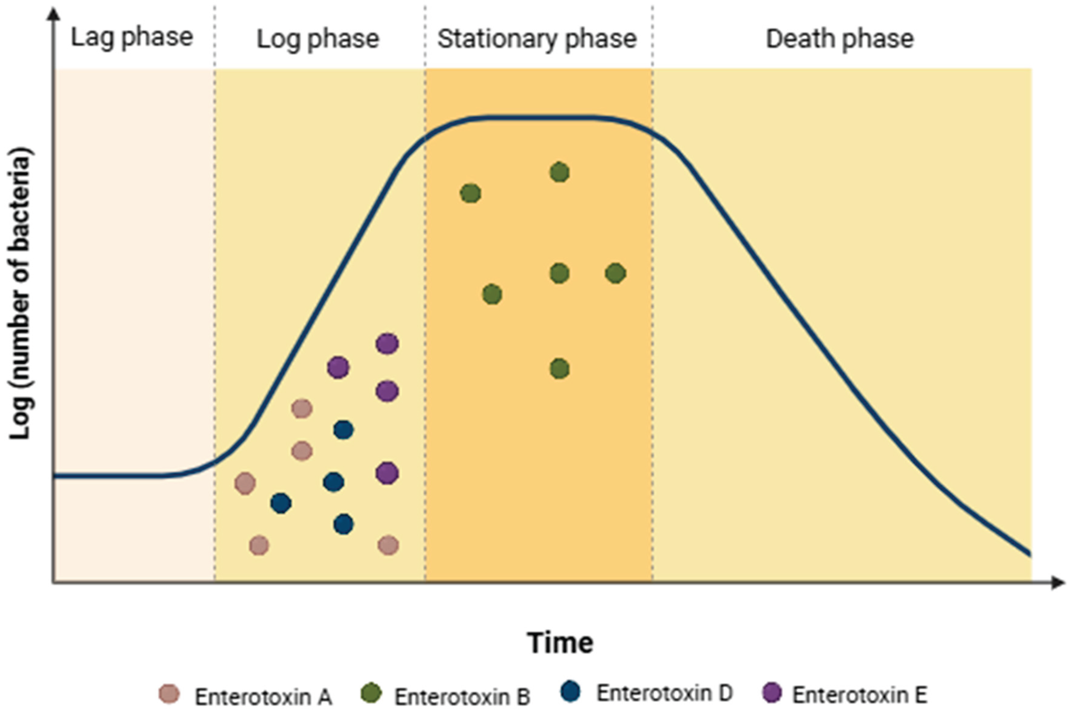
4.3. Expression of Enterotoxins Genes

Staphylococcal food poisoning (SFP) is caused by one or more enterotoxins produced by some species and strains of Staphylococcus. Although enterotoxin production is associated with CoPS and thermonuclease-positive S. aureus (TPS), some CoNS and species that are thermonuclease-negative also produce enterotoxins [46,169]. Within S. aureus, the regulation of virulence factors is subject to a complex network that integrates host and environmentally-derived signals into a coordinated response [170].
The genome of S. aureus harbors numerous toxin-encoding genes, which are primarily located on mobile genetic elements [148,171]. This arrangement results in significant variability in toxin production among different S. aureus strains [148,172]. Among the various known or strongly suspected toxins and virulence factors that cause specific diseases or symptoms, staphylococcal superantigens (SAgs), comprising SEs (Staphylococcus enterotoxins), SEls (Staphylococcus enterotoxin like), and TSST-1, are the most prominent [148,171,172].
SAgs are a group of potent immunostimulatory toxins produced by S. aureus. SAgs are characterized as pyrogenic toxin superantigens, with the ability to induce SFP and an infection known as Toxic Shock Syndrome (TSS). SAgs share many structural and functional similarities but have distinct characteristics. They are relatively resistant to heat and to proteolytic gastric enzymes such as pepsin and trypsin, allowing them to pass through the digestive tract and head to the site of action. In SFP, SAgs stimulate the vagus nerve endings in the stomach lining that control the emetic response, causing nausea, cramping, vomiting, and diarrhea, appearing abruptly 2–8 h after ingesting food containing these toxins. TSS is a potentially fatal disease characterized by fever, erythematous rash, hypotension, shock, multiple organ failure, and skin desquamation. This toxin-mediated systemic disease was first observed in non-systemic infections by SE producing S. aureus. Subsequently, another S. aureus toxin, designated as TSST-1 (formerly SEF), was shown to be associated with TSS in menstruating women and in non-menstrual cases [171].
Superantigens (SAgs) are single-chain proteins that interact with variable regions of T-cell receptors (TCR Vα or TCR Vβ), activating a large number of T-cells. This activation triggers massive proliferation and release of pro-inflammatory cytokines, such as IL-1, IL-2, IL-6, γ-interferon, and TNF, potentially leading to lethal TSS. SAgs can also interact with epithelial cells, promoting transepithelial transport and an inflammatory state. Due to their effects on the immune system and ability to induce SFP and TSS, SAgs are classified as pyrogenic toxin superantigens. Similar to TSST-1, they are super antigenic toxins, that activate T-cells in a predominantly nonspecific manner, resulting in an excessive immune response that includes polyclonal T-cell activation and massive cytokine release [3,171,173].
SEs belong to a large family of staphylococcal and streptococcal pyrogenic exotoxins, sharing common phylogenetic relationships, structure, function, and sequence homology. SEs are potent gastrointestinal toxins that cause emesis in a not completely understood manner that involves the induction of histamine release from intestinal mast cells [3,173].
SEs are heat-stable, low molecular weight (19,000–29,000 Da), single-chain proteins primarily produced by S. aureus, though not exclusively. These toxins belong to a major family of serological types, including SEA to SEE and SEIG to SEIJ. The classical enterotoxins, SEA, SEB, SEC1-3, SED, SEE, and SEH, are the main agents responsible for SFP [9,169,173,174]. These toxins function as SAgs, originally identified due to their emetic activity in SFP. This group includes SEs A, B, C, D, E, G, H, I, R, and T, as well as SElJ to SElX, which do not cause emesis or have not been tested in non-human primates. TSST-1, a pyrogenic exotoxin previously known as SEF, is also part of this SAgs group [23,175]. Although the role of certain SEs, SEls, and TSST-1 in foodborne diseases remains unclear and therefore cannot be ruled out, they share structural and functional similarities and have been associated not only with SFP- and TSS-like syndromes, but also with allergic and autoimmune disorders [9,169,173,174].
The expression of genes encoding SEB occurs primarily at the end of the stationary phase, while the production of SEA, SED, and SEE takes place throughout the logarithmic growth phase (Figure 1; [176]). The production of SEA and SEE is not regulated by the accessory gene regulator (agr) system [169,173]. In contrast, SEB, SEC, and SED require a functional agr system for maximal expression [173]. The agr system facilitates cell-to-cell communication through a quorum sensing mechanism, using autoinducing peptides (AIPs) as signaling molecules. Activation of the agr system leads to the expression of exo-toxins and exo-enzymes and is essential for virulence in animal models of skin infection, pneumonia, and endocarditis [170].
The synthesis of SEs depends on temperature, pH, water activity, and the presence/activity of other microorganisms with beneficial or antagonistic interactions. Generally, the production and accumulation of enterotoxins in food occurs when enterotoxigenic staphylococci are capable of proliferating, normally when populations are above 105 CFU/g [169,174,177].
The successful growth of S aureus in diverse environmental conditions is partly due to its ability to express different genes in response to changing conditions. Additionally, S. aureus has extraordinary adaptive power to ensure its success as a pathogen [171,178]. This microorganism is capable to detect different environmental signals and adjust the production of virulence factors critical for survival in the host, such as cell surface adhesins, extracellular enzymes, and toxins [24,170]. A virulence gene is often susceptible to transcirculatory control by more than one regulatory system and, there is cooperation or even competition between these systems to modulate the expression of a given virulence gene [179]. The accessory gene regulator (agr) quorum sensing mechanism is an important regulatory system of S. aureus and contributes to its pathogenicity, playing a key role in the expression of enterotoxins genes [170,179].
SaeRS, a two-component system in S. aureus responsible for the production of toxins, immunomodulators, and enzymes, was proven to be essential for virulence in animal models of skin infections and pneumonia [170,180]. The staphylococcal respiratory response regulator (SrrAB) is an oxygen-responsive two-component system that induces the expression of plc and ica, while repressing agr, TSST-1, and spa. It is essential for defense against neutrophils [170,181]. SrrAB activates ica operon transcription and promotes the expression of polysaccharide intercellular adhesin, helping S. aureus evade neutrophil-mediated killing during anaerobic growth conditions [181]. ArlRS, another two-component system that regulates autolysis and cell surface properties, promotes MgrA expression while repressing agr and autolysis. It is crucial for virulence in animal models of skin infection and endocarditis [182].
SarA is a cytoplasmic regulator that promotes the expression of extracellular proteins and represses spa, which encodes staphylococcal protein A. SarA is required for virulence in biofilm infection models [183]. Rot is a cytoplasmic regulator that controls the production of toxins and extracellular proteases in S. aureus. Its expression is regulated by the agr system, which, when active, prevents Rot from being translated. Interestingly, in certain conditions where agr is inactive (e.g., agr-null mutants), mutations in rot can restore the virulence of the bacteria. This was demonstrated in a rabbit model of endocarditis [184].
The transition of S. aureus from a commensal organism to a pathogen is strongly influenced by host-derived environmental signals, as described by Choueiry et al. [185]. In this study, significantly lower growth of the MRSA strain was observed under aerobic conditions, suggesting that these bacteria were subjected to oxidative stress, which impaired growth. Furthermore, supplementation of culture media with energy substrates and addition of carbon sources facilitated the ability of S. aureus to overcome environmental stress and grow, demonstrating a more robustly adaptive metabolism. These authors also noted that changes in growth environments may drive the regulation of virulence in S. aureus with the associations of changes in their metabolism with its virulence. Increased expression of the virulence factors agr-I, sea, seb, and eta was apparent in the supplemented S. aureus cultures [185].
Signal transduction systems that sense cell density, energy levels, and external stimuli facilitate S. aureus's remarkable adaptability to diverse environmental conditions [178,183]. These host environmental signals are crucial in promoting S. aureus colonization, allowing bacteria to adapt to different conditions and potentially switch to a pathogenic state when conditions are favorable. Understanding these host-pathogen interactions is critical for managing S. aureus infections in clinical settings and understanding enterotoxin expression in food [186].

5. Staphylococcal Food Poisoning from Cheese Consumption

SFP occurs due to the consumption of food containing preformed SEs, typically produced when S. aureus reaches concentrations of 106 CFU/g or mL in food matrix [187]. Although Bastos et al. [187] cite 100 ng as a general threshold dose to cause illness, other studies suggest that, for example, even 20–100 ng of SEA may be sufficient [188]. However, the dose response depends on the individual’s sensitivity, body weight, and the specific SE involved [188–191].
Documented SFP outbreaks associated with cheese consumption, categorized by country and year of occurrence, are summarized in Table 4. No further outbreaks related to cheese consumption were identified in the literature.
Regarding SFP outbreaks linked to cheeses, [192] reported that four individuals from the same family became ill after consuming fresh Minas cheese, in Brazil. The cheese contained high counts of S. aureus (9,3×10⁷ CFU/g), and the strains were capable of producing SEA, SEB, SED and SEE. which were likely responsible for the outbreak, the main symptoms were nausea, vomiting, diarrhea and abdominal pain, with no hospitalizations. The average incubation period was approximately one hour.
Pereira et al. [193] reported an outbreak that occurred in 1995 due to consumption of cheese produced in the Minas Gerais state, Brazil. A family of seven individuals consumed the cheese and began to present symptoms of vomiting and diarrhea approximately 4 hours later. Analysis of the cheese revealed a high population of S. aureus (2.9×10⁸ CFU/g) and the presence of SEH. There were no hospitalizations or deaths. In 1999, two additional outbreaks involving Staphylococcus and cheeses occurred in Minas Gerais state, Brazil, affecting around 700 people. One outbreak was linked to the consumption of Minas cheese and the other to raw milk. In the first outbreak, analysis of the cheese revealed S. aureus levels ranging from 2.4×10³ to 2.0×10⁸ CFU/g, with the production of SEA, SEB, and SEC. In the second outbreak, raw milk samples contained CoNS at counts exceeding 2.0×10⁸ CFU/g, along with the production of SEC and SED [194].
From 2014 to 2023, 6,874 foodborne disease outbreaks were reported in Brazil, leading to 110,614 illnesses and 12,346 hospitalizations and S. aureus was the second leading etiological agent, responsible for 9.7% of cases [195]. In these outbreaks dairy products were responsible for 6.7% of the total number of outbreaks. Unfortunately, no data is available on enterotoxins in these samples.
In Canada, in 1980, 62 individuals presented symptoms of nausea, vomiting, abdominal cramps, and diarrhea after consuming curd cheese, which was present in both boxed lunches and cheeses purchased at retail stores in cities near Montreal. The curd cheese was mainly produced in a cheese factory and distributed to retail stores for preparation of boxed lunches. When analyzed, the curd cheeses contained between 2.0 and 8.0×10⁷ S. aureus/g, in addition to SEA and SEC. No deaths were reported [196]. In United Kingdom, in 1981, a family of four consumed Halloumi cheese in brine imported from Cyprus. This cheese, traditionally made with goat's and sheep's milk, may also include cow's milk in some cases. After consumption, all family members developed symptoms typical of SFP. Although S. aureus was not isolated, SEA was detected in both the cheese and the brine [197]. Between December 1984 and January 1985, cheese made from raw ewe's milk at a dairy farm was linked to three outbreaks involving 27 people in the United Kingdom. The people who got ill had severe symptoms, such as violent vomiting and severe diarrhoea. SEA was detected in the cheese, although S. aureus was not identified. Subsequent testing of milk samples from the dairy revealed the presence of a SEA-producing strain [198].
Kérouanton et al. [199] investigated outbreaks associated with S. aureus in France, reporting that between 1981 and 2002, there were 13 incidents involving cheese. The analyzed matrices included raw milk semi-hard cheeses, raw milk soft cheeses, soft cheeses, raw milk cheeses, and sheep’s milk cheeses (raw and pasteurized). The enterotoxins detected in the cheeses were SEA, SEB, and SED. S. aureus populations ranged from 1.0×10⁴ to 3×10⁸ cfu/g, depending on the outbreak and cheese type. Reported symptoms among patients included nausea, vomiting, abdominal cramps and diarrhea, with no deaths. Ostyn et al. [200] reported six outbreaks occurred in France between October and November 2009 due to SEE in soft cheeses, resulting in 23 cases. The people got ill after consuming soft cheese from one producer with 1.5×10⁵ CFU/g and the only type of SE detected in all food samples was SEE (between 0.36 to more than 1.14 ng/g of cheese).
Filipello et al. [201] reported an outbreak in Lombardy, Italy, in 2018, caused by the consumption of artisanal Alm cheese containing SED. This outbreak involved three patients, and all individuals presented abdominal cramps, vomiting, and diarrhea.
In Northern Italy, in 2022, a family of eight reported gastrointestinal symptoms such as vomiting and diarrhea, as well as headaches, after eating sandwiches at a small local restaurant in the Alps region of Piedmont. Food safety agency inspectors collected samples of ham and cheese made with raw milk that the family members had consumed and found CoPS varying between 1.3×10³ and 8.1×10³ cfu/g in the cheeses, in addition to enterotoxins A to E, with SED estimated at 0.649 ng/g. There were no deaths [202].
In Switzerland, in 2007, five individuals presented nausea, abdominal cramps, vomiting and diarrhea after ingestion of a fresh goat milk cheese (Rabiola). The samples presented counts of CoPS between 6.7×10⁶ and 2.6×10⁷ CFU/g. The strains were positive for genes (seg, sei, sem, sen, and seo; [188]). Also in Switzerland, another outbreak affected 14 people, including children and adults, after consuming soft cheese produced from raw cow milk (Tomme cheese). The soft cheese contained SEA (>6 ng/g) and SED ( >200 ng/g). Counts of 10⁷ CFU/g of CoPS were detected. No deaths were reported [188].
According to the European Food Safety Authority and European Centre for Disease Prevention and Control, in 2022, S. aureus toxins were responsible for 137 outbreaks (0.02% of the total of 5763 reported cases), resulting in 148 hospitalizations and 4 deaths [203]. Between 2007 and 2018, 8,730 foodborne disease outbreaks caused by seven pathogens were reported in Japan. Among these, S. aureus was responsible for 448 outbreaks, none of which resulted in fatalities. Additionally, 2.6% of outbreaks linked to dairy product consumption were attributed to S. aureus [204].
Abiotic factors such as temperature, pH, water activity, redox potential, NaCl concentration and oxygen availability, in addition to bacterial antagonism, influence the growth and enterotoxin production by S. aureus in food [205,206]. These factors may help explain the relatively low number of S. aureus outbreaks associated with cheese. The optimal temperature range for both growth and enterotoxin production by S. aureus is 34–40 °C. The optimal pH for growth is 6 to 7, while for enterotoxin production it is 7 to 8. The ideal water activity for both growth and enterotoxin production is 0.99, although reports indicate enterotoxin production can occur between 0.86 and 0.99 [205]. Additionally, Schelin et al. [205] reported that the presence of lactic acid bacteria in cheese, such as Lactococcus lactis, can inhibit the transcription of genes responsible for enterotoxin production, such as sec, selk, seg, and seh. These characteristics suggest that although cheeses may provide conditions that support growth and toxin production, it does not offer the ideal environment. This likely contributes to the infrequent association of cheese with S. aureus-related foodborne outbreaks. While the characteristics of the cheese matrix may not be favourable for enterotoxin production the number of reported cases of staphylococcal foodborne diseases could also be attributed to underreporting. Given that the symptoms are often mild and the illness is self-limiting, affected individuals may not seek medical attention, making the true number of cases difficult to determine.

6. Control of Staphylococci in Cheeses

Controlling pathogens like enterotoxin-producing S. aureus is essential to ensure the safety of cheeses. At the farm level, maintaining animal health and adopting hygienic milking practices are critical to minimizing microbiological contamination of raw milk [207]. Preventing mastitis and implementing good agricultural practices significantly reduces the risk of contamination with spoilage and pathogenic bacteria, including those from the Staphylococcus group [88]. In cheese production facilities, strict hygiene protocols, good manufacturing practices (GMP), proper equipment design and maintenance, adequate production flow, as well as monitoring of production surfaces, raw materials and final products are important barriers to reducing cross-contamination with harmful bacteria. Additionally, the adoption of a hazard analysis and critical control points (HACCP) plan and a proactive food safety culture can minimize contamination, ensure product safety and promote a better working environment [208]. Controlled ripening conditions, such as proper temperature and pH, also help prevent S. aureus proliferation, while regular health checks for food handlers mitigate risks of contamination during handling.
Further down the supply chain, appropriate storage conditions at retail are important to hinder bacterial growth. Preventing cross-contamination during slicing and repackaging ensures that cheese remains safe for consumers. At home, proper handling, hygiene, and storage practices are key to reducing contamination and microbial growth, spoilage and to keep the product safe [209]. A comprehensive approach, from the farm to table, is necessary to effectively control S. aureus and other foodborne pathogens and safeguard public health.
In recent years, a growing number of studies have investigated both conventional and innovative strategies to reinforce these control measures, especially in response to the outbreaks involving SEs in cheeses. In addition to established interventions, such as cleaning-in-place (CIP) systems combined with peracetic acid or sodium hypochlorite sanitizers, novel approaches have emerged. These include the application of lytic bacteriophages targeting S. aureus [210], the use of competitive probiotic strains to reduce pathogen colonization [211], and nanotechnological solutions, such as functionalized magnetic microrobots capable of selectively removing S. aureus from milk without disrupting beneficial microbiota [212]. Furthermore, recent advances in rapid detection techniques, including real-time PCR, chromogenic media, and biosensors, have enhanced the early identification of enterotoxigenic strains, improving traceability and enabling more timely interventions [213]. While several of these innovations are still undergoing validation, their integration into the existing framework of GMP presents a promising avenue for strengthening microbial safety in both artisanal and industrial cheese production.

7. Microbiological Criteria for Staphylococcus and Enterotoxins in Cheeses

Microbiological criteria for CoPS, including S. aureus and their SEs in cheeses, are determined by regulatory bodies and differ among countries. In Brazil, the National Agency for Sanitary Vigilance (ANVISA) aligns its regulations with international standards, particularly those from Codex Alimentarius, focusing on controlling both CoPS and SEs in dairy products (Table 5). Detection of SEs in cheeses triggers corrective actions, including product destruction and potential product recalls.
In the European Union, Commission Regulation (EC) No 2073/2005, amended by Regulation (EC) No 1441/2007, specifies that the presence enterotoxins in cheese used as raw material should be monitored, especially when levels of CoPS exceed 10⁵ CFU/g of product [218]. This threshold is considered critical as it marks the potential for enterotoxin production, which can lead to foodborne illness outbreaks. The EFSA's guidelines for the detection of SEs focus on preventing contamination, particularly in raw milk cheeses, which are more susceptible to staphylococcal contamination during the production and ripening stages.
The FDA, through its Bacteriological Analytical Manual (BAM), provides specific methods for detecting SEs in foods, including cheese. While it does not set explicit limits for CoPS in cheeses, it emphasizes the importance of testing for enterotoxins, given their heat stability, which allows them to withstand pasteurization processes that eliminate the bacteria themselves [219]. The USDA recommendation is similar to the FDA.
These regulations aim to reduce the risk of SFP through a combination of good hygiene practices, appropriate processing conditions, and regular testing at various stages of cheese production, storage, and distribution.

8. Conclusions

As discussed in this review, the genera Staphylococcus spp. exhibit a dual role in cheese production, where certain CoNS can be seen as beneficial microbes contributing to ripening by enhancing flavor and texture, while pathogenic strains, especially S. aureus and other CoPS, pose food safety risks due to enterotoxin production. The persistence of antibiotic-resistant strains, including MRSA, are additional issues within the cheese production chain. At the farm level, pathogenic staphylococci, including CoPS and CoNS, are also of concern due to the potential to cause several diseases, including mastitis in the herd. Furthermore, given that low concentrations of SE can lead to food poisoning, strict control measures need to be put in place. This includes advancing research on the regulatory mechanisms of enterotoxin expression under various production conditions, so that better control measures are created, implementing stringent hygiene and GMP protocols, and maintaining rigorous temperature control throughout the production chain. Additionally, monitoring raw materials and livestock health is critical to prevent contamination, particularly reducing the risk of mastitis. Finally, balancing the applications of beneficial CoNS with the risks posed by pathogenic staphylococci requires ongoing research and improved control measures to ensure both product quality and food safety.

Author Contributions

Conceptualization, A.C.R., F.A.A. and U.M.P.; writing—original draft preparation, A.C.R., D.T.A., G.Z.C., T.G.C., F.A.A. and U.M.P.; writing—review and editing, A.C.R., D.T.A., G.Z.C., T.G.C., B.D.G.M.F., F.A.A. and U.M.P.; visualization, A.C.R., D.T.A., G.Z.C. and F.A.A.; supervision, F.A.A. and U.M.P. All authors have read and agreed to the published version of the manuscript.

Funding

This is a review article; therefore, this research was not funded. The APC was not funded.

Acknowledgments

The authors acknowledge the São Paulo Research Foundation (FAPESP) for financial support (grants 2013/07914-8, 2023/17090-4 and 2024/05158-6) and scholarships to A.C.R. (grant 2022/12535-5) and T.G.C. (grant 2022/16567-9). The authors acknowledge the support of the National Council for Scientific and Technological Development (CNPQ; grants 403661/2023-4, 306685/2022-1 and 311472/2020-6) and Minas Gerais State Research Support Foundation (FAPEMIG; grant PPE-00066-22). The authors acknowledge the support received from the Call for Proposals 02/2021 – Social Inclusion and Diversity at USP and in the Municipalities of Its Campuses, issued by the Office of the Vice Provost for Culture and University Extension of the University of São Paulo. During the preparation of this manuscript, the authors used ChatGPT (free version) or DeepSeek (free version) for the purposes of improving grammar or style of some phrases. The authors have reviewed and edited the output and take full responsibility for the content of this publication.

Conflicts of Interest

The authors declare that they have no known competing financial interests or personal relationships that could have appeared to influence the work reported in this paper.

Abbreviations

The following abbreviations are used in this manuscript:
CoNS coagulase-negative Staphylococcus species
CoPS Coagulase-positive staphylococci
CoPS coagulase-positive
CoVS coagulase-variable staphylococci
MRSA methicillin-resistant S. aureus
MSSA methicillin-susceptible S. aureus
SEs staphylococcal enterotoxins
SFP staphylococcal food poisoning
TSS-like toxic shock syndrome
TSST-1 like toxic shock syndrome toxin-1

References

  1. Silva, N.; Taniwaki, M.H.; Junqueira, V.C.A.; Silveira, N.; Okazaki, M.M.; Gomes, R.A.R. Microbiological Examination Methods of Food and Water, 2nd ed.; CRC Press: Boca Raton, FL, USA, 2013. [Google Scholar]
  2. Murray, E.; Draper, L.A.; Ross, R.P.; Hill, C. The advantages and challenges of using endolysins in a clinical setting. Viruses 2021, 13, 680. [Google Scholar] [CrossRef]
  3. Cheung, G.Y.C.; Bae, J.S.; Otto, M. Pathogenicity and virulence of Staphylococcus aureus. Virulence 2021, 12, 547–569. [Google Scholar] [CrossRef] [PubMed]
  4. González-Martín, M.; Corbera, J.A.; Suárez-Bonnet, A.; Tejedor-Junco, M.T. Virulence factors in coagulase-positive staphylococci of veterinary interest other than Staphylococcus aureus. Vet. Q. 2020, 40, 118–131. [Google Scholar] [CrossRef]
  5. Becker, K.; David, M.Z.; Skov, R.L. Staphylococcus, Micrococcus, and other catalase-positive cocci. In Manual of Clinical Microbiology, 13th ed.; ASM Press: Washington, DC, USA, 2023. [Google Scholar] [CrossRef]
  6. Bhattacharya, C.; Harsha, P.; Gupta, S.; Roy, S. Isolation and characterization of bacterial isolates from agricultural soil at drug district. Indian J. Sci. Res. 2014, 4, 221–226. [Google Scholar]
  7. National Center for Biotechnology Information (NCBI). Taxonomy browser: Staphylococcus.
  8. Grice, E.A.; Segre, J.A. The skin microbiome. Nat. Rev. Microbiol. 2011, 9. [Google Scholar] [CrossRef]
  9. Foster, T.J. Staphylococcus aureus—Molecular. In Encyclopedia of Dairy Sciences, 3rd ed.; Academic Press: London, UK, 2022. https://doi.org/10.1016/B978-0-08-100596-5.22969-7.aureus—Molecular. In Encyclopedia of Dairy Sciences, 3rd ed.; Academic Press: London, UK, 2022. [Google Scholar] [CrossRef]
  10. Casanova, C.; Iselin, L.; von Steiger, N.; Droz, S.; Sendi, P. Staphylococcus hyicus bacteremia in a farmer. J. Clin. Microbiol. 2011, 49. [Google Scholar] [CrossRef]
  11. da Silva, N.; Junqueira, V.C.A.; Silveira, N.F.A.; Taniwaki, M.H.; Gomes, R.A.R.; Okazaki, M.M. Manual de métodos de análise microbiológica de alimentos e água, 5th ed.; Blucher: São Paulo, Brazil, 2017. [Google Scholar]
  12. Santiago, M.C.L. Pesquisa de Staphylococcus coagulase positiva produtor da toxina 1 da síndrome do choque tóxico (TSST-1) em amostras de queijo Minas artesanal. Undergraduate Monograph, Federal University of Minas Gerais, Belo Horizonte, Brazil, 2019.
  13. Bennett, R.W.; Hait, J.M.; Tallent, S.M. Staphylococcus aureus. In Guide to Foodborne Pathogens, 2nd ed.; John Wiley & Sons, Ltd.: Hoboken, NJ, USA, 2013. [Google Scholar] [CrossRef]
  14. Kinoshita, M.; Kobayashi, N.; Nagashima, S.; Ishino, M.; Otokozawa, S.; Mise, K.; Sumi, A.; Tsutsumi, H.; Uehara, N.; Watanabe, N.; Endo, M. Diversity of staphylocoagulase and identification of novel variants of staphylocoagulase gene in Staphylococcus aureus. Microbiol. Immunol. 2008, 52, 334–348. [Google Scholar] [CrossRef]
  15. Wan, M.T.; Lauderdale, T.L.; Chou, C.C. Characteristics and virulence factors of livestock-associated ST9 methicillin-resistant Staphylococcus aureus with a novel recombinant staphylocoagulase type. Vet. Microbiol. 2013, 162, 779–784. [Google Scholar] [CrossRef] [PubMed]
  16. Shittu, A.O.; Lin, J. Antimicrobial susceptibility patterns and characterization of clinical isolates of Staphylococcus aureus in KwaZulu-Natal province, South Africa. BMC Infect. Dis. 2006, 6, 125. [Google Scholar] [CrossRef] [PubMed]
  17. Velázquez-Guadarrama, N.; Olivares-Cervantes, A.L.; Salinas, E.; Martínez, L.; Escorcia, M.; Oropeza, R.; Rosas, I. Presence of environmental coagulase-positive staphylococci, their clonal relationship, resistance factors and ability to form biofilm. Rev. Argent. Microbiol. 2017, 49, 15–23. [Google Scholar] [CrossRef]
  18. Foronda-García-Hidalgo, C. Staphylococcus and other catalase-positive cocci. In Rezaei, N. (Ed.), Encyclopedia of Infection and Immunity; Elsevier: Amsterdam, The Netherlands, 2022. [Google Scholar] [CrossRef]
  19. Madhaiyan, M.; Wirth, J.S.; Saravanan, V.S. Phylogenomic analyses of the Staphylococcaceae family suggest the reclassification of five species within the genus Staphylococcus as heterotypic synonyms, the promotion of five subspecies to novel species, the taxonomic reassignment of five Staphylococcus species to Mammaliicoccus gen. nov., and the formal assignment of Nosocomiicoccus to the family Staphylococcaceae. Int. J. Syst. Evol. Microbiol. 2020, 70, 5926–5936. [Google Scholar] [CrossRef]
  20. Le Loir, Y.; Baron, F.; Gautier, M. Staphylococcus aureus and food poisoning. Genet. Mol. Res. 2003, 2, 63–76. [Google Scholar] [PubMed]
  21. Jay, J.M.; Loessner, M.J.; Golden, D.A. Staphylococcal gastroenteritis. In Modern Food Microbiology, 7th ed.; Springer Science and Business Media, Inc.: New York, NY, USA, 2005. [Google Scholar]
  22. McGuire, E.; Boyd, A.; Woods, K. Staphylococcus aureus bacteremia. Clin. Infect. Dis. 2020, 71, 2765–2766. [Google Scholar] [CrossRef] [PubMed]
  23. Grispoldi, L.; Karama, M.; Armani, A.; Hadjicharalambous, C.; Cenci-Goga, B.T. Staphylococcus aureus enterotoxin in food of animal origin and staphylococcal food poisoning risk assessment from farm to table. Ital. J. Anim. Sci. 2021, 20, 677–690. [Google Scholar] [CrossRef]
  24. Oliveira, D.; Borges, A.; Simões, M. Staphylococcus aureus toxins and their molecular activity in infectious diseases. Toxins 2018, 10. [Google Scholar] [CrossRef]
  25. Barber, M. Methicillin-resistant staphylococci. J. Clin. Pathol. 1961, 14, 385–393. [Google Scholar] [CrossRef]
  26. Chalmers, S.J.; Wylam, M.E. Methicillin-resistant Staphylococcus aureus infection and treatment options. In Ji, Y. (Ed.), Methicillin-Resistant Staphylococcus aureus (MRSA) Protocols, Methods in Molecular Biology, vol. 2069; Humana: New York, NY, USA, 2020. [Google Scholar] [CrossRef]
  27. Morosini, M.I.; Cercenado, E.; Ardanuy, C.; Torres, C. Detección fenotípica de mecanismos de resistencia en microorganismos grampositivos. Enferm. Infecc. Microbiol. Clin. 2012, 30, 325–332. [Google Scholar] [CrossRef]
  28. Stefani, S.; Varaldo, P.E. Epidemiology of methicillin-resistant staphylococci in Europe. Clin. Microbiol. Infect. 2003, 9, 1179–1186. [Google Scholar] [CrossRef]
  29. Cassini, A.; Högberg, L.D.; Plachouras, D.; Quattrocchi, A.; Hoxha, A.; Simonsen, G.S.; Colomb-Cotinat, M.; Kretzschmar, M.E.; Devleesschauwer, B.; Cecchini, M.; Ouakrim, D.A.; Oliveira, T.C.; Struelens, M.J.; Suetens, C.; Monnet, D.L.; Strauss, R.; Mertens, K.; Struyf, T.; Catry, B.; et al. Attributable deaths and disability-adjusted life-years caused by infections with antibiotic-resistant bacteria in the EU and the European Economic Area in 2015: A population-level modelling analysis. Lancet Infect. Dis. 2019, 19, 56–66. [Google Scholar] [CrossRef]
  30. European Centre for Disease Prevention and Control (ECDC). Surveillance of antimicrobial resistance in Europe. Annual report of the European Antimicrobial Resistance Surveillance Network (EARS-Net) 2017; ECDC: Stockholm, Sweden, 2018. [Google Scholar]
  31. Ferrasso, M.M.; Gonzalez, H.L.; Timm, C.D. Staphylococcus hyicus. Arq. Inst. Biol. 2015, 82, 1–6. [Google Scholar] [CrossRef]
  32. Food Standards Australia New Zealand. Staphylococcus aureus. Staphylococcus aureus. Food Standards Australia New Zealand (FSANZ) 2013.
  33. Loeffler, A.; Lloyd, D.H. Staphylococcus intermedius: A comparative review of its role in canine disease, zoonosis, and toxigenesis. Vet. Microbiol. 1994, 39, 225–245. [Google Scholar]
  34. Ruscher, C.; Lübke-Becker, A.; Wleklinski, C.G.; Şoba, A.; Wieler, L.H.; Walther, B. Prevalence of methicillin-resistant Staphylococcus pseudintermedius isolated from clinical samples of companion animals and equidaes. Vet. Microbiol. 2009, 136, 197–201. [Google Scholar] [CrossRef]
  35. Scherer, C.B.; Botoni, L.S.; Coura, F.M.; Silva, R.O.; dos Santos, R.D.; Heinemann, M.B.; Costa-Val, A.P. Frequency and antimicrobial susceptibility of Staphylococcus pseudintermedius in dogs with otitis externa. Cienc. Rural 2018, 48, e20170738. [Google Scholar] [CrossRef]
  36. Devriese, L.A.; Hermans, K.; Baele, M.; Haesebrouck, F. Staphylococcus pseudintermedius versus Staphylococcus intermedius. Vet. Microbiol. 2009, 133, 206–207. [Google Scholar] [CrossRef]
  37. Sasaki, T.; Kikuchi, K.; Tanaka, Y.; Takahashi, N.; Kamata, S.; Hiramatsu, K. Reclassification of phenotypically identified Staphylococcus intermedius strains. J. Clin. Microbiol. 2007, 45, 2770–2778. [Google Scholar] [CrossRef]
  38. Morris, D.O.; Rook, K.A.; Shofer, F.S.; Rankin, S.C. Screening of Staphylococcus aureus, Staphylococcus intermedius, and Staphylococcus schleiferi isolates obtained from small companion animals for antimicrobial resistance: A retrospective review of 749 isolates (2003–2004). Vet. Dermatol. 2006, 17, 332–337. [Google Scholar] [CrossRef]
  39. Holt, D.C.; Holden, M.T.; Tong, S.Y.; Castillo-Ramirez, S.; Clarke, L.; Quail, M.A.; Currie, B.J.; Parkhill, J.; Bentley, S.D.; Feil, E.J.; Giffard, P.M. A very early-branching Staphylococcus aureus lineage lacking the carotenoid pigment staphyloxanthin. Genome Biol. Evol. 2011, 3, 881–895. [Google Scholar] [CrossRef]
  40. Schaumburg, F.; Alabi, A.S.; Köck, R.; Mellmann, A.; Kremsner, P.G.; Boesch, C.; Becker, K.; Leendertz, F.H.; Peters, G. Highly divergent Staphylococcus aureus isolates from African non-human primates. Environ. Microbiol. Rep. 2012, 4, 141–146. [Google Scholar] [CrossRef]
  41. Gao, J.; Ferreri, M.; Yu, F.; Liu, X.; Chen, L.; Su, J.; Han, B. Molecular types and antibiotic resistance of Staphylococcus aureus isolates from bovine mastitis in a single herd in China. Vet. J. 2012, 192, 550–552. [Google Scholar] [CrossRef]
  42. Stavropoulou, D.A.; de Vuyst, L.; Leroy, F. Nonconventional starter cultures of coagulase-negative staphylococci to produce animal-derived fermented foods: A SWOT analysis. J. Appl. Microbiol. 2018, 125, 1570–1586. [Google Scholar] [CrossRef]
  43. Hammer, P.; Jordan, J.; Jacobs, C.; Klempt, M. Characterization of coagulase-negative staphylococci from brining baths in Germany. J. Dairy Sci. 2019, 102, 8734–8744. [Google Scholar] [CrossRef] [PubMed]
  44. Artini, M.; Papa, R.; Scoarughi, G.L.; Galano, E.; Barbato, G.; Pucci, P.; Selan, L. Comparison of the action of different proteases on virulence properties related to the staphylococcal surface. J. Appl. Microbiol. 2013, 114, 266–277. [Google Scholar] [CrossRef]
  45. Fernandes, P.B.; Reed, P.; Monteiro, J.M.; Pinho, M.G. Revisiting the role of VraTSR in Staphylococcus aureus response to cell wall-targeting antibiotics. J. Bacteriol. 2022, 204, e00162–22. [Google Scholar] [CrossRef]
  46. Gajewska, J.; Zakrzewski, A.; Chajęcka-Wierzchowska, W.; Zadernowska, A. Meta-analysis of the global occurrence of S. aureus in raw cattle milk and artisanal cheeses. Food Control 2023, 147, 109603. [Google Scholar] [CrossRef]
  47. Morot-Bizot, S.C.; Leroy, S.; Talon, R. Staphylococcal community of a small unit manufacturing traditional dry fermented sausages. Int. J. Food Microbiol. 2006, 108, 210–217. [Google Scholar] [CrossRef]
  48. Zell, C.; Resch, M.; Rosenstein, R.; Albrecht, T.; Hertel, C.; Götz, F. Characterization of toxin production of coagulase-negative staphylococci isolated from food and starter cultures. Int. J. Food Microbiol. 2008, 127, 246–251. [Google Scholar] [CrossRef]
  49. Heo, S.; Lee, J.H.; Jeong, D.W. Food-derived coagulase-negative Staphylococcus as starter cultures for fermented foods. Food Sci. Biotechnol. 2020, 29, 1023–1035. [Google Scholar] [CrossRef] [PubMed]
  50. Martín, B.; Garriga, M.; Hugas, M.; Bover-Cid, S.; Veciana-Nogués, M.T.; Aymerich, T. Molecular, technological and safety characterization of Gram-positive catalase-positive cocci from slightly fermented sausages. Int. J. Food Microbiol. 2006, 107, 148–158. [Google Scholar] [CrossRef] [PubMed]
  51. McKay, A.M. Antimicrobial resistance and heat sensitivity of oxacillin-resistant, mecA-positive Staphylococcus spp. from unpasteurized milk. J. Food Prot. 2008, 71, 186–190. [Google Scholar] [CrossRef]
  52. Resch, M.; Nagel, V.; Hertel, C. Antibiotic resistance of coagulase-negative staphylococci associated with food and used in starter cultures. Int. J. Food Microbiol. 2008, 127, 99–104. [Google Scholar] [CrossRef]
  53. Nanoukon, C.; Argemi, X.; Sogbo, F.; Orekan, J.; Keller, D.; Affolabi, D.; Schramm, F.; Riegel, P.; Baba-Moussa, L.; Prévost, G. Pathogenic features of clinically significant coagulase-negative staphylococci in hospital and community infections in Benin. Int. J. Med. Microbiol. 2017, 307, 75–82. [Google Scholar] [CrossRef] [PubMed]
  54. Becker, K.; Both, A.; Weibelberg, S.; Heilmann, C.; Rohde, H. Emergence of coagulase-negative staphylococci. Expert Rev. Anti-Infect. Ther. 2020, 18, 349–366. [Google Scholar] [CrossRef]
  55. Pereira, V.C.; Romero, L.C.; Pinheiro-Hubinger, L.; Oliveira, A.; Martins, K.B.; da Cunha, M.L.R.S. Coagulase-negative staphylococci: A 20-year study on the antimicrobial resistance profile of blood culture isolates from a teaching hospital. Braz. J. Infect. Dis. 2020, 24, 160–169. [Google Scholar] [CrossRef]
  56. Sender, G.; Pawlik, A.; Korwin-Kossakowska, A. Current concepts on the impact of coagulase-negative staphylococci causing bovine mastitis as a threat to human and animal health: A review. Anim. Sci. Pap. Rep. 2017, 35, 123–135. [Google Scholar]
  57. Kadariya, J.; Smith, T.C.; Thapaliya, D. Staphylococcus aureus and staphylococcal food-borne disease: An ongoing challenge in public health. Biomed Res. Int. 2014, 2014, 827965. [Google Scholar] [CrossRef]
  58. Madhusoodanan, J.; Seo, K.S.; Remortel, B.; Park, J.Y.; Hwang, S.Y.; Fox, L.K.; Park, Y.H.; Deobald, C.F.; Wang, D.; Liu, S.; Daugherty, S.C.; Gill, A.L.; Bohach, G.A.; Gill, S.R. An enterotoxin-bearing pathogenicity island in Staphylococcus epidermidis. J. Bacteriol. 2011, 193, 1854. [Google Scholar] [CrossRef]
  59. Seitter, M.; Nerz, C.; Rosenstein, R.; Götz, F.; Hertel, C. DNA microarray-based detection of genes involved in safety and technologically relevant properties of food-associated coagulase-negative staphylococci. Int. J. Food Microbiol. 2011, 145, 449–458. [Google Scholar] [CrossRef]
  60. Lina, G.; Bohach, G.A.; Nair, S.P.; Hiramatsu, K.; Jouvin-Marche, E.; Mariuzza, R. Standard nomenclature for the superantigens expressed by Staphylococcus. J. Infect. Dis. 2004, 189, 2334–2336. [Google Scholar] [CrossRef]
  61. Cieza, M.Y.R.; Bonsaglia, E.C.R.; Rall, V.L.M.; Santos, M.V.; Silva, N.C.C. Staphylococcal enterotoxins: Description and importance in food. Pathogens 2024, 13, 676. [Google Scholar] [CrossRef] [PubMed]
  62. Blaiotta, G.; Ercolini, D.; Pennacchia, C.; Fusco, V.; Casaburi, A.; Pepe, O.; Villani, F. PCR detection of staphylococcal enterotoxin genes in Staphylococcus spp. strains isolated from meat and dairy products: Evidence for new variants of seG and seI in S. aureus AB-8802. J. Appl. Microbiol. 2004, 97, 719–730. [Google Scholar] [CrossRef]
  63. Nunes, R.S.C.; de Souza, C.P.; Pereira, K.S.; Del Aguila, E.M.; Paschoalin, V.M.F. Identification and molecular phylogeny of coagulase-negative staphylococci isolates from Minas Frescal cheese in southeastern Brazil: Superantigenic toxin production and antibiotic resistance. J. Dairy Sci. 2016, 99, 2641–2653. [Google Scholar] [CrossRef]
  64. Andrade, A.P.C.; Borges, M.F.; de Figueiredo, E.A.T.; Arcuri, E.F. Diversity of Staphylococcus coagulase-positive and negative strains of coalho cheese and detection of enterotoxin encoding genes. Bol. Cent. Pesqui. Process. Aliment. 2018, 36, e57553. [Google Scholar] [CrossRef]
  65. Chajęcka-Wierzchowska, W.; Gajewska, J.; Wiśniewski, P.; Zadernowska, A. Enterotoxigenic potential of coagulase-negative staphylococci from ready-to-eat food. Pathogens 2020, 9, 734. [Google Scholar] [CrossRef]
  66. von Eiff, C.; Peters, G.; Heilmann, C. Pathogenesis of infections due to coagulase-negative staphylococci. Lancet Infect. Dis. 2002, 2, 677–685. [Google Scholar] [CrossRef]
  67. Becker, K.; Heilmann, C.; Peters, G. Coagulase-negative staphylococci. Clin. Microbiol. Rev. 2014, 27, 870–926. [Google Scholar] [CrossRef]
  68. von Eiff, C.; Jansen, B.; Kohnen, W.; Becker, K. Infections associated with medical devices: pathogenesis, management and prophylaxis. Drugs 2005, 65, 179–214. [Google Scholar] [CrossRef]
  69. Kuroda, M.; Yamashita, A.; Hirakawa, H.; Kumano, M.; Morikawa, K.; Higashide, M.; Maruyama, A.; Inose, Y.; Matoba, K.; Toh, H.; Kuhara, S.; Hattori, M.; Ohta, T. Whole genome sequence of Staphylococcus saprophyticus reveals the pathogenesis of uncomplicated urinary tract infection. Proc. Natl. Acad. Sci. USA 2005, 102, 13272–13277. [Google Scholar] [CrossRef]
  70. Hammes, W.P.; Hertel, C. New developments in meat starter cultures. Meat Sci. 1998, 49 (Suppl. 1), S125–S138. [Google Scholar] [CrossRef]
  71. Barrière, C.; Centeno, D.; Lebert, A.; Leroy-Sétrin, S.; Berdagué, J.L.; Talon, R. Roles of superoxide dismutase and catalase of Staphylococcus xylosus in the inhibition of linoleic acid oxidation. FEMS Microbiol. Lett. 2001, 201, 181–185. [Google Scholar] [CrossRef]
  72. Jeong, D.W.; Lee, B.; Her, J.Y.; Lee, K.G.; Lee, J.H. Safety and technological characterization of coagulase-negative staphylococci isolates from traditional Korean fermented soybean foods for starter development. Int. J. Food Microbiol. 2016, 236, 9–16. [Google Scholar] [CrossRef]
  73. Götz, F. Staphylococcus carnosus: a new host organism for gene cloning and protein production. J. Appl. Bacteriol. 1990, 69 (Suppl. 19), 49S–53S. [Google Scholar] [CrossRef] [PubMed]
  74. Rosenstein, R.; Nerz, C.; Biswas, L.; Resch, A.; Raddatz, G.; Schuster, S.C.; Götz, F. Genome analysis of the meat starter culture bacterium Staphylococcus carnosus TM300. Appl. Environ. Microbiol. 2009, 75, 811–822. [Google Scholar] [CrossRef]
  75. Rosenstein, R.; Götz, F. Genomic differences between the food-grade Staphylococcus carnosus and pathogenic staphylococcal species. Int. J. Med. Microbiol. 2010, 300, 104–108. [Google Scholar] [CrossRef]
  76. Carnio, M.C.; Stachelhaus, T.; Francis, K.P.; Scherer, S. Pyridinyl polythiazole class peptide antibiotic micrococcin P1, secreted by foodborne Staphylococcus equorum WS2733, is biosynthesized nonribosomally. Eur. J. Biochem. 2001, 268, 6390–6401. [Google Scholar] [CrossRef]
  77. Deetae, P.; Bonnarme, P.; Spinnler, H.E.; Helinck, S. Production of volatile aroma compounds by bacterial strains isolated from different surface-ripened French cheeses. Appl. Microbiol. Biotechnol. 2007, 76, 1161–1171. [Google Scholar] [CrossRef]
  78. Ravyts, F.; Steen, L.; Goemaere, O.; Paelinck, H.; De Vuyst, L.; Leroy, F. The application of staphylococci with flavour-generating potential is affected by acidification in fermented dry sausages. Food Microbiol. 2010, 27, 945–954. [Google Scholar] [CrossRef] [PubMed]
  79. Flores, M.; Toldrá, F. Microbial enzymatic activities for improved fermented meats. Trends Food Sci. Technol. 2011, 22, 81–90. [Google Scholar] [CrossRef]
  80. Lee, J.H.; Heo, S.; Jeong, D.W. Genomic insights into Staphylococcus equorum KS1039 as a potential starter culture for the fermentation of high-salt foods. BMC Genomics 2018, 19, 4532. [Google Scholar] [CrossRef]
  81. Irlinger, F.; Loux, V.; Bento, P.; Gibrat, J.F.; Straub, C.; Bonnarme, P.; Landaud, S.; Monnet, C. Genome sequence of Staphylococcus equorum subsp. equorum Mu2, isolated from a French smear-ripened cheese. J. Bacteriol. 2012, 194, 5141–5142. [Google Scholar] [CrossRef]
  82. Jeong, D.W.; Lee, J.H. Complete genome sequence of Staphylococcus succinus 14BME20 isolated from a traditional Korean fermented soybean food. Genome Announc. 2017, 5, e01731–16. [Google Scholar] [CrossRef]
  83. Figueiredo, A.M.S.; Mamizuka, E.M.; McCulloch, J.A. Staphylococcus aureus. In Microbiologia, 7th ed.; Alterthum, F., Ed.; Atheneu: São Paulo, Brazil, 2024. [Google Scholar]
  84. Donlan, R.M. Biofilm formation: a clinically relevant microbiological process. Clin. Infect. Dis. 2001, 33, 1387–1392. [Google Scholar] [CrossRef] [PubMed]
  85. Araújo, P.; Lemos, M.; Mergulhão, F.; Melo, L.; Simões, M. Antimicrobial resistance to disinfectants in biofilms. In Science against Microbial Pathogens: Communicating Current Research and Technological Advances; Formatex: Badajoz, Spain, 2011; pp. 826–834. [Google Scholar]
  86. Fontes, C.O.; Silva, V.L.; de Paiva, M.R.B.; Garcia, R.A.; Resende, J.A.; Ferreira-Machado, A.B.; Diniz, C.G. Prevalence, antimicrobial resistance, and virulence characteristics of mecA-encoding coagulase-negative staphylococci isolated from soft cheese in Brazil. J. Food Sci. 2013, 78, M1–M8. [Google Scholar] [CrossRef]
  87. Friedriczewski, A.B.; Gandra, E.Á.; da Conceição, R.D.C.D.S.; Cereser, N.D.; Moreira, L.M.; Timm, C.D. Biofilm formation by coagulase-positive Staphylococcus aureus isolated from mozzarella cheese elaborated with buffalo milk and its effect on sensitivity to sanitizers. Acta Sci. Vet. 2018, 46, 6. [Google Scholar] [CrossRef]
  88. Pineda, A.P.A.; Chacón, R.D.; Costa, T.G.; Campos, G.Z.; Munive Nuñez, K.V.; Ramos, R.C.Z.; Camargo, C.H.; Lacorte, G.A.; Silva, N.C.C.; Pinto, U.M. Molecular characterization and virulence potential of Staphylococcus aureus from raw milk artisanal cheeses. Int. Dairy J. 2025, 160, 106097. [Google Scholar] [CrossRef]
  89. Carvalho, L.G.; Alvim, M.M.A.; Fabri, R.L.; Apolônio, A.C.M. Staphylococcus aureus biofilm formation in Minas Frescal cheese packaging. Int. J. Dairy Technol. 2021, 74, 575–580. [Google Scholar] [CrossRef]
  90. , J. ; Chajęcka-Wierzchowska, W. Biofilm formation ability and presence of adhesion genes among coagulase-negative and coagulase-positive staphylococci isolates from raw cow's milk. Pathogens 2020, 9, 654. [Google Scholar] [CrossRef]
  91. Goetz, C.; Tremblay, Y.D.N.; Lamarche, D.; Blondeau, A.; Gaudreau, A.M.; Labrie, J.; Malouin, F.; Jacques, M. Coagulase-negative staphylococci species affect biofilm formation of other coagulase-negative and coagulase-positive staphylococci. J. Dairy Sci. 2017, 100, 6454–6464. [Google Scholar] [CrossRef]
  92. Peng, Q.; Tang, X.; Dong, W.; Sun, N.; Yuan, W. A review of biofilm formation of Staphylococcus aureus and its regulation mechanism. Antibiotics 2023, 12, 12. [Google Scholar] [CrossRef]
  93. Wu, X.; Wang, H.; Xiong, J.; Yang, G.X.; Hu, J.F.; Zhu, Q.; Chen, Z. Staphylococcus aureus biofilm: Formulation, regulatory, and emerging natural products-derived therapeutics. Biofilm 2024, 7, 100175. [Google Scholar] [CrossRef]
  94. Schilcher, K.; Horswill, A.R. Staphylococcal biofilm development: Structure, regulation, and treatment strategies. Microbiol. Mol. Biol. Rev. 2020, 84, e00026–19. [Google Scholar] [CrossRef]
  95. Cheng, C.S.; Jiang, T.; Zhang, D.W.; Wang, H.Y.; Fang, T.; Li, C.C. Attachment characteristics and kinetics of biofilm formation by Staphylococcus aureus on ready-to-eat cooked beef contact surfaces. J. Food Sci. 2023, 88, 2595–2610. [Google Scholar] [CrossRef] [PubMed]
  96. Wolz, C.; Goerke, C.; Landmann, R.; Zimmerli, W.; Fluckiger, U. Transcription of clumping factor A in attached and unattached Staphylococcus aureus in vitro and during device-related infection. Infect. Immun. 2002, 70, 2758–2762. [Google Scholar] [CrossRef]
  97. Sabat, A.; Melles, D.C.; Martirosian, G.; Grundmann, H.; Van Belkum, A.; Hryniewicz, W. Distribution of the serine-aspartate repeat protein-encoding sdr genes among nasal-carriage and invasive Staphylococcus aureus strains. J. Clin. Microbiol. 2006, 44, 1135–1138. [Google Scholar] [CrossRef]
  98. O’Neill, E.; Pozzi, C.; Houston, P.; Humphreys, H.; Robinson, D.A.; Loughman, A.; Foster, T.J.; O’Gara, J.P. A novel Staphylococcus aureus biofilm phenotype mediated by the fibronectin-binding proteins, FnBPA and FnBPB. J. Bacteriol. 2008, 190, 3835–3850. [Google Scholar] [CrossRef]
  99. Maikranz, E.; Spengler, C.; Thewes, N.; Thewes, A.; Nolle, F.; Jung, P.; Bischoff, M.; Santen, L.; Jacobs, K. Different binding mechanisms of Staphylococcus aureus to hydrophobic and hydrophilic surfaces. Nanoscale 2020, 12, 19267–19275. [Google Scholar] [CrossRef] [PubMed]
  100. Burel, C.; Dreyfus, R.; Purevdorj-Gage, L. Physical mechanisms driving the reversible aggregation of Staphylococcus aureus and response to antimicrobials. Sci. Rep. 2021, 11, 94457. [Google Scholar] [CrossRef] [PubMed]
  101. Sedarat, Z.; Taylor-Robinson, A.W. Biofilm formation by pathogenic bacteria: Applying a Staphylococcus aureus model to appraise potential targets for therapeutic intervention. Pathogens 2022, 11, 388. [Google Scholar] [CrossRef]
  102. Souza, M.A.M.; da Silva, G.C.; Lopes, U.; Rosa, J.N.; Bazzolli, D.M.S.; Pimentel-Filho, N.J. Characterisation of Staphylococcus aureus isolated from artisanal unripened cheeses produced in São Paulo State, Brazil. Int. Dairy J. 2024, 149, 105825. [Google Scholar] [CrossRef]
  103. Schroeder, K.; Jularic, M.; Horsburgh, S.M.; Hirschhausen, N.; Neumann, C.; Bertling, A.; Schulte, A.; Foster, S.; Kehrel, B.E.; Peters, G.; Heilmann, C. Molecular characterization of a novel Staphylococcus aureus surface protein (SasC) involved in cell aggregation and biofilm accumulation. PLoS ONE 2009, 4. [Google Scholar] [CrossRef]
  104. Banner, M.A.; Cunniffe, J.G.; Macintosh, R.L.; Foster, T.J.; Rohde, H.; Mack, D.; Hoyes, E.; Derrick, J.; Upton, M.; Handley, P.S. Localized tufts of fibrils on Staphylococcus epidermidis NCTC 11047 are comprised of the accumulation-associated protein. J. Bacteriol. 2007, 189, 2793–2804. [Google Scholar] [CrossRef] [PubMed]
  105. Corrigan, R.M.; Rigby, D.; Handley, P.; Foster, T.J. The role of Staphylococcus aureus surface protein SasG in adherence and biofilm formation. Microbiology 2007, 153, 2435–2446. [Google Scholar] [CrossRef] [PubMed]
  106. Rohde, H.; Burdelski, C.; Bartscht, K.; Hussain, M.; Buck, F.; Horstkotte, M.A.; Knobloch, J.K.M.; Heilmann, C.; Herrmann, M.; Mack, D. Induction of Staphylococcus epidermidis biofilm formation via proteolytic processing of the accumulation-associated protein by staphylococcal and host proteases. Mol. Microbiol. 2005, 55, 1883–1895. [Google Scholar] [CrossRef]
  107. Reiter, K.C.; Villa, B.; Paim, T.G.S.; Sambrano, G.E.; Oliveira, C.F.; D’Azevedo, P.A. Enhancement of antistaphylococcal activities of six antimicrobials against sasG-negative methicillin-susceptible Staphylococcus aureus: An in vitro biofilm model. Diagn. Microbiol. Infect. Dis. 2012, 74, 101–105. [Google Scholar] [CrossRef]
  108. Savolainen, K.; Paulin, L.; Westerlund-Wikström, B.; Foster, T.J.; Korhonen, T.K.; Kuusela, P. Expression of pls, a gene closely associated with the mecA gene of methicillin-resistant Staphylococcus aureus, prevents bacterial adhesion in vitro. Infect. Immun. 2001, 69, 3013–3020. [Google Scholar] [CrossRef]
  109. Juuti, K.M.; Sinha, B.; Werbick, C.; Peters, G.; Kuusela, P.I. Reduced adherence and host cell invasion by methicillin-resistant Staphylococcus aureus expressing the surface protein Pls. J. Infect. Dis. 2004, 189, 1574–1584. [Google Scholar] [CrossRef]
  110. Hussain, M.; Schäfer, D.; Juuti, K.M.; Peters, G.; Haslinger-Löffler, B.; Kuusela, P.I.; Sinha, B. Expression of Pls (plasmin sensitive) in Staphylococcus aureus negative for pls reduces adherence and cellular invasion and acts by steric hindrance. J. Infect. Dis. 2009, 200, 107–117. [Google Scholar] [CrossRef]
  111. Artini, M.; Cellini, A.; Papa, R.; Tilotta, M.; Scoarughi, G.L.; Gazzola, S.; Fontana, C.; Tempera, G.; Cocconcelli, P.S.; Selan, L. Adhesive behaviour and virulence of coagulase negative staphylococci isolated from Italian cheeses. Int. J. Immunopathol. Pharmacol. 2015, 28, 341–350. [Google Scholar] [CrossRef] [PubMed]
  112. Heilmann, C.; Hartleib, J.; Hussain, M.S.; Peters, G. The multifunctional Staphylococcus aureus autolysin Aaa mediates adherence to immobilized fibrinogen and fibronectin. Infect. Immun. 2005, 73, 4793–4802. [Google Scholar] [CrossRef]
  113. Heilmann, C.; Thumm, G.; Chhatwal, G.S.; Hartleib, J.; Uekötter, A.; Peters, G. Identification and characterization of a novel autolysin (Aae) with adhesive properties from Staphylococcus epidermidis. Microbiology 2003, 149, 2769–2778. [Google Scholar] [CrossRef]
  114. Zoll, S.; Schlag, M.; Shkumatov, A.V.; Rautenberg, M.; Svergun, D.I.; Götz, F.; Stehle, T. Ligand-binding properties and conformational dynamics of autolysin repeat domains in staphylococcal cell wall recognition. J. Bacteriol. 2012, 194, 3789–3802. [Google Scholar] [CrossRef] [PubMed]
  115. Moormeier, D.E.; Bayles, K.W. Staphylococcus aureus biofilm: a complex developmental organism. Mol. Microbiol. 2017, 104, 365–376. [Google Scholar] [CrossRef]
  116. Mangwani, N.; Kumari, S.; Das, S. Bacterial biofilms and quorum sensing: fidelity in bioremediation technology. Biotechnol. Genet. Eng. Rev. 2016, 32, 43–73. [Google Scholar] [CrossRef]
  117. Otto, M. Staphylococcal biofilms. Curr. Top. Microbiol. Immunol. 2008, 322, 207–228. [Google Scholar] [CrossRef]
  118. Herman-Bausier, P.; El-Kirat-Chatel, S.; Foster, T.J.; Geoghegan, J.A.; Dufrêne, Y.F. Staphylococcus aureus fibronectin-binding protein A mediates cell-cell adhesion through low-affinity homophilic bonds. mBio 2015, 6, e00413–15. [Google Scholar] [CrossRef]
  119. Nguyen, H.T.T.; Nguyen, T.H.; Otto, M. The staphylococcal exopolysaccharide PIA – Biosynthesis and role in biofilm formation, colonization, and infection. Comput. Struct. Biotechnol. J. 2020, 18, 3324. [Google Scholar] [CrossRef] [PubMed]
  120. Mack, D.; Fischer, W.; Krokotsch, A.; Leopold, K.; Hartmann, R.; Egge, H.; Laufs, R. The intercellular adhesin involved in biofilm accumulation of Staphylococcus epidermidis is a linear β-1,6-linked glucosaminoglycan: purification and structural analysis. J. Bacteriol. 1996, 178, 175–183. [Google Scholar] [CrossRef]
  121. Cramton, S.E.; Gerke, C.; Schnell, N.F.; Nichols, W.W.; Götz, F. The intercellular adhesion (ica) locus is present in Staphylococcus aureus and is required for biofilm formation. Infect. Immun. 1999, 67. [Google Scholar] [CrossRef] [PubMed]
  122. Cramton, S.E.; Ulrich, M.; Götz, F.; Döring, G. Anaerobic conditions induce expression of polysaccharide intercellular adhesin in Staphylococcus aureus and Staphylococcus epidermidis. Infect. Immun. 2001, 69. [Google Scholar] [CrossRef]
  123. Cucarella, C.; Solano, C.; Valle, J.; Amorena, B.; Lasa, Í.; Penadés, J.R. Bap, a Staphylococcus aureus surface protein involved in biofilm formation. J. Bacteriol. 2001, 183, 2888–2896. [Google Scholar] [CrossRef]
  124. Schaeffer, C.R.; Woods, K.M.; Longo, G.M.; Kiedrowski, M.R.; Paharik, A.E.; Büttner, H.; Christner, M.; Boissy, R.J.; Horswill, A.R.; Rohde, H.; Fey, P.D. Accumulation-associated protein enhances Staphylococcus epidermidis biofilm formation under dynamic conditions and is required for infection in a rat catheter model. Infect. Immun. 2015, 83, 214–226. [Google Scholar] [CrossRef]
  125. Rohde, H.; Burandt, E.C.; Siemssen, N.; Frommelt, L.; Burdelski, C.; Wurster, S.; Scherpe, S.; Davies, A.P.; Harris, L.G.; Horstkotte, M.A.; Knobloch, J.K.M.; Ragunath, C.; Kaplan, J.B.; Mack, D. Polysaccharide intercellular adhesin or protein factors in biofilm accumulation of Staphylococcus epidermidis and Staphylococcus aureus isolated from prosthetic hip and knee joint infections. Biomaterials 2007, 28, 1711–1720. [Google Scholar] [CrossRef]
  126. Gill, S.R.; Fouts, D.E.; Archer, G.L.; Mongodin, E.F.; DeBoy, R.T.; Ravel, J.; Paulsen, I.T.; Kolonay, J.F.; Brinkac, L.; Beanan, M.; Dodson, R.J.; Daugherty, S.C.; Madupu, R.; Angiuoli, S.V.; Durkin, A.S.; Haft, D.H.; Vamathevan, J.; Khouri, H.; Utterback, T.; Fraser, C.M. Insights on evolution of virulence and resistance from the complete genome analysis of an early methicillin-resistant Staphylococcus aureus strain and a biofilm-producing methicillin-resistant Staphylococcus epidermidis strain. J. Bacteriol. 2005, 187, 2426–2438. [Google Scholar] [CrossRef]
  127. Rasamiravaka, T.; Labtani, Q.; Duez, P.; El Jaziri, M. The formation of biofilms by Pseudomonas aeruginosa: a review of the natural and synthetic compounds interfering with control mechanisms. Biomed Res. Int. 2015, 759348. [Google Scholar] [CrossRef]
  128. O’Toole, G.; Kaplan, H.B.; Kolter, R. Biofilm formation as microbial development. Annu. Rev. Microbiol. 2000, 54, 49–79. [Google Scholar] [CrossRef]
  129. Gupta, P.; Sarkar, S.; Das, B.; Bhattacharjee, S.; Tribedi, P. Biofilm, pathogenesis and prevention – a journey to break the wall: a review. Arch. Microbiol. 2016, 198, 1–15. [Google Scholar] [CrossRef]
  130. Costerton, J.W. Introduction to biofilm. Int. J. Antimicrob. Agents 1999, 11, 217–221. [Google Scholar] [CrossRef]
  131. Lister, J.L.; Horswill, A.R. Staphylococcus aureus biofilms: recent developments in biofilm dispersal. Front. Cell. Infect. Microbiol. 2014, 4, 178. [Google Scholar] [CrossRef]
  132. Boles, B.R.; Horswill, A.R. Staphylococcal biofilm disassembly. Trends Microbiol. 2011, 19, 449–455. [Google Scholar] [CrossRef]
  133. Vandana, D.S. Genetic regulation, biosynthesis and applications of extracellular polysaccharides of the biofilm matrix of bacteria. Carbohydr. Polym. 2022, 291, 119536. [Google Scholar] [CrossRef]
  134. Periasamy, S.; Joo, H.S.; Duong, A.C.; Bach, T.H.L.; Tan, V.Y.; Chatterjee, S.S.; Cheung, G.Y.C.; Otto, M. How Staphylococcus aureus biofilms develop their characteristic structure. Proc. Natl. Acad. Sci. USA 2012, 109, 1281–1286. [Google Scholar] [CrossRef]
  135. Cheung, G.Y.; Joo, H.S.; Chatterjee, S.S.; Otto, M. Phenol-soluble modulins – critical determinants of staphylococcal virulence. FEMS Microbiol. Rev. 2014, 38, 698–719. [Google Scholar] [CrossRef]
  136. Schwartz, K.; Syed, A.K.; Stephenson, R.E.; Rickard, A.H.; Boles, B.R. Functional amyloids composed of phenol soluble modulins stabilize Staphylococcus aureus biofilms. PLoS Pathog. 2012, 8. [Google Scholar] [CrossRef]
  137. Marinelli, P.; Pallares, I.; Navarro, S.; Ventura, S. Dissecting the contribution of Staphylococcus aureus α-phenol-soluble modulins to biofilm amyloid structure. Sci. Rep. 2016, 6, 1–13. [Google Scholar] [CrossRef]
  138. Pabst, B.; Pitts, B.; Lauchnor, E.; Stewart, P.S. Gel-entrapped Staphylococcus aureus bacteria as models of biofilm infection exhibit growth in dense aggregates, oxygen limitation, antibiotic tolerance, and heterogeneous gene expression. Antimicrob. Agents Chemother. 2016, 60, 6294–6301. [Google Scholar] [CrossRef]
  139. Flemming, H.C.; Wingender, J.; Szewzyk, U.; Steinberg, P.; Rice, S.A.; Kjelleberg, S. Biofilms: an emergent form of bacterial life. Nat. Rev. Microbiol. 2016, 14, 563–575. [Google Scholar] [CrossRef]
  140. Ciofu, O.; Moser, C.; Jensen, P.Ø.; Høiby, N. Tolerance and resistance of microbial biofilms. Nat. Rev. Microbiol. 2022, 20, 621–635. [Google Scholar] [CrossRef]
  141. Guo, H.; Tong, Y.; Cheng, J.; Abbas, Z.; Li, Z.; Wang, J.; Zhou, Y.; Si, D.; Zhang, R. Biofilm and small colony variants – an update on Staphylococcus aureus strategies toward drug resistance. Int. J. Mol. Sci. 2022, 23. [Google Scholar] [CrossRef]
  142. Conlon, B.P.; Rowe, S.E.; Gandt, A.B.; Nuxoll, A.S.; Donegan, N.P.; Zalis, E.A.; Clair, G.; Adkins, J.N.; Cheung, A.L.; Lewis, K. Persister formation in Staphylococcus aureus is associated with ATP depletion. Nat. Microbiol. 2016, 1. [Google Scholar] [CrossRef]
  143. Hart, J.W.; Waigh, T.A.; Lu, J.R.; Roberts, I.S. Microrheology and spatial heterogeneity of Staphylococcus aureus biofilms modulated by hydrodynamic shear and biofilm-degrading enzymes. Langmuir 2019, 35, 3553–3561. [Google Scholar] [CrossRef]
  144. Rivera, E.S.; Weiss, A.; Migas, L.G.; Freiberg, J.A.; Djambazova, K.V.; Neumann, E.K.; Van de Plas, R.; Spraggins, J.M.; Skaar, E.P.; Caprioli, R.M. Imaging mass spectrometry reveals complex lipid distributions across Staphylococcus aureus biofilm layers. J. Mass Spectrom. Adv. Clin. Lab 2022, 26, 36–46. [Google Scholar] [CrossRef] [PubMed]
  145. França, A.; Gaio, V.; Lopes, N.; Melo, L.D.R. Virulence factors in coagulase-negative staphylococci. Pathogens 2021, 10, 1–46. [Google Scholar] [CrossRef]
  146. Teuber, M. Veterinary use and antibiotic resistance. Curr. Opin. Microbiol. 2001, 4, 493–499. [Google Scholar] [CrossRef]
  147. Wang, R.; Braughton, K.R.; Kretschmer, D.; Bach, T.H.; Queck, S.Y.; Li, M.; Kennedy, A.D.; Dorward, D.W.; Klebanoff, S.J.; Peschel, A.; DeLeo, F.R.; Otto, M. Identification of novel cytolytic peptides as key virulence determinants for community-associated MRSA. Nat. Med. 2007, 13, 1510–1514. [Google Scholar] [CrossRef]
  148. Malachowa, N.; DeLeo, F.R. Mobile genetic elements of Staphylococcus aureus. Cell. Mol. Life Sci. 2010, 67. [Google Scholar] [CrossRef]
  149. Mlynarczyk-Bonikowska, B.; Kowalewski, C.; Krolak-Ulinska, A.; Marusza, W. Molecular mechanisms of drug resistance in Staphylococcus aureus. Int. J. Mol. Sci. 2022, 23. [Google Scholar] [CrossRef]
  150. Brdová, D.; Ruml, T.; Viktorová, J. Mechanism of staphylococcal resistance to clinically relevant antibiotics. Drug Resist. Updat. 2024, 77, 101147. [Google Scholar] [CrossRef]
  151. Velazquez-Meza, M.E.; Galarde-López, M.; Carrillo-Quiróz, B.; Alpuche-Aranda, C.M. Antimicrobial resistance: One Health approach. Vet. World 2022, 15, 743–749. [Google Scholar] [CrossRef] [PubMed]
  152. Aguiar, R.A.C.; Ferreira, F.A.; Rubio Cieza, M.Y.; Silva, N.C.C.; Miotto, M.; Carvalho, M.M.; Bazzo, B.R.; Botelho, L.A.B.; Dias, R.S.; De Dea Lindner, J. Staphylococcus aureus isolated from traditional artisanal raw milk cheese from Southern Brazil: Diversity, virulence, and antimicrobial resistance profile. J. Food Prot. 2024, 87. [Google Scholar] [CrossRef] [PubMed]
  153. Mlynarczyk, A.; Mlynarczyk, B.; Kmera-Muszynska, M.; Majewski, S.; Mlynarczyk, G. Mechanisms of the resistance and tolerance to beta-lactam and glycopeptide antibiotics in pathogenic gram-positive cocci. Mini Rev. Med. Chem. 2009, 9, 1527–1537. [Google Scholar] [CrossRef]
  154. Tulinski, P.; Fluit, A.C.; Wagenaar, J.A.; Mevius, D.; van de Vijver, L.; Duim, B. Methicillin-resistant coagulase-negative staphylococci on pig farms as a reservoir of heterogeneous staphylococcal cassette chromosome mec elements. Appl. Environ. Microbiol. 2012, 78, 299–304. [Google Scholar] [CrossRef]
  155. Miragaia, M. Factors contributing to the evolution of mecA-mediated β-lactam resistance in staphylococci: Update and new insights from whole genome sequencing (WGS). Front. Microbiol. 2018, 9, 2723. [Google Scholar] [CrossRef]
  156. Appelbaum, P.C.; Bozdogan, B. Vancomycin resistance in Staphylococcus aureus. Clin. Lab. Med. 2004, 24, 381–402. [Google Scholar] [CrossRef]
  157. Rehm, S.J.; Tice, A. Staphylococcus aureus: Methicillin-susceptible S. aureus to methicillin-resistant S. aureus and vancomycin-resistant S. aureus. Clin. Infect. Dis. 2010, 51 (Suppl. 2), S176–S182. [Google Scholar] [CrossRef]
  158. Saha, B.; Singh, A.K.; Ghosh, A.; Bal, M. Identification and characterization of a vancomycin-resistant Staphylococcus aureus isolated from Kolkata (South Asia). J. Med. Microbiol. 2008, 57 Pt 1, 72–79. [Google Scholar] [CrossRef]
  159. Zhu, W.; Clark, N.C.; McDougal, L.K.; Hageman, J.; McDonald, L.C.; Patel, J.B. Vancomycin-resistant Staphylococcus aureus isolates associated with Inc18-like vanA plasmids in Michigan. Antimicrob. Agents Chemother. 2008, 52, 452–457. [Google Scholar] [CrossRef]
  160. Limbago, B.M.; Kallen, A.J.; Zhu, W.; Eggers, P.; McDougal, L.K.; Albrecht, V.S. Report of the 13th vancomycin-resistant Staphylococcus aureus isolate from the United States. J. Clin. Microbiol. 2014, 52, 998–1002. [Google Scholar] [CrossRef] [PubMed]
  161. McGuinness, W.A.; Malachowa, N.; DeLeo, F.R. Vancomycin resistance in Staphylococcus aureus. Yale J. Biol. Med. 2017, 90, 269–281. [Google Scholar] [PubMed]
  162. Cameron, D.R.; Howden, B.P.; Peleg, A.Y. The interface between antibiotic resistance and virulence in Staphylococcus aureus and its impact upon clinical outcomes. Clin. Infect. Dis. 2011, 53, 576–582. [Google Scholar] [CrossRef]
  163. Alkuraythi, D.M.; Alkhulaifi, M.M.; Binjomah, A.Z.; Alarwi, M.; Mujallad, M.I.; Alharbi, S.A.; Alshomrani, M.; Gojobori, T.; Alajel, S.M. Comparative genomic analysis of antibiotic resistance and virulence genes in Staphylococcus aureus isolates from patients and retail meat. Front. Cell. Infect. Microbiol. 2023, 13, 1339339. [Google Scholar] [CrossRef] [PubMed]
  164. Shariati, A.; Dadashi, M.; Chegini, Z.; van Belkum, A.; Mirzaii, M.; Khoramrooz, S.S.; Darban-Sarokhalil, D. The global prevalence of daptomycin, tigecycline, quinupristin/dalfopristin, and linezolid-resistant Staphylococcus aureus and coagulase-negative staphylococci strains: A systematic review and meta-analysis. Antimicrob. Resist. Infect. Control 2020, 9, 56. [Google Scholar] [CrossRef]
  165. Podkowik, M.; Bystroń, J.; Bania, J. Genotypes, antibiotic resistance, and virulence factors of staphylococci from ready-to-eat food. Foodborne Pathog. Dis. 2012, 9, 91–93. [Google Scholar] [CrossRef]
  166. Klempt, M.; Franz, C.M.A.P.; Hammer, P. Characterization of coagulase-negative staphylococci and macrococci isolated from cheese in Germany. J. Dairy Sci. 2022, 105, 7951–7958. [Google Scholar] [CrossRef]
  167. Gajewska, J.; Chajęcka-Wierzchowska, W.; Zadernowska, A. Occurrence and Characteristics of Staphylococcus aureus strains along the production chain of raw milk cheeses in Poland. Molecules 2022, 27. [Google Scholar] [CrossRef]
  168. Allaion, J.R.; Barrionuevo, K.G.; Grande Burgos, M.J.; Gálvez, A.; Franco, B.D.G.M. Staphylococcus aureus from Minas artisanal cheeses: Biocide tolerance, antibiotic resistance and enterotoxin genes. Appl. Sci. 2022, 12. [Google Scholar] [CrossRef]
  169. Fox, E.M.; Jiang, Y.; Tinoco, M.B. Staphylococcus aureus – Dairy. In Encyclopedia of Dairy. In Encyclopedia of Dairy Sciences, 3rd ed.; Academic Press: Cambridge, MA, USA, 2022; pp. 123–135. [Google Scholar]
  170. Jenul, C.; Horswill, A.R. Regulation of Staphylococcus aureus virulence. Microbiol. Spectr. 2019, 7. [Google Scholar] [CrossRef] [PubMed]
  171. Liu, D. Enterotoxin-producing Staphylococcus aureus. In Molecular Medical Microbiology, 2nd ed.; Academic Press: Cambridge, MA, USA, 2015; pp. 1234–1245. [Google Scholar]
  172. Alibayov, B.; Baba-Moussa, L.; Sina, H.; Zdeňková, K.; Demnerová, K. Staphylococcus aureus mobile genetic elements. Mol. Biol. Rep. 2014, 41, 5005–5018. [Google Scholar] [CrossRef] [PubMed]
  173. Balaban, N.; Rasooly, A. Staphylococcal enterotoxins. Int. J. Food Microbiol. 2000, 61, 1–10. [Google Scholar] [CrossRef] [PubMed]
  174. Rodríguez-Sánchez, S.; Ramos, I.M.; Rodríguez-Pérez, M.; Poveda, J.M.; Seseña, S.; Palop, M.L. Lactic acid bacteria as biocontrol agents to reduce Staphylococcus aureus growth, enterotoxin production and virulence gene expression. LWT 2022, 170, 114025. [Google Scholar] [CrossRef]
  175. Fisher, E.L.; Otto, M.; Cheung, G.Y.C. Basis of virulence in enterotoxin-mediated staphylococcal food poisoning. Front. Microbiol. 2018, 9, 436. [Google Scholar] [CrossRef]
  176. Derzelle, S.; Dilasser, F.; Duquenne, M.; Deperrois, V. Differential temporal expression of the staphylococcal enterotoxins genes during cell growth. Food Microbiol. 2009, 26, 896–904. [Google Scholar] [CrossRef]
  177. Zeaki, N.; Johler, S.; Skandamis, P.N.; Schelin, J. The role of regulatory mechanisms and environmental parameters in staphylococcal food poisoning and resulting challenges to risk assessment. Front. Microbiol. 2019, 10, 1307. [Google Scholar] [CrossRef]
  178. Balasubramanian, D.; Harper, L.; Shopsin, B.; Torres, V.J. Staphylococcus aureus pathogenesis in diverse host environments. Pathog. Dis. 2017, 75, ftx005. [Google Scholar] [CrossRef]
  179. Thomas, M.S.; Wigneshweraraj, S. Regulation of virulence gene expression. Virulence 2014, 5, 832. [Google Scholar] [CrossRef]
  180. Olson, M.E.; Nygaard, T.K.; Ackermann, L.; Watkins, R.L.; Zurek, O.W.; Pallister, K.B.; Griffith, S.; Kiedrowski, M.R.; Flack, C.E.; Kavanaugh, J.S.; Kreiswirth, B.N.; Horswill, A.R.; Voyich, J.M. Staphylococcus aureus nuclease is an SaeRS-dependent virulence factor. Infect. Immun. 2013, 81, 1316–1324. [Google Scholar] [CrossRef]
  181. Ulrich, M.; Bastian, M.; Cramton, S.E.; Ziegler, K.; Pragman, A.A.; Bragonzi, A.; Memmi, G.; Wolz, C.; Schlievert, P.M.; Cheung, A.; Döring, G. The staphylococcal respiratory response regulator SrrAB induces ica gene transcription and polysaccharide intercellular adhesin expression, protecting Staphylococcus aureus from neutrophil killing under anaerobic growth conditions. Mol. Microbiol. 2007, 65, 1276–1287. [Google Scholar] [CrossRef]
  182. Walker, J.N.; Crosby, H.A.; Spaulding, A.R.; Salgado-Pabón, W.; Malone, C.L.; Rosenthal, C.B.; Schlievert, P.M.; Boyd, J.M.; Horswill, A.R. The Staphylococcus aureus ArlRS two-component system is a novel regulator of agglutination and pathogenesis. PLoS Pathog. 2013, 9, e1003819. [Google Scholar] [CrossRef]
  183. Hauserman, M.R.; Sullivan, L.E.; James, K.L.; Ferraro, M.J.; Rice, K.C. Response of Staphylococcus aureus physiology and Agr quorum sensing to low-shear modeled microgravity. J. Bacteriol. 2024, 206, e00272–24. [Google Scholar] [CrossRef]
  184. Benson, M.A.; Lilo, S.; Nygaard, T.; Voyich, J.M.; Torres, V.J. Rot and SaeRS cooperate to activate expression of the staphylococcal superantigen-like exoproteins. J. Bacteriol. 2012, 194, 4355–4365. [Google Scholar] [CrossRef]
  185. Choueiry, F.; Xu, R.; Zhu, J. Adaptive metabolism of Staphylococcus aureus revealed by untargeted metabolomics. J. Proteome Res. 2022, 21, 470–481. [Google Scholar] [CrossRef]
  186. Howden, B.P.; Giulieri, S.G.; Lung, T.W.F.; Baines, S.L.; Sharkey, L.K.; Lee, J.Y.H.; Hachani, A.; Monk, I.R.; Stinear, T.P. Staphylococcus aureus host interactions and adaptation. Nat. Rev. Microbiol. 2023, 21, 380. [Google Scholar] [CrossRef]
  187. Bastos, C.P.; Bassani, M.T.; Mata, M.M.; Lopes, G.V.; da Silva, W.P. Prevalence and expression of staphylococcal enterotoxin genes in Staphylococcus aureus isolated from food poisoning outbreaks. Can. J. Microbiol. 2017, 63, 834–840. [Google Scholar] [CrossRef]
  188. Johler, S.; Giannini, P.; Jermini, M.; Hummerjohann, J.; Baumgartner, A.; Stephan, R. Further evidence for staphylococcal food poisoning outbreaks caused by egc-encoded enterotoxins. Toxins 2015, 7, 997–1004. [Google Scholar] [CrossRef]
  189. Fricker, C.R.; Tompkin, R.B. Staphylococcus aureus. In Compendium of methods for the microbiological examination of foods, 4th ed.; Vanderzant, C., Splittstoesser, D.F., Eds.; APHA Press: [local], [país], 2005. [Google Scholar]
  190. Argudín, M.Á.; Mendoza, M.C.; Rodicio, M.R. Food poisoning and Staphylococcus aureus enterotoxins. Toxins 2010, 2, 1751–1773. [Google Scholar] [CrossRef]
  191. Chandrasekaran, S.; Jiang, D. A dose response model for Staphylococcus aureus. Sci. Rep. 2021, 11, 12279. [Google Scholar] [CrossRef]
  192. Sabioni, J.G.; Hirooka, E.Y.; de Souza, M.L. Intoxicação alimentar por queijo Minas contaminado com Staphylococcus aureus. Rev. Saúde Pública 1988, 22, 458–461. [Google Scholar] [CrossRef]
  193. Pereira, M.L.; do Carmo, L.S.; dos Santos, E.J.; Pereira, J.L.; Bergdoll, M.S. Enterotoxin H in staphylococcal food poisoning. J. Food Prot. 1996, 59, 559–561. [Google Scholar] [CrossRef]
  194. do Carmo, L.S.; Dias, R.S.; Linardi, V.R.; Sena, M.J.; Santos, D.A.; Faria, M.E.; Pena, E.C.; Jett, M.; Heneine, L.G. Food poisoning due to enterotoxigenic strains of Staphylococcus present in Minas cheese and raw milk in Brazil. Food Microbiol. 2002, 19, 9–14. [Google Scholar] [CrossRef]
  195. Brasil. Ministério da Saúde. Secretaria de Vigilância em Saúde e Ambiente. Surtos de doenças de transmissão hídrica e alimentar – informe, [: da Saúde, 2024.
  196. Todd, E.C.D. Foodborne and waterborne disease in Canada — 1980 annual summary. J. Food Prot. 1987, 50, 420–428. [Google Scholar] [CrossRef]
  197. Wieneke, A.A.; Roberts, D.; Gilbert, R.J. Staphylococcal food poisoning in the United Kingdom, 1969–90. Epidemiol. Infect. 1993, 110, 519–531. [Google Scholar] [CrossRef]
  198. Bone, F.J.; Bogie, D.; Morgan-Jones, S.C. Staphylococcal food poisoning from sheep milk cheese. Epidemiol. Infect. 1989, 103, 449–458. [Google Scholar] [CrossRef]
  199. Kérouanton, A.; Hennekinne, J.A.; Letertre, C.; Petit, L.; Chesneau, O.; Brisabois, A.; De Buyser, M.L. Characterization of Staphylococcus aureus strains associated with food poisoning outbreaks in France. Int. J. Food Microbiol. 2007, 115, 369–375. [Google Scholar] [CrossRef]
  200. Ostyn, A.; de Buyser, M.L.; Guillier, F.; Groult, J.; Félix, B.; Salah, S.; Delmas, G.; Hennekinne, J.A. First evidence of a food poisoning outbreak due to staphylococcal enterotoxin type E, France, 2009. Euro Surveill. 2010, 15, 10–13. [Google Scholar] [CrossRef]
  201. Filipello, V.; Bonometti, E.; Campagnani, M.; Bertoletti, I.; Romano, A.; Zuccon, F.; Campanella, C.; Losio, M.N.; Finazzi, G. Investigation and follow-up of a staphylococcal food poisoning outbreak linked to the consumption of traditional hand-crafted alm cheese. Pathogens 2020, 9, 1064. [Google Scholar] [CrossRef]
  202. Bianchi, D.M.; Romano, A.; Tramuta, C.; Distasio, P.; Decastelli, L. A foodborne outbreak caused by staphylococcal enterotoxins in cheese sandwiches in northern Italy. Ital. J. Food Sci. 2024, 36, 136–141. [Google Scholar] [CrossRef]
  203. European Food Safety Authority (EFSA); European Centre for Disease Prevention and Control (ECDC). The European Union One Health 2022 Zoonoses Report. EFSA J. 2023, 21, e8442.
  204. Kumagai, Y.; Pires, S.M.; Kubota, K.; Asakura, H. Attributing human foodborne diseases to food sources and water in Japan using analysis of outbreak surveillance data. J. Food Prot. 2020, 83, 2087–2094. [Google Scholar] [CrossRef]
  205. Schelin, J.; Wallin-Carlquist, N.; Cohn, M.T.; Lindqvist, R.; Barker, G.C.; Rådström, P. The formation of Staphylococcus aureus enterotoxin in food environments and advances in risk assessment. Virulence 2011, 2, 580. [Google Scholar] [CrossRef]
  206. Emiliano, J.V.S.; Fusieger, A.; Camargo, A.C.; Rodrigues, F.F.C.; Nero, L.A.; Perrone, Í.T.; Carvalho, A.F. Staphylococcus aureus in dairy industry: Enterotoxin production, biofilm formation, and use of lactic acid bacteria for its biocontrol. Foodborne Pathog. Dis. 2024, 21, [ID]. [Google Scholar] [CrossRef]
  207. Pineda, A.P.A.; Campos, G.Z.; Pimentel-Filho, N.J.; Franco, B.D.G.M.; Pinto, U.M. Brazilian artisanal cheeses: Diversity, microbiological safety, and challenges for the sector. Front. Microbiol. 2021, 12, 666922. [Google Scholar] [CrossRef]
  208. Lacorte, G.A.; Cruvinel, L.A.; Ávila, M.P.; Dias, M.F.; Pereira, A.A.; Nascimento, A.M.A.; Franco, B.D.G.M. Investigating the influence of food safety management systems (FSMS) on microbial diversity of Canastra cheeses and their processing environments. Food Microbiol. 2022, 105, 104023. [Google Scholar] [CrossRef]
  209. Finger, J.A.F.F.; Lima, E.M.F.; Coelho, K.S.; Behrens, J.H.; Landgraf, M.; Franco, B.D.G.M.; Pinto, U.M. Adherence to food hygiene and personal protection recommendations for prevention of COVID-19. Trends Food Sci. Technol. 2021, 112, 847–852. [Google Scholar] [CrossRef]
  210. Madera, C.; Montero, I.; Pérez, M.D.; Álvarez-Ordoñez, A.; Prieto, M. Evaluation of bacteriophage cocktails for biocontrol of Staphylococcus aureus in milk. Microorganisms 2024, 12, 347. [Google Scholar]
  211. El-Deeb, W.M.; Fayez, M.; Ibrahim, M.A. Use of probiotics to reduce intramammary infection by Staphylococcus aureus in dairy cows. Vet. World 2023, 16, 48–56. [Google Scholar]
  212. Doležalová, M.; Sedlaříková, M.; Krátký, M.; Neužil, J. Magnetic microbots for selective removal of Staphylococcus aureus from milk. Food Control 2023, 150, 109734. [Google Scholar]
  213. Kérouanton, A.; Derzelle, S.; Tenenhaus-Aziza, F. Advances in detection of staphylococcal enterotoxins in dairy products: Molecular and rapid methods. Front. Microbiol. 2024, 15, 1223450. [Google Scholar]
  214. Food Standards Australia New Zealand. Compendium of microbiological criteria for food. First published October 2016; last updated March 2022. Food Standards Australia New Zealand (FSANZ), 2022.
  215. Brazil, Ministry of Agriculture, Livestock, and Supply. Normative Instruction No. 161, of July 1, 2022. Official Gazette of the Union: Section 1, 2022.
  216. Health Canada. Draft – Table of microbiological criteria for food. Government of Canada, 2023.
  217. World Bank. Comparison of microbiological criteria in food products in the EU and China, The World Bank Group, 2017.
  218. European Commission. Guidance on cheese as raw material in the manufacture of food products; EDA/EUCOLAIT: [cidade], [país], 2018. [Google Scholar]
  219. U. S. Food and Drug Administration. Compliance Policy Guide Sec. 527.300 Dairy Products - Microbial Contaminants and Alkaline Phosphatase Activity; U.S. Food and Drug Administration: [cidade], EUA, 2010. [Google Scholar]
Figure 1. Regulation of enterotoxin production during bacterial growth phases. Based on Derzelle et al. [176].
Preprints 173107 g001
Table 1. Species of the coagulase-positive (CoPS), coagulase-negative (CoNS), and coagulase-variable (CoVS) staphylococci.
Coagulase-positive staphylococci (CoPS) species
Staphylococcus argenteus Staphylococcus intermedius
Staphylococcus aureus Staphylococcus lutrae
Staphylococcus coagulans Staphylococcus pseudintermedius
Staphylococcus cornubiensis Staphylococcus schweitzeri
Staphylococcus delphini -
Coagulase-negative staphylococci (CoNS) species
Staphylococcus americanisciuri Staphylococcus leei
Staphylococcus argensis Staphylococcus lloydii
Staphylococcus arlettae Staphylococcus lugdunensis
Staphylococcus auricularis Staphylococcus lyticans
Staphylococcus borealis Staphylococcus marylandisciuri
Staphylococcus brunensis Staphylococcus massiliensis
Staphylococcus caeli Staphylococcus microti
Staphylococcus caledonicus Staphylococcus muscae
Staphylococcus canis Staphylococcus nepalensis
Staphylococcus capitis Staphylococcus pasteuri
Staphylococcus caprae Staphylococcus petrasii
Staphylococcus carnosus Staphylococcus pettenkoferi
Staphylococcus casei Staphylococcus piscifermentans
Staphylococcus chromogenes Staphylococcus pragensis
Staphylococcus cohnii Staphylococcus pseudolugdunensis
Staphylococcus condimenti Staphylococcus pseudoxylosus
Staphylococcus croceilyticus Staphylococcus ratti
Staphylococcus debuckii Staphylococcus rostri
Staphylococcus devriesei Staphylococcus saccharolyticus
Staphylococcus durrellii Staphylococcus saprophyticus
Staphylococcus edaphicus Staphylococcus schleiferi
Staphylococcus epidermidis Staphylococcus shinii
Staphylococcus equorum Staphylococcus simiae
Staphylococcus felis Staphylococcus simulans
Staphylococcus gallinarum Staphylococcus succinus
Staphylococcus haemolyticus Staphylococcus taiwanensis
Staphylococcus hominis Staphylococcus ureilyticus
Staphylococcus hsinchuensis Staphylococcus warneri
Staphylococcus kloosii Staphylococcus xylosus
Coagulase-variable staphylococci (CoVS) species
Staphylococcus agnetis Staphylococcus roterodami
Staphylococcus hyicus Staphylococcus singaporensis
Adapted from Casanova et al. [10]; NCBI [7]; Velázquez-Guadarrama et al. [17]; Foronda-García-Hidalgo [18]; Madhaiyan et al. [19].
Table 2. Characterization of genetic element, activities, and source of staphylococcal enterotoxins (SEs), SEs-like (SEls), and toxic shock syndrome toxin-1 (TSST-1).
SEs/SEls/
TSST-1
Gene Genetic element Superantigenic activity Emetic activity Source
SEA sea Prophage Yes Yes Food poisoning, dairy products, human, bovine, caprine, ovine
SEB seb SaPI3, chromosome, plasmid Yes Yes Food poisoning, dairy products, human, bovine, caprine, ovine
SEC sec SaPI* Yes Yes Food poisoning, dairy products, human, bovine, caprine, ovine
SEC-1 sec SaPI Yes Yes Human
SEC-2 sec SaPI Yes Not evaluated Human
SEC-3 sec SaPI Yes Yes Human
SED sed Plasmid Yes Yes Food poisoning, bovine
SEE see Prophage (hypothetical location) Yes Yes Food poisoning, unpasteurized milk soft cheese
SEG seg egc1, egc2, egc3, egc4 Yes Yes Bovine
SEH seh Transposon Yes Yes Empyema human
SEI sei egc1, egc2, egc3 Yes Yes Mastitis cows, humans
SElJ selj Plasmid Yes Not evaluated Epidemiologically implicated in food poisoning
SElK selk SaPI1, SaPI3, SaPI5, SaPIbov1, prophages Yes Not evaluated Human
SElL sell SaPIn1, SaPIm1, SaPImw2, SaPIbov1 Yes Yes Human
SElM selm egc1, egc2 Yes Yes Bovine
SElN seln egc1, egc2, egc3, egc4 Yes Yes Human
SElO selo egc1, egc2, egc3, egc4, transposon Yes Yes Human
SElP selp Prophage Yes Yes Human, ulcer
SElQ selq SaPI1, SaPI3, SaPI5, prophage Yes Yes Human
SElR selr Plasmid Yes Yes Human
SElS sels Plasmid Yes Yes Not found
SElT selt Plasmid Yes Yes Not found
SElU selu egc2, egc3 Yes Not evaluated Human
SElV selv egc4 Yes Not evaluated Not found
SElW selw egc4 Yes Not evaluated Human
SElX selx Chromosome Yes Not evaluated Milk, raw meat, human
SElY sely Chromosome Yes Not evaluated Human
SElZ selz Chromosome Not evaluated Not evaluated Bovine
TSST-1 tst/TssT Chromosome Yes No Human
Adapted from Cieza et al. [61]. The structures of some enteroxins can be found in: http://www.ebi.ac.uk/ebisearch/search.ebi?db=macromolecularStructures&t=%22staphylococcal+enterotoxin%22&requestFrom=navigateYouResults. * SaPI - Staphylococcus aureus Pathogenicity Island.
Table 3. Resistance genes to different antibiotic classes and their encoded proteins in Staphylococcus spp.
Antibiotic class Resistance gene Encoded protein
Aminoglycoside aacA-aphD 6'-aminoglycoside N-acetyltransferase/2''-aminoglycoside phosphotransferase
aadA2 Spectinomycin 9-adenylyltransferase
aadA5 Aminoglycoside-3'-adenylyltransferase
ant(4’)-Ia Aminoglycoside adenyltransferase
aph(2’’)-Ih Aminoglycoside 2’’-phosphotransferase
aph(3’)-IIIa Aminoglycoside 3’-phosphotransferase
Beta-lactam mecA Penicillin-binding protein 2a (PBP2a)
mecA1
blaZ Beta-lactamase
blaTEM
Diaminopyrimidine dfrG Dihydrofolate reductase
Fusidane fusB 2-domain zinc-binding protein
fusC
Glycopeptide bleO Bleomycin resistant proteins
vanA Vancomycin/teicoplanin A-type resistance protein
Lincosamide lnuA Lincosamide nucleotidyltransferase
Lincosamide/ macrolide/ streptogramin ermC rRNA adenine N-6-methyltransferase
Lincosamide/ pleuromutilin/ streptogramin/ salA Iron-sulfur cluster carrier protein
vgaA-lc ABC transporter
Macrolide mphC Macrolide 2’- phosphotransferase
Macrolide/streptogramin msrA Peptide methionine sulfoxide reductase
Nucleoside sat-4 Streptothricin N-acetyltransferase and streptothricin
Phenicol fexA Chloramphenicol/florfenicol exporter
cmlA1 Bcr/CflA family efflux transporter
Phosphonic acid fosB-saur Metallothiol transferase
Quinolone gyrA DNA gyrase subunit A
Tetracycline tetK Tetracycline resistance protein
tetL
tet38 Tetracycline efflux MFS transporter
Trimethoprim dfrA12 Dihydrofolate reductase
dfr17
Adapted from Alkuraythi et al. [163].
Table 4. Occurrence of staphylococcal food poisoning (SFP) associated with cheese consumption in different locations.
Year Location Product Enterotoxin type Symptoms Number of patients (deaths) Reference
1980 Canada Curd Cheese SEA, SEC Nausea, vomiting, abdominal cramps and diarrhea 62 (0) [196]
1981 United Kingdom Halloumi cheese SEA Nausea, vomiting, abdominal cramps and diarrhea 4 (0) [198]
1981 France Raw milk semi-hard cheese SEA Unknown 4 (0) [199]
1983 France Raw milk semi-hard cheese SEA, SED Vomiting and abdominal cramps 20 (0) [199]
1983 France Raw milk soft cheese Absent Vomiting and diarrhea 4 (0) [199]
1985 France Soft cheese SEB Vomiting and diarrhea 2 (0) [199]
1985 France Soft cheese SEB Vomiting and diarrhea 3 (0) [199]
1985 United Kingdom Raw ewe's milk cheese SEA Nausea, vomiting, abdominal cramps and diarrhea 27 (0) [197]
1986 France Sheep’s milk cheese SEB Unknown Unknown [199]
1988 Brazil Fresh Minas cheese SEA, SEB, SED, SEE Nausea, vomiting, abdominal cramps and diarrhea 4 (0) [192]
1995 Brazil Minas cheese SEH Vomiting and diarrhea 7 (0) [193]
1997 France Raw milk cheese Present but not specified Unknown 43 (0) [199]
1998 France Raw milk cheese Present but not specified Vomiting, abdominal cramps and diarrhea 47 (0) [199]
1998 France Raw milk semi-hard cheese Absent Vomiting and abdominal cramps 10 (0) [199]
1999 Brazil Minas cheese SEA, SEB, SEC Vomiting, dizziness, chills, headaches and Diarrhea, 378 (0) [194]
2000 France Raw sheep’s milk cheese SEA Unknown Unknown [199]
2001 France Sliced soft cheese SEA Nausea, vomiting, abdominal cramps and diarrhea 2 (0) [199]
2001 France Raw milk semi-hard cheese SED Vomiting 17 (0) [199]
2002 France Raw sheep’s milk cheese SEA Nausea, vomiting, abdominal cramps and diarrhea 43 (0) [199]
2007 Switzerland Robiola cheese SEG, SEI, SEM, SEN, SEO Nausea, vomiting, abdominal cramps and diarrhea (in some cases) 5 (0) [188]
2009 France Soft cheese SEE Nausea, vomiting, abdominal cramps and diarrhoea and fever (in some cases) 23 (0) [200]
2014 Switzerland Tomme cheese SEA, SED Vomiting, Abdominal cramps, severe diarrhea and fever 14 (0) [188]
2018 Italy Alm cheese SED Vomiting, abdominal cramps and diarrhea 3 (0) [201]
2022 Italy Raw milk cheese SEA, SEB, SEC, SED Vomiting and diarrhea, headaches 8 (0) [202]
Table 5. Microbiological criteria for coagulase positive staphylococci (CoPS) and Staphylococcus enterotoxins (SEs) in different countries or regions of the world.
Country or region Microbiological criteria
CoPS or enterotoxins n c m M Notes Reference
Australia CoPS 5 2 100 1000 All types of cheese [214]
Brazil CoPS 5 2 100 1000 All types of cheese [215]
Staphylococcus Enterotoxin (SE) 5 0 absence - All types of cheese
Canada S. aureus 5 2 1000 10,000 Cheese made from an unpasteurized source [216]
China S. aureus 5 2 100 1000 All types of cheese [217]
European Union CoPS 5 2 10,000 100,000 Cheese made from raw milk [218]
European Union CoPS 5 2 100 1000 Cheese made from mild heat treated milk [218]
European Union CoPS 5 2 10 100 Unriped soft cheese made with pasteurized milk [218]
United States S. aureus - - - 10,000 All dairy products [219]
SE - - not detected - All dairy products
Disclaimer/Publisher’s Note: The statements, opinions and data contained in all publications are solely those of the individual author(s) and contributor(s) and not of MDPI and/or the editor(s). MDPI and/or the editor(s) disclaim responsibility for any injury to people or property resulting from any ideas, methods, instructions or products referred to in the content.
Copyright: This open access article is published under a Creative Commons CC BY 4.0 license, which permit the free download, distribution, and reuse, provided that the author and preprint are cited in any reuse.
Prerpints.org logo

Preprints.org is a free preprint server supported by MDPI in Basel, Switzerland.

Subscribe

Disclaimer

Terms of Use

Privacy Policy

Privacy Settings

© 2026 MDPI (Basel, Switzerland) unless otherwise stated