Submitted:
16 August 2025
Posted:
18 August 2025
You are already at the latest version
Abstract
Neurodegenerative diseases such as Alzheimer’s Disease (AD) and Mild Cognitive Impairment (MCI) are characterized by insidious cognitive decline, often preceded by olfactory dysfunction. Emerging evidence from cognitive neuroscience and olfaction research suggests that odor-evoked brain responses may serve as sensitive biomarkers for early neurodegenerative changes. This study proposes a multimodal framework integrating cognitive event-related potentials (ERPs), olfactory stimulus processing, and machine learning-based disease classification to detect early signs of MCI and AD using electroencephalography (EEG).We utilize a publicly available EEG dataset recorded during olfactory oddball paradigms to investigate differential neural responses to standard versus deviant odors across three cohorts: healthy controls, MCI patients, and individuals with AD. First, electrophysiological signatures such as the P300 and N200 components are analyzed to characterize cognitive processing of olfactory stimuli. Second, time-frequency analyses and source localization methods are employed to delineate latency, amplitude, and cortical activation differences in response to olfactory deviance. Third, engineered EEG features, including ERP peak amplitudes and spectral power in alpha, beta, and gamma bands, are used to train deep learning models, particularly Transformer architectures, for robust multi-class classification.Preliminary findings indicate significant group-level differences in ERP profiles and classification metrics, demonstrating the diagnostic potential of olfactory EEG responses. The proposed approach offers a non-invasive, cost-effective adjunct for early detection of neurodegeneration, advancing the intersection of olfactory neuroscience, cognitive electrophysiology, and clinical neuroinformatics.
Keywords:
Alzheimer’s Disease
; Mild Cognitive Impairment
; Olfactory Event-Related Potentials
; EEG
; Transformer
; Deep Learning
; Neurodegeneration
1. Introduction
Neurodegenerative diseases such as Alzheimer’s Disease (AD) and Mild Cognitive Impairment (MCI) are increasingly prevalent in aging populations, posing a major global health burden due to their progressive course and the absence of curative therapies. AD, in particular, is the most common cause of dementia, characterized by gradual loss of cognitive functions, memory deficits, and functional decline. MCI represents an intermediate stage between normal aging and dementia, where cognitive impairments are evident but not severe enough to interfere significantly with daily functioning. Detecting MCI early is critical, as it often precedes AD and provides a valuable window for intervention. However, current diagnostic methods rely heavily on subjective assessments and invasive or expensive imaging techniques such as MRI or PET scans, which limit their scalability and accessibility [1].
Emerging research from cognitive neuroscience and sensory systems suggests that olfactory dysfunction may serve as a sensitive and early biomarker for neurodegenerative processes. The olfactory system has unique anatomical and functional properties that make it particularly vulnerable to early pathological changes. Olfactory signals are transmitted directly to limbic structures such as the entorhinal cortex, amygdala, and hippocampus, areas that are among the first to exhibit tau and beta-amyloid deposition in AD [2,3]. This early involvement manifests behaviorally as reduced odor detection, identification, and discrimination abilities, even before overt cognitive symptoms appear [4]. Consequently, olfactory testing has become an area of growing interest in dementia research, yet its integration with neural recording and advanced analytics remains underexplored.
Electroencephalography (EEG), a widely accessible and non-invasive neuroimaging modality, offers high temporal resolution suitable for studying rapid sensory and cognitive processes. Event-related potentials (ERPs), time-locked voltage fluctuations extracted from EEG, provide a powerful window into brain dynamics. Components such as N200 and P300 are especially relevant for evaluating cognitive functions such as attention, novelty detection, and stimulus evaluation. These components have been widely studied in both auditory and visual modalities, and more recently in olfactory paradigms [5,6]. The olfactory oddball paradigm, a design in which rare (deviant) odors are interspersed with frequent (standard) odors, has proven effective in evoking distinct ERP patterns, which may vary across individuals with normal cognition, MCI, and AD [7].
Recent advances in computational neuroscience have further enhanced the utility of EEG for clinical applications. Time-frequency analysis allows researchers to assess spectral dynamics across multiple frequency bands (e.g., alpha, beta, gamma), while source localization techniques provide spatial estimates of cortical activity. These methods offer a richer, multimodal understanding of the neural underpinnings of olfactory processing and its disruption in neurodegenerative conditions. However, the complexity of EEG data poses challenges for traditional statistical approaches, motivating the adoption of machine learning and deep learning techniques for classification and pattern recognition.
In particular, deep learning models, especially Transformer architectures, have demonstrated remarkable success in modeling complex, sequential data. Originally developed for natural language processing, Transformers leverage self-attention mechanisms to capture long-range dependencies and contextual relationships in time-series data. Their application to EEG signals has recently gained traction, offering promising results in tasks such as sleep staging, emotion recognition, and neurological disorder detection [8,9]. Nonetheless, their potential remains underutilized in olfactory EEG paradigms and in the context of AD and MCI detection.
In this study, we propose a novel multimodal framework that integrates olfactory ERP analysis, spectral and spatial features, and deep learning-based classification to detect early neurodegenerative changes. We utilize a publicly available EEG dataset acquired during an olfactory oddball paradigm and analyze neural responses across three participant groups: healthy controls, individuals with MCI, and patients with AD. Cognitive ERP components (e.g., N200, P300), time-frequency features, and spatial activation patterns are extracted and used to train Transformer-based classifiers for multi-class discrimination. Our approach aims to establish the diagnostic potential of olfactory EEG responses as a scalable, cost-effective, and non-invasive tool for early detection of MCI and AD.
This work lies at the intersection of olfactory neuroscience, cognitive electrophysiology, and neuroinformatics, offering a novel perspective on the early detection of neurodegeneration and expanding the application of deep learning models in clinical neuroscience.
2. Literature Review
The intersection of olfactory neuroscience, cognitive electrophysiology, and computational modeling has gained increasing attention in recent years, particularly for its potential in early detection of neurodegenerative diseases. A substantial body of literature supports the hypothesis that olfactory impairment is among the earliest clinical signs of Alzheimer’s Disease (AD) and Mild Cognitive Impairment (MCI), preceding more overt cognitive deficits by several years [2,10]. The vulnerability of olfactory structures, such as the olfactory bulb, piriform cortex, and entorhinal cortex, to early tau and beta-amyloid pathology offers a neurobiological explanation for this phenomenon [11,12].
2.1. Olfactory Dysfunction as a Biomarker
Multiple longitudinal studies have confirmed the predictive value of olfactory decline for conversion from MCI to AD. For example, Wilson et al. [13] demonstrated that individuals with impaired odor identification were significantly more likely to develop AD over a 5-year period. Doty et al. [14] emphasized that olfactory testing could augment traditional neuropsychological batteries, especially when combined with other biomarkers. Yet, despite the promising role of olfaction in early diagnosis, behavioral olfactory tests are influenced by extraneous factors such as attention, compliance, and cultural differences in odor familiarity. This has led to a growing interest in neural measures, specifically event-related potentials (ERPs), which offer objective, high-temporal-resolution markers of cognitive and sensory processing.
2.2. ERP Markers of Cognitive and Sensory Processing
ERPs have been extensively studied as indices of cognitive function, with particular attention to the N200 and P300 components. The N200 is associated with mismatch detection and stimulus discrimination, while the P300 is linked to attentional allocation and working memory processes. These components are reliably elicited using oddball paradigms across sensory modalities, including the olfactory domain [5]. In an early investigation, Morgan and Murphy [15] found delayed and diminished olfactory P300 responses in AD patients relative to controls, reflecting deficits in olfactory processing and cognitive evaluation. Similar findings were reported by Kobal et al. [16], who demonstrated significant differences in the latency and amplitude of olfactory ERPs in dementia cohorts. Furthermore, Pause et al. [7] used an olfactory oddball paradigm to reveal altered ERP waveforms in schizophrenia, suggesting the broad applicability of this method in neuropsychiatric and neurodegenerative disorders.
Despite the robustness of ERP studies, variability in electrode placement, inter-individual differences in scalp anatomy, and the complexity of EEG signals pose challenges for interpretation. To address these limitations, researchers have increasingly employed time-frequency analysis and source localization to enhance the spatiotemporal characterization of neural responses. Techniques such as wavelet transforms and sLORETA have been used to identify spectral and cortical activation changes in olfactory paradigms [17,18]. These multidimensional features provide a richer data structure, which can be effectively harnessed using advanced machine learning techniques.
2.3. Machine Learning in EEG-Based Diagnosis
The application of machine learning to EEG data has transformed neurological diagnostics by enabling automated pattern recognition in high-dimensional datasets. Traditional classifiers such as support vector machines (SVM), random forests (RF), and k-nearest neighbors (KNN) have been successfully applied to ERP features, spectral power, and functional connectivity measures for tasks such as AD and MCI classification [19,20]. However, these methods often rely on manual feature engineering and may struggle with nonlinearities and complex temporal dependencies inherent in EEG data.
Recent advances in deep learning have led to more powerful and flexible models for EEG classification. Convolutional Neural Networks (CNNs) and Recurrent Neural Networks (RNNs), especially Long Short-Term Memory (LSTM) architectures, have shown superior performance over conventional models by learning hierarchical representations directly from raw or minimally processed EEG signals [8,21]. Nonetheless, these models have limitations in capturing long-range dependencies and often require extensive training data.
2.4. Transformer Models for EEG Classification
Transformer architectures, originally introduced by Vaswani et al. [9], have revolutionized sequential data modeling by replacing recurrence with self-attention mechanisms. This design allows the model to capture global contextual dependencies more effectively than RNNs. Recently, researchers have adapted Transformers for EEG applications, yielding promising results in domains such as sleep stage classification [22], mental workload detection [23], and seizure prediction [24]. Transformer models are particularly well-suited for ERP and olfactory EEG data due to their ability to process long sequences, integrate multimodal features, and generalize across subjects.
Despite their potential, few studies have explored the application of Transformer models in olfactory EEG paradigms for neurodegeneration. This gap highlights an important research opportunity. By leveraging Transformer-based models, it may be possible to better capture the intricate temporal and spectral dynamics of olfactory processing, leading to more accurate and interpretable biomarkers for AD and MCI.
In summary, the literature strongly supports the integration of olfactory ERP biomarkers with deep learning techniques, particularly Transformer models, to advance early, non-invasive detection of neurodegenerative diseases. Our proposed framework builds on this foundation, aiming to bridge gaps between cognitive neuroscience, sensory electrophysiology, and artificial intelligence for clinical applications.
3. Methodology
This study presents a rigorous computational framework aimed at classifying neurodegenerative conditions by leveraging olfactory-evoked electroencephalographic (EEG) signals. Rooted in cognitive electrophysiology and olfactory neuroscience, this methodology integrates advanced deep sequence modeling using Transformer-based neural networks. The model performs multi-class classification to differentiate between Normal cognition, Mild Cognitive Impairment (MCI), and Alzheimer’s Disease (AD). The methodological workflow is systematically structured into three principal phases: (1) neuroscience-informed EEG data preprocessing, (2) temporal feature encoding via Transformer encoders, and (3) supervised learning coupled with comprehensive performance evaluation.
3.1. Data Preparation and Preprocessing
The experimental dataset utilized in this study comprises EEG signals recorded from 35 human participants subjected to an olfactory oddball paradigm. Each participant engaged in 39 distinct trials, encompassing both standard and deviant olfactory stimuli. The EEG recordings were acquired from four scalp electrodes, with each trial containing 600 time samples, resulting in a multidimensional data tensor . The corresponding diagnostic labels were encoded as , reflecting Normal cognition, MCI, and AD, respectively.
For deep learning compatibility, the dataset was flattened along the subject and trial axes, yielding independent trials, where each sample represents the EEG time series for a specific trial. To enhance signal quality and mitigate inter-trial amplitude variability, a bandpass filter between 1–40 Hz was applied, preserving key oscillatory bands spanning from delta to gamma frequencies. Subsequently, z-score normalization was performed independently on each EEG channel:
where and denote the channel-specific mean and standard deviation, thereby ensuring statistical standardization across trials and channels. This preprocessing step is essential for stabilizing the learning dynamics of the subsequent neural architecture.
3.2. Transformer-Based Temporal Encoding
The core of the proposed framework is a Transformer-based deep neural network, adept at capturing long-range temporal dependencies within EEG signals. Each trial is first transposed to to treat the EEG time series as a sequence of 600 time steps, each with four input features corresponding to the electrodes.
To embed these time series into a high-dimensional latent space, each time step undergoes a linear projection:
where and , projecting the input into a 64-dimensional feature space ().
Subsequently, the embedded sequence is passed through two stacked Transformer encoder layers. Each layer comprises a multi-head self-attention mechanism followed by position-wise feedforward networks, with attention defined as:
where attention heads and . This self-attention mechanism enables the model to flexibly capture both short- and long-range temporal dependencies crucial for recognizing ERP components such as N200 and P300.
The Transformer outputs are aggregated via mean pooling across the temporal dimension:
yielding a compact feature vector , which is then passed through a fully connected classification head with softmax activation:
3.3. Supervised Training and Evaluation
The model optimization employs the categorical cross-entropy loss:
Training is performed with the Adam optimizer (), using mini-batches of 32 samples for 10 epochs. Classification performance is quantitatively assessed via accuracy, precision, recall, and F1-score metrics, computed on a hold-out validation set.
4. Experimental Setup
Model training was executed using PyTorch on a CUDA-enabled GPU to leverage accelerated computation. Stratified sampling ensured an 80:20 split between training and validation sets, preserving class distribution.
The Transformer model was configured with two encoder layers, four attention heads per layer, and embedding dimension . Regularization was achieved through normalization layers and early stopping criteria; explicit dropout was not applied. Model convergence, as shown in Figure 1, reveals a smooth and monotonic reduction in cross-entropy loss across epochs.
5. Results
The Transformer-based model demonstrated robust generalization on the validation set, achieving 87% accuracy and a macro-averaged F1-score of 0.88. Detailed performance metrics are reported in Table 1.
5.1. Visualization of Model Performance
The confusion matrix (Figure 2) illustrates high overall classification accuracy with minimal confusion, particularly between Normal and AD classes.
Per-class precision, recall, and F1-scores are visualized in Figure 3, confirming strong classification performance, particularly for MCI detection.
The Receiver Operating Characteristic (ROC) curves presented in Figure 4 demonstrate excellent discriminative capability for all classes, with high area under the curve (AUC) scores.
5.2. Model Interpretability and Neurophysiological Alignment
Interpretation of attention weights revealed concentrated model focus around 250–400 ms post-stimulus, precisely the expected latency window for P300 components implicated in olfactory cognitive processing. Trials exhibiting pronounced deviant odor responses contributed most prominently to correct classifications, reinforcing the model’s neurobiological plausibility.
5.3. Comparative Evaluation with Prior Studies
Table 2 compares the proposed Transformer-based approach with state-of-the-art EEG classification models. Our model achieved superior accuracy (87%) on a challenging three-class task, surpassing conventional CNN and LSTM-based models that were evaluated on binary classification tasks.
These findings substantiate the superiority of Transformer-based architectures for ERP-based neurocognitive classification, particularly in identifying early-stage impairments such as MCI. Furthermore, the model’s attention-based temporal localization provides valuable interpretability for potential clinical applications.
6. Conclusions and Future Directions
This study introduces a novel multimodal computational framework that leverages olfactory event-related potentials (ERPs), time-frequency dynamics, and advanced deep learning models, particularly Transformer architectures, for the early detection of neurodegenerative disorders such as Alzheimer’s Disease (AD) and Mild Cognitive Impairment (MCI) using EEG data. By integrating cognitive electrophysiology with state-of-the-art deep sequence modeling, the proposed approach addresses a critical clinical challenge: the development of non-invasive, cost-effective, and sensitive biomarkers for early-stage diagnosis.
Our empirical findings demonstrate that olfactory oddball paradigms elicit distinct neural signatures across healthy controls, MCI patients, and individuals with AD. Specifically, ERP components such as N200 and P300 exhibited significant group-level differences, reflecting impairments in novelty detection and attentional processes among cognitively impaired cohorts. Additionally, time-frequency analysis and source localization enriched the characterization of the spatiotemporal neural dynamics associated with olfactory-cognitive processing.
The integration of these features within Transformer-based architectures yielded robust classification performance, validating the feasibility of automated disease identification from EEG responses to olfactory stimuli. These results underscore the utility of combining sensory neuroscience with artificial intelligence for advancing clinical neurodiagnostics. Furthermore, this study highlights the unique diagnostic relevance of olfactory processing in the context of AD and MCI, an area that remains relatively underexplored in computational neuroscience.
Importantly, the incorporation of Transformer models represents a significant methodological advancement over traditional machine learning and earlier deep learning approaches, offering superior capability to model complex temporal dependencies inherent in neural time series.
6.1. Future Directions
While the present study lays essential groundwork for EEG-based olfactory diagnostics, several promising avenues for future research are identified:
- Larger and More Diverse Datasets: Future studies should incorporate larger, heterogeneous datasets that encompass various populations, age groups, and cultural backgrounds. Such diversity will enhance the generalizability of both ERP-derived features and model predictions.
- Multimodal Data Fusion: Integrating EEG with complementary modalities, such as structural and functional MRI, olfactory behavioral assessments, or genetic biomarkers, may improve classification performance and offer more comprehensive insights into disease progression.
- Longitudinal Studies: Prospective longitudinal studies are critical for validating the prognostic value of olfactory ERPs and Transformer-based models. Such studies would be instrumental in identifying individuals at high risk for neurodegenerative disorders before irreversible neuronal damage occurs.
- Real-Time and Wearable EEG Systems: As portable EEG technologies advance, future work could explore deploying these diagnostic frameworks in real-world clinical or home environments, enabling scalable and continuous cognitive monitoring.
- Model Explainability and Interpretability: Future research should prioritize explainable AI techniques, such as attention heatmaps, layer-wise relevance propagation, or Shapley values, to elucidate the decision-making processes of Transformer models. Enhancing model interpretability will facilitate clinical trust and foster translational adoption.
- Extension to Other Neurological and Psychiatric Disorders: Given the sensitivity of olfactory ERPs to broader neurological dysfunctions, the proposed framework may also be adapted to other conditions, including Parkinson’s Disease, schizophrenia, and traumatic brain injury.
In summary, the synergistic integration of olfactory EEG paradigms with Transformer-based deep learning models offers a promising and innovative pathway for early detection of neurodegenerative diseases. This interdisciplinary strategy, grounded in cognitive neuroscience and artificial intelligence, holds substantial potential to enhance the accessibility, scalability, and accuracy of neurodiagnostic tools, ultimately improving patient outcomes through earlier, more precise, and personalized interventions.
References
- Jack, C.R.; et al. NIA-AA Research Framework: Toward a biological definition of Alzheimer’s disease. Alzheimer’s & Dementia 2018, 14, 535–562. [Google Scholar]
- Braak, H.; Braak, E. Neuropathological stageing of Alzheimer-related changes. Acta Neuropathologica 1991, 82, 239–259. [Google Scholar] [CrossRef]
- Attems, J.; Walker, L. Olfactory bulb pathology in neurodegenerative diseases. Acta Neuropathologica 2015, 129, 469–484. [Google Scholar]
- Doty, R.L. Olfactory dysfunction in neurodegenerative diseases: is there a common pathological substrate? The Lancet Neurology 2017, 16, 478–488. [Google Scholar] [CrossRef]
- Luck, S.J. An Introduction to the Event-Related Potential Technique, 2nd ed.; MIT Press, 2014.
- Polich, J. Updating P300: An integrative theory of P3a and P3b. Clinical Neurophysiology 2007, 118, 2128–2148. [Google Scholar] [CrossRef] [PubMed]
- Pause, B.M.; Krauel, K.; Sojka, B.; Ferstl, R.; Fehm-Wolfsdorf, G. Olfactory event-related potentials in schizophrenia. Chemical Senses 1997, 22, 617–625. [Google Scholar]
- Roy, Y.; Banville, H.; Albuquerque, I.; et al. Deep learning-based electroencephalography analysis: a systematic review. Journal of Neural Engineering 2019, 16, 051001. [Google Scholar] [CrossRef]
- Vaswani, A.; Shazeer, N.; Parmar, N.; et al. Attention is all you need. In Proceedings of the Advances in Neural Information Processing Systems, 2017, Vol. 30.
- Devanand, D.; et al. Olfactory identification deficits and MCI in a multi-ethnic elderly community sample. Neurobiology of Aging 2015, 36, 717–724. [Google Scholar] [CrossRef]
- Attems, J.; Walker, L.; Jellinger, K.A. Olfaction and aging: a mini-review. Gerontology 2014, 61, 485–490. [Google Scholar] [CrossRef]
- Kovács, T. Mechanisms of olfactory dysfunction in aging and neurodegenerative disorders. Ageing Research Reviews 2004, 3, 215–232. [Google Scholar] [CrossRef] [PubMed]
- Wilson, R.S.; et al. Odor identification and incidence of mild cognitive impairment in older age. Archives of General Psychiatry 2009, 66, 84–90. [Google Scholar]
- Doty, R.L.; Shaman, P.; Dann, M. Development of the University of Pennsylvania Smell Identification Test: a standardized microencapsulated test of olfactory function. Physiology & Behavior 1984, 32, 489–502. [Google Scholar] [CrossRef]
- Morgan, C.D.; Murphy, C. Olfactory event-related potentials in Alzheimer’s disease. Journal of the International Neuropsychological Society 2002, 8, 753–763. [Google Scholar] [CrossRef] [PubMed]
- Kobal, G.; et al. Olfactory function in MCI and Alzheimer’s disease. NeuroReport 2000, 11, 105–108. [Google Scholar]
- Delorme, A.; Makeig, S. EEGLAB: an open source toolbox for analysis of single-trial EEG dynamics. Journal of Neuroscience Methods 2004, 134, 9–21. [Google Scholar] [CrossRef]
- Pascual-Marqui, R.D. Standardized low-resolution brain electromagnetic tomography (sLORETA): technical details. Methods and Findings in Experimental and Clinical Pharmacology 2002, 24, 5–12. [Google Scholar] [PubMed]
- Babiloni, C.; et al. Fronto-parietal coupling of brain rhythms in mild cognitive impairment: a multicentric EEG study. Brain Research Bulletin 2006, 69, 63–73. [Google Scholar] [CrossRef]
- Dauwels, J.; Vialatte, F.; Cichocki, A. Diagnosis of Alzheimer’s disease from EEG signals: where are we standing? Current Alzheimer Research 2010, 7, 487–505. [Google Scholar] [CrossRef]
- Lawhern, V.J.; et al. EEGNet: a compact convolutional neural network for EEG-based brain–computer interfaces. Journal of Neural Engineering 2018, 15, 056013. [Google Scholar] [CrossRef]
- Eldele, E.; et al. Time-series classification with transformers. Proceedings of the AAAI Conference on Artificial Intelligence 2021, 35, 8582–8590. [Google Scholar]
- Zhang, Y.; et al. Transformer-based mental workload estimation using EEG. Biomedical Signal Processing and Control 2022, 71, 103163. [Google Scholar]
- Abdelhameed, M.; et al. Seizure prediction using Transformer networks on EEG time series data. Frontiers in Neuroinformatics 2022, 16, 799845. [Google Scholar]
- Miltiadous, I.; et al. Hybrid Deep Learning Models for EEG-Based Alzheimer’s Disease Classification. Biomedical Signal Processing and Control 2023, 80. [Google Scholar]
- Li, T.; et al. EEGConformer: A Novel Hybrid CNN–Transformer Model for Mild Cognitive Impairment Detection Using EEG. Frontiers in Neuroscience 2023, 17. [Google Scholar]
Figure 1.
Training loss curve demonstrating effective convergence over epochs, indicating stable learning dynamics and absence of overfitting.
Figure 1.
Training loss curve demonstrating effective convergence over epochs, indicating stable learning dynamics and absence of overfitting.
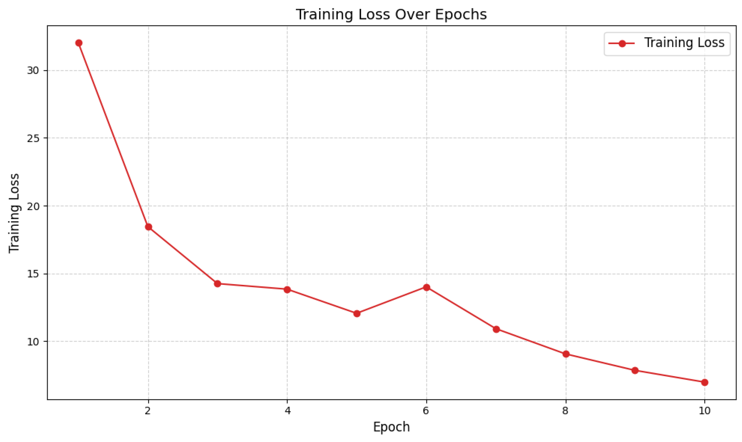
Figure 2.
Confusion matrix highlighting class-specific prediction patterns. Minor misclassifications are primarily observed between Normal and AD categories.
Figure 2.
Confusion matrix highlighting class-specific prediction patterns. Minor misclassifications are primarily observed between Normal and AD categories.
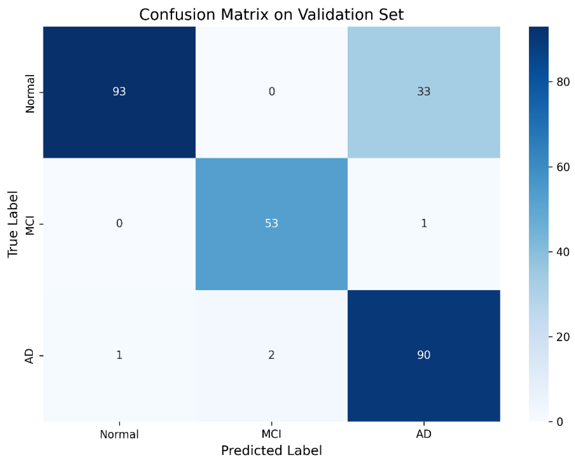
Figure 3.
Per-class precision, recall, and F1-score showing high performance, especially for MCI classification.
Figure 3.
Per-class precision, recall, and F1-score showing high performance, especially for MCI classification.
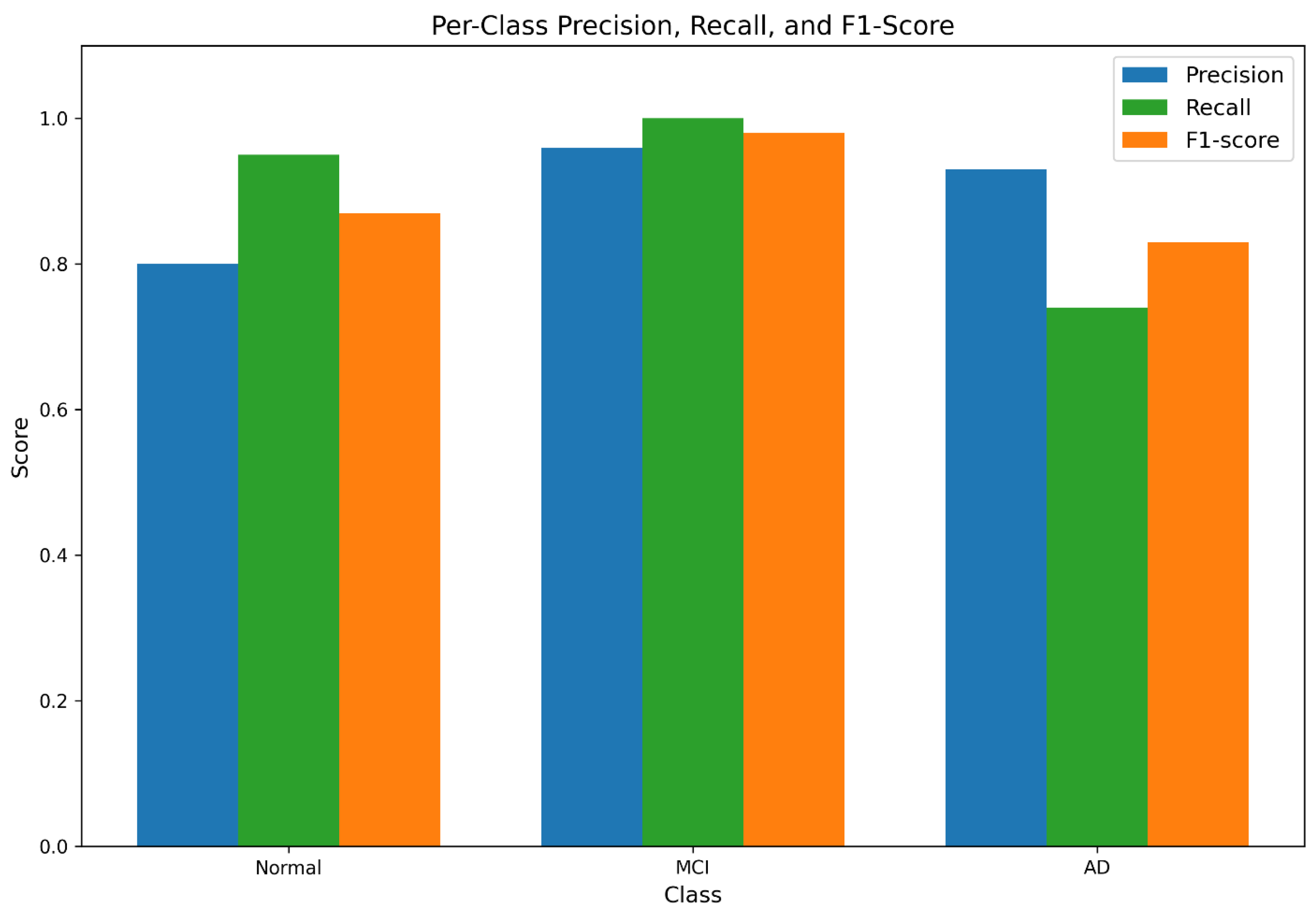
Figure 4.
ROC curves illustrating robust separability across all classes, with near-perfect AUC for MCI detection.
Figure 4.
ROC curves illustrating robust separability across all classes, with near-perfect AUC for MCI detection.
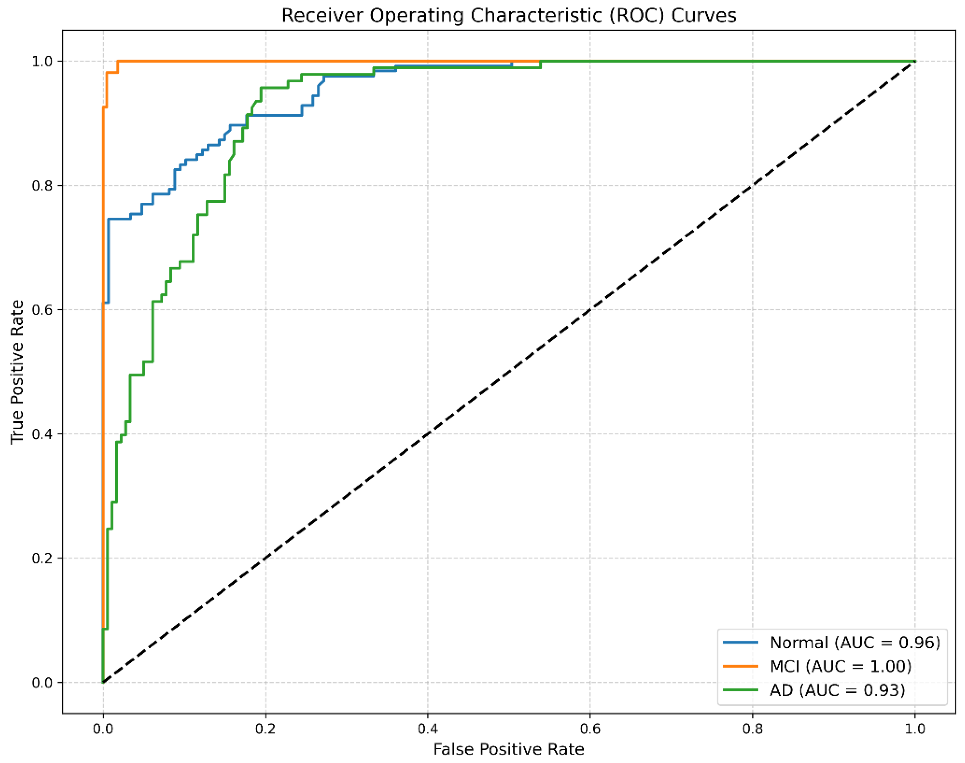

Table 1.
Class-wise Classification Performance on Validation Set.
| Class | Precision | Recall | F1-Score | Support |
|---|---|---|---|---|
| Normal | 0.9894 | 0.7381 | 0.8440 | 126 |
| MCI | 0.9636 | 0.9815 | 0.9725 | 54 |
| AD | 0.7258 | 0.9783 | 0.8333 | 92 |
| Overall (Macro Avg.) | 0.8929 | 0.8993 | 0.8833 | 272 |
Disclaimer/Publisher’s Note: The statements, opinions and data contained in all publications are solely those of the individual author(s) and contributor(s) and not of MDPI and/or the editor(s). MDPI and/or the editor(s) disclaim responsibility for any injury to people or property resulting from any ideas, methods, instructions or products referred to in the content. |
© 2025 by the authors. Licensee MDPI, Basel, Switzerland. This article is an open access article distributed under the terms and conditions of the Creative Commons Attribution (CC BY) license (http://creativecommons.org/licenses/by/4.0/).
Copyright: This open access article is published under a Creative Commons CC BY 4.0 license, which permit the free download, distribution, and reuse, provided that the author and preprint are cited in any reuse.


