
Submitted:
04 August 2025
Posted:
05 August 2025
You are already at the latest version
Abstract
Obesity concerns a wide range of the population, tending to become a major factor for diseases progression and fatality rate increase, with its implications concerning the cardiovascular system deterioration. Obesity is closely linked with metabolic derangements concerning the lipid storage and circulation and the cellular metabolism affecting most of the internal organs, especially liver and cellular function. In this current study, an analysis of the linking mechanisms between obesity, lipid deterioration, liver and lipid tissues homeostasis will be performed, with special attention on the pathophysiological characteristics of these detrimental effects on the NAFLD (non-alcoholic fatty liver disease) and the cellular function of the endothelial blood cells, with special reference on the additional burdening of the obesity on the autonomous nervous system signaling and the resulting hypertension. Despite the very complex and pluripotent pathogenic mechanisms with which obesity is intervening in these processes, it could be safely deduced that metabolic and lipid transport manipulation could serve as a crucial factor towards the cellular and tissue function improvement, with the interlinkages in the mechanisms, although highly analyzed, being until nowadays not completely deciphered.
Keywords:
obesity
; hyperlipidemia
; NAFLD
; oxidative stress
; endothelial cell function
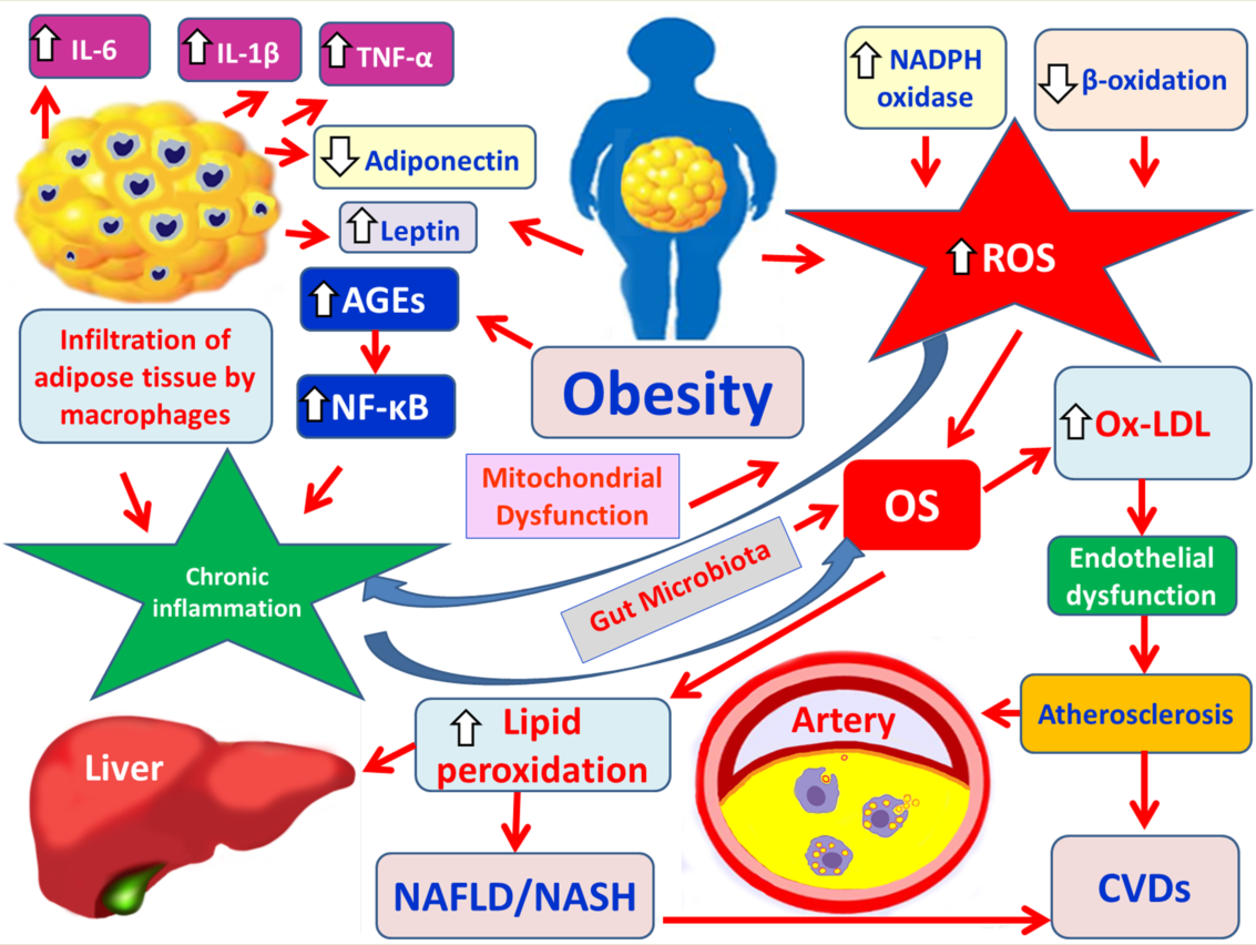
The role of obesity and oxidative stress in the development of NAFLD and CVDs. IL-6: interleukin-6; IL-1β: interleukin-1β; TNF-α: tumor necrosis factor alpha; AGEs: advanced glycation end products; NF-κB: nuclear factor kappa B; NALFD: non-alcoholic fatty liver disease; NASH: nonalcoholic steatohepatitis; OS: oxidative stress; Ox-LDL: Oxidized low-density lipoprotein; CVDs: cardiovascular disease.
1. Introduction
Obesity represents one of the most pressing public health challenges of the 21st century, with its prevalence rising steadily across both developed and developing nations. As a multifactorial condition characterized by excessive adiposity, obesity is closely linked with a variety of metabolic, hepatic, and cardiovascular disorders. Among these, non-alcoholic fatty liver disease (NAFLD) and cardiovascular diseases (CVDs) stand out as major causes of chronic illness and premature death [1,2]. NAFLD, the hepatic component of the metabolic syndrome, includes a continuum from benign hepatic steatosis to non-alcoholic steatohepatitis (NASH), fibrosis, cirrhosis, and potentially hepatocellular carcinoma [3]. In parallel, obesity markedly increases the risk of CVDs, including atherosclerosis, myocardial infarction, and heart failure, primarily through its adverse effects on systemic inflammation, insulin sensitivity, and lipid metabolism [4].
At the molecular level, obesity triggers a series of metabolic and immunological disturbances that underpin both NAFLD and CVDs. Adipose tissue, particularly visceral fat, becomes dysfunctional and secretes pro-inflammatory cytokines such as tumor necrosis factor-alpha (TNF-α), interleukin-6 (IL-6), and resistin, which promote hepatic fat accumulation, endothelial dysfunction, and vascular inflammation [5]. Insulin resistance plays a central role in this pathogenesis by exacerbating lipolysis and free fatty acid transfer to the liver, thus fostering hepatic steatosis and promoting atherogenic dyslipidemia [6]. Additionally, oxidative stress and mitochondrial dysfunction further contribute to hepatocellular injury and cardiovascular remodeling [7].
Given the overlapping pathophysiological pathways, understanding the molecular mechanisms, especially those implicating lipids deterioration, linking obesity, NAFLD, and CVDs, is crucial for the development of integrated therapeutic and preventive approaches. This review aims to elucidate the key molecular and pathological interconnections between these conditions, highlighting their clinical implications and future research directions.
2. Lipid Derangement and Non-Alcoholic Fatty Liver Disease
Nonalcoholic fatty liver disease (NAFLD) is a heterogeneous liver disease, which ranges from liver accumulation of lipids (steatosis) to non-alcoholic steatohepatitis (NASH) [8]. According to the guidelines of the American Association for the Study of Liver Diseases (AASLD) the definition of nonalcoholic fatty liver disease (NAFLD) requires (i) primary hepatic steatosis according to imaging or histology and (ii) exclusion of secondary hepatic fat accumulation, such as significant alcohol consumption, steatogenic medications and other hereditary medical disorders [9]. Hepatocellular steatosis means ectopic cytoplasmatic micro-, macro- or mixed lipid deposition into hepatocytes higher than 5% of the total liver weight. Hepatocellular steatosis may progress to the more severe form named nonalcoholic steatohepatitis (NASH) [10]. NASH is defined as the presence of primary hepatic steatosis and various degrees of inflammation with hepatocellular injury. NASH may progress to fibrosis, cirrhosis and hepatocellular carcinoma (HCC) [10]. The high frequency of concurrent primary hepatic steatosis in NAFLD with the obesity, hyperlipidemia and type 2 diabetes mellitus supports the premise that NAFLD is the hepatic representation of the metabolic syndrome [9,11].
Triglycerides (TGs, also called neutral fats, triacylglycerols, or triacylglycerides) are the major vegetable and animal fats from diet (exogenous triglycerides) and are the main constituent of the body’s fat stores. Plasma total TG concentration is determined to assess metabolic disorders. TGs are hydrolysed in the gut by lipases releasing fatty acids (FAs) and monoglycerides through lipolysis. Only short- and medium-chain FAs (up to 12 carbons) can be absorbed by enterocytes directly and then are transfered into the bloodstream by serum albumin. Long-chain FAs (over 12 carbons) are reconverted to TGs after absorption and are transported by lipoprotein called HDL, LDL, very low-density lipoprotien (VLDL) and chylomicrons (also known as ultra low-density lipoprotein, ULDL), depending on their size [12]. Lipoprotein lipase present at the surface of cells cleaves TGs to FFAs that are absorbed by cells [12]. The monoglycerides in the smooth endoplasmic reticulum of the enterocytes are re-esterified to triglycerides, which are packaged into chylomicrons and are delivered primarily to muscle and adipose tissue. The remaining TGs present in chylomicron-remnants are transported to the liver and lead to FA release within hepatocytes [13]. The major sites of endogenous TG synthesis are the liver and the adipose tissue. Also, carbohydrates from diet (e.g., glucose and fructose), are utilized in hepatic de novo lipogenesis (DNL) for the production of FAs [13]. Hepatic de novo synthesized TGs can be stored in intracellular lipid droplets or packed very low-density lipoproteins (VLDL) and secreted to the plasma [13,14] (Figure 1). Apolipoprotein B100 (ApoB100) is the critical protein for the assembly and secretion of VLDL-TG particles [14]. Once FFAs are taken up by cells or synthesized by de novo lipogenesis, they are transported by intracellular fatty-acid binding proteins (FABPs) to the hepatic mitochondria where they undergo β-oxidation to release energy in the form of ATP for preservation of the homeostasis of cells and tissues [12,15]. The acetyl-CoA produced during β-oxidation is converted to ketone bodies, i.e., acetoacetate, beta-hydroxybutyrate (BOH), and acetone during the aerobic respiration. Ketone bodies are released and then taken up by other tissues such as the brain, muscle and heart where they are converted to acetyl-CoA to serve as an energy source [15]. The three major sources of hepatic fatty acids (FA) are dietary lipids, endogenous de novo synthesis and adipose tissue derived FAs [13]. Also, FAs are important component of all cellular membranes, in the form of phospholipids. In adipose tissue, FAs are re-esterified to form TGs in lipid droplets for energy storage [12,16]. The endogenous FAs are mobilized from adipose tissue TGs by the action of hormone-sensitive lipase (HSL), which is activated by glucagon and adrenaline and inhibited by insulin, in cases such as fasting or during prolonged exercise. This ensures a steady basal supply of fuel for skeletal muscles and the heart muscle independent of food intake [17,18]. FAs circulating in the plasma, not in their ester form, are known as free fatty acids (FFAs). FFAs are always bound to a transport protein, such as albumin. In the setting of increased dietary intake of FAs, excessive adipose tissue and insulin resistance, the hepatic FA levels are increased due to enhanced peripheral lipolysis within adipocytes. Excess FAs cannot be consumed by oxidative pathways and FAs are directed towards the synthesis of TGs, leading to increased hepatic TG storage and VLDL overproduction [13].
2.1. Pathophysiological Mechanisms of NAFLD and the Role of Oxidative Stress
The pathogenesis of NAFLD is a complex mechanism with the “two-hit model” being the most widespread and prevailing theory. The “first hit”, as the initial stage, is characterized by concomitant (i) increased hepatic steatosis and (ii) insulin resistance [19]. Oxidative stress is the initiator of the “second hit” [20]. A large number of adipokines, such as leptin, adiponectin and resistin regulates free fatty acids (FFAs) to induce reactive oxygen species (ROS)-mediated injury to liver [21,22]. In addition, it seems to be involved in the pathogenesis of NAFLD, a complex interplay between several interaction mechanisms, which occur in parallel. These mechanisms include: (a) genetic predisposition, (b) lifestyle risk factors (e.g., diet, lifestyle, and smoking) and (c) metabolic deregulation (e.g., hyperglycemia, insulin resistance, dyslipidemia and lipotoxicity) and (d) alterations in gut microbiota or dysbiosis [23,24,25].
Lipid accumulation in the hepatocytes increases mitochondrial fatty-acid oxidation capacity and increases the electron trasport in the mitochondrial respiratory chain, stimulating peroxisomal and microsomal activity, which is associated with increased reactive oxygen species (ROS) formation and this may contribute to the liver fibrosis, cirrhosis and cancer observed in NASH [26]. Additionally, there is a close link between hepatic lipid storage and mitochondrial dysfunction [27]. Mitochondrial dysfunction can directly lead to enhanced ROS formation by the organelle itself and particularly from its endoplasmic reticulum surface [28]. If electron flow is interrupted at any point in the respiratory electron transport chain, the previous respiratory intermediates can transfer electrons to free oxygen molecules to produce superoxide anions and hydrogen peroxide (H2O2) [29]. Then, superoxide anions and hydrogen peroxide (H2O2) induce ROS production. It has been found that ROS generation is associated with activation of Fas transmembrane death receptor [30]. This activation of the Fas ligand/Fas system raises the down-stream caspase family members to form the protease pro-cascade reaction, resulting in cellular disorganization and apoptosis [22]. Additionally, high levels of ROS can cause oxidative modifications to the proteins, lipids and DNA, which further promote OS and cell death [31]. Therefore, mitochondrial dysfunction contributes to the development of NAFLD and NASH [32]. Moreover, ROS and lipid peroxidation products mediate hepatic fibrosis by activating hepatic stellate cells, which secrete collagen and are involved in inflammatory responses and hepatic immunology contributing to the pathogenesis of fibrosis [33]. In addition, hepatic fibrosis worsens when obesity is associated with chronic low-grade systemic inflammation. Studies have also demonstrated that hepatic steatosis leads to increased NF-κB (nuclear factor kappa B). NF-κB induces further production of local and systemic inflammatory mediators, such as TGF-β, Fas ligand, TNF-α, leptin, IL-1β, IL-6, ΙL-8, which are involved in the development of ROS-mediated hepatocellular injury [34,35]. In addition, a liver with steatosis is more vulnerable to inflammatory mediators, due to decreased antioxidant mechanisms, favoring OS-related obesity [36]. Moreover, low levels of adiponectin are associated with more severe liver fibrosis [37]. Finally, alterations in gut microbiota or dysbiosis play a large role in the development of NAFLD and NASH in obese patients, through different pathways, including its influence in energy storage, lipid metabolism, ethanol production, immune balance and inflammation [35]. It has been found that changes in gut microbiota homeostasis deregulate the gut-liver axis leading to increased intestinal permeability, thus promoting the translocation of bacterial endotoxins into systemic circulation and facilitating the hepatic endotoxemia and low-grade intestinal inflammation [38]. Therefore, alterations in the gut microbiota are strongly linked to increased ROS products and oxidative stress (OS), which are involved in the development of NAFLD and NASH [39] (Figure 2).
2.2. Obesity, Lipid Derangement, Oxidative Stress and Cardiovascular Disease (CVD)
Dyslipidemia, also known as hyperlipidemia, describes an abnormal amount of lipids in the blood that can increase the risk of CVD and heart stroke [40]. The typical dyslipidemia of obesity consists of increased circulating triglycerides (TG) and FFAs, decreased high density lipoprotein (HDL) cholesterol and normal or slightly increased low-density lipoprotein (LDL) cholesterol with increased small dense low density lipoprotein (sdLDL). Elevated levels of plasma apolipoprotein (apo) B are usually the result of hepatic overproduction of apo B containing lipoproteins [41]. Diagnostic criteria for obesity related dyslipidemia are defined as HDL cholesterol less than 1.0 mmol/l (40 mg/dl) in men and less than 1.3 mmol/L (50 mg/dl) in women, or blood triglycerides greater than 1.7 mmol/l (150 mg/dl) [41,42]. High levels of triglycerides and low levels of high-density lipoprotein (HDL) cholesterol are major risk factors of atherosclerosis by a buildup of plaques in the inner lining of all large and medium-sized arteries, which are deposits of fatty materials.
Patients with obesity and dyslipidemia are at increased risk of developing cardiovascular disease (CVD) [2]. Obesity is characterized by hypertrophy of adipocytes and the development of a chronic sub-clinical pro-inflammatory environment in adipose tissue, leading to increased infiltration of immune cells [43]. It has been found that FAs in hypertrophic adipocytes trigger specific serine-kinases that attenuate insulin receptor function. Adipose tissue insulin resistance consequently causes adipose tissue to release excessive FFAs into the bloodstream leading to accumulation of ectopic fat in the liver and skeletal muscles [44]. Also, obesity-induced hepatic insulin resistance is characterized by an impairment of insulin ability to inhibit glucose output via hepatic endoplasmic reticulum (ER) stress, reactive oxygen species (ROS) and inflammation [45]. These processes stimulate gluconeogenesis and lead to hyperglycemia and consequent hyperinsulinemia [46]. Mechanisms that also connect obesity with insulin resistance, dyslipidemia and cardiovascular disease and stimulate ROS production in the vascular endothelium are (i) Low levels of high-density lipoprotein (HDL) cholesterol; (ii) Enhanced clearance of HDL (HDL) cholesterol; (iii) Increased post-prandial TG values; (iv) Elevated plasma very low density lipoprotein (VLDL); (v) Elevated levels of plasma TGs; (vi) Increased Ox-LDL [47].
Elevated levels of plasma triglyceride rich lipoproteins (TGRL) are due to their delayed hepatic clearance as a result of insulin resistance leading to TGs accumulation in arteries, promoting macrophage foam cell formation and inflammation [48]. Under these circumstances, cholesteryl ester transfer protein (CETP) activity facilitates the exchange of cholesteryl esters and triglycerides (TGs) between high-density lipoprotein (HDL) cholesterol and TG-rich, apoB-containing lipoproteins [49] leading to low levels of high-density lipoprotein (HDL) cholesterol and the generation of small, dense, low-density lipoproteins (sdLDL), which are the key components of metabolic syndrome (MS) [50]. Low levels of HDL are associated with reduced unesterified cholesterol efflux from macrophage foam cells in arteries, aggravating atherosclerosis and are therefore risk factor for cardiovascular disease [48]. Also, the high levels of low-density lipoprotein (LDL) cholesterol are involved in the development of atherosclerosis and LDL cholesterol remains the primary target of therapy for the prevention of coronary heart disease [51]. Smaller, denser LDL (sdLDL) particles are pivotal for the formation and progression of atheromatic plaques than the larger, lighter LDL particles, because circulating sdLDL particles undergo multiple atherogenic modifications in blood plasma, making them strong inducers of inflammation resulting therefore in atherosclerotic plaques formation. These modifications include desialylation, glycation and oxidation [52,53]. Also, sdLDL particles have reduced affinity for the LDL receptors and lower catabolic rate. Moreover, sdLDL have a greater susceptibility to oxidation and higher concentration of polyunsaturated fatty acids (rendering their oxidized forms to have greater affinity for the scavenger receptors of macrophages, giving further rise to the inflammatory atherosclerotic lessions). In addition, desialylation of sdLDL particles increases their affinity to proteoglycans in the arterial wall and therefore exhibit greater permeability in the endothelium of arterial walls [54,55]. Apart from all the above mechanisms sdLDL cholesterol is able to increase the atherogenic effect by regulating the activity of gene networks, monocytes and enzymes [55].
Insulin resistance at the adipose tissue results in increase of fatty acids into the circulation. Increased FAs flux to the liver stimulates the assembly and secretion of VLDL resulting in hypertriglyceridemia [56]. Cholesterol ester transfer protein (CETP) in the bloodstream catalyzes the exchange of triglycerides and cholesterol ester between VLDL and chylomicron (TG-rick lipoproteins, TGL) and HDL, respectively. This exchange results in cholesterol ester (CE) depletion and TG enrichment of HDL. As HDL accumulates triglycerids, it is a substrate for lipoprotein lipase and hepatic lipase. Hepatic lipase modification of TG-rich HDL releases lipid-poor apoA-I and HDL remnant particles. Apo-I is filtered rapidly by the renal glomerus and then degraded by proximal tubular cell receptors, such as cubilin/megalin system (Figure 3). HDL remnants may bind to putative receptors in the liver that mediate HDL degradation. Therefore, the availability of HDL for reverse cholesterol transport is reduced. Moreover, the interchange between TRL (triglyceride-rich lipoproteins) and HDL is mediated by phospholipid transfer protein (PLTP), which transfers surface phospholipids (PL) from TRL to HDL during lipolysis. Insulin resistance is associated with TG enrichment of HDL particles and increased hepatic lipase enzyme activity and lowering of HDL. Then, LDL lipoproteins with the action of hepatic lipase generate the small dense LDL lipoproteins (sdLDL) [48,54,57]. There is an inverse relationship between plasma triglycerides secreted either from the liver (VLDL) or intestine (chylomicrons) and HDL levels. A higher level of VLDL and chlylomicrons correlates with lower HDL levels. Low levels of HDL are associated with reduced unesterified cholesterol efflux from macrophage foam cells in arteries, aggravating atherosclerosis. Also, the high levels of low-density lipoprotein (LDL) cholesterol and the presence of small dense LDL are involved in the development of cardiovascular disease [56]. Endothelial dysfuction allows LDL particles, which are rich in cholesteryl esters (LDL-c) to enter the tunica intima of the arteries. Blood monocytes enter into the intima producing free radicals, which oxidize the LDL and become macrophages. Macrophages engulf ox-LDL particles and turn into foam cells. Necrosis of foam cells release its content outside that is engulfed by other macrophages, which eventually build a large lesion area. Progression into this lesion, turns into plaque gradually accumulating calcium slats, smooth muscle cells from tunica media, collagen and the foam cells [58]. A typical adverse plaque is characterized by macrophage accumulation, a large lipid core, a thin fibrous cap and microcalcifications. Intraplaque microvessels result from angiogenesis driven by hypoxia and inflammatory stimuli within the necrotic core. These vessels can result in intraplaque hemorrhage. The clot attached to the vessel wall would make a thrombus, which increases the risk of plaque destabilization, with the break of the thrombus being able cause stroke or myocardial infarction [58,59].
2.3. Obesity, Oxidative Stress and Endothelial Blood Cells Function
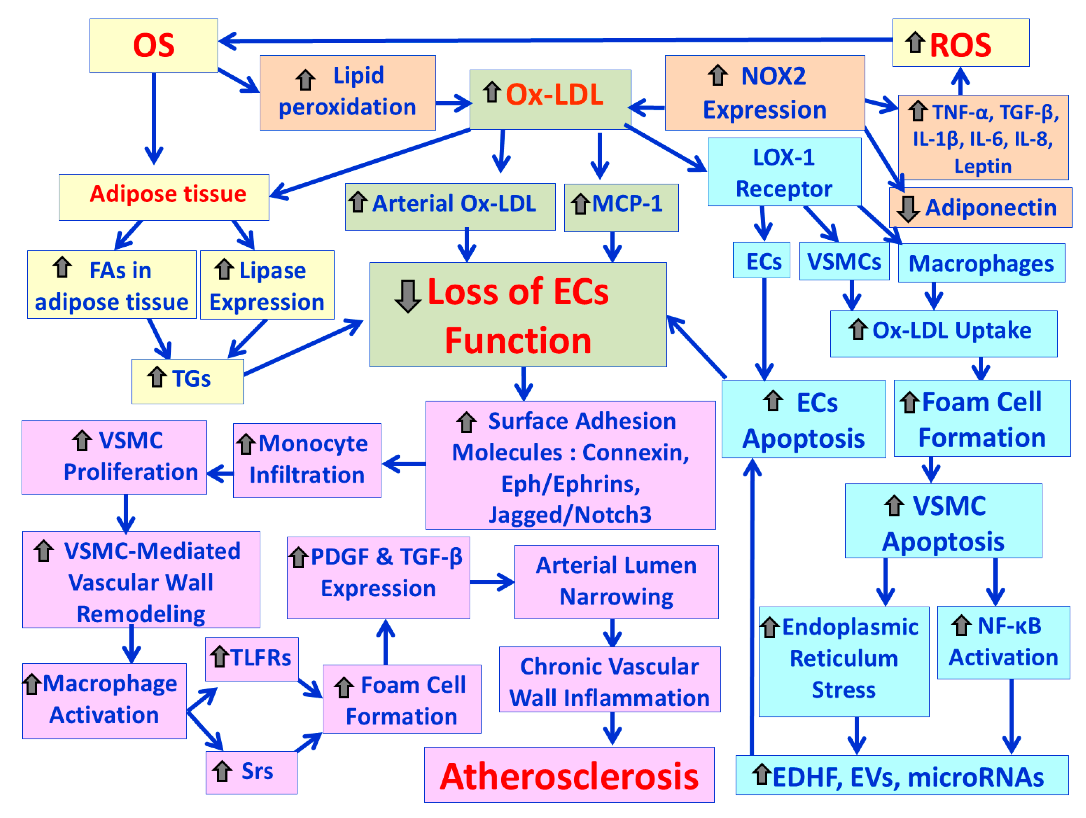
Oxidative stress plays a crucial role in oxidation of LDL particles by the process of lipid peroxidation [60]. Accumulation of oxidized low-density lipoproteins (ox-LDLs) in the tunica intima of arteries triggers the onset of atherosclerosis [58]. Moreover, an increase in ox-LDL could also be due to increased oxidant capacity, by elevated expression of NOX2 (NADPH oxidase 2) [61,62]. Increased expression of NOX2 in turn, induces further increased pro-inflammatory cytokine levels, decreased production of adiponectin and generation of ROS in vascular and immune cells circulating in blood vessels, which can lead to further OS and development of atherosclerosis [63,64]. Another possible mechanism of the Ox-LDL implication in the development of atherogenesis involves the release of monocyte chemoattractant protein 1 (MCP-1) from the endothelial cells (Ecs) and the vascular smooth muscle cells (VSMCs) [65]. Specifically, Ox-LDL, particles stimulate the secretion of MCP-1, which further facilitates the dysfunction of endothelial cells (Ecs) and smooth muscle cells (SMCs) of the arterial media layer [66]. Loss of endothelial cell (EC) function induces activation of several cell surface adhesion molecules, such as connexin, Eph/ephrins, Jagged/Notch3, which recruit monocytes from the blood into the endothelial space and these monocytes promote VSMC proliferation and vascular wall remodeling [67]. Monocytes then differentiate into macrophages that upregulate both (i) toll-like receptors (TLFRs), which play a central role in macrophage activation and (ii) scavenger receptors (Srs), which remove apoptotic cell fragments, bacterial endotoxins and Ox-LDL. This leads to lipid accumulation and foam cell formation [68]. Then, cholesterol builds up in the inner lining of the artery and an arterial plaque develops and forms a complex lesion with migration and proliferation of vascular smooth muscle cells (VSMCs), which secrete extracellular matrix, such as collagen accumulation in the arterial plaques [69]. Progressive narrowing of the arterial lumen causes the expression of platelet derived growth factors (PDGF) and transforming growth factor-β (TGF-β), which further increase VSMC proliferation and collagen deposition in the arterial plaque [67]. Macrophage activation leads to the release of pro-inflammatory cytokines, proteases, reactive oxygen species (ROS), which promote atherosclerosis (68,70,71). Also, it has been found that macrophages exposed to Ox-LDL up-regulate the expression of caveolin-1, which plays an important role in the development of atherosclerosis [61,62]. However, the EC-VSMC communication is bi-directional meaning that changes occurring in VSMCs may ultimately affect ECs function. Suppression of VSMC proliferation causes elevated endoplasmic reticulum stress and NF-κB activation, which stimulate the release of factors, such as EDHF (endothelium-derived hyperpolarizing factor), Evs (extracellular vesicles), and microRNAs, which leads to ECs apoptosis and subsequent atherosclerosis [70]. In addition, Ox-LDL increases triglyceride production by inducing the expression of lipoprotein lipase [72] and by inducing the accumulation of fatty acids in adipocytes [73]. This increased lipotoxicity directly causes ECs dysfunction and subsequent atherosclerosis. Moreover, Ox-LDL binds to lectin-like oxidized low-density lipoprotein receptor-1 (LOX-1) [74]. The binding of Ox-LDL to LOX-1 receptor enhances Ox-LDL uptake in both macrophages and SMCs and stimulates foam cell formation. It also contributes to endothelial cell (EC) activation and EC-induced apoptosis (programmed cell death) of vascular smooth muscle cells (VSMCs) [61,70]. Also, the excessive formation of Ox-LDL induces increased expression of the proapoptotic protein Bax, and the anti-apoptotic protein Bcl-2, suggesting the involvement of LOX-1 in the atherosclerotic plaque destabilization [75,76]. Figure 4 demonstrates the possible mechanisms involved in atherosclerosis development by the oxidized low-density lipoproteins (Ox-LDLs).
2.4. Obesity, Oxidative Stress and Hypertension
Hypertension is strongly associated with the metabolic syndrome and predisponses to increased cardiovascular morbidity and mortality [77,78]. Also, hyperinsulinemia and insulin resistance are found in hypertensive patients with type II diabetes [79]. Hypertension and obesity interact with cardiac function, leading to larger heart volume, left ventricular hypertrophy and thus greater likelihood of cardiac failure [79,80]. Specifically, increased peripheral resistance due to arterial wall thickening and abnormal vascular tone is the main hemodynamic situation in hypertension [77]. Multiple interacting factors, such as activation of the renin-angiotensin-aldosterone system (RAAS), oxidative stress, activation of the sympathetic nervous system, hemodynamic changes and mechanical forces stimulate vascular smooth muscle cells signaling, which promotes vasoconstriction, vascular hypertrophy, fibrosis, inflammation and calcification, processes that contribute to vascular remodeling in hypertension [77,81,82].
A free radical is the product of normal cellular metabolism, which has one or more unpaired electrons. A free radical can be defined as an atom or molecule, which is chemically highly reactive [83,84]. Reactive oxygen species (ROS) such as superoxide anion (·O2−), hydroxyl radical (·OH-), hydrogen peroxide (H2O2) and singlet oxygen (1O2) are highly reactive and unstable chemicals formed from diatomic oxygen (O2). Free radicals and other ROS are formed continuously in normal essential physiological processes [85]. Oxidative stress occurs when there is an imbalance between production of free radicals and ROS and antioxidant defence system, which may results from increased production of free radicals and ROS or diminished levels of antioxidants or both [86]. ROS can cause direct damage to various cellular componets including DNA, proteins and lipids [87]. Lipid peroxidation preferentially oxidizes polyunsaturated fatty acids (PUFAs) and causes structural modifications of lipids in plasma membranes [85]. Increase in free radicals and ROS eliminates nitrogen monoxide (NO), decreasing vascular relaxation and vasodilation, which results in increase in the systemic vascular resistance (SVR) [83]. An decreased nitrogen monoxide (NO) production or an increased inactivation of NO because of its interaction with the superoxide anion (O2·−) or an imbalance between the superoxide (O22-) and the NO production may account for reduced vasodilation and may promote endothelial dysfunction, leading to development of hypertension and cardiovascular complications [88,89].
2.4.1. Physiology and Pathophysiology of the Renin-Angiotensin-Aldosterone System (RAAS)
The renin-angiotensin-aldosterone system (RAAS) plays an important role in the regulation of arterial blood pressure. RAAS interacts with the sympathetic nervous system (SNS) and the baroreceptor reflexes [90]. In response to low blood pressure, the sympathetic nervous system (SNS) is the major determinant for conversion of prorenin to active renin from juxtaglomerular cells within the afferent arterioles of the kidneys via norepinephrine actions on β1-adrenergic receptor-AMP pathway [91,92]. The enzyme, renin in the bloodstream cleaves angiotensin I from angiotensinogen, a glycoprotein produced in the liver and fat cells [91]. Angiotensin I is a peptide hormone, physiologically inactive, and works as a precursor for angiotensin II [rapidly cleaved by angiotensin-converting enzyme (ACE) to form angiotensin II, which is the physiological active component of the RAAS] [90]. Angiotensin II has effects on the kidney, adrenal gland, arterioles and brain by binding to angiotensin type I and angiotensin type II receptors, which are G protein-coupled receptors [93]. The effect of angiotensin II on vasoconstriction takes places in renal and systemic arterioles through its effects on vascular smooth muscle, increasing heart rate and contractility through activation of the sympathetic nervous system. Therefore, angiotensin II increases total peripheral resistance, and blood pressure (91,94]. Also, angiotensin II acts on the glomerulosa cells of the adrenal cortex and stimulates aldosterone secretion, which is a steroid hormone [95]. Aldosterone has multiple effects that regulate blood pressure [96]. The fundamental role of aldosterone is to support sodium reabsorption with associated passive water reabsorption and potassium excretion in the distal tubule and collecting duct of nephrons in kidney [91,97]. Moreover, aldosterone acts in the central nervous system (CNS) and sensitizes angiotensin II-induced hypertension [98,99]. When the RAAS is pathologically activated results in excessive vasoconstriction causing hypertension [91]. Abundant evidence supports the role of activation of the sympathetic nervous system in the pathogenesis of obesity-related hypertension (SNS) [79,100]. Also, increased renal sodium reabsorption, impaired pressure natriuresis and physical compression of kidneys, especially when there is increased visceral adiposity play a major role in initiating the rise in blood pressure (BP) associated with excessive adiposity and obese subjects [101]. The activation of the renin-angiotensin-aldosterone system (RAAS) is also an important mediator of obesity-induced hypertension [77,102]. Particularly, increased Ang II levels due to adipose tissue dysfunction can enhance microvascular vasoconstriction by (i) decreasing the synthesis and release of endothelium-derived nitric oxide (NO), (ii) stimulating the secretion and action of endothelium-derived vasoconstrictors such as endothelin-1 (ET-1) and prostanoids, (iii) promoting the contraction of vascular smooth muscle cells (VSMC) and (iv) increasing sympathetic nervous system activity [103,104]. Ang II induces oxidative stress via activation of nicotinamide adenine dinucleotide phosphate (NADPH) oxidase and the production of ROS such as superoxide anion (O2·−) [105]. Also, Ang II induces lipid peroxidation and the combination of increased lipid peroxidation and reduced antioxidant status are associated with hypertension [106]. Moreover, Ang II stimulates the release of inflammatory cytokines [107], whilst growing evidence supports the vital pathophysiological role of oxidative stress (OS) in the development of hypertension, especially in obesity [108,109].
2.4.2. Hypertension Due to Over-Activation of Sympathetic Nervous System (SNS) in Obesity
Obesity activates SNS in tissues such as heart, kidneys and skeletal muscles [110]. Especially, obese individuals have increased renal and cardiac SNS activity compared with healthy individuals, which is involved in the development of hypertension in these subjects [111]. Several potential factors have been proposed to promote obesity-associated hypertension by activating the SNS. These factors include alterations in adipose tissue secretion of adipokines and cytokines. Specifically, low adiponectin levels as well as low apelin levels are associated with SNS activation and arterial hypertension [111,112,113]. Also, increased leptin secretion from adipose tissue raises SNS activity causing increased renal sodium retention and subsequent increased blood pressure [111,114]. Moreover, sympathetic activation is associated with increased levels of the pro-inflammatory IL-6, which are implicated with increased blood pressure linking therefore systemic inflammation with increased risk for hypertension [115]. In addition, TNF-α contributes to Ang II-induced hypertension [116]. Additionally, the activation of the renin-angiotensin-aldosterone system (RAAS) and the formation of Ang II is implicated. In obese individuals, significant amounts of Ang II are secreted from abdominal subcutaneous adipose tissue [117]. Also, adipocytes are capable of aldosterone production, which is partially Ang II dependent [118]. Increased Ang II levels due to adipose tissue dysfunction can increase sympathetic nervous system activity [103,104]. Angiotensin II signaling is enhanced in different brain sites such as the paraventricular nucleus, the rostral ventrolateral medulla (RVLM) and the area postrema [119]. The rostral ventrolateral medulla (RVLM) is the key region in cardiovascular regulation [120]. The action of Ang II on SNS includes (i) activation of adrenergic pathways within the brain to enhance sympathetic outflow, (ii) stimulatory effects on sympathetic ganglia and the adrenal medulla, which is a modified ganglion of the sympathetic nervous system and (iii) actions into sympathetic nerve terminals that facilitate sympathetic neurotransmission [90,121]. Also, there is evidence that high aldosterone levels can increase blood pressure by acting directly to the CNS to increase sodium appetite, sympathetic nerve activity, and vasopressin release and impair pressoreflex (or baroreflex) sensitivity [103,122]. Dysfunction of the pressoreceptor reflexes may contribute to excessive renal sympathetic nerve activity (RSNA) in cases with obesity-related hypertension [100], whilst activation of chemoreceptor-mediated reflexes associated with obstructive sleep apnea (OSA) and intermittent hypoxemia may also be an important factor. Obstructive breathing events in obese patients cause repetitive events of hypoxemia and sometimes hypercapnia, which stimulate the central chemoreflex response and in that way contribute to elevated blood pressure levels through SNS activation and sympathetic vasoconstriction [111,123,124].
3. Conclusions and Future Perspectives
The increasingly discovered and determined, shared pathophysiological mechanisms, between obesity, non-alcoholic fatty liver disease (NAFLD), and cardiovascular diseases (CVDs), show their intricate interplay, among which oxidative stress seems to play a central role. Excess adipose tissue, particularly visceral fat, promotes chronic low-grade inflammation that in combination with the parallel overproduction of reactive oxygen, nitrogen, sulfur and carbon species, contribute to hepatocellular injury and endothelial dysfunction. These inflammatory and oxidative insults disrupt lipid metabolism, in the liver and their circulation in the cardiovascular system, accelerating NAFLD progression, while simultaneously giving rise to atherosclerotic changes in the cardiovascular system. Understanding of these overlapping pathways, opens new ways for early identification and targeted interventions of these conditions, with the antioxidant strategies, either dietary or pharmacological (or even lifestyle modifications) may assisting to the attenuation of the oxidative stress and its downstream effects, leading to agents with multi-functionality, acting pleiotropically in these conditions and paving also the way towards potential repurposing of compounds against the one condition for the treatment of the other. However, further studies are required to determine the efficacy and long-term benefits of such interventions in diverse patient populations.
Future research should focus on further identification of specific biomarkers of oxidative damage that could aid in the stratification of individuals being at-risk and guide personalized therapies. Additionally, exploring the liver-adipose-nervous system axis and its influence on endothelial cells function may reveal novel therapeutic targets. These directions will ultimately lead to a multidisciplinary approach that integrates metabolic, inflammatory, and oxidative perspectives that will be essential for preventing and managing obesity-related NAFLD and CVDs more effectively.
Author Contributions
The authors confirm their contribution to the paper as follows: study conception and study design: M.V., P.T.N.; literature review F.N.V., V.K.V.; draft manuscript: F.N.V., M.V., V.K.V., P.T.N.; editing and critical revision of the manuscript: F.N.V., M.V., P.T.N. All authors reviewed and approved the final version of the manuscript.
Funding
This review did not receive any financial support.
Conflicts of Interest
The authors declare no conflict of interest.
References
- Younossi, Z.; Anstee, Q.M.; Marietti, M.; Hardy, T.; Henry, L.; Eslam, M.; George, J.; Bugianesi, E. Global burden of NAFLD and NASH: Trends, predictions, risk factors and prevention. Nature Reviews Gastroenterology & Hepatology 2018, 15, 11–20. [Google Scholar]
- Powell-Wiley, T.M.; Poirier, P.; Burke, L.E.; Després, J.P.; Gordon-Larsen, P.; Lavie, C.J.; Lear, S.A.; Ndumele, C.E.; Neeland, I.J.; Sanders, P. Obesity and cardiovascular disease: A scientific statement from the American Heart Association. Circulation 2021, 143, e984–e1010. [Google Scholar] [CrossRef]
- Chalasani, N.; Younossi, Z.; Lavine, J.E.; Diehl, A.M.; Brunt, E.M.; Cusi, K.; Charlton, M.; Sanyal, A.J. The diagnosis and management of non-alcoholic fatty liver disease: Practice guidance from the American Association for the Study of Liver Diseases. Hepatology 2018, 67, 328–357. [Google Scholar] [CrossRef] [PubMed]
- Herrington, W.; Lacey, B.; Sherliker, P.; Armitage, J.; Lewington, S. Epidemiology of atherosclerosis and the potential to reduce the global burden of atherothrombotic disease. Circulation Research 2016, 118, 535–546. [Google Scholar] [CrossRef]
- Saltiel, A.R.; Olefsky, J.M. Inflammatory mechanisms linking obesity and metabolic disease. The Journal of Clinical Investigation 2017, 127, 1–4. [Google Scholar] [CrossRef] [PubMed]
- Tilg, H.; Moschen, A.R.; Roden, M. NAFLD and diabetes mellitus. Nature Reviews Gastroenterology & Hepatology 2021, 18, 32–42. [Google Scholar]
- Sanyal, A.J. Past, present and future perspectives in nonalcoholic fatty liver disease. Nature Reviews Gastroenterology & Hepatology 2019, 16, 377–386. [Google Scholar] [CrossRef] [PubMed]
- Miyake, T.; Kumagi, T.; Hirooka, M.; Furukawa, S.; Kawasaki, K.; Koizumi, M.; Todo, Y.; Yamamoto, S.; Nunoi, H.; Tokumoto, Y.; et al. Significance of exercise in nonalcoholic fatty liver disease in men: A community-based large crosss-sectional study. J Gastroenterol 2015, 50, 230–237. [Google Scholar] [CrossRef]
- Chalasani, N.; Younossi, Z.; Lavine, J.E.; Diehl, A.M.; Brunt, E.M.; Cusi, K.; Charlton, M.; Sanyal, A.J. The diagnosis and management of non-alcoholic fatty liver disease: Practice Guideline by the American Association for the study of liver diaseases, American College of Gastroenterology, and the American Gastroenterological Association. Hepatology 2012, 55, 2005–2023. [Google Scholar] [CrossRef]
- Burnt, E.M.; Tiniakos, D.G. Histopatholygy of nonalcoholic fatty liver disease. World J Gastroenterol 2010, 16, 5286–5296. [Google Scholar] [CrossRef]
- Cariou, B.; Byrne, C.D.; Loomba, R.; Sanyal, A. Nonalcoholic fatty liver disease as a metabolic disease in humans: A literature review. Diabetes Obes Metab 2021, 23, 1069–1083. [Google Scholar] [CrossRef]
- Sobczak, A.I.S.; Blindauer, C.A.; Stewart, A.J. Changes in plasma free fatty acids associated with type-2 diabetes. Nutrients 2019, 11, 2022. [Google Scholar] [CrossRef]
- Alves-Bezerra, M.; Cohen, D.E. Triglyceride metabolism in the liver. Comp Physiol 2017, 1–8. [Google Scholar]
- Fisher, E.A.; Feig, J.E.; Hewing, B.; Hazen, S.L.; Smith, J.D. High-density lipoprotein function, dysfunction, and reserve cholesterol trasport. Arterioscler Thromb Vasc Biol 2012, 32, 2813–2820. [Google Scholar] [CrossRef]
- Sapanaro, C.; Gaggini, M.; Carli, F.; Gastaldelli, A. The subtle balance between lipolysis and lipogenesis: A critical point in metabolic homeostasis. Nutrients 2015, 7, 9453–9474. [Google Scholar] [CrossRef] [PubMed]
- Price, E.R. The physiology of lipid storage and use in reptiles. Biol Rev Cam Philos Soc 2017, 92, 1406–1426. [Google Scholar] [CrossRef]
- Nielsen, T.S.; Jessen, N.; Jørgensen, J.O.L.; Møller, N.; Lund, S. Dissecting adipose tissue lipolysis: Molecular regulation and implications for metabolic disease. J Mol Endocrinol 2014, 52, R199–R222. [Google Scholar] [CrossRef] [PubMed]
- Rui, L. Energy metabolism in the liver. Compr Physiol 2014, 4, 17–197. [Google Scholar] [CrossRef]
- Peverill, W.; Powell, L.W.; Skoien, R. Evolving concepts in the pathgenesis of NASH: Beyond steatosis and inflammation. Int J Mol Sci 2014, 15, 8591–8638. [Google Scholar] [CrossRef]
- Ma, Y.; Lee, G.; Heo, S.-Y.; Roh, Y.-S. Oxidative stress is a key modulator in the development of nonalcoholic fatty liver disease. Antioxidants (Basel) 2022, 11, 91. [Google Scholar] [CrossRef]
- Mavilia, M.G.; Wu, G.Y. Liver and serum adiponectin levels in non-alcoholic fatty liver disease. J Dig Dis 2021, 22, 214–221. [Google Scholar] [CrossRef]
- Zeng, L.; Tang, W.J.; Yin, J.J.; Zhou, B. Signal transductions and nonalcoholic fatty liver: A mini-review. Int J Clin Exp Med 2014, 7, 1624–1631. [Google Scholar]
- Doulberis, M.; Kotronis, G.; Gialamprinou, D.; Kountouras, J.; Katsinelos, P. Non-alcoholic fatty liver disease: An update with special focus on the role of gut microbiota. Metabolism 2017, 71, 182–197. [Google Scholar] [CrossRef]
- Diehl, A.M.; Day, C. Cause, Pathogenesis, and Treatment of Nonalcoholic Steatohepatitis. N Engl Med 2017, 377, 2063–2072. [Google Scholar] [CrossRef]
- Sun, Z.; Lazar, M.A. Dissociating fatty liver and diabetes. Trends Endocrinol Metab 2013, 24, 4–12. [Google Scholar] [CrossRef]
- Pérez-Carreras, M.; Del Hoyo, P.; Martín, M.A.; Rudio, J.C.; Martín, A.; Castellano, G.; Colina, F.; Arenas, J.; Solis-Herruzo, J.A. Defective hepatic mitochondrial respiratory chain in patients with nonalcoholic steatohepatitis. Hepatology 2003, 38, 999–1007. [Google Scholar] [CrossRef] [PubMed]
- Peng, K.-Y.; Watt, M.J.; Pensen, S.; Greve, J.W.; Huynh, K.; Jayawardana, K.S.; Meikle, P.J.; Meex, R.C.R. Mitochondrial dysfunction-related lipid changes occur in nonalcoholic fatty liver disease progression. J Lipid Res 2018, 59, 1977–1986. [Google Scholar] [CrossRef] [PubMed]
- Leadsham, J.E.; Sanders, G.; Giannaki, S.; Bastow, E.L.; Hutton, R.; Naeimi, W.R.; Breitenbach, M.; Gourlay, C.W. Loss of cytochrome c oxidase promotes RAS-dependent ROS production from the ER resident NADPH oxidase, Yno1p, in yeast. Cell Metab. 2013, 18, 279–286. [Google Scholar] [CrossRef]
- Hensley, K.; Robinson, K.A.; Gabbita, S.P.; Salsman, S.; Flyod, R.A. Reactive oxygen species, cell signaling, and cell injury. Free Radic Biol Med 2000, 28, 1456–1462. [Google Scholar] [CrossRef]
- Redza-Dutordoir, M.; Averill-Bates, D.A. Activation of apoptosis signalling pathways by reactive oxygen species. Biochim Biphys Acta 2016, 1863, 2977–2992. [Google Scholar] [CrossRef] [PubMed]
- Klimova, N.; Fearnow, A.; Kristiann, T. Role of NAD+-modulated free fadical generation in mechanisms o facute brain injury. Brain Sci 2020, 10, 449. [Google Scholar] [CrossRef]
- Begriche, K.; Massart, J.; Robin, M.A.; Bonnet, F.; Fromenty, B. Mitochondrial adaptation and dysfunctions in nonalcoholic fatty liver disease. Hepatology 2013, 58, 1497–1507. [Google Scholar] [CrossRef] [PubMed]
- Ramos-Tovar, E.; Muriel, P. Molecular mechanisms that link oxidative stress, inflammation, and fibrosis in the liver. Antioxidants 2020, 9, 1279. [Google Scholar] [CrossRef]
- Manti, S.; Maseglia, L.; D’Angelo, G.; Filippelli, M.; Cuppari, C.; Gitto, E.; Romano, C.; Arrigo, T.; Salpietro, C. Portal hypertension as immune mediate disease. Hepat Mon 2014, 14, e18625. [Google Scholar] [CrossRef]
- Takaki, A.; Kawai, D.; Yamamoto, K. Multiple hits, including oxidative stress, as pathogenesis and treatment target in non-alcoholic treatohepatitis (NASH). Int J Mole Sci 2013, 14, 20704–20728. [Google Scholar] [CrossRef]
- Basaronoglu, M.; Kayacetin, S.; Yilmaz, N.; Kayacetin, E.; Tarcin, O.; Sonsuz, A. Understanding mechanisms o the pathogenesis of nonalcoholic fatty liver disease. World J Gastroenterol 2010, 16, 2223–2226. [Google Scholar] [CrossRef]
- Mohamed, M.S.; Youssef, T.M.; Abdullah, E.E.; Ahmed, A.E. Correlation between adiponectin level and the degree of fibrosis in patients with non-alcoholic fatty liver disease. Egupt Liver J 2021, 2021, 78. [Google Scholar] [CrossRef]
- Song, Q.; Zhang, X. The role of gut-liver axis in gut microbiome dysbiosis associated NAFL and NAFLD-HCC. Biomedicines 2022, 10, 524. [Google Scholar] [CrossRef] [PubMed]
- Turnbaugh, P.J.; Ley, R.E.; Mahowald, M.A.; Magrini, V.; Mardis, E.R.; Gordon, J.I. An obesity-associated gut microbiome with increased capacity for energy harvest. Nature 2006, 444, 1027–1031. [Google Scholar] [CrossRef] [PubMed]
- Bolkent, S.; Yanardag, R.; Bolkent, S.; Doger, M.M. Beneficial effects of combined treatment with niacin and chromium on the liver of hyperlipemic rats. Biol Trace Elem Res 2004, 101:219-229.
- Jellinger, P.S.; Handelsman, Y.; Rosenblit, P.D.; Bloomgarden, Z.T.; Fonseca, V.A.; Garber, A.J.; Grunberger, G.; Guerin, C.K.; Bell, D.S.H.; Mechanick, J.I.; et al. American Association of Clinical Endocrinologists and American College of Endocrinologyy Guidelines for Management of Dyslipidemia and Prevention of Cardiovascular Disease. Endocr Pract 2017, 23(Suppl2):1-87.
- Matey-Hernandez, M.; Williams, F.M.K.; Potter, T.; Valdes, A.M.; Spector, T.D.; Menni, C. Genetic and microbiome influence on lipid metabolism and dyslipidemia. Physiol Genomics 2018, 50, 117–126. [Google Scholar] [CrossRef] [PubMed]
- Bijland, S.; Mancini, S.J.; Salt, I.P. Role of AMP-activated protein kinase in adipose tissue metabolism and inflammation. Clin Sci (Lond) 2013, 124, 491–507. [Google Scholar] [CrossRef]
- Hammarstedt, A.; Gogg, S.; Hedjazifar, S.; Nerstedt, A.; Smith, U. Impaired Adipogenesis and Dysfunctional Adipose Tissue in Human Hypertrophic Obesity. Physiol Rev 98: 1911–1941, 2018.
- Lee, S.-H.; Park, S.-Y.; Coi, C.S. Insulin resistance: From mechanisms to therapeutic strategies. Diabetes Metab 2022, 46:15-37.
- Kim, O.-K.; Jun, W.; Lee, J. Mechanism of ER Stress and Inflammation for Hepatic Insulin Resistance in Obesity. Ann Nutr Metab 2015, 67, 218–227. [Google Scholar] [CrossRef]
- Cariello, A.; Toboga, C.; Tonutti, L.; Quagliaro, L.; Piconi, L.; Bais, D.; da Ros, R.; Motz, E. Evidence for an independent and cumulative effect of postprandial hypertriglyceridemia and hyper-glycemia on endothelia dysfunction and oxidative stress generation: Effects of short- and long-term simvastatin treatment. Circulation 2002, 106:1211-1218.
- Tall, A.R.; Thomas, D.G.; Gonzalez-Cabodevilla, A.G.; Goldberg, I.J. Adrressing dyslipidemic risk beyond LDL-cholesterol. J Clin Invest 2022, 132, e148559. [Google Scholar] [CrossRef]
- Nelson, A.J.; Sniderman, A.D.; Ditmarsch, M.; Dicklin, M.R.; Nicholls, S.J.; Davidson, M.H.; Kastelein, J.J.P. Cholesteryl ester transfer protein inhibition reduces major adverse cardiovascular events by lowering apolipoprotein B levels. Int J Mol Sci 2022, 23, 9417. [Google Scholar] [CrossRef] [PubMed]
- Sandhofer, A.; Kaser s Ritch, A.; Laimer, M.; Engl, J.; Paulweber, B.; Pathch, J.R.; Ebenbichler, C.F. Cholesteryl ester transfer protein in metabolic syndrome. Obesity (Silver Spring) 2006, 14, 812–818. [Google Scholar] [CrossRef] [PubMed]
- Carmena, R.; Duriez, P.; Jean-Charles Fruchart, J.-C. Atherogenic lipoprotein particles in atherosclerosis. Circulation 2004, 109 (Suppl. 1), III2-7. [Google Scholar] [CrossRef]
- Ivanova, E.A.; Myasoedova, V.A.; Melnichenko, A.A.; Grechko, A.V.; Orekhov, A.N. Small dense low-density lipoprotein as biomarker for atherosclerotic diseases. Oxid Med Cell Longev 2017, 2017, 1273042. [Google Scholar] [CrossRef] [PubMed]
- Nazar, A.; Anush, J.; Mathew, R.; Prabhakar, P.K. Clinical Utility of small, dense LDL as an atherogenic risk marker. Biointerface Res Appl Chem 2023, 13, 199. [Google Scholar]
- Vekic, J.; Zeljkovic, A.; Cicero, A.R.G.; Janez, A.; Stoian, A.P.; Sonmez, A.; Rizzo, M. Atherosclerosis development and progression:the role of atherogenic small, dense LDL. Medicina 2022, 58, 299. [Google Scholar] [CrossRef]
- Jin, X.; Yang, S.; Lu, J.; Wu, M. Small, dense low-density lipoprotein-cholesterol and atherosclerosis: Relationship and therapeutic strategies. Front Cardiovasc Med 2021, 8, 804214. [Google Scholar] [CrossRef]
- Ginsberg, H.N. Insulin resistance and cardiovascular disease. J Clin Invest 2000, 106, 453–458. [Google Scholar] [CrossRef]
- Hirano, T. Pathophysiology of diabetic dyslipidemia. J Atheroscler Thromb 2018, 25, 771–782. [Google Scholar] [CrossRef]
- Khatana, C.; Sain, N.K.; Chakrabarti, S.; Saini, V.; Sharma, A.; Saini, R.V.; Saini, A.K. Mechanistic insights into the oxidized low-density lipoprotein-induced atherosclerosis. Oxid Med Cell Long 2020, 2020, 5245308. [Google Scholar] [CrossRef]
- Daghem, M.; Bing, R.; Fayad, Z.A.; Dweck, M.R. Noninvasive imaging to assess atherosclerotic plaque composition and disease acrivity. Coronary and carotid applications. J Am Coll Cardiol Img 2020, 13, 1055–1068. [Google Scholar] [CrossRef] [PubMed]
- Parthasarathy, S.; Raghavamenon, A.; Garelnabi, M.O.; Santanam, N. Oxidized low–density lipoprotein. Methods Mol Biol 2010, 610, 403–417. [Google Scholar]
- Poznyak, A.V.; Nikoforov, N.G.; Markin, A.M.; Kashirskikh, D.A.; Myasoedova, V.A.; Gerasimova, E.V.; Orekhov, A.N. Overview of OxLDL and its impact on cardiovascular health: Focus on atherosclerosis. Front Pharmacol 2021, 11, 613780. [Google Scholar] [CrossRef]
- Wang, J.; Bai, Y.; Zhao, X.; Ru, J.; Kang, N.; Tian, T.; Tang, L.; An, Y.; Li, P. oxLDL-mediated cellular senescence is associated with increased NADPH oxidase p47phox recruitment to caveolae. Bioscie Rep 2018, 38, BSR20180283. [Google Scholar] [CrossRef]
- Manea, A. , Simionescu, M. Nox enzymes and oxidative stress in atherosclerosis. Front Biosci 2012, 4, 651–670. [Google Scholar]
- Gaens, K.H.; Sthouwer, C.D.; Schalkwijk, C.G. Advanced glycation endproducts and its receprot for advanced glycation endproducts in obesity. Curr Opin Lipidol 2013, 24, 4–11. [Google Scholar] [CrossRef] [PubMed]
- Jebari-Benslaiman, S.; Galicia-Garcia, U.; Larrea-Sebal, A.; Olaetxea, J.R.; Alloza, I.; Vandenbroeck, K.; Benito-Vicente, A.; Martin, C. Pathophysiology of atherosclerosis. Int J Mol Scie 2022, 23, 3346. [Google Scholar] [CrossRef]
- Jiang, H.; Zhou, Y.; Nabavi, S.M.; Sahabkar, A.; Little, P.J.; Xu, S.; Weng, J.; Ge, J. Mechanisms of oxidized LDL-mediated endothelial dysfunction and its consequences for the development of atherosclerosis. Front Cardiovasc Med 2022, 9, 925923. [Google Scholar] [CrossRef]
- Beck-Joseph, J.; Lehoux, S. Molecular interactions between vascular smooth muscle cells and macrophages in atherosclerosis. Front Cardiovasc Med 2021, 8, 737934. [Google Scholar] [CrossRef]
- Pirillo, A.; Notara, G.D.; Catapano, A.L. LOX-1, OxLDL and atherosclerosis. Mediators Inflamm 2013, 2013, 152786. [Google Scholar] [CrossRef]
- Li, W.; Jin, K.; Luo, J.; Xu, W.; Wu, Y.; Zhou, J.; Wang, Y.; Xu, R.; Jiao, L.; Wang, T.; et al. NF-κB and its crosstalk with endoplasmic reticulum stress in atherosclerosis. Front Cardiovasc Med 2022, 9, 988266. [Google Scholar] [CrossRef] [PubMed]
- Li, M.; Qian, M.; Kyler KXu, J. Endothelial–Vascular smooth muscle cells interactions in atherosclerosis. Front Cardiovasc Med 2018, 5, 151. [Google Scholar] [CrossRef]
- Wu, M.-Y.; Li, C.-J.; Hou, M.-F.; Chu, P.-Y. New insights into the role of inflammation in the pathogenesis of atherosclerosis. Int J Mol Sci 2017, 18, 2034. [Google Scholar] [CrossRef] [PubMed]
- Stengel, D.; Antonucci, M.; Groua, W.; Dachet, C.; Lesnik, P.; Hourton, D.; Ninio, E.; Chapman, M.J.; Griglio, S. Inhibition of LPL expression in human monocyte-derived macrophages is dependent on LDL oxidation state: A key role for lysophosphatidylcholine. Arterioscler Thromb Vasc Biol 1998, 18, 1172–1180. [Google Scholar] [CrossRef]
- Merkel, M.; Heeren, J.; Dudeck, W.; Rinninger, F.; Radner, H.; Breslow, J.L.; Goldberg, I.J.; Zachner, R.; Greten, H. Inactive lipoprotein lipase (LPL) alone increases selective cholesterol ester uptake in vivo, whereas in the presence of active LPL it also increases triglyceride hydroysis and whole particle lipoprotein uptake. J Biol Chem 2002, 277, 7405–7411. [Google Scholar] [CrossRef] [PubMed]
- Zani, I.A.; Stephen, S.L.; Mughal, N.A.; Russell, D.; Homer-Vanniasinkam, S.; Wheatcroft, S.B.; Ponnambalam, S. Scavenger report structure and function in health and disease. Cells 2015, 4, 178–201. [Google Scholar] [CrossRef] [PubMed]
- Toledo-Ibelles, P.; Mas-Oliva, J. Antioxidants in the fight against atherosclerosis: Is this a dead end? Curr Atheroscl Rep 2018, 20, 36. [Google Scholar] [CrossRef]
- Kataoka, H.; Kume, N.; Miyamoto, S.; Minami, M.; Morimoto, M.; Hayashida, K.; Hashimoto, N.; Kita, T. Oxidized LDL modulates Bax/Bcl-2 through the lectinlike Ox-LDL receptor-1 in vascular smooth muscle cells. Arterioscler Thromb Vasc Biol 2001, 21, 955–960. [Google Scholar] [CrossRef]
- Vykoukal, D.; Davies, M.G. Vascular biology of metabolic syndrome. J Vasc Surg 2011, 54, 819–831. [Google Scholar] [CrossRef]
- Franklin, S.S. Hypertension in the metabolic syndrome. Metab Syndr Relat Disord 2006, 4, 287–298. [Google Scholar] [CrossRef]
- Bray, G.A.; Bellanger, T. Epidemiology, trends and morbidities of obesity and the metabolic syndrome. Endocrine 2006, 29, 109–117. [Google Scholar] [CrossRef] [PubMed]
- daSilva-deAbreu, A.; Alhafez, B.A.; Lavie, C.J.; Milani, R.V.; Ventura, H.O. Interactions of hypertension, obesity, left ventricular hypertrophy, and heart failure. Cur Opin Cardiol 2021, 36, 453–460. [Google Scholar] [CrossRef] [PubMed]
- Intengan, H.D.; Schiffrin, E.L. Vascular remodeling in hypertension. Roles of apoptosis, inflammation and fibrosis. Hypertension 2001, 38(Pt2), 581–587. [Google Scholar] [CrossRef] [PubMed]
- Touyz, R.M.; Alves-Lopes, R.; Rios, F.J.; Camargo, L.L.; Anagnostopoulou, A.; Arner, A.; Montezano, A.C. Vascular smooth muscle contraction in hypertension. Cardiovasc Res 2018, 114, 529–539. [Google Scholar] [CrossRef]
- Phaniendra, A.; Jestadi, D.B.; Periyasamy, L. Free redicals: Properties, sources, targets and their implication in various diseases. Indian J Clin Biochem 2015, 30, 11–26. [Google Scholar] [CrossRef]
- Hayyan, M.; Hashim, M.A.; AlNashef, I.M. Superoxide ion: Generation and chemical implications. Chem Rev 2016, 116, 3029–3085. [Google Scholar] [CrossRef] [PubMed]
- Leong, X.-F.; Mustafa, M.R.; Jaarin, K. Nigella sativa and its protective role in oxidative stress and hypertension. Evid Based Complement Alternat Med 2013, 2013, 120732. [Google Scholar] [CrossRef] [PubMed]
- Jefimow, M.; Przybylska-Piech, A.S.; Wojciechowski, M.S. Predictive and reactive changes in antioxidant defence system in a heterothermic rodent. J Comp Physiol B 2020, 190, 479–492. [Google Scholar] [CrossRef]
- Shields, H.J.; Traa, A.; Van Raamsdonk, J.M. Beneficial and detrimental effects of reactive oxygen species of lifespan: A comprehensive review of comparative and experimental studies. Front Cell Dev Biol 2021, 9, 628157. [Google Scholar] [CrossRef]
- Higashi, Y.; Maruhashi, T.; Noma, K.; Kihara, Y. Oxidative stress and endothelial dysfunction: Clinical evidence and therapeutic implications. Treds Cardiovasc Med 2014, 24, 165–169. [Google Scholar] [CrossRef]
- Elmasry, S.A.; Al-Azzawi, M.A.; Ghoneim, A.H.; Nasr, M.Y.; AboZaid, M.M.N. Role of oxidant-antioxidant imbalance in the pathogenesis of chronic obstructive pulmonary disease. Egypt J Chest Dis Tuberc 2015, 64, 813–820. [Google Scholar] [CrossRef]
- Reid, I.A. Interanctions between ANG II, sympathetic nervous system, and baroreceptor reflexes in regulation of blood pressure. Am J Physiol 1992, 262(Pt1), E763–E778. [Google Scholar]
- Ranjbar, T.; Oza, P.P.; Kashfi, K. The renin-angiotensin-aldosterone system, nitric oxide, and hydrogen sulfide at the crossroads of hypertension and COVID-19: Radical disparities and outcomes. Int J Mol Sci 2022, 23, 13895. [Google Scholar] [CrossRef]
- Miller, A.J.; Arnold, A.C. The renin-angiotensin system in cardiovascular autonomic control: Recent development and clinical implications. Clin Auton Res 2019, 29, 231–234. [Google Scholar] [CrossRef]
- Bernstein, K.E.; Khan, Z.; Giani, J.F.; Cao, D.Y.; Bernstein, E.A.; Shen, X.Z. Angiotensin-converting enzyme in innate and adaptive immunity. Nat Rev Nephrol 2018, 14, 325–336. [Google Scholar] [CrossRef] [PubMed]
- Harrison-Bernard, L.M. The renal renin-angiotensin system. Adv Physiol Educ 2009, 33, 270–274. [Google Scholar] [CrossRef] [PubMed]
- Rossi, G.P.; Lenzini, L.; Caroccia, B.; Rossitto, G.; Seccia, T.M. Angiotensin peptides in the regulation of adrenal cortical function. Explor Med 2021, 2, 294–304. [Google Scholar] [CrossRef]
- Weiner, I.D. Endocrine and hypetensive disorders of potassium regulation: Primary aldosterone. Semin Nephrol 2013, 33, 265–276. [Google Scholar] [CrossRef]
- Meneton, P.; Loffing, J.; Warnock, D.G. Sodium and potassium hadling by the aldosterone-sensitive distal nephron: The pivotal role of the distal and connectin tubule. Am J Physiol Renal Physiol 2004, 287, F593–F601. [Google Scholar] [CrossRef]
- Tsuda, K. Renin-angiotensin system and sympathetic neurotransmitter release in the central nervous system of hypertension. Int J Hypertens 2012, 2012, 474870. [Google Scholar] [CrossRef] [PubMed]
- Xue, B.; Zhang, Z.; Roncari, C.F.; Guo, F.; Johnson, A.K. Aldosterone acting through the central nervous system sensitized angiotensin II-induced hypertension. Hypertension 2012, 60, 1023–1030. [Google Scholar] [CrossRef] [PubMed]
- Lohmeier, T.E.; Iliescu, R. The sympathetic nervous system in obesity hypertension. Curr Hypertens Rep 2013, 15, 409–416. [Google Scholar] [CrossRef]
- Hall, J.E.; do Carmo, J.M.; da Silva, A.A.; Wang, Z.; Hall, M.E. Obesity-induced hypertension: Interaction of neurohumoral and renal mechanisms. Circ Res 2015, 116, 991–1006. [Google Scholar] [CrossRef]
- Thethi, T.; Kamiyama, M.; Kobori, H. The link between renin-angiotensin-aldosterone system and renal injury in obesity and the metabolic syndrome. Curr Hypertens Rep 2012, 14, 160–169. [Google Scholar] [CrossRef]
- Schütten, M.T.J.; Houben, A.J.H.M.; de Leeuw, P.W.; Stehouwer, C.D.A. The link between adipose tissue renin-angiotensine-aldosterone system signaling and obesity-associated hypertension. Physiology (Bethesda) 2017, 32, 197–209. [Google Scholar]
- Muniyappa, R.; Yavuz, S. Metabolic actions of angiotensin II and insulin: A microvascular endothelial balancing act. Mol Cell Endocrinol 2013, 378, 59–69. [Google Scholar] [CrossRef] [PubMed]
- Touyz, R.M. Reactive oxygen species, vascular oxidative stress, and redox signaling in hypertension: What is the clinical significance? Hypertension 2004, 44, 248–252. [Google Scholar] [CrossRef]
- Yavuzer, H.; Yavuzer, S.; Cengiz, M.; Erman, H.; Doventas, A.; Balci, H.; Erdincler, D.S.; Uzun, H. Biomarkers of lipid peroxidation related to hypertension in aging. Hypertens Res 2016, 39, 342–348. [Google Scholar] [CrossRef]
- Guo Z-y Hao X-h Tan F-f Pei, X.; Shang, L.-M.; Jiang, X.-I. Yang, F. The elements of human cyclin D1 promoter and regulation involved. Clin Epigenetics 2011, 2, 63–76. [Google Scholar]
- Wang, M.-L.; Yu, X.-J.; Li, X.-G.; Pang, D.-Z.; Su, Q.; Saahene, R.O.; Li, H.-B.; Mao, X.-Y.; Liu, K.-L.; Fu, L.-Y.; et al. Blockade of TLR4 within the paraventricular nucleus attenuates blood pressure by regulating ROS and inflammatory cytokines in prehypertensive rats. Am J Hypertens 2018, 32, 1013–1023. [Google Scholar] [CrossRef]
- Touyz, R.M.; Rios, F.J.; Alves-Lopes, R.; Neves, K.B.; Camargo, L.L.; Montezano, A.C. Oxidative stress: A unifying paradigm in hypertension. Can J Cardiol 2020, 36, 659–670. [Google Scholar] [CrossRef]
- Canale, M.P.; di Villahermosa, S.M.; Martino, G.; Rovella, V.; Noce, A.; de Lorenzo, A.; Daniele, N.D. Obesity-related metabolic syndrome: Mechanisms of sympathetic overactivity. Int J Endocrinol 2013, 2013, 865965. [Google Scholar] [CrossRef]
- DeMarco, V.G.; Aroor, A.R.; Sowers, J.R. The pathophysiology of hypertension in patients with obesity. Nat Rev Endocrinol 2014, 10, 364–376. [Google Scholar] [CrossRef]
- Liakos, C.I.; Sanidas, E.A.; Perrea, D.N.; Grassos, C.A.; Chantziara, V.; Viniou, N.-A.; Barbetseas, J.D.; Papadopoulos, D.P. Apelin and visfatin plasma levels in healthy individuals with high normal blood pressure. Am J Hypertens 2016, 29, 549–552. [Google Scholar] [CrossRef] [PubMed]
- Smith, M.M.; Minson, C.T. Obesity and adipokines: Effects on sympathetic overactivity. J Physiol 2012, 590, 1787–1801. [Google Scholar] [CrossRef] [PubMed]
- Rahmouni, K. Leptin-induced sympathetic nerve activation: Signaling mechanisms and cardiovascular consequences in obesity. Curr Hypertens Rev 2010, 6, 104–109. [Google Scholar] [CrossRef]
- Okamoto, L.E.; Raj, S.R.; Gamboa, A.; Shibao, C.A.; Arnold, A.C.; Garland, E.M.; Black, B.K.; Farley, G.; Giedrich, A.; Biaggioni, I. Am J Physiol Heart Circ Physiol 2015, 309, 112098–112107.
- Huang, B.; Cheng, Y.; Usa, K.; Liu, Y.; Baker, M.A.; Mattson, D.L.; He, Y.; Wang, N.; Liang, M. Renal tumor necrosis factor α contributes to hypertension in Dahl salt-sensitive rats. Sci Rep 2016, 6, 21960. [Google Scholar] [CrossRef]
- Harte, A.; McTernal, P.; Chetty, R.; Coppack, S.; Kartz, J.; Smith, S.; Kumar, S. Insulin-mediated upregulation of the renin angiotensin system in human subcutaneous adipocytes is reduced by rosiglitazone. Circulation 2005, 111, 1954–1961. [Google Scholar] [CrossRef]
- Briones, A.M.; Nguyen Dinh Cat, A.; Callera, G.E.; Yogi, A.; Burger, D.; He, Y.; Corrêa, J.W.; Gagnon, A.M.; Gomez-Sanchez, C.E.; Gomez-Sanchez, E.P.; et al. Adipocytes produce aldosterone through calcineurin-dependent signaling pathways: Implications in diabetes mellitus-associated obesity and vascular dysfunction. Hypertension 2012, 59, 1069–1078. [Google Scholar] [CrossRef]
- Wang, Y.; Seto, S.-W.; Golledge, J. Angiotensin II, sympathetic nerve activity and chronic heart failure. Heart Fail Rev 2014, 19, 187–198. [Google Scholar] [CrossRef]
- Deng, Y.; Ma, L.; Wu, B.; Wang, G.; Zhan, X.; Wang, Q.; Bayorh, M.A.; Song, Q. C-reactive protein causes adult-onset obesity through chronic inflammatory mechanism. Front Cell Dev Biol 2020, 8, 18. [Google Scholar]
- Tank, J.; Diedrich, A.; Schoeder, C.; Stoffels, M.; Franke, G.; Sharma, A.M.; Luft, F.C.; Jordan, J. Limited effect of systemic β-blockade on sympathetic outflow. Hypertension 2001, 38, 1377–1381. [Google Scholar] [CrossRef] [PubMed]
- Nakagaki, T.; Hirooka, Y.; Matsukawa, R.; Nishihara, M.; Nakano, M.; Ito, K.; Hoka, S.; Sunagawa, K. Activation of mineralocorticoid receptors in the rostral ventrolateral medulla is involved in hypertensive mechanisms in stroke-prone spontaneously hypertensive rats. Hypertens Res 2012, 35, 470–476. [Google Scholar] [CrossRef] [PubMed]
- Mansukhani, M.P.; Wangs Somers, V.K. Chemoreflex physiology and implications for sleep apnea-insights from studies in humans. Exp Physiol 2015, 100, 130–135. [Google Scholar] [CrossRef] [PubMed]
- Narkiewicz, K.; Somers, V.K. Sympathetic nerve activity in obstructive sleep apnoea. Acta Physiol Scand 2003, 177, 385–390. [Google Scholar] [CrossRef]
Figure 1.
The metabolism of TGs and carbohydrates in the liver and their implication into liver and metabolic abnormalities. VLDL: very low-density lipoproteins; TGs: triglycerides; FAs: fatty acids; FFAs: free fatty acids; IR: insulin resistance; DNL: de novo lipogenesis.
Figure 1.
The metabolism of TGs and carbohydrates in the liver and their implication into liver and metabolic abnormalities. VLDL: very low-density lipoproteins; TGs: triglycerides; FAs: fatty acids; FFAs: free fatty acids; IR: insulin resistance; DNL: de novo lipogenesis.
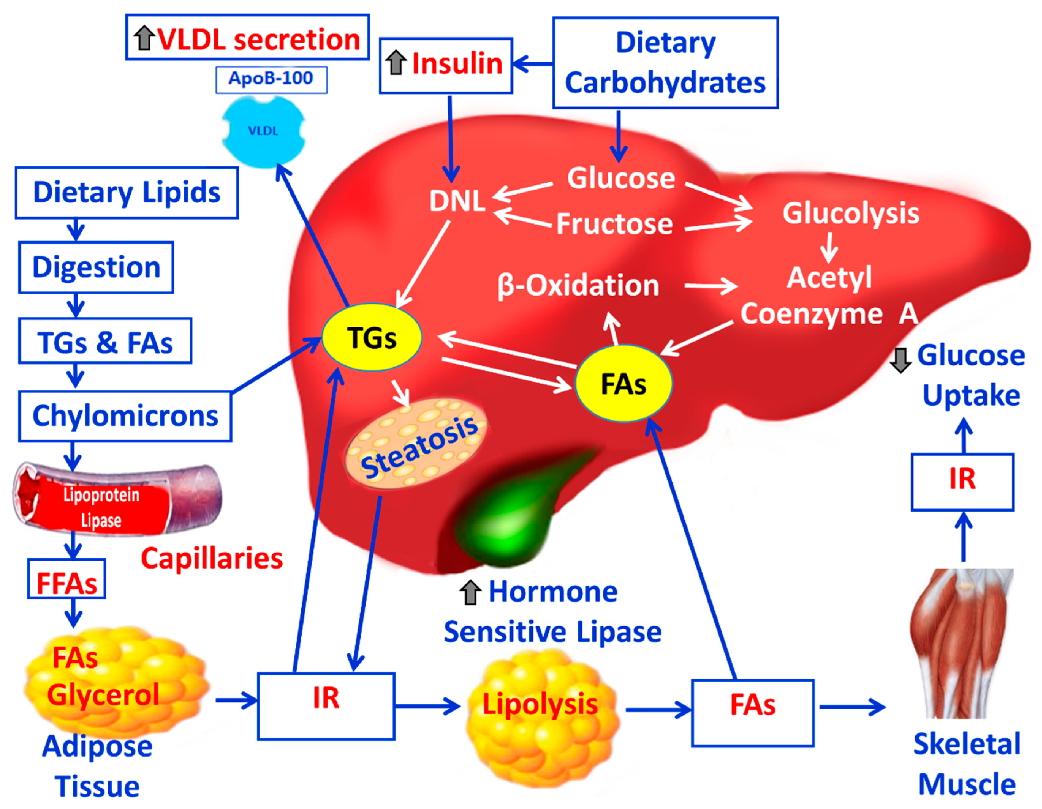
Figure 2.
Illustration of the pathogenetic mechanisms for the development of NALFD and NASH. T2DM: type 2 diabetes mellitus; IR: insulin resistnace; VLDL: very low-density lipoproteins; NAFLD: non-alcoholic fatty liver disease; NASH: non-alcoholic steatohepatitis; FFAs: free fatty acids; ROS: reactive oxygen species; OS: oxidative stress; NF-κB: nuclear factor-kappa B; TNF-α: tumor necrosis factor alpha; TGF-β: transforming growth factor beta; IL-1β: interleukin-1β; IL-6: interleukin-6; IL-8: interleukin-8.
Figure 2.
Illustration of the pathogenetic mechanisms for the development of NALFD and NASH. T2DM: type 2 diabetes mellitus; IR: insulin resistnace; VLDL: very low-density lipoproteins; NAFLD: non-alcoholic fatty liver disease; NASH: non-alcoholic steatohepatitis; FFAs: free fatty acids; ROS: reactive oxygen species; OS: oxidative stress; NF-κB: nuclear factor-kappa B; TNF-α: tumor necrosis factor alpha; TGF-β: transforming growth factor beta; IL-1β: interleukin-1β; IL-6: interleukin-6; IL-8: interleukin-8.
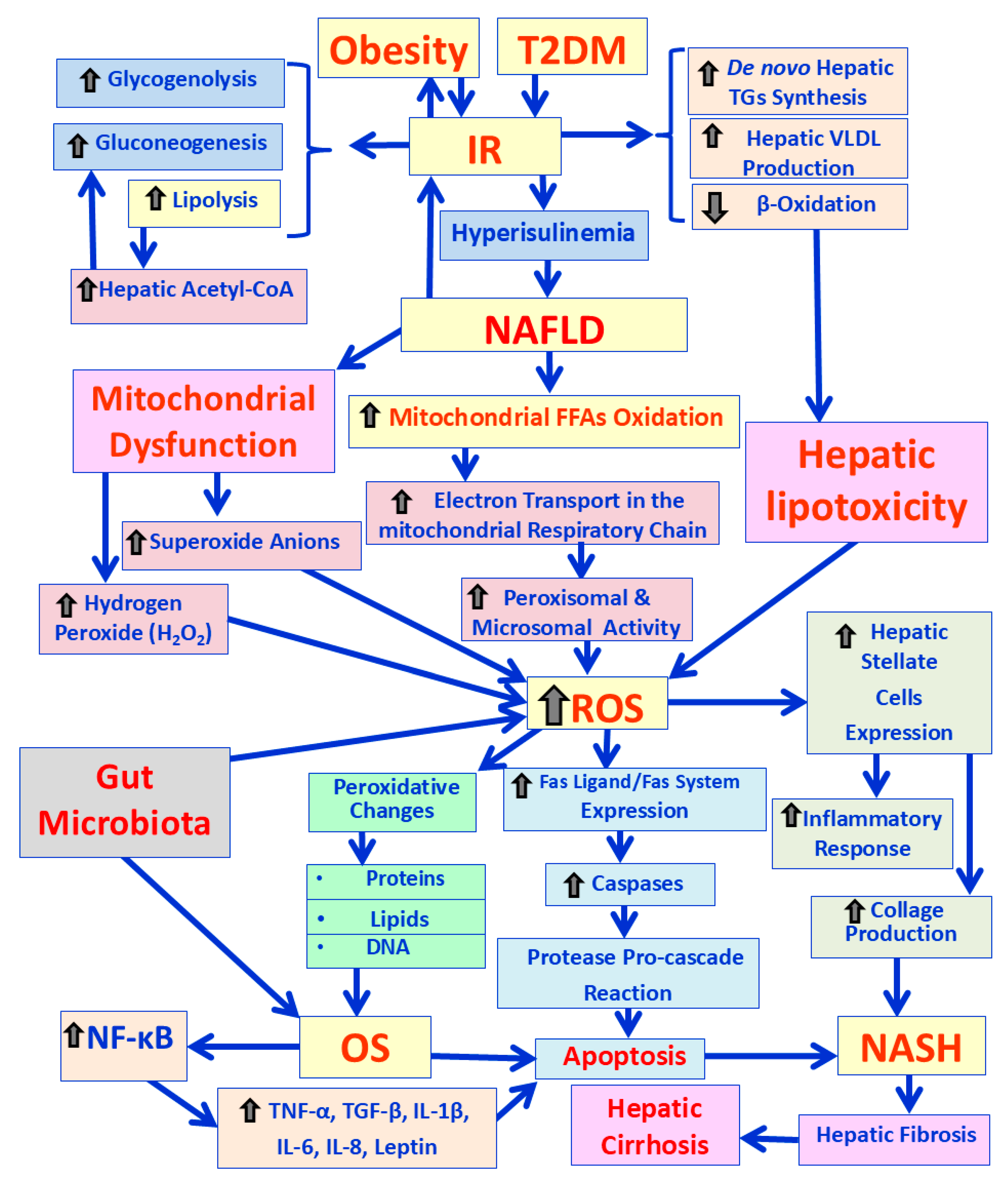
Figure 3.
A schematic representation of the changes in lipid metabolism, which lead to the development of dyslipidemia and the atheromatic plaque process in obesity. Legend: VLDL: very low density lipoproteins; LDL: low density lipoproteins; HDL: high-density lipoproteins; TG: triglycerides; CE: cholesterole esters; HP: hepatic lipase; CETP: cholesterol-ester trasfer protein; FC: free clolesterole; Ox-LDL: Oxidized low-density lipoprotein.
Figure 3.
A schematic representation of the changes in lipid metabolism, which lead to the development of dyslipidemia and the atheromatic plaque process in obesity. Legend: VLDL: very low density lipoproteins; LDL: low density lipoproteins; HDL: high-density lipoproteins; TG: triglycerides; CE: cholesterole esters; HP: hepatic lipase; CETP: cholesterol-ester trasfer protein; FC: free clolesterole; Ox-LDL: Oxidized low-density lipoprotein.
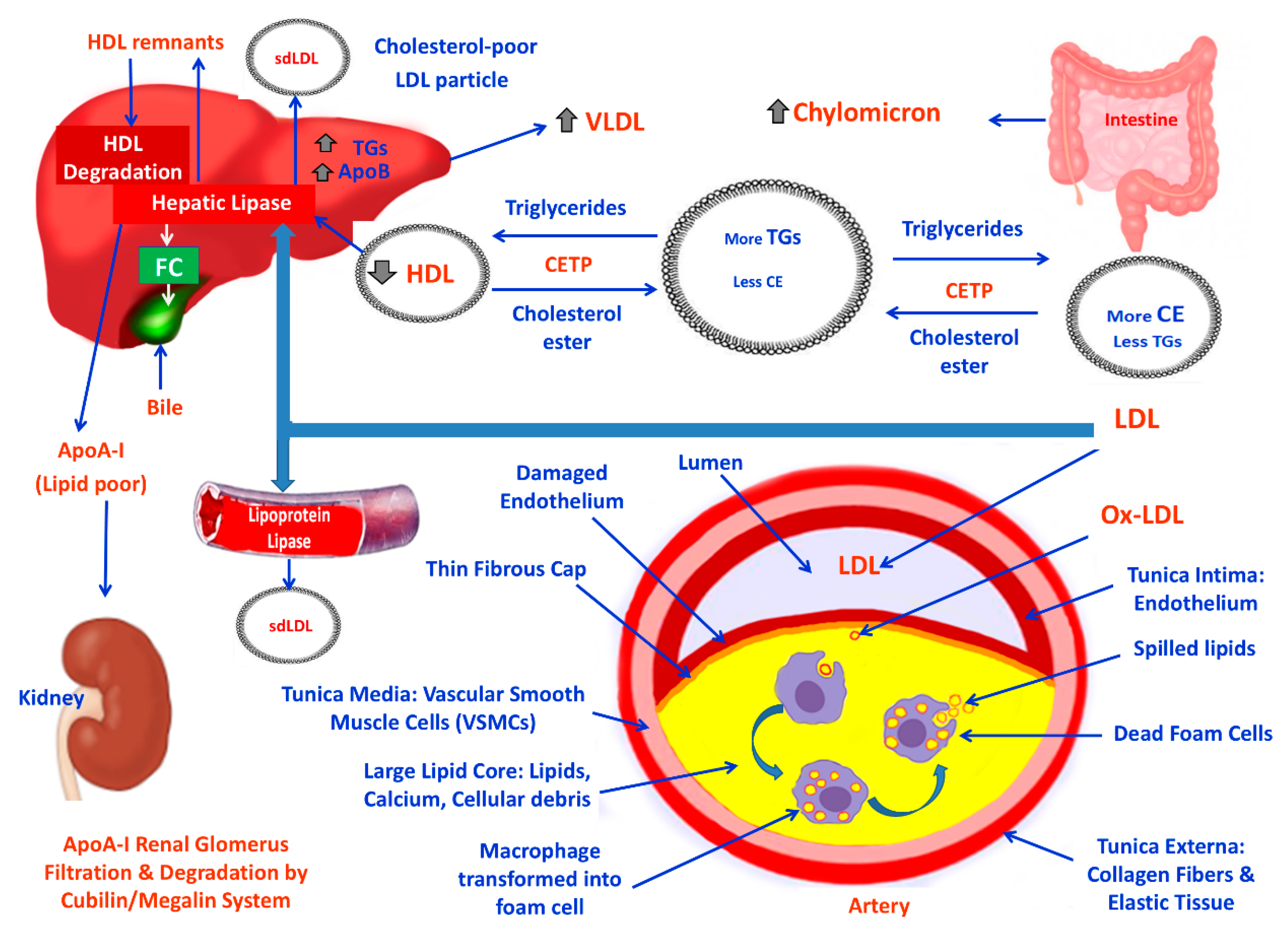
Figure 4.
Illustration of the mechanisms involved in development of atherosclerosis induced by Ox-LDL in obesity. OS: oxidative stress; Ox-LDL: oxidized low-density lipoproteins; NOX2: NADPH oxidase 2; LOX-1: lectin-like oxidized low-density lipoprotein receptor-1; TNF-α: tumor necrosis factor-alpha; IL-1β: interleukin-1β; IL-6: interleukin-6; IL-8: interleukin-8; ECs: endothelial cells; VSMCs: vascular smooth muscle cells; NF-κB: nuclear factor-kappa B; EDHF: endothelium-derived hyperpolarizing factor; EVs: extracellular vesicles; MCP-1: monocyte chemoattractant protein 1; TGs: triglycerids; Eph/Ephrins: receptor tyrocine kinases; Notch3: neurogenic locus notch homolog protein 3; PDGF: platelet derived growth factors; TLRs: Toll-like receptors ; Srs: scavenger receptors.
Figure 4.
Illustration of the mechanisms involved in development of atherosclerosis induced by Ox-LDL in obesity. OS: oxidative stress; Ox-LDL: oxidized low-density lipoproteins; NOX2: NADPH oxidase 2; LOX-1: lectin-like oxidized low-density lipoprotein receptor-1; TNF-α: tumor necrosis factor-alpha; IL-1β: interleukin-1β; IL-6: interleukin-6; IL-8: interleukin-8; ECs: endothelial cells; VSMCs: vascular smooth muscle cells; NF-κB: nuclear factor-kappa B; EDHF: endothelium-derived hyperpolarizing factor; EVs: extracellular vesicles; MCP-1: monocyte chemoattractant protein 1; TGs: triglycerids; Eph/Ephrins: receptor tyrocine kinases; Notch3: neurogenic locus notch homolog protein 3; PDGF: platelet derived growth factors; TLRs: Toll-like receptors ; Srs: scavenger receptors.
Disclaimer/Publisher’s Note: The statements, opinions and data contained in all publications are solely those of the individual author(s) and contributor(s) and not of MDPI and/or the editor(s). MDPI and/or the editor(s) disclaim responsibility for any injury to people or property resulting from any ideas, methods, instructions or products referred to in the content. |
© 2025 by the authors. Licensee MDPI, Basel, Switzerland. This article is an open access article distributed under the terms and conditions of the Creative Commons Attribution (CC BY) license (http://creativecommons.org/licenses/by/4.0/).
Copyright: This open access article is published under a Creative Commons CC BY 4.0 license, which permit the free download, distribution, and reuse, provided that the author and preprint are cited in any reuse.