Submitted:
04 August 2025
Posted:
05 August 2025
You are already at the latest version
Abstract
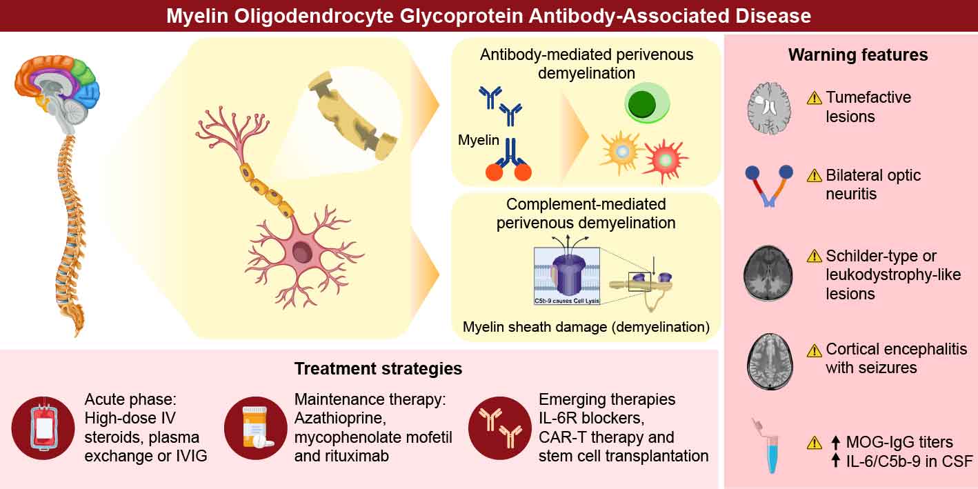
Myelin oligodendrocyte glycoprotein (MOG) antibody-associated disease (MOGAD) is characterized by the predominance of optic neuritis, myelitis, acute disseminated encephalomyelitis, and cortical encephalitis, which can be diagnosed by the presence of pathogenic immunoglobulin G (IgG) antibodies targeting the extracellular domain of MOG in the serum and cerebrospinal fluid (CSF). Initially considered a variant of multiple sclerosis (MS) or neuromyelitis optica spectrum disorder (NMOSD), it is now widely recognized as a separate entity, supported by converging evidence from serological, pathological, and clinical studies. Pa-tients with MOGAD often exhibit better recovery from acute attacks, but their clinical and pathological features vary based on the immunological role of MOG-IgG via antibody-mediated or complement-mediated perivenous demyelinating pathology in addition to MOG-specific cellular immunity, resulting in heterogeneous demyelinated lesions from vanishing benign form to tissue necrosis. The key is the immunological mechanism of devastating lesion coalescence and long-term degenerating mechanisms, which may still accrue, particularly in the relapsing, progressing, and aggressive clinical course of encephalomyelitis. The warning features of the severe forms are: 1. fulminant acute multifocal lesions transitioning to diffuse or tumefactive lesions; 2. cortical lesions related to status epilepsy; 3. Longitudinally extended spinal cord lesions not well subsided. Persistent MOG-IgG high-titration, intrathecal production of MOG-IgG, and suggestive markers of higher disease activity like cerebrospinal fluid inter-leukin-6 and complement C5b-9 could be identified as promising markers of higher disease activity, disability worsening, and poor prognosis, for identifying sign of escalating treatment strategies.
Keywords:
myelin oligodendrocyte glycoprotein
; demyelination
; biologics
; complement
; autoimmunity
1. Introduction
Myelin oligodendrocyte glycoprotein antibody-associated disease (MOGAD) is a distinct demyelinating disorder of the central nervous system (CNS), characterized by the presence of pathogenic immunoglobulin G (IgG) antibodies targeting the extracellular domain of myelin oligodendrocyte glycoprotein (MOG), a glycoprotein expressed on the surface of oligodendrocytes and the outermost lamellae of CNS myelin sheaths. Initially considered a variant of multiple sclerosis (MS) or neuromyelitis optica spectrum disorder (NMOSD), MOGAD is now widely recognized as a separate nosological entity, supported by converging evidence from serological, pathological, and clinical studies [1,2].
Clinically, MOGAD manifests as diverse phenotypes, including optic neuritis (ON), transverse myelitis (TM), acute disseminated encephalomyelitis (ADEM), and brainstem encephalitis. Notably, disease presentation can vary by age group; children more commonly exhibit ADEM-like symptoms, whereas adults typically present with ON or TM [3,4,5]. These attacks are often monophasic, but may recur, with some patients experiencing frequent and disabling relapses. Unlike patients with AQP4-IgG+ NMOSD, patients with MOGAD often exhibit better recovery from acute attacks. However, long-term disability may still occur, particularly in relapsing forms [6,7].
Serologically, MOG-IgG is most reliably detected using cell-based assays that employ full-length human MOG as the target antigen. These antibodies are predominantly of the IgG1 subtype and thought to be pathogenic via complement- and antibody-dependent cellular cytotoxicity [8,9]. However, the immunopathological mechanisms underlying MOGAD remain unclear. Autopsy and biopsy findings reveal perivenous demyelination, axon preservation, and a relative lack of astrocyte loss, contrasting with AQP4-NMOSD [10,11]. These findings, along with the distinct cerebrospinal fluid (CSF) profiles and magnetic resonance imaging (MRI) characteristics, further reinforce the nosological distinction between MS and NMOSD.
Epidemiological studies suggest that MOGAD accounts for a significant proportion of acquired demyelinating syndromes, particularly in children and AQP4-IgG− adults [12,13]. The annual incidence is approximately 1.6–3.4 per million, although the true prevalence is likely underestimated due to diagnostic challenges and evolving serological testing standards [8,14]. MOGAD burden is compounded by its unpredictability: while many patients respond well to steroids and immunotherapies, others suffer from recurrent or severe attacks that are difficult to control [8,15].
Owing to this clinical heterogeneity, growing attention is being paid to severe and treatment-refractory subtypes of MOGAD. These forms may be associated with persistent or relapsing disease, poor steroid responsiveness, or atypical imaging patterns. A deep understanding of immunopathological mechanisms, including the role of intrathecal MOG-IgG synthesis and complement activation, is essential for improving diagnostic precision and tailoring therapy. This review aims to synthesize the current knowledge on the pathophysiological basis, imaging characteristics, immunological biomarkers, and therapeutic challenges of refractory and severe MOGAD, with the goal of guiding future clinical and translational research.
2. Pathological Insights
2.1. Pathological Features of MOGAD
MOGAD is pathologically distinct from MS and aquaporin-4 (AQP4)-IgG+ NMOSD and exhibits characteristic features that reflect its unique immunopathogenesis [16]. Perivenous demyelination and the dominance of CD4+ T-cell infiltration [10,17], along with granulocytes such as neutrophils and eosinophils, often reminiscent of monophasic ADEM, are considered the hallmarks of MOGAD in acute stage [18]. These lesions typically originate around small venules, and complement deposition is relatively rare in MOG antibody+ ADEM cases [10] than in NMOSD cases; however, some ADEM-like cases resemble type II MS pathology [19]. Unlike the confluent, sharply demarcated plaques seen in prototypic MS, MOGAD lesions tend to be ill-defined and patchy, with relative preservation of axons and oligodendrocytes, particularly in early lesions [10,11]. This contrasts with the pathology in MS, where CD8+ T cells are predominant and chronic microglial activation occurs in chronic expanding lesion limbs, and that in AQP4-NMOSD, which shows marked astrocyte loss and complement-mediated tissue necrosis [20]. These histopathological differences are often correlated with favorable clinical recovery in patients with MOGAD.
The smoldering confluent MS-like lesion is rare in MOGAD
An important aspect of MOGAD pathology is the rapid centrifugal expansion of demyelinating lesions from a perivenous epicenter, driven by T-cell infiltration and secondary activation of innate immune cells. Several multiple perivenous demyelinating lesions often coalesce and grow into larger lesions [10,11]. This radial lesion growth resembles the "smoldering" confluent lesions described in progressive MS, but the progression scale is different and lacks the chronic microglial rim that typifies such MS plaques [16]. Moreover, while MS is associated with oligodendrocyte apoptosis and axonal transection, MOGAD exhibits limited oligodendrocyte damage, potentially due to antibody-mediated myelin disruption, without direct cytotoxicity to myelin-producing cells [11,16].
Pathogenesis of MOG-IgG related to complement and antibody related cytotoxicity
2.2. Complement-Mediated
The isotypes of autoantibodies against MOG and AQP4 are mainly IgG1 can mediate complement-dependent cytotoxicity (CDC) in addition to antibody-mediated cytotoxicity (ADCC). The assembly of AQP4 into orthogonal arrays of particles (OAPs), consistent with tetrameric AQP4-M23 components, is required for complement activation, which is influenced by the interaction of the Fc portion of AQP4-IgG with the OAP structure [21]. The binding of C1q to Fc portion of AQP4-IgG on astrocytes activates C1r and C1s, which then cleave C4 and C2. These complement proteins are also cleaved by lectin-associated serine proteases [22] and the cleaved products combine to form C3 convertase, which further cleaves C3 into C3b and C3a. C3b then associates with C4bC2a to form the C5 convertase, finally forming cascades of membrane attack complex (MAC) [22]. This pathway has been well observed in clinical, experimental NMOSD pathology [20,23], and MAC biomarkers are associated with disease activity [24,25]. In addition, the alternative pathway is activated when C3 spontaneously hydrolyzes to form the C3 convertase, C3(H2O)Bb, in the presence of factors B and D, leading to additional C3 cleavage and eventual formation of C3 and C5 convertases [22]. This pathway is also involved in MAC formation. These three pathways work in concert to protect the host from pathogenic invasions by activated components such as C3aR orC5aR, including activated macrophage-induced phagocytosis; anaphylatoxins, including C3a and C5a, inducing neutrophil recruitment; and activation of innate and adaptive immunity, including B and T cells [22], some of which are reported to be strongly or moderately associated with NMOSD pathology[21,26,27]. Interestingly, although complement deposition is not always prominent in MOGAD and is stage-dependent [10,11], some necrotizing lesions exhibit C9neo deposition, indicating complement activation and MAC formation [11,28]. An in vitro study suggested that MOG-IgG-induced complement-mediated cytotoxicity[29] and MOGAD encompasses a spectrum of several histological phenotypes, including benign and malignant ones. Autopsy and biopsy revealed widespread confluent demyelination, perivascular necrosis, and complement-mediated cytotoxicity, akin to pattern II or III MS lesions, in a subset of patients, particularly those with refractory disease [30,31]. Autopsy also showed diffuse multi-coalesced demyelinated lesions with deep complement deposition in a patient with progressive extension of diffuse and multifocal white matter lesions [11]. These cases may underlie the severe clinical presentations and reduced responsiveness to immunotherapy, although definitive biomarkers for predicting such pathologies remain to be elucidated.
2.3. Antibody-Mediated
AQP4-IgG showed markedly higher levels of CDC than MOG-IgG in vitro [32] together with marked deposition of MAC in pathological studies [33], which could be due to the difference in C1q binding to targeted molecules between AQP4-IgG and MOG-IgG, initiating a classical complement pathway[34]. In addition, MOG-IgG-induced demyelination was equally mediated by CDC and ADCC, suggesting the diverse mechanism of pathological characteristics involved in MOGAD with or without complement-induced demyelination and tissue necrosis. Moreover, its ADCC could be activated by FcγR activation [35], which could also be essential for cognate T-cell activation via antigen presenting cells, resulting in MOG-specific T-cell proliferation [36,37]. ADCC induces the alteration of oligodendrocytes into thin filaments and microtubule cytoskeleton [38]. Other aspect of MOG-IgG cytotoxicity is antibody-dependent cellular phagocytosis (ADCP) which could be identified by its functional role on in vitro MOG-expressing cells [39]. MOG-IgG includes not only IgG1 but also IgG2, IgG3, and IgG4, all of which could induce ADCP, in contrast CDC could be observed by IgG1 and IgG3, suggesting patients’ derived antibodies must have multiple mechanism of cytotoxic effects against targeted myelin sheaths [39], possibly links to a unique accumulation of macrophages in perivenous demyelinating lesions in ADEM phenotype of MOGAD[10].
3. Clinical Features of MOGAD
The clinical manifestations of MOGAD are heterogeneous, ranging from ON [40], myelitis [41], tumefactive disease [42], multifocal ADEM, or cortical encephalitis [43,44]; however, isolated ON was the most frequent (approximately 50% cases) clinical presentation in both children and adults [45]; the first episode of ON reduced the risk of expanded disability scale score (EDSS) progression than that of myelitis [46]. Two-thirds of the patients had prodromal symptoms such as rhinorrhea, sore throat, low-grade fever, or cough due to a presumed or confirmed infection [47], which is often encountered in pediatric patients and patients after several kinds of vaccinations, including for SARS-CoV-2. ADEM was more frequent and had better prognosis in children (36.7%) [5] than in adults (5.6%) [45]. In contrast, the relapse in adults, particularly with manifestations of ON and myelitis [48], is relatively higher than that in children [45], with the probability of reaching a first relapse after 2 and 5 years being 44.8% and 61.8%, respectively [4]. At 2 years, monophasic ADEM often becomes MOG-IgG− (64.2%); in contrast, most relapsing cases have persistent MOG-IgG [45]. Therefore, MOGAD is generally considered better than NMOSD, though MOGAD is not a mild disease.
- (1)
- Red flags for severe MOGAD
Severe MOGAD presentations often include bilateral ON, MOG-associated encephalitis or cortical encephalitis with seizures, and longitudinally extensive transverse myelitis (LETM) resulting in persistent motor deficits [49,50]. Moderate to poor recovery after acute immunotherapy is frequently observed in patients with LETM (40%), bilateral ON (32%), ADEM (17%), short myelitis (11%), and unilateral ON (11%), with its ratio being relatively higher in older onset group [41] (Table 1).
3.1. Transverse Myelitis
Incomplete recovery from paraplegia and bladder dysfunction may necessitate aggressive interventions [51]. Myelitis attacks in acute MOGAD are often severe, with transverse symptoms including motor weakness (83%), numbness (89%), sphincter symptoms (83%), needs cane (41%), or wheel chair (33%), often with longitudinally extended lesions over three vertebral segments (79%), and often categorized as EDSS 6.0. [47]. In addition, neurological findings in the acute stage involve both spasticity/hyperreflexia and flaccid areflexia due to LETM, some of which meet the criteria for acute flaccid myelitis with preceding prodromal infectious symptoms and MRI findings of the central gray matter, which could be of prognostic value [52]. Particularly, neurogenic bladder is due to the frequent involvement of conus lesions on MRI, resulting in the frequent residual autonomic dysfunction over 60% in MOGAD than that in NMOSD [47,53]; some of these patients need long-term catheter requirement (20–44%). In addition, 6–7% patients needed gait aid (EDSS >6.0) at last follow-up [47,53].
3.2. Optic Neuritis
Poor visual recovery with recurrence despite high-dose steroids or progression to bilateral involvement often signifies refractory disease. Maintenance therapy with intravenous immunoglobulin (IVIG), rituximab, or tocilizumab has shown variable success [54]. Acute symptoms of ON are often severe, with vision loss less than 20/200, often bilateral (40%), papillary edema (80%), and eye ball pain (90%), but corticosteroid treatment is relatively effective [41], particularly in the milder forms than AQP4-IgG+ NMOSD and double negative NMOSD [46] in EDSS-based cohort. However, 12 of 75 patients experienced moderate to severe permanent visual dysfunction [41]. Poor (6.2%) and incomplete (19.4%) visual acuity recovery were previously observed with poor visual field abnormalities (16.9%), but the lack of or milder optic nerve sheath enhancement was associated with incomplete recovery, which could be a prognostic factor for MOGAD [55]. The peripapillary retinal nerve fiber layer (pRNFL) thickness measured acutely frequently demonsrated swelling, in contrast a negative correlation between follow-up pRNFL thickness and latest follow-up visual acuity was noted, which suggested a degenerating mechanism also observed in MOGAD[56].
3.3. Encephalopathy and Seizures
In children and young adults, ADEM and MOG-associated encephalitis may present with monophasic or multiphasic ADEM presenting with cerebral symptoms of encephalopathy, including decreased level of consciousness (100%) and seizures (30 to 60%) [6,42], but most monophasic ADEM cases are treated with first-line immunotherapy resulted in symptom and MRI lesion resolution [57]. In contrast, in a pediatric ADEM cohort (n=46), mRS >2 was observed in 6 cases (13%) at last follow-up, 8 were multiphasic ADEM (MDEM), and 5 were ADEM followed by relapses in ON or myelitis. Half of the MDEM cases resulted in severe disability (mRS 4 and 2 in two cases each), suggesting MDEM as a red flag [58]. In addition, persistent epilepsy or cognitive impairment may occur, particularly in patients with extensive cortical involvement of leukodystrophy-like or predominant cortical involvement [42,44], some of whom have persistent brain atrophy and severe cognitive impairment [42,59]. Other refractory cases included acute symptomatic seizures and new-onset refractory status epilepticus (NORSE) due to MOG-IgG-induced ADEM or hemispheric cortical encephalitis, for which several lines of immunotherapy and antiepileptic treatments could not subside seizures [60,61], resulting in refractory seizure-induced hemispheric brain atrophy, such as Rasmussen’s encephalitis. Most seizures that subsided with immunotherapy had focal patterns due to a unilateral type of cortical encephalitis called FLAIR-hyperintense lesions in anti-MOG-associated encephalitis with seizures (FLAMES) [43,62], and a combination of immunotherapy and antiepileptic drugs is often needed for long-term remission.
- (2)
- Clinical indicators of refractory activity
While most cases of MOGAD respond favorably to corticosteroids and immunotherapy, a subset of patients experience severe or refractory disease, defined by criteria such as poor functional recovery; early relapses during sequential acute immunotherapy, including IVIG and plasma exchange (PLEX); and resistance to corticosteroids and immunosuppressants such as azathioprine, tacrolimus, mycophenolate mofetil, or other maintenance therapy [5,49,50]. Moreover, ongoing disease activity in maintenance immunotherapy by rituximab and IVIG is considered to be the motivation for tocilizumab trials [54,63]. Beyond these aggressive treatments, one case report attempted hematopoietic stem cell transplantation [64] resulting in release from disease activity. It has also been suggested that complications of other autoimmune diseases and autoimmune encephalitis must be influenced by refractory to specific treatment MOGAD [65,66]. It is generally considered the signs of refractory MOGAD are as follows:
Relapsing trends at dose of prednisolone < 10mg daily within 2 months from onset [50]
Ineffective to first-line immunotherapy in acute stage within 2 to 4 weeks [6]
Residual symptoms under optimal treatments at the first onset [41]
Symptoms of seizure and consciousness disturbance in encephalitis [61]
Pediatric vs. adult presentations
Children with MOGAD tend to have a high frequency of ADEM-like episodes and increased relapse recovery; however, severe leukodystrophy-like phenotypes with diffuse white matter changes and developmental delays have also been reported [6,68]. In contrast, adults are more prone to ON-dominant forms. However, those with recurrent brainstem encephalitis or MOG encephalitis may experience significant residual disability[5]. The EDSS at onset nadir over 6.0 is higher in pediatric patients (35.7%) than in adult patients (26.7%). In contrast, the EDSS over 3.0 at last follow-up was higher in adults (20%) than in children (6.3%), suggesting a relatively good response to immunotherapy in pediatric patients [45]. However, the prognosis of well-known major features, including ON and myelitis, is similar in several cohorts in children and adults. Interestingly, the rate of ADEM encephalopathy or brain lesions is around 50% in children [12] worldwide, but less than 10% in adults. Therefore, severe and refractory cases due to brain lesions have also been observed in pediatric patients [60,69]. In a cohort of pediatric patients with MOGAD, early immunotherapy less than 1 week from the onset was predictive of a monophasic course; in contrast, MOGAD with a relapsing course had a higher proportion of final EDSS over 1 and 2 [70] and should not be handled with good prognosis.
4. MRI and Other Biomarkers Predicting Severity
4.1. Typical MRI Features
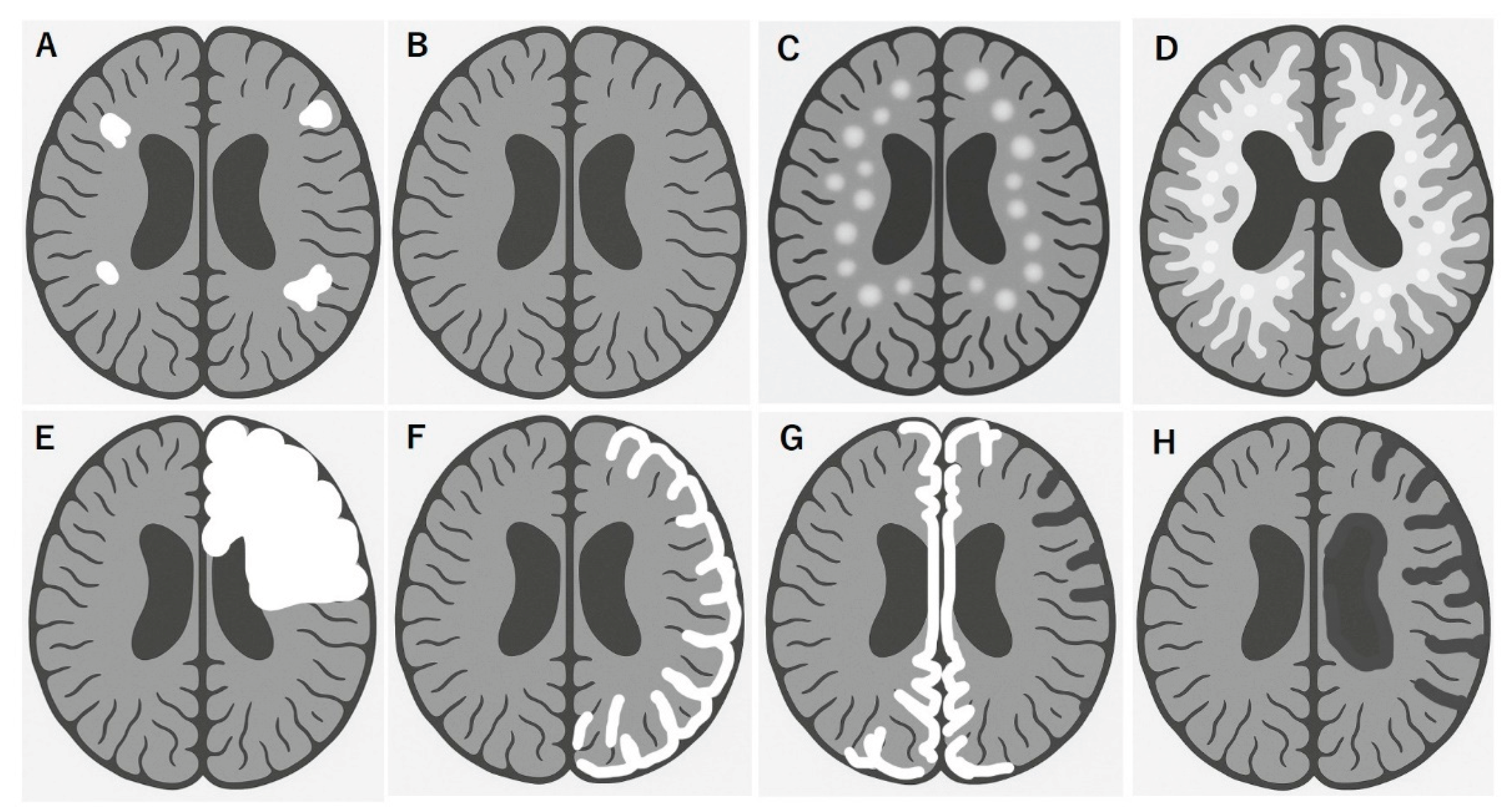
MOGAD exhibits distinct MRI patterns essential for differentiation from MS and AQP4+ NMOSD. Although these conditions often overlap in clinical presentation, they can be distinguished through careful evaluation of lesion distribution, morphology, and enhancement patterns. Unlike MS, the multiple isolated lesions can be often seen but juxtacortical U-fiber sparing and the absence of Dawson’s fingers are typical features (Fig.1A). These lesions tend to involve the deep gray nuclei, brainstem, and supratentorial white matter, often symmetrically [71,72] and completely subsided in benign cases (Fig. 1B). MOGAD frequently presents as multifocal small to large lesions, poorly demarcated, bilateral ADEM-like lesions in the brain (Fig. 1C and 1D), particularly in children. Some of these lesions can be seen with focal or multifocal tumefactive lesions (Fig.1E). A hallmark of cortical presentation is called FLAMES, showing unilateral [43] (Fig. 1F) or bilateral medial [44] (Fig. 1G) cortical swelling, often associated with seizures and a relatively benign prognosis [62]; however, there are severe cases refractory to inflammatory and anti-epileptic treatments [60,61]. Spinal cord lesions in MOGAD are usually longitudinally extensive and centrally located, frequently involving the conus medullaris and thoracic segments. MOGAD lesions often had the gray matter “H sign” [51,73]. Brainstem and cerebellar peduncular lesions are more frequent in MOGAD than in NMOSD, with lesions often appearing in the middle cerebellar peduncles [4,74,75] In contrast, area postrema lesions are relatively rare compared in NMOSD [4,33,76]. ON in MOGAD tends to be bilateral, with anterior optic nerve and optic disk involvement often associated with disk edema [40]. Orbital fat inflammation and perineural enhancement are more common than in MS or AQP4-NMOSD[77], and optic chiasm involvement, while possible, is typically partial [72,78]. Importantly, MRI misinterpretation can lead to misdiagnosis of MOGAD as MS, particularly when corpus callosum or periventricular lesions are present. However, MOGAD typically lacks the chronic black holes, callosal thinning, central vein sign, and progressive diffuse atrophy seen in MS [71,78].
4.2. MRI Features Predicting Severe and Refractory MOGAD
While MOGAD is generally monophasic and radiologically responsive to treatment like vanishing tumor, various atypical MRI features may correlate with treatment-resistant or severe phenotypes. These include:
Quantitative imaging has demonstrated the relative preservation of brain and spinal cord volume in MOGAD than in MS and NMOSD, supporting its generally non-degenerative course [79] in MOGAD; however, repeated attacks and residual lesions can result in cumulative tissue damage causing atrophy, particularly in the spinal cord [51,73]. In addition, progressive atrophy in MOGAD could be observed with refractory encephalitis, possibly with other autoimmune diseases like NMDA-R encephalitis [61,67], and refractory status epilepsy [81]. Accurate recognition of these features is critical for not only diagnosis, but also identifying patients at risk for severe disease courses.
Figure 1.
Brain MRI features of MOGAD links to disease worsening and poor recovery.
4.3. Biomarkers Predicting Severe and Refractory MOGAD
The identification of biomarkers to guide treatment decisions in MOGAD is an evolving field, especially the titration of MOG-IgG in serum and CSF, inflammatory cytokines, and tissue damage markers [82]. Some candidates are as follows:
- Serum and CSF cytokine and chemokine profiles, such as elevated interleukin-6 (IL-6) [85,86,87], IL-8 [86,87] , and B-cell activating factor (BAFF) [87] levels, reflect active B-cell-mediated inflammation and predict the need for aggressive therapy [85,86,87,88], associated with disease severity in general, of which BAFF levels predicted lower risk of relapse [89]
Altogether, these indicators support a stratified treatment approach that allows early escalation to second-line agents such as rituximab or tocilizumab in high-risk patients.
5. MOGAD Treatment
- (1)
- Acute-phase treatments
The cornerstone of acute-phase MOGAD remains high-dose intravenous methylprednisolone (IVMP), typically administered at 1000 mg/day for 3–5 days, followed by a gradual oral steroid taper over weeks to months [41]. This approach often effectively suppresses acute inflammation and induces neurological recovery, particularly during initial episodes. However, emerging data suggest that a subset of patients is relatively unresponsive to corticosteroid therapy, failing to achieve functional recovery or experiencing relapse during tapering or shortly after withdrawal [49]. Steroid unresponsiveness has been reported in 20–30% patients, depending on the cohort and disease phenotype [50]. These observations underscore the need for timely treatment and alternative strategies in poor responders.
Adjunct therapies: PLEX and IVIG
PLEX
PLEX has demonstrated efficacy in rapidly reducing circulating autoantibodies and improving neurological outcomes in cases in which corticosteroids are insufficient. It is especially beneficial in patients with severe ON, TM [101], or fulminant encephalitis [42]. PLEX is typically initiated within 7–10 days of steroid failure and involves 5–7 sessions.
IVIG
Either as an adjunct or a monotherapy, IVIG has gained attention for its potential superiority in certain refractory or pediatric-onset cases. Several studies, including randomized pediatric trials, have shown that IVIG may lead to faster functional recovery and reduced relapse rates than steroids alone [102,103,104]. Its mechanisms likely include Fc receptor blockade, neutralization of inflammatory cytokines, and modulation of autoreactive B and T cells, where patients relapse during steroid tapering. Moreover, monthly IVIG has also shown promise in preventing attacks and reducing steroid exposure [105].
- (2)
- Maintenance therapy
Traditional immunosuppressants: Azathioprine and mycophenolate mofetil
Azathioprine and mycophenolate mofetil: These are oral immunosuppressants widely used in NMOSD, and by extension, have been trialed in MOGAD. Observational studies and meta-analyses suggest that both agents can reduce the relapse frequency in MOGAD, particularly when used early and in combination with oral corticosteroids during initiation [4,5]. However, relapse despite treatment is common, particularly after steroid withdrawal. Furthermore, the delayed onset of efficacy (typically >3 months), potential adverse effects, and the need for regular laboratory monitoring limit its use, particularly in pediatric populations [6].
Rituximab: A monoclonal antibody targeting CD20+ B cells is highly effective in AQP4-IgG+ NMOSD, but shows inconsistent benefits in MOGAD [106]. While some patients respond well, others relapse despite complete B-cell depletion [107,108]. This paradox may reflect the fact that MOG-IgG production is often extrafollicular or partially T-cell-driven, and rituximab does not eliminate long-lived plasma cells or influence T-cell–mediated mechanisms [35]. Recent cohort studies have noted that rituximab reduced relapse rates by 37%, but up to 67% patients had relapses within 2 years of rituximab initiation, nevertheless of apparent robust B-cell depletion, raising concerns about its role as a first-line agent in MOGAD maintenance [106].
IVIG: An increasing evidence supports IVIG as a favorable long-term treatment, particularly for children and patients intolerant or unresponsive to traditional immunosuppressants. Several cohort studies and meta-analyses have reported that monthly IVIG administration significantly reduces relapse rates in pediatric and adult patients with MOGAD, with a favorable safety profile and fewer systemic adverse effects [102,103,104]. Mechanistically, IVIG exerts pleiotropic immunomodulatory effects, including Fc receptor blockade, pathogenic autoantibody neutralization, interference with the effector functions of B and T cells, and cytokine network rebalancing, which may explain its efficacy in MOGAD [109].
- (3)
- Emerging therapies in clinical trials and others
However, a subset of patients remains refractory to conventional immunotherapy. To address this issue, novel therapeutic strategies targeting diverse immune pathways, including inhibitors of interleukin-6 (IL-6) receptors, neonatal Fc receptors (FcRn), the complement cascade, and pan-B-cell depletion therapies, are being investigated.
IL-6 receptor inhibitors: Tocilizumab and satralizumab
IL-6 plays a central role in B-cell differentiation, T-cell activation, and pro-inflammatory signaling. Case series have reported favorable outcomes with tocilizumab, an IL-6 receptor blocker, in patients with highly relapsing MOGAD unresponsive to steroids, rituximab, or IVIG [54,63,85]. These patients often experience a marked reduction in relapse rate and stabilization of neurological function. More recently, satralizumab, a long-acting subcutaneous IL-6R inhibitor approved for treating AQP4+ NMOSD, has been evaluated in MOGAD cohorts. Although controlled trials are lacking, real-world data support the safety and potential efficacy of these drugs.
FcRn antagonists: Rozanolixizumab and efgartigimod
FcRn inhibitors block IgG recycling, leading to the rapid and sustained depletion of pathogenic autoantibodies, including MOG-IgG. Rozanolixizumab and efgartigimod have shown promising results in treating myasthenia gravis and other IgG-mediated diseases. Since MOGAD is antibody-mediated and frequently relapses, blocking FcRn is a rational therapeutic strategy. A phase 2 trial of rozanolixizumab (NCT05835420) is currently enrolling patients with MOGAD, and its preliminary clinical and experimental results suggest a rapid decline in serum MOG-IgG titers [110,111].
Complement inhibition and anti-CD19 therapies
Although complement activation is less prominent in MOGAD than in AQP4+ NMOSD, necrotizing lesions and C9neo deposition have been documented in severe MOGAD pathology [10,11], suggesting a possible role in a subset of refractory or fulminant cases. Now limited in AQP4+ NMOSD, it is tried in non-AQP4+NMOSD showing promising treatment effect[112]. In parallel, B-cell-targeting agents, such as ocrelizumab (anti-CD20) and inebilizumab (anti-CD19), are under consideration. CD20-depleting therapy has yielded mixed results in MOGAD, potentially due to the persistence of CD20− plasma cells. In contrast, anti-CD19 agents target a broad range of B-lineage cells, including plasmablasts, and are being explored for multiphasic or treatment-resistant MOGAD, although evidence remains limited [113].
- (4)
- Chimeric antigen receptor T-cell Therapy and autologous hematopoietic stem cell transplantation
A particularly aggressive case was reported in response to aaHSCT, highlighting its potential role in select patients [64]. Most notably, CD19-targeted CAR T-cell therapy has been applied to treat refractory MOGAD, showing sustained suppression of relapse activity and immunological response for over 1 year [114,115]. Although MOGAD is often considered a benign or monophasic illness, a subset of patients, particularly those with bilateral ON, cortical encephalitis, or conus-involving myelitis, require intensive immunosuppression and long-term management. Identifying early clinical and imaging markers of disease severity is critical for optimizing outcomes.
6. Discussion: Toward a Clinical Algorithm for Difficult-to-Treat MOGAD
Although most patients with MOGAD respond to standard immunotherapy, a distinct subset exhibits primary resistance or highly relapsing disease. For these patients, a structured clinical algorithm is essential to guide timely diagnosis, treatment escalation, and individualized care.
Proposed escalation pathway: Acute to maintenance
A pragmatic treatment algorithm for refractory MOGAD should begin with:
- First-line acute therapy: IV methylprednisolone (1 g/day × 3–5 days), followed by oral steroid taper over ≥3 months
- If response is incomplete: consider IVIG (2 g/kg) or PLEX (5–7 sessions)
- Persistent or early relapse: initiation of maintenance immunotherapy, often beginning with monthly IVIG or oral azathioprine/MMF/Tacrolims
- Second-line biologics: Tocilizumab, satralizumab, and rituximab for patients with frequent relapses.
- Other options: In severe multiphasic cases, options include aHSCT or anti-CD19 CAR T cells under specialized care.
Tailoring therapy to phenotype and biomarkers
Personalized medicine is becoming increasingly viable in MOGAD owing to growing insights into various biological and radiological phenotypes, immune markers, and therapeutic response patterns. Persistent MOG-IgG seropositivity or escalated MOG-IgG titration predicts relapse activity, immunosuppressant dose increase, or initiation of biologics [46]. Intrathecal MOG-IgG detection predicts refractory phenotypes of MOGAD derived from limited biological efficacy [91]. Elevated IL-6 or complement C5b-9 levels that predict disease activity and worsening of disability may support escalation to second-line biologics [64,114]. The cortical encephalitis phenotype requires more aggressive, prolonged immunotherapy and seizure treatment and requires electroencephalography and MRI during clinical worsening [43]. Leukodystrophy-like presentation requires early immunotherapy from biological treatments and neurodevelopmental follow-up, particularly in pediatric cases [59].
1. Future directions and research priorities
Despite the growing recognition of MOGAD as a distinct CNS autoimmune demyelinating condition, several clinical questions remain unanswered, particularly concerning treatment-refractory or severe phenotypes. Coordinated international efforts are crucial for advancing this field through improved data sharing, biomarker discovery, and therapeutic innovation. Currently, data on severe or refractory MOGAD cases are fragmented across case reports, small series, and retrospective cohorts. The establishment of international registries is critical for designing prospective trials and establishing evidence-based guidelines. Most therapeutic data on MOGAD have been derived from uncontrolled retrospective studies, and randomized controlled trials (RCTs) that specifically target high-risk populations are urgently needed. In addition to IL-6R inhibitors and anti-FcRn trials, RCTs evaluating agents such as B-cell depletion, and cell-based therapies in these populations will be critical for refining the current algorithms [114].
7. Conclusion
To improve the outcomes in patients with severe or refractory MOGAD, future efforts must combine clinical standardization through patient registries, rigorous clinical trials, and translational research on immunological and imaging biomarkers. Precision medicine frameworks and prospective data capture hold promise in transforming MOGAD care.
Conflicts of Interest
T. Misu received speaker honoraria from Tanabe Mitsubishi, Novartis, Alexion, Viela Bio, Teijin, Chugai, Sanofi, GE Health Care Japan, CSL Behring and Biogen Japan, and research support from Cosmic Corporation and Medical Biological Laboratories received a Grant-in-Aid for Scientific Research from the Ministry of Education, Culture, Sports, Science and Technology of Japan.
Abbreviations
ADCC: Antibody-dependent cytotoxicity; ADEM: Acute disseminated encephalomyelitis; BAFF: B-cell activating factor; CDC: Complement-dependent cytotoxicity; CNS: Central nervous system; CSF: Cerebrospinal fluid; EDSS: expanded disability status scale; FLAMES: FLAIR-hyperintense lesions in anti-MOG-associated encephalitis with seizures; IgG; Immunoglobulin G; IVIG: Intravenous immunoglobulin; IVMP: Intravenous methylprednisolone; LETM: Longitudinally extensive transverse myelitis; MAC: Membrane attack complex; MDEM: Multiphasic ADEM; MOG: Myelin oligodendrocyte glycoprotein; MOGAD: Myelin oligodendrocyte glycoprotein antibody-associated disease; MRI: Magnetic resonance imaging; MS: Multiple sclerosis; NMOSD: Neuromyelitis optica spectrum disorder; NORSE: New-onset refractory status epilepticus; OAP: Orthogonal arrays of particles; ON: Optic neuritis; PLEX: Plasma exchange; TM: Transverse myelitis.
References
- Reindl, M.; Di Pauli, F.; Rostasy, K.; Berger, T. The spectrum of MOG autoantibody-associated demyelinating diseases. Nat Rev Neurol 2013, 9, 455-461. [CrossRef]
- Marignier, R.; Hacohen, Y.; Cobo-Calvo, A.; Pröbstel, A.K.; Aktas, O.; Alexopoulos, H.; Amato, M.P.; Asgari, N.; Banwell, B.; Bennett, J.; et al. Myelin-oligodendrocyte glycoprotein antibody-associated disease. Lancet Neurol 2021, 20, 762-772. [CrossRef]
- Fadda, G.; Armangue, T.; Hacohen, Y.; Chitnis, T.; Banwell, B. Paediatric multiple sclerosis and antibody-associated demyelination: Clinical, imaging, and biological considerations for diagnosis and care. Lancet Neurol 2021, 20, 136-149. [CrossRef]
- Cobo-Calvo, A.; Ruiz, A.; Maillart, E.; Audoin, B.; Zephir, H.; Bourre, B.; Ciron, J.; Collongues, N.; Brassat, D.; Cotton, F.; et al. Clinical spectrum and prognostic value of CNS MOG autoimmunity in adults: The MOGADOR study. Neurology 2018, 90, e1858-e1869. [CrossRef]
- Cobo-Calvo, A.; Ruiz, A.; Rollot, F.; Arrambide, G.; Deschamps, R.; Maillart, E.; Papeix, C.; Audoin, B.; Lepine, A.F.; Maurey, H.; et al. Clinical Features and Risk of Relapse in Children and Adults with Myelin Oligodendrocyte Glycoprotein Antibody-Associated Disease. Ann Neurol 2021, 89, 30-41. [CrossRef]
- Hacohen, Y.; Wong, Y.Y.; Lechner, C.; Jurynczyk, M.; Wright, S.; Konuskan, B.; Kalser, J.; Poulat, A.L.; Maurey, H.; Ganelin-Cohen, E.; et al. Disease Course and Treatment Responses in Children With Relapsing Myelin Oligodendrocyte Glycoprotein Antibody-Associated Disease. JAMA Neurol 2018, 75, 478-487. [CrossRef]
- Armangue, T.; Olivé-Cirera, G.; Martínez-Hernandez, E.; Sepulveda, M.; Ruiz-Garcia, R.; Muñoz-Batista, M.; Ariño, H.; González-Álvarez, V.; Felipe-Rucián, A.; Jesús Martínez-González, M.; et al. Associations of paediatric demyelinating and encephalitic syndromes with myelin oligodendrocyte glycoprotein antibodies: A multicentre observational study. The Lancet Neurology 2020, 19, 234-246. [CrossRef]
- de Mol, C.L.; Wong, Y.; van Pelt, E.D.; Wokke, B.; Siepman, T.; Neuteboom, R.F.; Hamann, D.; Hintzen, R.Q. The clinical spectrum and incidence of anti-MOG-associated acquired demyelinating syndromes in children and adults. Mult Scler 2020, 26, 806-814. [CrossRef]
- Jarius, S.; Pellkofer, H.; Siebert, N.; Korporal-Kuhnke, M.; Hummert, M.W.; Ringelstein, M.; Rommer, P.S.; Ayzenberg, I.; Ruprecht, K.; Klotz, L.; et al. Cerebrospinal fluid findings in patients with myelin oligodendrocyte glycoprotein (MOG) antibodies. Part 1: Results from 163 lumbar punctures in 100 adult patients. Journal of neuroinflammation 2020, 17, 261. [CrossRef]
- Takai, Y.; Misu, T.; Kaneko, K.; Chihara, N.; Narikawa, K.; Tsuchida, S.; Nishida, H.; Komori, T.; Seki, M.; Komatsu, T.; et al. Myelin oligodendrocyte glycoprotein antibody-associated disease: An immunopathological study. Brain 2020, 143, 1431-1446. [CrossRef]
- Hoftberger, R.; Guo, Y.; Flanagan, E.P.; Lopez-Chiriboga, A.S.; Endmayr, V.; Hochmeister, S.; Joldic, D.; Pittock, S.J.; Tillema, J.M.; Gorman, M.; et al. The pathology of central nervous system inflammatory demyelinating disease accompanying myelin oligodendrocyte glycoprotein autoantibody. Acta Neuropathol 2020, 139, 875-892. [CrossRef]
- Banwell, B.; Bennett, J.L.; Marignier, R.; Kim, H.J.; Brilot, F.; Flanagan, E.P.; Ramanathan, S.; Waters, P.; Tenembaum, S.; Graves, J.S.; et al. Diagnosis of myelin oligodendrocyte glycoprotein antibody-associated disease: International MOGAD Panel proposed criteria. Lancet Neurol 2023, 22, 268-282. [CrossRef]
- Hacohen, Y.; Mankad, K.; Chong, W.K.; Barkhof, F.; Vincent, A.; Lim, M.; Wassmer, E.; Ciccarelli, O.; Hemingway, C. Diagnostic algorithm for relapsing acquired demyelinating syndromes in children. Neurology 2017, 89, 269-278. [CrossRef]
- Hor, J.Y.; Fujihara, K. Epidemiology of myelin oligodendrocyte glycoprotein antibody-associated disease: A review of prevalence and incidence worldwide. Front Neurol 2023, 14, 1260358. [CrossRef]
- López-Chiriboga, A.S.; Majed, M.; Fryer, J.; Dubey, D.; McKeon, A.; Flanagan, E.P.; Jitprapaikulsan, J.; Kothapalli, N.; Tillema, J.M.; Chen, J.; et al. Association of MOG-IgG Serostatus With Relapse After Acute Disseminated Encephalomyelitis and Proposed Diagnostic Criteria for MOG-IgG-Associated Disorders. JAMA Neurol 2018, 75, 1355-1363. [CrossRef]
- Lassmann, H. The contribution of neuropathology to multiple sclerosis research. Eur J Neurol 2022, 29, 2869-2877. [CrossRef]
- Misu, T.; Takai, Y.; Takahashi, T.; Nakashima, I.; Fujihara, K.; Aoki, M. Perivenous demyelination: Association with anti-myelin oligodendrocyte glycoprotein antibody. Clinical and Experimental Neuroimmunology 2020, 11, 22-27. [CrossRef]
- Young, N.P.; Weinshenker, B.G.; Parisi, J.E.; Scheithauer, B.; Giannini, C.; Roemer, S.F.; Thomsen, K.M.; Mandrekar, J.N.; Erickson, B.J.; Lucchinetti, C.F. Perivenous demyelination: Association with clinically defined acute disseminated encephalomyelitis and comparison with pathologically confirmed multiple sclerosis. Brain 2010, 133, 333-348. [CrossRef]
- Körtvélyessy, P.; Breu, M.; Pawlitzki, M.; Metz, I.; Heinze, H.-J.; Matzke, M.; Mawrin, C.; Rommer, P.; Kovacs, G.G.; Mitter, C.; et al. ADEM-like presentation, anti-MOG antibodies, and MS pathology: TWO case reports. Neurology® Neuroimmunology & Neuroinflammation 2017, 4, e335. [CrossRef]
- Misu, T.; Höftberger, R.; Fujihara, K.; Wimmer, I.; Takai, Y.; Nishiyama, S.; Nakashima, I.; Konno, H.; Bradl, M.; Garzuly, F.; et al. Presence of six different lesion types suggests diverse mechanisms of tissue injury in neuromyelitis optica. Acta Neuropathologica 2013, 125, 815-827. [CrossRef]
- Asavapanumas, N.; Tradtrantip, L.; Verkman, A.S. Targeting the complement system in neuromyelitis optica spectrum disorder. Expert Opin Biol Ther 2021, 21, 1073-1086. [CrossRef]
- Dunkelberger, J.R.; Song, W.-C. Complement and its role in innate and adaptive immune responses. Cell Research 2010, 20, 34-50. [CrossRef]
- Bradl, M.; Misu, T.; Takahashi, T.; Watanabe, M.; Mader, S.; Reindl, M.; Adzemovic, M.; Bauer, J.; Berger, T.; Fujihara, K.; et al. Neuromyelitis optica: Pathogenicity of patient immunoglobulin in vivo. Ann Neurol 2009, 66, 630-643. [CrossRef]
- Kuroda, H.; Fujihara, K.; Takano, R.; Takai, Y.; Takahashi, T.; Misu, T.; Nakashima, I.; Sato, S.; Itoyama, Y.; Aoki, M. Increase of complement fragment C5a in cerebrospinal fluid during exacerbation of neuromyelitis optica. J Neuroimmunol 2013, 254, 178-182. [CrossRef]
- Miyamoto, K.; Murakami, K.; Sakata, M.; Nakayama, Y.; Kuwahara, M.; Inoue, N. Predictive complement biomarkers for relapse in neuromyelitis optica spectrum disorders. Mult Scler Relat Disord 2025, 94, 106282. [CrossRef]
- Wolf, H.N.; Guempelein, L.; Schikora, J.; Pauly, D. C3a Mediates Endothelial Barrier Disruption in Brain-Derived, but Not Retinal, Human Endothelial Cells. Int J Mol Sci 2024, 25. [CrossRef]
- Kaneko, K.; Kuroda, H.; Matsumoto, Y.; Sakamoto, N.; Yamazaki, N.; Yamamoto, N.; Umezawa, S.; Namatame, C.; Ono, H.; Takai, Y.; et al. Different Complement Activation Patterns Following C5 Cleavage in MOGAD and AQP4-IgG+NMOSD. Neurol Neuroimmunol Neuroinflamm 2024, 11, e200293. [CrossRef]
- Spadaro, M.; Winklmeier, S.; Beltrán, E.; Macrini, C.; Höftberger, R.; Schuh, E.; Thaler, F.S.; Gerdes, L.A.; Laurent, S.; Gerhards, R.; et al. Pathogenicity of human antibodies against myelin oligodendrocyte glycoprotein. Ann Neurol 2018, 84, 315-328. [CrossRef]
- Kohyama, K.; Nishida, H.; Kaneko, K.; Misu, T.; Nakashima, I.; Sakuma, H. Complement-dependent cytotoxicity of human autoantibodies against myelin oligodendrocyte glycoprotein. Front Neurosci 2023, 17, 1014071. [CrossRef]
- Spadaro, M.; Gerdes, L.A.; Mayer, M.C.; Ertl-Wagner, B.; Laurent, S.; Krumbholz, M.; Breithaupt, C.; Högen, T.; Straube, A.; Giese, A.; et al. Histopathology and clinical course of MOG-antibody-associated encephalomyelitis. Annals of clinical and translational neurology 2015, 2, 295-301. [CrossRef]
- Takai, Y.; Misu, T.; Fujihara, K.; Aoki, M. Pathology of myelin oligodendrocyte glycoprotein antibody-associated disease: A comparison with multiple sclerosis and aquaporin 4 antibody-positive neuromyelitis optica spectrum disorders. Front Neurol 2023, 14, 1209749. [CrossRef]
- Lerch, M.; Schanda, K.; Lafon, E.; Würzner, R.; Mariotto, S.; Dinoto, A.; Wendel, E.M.; Lechner, C.; Hegen, H.; Rostásy, K.; et al. More Efficient Complement Activation by Anti-Aquaporin-4 Compared With Anti-Myelin Oligodendrocyte Glycoprotein Antibodies. Neurol Neuroimmunol Neuroinflamm 2023, 10, e200059. [CrossRef]
- Misu, T.; Fujihara, K.; Nakashima, I.; Sato, S.; Itoyama, Y. Intractable hiccup and nausea with periaqueductal lesions in neuromyelitis optica. Neurology 2005, 65, 1479-1482. [CrossRef]
- Macrini, C.; Gerhards, R.; Winklmeier, S.; Bergmann, L.; Mader, S.; Spadaro, M.; Vural, A.; Smolle, M.; Hohlfeld, R.; Kumpfel, T.; et al. Features of MOG required for recognition by patients with MOG antibody-associated disorders. Brain 2021, 144, 2375-2389. [CrossRef]
- Mader, S.; Ho, S.; Wong, H.K.; Baier, S.; Winklmeier, S.; Riemer, C.; Rübsamen, H.; Fernandez, I.M.; Gerhards, R.; Du, C.; et al. Dissection of complement and Fc-receptor-mediated pathomechanisms of autoantibodies to myelin oligodendrocyte glycoprotein. Proc Natl Acad Sci U S A 2023, 120, e2300648120. [CrossRef]
- Hofer, L.S.; Ramberger, M.; Gredler, V.; Pescoller, A.S.; Rostásy, K.; Sospedra, M.; Hegen, H.; Berger, T.; Lutterotti, A.; Reindl, M. Comparative Analysis of T-Cell Responses to Aquaporin-4 and Myelin Oligodendrocyte Glycoprotein in Inflammatory Demyelinating Central Nervous System Diseases. Frontiers in Immunology 2020, Volume 11 - 2020, 1188. [CrossRef]
- Ono, H.; Misu, T.; Namatame, C.; Matsumoto, Y.; Takai, Y.; Nishiyama, S.; Kuroda, H.; Takahashi, T.; Nakashima, I.; Fujihara, K.; et al. CD4-Positive T-Cell Responses to MOG Peptides in MOG Antibody-Associated Disease. Int J Mol Sci 2025, 26, 3606. [CrossRef]
- Dale, R.C.; Tantsis, E.M.; Merheb, V.; Kumaran, R.Y.; Sinmaz, N.; Pathmanandavel, K.; Ramanathan, S.; Booth, D.R.; Wienholt, L.A.; Prelog, K.; et al. Antibodies to MOG have a demyelination phenotype and affect oligodendrocyte cytoskeleton. Neurol Neuroimmunol Neuroinflamm 2014, 1, e12. [CrossRef]
- Yandamuri, S.S.; Filipek, B.; Obaid, A.H.; Lele, N.; Thurman, J.M.; Makhani, N.; Nowak, R.J.; Guo, Y.; Lucchinetti, C.F.; Flanagan, E.P.; et al. MOGAD patient autoantibodies induce complement, phagocytosis, and cellular cytotoxicity. JCI Insight 2023, 8. [CrossRef]
- Bennett, J.L.; Costello, F.; Chen, J.J.; Petzold, A.; Biousse, V.; Newman, N.J.; Galetta, S.L. Optic neuritis and autoimmune optic neuropathies: Advances in diagnosis and treatment. The Lancet Neurology 2023, 22, 89-100. [CrossRef]
- Jurynczyk, M.; Messina, S.; Woodhall, M.R.; Raza, N.; Everett, R.; Roca-Fernandez, A.; Tackley, G.; Hamid, S.; Sheard, A.; Reynolds, G.; et al. Clinical presentation and prognosis in MOG-antibody disease: A UK study. Brain 2017, 140, 3128-3138. [CrossRef]
- Armangue, T.; Olive-Cirera, G.; Martinez-Hernandez, E.; Sepulveda, M.; Ruiz-Garcia, R.; Munoz-Batista, M.; Arino, H.; Gonzalez-Alvarez, V.; Felipe-Rucian, A.; Jesus Martinez-Gonzalez, M.; et al. Associations of paediatric demyelinating and encephalitic syndromes with myelin oligodendrocyte glycoprotein antibodies: A multicentre observational study. Lancet Neurol 2020, 19, 234-246. [CrossRef]
- Ogawa, R.; Nakashima, I.; Takahashi, T.; Kaneko, K.; Akaishi, T.; Takai, Y.; Sato, D.K.; Nishiyama, S.; Misu, T.; Kuroda, H.; et al. MOG antibody-positive, benign, unilateral, cerebral cortical encephalitis with epilepsy. Neurol Neuroimmunol Neuroinflamm 2017, 4, e322. [CrossRef]
- Fujimori, J.; Takai, Y.; Nakashima, I.; Sato, D.K.; Takahashi, T.; Kaneko, K.; Nishiyama, S.; Watanabe, M.; Tanji, H.; Kobayashi, M.; et al. Bilateral frontal cortex encephalitis and paraparesis in a patient with anti-MOG antibodies. J Neurol Neurosurg Psychiatry 2017, 88, 534-536. [CrossRef]
- Cobo-Calvo, A.; Ruiz, A.; Rollot, F.; Arrambide, G.; Deschamps, R.; Maillart, E.; Papeix, C.; Audoin, B.; Lépine, A.F.; Maurey, H.; et al. Clinical Features and Risk of Relapse in Children and Adults with Myelin Oligodendrocyte Glycoprotein Antibody-Associated Disease. Ann Neurol 2021, 89, 30-41. [CrossRef]
- Duchow, A.; Bellmann-Strobl, J.; Friede, T.; Aktas, O.; Angstwurm, K.; Ayzenberg, I.; Berthele, A.; Dawin, E.; Engels, D.; Fischer, K.; et al. Time to Disability Milestones and Annualized Relapse Rates in NMOSD and MOGAD. Annals of Neurology 2024, 95, 720-732. [CrossRef]
- Dubey, D.; Pittock, S.J.; Krecke, K.N.; Morris, P.P.; Sechi, E.; Zalewski, N.L.; Weinshenker, B.G.; Shosha, E.; Lucchinetti, C.F.; Fryer, J.P.; et al. Clinical, Radiologic, and Prognostic Features of Myelitis Associated With Myelin Oligodendrocyte Glycoprotein Autoantibody. JAMA Neurol 2019, 76, 301-309. [CrossRef]
- Satukijchai, C.; Mariano, R.; Messina, S.; Sa, M.; Woodhall, M.R.; Robertson, N.P.; Ming, L.; Wassmer, E.; Kneen, R.; Huda, S.; et al. Factors Associated With Relapse and Treatment of Myelin Oligodendrocyte Glycoprotein Antibody-Associated Disease in the United Kingdom. JAMA Netw Open 2022, 5, e2142780. [CrossRef]
- Sechi, E.; Cacciaguerra, L.; Chen, J.J.; Mariotto, S.; Fadda, G.; Dinoto, A.; Lopez-Chiriboga, A.S.; Pittock, S.J.; Flanagan, E.P. Myelin Oligodendrocyte Glycoprotein Antibody-Associated Disease (MOGAD): A Review of Clinical and MRI Features, Diagnosis, and Management. Front Neurol 2022, 13, 885218. [CrossRef]
- Ramanathan, S.; Mohammad, S.; Tantsis, E.; Nguyen, T.K.; Merheb, V.; Fung, V.S.C.; White, O.B.; Broadley, S.; Lechner-Scott, J.; Vucic, S.; et al. Clinical course, therapeutic responses and outcomes in relapsing MOG antibody-associated demyelination. J Neurol Neurosurg Psychiatry 2018, 89, 127-137. [CrossRef]
- Fadda, G.; Flanagan, E.P.; Cacciaguerra, L.; Jitprapaikulsan, J.; Solla, P.; Zara, P.; Sechi, E. Myelitis features and outcomes in CNS demyelinating disorders: Comparison between multiple sclerosis, MOGAD, and AQP4-IgG-positive NMOSD. Front Neurol 2022, 13, 1011579. [CrossRef]
- Wang, C.; Narayan, R.; Greenberg, B. Anti-Myelin Oligodendrocyte Glycoprotein Antibody Associated With Gray Matter Predominant Transverse Myelitis Mimicking Acute Flaccid Myelitis: A Presentation of Two Cases. Pediatr Neurol 2018, 86, 42-45. [CrossRef]
- Mariano, R.; Messina, S.; Kumar, K.; Kuker, W.; Leite, M.I.; Palace, J. Comparison of Clinical Outcomes of Transverse Myelitis Among Adults With Myelin Oligodendrocyte Glycoprotein Antibody vs Aquaporin-4 Antibody Disease. JAMA Netw Open 2019, 2, e1912732. [CrossRef]
- Ringelstein, M.; Ayzenberg, I.; Lindenblatt, G.; Fischer, K.; Gahlen, A.; Novi, G.; Hayward-Könnecke, H.; Schippling, S.; Rommer, P.S.; Kornek, B.; et al. Interleukin-6 Receptor Blockade in Treatment-Refractory MOG-IgG-Associated Disease and Neuromyelitis Optica Spectrum Disorders. Neurol Neuroimmunol Neuroinflamm 2022, 9, e1100. [CrossRef]
- Handzic, A.; Xie, J.S.; Tisavipat, N.; O'Cearbhaill, R.M.; Tajfirouz, D.A.; Chodnicki, K.D.; Flanagan, E.P.; Chen, J.J.; Micieli, J.; Margolin, E. Radiologic Predictors of Visual Outcome in Myelin Oligodendrocyte Glycoprotein-Related Optic Neuritis. Ophthalmology 2025, 132, 170-180. [CrossRef]
- Trewin, B.P.; Brilot, F.; Reddel, S.W.; Dale, R.C.; Ramanathan, S. MOGAD: A comprehensive review of clinicoradiological features, therapy and outcomes in 4699 patients globally. Autoimmun Rev 2025, 24, 103693. [CrossRef]
- Hennes, E.-M.; Baumann, M.; Schanda, K.; Anlar, B.; Bajer-Kornek, B.; Blaschek, A.; Brantner-Inthaler, S.; Diepold, K.; Eisenkölbl, A.; Gotwald, T.; et al. Prognostic relevance of MOG antibodies in children with an acquired demyelinating syndrome. Neurology 2017, 89, 900-908, doi:doi:10.1212/WNL.0000000000004312.
- Baumann, M.; Hennes, E.-M.; Schanda, K.; Karenfort, M.; Kornek, B.; Seidl, R.; Diepold, K.; Lauffer, H.; Marquardt, I.; Strautmanis, J.; et al. Children with multiphasic disseminated encephalomyelitis and antibodies to the myelin oligodendrocyte glycoprotein (MOG): Extending the spectrum of MOG antibody positive diseases. Multiple Sclerosis Journal 2016, 22, 1821-1829. [CrossRef]
- Wang, X.; Zhao, R.; Yang, H.; Liu, C.; Zhao, Q. Two rare cases of myelin oligodendrocyte glycoprotein antibody-associated disorder in children with leukodystrophy-like imaging findings. BMC Neurol 2023, 23, 247. [CrossRef]
- Kaur, R.; Singh, R.K.; Vibha, D.; Gaikwad, S.; Tripathi, M. Drug refractory epilepsy in MOGAD: An evolving spectrum. Neurol Sci 2024, 45, 1779-1781. [CrossRef]
- Montalvo, M.; Khattak, J.F.; Redenbaugh, V.; Britton, J.; Sanchez, C.V.; Datta, A.; Tillema, J.-M.; Chen, J.; McKeon, A.; Pittock, S.J.; et al. Acute symptomatic seizures secondary to myelin oligodendrocyte glycoprotein antibody-associated disease. Epilepsia 2022, 63, 3180-3191. [CrossRef]
- Budhram, A.; Mirian, A.; Le, C.; Hosseini-Moghaddam, S.M.; Sharma, M.; Nicolle, M.W. Unilateral cortical FLAIR-hyperintense Lesions in Anti-MOG-associated Encephalitis with Seizures (FLAMES): Characterization of a distinct clinico-radiographic syndrome. Journal of Neurology 2019, 266, 2481-2487. [CrossRef]
- Kang, Y.R.; Kim, K.H.; Hyun, J.W.; Kim, S.H.; Kim, H.J. Efficacy of tocilizumab in highly relapsing MOGAD with an inadequate response to intravenous immunoglobulin therapy: A case series. Mult Scler Relat Disord 2024, 91, 105859. [CrossRef]
- Sbragia, E.; Boffa, G.; Varaldo, R.; Raiola, A.M.; Ghiso, A.; Gambella, M.; Benedetti, L.; Angelucci, E.; Inglese, M. An aggressive form of MOGAD treated with aHSCT: A case report. Mult Scler 2024, 30, 612-616. [CrossRef]
- Caicedo, C.N.; Jimenez, S.; Li, X.; Fang, X. Anti-NMDA Receptor Encephalitis with Elevated MOG Antibodies: A Case of Overlapping Autoimmune Syndromes (P10-8.008). Neurology 2025, 104, 5294, doi:doi:10.1212/WNL.0000000000212268.
- Kaneko, K.; Sato, D.K.; Kurosawa, K.; Misu, T.; Nakashima, I.; Fujihara, K.; Aoki, M. Case of autoantibodies against N-methyl-D-aspartate receptor+/antibodies against myelin-oligodendrocyte glycoprotein+ multiphasic acute disseminated encephalomyelitis (ADEM). Clinical and Experimental Neuroimmunology 2014, 5, 49-51. [CrossRef]
- Titulaer, M.J.; Hoftberger, R.; Iizuka, T.; Leypoldt, F.; McCracken, L.; Cellucci, T.; Benson, L.A.; Shu, H.; Irioka, T.; Hirano, M.; et al. Overlapping demyelinating syndromes and anti-N-methyl-D-aspartate receptor encephalitis. Ann Neurol 2014, 75, 411-428. [CrossRef]
- Jiang, Y.; Tan, C.; Li, X.; Jiang, L.; Hong, S.; Yuan, P.; Zheng, H.; Fan, X.; Han, W. Clinical features of the first attack with leukodystrophy-like phenotype in children with myelin oligodendrocyte glycoprotein antibody-associated disorders. Int J Dev Neurosci 2023, 83, 267-273. [CrossRef]
- Kroenke, E.; Ankar, A.; Malani Shukla, N. Refractory MOG-Associated Demyelinating Disease in a Pediatric Patient. Child Neurol Open 2022, 9, 2329048x221079093. [CrossRef]
- Nosadini, M.; Eyre, M.; Giacomini, T.; Valeriani, M.; Della Corte, M.; Praticò, A.D.; Annovazzi, P.; Cordani, R.; Cordelli, D.M.; Crichiutti, G.; et al. Early Immunotherapy and Longer Corticosteroid Treatment Are Associated With Lower Risk of Relapsing Disease Course in Pediatric MOGAD. Neurol Neuroimmunol Neuroinflamm 2023, 10, e200065. [CrossRef]
- Cortese, R.; Prados Carrasco, F.; Tur, C.; Bianchi, A.; Brownlee, W.; De Angelis, F.; De La Paz, I.; Grussu, F.; Haider, L.; Jacob, A.; et al. Differentiating Multiple Sclerosis From AQP4-Neuromyelitis Optica Spectrum Disorder and MOG-Antibody Disease With Imaging. Neurology 2023, 100, e308-e323. [CrossRef]
- Salama, S.; Khan, M.; Shanechi, A.; Levy, M.; Izbudak, I. MRI differences between MOG antibody disease and AQP4 NMOSD. Mult Scler 2020, 26, 1854-1865. [CrossRef]
- Mariano, R.; Messina, S.; Roca-Fernandez, A.; Leite, M.I.; Kong, Y.; Palace, J.A. Quantitative spinal cord MRI in MOG-antibody disease, neuromyelitis optica and multiple sclerosis. Brain 2020, 144, 198-212. [CrossRef]
- Carandini, T.; Sacchi, L.; Bovis, F.; Azzimonti, M.; Bozzali, M.; Galimberti, D.; Scarpini, E.; Pietroboni, A.M. Distinct patterns of MRI lesions in MOG antibody disease and AQP4 NMOSD: A systematic review and meta-analysis. Mult Scler Relat Disord 2021, 54, 103118. [CrossRef]
- Matsumoto, Y.; Ohyama, A.; Kubota, T.; Ikeda, K.; Kaneko, K.; Takai, Y.; Warita, H.; Takahashi, T.; Misu, T.; Aoki, M. MOG Antibody-Associated Disorders Following SARS-CoV-2 Vaccination: A Case Report and Literature Review. Front Neurol 2022, 13, 845755. [CrossRef]
- Matsumoto, Y.; Misu, T.; Mugikura, S.; Takai, Y.; Nishiyama, S.; Kuroda, H.; Takahashi, T.; Fujimori, J.; Nakashima, I.; Fujihara, K.; et al. Distinctive lesions of brain MRI between MOG-antibody-associated and AQP4-antibody-associated diseases. J Neurol Neurosurg Psychiatry 2020, 10.1136/jnnp-2020-324818, 682–684. [CrossRef]
- Chen, J.J.; Flanagan, E.P.; Jitprapaikulsan, J.; López-Chiriboga, A.S.S.; Fryer, J.P.; Leavitt, J.A.; Weinshenker, B.G.; McKeon, A.; Tillema, J.M.; Lennon, V.A.; et al. Myelin Oligodendrocyte Glycoprotein Antibody-Positive Optic Neuritis: Clinical Characteristics, Radiologic Clues, and Outcome. Am J Ophthalmol 2018, 195, 8-15. [CrossRef]
- Geraldes, R.; Arrambide, G.; Banwell, B.; Rovira, À.; Cortese, R.; Lassmann, H.; Messina, S.; Rocca, M.A.; Waters, P.; Chard, D.; et al. The influence of MOGAD on diagnosis of multiple sclerosis using MRI. Nat Rev Neurol 2024, 20, 620-635. [CrossRef]
- Cacciaguerra, L.; Morris, P.; Tobin, W.O.; Chen, J.J.; Banks, S.A.; Elsbernd, P.; Redenbaugh, V.; Tillema, J.M.; Montini, F.; Sechi, E.; et al. Tumefactive Demyelination in MOG Ab-Associated Disease, Multiple Sclerosis, and AQP-4-IgG-Positive Neuromyelitis Optica Spectrum Disorder. Neurology 2023, 100, e1418-e1432. [CrossRef]
- Sato, D.K.; Callegaro, D.; Lana-Peixoto, M.A.; Waters, P.J.; de Haidar Jorge, F.M.; Takahashi, T.; Nakashima, I.; Apostolos-Pereira, S.L.; Talim, N.; Simm, R.F.; et al. Distinction between MOG antibody-positive and AQP4 antibody-positive NMO spectrum disorders. Neurology 2014, 82, 474-481. [CrossRef]
- Stredny, C.M.; Steriade, C.; Papadopoulou, M.T.; Pujar, S.; Kaliakatsos, M.; Tomko, S.; Wickström, R.; Cortina, C.; Zhang, B.; Bien, C.G. Current practices in the diagnosis and treatment of Rasmussen syndrome: Results of an international survey. Seizure - European Journal of Epilepsy 2024, 122, 153-164. [CrossRef]
- Kim, H.; Lee, E.-J.; Kim, S.; Choi, L.-K.; Kim, K.; Kim, H.W.; Kim, K.-K.; Lim, Y.-M. Serum biomarkers in myelin oligodendrocyte glycoprotein antibody–associated disease. Neurology Neuroimmunology & Neuroinflammation 2020, 7, e708, doi:doi:10.1212/NXI.0000000000000708.
- Waters, P.; Fadda, G.; Woodhall, M.; O’Mahony, J.; Brown, R.A.; Castro, D.A.; Longoni, G.; Irani, S.R.; Sun, B.; Yeh, E.A.; et al. Serial Anti–Myelin Oligodendrocyte Glycoprotein Antibody Analyses and Outcomes in Children With Demyelinating Syndromes. JAMA Neurology 2020, 77, 82-93. [CrossRef]
- Gastaldi, M.; Foiadelli, T.; Greco, G.; Scaranzin, S.; Rigoni, E.; Masciocchi, S.; Ferrari, S.; Mancinelli, C.; Brambilla, L.; Mancardi, M.; et al. Prognostic relevance of quantitative and longitudinal MOG antibody testing in patients with MOGAD: A multicentre retrospective study. Journal of Neurology, Neurosurgery & Psychiatry 2023, 94, 201-210. [CrossRef]
- Virupakshaiah, A.; Moseley, C.E.; Elicegui, S.; Gerwitz, L.M.; Spencer, C.M.; George, E.; Shah, M.; Cree, B.A.C.; Waubant, E.; Zamvil, S.S. Life-Threatening MOG Antibody-Associated Hemorrhagic ADEM With Elevated CSF IL-6. Neurology Neuroimmunology & Neuroinflammation 2024, 11, e200243, doi:doi:10.1212/NXI.0000000000200243.
- Kaneko, K.; Sato, D.K.; Nakashima, I.; Ogawa, R.; Akaishi, T.; Takai, Y.; Nishiyama, S.; Takahashi, T.; Misu, T.; Kuroda, H.; et al. CSF cytokine profile in MOG-IgG+ neurological disease is similar to AQP4-IgG+ NMOSD but distinct from MS: A cross-sectional study and potential therapeutic implications. J Neurol Neurosurg Psychiatry 2018, 89, 927-936. [CrossRef]
- Villacieros-Álvarez, J.; Espejo, C.; Arrambide, G.; Dinoto, A.; Mulero, P.; Rubio-Flores, L.; Nieto, P.; Alcalá, C.; Meca-Lallana, J.E.; Millan-Pascual, J.; et al. Profile and Usefulness of Serum Cytokines to Predict Prognosis in Myelin Oligodendrocyte Glycoprotein Antibody-Associated Disease. Neurol Neuroimmunol Neuroinflamm 2025, 12, e200362. [CrossRef]
- Horellou, P.; Wang, M.; Keo, V.; Chrétien, P.; Serguera, C.; Waters, P.; Deiva, K. Increased interleukin-6 correlates with myelin oligodendrocyte glycoprotein antibodies in pediatric monophasic demyelinating diseases and multiple sclerosis. J Neuroimmunol 2015, 289, 1-7. [CrossRef]
- Villacieros-Álvarez, J.; Lunemann, J.D.; Sepulveda, M.; Valls-Carbó, A.; Dinoto, A.; Fernández, V.; Vilaseca, A.; Castillo, M.; Arrambide, G.; Bollo, L.; et al. Complement Activation Profiles Predict Clinical Outcomes in Myelin Oligodendrocyte Glycoprotein Antibody-Associated Disease. Neurol Neuroimmunol Neuroinflamm 2025, 12, e200340. [CrossRef]
- Akaishi, T.; Takahashi, T.; Misu, T.; Kaneko, K.; Takai, Y.; Nishiyama, S.; Ogawa, R.; Fujimori, J.; Ishii, T.; Aoki, M.; et al. Difference in the Source of Anti-AQP4-IgG and Anti-MOG-IgG Antibodies in CSF in Patients With Neuromyelitis Optica Spectrum Disorder. Neurology 2021, 97, e1-e12. [CrossRef]
- Matsumoto, Y.; Kaneko, K.; Takahashi, T.; Takai, Y.; Namatame, C.; Kuroda, H.; Misu, T.; Fujihara, K.; Aoki, M. Diagnostic implications of MOG-IgG detection in sera and cerebrospinal fluids. Brain 2023, 146, 3938-3948. [CrossRef]
- Kwon, Y.N.; Kim, B.; Kim, J.S.; Mo, H.; Choi, K.; Oh, S.I.; Kim, J.E.; Nam, T.S.; Sohn, E.H.; Heo, S.H.; et al. Myelin Oligodendrocyte Glycoprotein-Immunoglobulin G in the CSF: Clinical Implication of Testing and Association With Disability. Neurol Neuroimmunol Neuroinflamm 2022, 9, e1095. [CrossRef]
- Greco, G.; Risi, M.; Masciocchi, S.; Businaro, P.; Rigoni, E.; Zardini, E.; Scaranzin, S.; Morandi, C.; Diamanti, L.; Foiadelli, T.; et al. Clinical, prognostic and pathophysiological implications of MOG-IgG detection in the CSF: The importance of intrathecal MOG-IgG synthesis. J Neurol Neurosurg Psychiatry 2024, 95, 1176-1186. [CrossRef]
- Keller, C.W.; Lopez, J.A.; Wendel, E.M.; Ramanathan, S.; Gross, C.C.; Klotz, L.; Reindl, M.; Dale, R.C.; Wiendl, H.; Rostásy, K.; et al. Complement Activation Is a Prominent Feature of MOGAD. Ann Neurol 2021, 90, 976-982. [CrossRef]
- Mariotto, S.; Gastaldi, M.; Grazian, L.; Mancinelli, C.; Capra, R.; Marignier, R.; Alberti, D.; Zanzoni, S.; Schanda, K.; Franciotta, D.; et al. NfL levels predominantly increase at disease onset in MOG-Abs-associated disorders. Multiple Sclerosis and Related Disorders 2021, 50, 102833. [CrossRef]
- Hyun, J.-W.; Kim, S.Y.; Kim, Y.; Park, N.Y.; Kim, K.H.; Kim, S.-H.; Kim, H.J. Absence of attack-independent neuroaxonal injury in MOG antibody-associated disease: Longitudinal assessment of serum neurofilament light chain. Multiple Sclerosis Journal 2022, 28, 993-999. [CrossRef]
- Luo, W.; Chen, Y.; Mao, S.; Jin, J.; Liu, C.; Zhong, X.; Sun, X.; Kermode, A.G.; Qiu, W. Serum neurofilament light chain in adult and pediatric patients with myelin oligodendrocyte glycoprotein antibody-associated disease: Correlation with relapses and seizures. Journal of Neurochemistry 2022, 160, 568-577. [CrossRef]
- Wendel, E.-M.; Bertolini, A.; Kousoulos, L.; Rauchenzauner, M.; Schanda, K.; Wegener-Panzer, A.; Baumann, M.; Reindl, M.; Otto, M.; Rostásy, K. Serum neurofilament light-chain levels in children with monophasic myelin oligodendrocyte glycoprotein-associated disease, multiple sclerosis, and other acquired demyelinating syndrome. Multiple Sclerosis Journal 2022, 28, 1553-1561. [CrossRef]
- Kaneko, K.; Sato, D.K.; Nakashima, I.; Nishiyama, S.; Tanaka, S.; Marignier, R.; Hyun, J.W.; Oliveira, L.M.; Reindl, M.; Seifert-Held, T.; et al. Myelin injury without astrocytopathy in neuroinflammatory disorders with MOG antibodies. J Neurol Neurosurg Psychiatry 2016, 87, 1257-1259. [CrossRef]
- Chang, X.; Huang, W.; Wang, L.; ZhangBao, J.; Zhou, L.; Lu, C.; Wang, M.; Yu, J.; Li, H.; Li, Y.; et al. Serum Neurofilament Light and GFAP Are Associated With Disease Severity in Inflammatory Disorders With Aquaporin-4 or Myelin Oligodendrocyte Glycoprotein Antibodies. Frontiers in Immunology 2021, Volume 12 - 2021. [CrossRef]
- Chen, J.J.; Flanagan, E.P.; Pittock, S.J.; Stern, N.C.; Tisavipat, N.; Bhatti, M.T.; Chodnicki, K.D.; Tajfirouz, D.A.; Jamali, S.; Kunchok, A.; et al. Visual Outcomes Following Plasma Exchange for Optic Neuritis: An International Multicenter Retrospective Analysis of 395 Optic Neuritis Attacks. Am J Ophthalmol 2023, 252, 213-224. [CrossRef]
- MacRae, R.; Race, J.; Schuette, A.; Waltz, M.; Casper, T.C.; Rose, J.; Abrams, A.; Rensel, M.; Waubant, E.; Virupakshaiah, A.; et al. Limited early IVIG for the treatment of pediatric myelin oligodendrocyte glycoprotein antibody-associated disease. Mult Scler Relat Disord 2025, 97, 106345. [CrossRef]
- Chen, J.J.; Flanagan, E.P.; Bhatti, M.T.; Jitprapaikulsan, J.; Dubey, D.; Lopez Chiriboga, A.S.S.; Fryer, J.P.; Weinshenker, B.G.; McKeon, A.; Tillema, J.M.; et al. Steroid-sparing maintenance immunotherapy for MOG-IgG associated disorder. Neurology 2020, 95, e111-e120. [CrossRef]
- Chen, J.J.; Huda, S.; Hacohen, Y.; Levy, M.; Lotan, I.; Wilf-Yarkoni, A.; Stiebel-Kalish, H.; Hellmann, M.A.; Sotirchos, E.S.; Henderson, A.D.; et al. Association of Maintenance Intravenous Immunoglobulin With Prevention of Relapse in Adult Myelin Oligodendrocyte Glycoprotein Antibody-Associated Disease. JAMA Neurol 2022, 79, 518-525. [CrossRef]
- Kamijo, Y.; Usuda, M.; Matsuno, A.; Katoh, N.; Morita, Y.; Tamaru, F.; Kasamatsu, H.; Sekijima, Y. Successful Maintenance Therapy with Intravenous Immunoglobulin to Reduce Relapse Attacks and Steroid Dose in a Patient with Refractory Myelin Oligodendrocyte Glycoprotein Antibody-positive Optic Neuritis. Intern Med 2025, 64, 775-779. [CrossRef]
- Whittam, D.H.; Cobo-Calvo, A.; Lopez-Chiriboga, A.S.; Pardo, S.; Gornall, M.; Cicconi, S.; Brandt, A.; Berek, K.; Berger, T.; Jelcic, I.; et al. Treatment of MOG-IgG-associated disorder with rituximab: An international study of 121 patients. Mult Scler Relat Disord 2020, 44, 102251. [CrossRef]
- Bai, P.; Zhang, M.; Yuan, J.; Zhu, R.; Li, N. A comparison of the effects of rituximab versus other immunotherapies for MOG-IgG-associated central nervous system demyelination: A meta-analysis. Mult Scler Relat Disord 2021, 53, 103044. [CrossRef]
- Barreras, P.; Vasileiou, E.S.; Filippatou, A.G.; Fitzgerald, K.C.; Levy, M.; Pardo, C.A.; Newsome, S.D.; Mowry, E.M.; Calabresi, P.A.; Sotirchos, E.S. Long-term Effectiveness and Safety of Rituximab in Neuromyelitis Optica Spectrum Disorder and MOG Antibody Disease. Neurology 2022, 99, e2504-e2516. [CrossRef]
- Bayry, J.; Misra, N.; Latry, V.; Prost, F.; Delignat, S.; Lacroix-Desmazes, S.; Kazatchkine, M.D.; Kaveri, S.V. Mechanisms of action of intravenous immunoglobulin in autoimmune and inflammatory diseases. Transfus Clin Biol 2003, 10, 165-169. [CrossRef]
- Remlinger, J.; Madarasz, A.; Guse, K.; Hoepner, R.; Bagnoud, M.; Meli, I.; Feil, M.; Abegg, M.; Linington, C.; Shock, A.; et al. Antineonatal Fc Receptor Antibody Treatment Ameliorates MOG-IgG–Associated Experimental Autoimmune Encephalomyelitis. Neurology Neuroimmunology & Neuroinflammation 2022, 9, e1134, doi:doi:10.1212/NXI.0000000000001134.
- Redenbaugh, V.; Flanagan, E.P. Monoclonal Antibody Therapies Beyond Complement for NMOSD and MOGAD. Neurotherapeutics 2022, 19, 808-822. [CrossRef]
- Digala, L.; Katyal, N.; Narula, N.; Govindarajan, R. Eculizumab in the Treatment of Aquaporin-4 Seronegative Neuromyelitis Optica Spectrum Disorder: A Case Report. Frontiers in Neurology 2021, Volume 12 - 2021. [CrossRef]
- Zhang, J.; Hu, M.; Wang, C.; Guo, S. Successful sequential therapy with rituximab and telitacicept in refractory Anti-NMDA receptor encephalitis and MOG-associated demyelination: A case report and literature review. Front Immunol 2025, 16, 1509143. [CrossRef]
- Derdelinckx, J.; Reynders, T.; Wens, I.; Cools, N.; Willekens, B. Cells to the Rescue: Emerging Cell-Based Treatment Approaches for NMOSD and MOGAD. Int J Mol Sci 2021, 22, 7925. [CrossRef]
- Cabrera-Maqueda, J.M.; Sepulveda, M.; García, R.R.; Muñoz-Sánchez, G.; Martínez-Cibrian, N.; Ortíz-Maldonado, V.; Lorca-Arce, D.; Guasp, M.; Llufriu, S.; Martinez-Hernandez, E.; et al. CD19-Directed CAR T-Cells in a Patient With Refractory MOGAD: Clinical and Immunologic Follow-Up for 1 Year. Neurol Neuroimmunol Neuroinflamm 2024, 11, e200292. [CrossRef]

Table 1.
Candidates for severe prognostic factors in MOGAD.
| benign factors | malignant factors | |
|---|---|---|
| Onset age | Younger less than 10 years (monophasic > multiphasic) | Older age in adults (relapsing and poor recovery) |
| Clinical course | monophasic | multiphasic/relapsing |
| Onset symptoms | optic neuritis | |
| myelitis/encephalitis/brainstem | ||
| (mostly monophasic) | (incomplete recovery from onset) | |
| Cognition | none | consciousness disturbance/epilepsy |
| Therapeutic response | markedly improved in 1st IVMP | Refractory to 1st and 2nd IVMP |
| No relapse in maintenance treatment | disease activity in 2nd line treatment | |
| No residual symptoms after onset | Residual symptoms after treatment | |
| IVMP within 1 week (monophasic) | delayed IVMP (multiphasic course) | |
| MOG-IgG titers | seronegative conversion | high in ADEM, cortical encephalitis |
| isolated serum for optic neuritis | CSF persistent for disease worsening | |
| MRI | Vanishing lesion in initial treatment | residual lesions after treatment |
| Gd-enhancement initially subsided | Persistent Gd-enhancement | |
| No atrophy | Progressive atrophy | |
| ADEM like monophasic | Multiphasic ADEM | |
| Isolated multiple lesions | Leukodystrophy-like diffuse lesion | |
| (Transitional type/Schilder type) | ||
| Biomarkers | ||
| Nf-L/Tau | low in serum | high in acute stage |
| IL-6 | low in serum and CSF | high in acute stage severely disabled |
| C5b-9 | low in serum and CSF | high in patients with EDSS > 3.0. |
Disclaimer/Publisher’s Note: The statements, opinions and data contained in all publications are solely those of the individual author(s) and contributor(s) and not of MDPI and/or the editor(s). MDPI and/or the editor(s) disclaim responsibility for any injury to people or property resulting from any ideas, methods, instructions or products referred to in the content. |
© 2025 by the authors. Licensee MDPI, Basel, Switzerland. This article is an open access article distributed under the terms and conditions of the Creative Commons Attribution (CC BY) license (http://creativecommons.org/licenses/by/4.0/).
Copyright: This open access article is published under a Creative Commons CC BY 4.0 license, which permit the free download, distribution, and reuse, provided that the author and preprint are cited in any reuse.


