
Submitted:
07 August 2025
Posted:
08 August 2025
You are already at the latest version
Abstract
During diabetic retinopathy (DR), cell death has been characterized in all of the major retinal cell types but was observed initially in the microvasculature, particularly the mural cells: pericytes and vascular smooth muscle cells (VSMCs). Indeed, our ability to identify mural cell corpses called “ghost cells” within the vascular basement membranes (BMs) in eyes from diabetic patients and animal models, is indicative that removal of dead cells, or efferocytosis (EF), is dysfunctional during this disease. EF is the process whereby apoptotic cells are eliminated through phagocytic engulfment and digestion and is essential to maintain tissue integrity and immune homeostasis. The process occurs in three distinct phases: finding and recognition, engulfment and digestion, under the direction of “find me” and “eat me” signals and a large array of their cognate receptors and bridging molecules. Efferocytosis can be performed by many cell types, but most efficiently by professional phagocytes, and with such rapidity that the process is extremely difficult to detect in healthy tissues. As delayed EF is a recognized cause of autoimmune and inflammatory disease, mural cell death in DR may create inflammatory foci in the neurovascular unit (NVU). Here we discuss the basic mechanisms of EF in the context of DR and the impact of diabetic metainflammation on EF effector cell dysfunction.
Keywords:
diabetic retinopathy
; efferocytosis
; phagocytosis
; vasculature
; juxtavascular microglia
; apoptosis
; cell death
; inflammation
; neurovascular unit
; autophagy
1. Introduction
Efferocytosis: An Essential Process in Tissue and Immune Homeostasis
Programmed cell death and its consequences are not only determined by the mode of death but also by the timing and efficiency of corpse removal, a process now known as efferocytosis (EF) [1]. Indeed, it has been proposed that apoptosis, the principal mode of homeostatic cell death is not complete until efferocytosis has been successfully accomplished [2].
The term EF encompasses all phases in the detection, identification and phagocytic removal of dead cells and is a crucial background function for physiological cell turnover, prevention of autoimmunity, and resolution of inflammation after injury or disease [3,4,5,6]. Caspase-dependent apoptosis represents the preferred mode of programmed cell death in homeostatic cell replacement and is characterized as “clean” and non-inflammatory in comparison to necrosis and other modes of cellular demise. However, the benign reputation of apoptosis relies on efficient purging of apoptotic bodies before progression to post-apoptotic or secondary necrosis can occur [2]. Therefore, efferocytosis constitutes an essential last step in apoptosis-based cell turnover and similar mechanisms are recognized even in primitive metazoan organisms [7]. Indeed, rapid and efficient efferocytosis carries such a high priority in normal tissues that many parenchymal cells can perform the function in an assumed role as non-professional phagocytes, although not as efficiently as when effected by dedicated phagocytes such as tissue macrophages [4,6]. The need for efficient EF is intimately tied to its dual actions in preservation of immune homeostasis and prevention of autoimmune and chronic inflammatory disease [8,9]: Firstly, to remove the threat of post-apoptotic necrosis and secondly to initiate the anti-inflammatory signalling that is an inherent component of EF. Therefore, elucidation of the basic biology of EF and manipulation of its downstream immune mechanisms are currently under intensive investigation as potential avenues for therapeutic intervention in diseases such as systemic lupus erythematosus (SLE), multiple sclerosis (MS) and Alzheimer’s disease [10,11,12,13].
2. Efferocytosis in the CNS
In the CNS, including the retina, EF is chiefly performed by the microglia, the resident tissue macrophages and representatives of the innate immune response within the blood-brain barrier. Astrocytes also have a demonstratable capacity for phagocytosis and their use of EF-related signalling and phagocytic engulfment in synaptic pruning is now well-recognized [14,15,16]. They share this function with microglia [17,18] employing some of the same receptors for the “eat me” signals displayed by redundant synapses and dead cells [15,19]. However, in EF of apoptotic neurons astrocytes appear to be utilized chiefly for engulfment of relatively discrete fragments of apoptotic cells within their immediate locality, while microglia engulf the soma and larger segments of major cell processes [20]. In this regard the intimate morphological association of astrocytes with synapses [21] and neural processes [22] affords them extensive access for clean-up of apoptotic remnants derived from the spatially expansive processes of large neurons, while the major workload of digestion and processing is handled by the microglia with their enormous capacity for phagocytosis and digestion [23]. In comparison, microglia, while lacking the extensive access of astrocytes to distant portions of neuronal cytoplasm, are freely mobile cells. As such, microglia may either extend their processes to reach EF targets, or when necessary, adopt an amoeboid morphology and engage in chemotaxis to home on the somata and other major portions of dead or dying neurons. Within the retina the Muller cells are the major macroglial cells and fulfil all the phagocytic functions that astrocytes perform in the brain, and are likewise known to share their phagocytic duties with the local microglia [24].
3. Efferocytosis in CNS Development
Microglia, have a similar, though not identical phenotype and ontogeny with all tissue macrophages and a common embryonic origin in the yolk sac [25,26,27]. Importantly, like other tissue-specific macrophages, microglia maintain a self-sustaining population throughout adult life with an insignificant contribution from circulating mononuclear cells [28,29]. Also, in common with other myeloid macrophages, microglial cells demonstrate a massive phagocytic capacity and an elaborate lysosomal apparatus (Figure 1A) that permits the phagocytic engulfment and intracellular degradation of several apoptotic cells simultaneously (Figure 1B). However, it has recently been shown that although digestion of enclosed dead cells may continue for an extended period, microglia may only engulf one additional apoptotic neuron at any given time [30]. During early development of the CNS, microglia are the only professional phagocytes within the neural ectoderm as they enter the primitive neural tube prior to neurogenesis and are therefore on site during a period of high apoptosis in neural precursors [31,32]. However, it should be recognized that during the very early stages of neural development, the role of non-professional phagocytes is central and in the primitive retina the neuroepithelial cells function in this role [33].
Microglia infiltrate the neuroectoderm from local extensions of the primitive vasculature in an IL34/CSF1R dependent manner [32] and soon after the onset of neurogenesis [32]. Importantly, the inception of neurogenesis is closely followed by the commencement of programmed cell death and Wu et al. cite IL34/CSF1R signalling and neuronal apoptosis as synergistic factors in microglial colonization of the CNS in zebrafish [32]. Therefore, it appears that “find me” signals released by apoptotic neurons may be partially responsible for the initial microglial colonization of the developing CNS. Although microglial ingress to the embryonic retina occurs a little later than when first noted elsewhere in the neural tube, they are known to enter both from the ciliary margin and from the primitive hyaloid close to the initiation of neurogenesis, an event associated with the commencement of apoptotic death in retinal ganglion cells [34,35]. The cell dynamics and chronology of EF has been extensively investigated in the developing retina and after injury although specific knowledge concerning the phagocytic receptors involved remains unclear [33]. However, as the same menu of EF-related receptors appears reasonably well conserved across different classes of macrophages, those employed by retinal microglia will presumably be similar, but selected according to the situation and inflammatory environment [36].
4. Recognized Phases in EF
There are three definable phases of EF: finding and recognition of the cell corpse, phagocytic engulfment, and digestion. Each phase is precisely controlled by “find me” signalling molecules released by dying cells and the display of “eat me” molecules on their surface that may be recognized by several classes of cognate receptors expressed both by somatic cells and professional phagocytes. Of course, the decision to engage an EF target cell requires the safety check of “don’t eat me” membrane markers, notably CD47, to prevent engulfment of healthy cells [37], a real possibility when professional phagocytes are deployed [38,39,40].
5. “Find Me” Signals
The “find me” signals that have been best characterized are the nucleotides ATP and UTP, the signalling lipids sphingosine-1 phosphate (S1P) and lysophosphatidylcholine (LPS), and the chemokine CX3CL1/ fractalkine [5,41,42,43]. Importantly, the “find me” signals tend to be detected by a relatively small group of G-protein coupled receptors, the diverse downstream signalling of which mediate profound changes in the gene expression profile of the phagocyte, preparing them for subsequent steps in the progression of EF [5,10,43]. Fractalkine is of particular importance in CNS as it mediates a broad array of signalling responses between neurons and microglia [44]. In addition to its role as a “find me” chemokine [45], fractalkine has been shown to reduce microglial-mediated inflammation and exert neuroprotection in an animal model of DR [46].
6. “Eat Me” Molecules
In accord with the deprived energy status of dying/dead cells, “eat me” molecules are relatively few and represent normal cell components not displayed on the surface of healthy cells. Calreticulin, a calcium-binding protein with diverse roles in calcium homeostasis, cell adhesion and as an endoplasmic reticulin chaperone, also has immune roles in MHC display and as an “eat me” molecule as an activator of immunogenic forms of cell death. As such, calreticulin chiefly serves to attract antigen presenting cells [47] and its externalization is not conducive to the homeostatic removal of apoptotic cells. In contrast, the principal “eat me” receptor in homeostatic efferocytosis is the negatively charged phospholipid phosphatidylserine (PS) [48,49], which can be bound by several different classes of EF receptors, some directly and others via soluble opsonins or bridging molecules [5,41,43,48]. PS is maintained on the cytoplasmic face of the plasma membrane (PM) through the action of ATP-dependent flippase enzymes that maintain phospholipid asymmetry in normal cells. However, during apoptosis PS is flipped to the external leaflet of the PM by scrambalases that are ATP-independent, appropriate to the low energy status of apoptotic cells [49,50].
7. Phagocytic Engulfment Receptors that Recognize PS
An elaborate array of engulfment receptors that bind to the “eat me” molecules are employed by different classes of phagocytes and varies according to the cell and tissue context, and the immune status of the immediate environment [5,6,41,43]. In microglia the main groups of phagocytic receptors that bind PS directly can be grouped as follows: Those with extracellular immunoglobulin domains: TIM family members TIM1/4 [43,48,51,52] and the scavenger receptors CD300, stabilin-1 and RAGE (receptor for advanced glycation endproducts [11,53,54,55]). Likewise, the G-protein coupled receptor BAI1 also binds PS directly and seems to be more important in microglia than for other classes of macrophage [48,52].
8. TAM Receptors
Of those phagocytic receptors that utilize opsonins/bridging molecules, the TAM family of Tyro3, Axl and Mer are receptor tyrosine kinases [56] and bind to PS coated apoptotic cells via the bridging molecules Gas6 and Protein-S and have multiple downstream signalling targets [11,48]. Interestingly, galectin-3 (Gal-3) represents a less recognized third opsonin for MerTK-mediated phagocytosis that binds to glycoproteins on the surface of dead cells, rather than PS [57,58]. Gal-3 is of particular interest in the context of microglial phagocytosis [58], especially in retinal photoreceptor damage and DR [57,59,60,61], where its knockout is neuroprotective [59].
Axl and MerTK have overarching anti-inflammatory roles in the immune system in which their dysfunction is associated with autoimmunity [62]. Within the immune system Axl appears to predominate in dendritic cells, with MerTK as the principal TAM receptor expressed in macrophages and Tyro3 in neither [63]. Also, interesting studies in bone-marrow derived macrophages revealed that while both Axl and MerTK had overall anti-inflammatory effects, the expression of MerTK was increased in anti-inflammatory situations while Axl was reduced, a reciprocal relationship that held true for proinflammatory environments, where Axl increased and MerTK declined. Importantly, Axl expression increased in macrophages showing either M1 or M2 phenotypes, indicating that it responds to any form of inflammation or injury [63].
TAM receptors are also important in microglia, especially MerTK as its dysfunction is associated with development of multiple sclerosis [64]. However, it remains to be seen whether the reciprocal expression of Axl and MerTK described in marrow-derived macrophages is reflected in microglia [63].
MerTK has emerged as the most important member Of the TAM receptor trio in homeostatic EF, as it is the dominant form expressed in microglia and shows the most dysfunctional phenotypes in gene knockout animals, including outer retinal degeneration akin to RCS rats and photoreceptor degeneration similar to retinitis pigmentosa [43,65,66,67,68,69]. Similarly, polymorphisms in the MerTK gene have been strongly linked to MS and SLE [56]. Consistent with its status as an MS risk gene, studies in knockout mice show that MerTK is required for clearance of myelin debris and efficient remyelination following a demyelinating event [64].
In contrast to MerTK expression by microglia, AXL is upregulated in astrocytes after traumatic brain injury, where it promotes astrocyte switching to a phenotype that limits neuroinflammation and promotes efficient phagocytosis of dead cells and debris [70].
9. Triggering Receptor Expressed on Myeloid Cells-2 (TREM2)
Discussion of phagocyte receptors that recognize phosphatidylserine requires mention of TREM2 as it has pervasive roles in microglial biology [71,72] and during neurodegenerative disease [73,74]. Importantly, dysfunctional genetic variants represent recognized risk factors for the development of Alzheimer’s disease (AD) [75,76,77] and identification of this disease connection proved instrumental in initiation of the current research focused on this receptor [72]. In AD dysfunction of TREM2 impairs the compaction and removal of amyloid plaques [78,79] consistent with its function as receptor for amyloid beta [80]. In regard to EF, TREM2 is widely quoted as a PS receptor, although the two studies cited as support for the notion only identified the importance of TREM2 in phagocytosis of apoptotic cells and did not specifically study TREM2 binding to PS [81,82]. TREM2 does bind a broad class of damage-associated lipid molecules [83], however a 2015 study by Bailey et al. showed that although TREM2 demonstrated some affinity for PS, it did not bind to the membranes of apoptotic cells [84]. These authors suggested that the activity of TREM2 in the clearance of apoptotic cells was due to its binding of apolipoprotein-E (ApoE) [85] that has been shown to associate with a broad range of lipid and membranous cell debris and facilitate their phagocytosis [86]. Therefore in the context of EF, TREM2 is obviously central to the promotion of multiple EF related function including, chemotaxis [87], cell migration, and phagocytosis [71], but not as a specific PS receptor.
10. Engulfment Receptors Involved in EF Target Cell Adhesion
An issue sometimes overlooked in reviews of the topic is that phagocytic engulfment in EF requires mechanical adhesion and for dealing with large cargos, strong plasma membrane attachment to the actin cytoskeleton may be required [88]. Therefore, subtle differences in the nature of the initial attachment of PS coated apoptotic cells by tethering receptors may require recruitment of coreceptors that provide essential downstream signalling and/or additional mechanical adhesion, depending on the size of the target and the nature of the opsonin [48,88,89,90,91,92,93].
In this regard, integrin receptors are of particular importance as they represent a class of phagocytic receptors with established roles in robust cell adhesion that can bind to PS on the surface of the apoptotic cell but with a firm anchorage of their cytoplasmic tails to the actin cytoskeleton [5,6,42]. Microglia are known to express αvβ3 and αvβ5 integrins and to utilize these in EF of apoptotic neurons via the bridging molecule MFG-E8 (milk-fat globule-EGF factor E8) [11,48] secreted by astrocytes [94]. Also in regard to integrin-mediated binding to the cell corpse during EF, the importance of the complement system should be emphasized as the complement receptor CR3 is also known as the integrin receptor αMβ2 [95] (alternative names Mac-1 and CD11b-CD18 [95]) and plays a major role in both microglial-mediated synaptic pruning [18] and EF [6,10,42]. Release of the complement component C1q by neurons binds to PS that may be locally externalized on redundant synapses [96], initiating function-related pruning, or over the entire plasma membrane of cell corpses, precipitating deposition of C3 that is bound by CR3/ αMβ2 [18,97] on microglia. In this regard it should be noted that microglia themselves represent the main source of C1q in the CNS [98].
Interestingly, there is now some evidence that MerTK signalling may be important in integrin localization and the “inside-out” activation [99] typical of this class of adhesion molecules [100]. Also, in regard to its role in tethering apoptotic cells to the surface of the phagocyte, MerTK may have an advantage in bearing structural similarities to the cell adhesion molecule NCAM [93,101].
11. Spoiled for Choice: Engulfment Receptor Diversity in EF
Although it is unclear why such a large spectrum of receptors are used in EF [6], it may be assumed that the diversity can lend specificity to each situation and elicit appropriate downstream signalling. Some level of redundancy could also be expected with so many receptors targeted to PS, however the TAM receptors and MerTK in particular appears indispensable [43,56] in the CNS. Also, the PS receptors BAI1 and Tim-4 have been shown to have distinctive and co-dependent roles in microglial phagocytosis of apoptotic neurons [52]. The situation is made more complex by the fact that not all receptors are expressed by all classes of phagocytes [43], although professional phagocytes appear to have a wider range of possibilities [6]. For instance, retinal pigment epithelial cells, that serve as specialized [6], though not professional phagocytes show an absolute dependency on MerTK for the circadian phagocytosis of photoreceptor outer segments [67]. Whatever the choice of EF receptors deployed, they must be able to distinguish dead from living cells, engage in membrane adhesion, and activate signalling cascades that control the membrane dynamics and cytoskeletal changes required for phagocytic engulfment [4,5,6,41]. Receptor signalling following ligation of “eat me” molecules also increases the capacity for post-engulfment digestion, lysosomal processing of the products, and extrusion of those products that are surplus to the metabolic needs of the phagocyte, such as cholesterol derived from ingested cell membranes [6,42].
One important distinction that can be made in the classes of phagocytic receptors selected in any particular situation may be determined by whether the target has been opsonized by coating with immunoglobulins or complement in a proinflammatory environment. In this situation macrophage Fc receptors are predominant and accompanied by other receptors only peripherally related to EF and so not included in the present discussion [36,102].
12. Efferocytosis-Mediated Immune Modulation
Parallel to the internal signalling pathways responsible for the physical processes of cell disposal in EF, are those that induce a pro-regenerative anti-inflammatory gene expression profile in the phagocyte, coupled with the release of anti-inflammatory cytokines, notably transforming growth factor beta (TGFβ) and interleukin-10 (IL10) [5,6,10,13,42,103]. Such anti-inflammatory signalling associated with EF is clearly its most important feature for health, and of enormous consequence for disease causation if dysfunctional [4,5,6,8,9,12,13,42]. The anti-inflammatory actions of EF signalling in phagocytes commence early in the process, with the “find me” chemokine CX3CL1/fractalkine showing both anti-inflammatory and neuroprotective effects in microglia, even before engagement of engulfment receptor signalling [104,105]. Beyond the resolution of inflammation, the secretome of macrophages has been shown to actively promote wound healing and regeneration [13,106].
13. Impaired Efferocytosis in Diabetic Retinopathy
In the field of DR, we have been rightly preoccupied in identification of the pathogenetic mechanisms responsible for the dysfunction and death of retinal cells, with less attention given to what happens to the products of such death [107,108]. Cell death has been reported in both neural and vascular cells during DR [109], although the first notable diabetes-related cell loss was observed in capillary pericytes [110]. Many studies have reported increased cell death of retinal neurons in animal models of DR, using various markers of apoptosis [111,112,113,114,115,116,117,118]. However, EF of the apoptotic neural cells has not been described, either by microglia or Muller cells, both of which can perform this function in retinal injury or disease [24,119]. Interestingly pericyte loss was registered by the failure of EF in DR, resulting in persistence of uncleared corpses of dead pericytes described as “pericyte ghosts” within the capillary basement membranes in flat-mounted digest preparations of the whole retinal vasculature [110] (Figure 2A). Later studies in diabetic dog and human retina revealed the corpses of vascular smooth muscle cells (VSMC) in the walls of retinal arterioles [120,121] (Figure 2B). As the corpses of each type of mural cell were most frequently observed in microscopic preparations as pockets of cell debris encased by discontinuous plasma membranes and basal lamina, they were initially considered as the remnants of cell death through primary necrosis [122]. However, our retrospective reappraisal of electron microscopic data from diabetic patients and animal models, presented in two previous reports [122,123], showed that retinal mural cell “ghosts” demonstrate ultrastructural features that are inconsistent with death by primary necrosis [122]. As expected, the pericyte and VSMC ghosts showed a spectrum of preservation states, but most contained abundant membranous profiles (Figure 3A,B) with the better-preserved examples offering convincing evidence of excessive autophagy [122] (Figure 3C,D). For a time, such autophagy was classified as a distinct mode of cell death [124,125], however as a constitutive survival mechanism employed by many cell types during starvation or other forms of stress [126], autophagy may constitute a final attempt to defer cell death before ATP depletion triggers either apoptosis or necrosis [122,127,128]. In fact, later recommendations by some of the investigators who initially proposed autophagy as a distinct mode of cell death, caution against labelling any cell death situation as dependent on autophagy, outside experimental situations where strict mechanistic criteria can be met [129], and so the situation remains controversial [130]. It appears then that the status of cells showing excessive autophagy is ambiguous, with the situation made more uncertain by the mysterious fate of cell remnants in autophagic death, as the involvement of phagocytes appears to be excluded [124,126]. Interestingly, although the majority of mural cell corpses had progressed to secondary necrosis [2,122], those detected during phagocytic engulfment appeared to have been engaged in excessive autophagy and cytoplasmic cleavage prior to phagocytosis [123]. In this regard it should be noted that plasma membrane blebbing and cytoplasmic cleavage represent characteristic morphological changes of apoptosis [131]. Therefore pericyte/VSMC death in vivo shows features of both autophagic and apoptotic cell death, suggesting that extreme autophagy preceded the initiation of apoptosis in DR-related mural cell loss [123]. This conclusion is consistent with the finding of apoptosis as the only mode of pericyte cell death demonstrated in human DR [132].
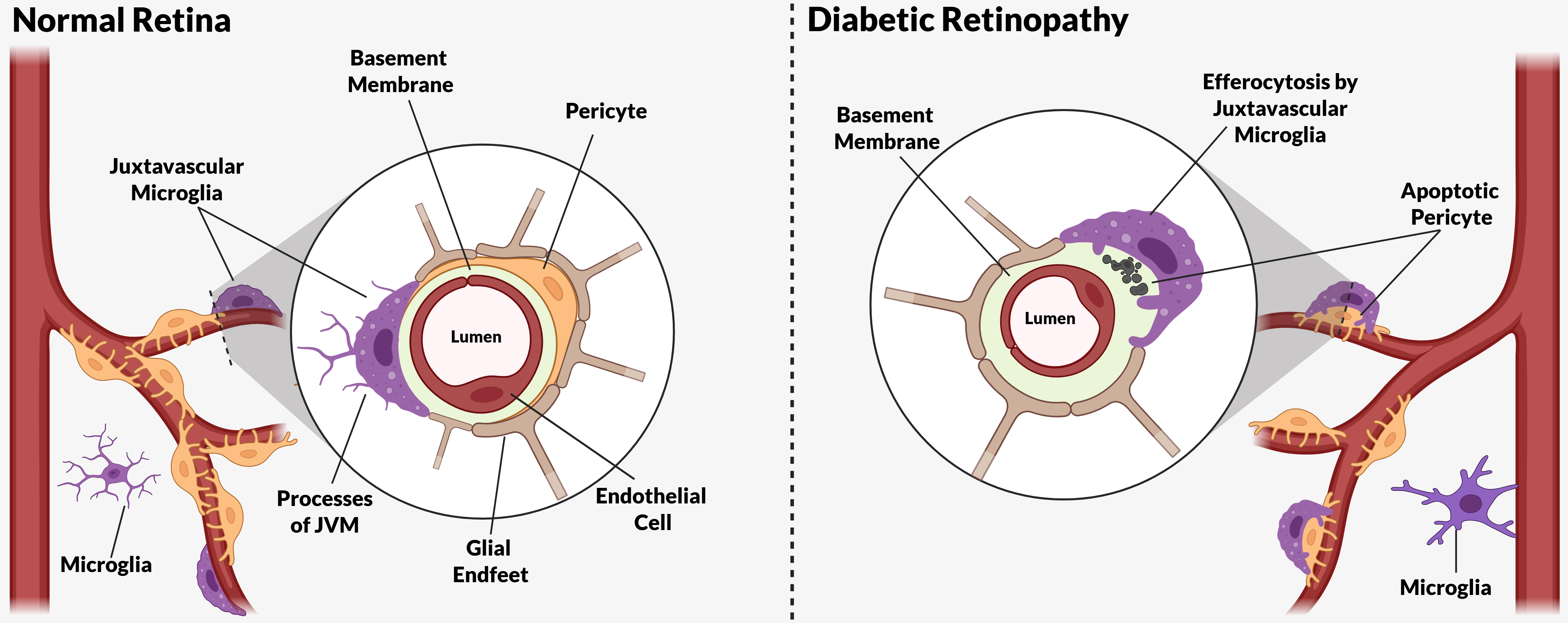
14. Juxtavascular Microglia (JVM) as Efferocytes of Dead Pericytes/VSMCs and Their Inhibition in DR
Our electron microscopic evidence identified the EF effector cells as juxtavascular microglia (JVM), a specialized population of microglial cells that were previously characterized in the brain but not hitherto described in the retina [123] (Box 1). It appears significant that although the majority of cell corpses identified as “ghost” cells within the vascular basement membranes in DR had progressed to post-apoptotic necrosis (Figure 3A,B), those observed while undergoing phagocytic engulfment by JVM (Figure 4) showed superior ultrastructural preservation and appeared to have been engaged at an earlier stage in the death process (Figure 5) [123]. As cells dying by apoptosis have been shown to release “find me” signals such as ATP and UTP very early in cell death [133], the presence of a JVM cell within the immediate or an adjacent neurovascular unit (NVU) may have overcome diabetes-related inhibitory factors in “find me” signalling or chemotaxis [134].
Specifically, the metainflammation induced by diabetes in the tissue environment of the CNS, including the retina [135,136], has been shown to downregulate expression of the P2Y12 receptor for purinergic “find me” signals (ATP & UTP) and impair JVM chemotaxis and the level of cell recruitment to sites of vascular injury [134,137]. Reduction of interferon gamma (IFγ) by anti-inflammatory treatment with dexamethasone restored P2Y12 levels and the normal vascular repair function of JVM [134,137]. Likewise, microglial responses were partially restored by insulin treatment and blood glucose levels approaching that of non-diabetic controls [134]. Therefore, JVM proximity and access could represent the deciding factors in whether successful EF can be performed on dying mural cells [123].
Also, in relation to inflammation-related inhibition of microglial responses in DR, a recent study by Pathak et al. has demonstrated additional recruitment of JVM in experimental DR that was significantly reduced in the absence of pentraxin-3 gene expression [138]. PTX3 is a pattern recognition molecule and modulator of innate immunity and inflammation, with multiple effects including either enhancement or inhibition of phagocytosis, depending on the context [139]. Pathak et al. showed a marked increase in pentraxin-3 (PTX3) expression by the Muller glia during experimental DR [138]. Somewhat counterintuitively this study also demonstrated that phagocytosis of apoptotic bodies was inhibited in human microglial cells treated with PTX3; the apoptotic bodies were derived from astrocytes and vascular endothelial cells induced with staurosporine [138]. Therefore, although PTX3 expression increased the number of JVM, it inhibited the phagocytosis of apoptotic bodies, consistent with the dual outcomes produced by this immunomodulator [140,141].
Despite of the importance of EF in limiting inflammation in diseases associated with cell death, studies of microglial EF engulfment receptors in DR are lacking. However, a recent study by Qi et al. reports MerTK impairment in experimental DR, due to increased expression of ADAM17 [142]. ADAM17 is the principal matrix metalloproteinase for MerTK shedding that in addition to decreasing the level of functional receptor on the efferocyte plasma membrane, further reduces its activity by liberating soluble MerTK (sMER) [143]. The soluble receptor has been shown to inhibit EF engulfment through binding and inactivation of the MerTK bridging ligand GAS6 [144].
- Box 1. Juxtavascular Microglia.
Retinal JVM reside in close apposition to the blood vessels with their cell soma and nucleus immediately adjacent to the vascular wall and can either penetrate the glia limitans (GL) of the astrocyte/Muller cell endfeet, or in some instances constitute an integral component of the GL [123] (Figure 6A). It is important to differentiate JVM from the perivascular macrophages that have been previously described and occupy an intravascular niche within the basement membranes of the retinal vasculature [145]. These cells were distinguished from microglia based on non-staining with the microglial immunomarker Iba-1 and electron microscopic confirmation of their exact location on the vascular side of the GL [145]. In comparison, the retinal JVM cells we observed in the phagocytic engulfment of dead or dying pericytes and VSMCs in DR were unambiguously located on the parenchymal side of the GL and also identified as a normal participant in the GL around normal retinal vessels [123]. Therefore, retinal JVM cells occupy precisely the same location as those previously described in the brain [146,147]. JVM in the brain have been investigated over a period of at least 30 years [137,146,147,148,149,150,151], and like other microglia have a stable population showing little exchange with marrow-derived macrophages [148,151]. They are dynamic cells that can migrate along the microvessels to access sites of injury [149] and have been shown to physically seal sites of endothelial damage to restore the integrity of the blood-brain barrier [137]. However, it is now clear that although a certain population of microglial cells are always in contact with the vasculature, a recent study by Pathak et al. has shown that additional microglial cells may be recruited to the juxtavascular compartment in experimental DR [138]. Whether the role of JVM cells in microvascular repair described in the brain [137] is common to all microglia or attributable to a specific gene expression profile acquired by local signalling in the juxtavascular compartment is currently unknown. Joost et al. have proposed that JVM represent an integral component of the NVU in the brain [146] and we would extend that classification to the retina (Figure 6) [123] as they show cell junctions with adjacent astrocyte/Muller cell footplates and unique matrix attachments to the vascular basement membrane (Figure 6A).
15. The Challenge of Efferocyte Access to the Vascular Basement Membranes in DR
JVMs also show the capacity to infiltrate the thickened vascular BMs [123] that characterize the retinal blood vessels in DR [108,120,152,153,154]. However, it is likely that such infiltration would be slower than in normal retinal vessels as the JVM must overcome the inhibitory nature of matrix protein crosslinking and other modifications induced in the vascular BMs by advanced glycation endproducts (AGEs) during diabetes [152,155,156,157]. AGE-adducted basement membrane proteins are more resistant to the protease digestion that facilitates access to the matrix and can block the critical motifs necessary for cell attachment, migration and survival [155,158,159,160]. Nevertheless, JVM cells appear to have a superior capacity for matrix infiltration than the local non-professional phagocytes that could theoretically fulfil the role of efferocytes for dead/dying mural cells.
16. Other Candidates for the Role of Efferocytes in EF of Apoptotic Pericytes and VSMCs
The other possible candidates for the EF of apoptotic mural cells are their partner endothelial cells within the vasculature as they exhibit significant phagocytic properties in situations of disease or injury [161,162]. Likewise, the Muller cells play major roles in EF during development and injury and have extensive access through their vascular endfeet at the glia limitans [24,33,163,164,165]. However, the previously mentioned increased expression of PTX3 or other pro-inflammatory changes induced in Muller cells by DR may alter EF-related genes [166,167,168]. Similarly, Muller cells are known to employ Gal-3 as a MerTK opsonin for phagocytosis in retinal degeneration, where it plays an anti-inflammatory role by reducing Muller cell activation [61]. However, in the context of DR, Gal-3 plays a pathological role in neuroinflammation and blood-retinal barrier dysfunction [59,169], a duality of outcome that may be due to the proinflammatory activation of Muller cells by diabetes-related metainflammation and AGEs [166,167,168]. The situation is further complicated by the presence of AGEs in DR as Gal-3 is also a well-characterized member of the AGE receptor complex [155,170] and so it is possible that the EF-related role of soluble Gal-3 as apoptotic cell opsonin for MerTK by Muller cells [61] is reduced by competition with AGEs in the diabetic retina.
Although microglial cells employ RAGE as a scavenger receptor for PS in phagocytosis of dead cells, there is no evidence that Muller cells do so, in spite of the fact that RAGE expression in Muller cells is upregulated during DR [168]. RAGE signalling represents an ongoing source of vascular inflammation in diabetes [171] with its role made more complex through interaction with the increased production of its ligand S100B [168] and the secreted form of the nuclear chaperone high mobility group box-1 (HMGB1) [172] protein that has been implicated in inflammation and vascular injury in DR [173,174,175], (see review by Steinle [173]). Importantly, HMBG1 has been shown to inhibit EF through binding of αvβ3 integrins and blocking their interaction with their opsonin MFG-E8 [176]. However, whether HMGB1-mediated inhibition of EF affects microglial cells during DR is currently unknown.
One last class of cells that represent credible candidates for the role of efferocytes for retinal vascular cells are the perivascular macrophages (PVM) that lie within the BM envelope and on the endothelial aspect of the GL. Although our ultrastructural study provided no evidence of their engagement with mural cell corpses in DR, PVM are perfectly located and have the capacity to function as efficient efferocytes in the vasculature [145,177,178]. PVM have a scavenger function at the blood-retinal barrier and their precise location within the vascular basement membrane is well-described in a 2009 study by Mendes--Jorge et al. [179] (see comparison to JVM in our previous paper [123]). However, as PVM are also myeloid macrophages it may be presumed that they suffer the same diabetes-related impairments as microglia and peripheral macrophages in diabetic wound healing [180].
17. Proinflammatory Consequences of Failed EF in DR
Unfortunately, it is currently unknown whether EF is dysfunctional within the retinal neuropile. However, it can be speculated that EF of neuronal and glial cells also suffers some level of impairment, but without the additional matrix-related inhibitions imposed on EF of mural cells within the vascular BM.
As the retinal vascular cells represent a remarkably stable population with an extremely low level of turnover under physiological conditions [181], there is probably little need for homeostatic EF, with the need only apparent in situations of injury or disease. Although there is evidence for accelerated apoptosis in retinal vascular cells in DR [132] , only the endothelial cells show evidence of regenerative capacity during this disease [181] and their increased turnover may not require local EF as apoptotic endothelial cells are probably removed by the circulation and scavenged in the liver. However, failed or dysfunctional EF of retinal pericytes and VSMCs has potentially deleterious immune consequences for the vessels harbouring them in DR. Firstly, the release of necrotic cell products can incite local inflammation, with possible consequences such as endothelial dysfunction and breakdown of the blood-retinal barrier. Secondly, the release of anti-inflammatory mediators by the efferocytes is negated, rendering sites of mural cell loss as proinflammatory foci within a host of retinal NVUs. Such inflammatory “hot spots” may increase local endothelial expression of adhesion molecules such as ICAM-1, thus creating potential sites for leukostasis-mediated endothelial injury [182,183]. Therefore, failure of EF in DR represents yet another function in which the vasculature suffers through diabetes-related dysfunction in other members of the NVU.
18. Further Insights from Published Data
Interrogation of data derived from publicly available datasets (retina – PMID: 33674409, brain—PMID: 37351595) from studies of the established DB/DB mouse model of type-2 diabetes, showed interesting similarities and notable differences between retinal microglia and those from the neural cortex (Figure 7).
Significantly, the expression of TREM2, a gene with multiple roles in microglial biology, including chemotaxis, migration and phagocytosis, was relatively reduced by diabetes in retinal compared to cortical microglia.
Several significant alterations were evident in genes associated with “find me” signalling: Expression of P2ry12, the gene that encodes the P2Y12 purinergic receptor was more depressed by diabetes in retinal compared to cortical microglia, a change paralleled by reduced expression of a second purinergic receptor gene, P2ry6 in retinal microglia. As P2Y12 triggers microglial activation in response to the “find me” signals ATP and UTP in EF and P2Y6 increases phagocytosis in response to UTP [184,185], both changes could contribute to slowed and inefficient EF during DR. Similarly, reduced expression of the fractalkine receptor gene Cx3CR1 was evident in retinal microglia during diabetes compared to those from the cortex, a change that in addition to impairment of “find me” signal detection could have proinflammatory effects in the microglial cells [45,46].
A number of differences were noted in microglial expression of genes related to PS receptors or their opsonins. Cortical microglia showed significantly lower expression of MerTK in the diabetic mice compared to controls, a change that was reflected in retinal microglia, but was statistically insignificant due to the relatively smaller population of microglial cells available in retinal tissue.
The gene coding for a second TAM receptor, the AXL kinase showed a low level of expression in the cortical microglia of diabetic animals but was almost absent in controls. However, retinal microglia showed a low but definite expression of AXL in both diabetic and control retina, but no measurable difference accountable to diabetes.
In contrast to TAM receptor expression in microglial cells, both MerTK and AXL showed relatively high expression in the cortical astrocytes and Muller glia of the retina that was not significantly altered by diabetes, consistent with their role as non-professional phagocytes.
The scavenger receptor stabilin-1 that binds directly to PS was expressed by both cortical and retinal microglia but was relatively higher in those of the cortex. Stabilin-2 receptor showed no expression in microglia from either location.
The soluble opsonin/bridging molecules for PS tended to be increased in microglia. GAS6 and MFG-E8 were marginally increased in diabetic microglia, as was the integrin opsonin MFG-E8. However, the levels of MFG-E8 expression in Muller cells and astrocytes suggest that the Muller cells and astrocytes represent more significant sources of MFG-E8 than the microglia. MFG-E8 was highly expressed by Muller cells in control retina and increased during diabetes. In contrast, diabetes significantly reduced MFG-E8 expression in cortical astrocytes. Interestingly, treatment with exogenous MFG-E8 after traumatic or hemorrhagic brain injury is both anti-apoptotic and anti-inflammatory [186,187,188,189]. Although these studies did not seek to dissect the benefits of enhanced EF by MFG-E8, a recent study showing reduced inflammation and increased neuroprotection when microglial EF is potentiated by exogenous MFG-E8, suggests that the level of MFG-E8 may be limiting in situations with high levels of cell death [190]. The related issue of EF-related microglial cell expression of the αvβ3 and αvβ5 integrin receptors that bind MFG-E8 is interesting: The genes encoding the beta subunits of the αvβ3 and αvβ5 (Itgb3 and Itgb5 respectively) are expressed by cortical microglia, although only Itgb5 was detected in those from the retina, and neither was altered in diabetic mice. However, Itgav, the gene coding for the alpha subunit, designated CD51, and shared by αvβ3 and αvβ5 was undetected in microglia from either the brain or the retina (therefore not displayed in Figure-7). This apparent discrepancy can be explained by the observation that CD51 may only be induced in certain populations of macroglia or during inflammation [191]. Also, CD51 is expressed by both non-professional phagocytes (Leydig cells) and macrophages in the seminiferous tubules of the testis [192], a site of constant apoptosis and an associated need for EF [193,194]. Therefore, in the brain, it would be interesting to examine sites of adult neurogenesis for CD51-positive microglia, as these are locations with ongoing apoptosis with a continual need for EF [195]. Therefore, the expression of CD51 may represent a marker for microglia engaged in EF. Of course, if CD51 was only expressed in situations of cell death, it would probably require induction by the early” find me” signals to be available at the stage where phagocytosis commenced.
Expression of all three genes encoding the complement factor C1q component peptides C1qA-C were reduced by diabetes in retinal microglia compared to those from the cortex, although expression of the CR3 receptor genes (Itgam &Itgb2) was unaltered by diabetes in either the brain or retina. Remarkably, MEGF10 a scavenger receptor employed by astrocytes for synaptic pruning and efferocytosis [15,196], but not expressed by microglia [196,197] showed a relatively high level of expression in Muller cells compared to cortical astrocytes. MEGF10 is a high affinity receptor for C1q bound to PS on the surface of redundant synapsed or apoptotic cells and although its expression was unaltered in diabetic mice, its use by Muller cells deserves further investigation.
In summary, the data described are consistent with impaired EF during DR in relation to both “find me” and “eat me” signalling and attributable to the metainflammation of the diabetic state and the influence of a proteome modified by non-enzymatic glycation. However, transcriptional alterations in particular genes occurring within the metabolome of the diabetic retina require cautious interpretation as they could result from factors unrelated to EF. Metabolic alterations or compensatory feedback due to dysfunction in the protein product caused by the addition of AGEs can have profound influences. It is also needful to examine the other roles played by proteins encoded by the altered genes. A pertinent example is the scavenger receptor stabilin-1 that shows increased expression in cortical microglia within our analysis. Stabilin-1 is a known receptor for AGEs and has important roles in the biosecretory pathway, endocytosis and lysosomal function (see discussion by Twarda-Clapa et al. [198]), all of which are involved in the phagocytic functions engaged during EF. In short, the situation in regard to Stabilin-1 embodies significant complexity that precludes any attempt to directly relate alterations in its transcription to EF in the diabetic retina. Therefore, there is an obvious need for more experimental data using models that permit dissection of the roles of the various players in EF and weighing of their relative contributions.
19. Conclusions
In general, the tendency for microglia in the diabetic retina is to assume a proinflammatory phenotype [199,200] akin to M1 macrophages that show impaired chemotaxis and phagocytosis of dead cells in diabetic wounds [180].
There is now significant evidence that hyperglycemia and the associated oxidative stress induce a persistent pro-inflammatory state in marrow-derived myeloid macrophages [201,202] that is refractory to restoration of euglycaemia and may be accountable to epigenetic changes manifested in metabolic memory [203] and “trained immunity” [204,205]. However, as the microglia are not derived from marrow-based progenitors they may be less susceptible to such metainflammatory conditioning offering hope of successful restoration of functionality with appropriate treatments. Also, the period of exposure to chronic inflammatory stimuli required to induce such epigenetic change is unclear. This is especially true in diabetic patients for whom hyperglycemia is episodic, unlike the unremitting high-glucose environment of the animal models in which metabolic memory was characterized [203,206]. Interestingly, a recent study by Zhang et al. has shown that intraventricular injection of the opsonin MFG-E8 was able to promote microglial EF and exert neuroprotection after an acute ischemic insult [190]. This treatment also induced a generalized and lasting change in the microglial gene expression profile from a proinflammatory phenotype to one that was anti-inflammatory and neuroprotective [190]. The authors emphasized the importance of interferon regulatory factor-7 (IRF7) upregulation in the post-treatment microglial gene profile [207]. IRF7 is associated with the transition from a proinflammatory M1 macrophage phenotype to an anti-inflammatory regenerative M2 profile [207,208,209]. Indeed, at a fundamental level in both development and adulthood, microglia can radically alter their phenotype in response to paracrine signals from their microenvironment [178,210] and as such present themselves as suitable targets for treatment.
Currently, it remains to be seen whether broad anti-inflammatory strategies or novel approaches targeting specific EF-related gene products may prove useful in chronic diseases such as DR. Nevertheless, the phenomenon of ongoing cell death in all cells of the retinal NVU during DR requires intervention to steer proinflammatory cell death events to pro-regenerative anti-inflammatory outcomes.
Author Contributions
Writing—original draft preparation, T.A.G.; writing—review and editing, A.W.S, K L.; formal analysis — KL, T.A.G. All authors have read and agreed to the published version of the manuscript.
Funding
This research received no external funding.
Institutional Review Board Statement
Not applicable.
Data Availability Statement
No new data was created for this work.
Acknowledgments
The graphical abstract was created with BioRender.com. The authors have reviewed and edited the output and take full responsibility for the content of this publication.
Conflicts of Interest
The authors declare no conflicts of interest.
Abbreviations
The following abbreviations are used in this manuscript:
| ADAM17 | A Disintegrin and Metalloprotease 17 |
| AGE | advanced glycation endproduct |
| ApoE | apolipoprotein-E |
| ATP | Adenosine triphosphate |
| AXL | AXL receptor tyrosine kinase |
| BAI1 | brain-specific angiogenesis inhibitor 1 |
| BM | basement membrane |
| C1q | complement component-1q |
| C3 | Complement component-3 |
| CNS | central nervous system |
| CSF1R | colony-stimulating factor-1 receptor |
| CX3CL1 | CX3C motif chemokine ligand-1 (fractalkine) |
| GAL3 | Galectin-3 |
| GAS6 | growth arrest specific 6 |
| GL | glia limitans |
| HMGB1 | high mobility group box-1 |
| IL10 | Interleukin-10 |
| IL34 | interleukin-34 |
| IRF7 | interferon regulatory factor-7 |
| JVM | juxtavascular microglia |
| LPS | lysophosphatidylcholine |
| PS | phosphatidylserine |
| MEGF10 | multiple epidermal growth factor-like domains protein 10 |
| MerTK | mer proto-oncogene tyrosine kinase |
| MFGE8 | milk-fat globule-EGF factor E8 |
| MS | multiple sclerosis |
| NCAM | neural cell adhesion molecule |
| NVU | neurovascular unit |
| PTX3 | pentraxin-3 |
| PVM | perivascular macrophages |
| RAGE | receptor for advanced glycation endproducts |
| SLE | systemic lupus erythematosus |
| TAM | Tyro, Axl & MerTK receptor family |
| TGFβ | transforming growth factor beta |
| TREM2 | triggering receptor expressed on myeloid cells-2 |
| TYRO3 | tyrosine protein kinase receptor 3 |
| S1P | sphingosine-1 phosphate |
| TIM1/4 | T-cell immunoglobulin and mucin domain-containing protein 1/4 |
| UTP | uridine-5-triphosphate |
| VSMC | vascular smooth muscle cell |
References
- deCathelineau, A.M.; Henson, P.M. The final step in programmed cell death: phagocytes carry apoptotic cells to the grave. Essays Biochem. 2003, 39, 105–117. [Google Scholar] [CrossRef]
- Silva, M.T. Secondary necrosis: the natural outcome of the complete apoptotic program. FEBS Lett 2010, 584, 4491–4499. [Google Scholar] [CrossRef]
- Zhang, J.; Ding, W.; Zhao, M.; Liu, J.; Xu, Y.; Wan, J.; Wang, M. Mechanisms of efferocytosis in determining inflammation resolution: Therapeutic potential and the association with cardiovascular disease. Br. J. Pharmacol. 2022, 179, 5151–5171. [Google Scholar] [CrossRef]
- Morioka, S.; Maueröder, C.; Ravichandran, K.S. Living on the Edge: Efferocytosis at the Interface of Homeostasis and Pathology. Immunity 2019, 50, 1149–1162. [Google Scholar] [CrossRef]
- Elliott, M.R.; Koster, K.M.; Murphy, P.S. Efferocytosis Signaling in the Regulation of Macrophage Inflammatory Responses. J Immunol 2017, 198, 1387–1394. [Google Scholar] [CrossRef]
- Arandjelovic, S.; Ravichandran, K.S. Phagocytosis of apoptotic cells in homeostasis. Nat. Immunol. 2015, 16, 907–917. [Google Scholar] [CrossRef]
- Bajgar, A.; Krejčová, G. On the origin of the functional versatility of macrophages. Front Physiol 2023, 14, 1128984. [Google Scholar] [CrossRef] [PubMed]
- Ge, Y.; Huang, M.; Yao, Y.-m. Efferocytosis and Its Role in Inflammatory Disorders. Front. Cell Dev. Biol. 2022, 10. [Google Scholar] [CrossRef]
- Abdolmaleki, F.; Farahani, N.; Gheibi Hayat, S.M.; Pirro, M.; Bianconi, V.; Barreto, G.E.; Sahebkar, A. The Role of Efferocytosis in Autoimmune Diseases. Front. Immunol. 2018, 9. [Google Scholar] [CrossRef] [PubMed]
- Mehrotra, P.; Ravichandran, K.S. Drugging the efferocytosis process: concepts and opportunities. Nat. Rev. Drug Discov. 2022, 21, 601–620. [Google Scholar] [CrossRef] [PubMed]
- Chen, Y.; Kou, Y.; Ni, Y.; Yang, H.; Xu, C.; Fan, H.; Liu, H. Microglia efferocytosis: an emerging mechanism for the resolution of neuroinflammation in Alzheimer’s disease. J. Neuroinflammation 2025, 22, 96. [Google Scholar] [CrossRef]
- Xing, J.; Wang, K.; Xu, Y.-c.; Pei, Z.-j.; Yu, Q.-x.; Liu, X.-y.; Dong, Y.-l.; Li, S.-f.; Chen, Y.; Zhao, Y.-j.; et al. Efferocytosis: Unveiling its potential in autoimmune disease and treatment strategies. Autoimmun. Rev. 2024, 23, 103578. [Google Scholar] [CrossRef]
- Li, F.; Huang, Q.; Chen, J.; Peng, Y.; Roop, D.R.; Bedford, J.S.; Li, C.-Y. Apoptotic Cells Activate the “Phoenix Rising” Pathway to Promote Wound Healing and Tissue Regeneration. Sci. Signal. 2010, 3, ra13. [Google Scholar] [CrossRef]
- Faust, T.E.; Gunner, G.; Schafer, D.P. Mechanisms governing activity-dependent synaptic pruning in the developing mammalian CNS. Nat. Rev. Neurosci. 2021, 22, 657–673. [Google Scholar] [CrossRef] [PubMed]
- Chung, W.-S.; Clarke, L.E.; Wang, G.X.; Stafford, B.K.; Sher, A.; Chakraborty, C.; Joung, J.; Foo, L.C.; Thompson, A.; Chen, C.; et al. Astrocytes mediate synapse elimination through MEGF10 and MERTK pathways. Nature 2013, 504, 394–400. [Google Scholar] [CrossRef] [PubMed]
- Mike, J.K.; Ferriero, D.M. Efferocytosis Mediated Modulation of Injury after Neonatal Brain Hypoxia-Ischemia. Cells 2021, 10. [Google Scholar] [CrossRef]
- Schafer, Dorothy P. ; Lehrman, Emily K.; Kautzman, Amanda G.; Koyama, R.; Mardinly, Alan R.; Yamasaki, R.; Ransohoff, Richard M.; Greenberg, Michael E.; Barres, Ben A.; Stevens, B. Microglia Sculpt Postnatal Neural Circuits in an Activity and Complement-Dependent Manner. Neuron 2012, 74, 691–705. [Google Scholar] [CrossRef]
- Stevens, B.; Allen, N.J.; Vazquez, L.E.; Howell, G.R.; Christopherson, K.S.; Nouri, N.; Micheva, K.D.; Mehalow, A.K.; Huberman, A.D.; Stafford, B.; et al. The Classical Complement Cascade Mediates CNS Synapse Elimination. Cell 2007, 131, 1164–1178. [Google Scholar] [CrossRef]
- Park, J.; Choi, Y.; Jung, E.; Lee, S.H.; Sohn, J.W.; Chung, W.S. Microglial MERTK eliminates phosphatidylserine-displaying inhibitory post-synapses. Embo J 2021, 40, e107121. [Google Scholar] [CrossRef]
- Damisah, E.C.; Hill, R.A.; Rai, A.; Chen, F.; Rothlin, C.V.; Ghosh, S.; Grutzendler, J. Astrocytes and microglia play orchestrated roles and respect phagocytic territories during neuronal corpse removal in vivo. Sci. Adv. 2020, 6, eaba3239. [Google Scholar] [CrossRef]
- Halassa, M.M.; Fellin, T.; Haydon, P.G. The tripartite synapse: roles for gliotransmission in health and disease. Trends Mol. Med. 2007, 13, 54–63. [Google Scholar] [CrossRef]
- Freeman, M.R. Specification and Morphogenesis of Astrocytes. Science 2010, 330, 774–778. [Google Scholar] [CrossRef] [PubMed]
- Galloway, D.A.; Phillips, A.E.M.; Owen, D.R.J.; Moore, C.S. Phagocytosis in the Brain: Homeostasis and Disease. Front. Immunol. 2019, 10. [Google Scholar] [CrossRef] [PubMed]
- Bejarano-Escobar, R.; Sánchez-Calderón, H.; Otero-Arenas, J.; Martín-Partido, G.; Francisco-Morcillo, J. Müller glia and phagocytosis of cell debris in retinal tissue. J. Anat. 2017, 231, 471–483. [Google Scholar] [CrossRef]
- Ginhoux, F.; Prinz, M. Origin of Microglia: Current Concepts and Past Controversies. Cold Spring Harb. Perspect. Biol. 2015, 7. [Google Scholar] [CrossRef]
- Alliot, F.; Godin, I.; Pessac, B. Microglia derive from progenitors, originating from the yolk sac, and which proliferate in the brain. Dev. Brain Res. 1999, 117, 145–152. [Google Scholar] [CrossRef]
- Ginhoux, F.; Greter, M.; Leboeuf, M.; Nandi, S.; See, P.; Gokhan, S.; Mehler, M.F.; Conway, S.J.; Ng, L.G.; Stanley, E.R.; et al. Fate mapping analysis reveals that adult microglia derive from primitive macrophages. Science 2010, 330, 841–845. [Google Scholar] [CrossRef]
- Hashimoto, D.; Chow, A.; Noizat, C.; Teo, P.; Beasley, Mary B. ; Leboeuf, M.; Becker, Christian D.; See, P.; Price, J.; Lucas, D.; et al. Tissue-Resident Macrophages Self-Maintain Locally throughout Adult Life with Minimal Contribution from Circulating Monocytes. Immunity 2013, 38, 792–804. [Google Scholar] [CrossRef]
- Prinz, M.; Jung, S.; Priller, J. Microglia Biology: One Century of Evolving Concepts. Cell 2019, 179, 292–311. [Google Scholar] [CrossRef] [PubMed]
- Möller, K.; Brambach, M.; Villani, A.; Gallo, E.; Gilmour, D.; Peri, F. A role for the centrosome in regulating the rate of neuronal efferocytosis by microglia in vivo. eLife 2022, 11, e82094. [Google Scholar] [CrossRef]
- Thion, M.S.; Garel, S. On place and time: microglia in embryonic and perinatal brain development. Curr. Opin. Neurobiol. 2017, 47, 121–130. [Google Scholar] [CrossRef] [PubMed]
- Wu, S.; Xue, R.; Hassan, S.; Nguyen, T.M.L.; Wang, T.; Pan, H.; Xu, J.; Liu, Q.; Zhang, W.; Wen, Z. Il34-Csf1r Pathway Regulates the Migration and Colonization of Microglial Precursors. Dev. Cell 2018, 46, 552–563.e554. [Google Scholar] [CrossRef] [PubMed]
- Francisco-Morcillo, J.; Bejarano-Escobar, R.; Rodríguez-León, J.; Navascués, J.; Martín-Partido, G. Ontogenetic Cell Death and Phagocytosis in the Visual System of Vertebrates. Dev. Dyn. 2014, 243, 1203–1225. [Google Scholar] [CrossRef]
- Ranawat, N.; Masai, I. Mechanisms underlying microglial colonization of developing neural retina in zebrafish. Elife 2021, 10. [Google Scholar] [CrossRef]
- Santos, A.M.; Calvente, R.; Tassi, M.; Carrasco, M.-C.; Martín-Oliva, D.; Marín-Teva, J.L.; Navascués, J.; Cuadros, M.A. Embryonic and postnatal development of microglial cells in the mouse retina. J. Comp. Neurol. 2008, 506, 224–239. [Google Scholar] [CrossRef] [PubMed]
- Flannagan, R.S.; Jaumouillé, V.; Grinstein, S. The Cell Biology of Phagocytosis. Annu. Rev. Pathol. : Mech. Dis. 2012, 7, 61–98. [Google Scholar] [CrossRef]
- Kelley, S.M.; Ravichandran, K.S. Putting the brakes on phagocytosis: “don’t-eat-me” signaling in physiology and disease. EMBO Rep. 2021, 22, e52564. [Google Scholar] [CrossRef]
- Brown, G.C.; Neher, J.J. Microglial phagocytosis of live neurons. Nat. Rev. Neurosci. 2014, 15, 209–216. [Google Scholar] [CrossRef]
- Neher, J.J.; Emmrich, J.V.; Fricker, M.; Mander, P.K.; Théry, C.; Brown, G.C. Phagocytosis executes delayed neuronal death after focal brain ischemia. Proc. Natl. Acad. Sci. 2013, 110, E4098–E4107. [Google Scholar] [CrossRef]
- Brown, G.C.; Neher, J.J. Eaten alive! Cell death by primary phagocytosis: ‘phagoptosis’. Trends Biochem. Sci. 2012, 37, 325–332. [Google Scholar] [CrossRef]
- Park, S.-Y.; Kim, I.-S. Engulfment signals and the phagocytic machinery for apoptotic cell clearance. Exp. Mol. Med. 2017, 49, e331–e331. [Google Scholar] [CrossRef]
- Doran, A.C.; Yurdagul, A.; Tabas, I. Efferocytosis in health and disease. Nat. Rev. Immunol. 2020, 20, 254–267. [Google Scholar] [CrossRef]
- Lam, A.L.; Heit, B. Having an Old Friend for Dinner: The Interplay between Apoptotic Cells and Efferocytes. Cells 2021, 10, 1265. [Google Scholar] [CrossRef]
- Angelopoulou, E.; Paudel, Y.N.; Shaikh, M.F.; Piperi, C. Fractalkine (CX3CL1) signaling and neuroinflammation in Parkinson’s disease: Potential clinical and therapeutic implications. Pharmacol. Res. 2020, 158, 104930. [Google Scholar] [CrossRef]
- Sokolowski, J.D.; Chabanon-Hicks, C.N.; Han, C.Z.; Heffron, D.S.; Mandell, J.W. Fractalkine is a “find-me” signal released by neurons undergoing ethanol-induced apoptosis. Front Cell Neurosci 2014, 8, 360. [Google Scholar] [CrossRef]
- Rodriguez, D.; Church, K.A.; Pietramale, A.N.; Cardona, S.M.; Vanegas, D.; Rorex, C.; Leary, M.C.; Muzzio, I.A.; Nash, K.R.; Cardona, A.E. Fractalkine isoforms differentially regulate microglia-mediated inflammation and enhance visual function in the diabetic retina. J. Neuroinflammation 2024, 21, 42. [Google Scholar] [CrossRef] [PubMed]
- Kielbik, M.; Szulc-Kielbik, I.; Klink, M. Calreticulin-Multifunctional Chaperone in Immunogenic Cell Death: Potential Significance as a Prognostic Biomarker in Ovarian Cancer Patients. Cells 2021, 10. [Google Scholar] [CrossRef]
- Vorselen, D. Dynamics of phagocytosis mediated by phosphatidylserine. Biochem. Soc. Trans. 2022, 50, 1281–1291. [Google Scholar] [CrossRef] [PubMed]
- Fadok, V.A.; Voelker, D.R.; Campbell, P.A.; Cohen, J.J.; Bratton, D.L.; Henson, P.M. Exposure of phosphatidylserine on the surface of apoptotic lymphocytes triggers specific recognition and removal by macrophages. J. Immunol. 1992, 148, 2207–2216. [Google Scholar] [CrossRef]
- Lemke, G. How macrophages deal with death. Nat. Rev. Immunol. 2019, 19, 539–549. [Google Scholar] [CrossRef] [PubMed]
- Freeman, G.J.; Casasnovas, J.M.; Umetsu, D.T.; DeKruyff, R.H. TIM genes: a family of cell surface phosphatidylserine receptors that regulate innate and adaptive immunity. Immunol. Rev. 2010, 235, 172–189. [Google Scholar] [CrossRef] [PubMed]
- Mazaheri, F.; Breus, O.; Durdu, S.; Haas, P.; Wittbrodt, J.; Gilmour, D.; Peri, F. Distinct roles for BAI1 and TIM-4 in the engulfment of dying neurons by microglia. Nat. Commun. 2014, 5, 4046. [Google Scholar] [CrossRef]
- Hudson, B.I.; Lippman, M.E. Targeting RAGE Signaling in Inflammatory Disease. Annu. Rev. Med. 2018, 69, 349–364. [Google Scholar] [CrossRef]
- Derk, J.; MacLean, M.; Juranek, J.; Schmidt, A.M. The Receptor for Advanced Glycation Endproducts (RAGE) and Mediation of Inflammatory Neurodegeneration. J Alzheimers Dis Park. 2018, 8. [Google Scholar] [CrossRef]
- Alí-Ruiz, D.; Vitureira, N.; Peluffo, H. Microglial CD300f immune receptor contributes to the maintenance of neuron viability in vitro and after a penetrating brain injury. Sci. Rep. 2023, 13, 16796. [Google Scholar] [CrossRef]
- Lemke, G. Biology of the TAM receptors. Cold Spring Harb Perspect Biol 2013, 5, a009076. [Google Scholar] [CrossRef]
- Caberoy, N.B.; Alvarado, G.; Bigcas, J.-L.; Li, W. Galectin-3 is a new MerTK-specific eat-me signal. J. Cell. Physiol. 2012, 227, 401–407. [Google Scholar] [CrossRef]
- Nomura, K.; Vilalta, A.; Allendorf, D.H.; Hornik, T.C.; Brown, G.C. Activated Microglia Desialylate and Phagocytose Cells via Neuraminidase, Galectin-3, and Mer Tyrosine Kinase. J. Immunol. 2017, 198, 4792–4801. [Google Scholar] [CrossRef]
- Mendonça, H.R.; Carvalho, J.N.A.; Abreu, C.A.; Mariano de Souza Aguiar dos Santos, D.; Carvalho, J.R.; Marques, S.A.; da Costa Calaza, K.; Martinez, A.M.B. Lack of Galectin-3 attenuates neuroinflammation and protects the retina and optic nerve of diabetic mice. Brain Res. 2018, 1700, 126–137. [Google Scholar] [CrossRef] [PubMed]
- Uehara, F.; Ohba, N.; Ozawa, M. Isolation and Characterization of Galectins in the Mammalian Retina. Investig. Ophthalmol. Vis. Sci. 2001, 42, 2164–2172. [Google Scholar]
- Lew, D.S.; McGrath, M.J.; Finnemann, S.C. Galectin-3 Promotes Müller Glia Clearance Phagocytosis via MERTK and Reduces Harmful Müller Glia Activation in Inherited and Induced Retinal Degeneration. Front. Cell. Neurosci. 2022, 16. [Google Scholar] [CrossRef]
- Lemke, G.; Rothlin, C.V. Immunobiology of the TAM receptors. Nat. Rev. Immunol. 2008, 8, 327–336. [Google Scholar] [CrossRef]
- Zagórska, A.; Través, P.G.; Lew, E.D.; Dransfield, I.; Lemke, G. Diversification of TAM receptor tyrosine kinase function. Nat. Immunol. 2014, 15, 920–928. [Google Scholar] [CrossRef]
- Shen, K.; Reichelt, M.; Kyauk, R.V.; Ngu, H.; Shen, Y.-A.A.; Foreman, O.; Modrusan, Z.; Friedman, B.A.; Sheng, M.; Yuen, T.J. Multiple sclerosis risk gene Mertk is required for microglial activation and subsequent remyelination. Cell Rep. 2021, 34. [Google Scholar] [CrossRef] [PubMed]
- Fourgeaud, L.; Través, P.G.; Tufail, Y.; Leal-Bailey, H.; Lew, E.D.; Burrola, P.G.; Callaway, P.; Zagórska, A.; Rothlin, C.V.; Nimmerjahn, A.; et al. TAM receptors regulate multiple features of microglial physiology. Nature 2016, 532, 240–244. [Google Scholar] [CrossRef] [PubMed]
- Gautier, E.L.; Shay, T.; Miller, J.; Greter, M.; Jakubzick, C.; Ivanov, S.; Helft, J.; Chow, A.; Elpek, K.G.; Gordonov, S.; et al. Gene-expression profiles and transcriptional regulatory pathways that underlie the identity and diversity of mouse tissue macrophages. Nat. Immunol. 2012, 13, 1118–1128. [Google Scholar] [CrossRef] [PubMed]
- Duncan, J.L.; LaVail, M.M.; Yasumura, D.; Matthes, M.T.; Yang, H.; Trautmann, N.; Chappelow, A.V.; Feng, W.; Earp, H.S.; Matsushima, G.K.; et al. An RCS-Like Retinal Dystrophy Phenotype in Mer Knockout Mice. Investig. Ophthalmol. Vis. Sci. 2003, 44, 826–838. [Google Scholar] [CrossRef]
- Gal, A.; Li, Y.; Thompson, D.A.; Weir, J.; Orth, U.; Jacobson, S.G.; Apfelstedt-Sylla, E.; Vollrath, D. Mutations in MERTK, the human orthologue of the RCS rat retinal dystrophy gene, cause retinitis pigmentosa. Nat. Genet. 2000, 26, 270–271. [Google Scholar] [CrossRef]
- D’Cruz, P.M.; Yasumura, D.; Weir, J.; Matthes, M.T.; Abderrahim, H.; LaVail, M.M.; Vollrath, D. Mutation of the receptor tyrosine kinase gene Mertk in the retinal dystrophic RCS rat. Hum. Mol. Genet. 2000, 9, 645–651. [Google Scholar] [CrossRef]
- Zhou, H.; Hu, L.; Li, J.; Ruan, W.; Cao, Y.; Zhuang, J.; Xu, H.; Peng, Y.; Zhang, Z.; Xu, C.; et al. AXL kinase-mediated astrocytic phagocytosis modulates outcomes of traumatic brain injury. J. Neuroinflammation 2021, 18, 154. [Google Scholar] [CrossRef]
- Zhang, L.; Xiang, X.; Li, Y.; Bu, G.; Chen, X.-F. TREM2 and sTREM2 in Alzheimer’s disease: from mechanisms to therapies. Mol. Neurodegener. 2025, 20, 43. [Google Scholar] [CrossRef]
- Colonna, M. The biology of TREM receptors. Nat. Rev. Immunol. 2023, 23, 580–594. [Google Scholar] [CrossRef]
- Gao, H.; Di, J.; Clausen, B.H.; Wang, N.; Zhu, X.; Zhao, T.; Chang, Y.; Pang, M.; Yang, Y.; He, R.; et al. Distinct myeloid population phenotypes dependent on TREM2 expression levels shape the pathology of traumatic versus demyelinating CNS disorders. Cell Rep. 2023, 42. [Google Scholar] [CrossRef] [PubMed]
- Deczkowska, A.; Weiner, A.; Amit, I. The Physiology, Pathology, and Potential Therapeutic Applications of the TREM2 Signaling Pathway. Cell 2020, 181, 1207–1217. [Google Scholar] [CrossRef] [PubMed]
- Karch, C.M.; Goate, A.M. Alzheimer’s disease risk genes and mechanisms of disease pathogenesis. Biol Psychiatry 2015, 77, 43–51. [Google Scholar] [CrossRef] [PubMed]
- Jonsson, T.; Stefansson, H.; Steinberg, S.; Jonsdottir, I.; Jonsson, P.V.; Snaedal, J.; Bjornsson, S.; Huttenlocher, J.; Levey, A.I.; Lah, J.J.; et al. Variant of TREM2 associated with the risk of Alzheimer’s disease. N Engl J Med 2013, 368, 107–116. [Google Scholar] [CrossRef]
- Guerreiro, R.; Wojtas, A.; Bras, J.; Carrasquillo, M.; Rogaeva, E.; Majounie, E.; Cruchaga, C.; Sassi, C.; Kauwe, J.S.K.; Younkin, S.; et al. TREM2 Variants in Alzheimer’s Disease. New Engl. J. Med. 2013, 368, 117–127. [Google Scholar] [CrossRef]
- Yuan, P.; Condello, C.; Keene, C.D.; Wang, Y.; Bird, Thomas D. ; Paul, Steven M.; Luo, W.; Colonna, M.; Baddeley, D.; Grutzendler, J. TREM2 Haplodeficiency in Mice and Humans Impairs the Microglia Barrier Function Leading to Decreased Amyloid Compaction and Severe Axonal Dystrophy. Neuron 2016, 90, 724–739. [Google Scholar] [CrossRef]
- Wang, Y.; Ulland, T.K.; Ulrich, J.D.; Song, W.; Tzaferis, J.A.; Hole, J.T.; Yuan, P.; Mahan, T.E.; Shi, Y.; Gilfillan, S.; et al. TREM2-mediated early microglial response limits diffusion and toxicity of amyloid plaques. J Exp Med 2016, 213, 667–675. [Google Scholar] [CrossRef]
- Zhao, Y.; Wu, X.; Li, X.; Jiang, L.L.; Gui, X.; Liu, Y.; Sun, Y.; Zhu, B.; Piña-Crespo, J.C.; Zhang, M.; et al. TREM2 Is a Receptor for β-Amyloid that Mediates Microglial Function. Neuron 2018, 97, 1023–1031.e1027. [Google Scholar] [CrossRef]
- Hsieh, C.L.; Koike, M.; Spusta, S.C.; Niemi, E.C.; Yenari, M.; Nakamura, M.C.; Seaman, W.E. A role for TREM2 ligands in the phagocytosis of apoptotic neuronal cells by microglia. J. Neurochem. 2009, 109, 1144–1156. [Google Scholar] [CrossRef] [PubMed]
- Takahashi, K.; Rochford, C.D.P.; Neumann, H. Clearance of apoptotic neurons without inflammation by microglial triggering receptor expressed on myeloid cells-2. J. Exp. Med. 2005, 201, 647–657. [Google Scholar] [CrossRef]
- Wang, Y.; Cella, M.; Mallinson, K.; Ulrich, Jason D. ; Young, Katherine L.; Robinette, Michelle L.; Gilfillan, S.; Krishnan, Gokul M.; Sudhakar, S.; Zinselmeyer, Bernd H.; et al. TREM2 Lipid Sensing Sustains the Microglial Response in an Alzheime’s Disease Model. Cell 2015, 160, 1061–1071. [Google Scholar] [CrossRef]
- Bailey, C.C.; DeVaux, L.B.; Farzan, M. The Triggering Receptor Expressed on Myeloid Cells 2 Binds Apolipoprotein E. J Biol Chem 2015, 290, 26033–26042. [Google Scholar] [CrossRef] [PubMed]
- Atagi, Y.; Liu, C.-C.; Painter, M.M.; Chen, X.-F.; Verbeeck, C.; Zheng, H.; Li, X.; Rademakers, R.; Kang, S.S.; Xu, H.; et al. Apolipoprotein E Is a Ligand for Triggering Receptor Expressed on Myeloid Cells 2 (TREM2) *. J. Biol. Chem. 2015, 290, 26043–26050. [Google Scholar] [CrossRef]
- Grainger, D.J.; Reckless, J.; McKilligin, E. Apolipoprotein E Modulates Clearance of Apoptotic Bodies In Vitro and In Vivo, Resulting in a Systemic Proinflammatory State in Apolipoprotein E-Deficient Mice1. J. Immunol. 2004, 173, 6366–6375. [Google Scholar] [CrossRef]
- Mazaheri, F.; Snaidero, N.; Kleinberger, G.; Madore, C.; Daria, A.; Werner, G.; Krasemann, S.; Capell, A.; Trümbach, D.; Wurst, W.; et al. TREM2 deficiency impairs chemotaxis and microglial responses to neuronal injury. EMBO Rep. 2017, 18, 1186–1198. [Google Scholar] [CrossRef]
- Jaumouillé, V.; Waterman, C.M. Physical Constraints and Forces Involved in Phagocytosis. Front. Immunol. 2020, 11. [Google Scholar] [CrossRef]
- Henson, P.M.; Hume, D.A. Apoptotic cell removal in development and tissue homeostasis. Trends Immunol. 2006, 27, 244–250. [Google Scholar] [CrossRef] [PubMed]
- Flannagan, R.S.; Canton, J.; Furuya, W.; Glogauer, M.; Grinstein, S. The phosphatidylserine receptor TIM4 utilizes integrins as coreceptors to effect phagocytosis. Mol Biol Cell 2014, 25, 1511–1522. [Google Scholar] [CrossRef] [PubMed]
- Mylvaganam, S.; Freeman, S.A.; Grinstein, S. The cytoskeleton in phagocytosis and macropinocytosis. Curr. Biol. 2021, 31, R619–R632. [Google Scholar] [CrossRef]
- Park, B.; Lee, J.; Moon, H.; Lee, G.; Lee, D.H.; Hoon Cho, J.; Park, D. Co-receptors are dispensable for tethering receptor-mediated phagocytosis of apoptotic cells. Cell Death Dis. 2015, 6, e1772–e1772. [Google Scholar] [CrossRef]
- Dransfield, I.; Zagórska, A.; Lew, E.D.; Michail, K.; Lemke, G. Mer receptor tyrosine kinase mediates both tethering and phagocytosis of apoptotic cells. Cell Death Dis. 2015, 6, e1646–e1646. [Google Scholar] [CrossRef]
- Sokolova, D.; Ghansah, S.A.; Puletti, F.; Georgiades, T.; De Schepper, S.; Zheng, Y.; Crowley, G.; Wu, L.; Rueda-Carrasco, J.; Koutsiouroumpa, A.; et al. Astrocyte-derived MFG-E8 facilitates microglial synapse elimination in Alzheimer’s disease mouse models. bioRxiv 2024. [Google Scholar] [CrossRef]
- Vandendriessche, S.; Cambier, S.; Proost, P.; Marques, P.E. Complement Receptors and Their Role in Leukocyte Recruitment and Phagocytosis. Front Cell Dev Biol 2021, 9, 624025. [Google Scholar] [CrossRef] [PubMed]
- Scott-Hewitt, N.; Perrucci, F.; Morini, R.; Erreni, M.; Mahoney, M.; Witkowska, A.; Carey, A.; Faggiani, E.; Schuetz, L.T.; Mason, S.; et al. Local externalization of phosphatidylserine mediates developmental synaptic pruning by microglia. EMBO J. 2020, 39, e105380. [Google Scholar] [CrossRef] [PubMed]
- Païdassi, H.; Tacnet-Delorme, P.; Garlatti, V.; Darnault, C.; Ghebrehiwet, B.; Gaboriaud, C.; Arlaud, G.r.J.; Frachet, P. C1q Binds Phosphatidylserine and Likely Acts as a Multiligand-Bridging Molecule in Apoptotic Cell Recognition1. J. Immunol. 2008, 180, 2329–2338. [Google Scholar] [CrossRef] [PubMed]
- Fonseca, M.I.; Chu, S.-H.; Hernandez, M.X.; Fang, M.J.; Modarresi, L.; Selvan, P.; MacGregor, G.R.; Tenner, A.J. Cell-specific deletion of C1qa identifies microglia as the dominant source of C1q in mouse brain. J. Neuroinflammation 2017, 14, 48. [Google Scholar] [CrossRef]
- Dickson, B.H.; Tasnim, T.; Lam, A.L.; Vreize, A.; Blythe, E.N.; Dekaban, G.A.; Heit, B. MERTK Coordinates Efferocytosis by Regulating Integrin Localization and Activation. bioRxiv, 2007. [Google Scholar] [CrossRef]
- Flannagan, R.S.; Jaumouillé, V.; Grinstein, S. The cell biology of phagocytosis. Annu Rev Pathol 2012, 7, 61–98. [Google Scholar] [CrossRef]
- Ling, L.; Templeton, D.; Kung, H.-J. Identification of the Major Autophosphorylation Sites of Nyk/Mer, an NCAM-related Receptor Tyrosine Kinase*. J. Biol. Chem. 1996, 271, 18355–18362. [Google Scholar] [CrossRef]
- Gordon, S. Phagocytosis: An Immunobiologic Process. Immunity 2016, 44, 463–475. [Google Scholar] [CrossRef]
- Fadok, V.A.; Bratton, D.L.; Konowal, A.; Freed, P.W.; Westcott, J.Y.; Henson, P.M. Macrophages that have ingested apoptotic cells in vitro inhibit proinflammatory cytokine production through autocrine/paracrine mechanisms involving TGF-beta, PGE2, and PAF. J. Clin. Investig. 1998, 101, 890–898. [Google Scholar] [CrossRef]
- Noda, M.; Doi, Y.; Liang, J.; Kawanokuchi, J.; Sonobe, Y.; Takeuchi, H.; Mizuno, T.; Suzumura, A. Fractalkine Attenuates Excito-neurotoxicity via Microglial Clearance of Damaged Neurons and Antioxidant Enzyme Heme Oxygenase-1 Expression. J. Biol. Chem. 2011, 286, 2308–2319. [Google Scholar] [CrossRef]
- Mizuno, T.; Kawanokuchi, J.; Numata, K.; Suzumura, A. Production and neuroprotective functions of fractalkine in the central nervous system. Brain Res. 2003, 979, 65–70. [Google Scholar] [CrossRef] [PubMed]
- Bonnefoy, F.; Gauthier, T.; Vallion, R.; Martin-Rodriguez, O.; Missey, A.; Daoui, A.; Valmary-Degano, S.; Saas, P.; Couturier, M.; Perruche, S. Factors Produced by Macrophages Eliminating Apoptotic Cells Demonstrate Pro-Resolutive Properties and Terminate Ongoing Inflammation. Front. Immunol. 2018. [Google Scholar] [CrossRef]
- Stitt, A.W.; Curtis, T.M.; Chen, M.; Medina, R.J.; McKay, G.J.; Jenkins, A.; Gardiner, T.A.; Lyons, T.J.; Hammes, H.-P.; Simó, R.; et al. The progress in understanding and treatment of diabetic retinopathy. Prog. Retin. Eye Res. 2016, 51, 156–186. [Google Scholar] [CrossRef]
- Curtis, T.M.; Gardiner, T.A.; Stitt, A.W. Microvascular lesions of diabetic retinopathy: clues towards understanding pathogenesis? Eye (Lond) 2009, 23, 1496–1508. [Google Scholar] [CrossRef] [PubMed]
- Feenstra, D.J.; Yego, E.C.; Mohr, S. Modes of Retinal Cell Death in Diabetic Retinopathy. J Clin Exp Ophthalmol 2013, 4, 298. [Google Scholar] [CrossRef]
- Cogan, D.G.; Toussaint, D.; Kuwabara, T. Retinal vascular patterns. IV. Diabetic retinopathy. Arch Ophthalmol 1961, 66, 366–378. [Google Scholar] [CrossRef] [PubMed]
- Barber, A.J.; Gardner, T.W.; Abcouwer, S.F. The Significance of Vascular and Neural Apoptosis to the Pathology of Diabetic Retinopathy. Investig. Ophthalmol. Vis. Sci. 2011, 52, 1156–1163. [Google Scholar] [CrossRef]
- Gastinger, M.J.; Singh, R.S.J.; Barber, A.J. Loss of Cholinergic and Dopaminergic Amacrine Cells in Streptozotocin-Diabetic Rat and Ins2Akita-Diabetic Mouse Retinas. Investig. Ophthalmol. Vis. Sci. 2006, 47, 3143–3150. [Google Scholar] [CrossRef]
- El-Remessy, A.B.; Al-Shabrawey, M.; Khalifa, Y.; Tsai, N.-T.; Caldwell, R.B.; Liou, G.I. Neuroprotective and Blood-Retinal Barrier-Preserving Effects of Cannabidiol in Experimental Diabetes. Am. J. Pathol. 2006, 168, 235–244. [Google Scholar] [CrossRef] [PubMed]
- Barber, A.J.; Antonetti, D.A.; Kern, T.S.; Reiter, C.E.N.; Soans, R.S.; Krady, J.K.; Levison, S.W.; Gardner, T.W.; Bronson, S.K. The Ins2Akita Mouse as a Model of Early Retinal Complications in Diabetes. Investig. Ophthalmol. Vis. Sci. 2005, 46, 2210–2218. [Google Scholar] [CrossRef]
- Martin, P.M.; Roon, P.; Van Ells, T.K.; Ganapathy, V.; Smith, S.B. Death of Retinal Neurons in Streptozotocin-Induced Diabetic Mice. Investig. Ophthalmol. Vis. Sci. 2004, 45, 3330–3336. [Google Scholar] [CrossRef]
- Kanamori, A.; Makoto, N.; Hirokazu, M.; Hidetaka, M.; and Negi, A. Diabetes has an additive effect on neural apoptosis in rat retina with chronically elevated intraocular pressure. Curr. Eye Res. 2004, 28, 47–54. [Google Scholar] [CrossRef] [PubMed]
- Zeng, X.-X.; Ng, Y.-K.; Ling, E.-A. Neuronal and microglial response in the retina of streptozotocin-induced diabetic rats. Vis. Neurosci. 2000, 17, 463–471. [Google Scholar] [CrossRef]
- Barber, A.J.; Lieth, E.; Khin, S.A.; Antonetti, D.A.; Buchanan, A.G.; Gardner, T.W. Neural apoptosis in the retina during experimental and human diabetes. Early onset and effect of insulin. J. Clin. Investig. 1998, 102, 783–791. [Google Scholar] [CrossRef]
- Shahror, R.A.; Shosha, E.; Morris, C.; Wild, M.; Mu, S.; Csanyi, G.; Boerma, M.; Rusch, N.J.; Fouda, A.Y. Deletion of myeloid HDAC3 promotes efferocytosis to ameliorate retinal ischemic injury. J. Neuroinflammation 2024, 21, 170. [Google Scholar] [CrossRef]
- Gardiner, T.A.; Archer, D.B.; Curtis, T.M.; Stitt, A.W. Arteriolar involvement in the microvascular lesions of diabetic retinopathy: implications for pathogenesis. Microcirculation 2007, 14, 25–38. [Google Scholar] [CrossRef]
- Gardiner, T.A.; Stitt, A.W.; Anderson, H.R.; Archer, D.B. Selective loss of vascular smooth muscle cells in the retinal microcirculation of diabetic dogs. Br J Ophthalmol 1994, 78, 54–60. [Google Scholar] [CrossRef]
- Gardiner, T.A.; Stitt, A.W. Pericyte and Vascular Smooth Muscle Death in Diabetic Retinopathy Involves Autophagy. Int. J. Transl. Med. 2022, 2, 26–40. [Google Scholar] [CrossRef]
- Gardiner, T.A.; Stitt, A.W. Juxtavascular Microglia Scavenge Dying Pericytes and Vascular Smooth Muscle Cells in Diabetic Retinopathy. Int. J. Transl. Med. 2022, 2, 41–50. [Google Scholar] [CrossRef]
- Kroemer, G.; Galluzzi, L.; Vandenabeele, P.; Abrams, J.; Alnemri, E.S.; Baehrecke, E.H.; Blagosklonny, M.V.; El-Deiry, W.S.; Golstein, P.; Green, D.R.; et al. Classification of cell death: recommendations of the Nomenclature Committee on Cell Death 2009. Cell Death Differ 2009, 16, 3–11. [Google Scholar] [CrossRef]
- Kroemer, G.; El-Deiry, W.S.; Golstein, P.; Peter, M.E.; Vaux, D.; Vandenabeele, P.; Zhivotovsky, B.; Blagosklonny, M.V.; Malorni, W.; Knight, R.A.; et al. Classification of cell death: recommendations of the Nomenclature Committee on Cell Death. Cell Death Differ 2005, 12 Suppl 2, 1463–1467. [Google Scholar] [CrossRef] [PubMed]
- Baehrecke, E.H. Autophagy: dual roles in life and death? Nat Rev Mol Cell Biol 2005, 6, 505–510. [Google Scholar] [CrossRef]
- Lieberthal, W.; Menza, S.A.; Levine, J.S. Graded ATP depletion can cause necrosis or apoptosis of cultured mouse proximal tubular cells. Am J Physiol 1998, 274, F315–327. [Google Scholar] [CrossRef]
- Lelli, J.L.; Becks, L.L.; Dabrowska, M.I.; Hinshaw, D.B. ATP converts necrosis to apoptosis in oxidant-injured endothelial cells. Free Radic. Biol. Med. 1998, 25, 694–702. [Google Scholar] [CrossRef]
- Galluzzi, L.; Vitale, I.; Aaronson, S.A.; Abrams, J.M.; Adam, D.; Agostinis, P.; Alnemri, E.S.; Altucci, L.; Amelio, I.; Andrews, D.W.; et al. Molecular mechanisms of cell death: recommendations of the Nomenclature Committee on Cell Death 2018. Cell Death Differ 2018, 25, 486–541. [Google Scholar] [CrossRef]
- Schwartz, L.M. Autophagic Cell Death During Development – Ancient and Mysterious. Front. Cell Dev. Biol. 2021. [Google Scholar] [CrossRef]
- Mills, J.C.; Stone, N.L.; Erhardt, J.; Pittman, R.N. Apoptotic membrane blebbing is regulated by myosin light chain phosphorylation. J Cell Biol 1998, 140, 627–636. [Google Scholar] [CrossRef]
- Mizutani, M.; Kern, T.S.; Lorenzi, M. Accelerated death of retinal microvascular cells in human and experimental diabetic retinopathy. J Clin Invest 1996, 97, 2883–2890. [Google Scholar] [CrossRef]
- Elliott, M.R.; Chekeni, F.B.; Trampont, P.C.; Lazarowski, E.R.; Kadl, A.; Walk, S.F.; Park, D.; Woodson, R.I.; Ostankovich, M.; Sharma, P.; et al. Nucleotides released by apoptotic cells act as a find-me signal to promote phagocytic clearance. Nature 2009, 461, 282–286. [Google Scholar] [CrossRef] [PubMed]
- Taylor, S.; Mehina, E.; White, E.; Reeson, P.; Yongblah, K.; Doyle, K.P.; Brown, C.E. Suppressing Interferon-γ Stimulates Microglial Responses and Repair of Microbleeds in the Diabetic Brain. J. Neurosci. 2018, 38, 8707–8722. [Google Scholar] [CrossRef]
- Tang, J.; Kern, T.S. Inflammation in diabetic retinopathy. Prog. Retin. Eye Res. 2011, 30, 343–358. [Google Scholar] [CrossRef] [PubMed]
- Forrester, J.V.; Kuffova, L.; Delibegovic, M. The Role of Inflammation in Diabetic Retinopathy. Front Immunol 2020, 11, 583687. [Google Scholar] [CrossRef] [PubMed]
- Lou, N.; Takano, T.; Pei, Y.; Xavier, A.L.; Goldman, S.A.; Nedergaard, M. Purinergic receptor P2RY12-dependent microglial closure of the injured blood-brain barrier. Proc Natl Acad Sci U S A 2016, 113, 1074–1079. [Google Scholar] [CrossRef]
- Pathak, V.; Bertelli, P.M.; Pedrini, E.; Harkin, K.; Peixoto, E.; Allen, L.-D.; Mcloughlin, K.; Chavda, N.D.; Hamill, K.J.; Guduric-Fuchs, J.; et al. Modulation of diabetes-related retinal pathophysiology by PTX3. Proc. Natl. Acad. Sci. 2024, 121, e2320034121. [Google Scholar] [CrossRef]
- Ma, L.; Li, D.; Wen, Y.; Shi, D. Advances in understanding the role of pentraxin-3 in lung infections. Front. Immunol. 2025. [Google Scholar] [CrossRef]
- van Rossum, A.P.; Fazzini, F.; Limburg, P.C.; Manfredi, A.A.; Rovere-Querini, P.; Mantovani, A.; Kallenberg, C.G.M. The prototypic tissue pentraxin PTX3, in contrast to the short pentraxin serum amyloid P, inhibits phagocytosis of late apoptotic neutrophils by macrophages. Arthritis Rheum. 2004, 50, 2667–2674. [Google Scholar] [CrossRef]
- Rovere, P.; Peri, G.; Fazzini, F.; Bottazzi, B.; Doni, A.; Bondanza, A.; Zimmermann, V.S.; Garlanda, C.; Fascio, U.; Sabbadini, M.G.; et al. The long pentraxin PTX3 binds to apoptotic cells and regulates their clearance by antigen-presenting dendritic cells. Blood 2000, 96, 4300–4306. [Google Scholar] [CrossRef]
- Qi, X.; Guo, H.; Xia, X.; Liu, Y.; Qiu, S.; Lin, T.; He, W.; Jin, L.; Cheng, J.; Hao, L.; et al. Paeoniflorin alleviated STZ-induced diabetic retinopathy via regulation of the PDI/ADAM17/MerTK pathway. Int. Immunopharmacol. 2025, 155, 114571. [Google Scholar] [CrossRef] [PubMed]
- Thorp, E.; Vaisar, T.; Subramanian, M.; Mautner, L.; Blobel, C.; Tabas, I. Shedding of the Mer tyrosine kinase receptor is mediated by ADAM17 protein through a pathway involving reactive oxygen species, protein kinase Cδ, and p38 mitogen-activated protein kinase (MAPK). J Biol Chem 2011, 286, 33335–33344. [Google Scholar] [CrossRef]
- Cai, B.; Thorp, E.B.; Doran, A.C.; Subramanian, M.; Sansbury, B.E.; Lin, C.-S.; Spite, M.; Fredman, G.; Tabas, I. MerTK cleavage limits proresolving mediator biosynthesis and exacerbates tissue inflammation. Proc. Natl. Acad. Sci. 2016, 113, 6526–6531. [Google Scholar] [CrossRef]
- Mendes-Jorge, L.; Ramos, D.; Luppo, M.; Llombart, C.; Alexandre-Pires, G.; Nacher, V.; Melgarejo, V.; Correia, M.; Navarro, M.; Carretero, A.; et al. Scavenger Function of Resident Autofluorescent Perivascular Macrophages and Their Contribution to the Maintenance of the Blood–Retinal Barrier. Investig. Ophthalmol. Vis. Sci. 2009, 50, 5997–6005. [Google Scholar] [CrossRef]
- Joost, E.; Jordao, M.J.C.; Mages, B.; Prinz, M.; Bechmann, I.; Krueger, M. Microglia contribute to the glia limitans around arteries, capillaries and veins under physiological conditions, in a model of neuroinflammation and in human brain tissue. Brain Struct Funct 2019, 224, 1301–1314. [Google Scholar] [CrossRef]
- Lassmann, H.; Zimprich, F.; Vass, K.; Hickey, W.F. Microglial cells are a component of the perivascular glia limitans. J Neurosci Res 1991, 28, 236–243. [Google Scholar] [CrossRef]
- Mondo, E.; Becker, S.C.; Kautzman, A.G.; Schifferer, M.; Baer, C.E.; Chen, J.; Huang, E.J.; Simons, M.; Schafer, D.P. A Developmental Analysis of Juxtavascular Microglia Dynamics and Interactions with the Vasculature. J Neurosci 2020, 40, 6503–6521. [Google Scholar] [CrossRef] [PubMed]
- Grossmann, R.; Stence, N.; Carr, J.; Fuller, L.; Waite, M.; Dailey, M.E. Juxtavascular microglia migrate along brain microvessels following activation during early postnatal development. Glia 2002, 37, 229–240. [Google Scholar] [CrossRef] [PubMed]
- Mato, M.; Ookawara, S.; Sakamoto, A.; Aikawa, E.; Ogawa, T.; Mitsuhashi, U.; Masuzawa, T.; Suzuki, H.; Honda, M.; Yazaki, Y.; et al. Involvement of specific macrophage-lineage cells surrounding arterioles in barrier and scavenger function in brain cortex. Proc Natl Acad Sci U S A 1996, 93, 3269–3274. [Google Scholar] [CrossRef]
- Gehrmann, J.; Matsumoto, Y.; Kreutzberg, G.W. Microglia: intrinsic immuneffector cell of the brain. Brain Res Brain Res Rev 1995, 20, 269–287. [Google Scholar] [CrossRef] [PubMed]
- Gardiner, T.A.; Anderson, H.R.; Stitt, A.W. Inhibition of advanced glycation end-products protects against retinal capillary basement membrane expansion during long-term diabetes. J Pathol 2003, 201, 328–333. [Google Scholar] [CrossRef]
- Stitt, A.W.; Anderson, H.R.; Gardiner, T.A.; Archer, D.B. Diabetic retinopathy: quantitative variation in capillary basement membrane thickening in arterial or venous environments. Br J Ophthalmol 1994, 78, 133–137. [Google Scholar] [CrossRef]
- Roy, S.; John, H.; Kyle, T.; and Beglova, E. Vascular Basement Membrane Thickening in Diabetic Retinopathy. Curr. Eye Res. 2010, 35, 1045–1056. [Google Scholar] [CrossRef]
- Stitt, A.W. AGEs and Diabetic Retinopathy. Investig. Ophthalmol. Vis. Sci. 2010, 51, 4867–4874. [Google Scholar] [CrossRef]
- DeGroot, J.; Verzijl, N.; Budde, M.; Bijlsma, J.W.J.; Lafeber, F.P.J.G.; TeKoppele, J.M. Accumulation of Advanced Glycation End Products Decreases Collagen Turnover by Bovine Chondrocytes. Exp. Cell Res. 2001, 266, 303–310. [Google Scholar] [CrossRef]
- Sloseris, D.; Forde, N.R. AGEing of collagen: The effects of glycation on collagen’s stability, mechanics and assembly. Matrix Biol. 2025, 135, 153–160. [Google Scholar] [CrossRef]
- McDonald, D.M.; Coleman, G.; Bhatwadekar, A.; Gardiner, T.A.; Stitt, A.W. Advanced glycation of the Arg-Gly-Asp (RGD) tripeptide motif modulates retinal microvascular endothelial cell dysfunction. Mol Vis 2009, 15, 1509–1520. [Google Scholar]
- Li, H.; Zhang, X.; Guan, X.; Cui, X.; Wang, Y.; Chu, H.; Cheng, M. Advanced glycation end products impair the migration, adhesion and secretion potentials of late endothelial progenitor cells. Cardiovasc. Diabetol. 2012, 11, 46. [Google Scholar] [CrossRef] [PubMed]
- Kuzuya, M.; Asai, T.; Kanda, S.; Maeda, K.; Cheng, X.W.; Iguchi, A. Glycation cross-links inhibit matrix metalloproteinase-2 activation in vascular smooth muscle cells cultured on collagen lattice. Diabetologia 2001, 44, 433–436. [Google Scholar] [CrossRef] [PubMed]
- Chang, R.T.; Fisher, M.J.; Sumbria, R.K. Brain endothelial cells as phagocytes: mechanisms and implications. Fluids Barriers CNS 2025, 22, 30. [Google Scholar] [CrossRef] [PubMed]
- Zhou, T.; Zheng, Y.; Sun, L.; Badea, S.R.; Jin, Y.; Liu, Y.; Rolfe, A.J.; Sun, H.; Wang, X.; Cheng, Z.; et al. Microvascular endothelial cells engulf myelin debris and promote macrophage recruitment and fibrosis after neural injury. Nat. Neurosci. 2019, 22, 421–435. [Google Scholar] [CrossRef]
- Morales, M.; Findley, A.P.; Mitchell, D.M. Intercellular contact and cargo transfer between Müller glia and to microglia precede apoptotic cell clearance in the developing retina. Development 2024, 151. [Google Scholar] [CrossRef]
- Thiel, W.A.; Blume, Z.I.; Mitchell, D.M. Compensatory engulfment and Müller glia reactivity in the absence of microglia. Glia 2022, 70, 1402–1425. [Google Scholar] [CrossRef]
- Vecino, E.; Rodriguez, F.D.; Ruzafa, N.; Pereiro, X.; Sharma, S.C. Glia–neuron interactions in the mammalian retina. Prog. Retin. Eye Res. 2016, 51, 1–40. [Google Scholar] [CrossRef]
- Carpi-Santos, R.; de Melo Reis, R.A.; Gomes, F.C.A.; Calaza, K.C. Contribution of Müller Cells in the Diabetic Retinopathy Development: Focus on Oxidative Stress and Inflammation. Antioxid. (Basel) 2022, 11. [Google Scholar] [CrossRef]
- Coughlin, B.A.; Feenstra, D.J.; Mohr, S. Müller cells and diabetic retinopathy. Vis. Res. 2017, 139, 93–100. [Google Scholar] [CrossRef]
- Zong, H.; Ward, M.; Madden, A.; Yong, P.H.; Limb, G.A.; Curtis, T.M.; Stitt, A.W. Hyperglycaemia-induced pro-inflammatory responses by retinal Müller glia are regulated by the receptor for advanced glycation end-products (RAGE). Diabetologia 2010, 53, 2656–2666. [Google Scholar] [CrossRef]
- Canning, P.; Glenn, J.V.; Hsu, D.K.; Liu, F.T.; Gardiner, T.A.; Stitt, A.W. Inhibition of advanced glycation and absence of galectin-3 prevent blood-retinal barrier dysfunction during short-term diabetes. Exp Diabetes Res 2007, 2007, 51837. [Google Scholar] [CrossRef]
- Vlassara, H.; Li, Y.M.; Imani, F.; Wojciechowicz, D.; Yang, Z.; Liu, F.-T.; Cerami, A. Identification of Galectin-3 As a High-Affinity Binding Protein for Advanced Glycation End Products (AGE): A New Member of the AGE-Receptor Complex. Mol. Med. 1995, 1, 634–646. [Google Scholar] [CrossRef]
- Manigrasso, M.B.; Juranek, J.; Ramasamy, R.; Schmidt, A.M. Unlocking the biology of RAGE in diabetic microvascular complications. Trends Endocrinol. Metab. 2014, 25, 15–22. [Google Scholar] [CrossRef] [PubMed]
- Chen, R.; Kang, R.; Tang, D. The mechanism of HMGB1 secretion and release. Exp. Mol. Med. 2022, 54, 91–102. [Google Scholar] [CrossRef] [PubMed]
- Steinle, J.J. Role of HMGB1 signaling in the inflammatory process in diabetic retinopathy. Cell Signal 2020, 73, 109687. [Google Scholar] [CrossRef] [PubMed]
- Mohammad, G.; Siddiquei, M.M.; Othman, A.; Al-Shabrawey, M.; Abu El-Asrar, A.M. High-mobility group box-1 protein activates inflammatory signaling pathway components and disrupts retinal vascular-barrier in the diabetic retina. Exp. Eye Res. 2013, 107, 101–109. [Google Scholar] [CrossRef]
- Santos, A.R.; Dvoriantchikova, G.; Li, Y.; Mohammad, G.; Abu El-Asrar, A.M.; Wen, R.; Ivanov, D. Cellular mechanisms of high mobility group 1 (HMGB-1) protein action in the diabetic retinopathy. PLoS One 2014, 9, e87574. [Google Scholar] [CrossRef] [PubMed]
- Friggeri, A.; Yang, Y.; Banerjee, S.; Park, Y.-J.; Liu, G.; Abraham, E. HMGB1 inhibits macrophage activity in efferocytosis through binding to the αvβ3-integrin. Am. J. Physiol. -Cell Physiol. 2010, 299, C1267–C1276. [Google Scholar] [CrossRef]
- Yang, T.; Guo, R.; Zhang, F. Brain perivascular macrophages: Recent advances and implications in health and diseases. CNS Neurosci Ther 2019, 25, 1318–1328. [Google Scholar] [CrossRef]
- Silvin, A.; Qian, J.; Ginhoux, F. Brain macrophage development, diversity and dysregulation in health and disease. Cell. Mol. Immunol. 2023, 20, 1277–1289. [Google Scholar] [CrossRef]
- Mendes-Jorge, L.; Ramos, D.; Luppo, M.; Llombart, C.; Alexandre-Pires, G.; Nacher, V.; Melgarejo, V.; Correia, M.; Navarro, M.; Carretero, A.; et al. Scavenger function of resident autofluorescent perivascular macrophages and their contribution to the maintenance of the blood-retinal barrier. Invest Ophthalmol Vis Sci 2009, 50, 5997–6005. [Google Scholar] [CrossRef] [PubMed]
- Sharifiaghdam, M.; Shaabani, E.; Faridi-Majidi, R.; De Smedt, S.C.; Braeckmans, K.; Fraire, J.C. Macrophages as a therapeutic target to promote diabetic wound healing. Mol. Ther. 2022, 30, 2891–2908. [Google Scholar] [CrossRef]
- Sharma, N.K.; Gardiner, T.A.; Archer, D.B. A morphologic and autoradiographic study of cell death and regeneration in the retinal microvasculature of normal and diabetic rats. Am. J. Ophthalmol. 1985, 100, 51–60. [Google Scholar] [CrossRef] [PubMed]
- Joussen, A.M.; Poulaki, V.; Le, M.L.; Koizumi, K.; Esser, C.; Janicki, H.; Schraermeyer, U.; Kociok, N.; Fauser, S.; Kirchhof, B.; et al. A central role for inflammation in the pathogenesis of diabetic retinopathy. FASEB J 2004, 18, 1450–1452. [Google Scholar] [CrossRef]
- Moore, T.C.; Moore, J.E.; Kaji, Y.; Frizzell, N.; Usui, T.; Poulaki, V.; Campbell, I.L.; Stitt, A.W.; Gardiner, T.A.; Archer, D.B.; et al. The role of advanced glycation end products in retinal microvascular leukostasis. Invest Ophthalmol Vis Sci 2003, 44, 4457–4464. [Google Scholar] [CrossRef]
- Wen, R.X.; Shen, H.; Huang, S.X.; Wang, L.P.; Li, Z.W.; Peng, P.; Mamtilahun, M.; Tang, Y.H.; Shen, F.X.; Tian, H.L.; et al. P2Y6 receptor inhibition aggravates ischemic brain injury by reducing microglial phagocytosis. CNS Neurosci Ther 2020, 26, 416–429. [Google Scholar] [CrossRef] [PubMed]
- Anwar, S.; Pons, V.; Rivest, S. Microglia Purinoceptor P2Y6: An Emerging Therapeutic Target in CNS Diseases. Cells 2020, 9. [Google Scholar] [CrossRef]
- Li, Z.-Y.; Yang, X.; Wang, J.-K.; Yan, X.-X.; Liu, F.; Zuo, Y.-C. MFGE8 promotes adult hippocampal neurogenesis in rats following experimental subarachnoid hemorrhage via modifying the integrin β3/Akt signaling pathway. Cell Death Discov. 2024, 10, 359. [Google Scholar] [CrossRef]
- Gao, Y.Y.; Zhang, Z.H.; Zhuang, Z.; Lu, Y.; Wu, L.Y.; Ye, Z.N.; Zhang, X.S.; Chen, C.L.; Li, W.; Hang, C.H. Recombinant milk fat globule-EGF factor-8 reduces apoptosis via integrin β3/FAK/PI3K/AKT signaling pathway in rats after traumatic brain injury. Cell Death Dis 2018, 9, 845. [Google Scholar] [CrossRef]
- Liu, F.; Chen, Y.; Hu, Q.; Li, B.; Tang, J.; He, Y.; Guo, Z.; Feng, H.; Tang, J.; Zhang, J.H. MFGE8/Integrin β3 pathway alleviates apoptosis and inflammation in early brain injury after subarachnoid hemorrhage in rats. Exp. Neurol. 2015, 272, 120–127. [Google Scholar] [CrossRef]
- Liu, F.; Hu, Q.; Li, B.; Manaenko, A.; Chen, Y.; Tang, J.; Guo, Z.; Tang, J.; Zhang, J.H. Recombinant Milk Fat Globule–EGF Factor-8 Reduces Oxidative Stress via Integrin β3/Nuclear Factor Erythroid 2–Related Factor 2/Heme Oxygenase Pathway in Subarachnoid Hemorrhage Rats. Stroke 2014, 45, 3691–3697. [Google Scholar] [CrossRef] [PubMed]
- Zhang, K.; Zhang, Y.; Li, Z.; Chen, J.; Chang, Y.; Li, Y.; Zeng, S.; Pan, S.; Pan, S.; Huang, K. Potentiating microglial efferocytosis by MFG-E8 improves survival and neurological outcome after successful cardiopulmonary resuscitation in mice. Brain Pathol. 2025, 35, e13327. [Google Scholar] [CrossRef] [PubMed]
- Haage, V.; Bautista, A.R.; Tuddenham, J.; Marshe, V.; Chiu, R.; Liu, Y.; Lama, T.; Kelly, S.S.; Parghi, N.A.; Park, J.; et al. A proteogenomic tool uncovers protein markers for human microglial states. bioRxiv, 2003. [Google Scholar] [CrossRef]
- Chen, P.; Guan, X.; Zhao, X.; Chen, F.; Yang, J.; Wang, Y.; Hu, Y.; Lian, Q.; Chen, H. Characterization and differentiation of CD51+ Stem Leydig cells in adult mouse testes. Mol. Cell. Endocrinol. 2019, 493, 110449. [Google Scholar] [CrossRef]
- Elliott, M.R.; Zheng, S.; Park, D.; Woodson, R.I.; Reardon, M.A.; Juncadella, I.J.; Kinchen, J.M.; Zhang, J.; Lysiak, J.J.; Ravichandran, K.S. Unexpected requirement for ELMO1 in clearance of apoptotic germ cells in vivo. Nature 2010, 467, 333–337. [Google Scholar] [CrossRef]
- Elliott, Michael R. ; Ravichandran, Kodi S. The Dynamics of Apoptotic Cell Clearance. Dev. Cell 2016, 38, 147–160. [Google Scholar] [CrossRef] [PubMed]
- Sierra, A.; Encinas, J.M.; Deudero, J.J.P.; Chancey, J.H.; Enikolopov, G.; Overstreet-Wadiche, L.S.; Tsirka, S.E.; Maletic-Savatic, M. Microglia Shape Adult Hippocampal Neurogenesis through Apoptosis-Coupled Phagocytosis. Cell Stem Cell 2010, 7, 483–495. [Google Scholar] [CrossRef]
- Iram, T.; Ramirez-Ortiz, Z.; Byrne, M.H.; Coleman, U.A.; Kingery, N.D.; Means, T.K.; Frenkel, D.; El Khoury, J. Megf10 Is a Receptor for C1Q That Mediates Clearance of Apoptotic Cells by Astrocytes. J Neurosci 2016, 36, 5185–5192. [Google Scholar] [CrossRef]
- Hickman, S.E.; Kingery, N.D.; Ohsumi, T.K.; Borowsky, M.L.; Wang, L.-c.; Means, T.K.; El Khoury, J. The microglial sensome revealed by direct RNA sequencing. Nat. Neurosci. 2013, 16, 1896–1905. [Google Scholar] [CrossRef]
- Twarda-Clapa, A.; Olczak, A.; Białkowska, A.M.; Koziołkiewicz, M. Advanced Glycation End-Products (AGEs): Formation, Chemistry, Classification, Receptors, and Diseases Related to AGEs. Cells 2022, 11. [Google Scholar] [CrossRef] [PubMed]
- Harley, O.; Amelia, Y.S.; Gustianty, E.; Soetedjo, N.N.M.; Kartasasmita, A.S. Retinal Microglia: Revealing New Opportunities for Identifying Early Biomarkers of Diabetic Retinopathy. Curr. Eye Res. 2025, 1–9. [Google Scholar] [CrossRef] [PubMed]
- Li, X.; Yu, Z.-W.; Li, H.-Y.; Yuan, Y.; Gao, X.-Y.; Kuang, H.-Y. Retinal microglia polarization in diabetic retinopathy. Vis. Neurosci. 2021, 38, E006. [Google Scholar] [CrossRef] [PubMed]
- Silveira, A.S.d.A.; Alves, A.C.d.A.; Gimenes, G.M.; Quessada, P.d.S.; Lobato, T.B.; Dias, B.B.; Pereira, A.C.G.; Iser-Bem, P.N.; Pereira, J.N.B.; Hatanaka, E.; et al. Evidence for a Pro-Inflammatory State of Macrophages from Non-Obese Type-2 Diabetic Goto-Kakizaki Rats. Int. J. Mol. Sci. 2024, 25, 10240. [Google Scholar] [CrossRef]
- Torres-Castro, I.; Arroyo-Camarena, Ú.D.; Martínez-Reyes, C.P.; Gómez-Arauz, A.Y.; Dueñas-Andrade, Y.; Hernández-Ruiz, J.; Béjar, Y.L.; Zaga-Clavellina, V.; Morales-Montor, J.; Terrazas, L.I.; et al. Human monocytes and macrophages undergo M1-type inflammatory polarization in response to high levels of glucose. Immunol. Lett. 2016, 176, 81–89. [Google Scholar] [CrossRef]
- Kowluru, R.A.; Kowluru, A.; Mishra, M.; Kumar, B. Oxidative stress and epigenetic modifications in the pathogenesis of diabetic retinopathy. Prog Retin Eye Res 2015, 48, 40–61. [Google Scholar] [CrossRef]
- Edgar, L.; Akbar, N.; Braithwaite, A.T.; Krausgruber, T.; Gallart-Ayala, H.; Bailey, J.; Corbin, A.L.; Khoyratty, T.E.; Chai, J.T.; Alkhalil, M.; et al. Hyperglycemia Induces Trained Immunity in Macrophages and Their Precursors and Promotes Atherosclerosis. Circulation 2021, 144, 961–982. [Google Scholar] [CrossRef]
- Mitroulis, I.; Ruppova, K.; Wang, B.; Chen, L.-S.; Grzybek, M.; Grinenko, T.; Eugster, A.; Troullinaki, M.; Palladini, A.; Kourtzelis, I.; et al. Modulation of Myelopoiesis Progenitors Is an Integral Component of Trained Immunity. Cell 2018, 172, 147–161.e112. [Google Scholar] [CrossRef]
- Engerman, R.L.; Kern, T.S. Progression of Incipient Diabetic Retinopathy During Good Glycemic Control. Diabetes 1987, 36, 808–812. [Google Scholar] [CrossRef] [PubMed]
- Ma, W.; Huang, G.; Wang, Z.; Wang, L.; Gao, Q. IRF7: role and regulation in immunity and autoimmunity. Front. Immunol. 2023. [Google Scholar] [CrossRef]
- Cohen, M.; Matcovitch, O.; David, E.; Barnett-Itzhaki, Z.; Keren-Shaul, H.; Blecher-Gonen, R.; Jaitin, D.A.; Sica, A.; Amit, I.; Schwartz, M. Chronic exposure to TGFβ1 regulates myeloid cell inflammatory response in an IRF7-dependent manner. EMBO J. 2014, 33, 2906–2921. [Google Scholar] [CrossRef]
- Stevens, S.L.; Leung, P.Y.; Vartanian, K.B.; Gopalan, B.; Yang, T.; Simon, R.P.; Stenzel-Poore, M.P. Multiple Preconditioning Paradigms Converge on Interferon Regulatory Factor-Dependent Signaling to Promote Tolerance to Ischemic Brain Injury. J. Neurosci. 2011, 31, 8456–8463. [Google Scholar] [CrossRef] [PubMed]
- Barry-Carroll, L.; Gomez-Nicola, D. The molecular determinants of microglial developmental dynamics. Nat. Rev. Neurosci. 2024, 25, 414–427. [Google Scholar] [CrossRef] [PubMed]
Figure 1.
(A): Electron micrograph of retinal microglial (MG) cell from normal adult rat shows a perinuclear profile of the cell including the cytocentrum containing the nucleus (N), centrioles (CC) and Golgi apparatus (G), and highlights the elaborate lysosomal apparatus that characterizes such cells, but is typically excluded from the CC. Some of the large secondary lysosomes contain amorphous cell products, while others enclose numerous cell membranes and membrane-bound remnants. Multiple groups of mitochondria (M) are distributed throughout the cytoplasm, some in close apposition to the surface of the lysosomes. An extension of the rough endoplasmic reticulum is present (rer). (B): Confocal micrograph of normal mouse retina at postnatal day one (P1) depicts a MG cell (red) showing the amoeboid morphology typical of MG in early postnatal retinal development. The MG surface is stained red with fluorescently labelled IB4 lectin and outlines cell processes that have enclosed several apoptotic cells (arrows). The enclosed cells show the highly condensed chromatin of late apoptosis. Numerous apoptotic cells showing the characteristic rounded nuclei and hypercondensed chromatin of apoptosis (stained bright blue with DAPI) remain free in the developing neuropile. Some apoptotic cells have undergone cleavage to subnuclear apoptotic bodies. (Original micrographs by Dr Tom Gardiner).
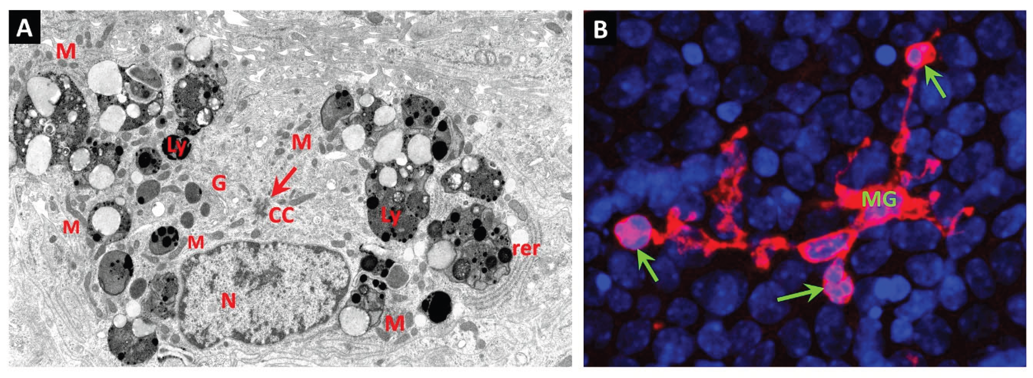
Figure 2.
(A): Digest preparation of retinal vasculature from 5-year diabetic dog depicts capillary bed showing a pericyte “ghost” within the vascular basement membrane (arrow). The ghost cell is stained magenta with the PAS reaction and contains a hematoxylin-stained remnant of nuclear chromatin. Darkly-stained nuclei of viable pericytes (P) and large pale nuclei of endothelial cells (E). (B): Digest of retinal vasculature from 7-year spontaneously diabetic dog featuring a retinal arteriole (A). A dilated portion of the arteriolar wall contains several fusiform remnants of vascular smooth muscle cells, stained magenta with PAS. (From Gardiner et al. 1994 [121]).
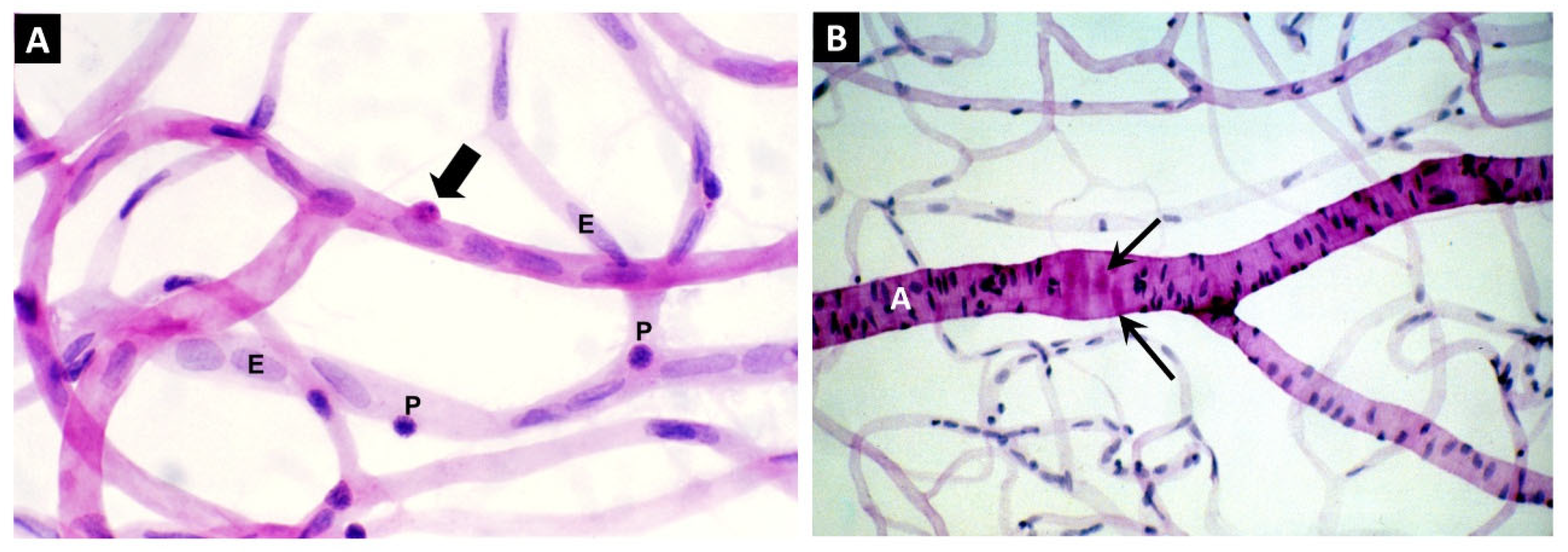
Figure 3.
(A&B): Electron micrographs of diabetic human donor retina depicts a capillary blood vessel with an intact endothelial cell (EC) and pericyte “Ghost” (PG) within the basement membrane; endothelial cell nucleus (N), vessel lumen (L), glial cell (G). The basement membrane (BM) is grossly thickened and laminated, and the pericyte “ghost” (PG) is filled with vesicular and granular material. The plasma membrane of the ghost cell appears to have disintegrated, apart from that surrounding a small pericyte process (pp). (C&D): Retinal capillary from diabetic human donor shows pericyte processes (pp) with intact plasma membranes (arrow heads in panel C) containing multiple autophagosomes. The autophagosomes contain enclosed cytoplasm and organelles (arrows). Bars 1.0 µm. (From Gardiner & Stitt 2022 [122]).
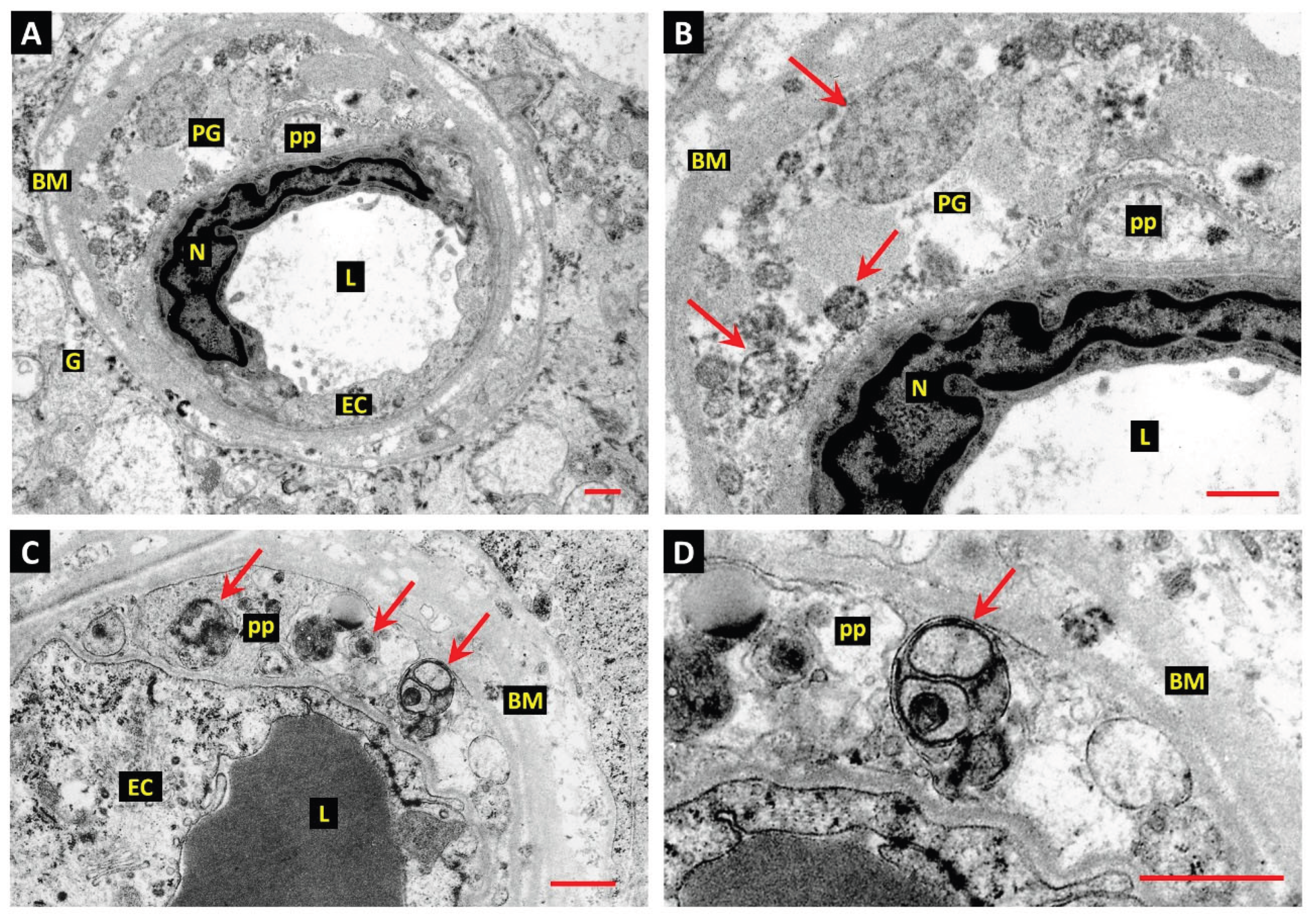
Figure 4.
(A&B): Electron micrograph of a retinal capillary from a diabetic donor eye shows a pericyte “ghost” (PG) filled with vacuolar remnants of extreme autophagic activity within the vascular basement membrane (BM). The PG is undergoing phagocytosis by a JVM cell. The process of the JVM cell engaged in phagocytosis has infiltrated the BM but shows cytoplasmic continuity with the cell of origin (thick arrow). An adjacent process of the JVM cell shows anchorage to the BM via a well-developed hemi-desmosome-like attachment plaque (arrow, panel B). The JVM shows aggregates of rough endoplasmic reticulum at the periphery of the cell soma (RER). Vascular lumen—L; endothelial cells—EC; pericyte processes—pp; mitochondria—M; bars 1.0 µm. (C&D): A deeper Z-plane of the same retinal capillary depicted in panels A and B provides additional detail of PG phagocytosis by the JVM cell. The extremity of the JVM process engaged in PG engulfment shows the tapered double-membrane ends of phagocytic pseudopodia (thick arrows). This profile also shows a unique form of matrix attachment by the JVM: 2 of the hemidesmosome-like attachments observed in panels A and B are anchored on the same tag of basal lamina (arrows). Bars 1.0 µm. (From Gardiner & Stitt 2022 [123]).
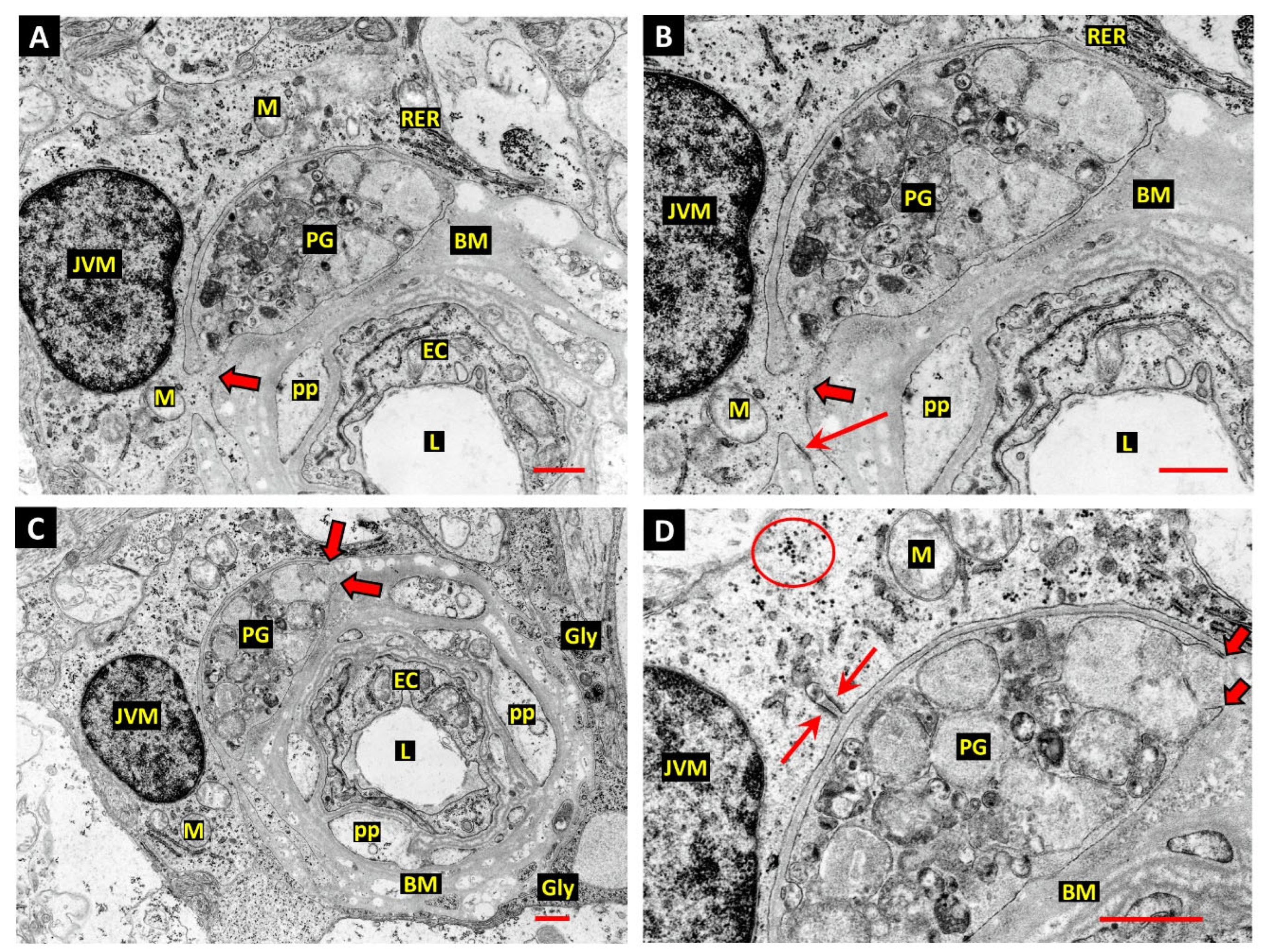
Figure 5.
(A): The wall of a retinal arteriole from a diabetic donor eye shows a JVM cell during engulfment of an apoptotic body derived from a cell thought to be engaged in apoptosis with autophagy (thick arrow). Glycogen deposits in vascular smooth muscle cells (VSMCs) are circled—see enlargements in panel E below. Bar 3.0 µm. (B): The JVM cell appears to have drawn the dying cell through a break in the outer lamina of the vascular BM (arrow, also in panel C). The cytoplasm of the dying cell contains autophagosomes containing degraded chromatin (Ch), other more typically heterogeneous material (Ap) and coarse aggregates of glycogen granules (Gy), similar to those in the adjacent viable VSMCs. Glial process—G; bar 1.0 µm. (C): This profile shows the nucleus of the phagocytic JVM cell with a group of the presumptive storage granules in the cytoplasm adjacent to the forming phagosome. Glial process—G; storage granules—sg; bar 1.0 µm. (D): Enlargement of the profile of the dying cell from panel C shows a C-shaped double membrane structure (arrow in panel D), suggesting that phagophores were still forming at the time of fixation. The JVM cell and the apoptotic body undergoing engulfment show several zones of intimate plasma membrane contact indicating the position of temporary adhesion complexes (arrow heads). Bar 1.0 µm. (From Gardiner & Stitt 2022 [123]).
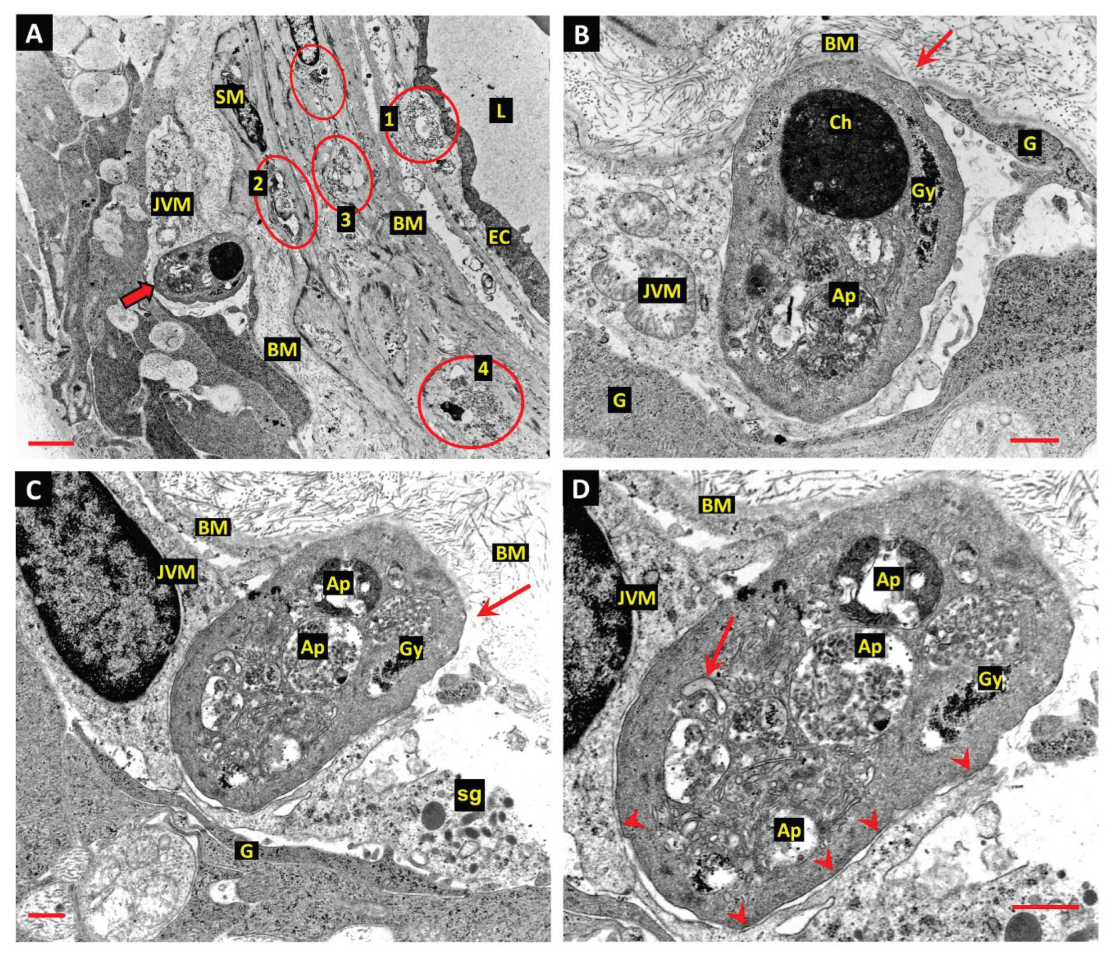
Figure 6.
(A): Normal donor retina shows the nucleus and perinuclear cytoplasm of a JVM cell in the same profile as a vascular pericyte (P). As in the diabetic retina, the cell soma and nucleus of the JVM cell typically lies adjacent to the vascular BM and the extensive participation of the JVM in the glia limitans is indicated by arrowheads. The matrices of the mitochondria in the JVM cell are unusually electron-opaque, which may be a result of fixation-delay in this particular specimen (M), and age-related BM vacuoles typical of human retinal capillaries are present (Vac). The matrices of the mitochondria in the JVM cell are unusually electron-opaque, which may be a result of fixation-delay in this particular specimen (M). Arrow—lysosome-related dense body; pericyte centrioles—C; macroglial processes—G; bar 1.0 µm. (From Gardiner & Stitt 2022 [123]). (B&C): Confocal micrographs of JVM cells in normal mouse retina stained red with anti-Iba1 and retinal capillaries stained green with Alexa-488 labelled GS isolectin-B4. The cell depicted in image-B has the soma in close apposition to the capillary basement membrane and approximates to the location of that shown in image-A. The soma of the cell in image-C lies close to the capillary wall but only has processes in contact with the vessel. Also, 2 additional microglial processes of cell/cells with soma residing outside the plane of section contact the vessel wall and appear to spread laterally to produce foot-plates similar to those of the macroglia (arrows). Bar- 20 µm. (Images C&D are original micrographs by Dr Karis Little.
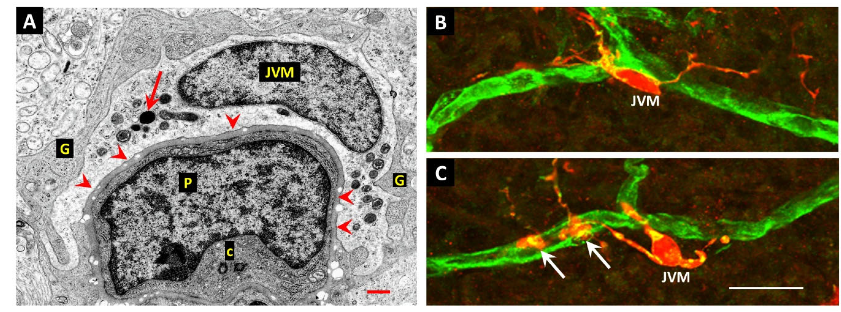
Figure 7.
Expression of efferocytosis-related gene expression in microglia and glia cell populations of the diabetic retina (8-month-old db/db—PMID: 33674409) and brain (4-month-old db/db mice- PMID: 37351595). Single-cell RNAseq datasets were accessed from the corresponding databases and analyzed using Seurat_4.3.0 (https://satijalab.org/seurat/; developed by P. Hoffman, S. Lab and collaborators, New York City, NY, USA). . (a) Retinal microglia, (b) retinal Müller cells, (c) brain microglia, (d) brain astrocytes. Changes reported as statistically significant had a Log2Fold Change >0.25. * p<0.05, ****p<0.001. Blue = diabetic group (db/db), orange = control group (db/+).
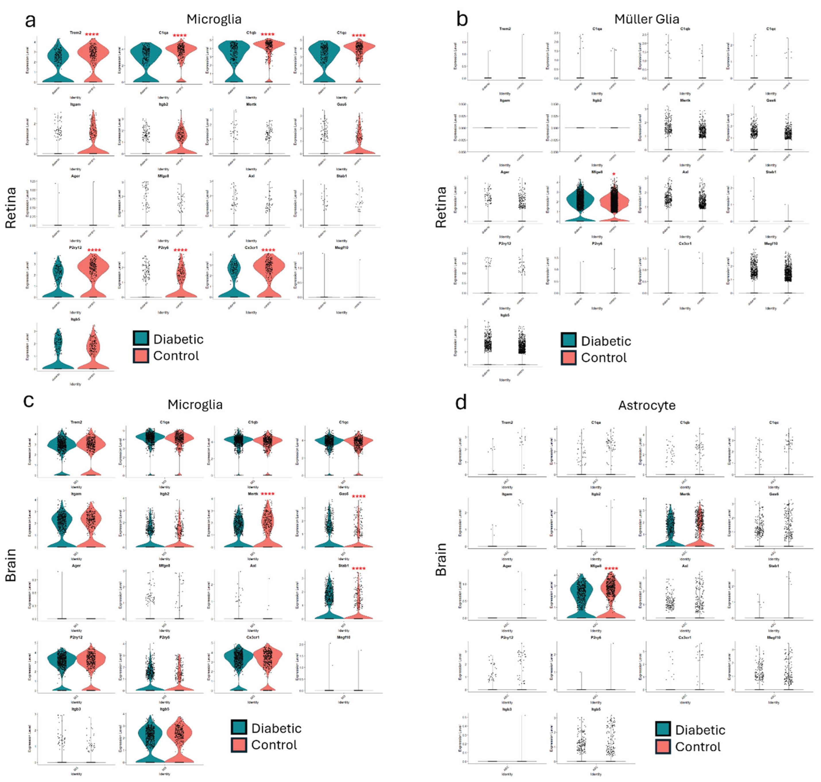
Disclaimer/Publisher’s Note: The statements, opinions and data contained in all publications are solely those of the individual author(s) and contributor(s) and not of MDPI and/or the editor(s). MDPI and/or the editor(s) disclaim responsibility for any injury to people or property resulting from any ideas, methods, instructions or products referred to in the content. |
© 2025 by the authors. Licensee MDPI, Basel, Switzerland. This article is an open access article distributed under the terms and conditions of the Creative Commons Attribution (CC BY) license (http://creativecommons.org/licenses/by/4.0/).
Copyright: This open access article is published under a Creative Commons CC BY 4.0 license, which permit the free download, distribution, and reuse, provided that the author and preprint are cited in any reuse.


