
Submitted:
24 July 2025
Posted:
25 July 2025
You are already at the latest version
Abstract
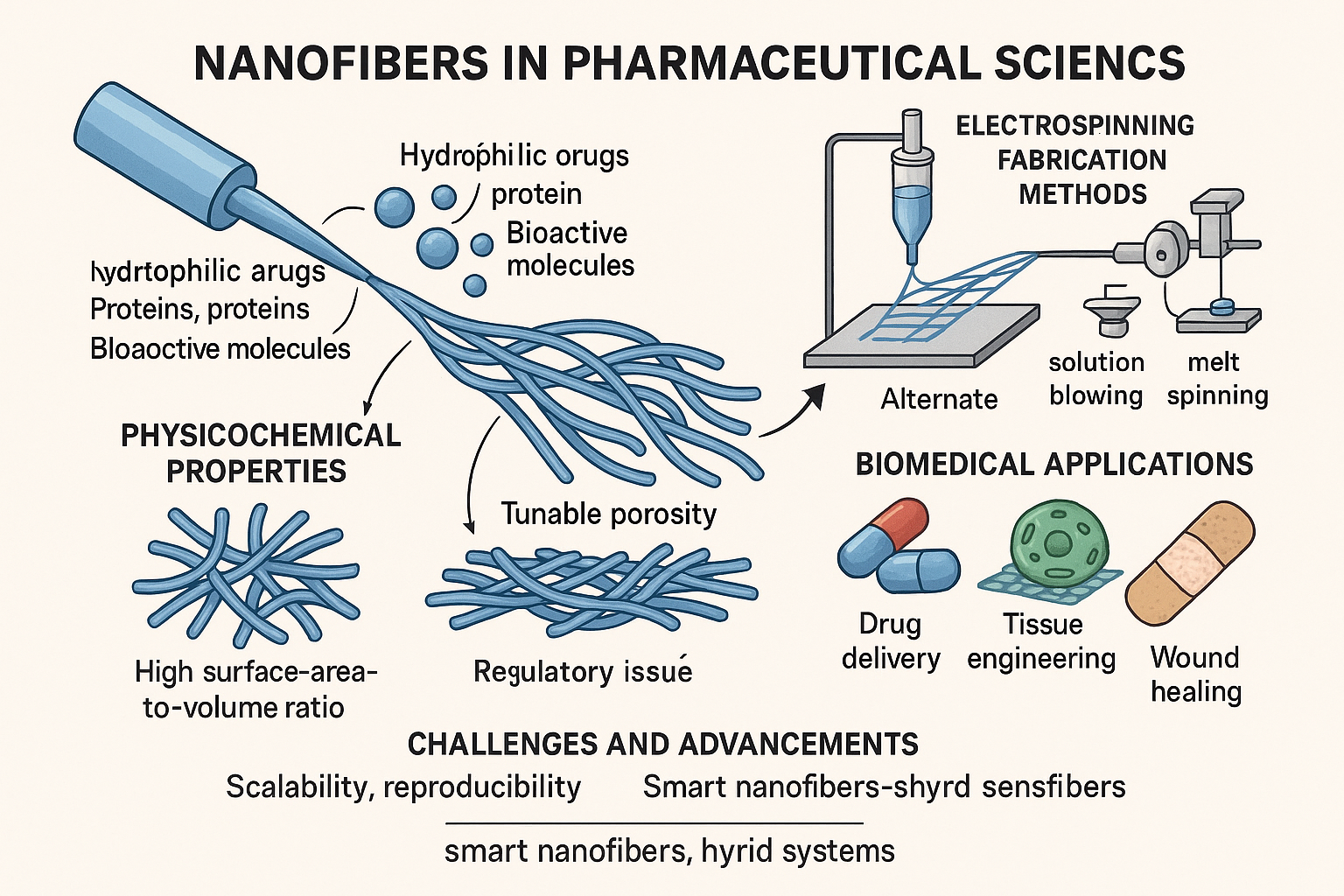
Nanofibers have emerged as a transformative platform in pharmaceutical sciences, offering unique advantages for drug delivery, tissue engineering, and wound healing. Their nanoscale structure, characterized by high surface-area-to-volume ratios and tunable porosity, enables efficient encapsulation of both hydrophilic and hydrophobic drugs, proteins, and bioactive molecules. Electrospinning remains the predominant fabrication technique, allowing for precise control over fiber morphology and drug release kinetics. Additionally, alternative methods such as solution blowing, melt spinning, and self-assembly have expanded the scope of nanofiber applications, particularly in industrial and clinical contexts.This review provides a comprehensive overview of the physicochemical properties, fabrication strategies, and biomedical applications of nanofibers, with a focus on their role in controlled and stimuli-responsive drug delivery systems. It also addresses the current limitations—such as scalability, reproducibility, and regulatory barriers—and highlights recent advancements, including smart nanofibers and hybrid constructs. Future directions emphasize the integration of nanofibers into multifunctional and patient-specific systems, underscoring their potential to reshape modern pharmaceutical and regenerative therapies.
Keywords:
Nanofibers
; Drug Delivery Systems
; Electrospinning
; Tissue Engineering
; Controlled Release
; Biopolymers
; Biomedical Applications
; Smart Nanomaterials
; Pharmaceutical Nanotechnology
; Stimuli-Responsive Systems
1. Introduction
- Introduction to Nanofibers in Pharmaceutical Applications
The advent of nanotechnology has revolutionized the landscape of pharmaceutical sciences by introducing innovative strategies for drug delivery, disease management, and tissue engineering. Among the various nanoscale drug delivery systems, nanofibers have emerged as one of the most promising candidates due to their unique physicochemical, mechanical, and biological characteristics. These one-dimensional structures, typically with diameters in the nano meter range (50–500 nm), exhibit a high surface-area-to-volume ratio, tunable porosity, and superior mechanical strength, which significantly enhance their utility in drug delivery systems[1].
Nanofibers can be engineered using various synthetic and natural polymers to achieve desirable properties such as biodegradability, biocompatibility, and controlled degradation rates, which are critical for clinical translation. One of their most notable advantages lies in their structural similarity to the extracellular matrix (ECM) of biological tissues. This ECM-like morphology facilitates cell adhesion, proliferation, and differentiation, making nanofibers excellent scaffolds for tissue regeneration, wound healing, and localized drug delivery[2].
In pharmaceutical applications, nanofibers are commonly fabricated using the electrospinning technique, which enables the production of uniform fibers from a wide variety of polymers. Drugs can be loaded into nanofibers by blending with the polymer solution prior to spinning (encapsulation) or by post-spinning surface modification. These delivery platforms are capable of encapsulating both hydrophilic and hydrophobic drugs, proteins, peptides, and even nucleic acids, thereby expanding their therapeutic utility[3].
One of the key benefits of nanofiber-based drug delivery systems is their ability to provide controlled and sustained release of therapeutic agents. By modifying polymer composition, fiber diameter, and drug-polymer interactions, the release kinetics can be finely tuned to match the pharmacological needs of the patient. Additionally, targeted delivery can be achieved by functionalizing the surface of nanofibers with ligands, peptides, or antibodies that recognize specific cellular markers. This not only enhances therapeutic efficacy but also minimizes systemic toxicity and side effects, which are commonly associated with conventional dosage forms[4].
Moreover, the versatility of nanofibers extends beyond drug delivery. They are also used in diagnostic systems, bioactive wound dressings, and as platforms for tissue engineering, particularly in areas such as bone regeneration, neural repair, and cardiac patching. The integration of stimuli-responsive materials has further advanced their functionality, enabling on-demand drug release in response to environmental triggers like pH, temperature, or enzymatic activity[5].
In conclusion, nanofibers represent a transformative advancement in pharmaceutical technology. Their multifunctional capabilities, combined with scalability and customizable properties, make them a pivotal tool in the development of next-generation therapeutic systems. As research continues to evolve, it is anticipated that nanofiber-based systems will become increasingly prevalent in clinical settings, offering personalized, effective, and safer treatment options for a wide range of diseases.
2. Fabrication Techniques of Nanofibers
2.1. Electrospinning[6,7,8]
Electrospinning is the most common and versatile method for nanofiber production. It involves applying a high-voltage electric field to a polymer solution, which forms a Taylor cone and jets out fine fibers that solidify on a collector of electrospinning parameters and their effect were in Table 1.
2.2. Alternative Fabrication Techniques for Nanofibers
While electrospinning remains the most widely used and studied technique for nanofiber fabrication, several alternative methods have gained prominence due to their unique advantages in scalability, cost-effectiveness, and solvent-free processing. Among these, solution blowing, melt spinning, and phase separation/self-assembly techniques are gaining traction in pharmaceutical and biomedical fields.
- Solution Blowing[9]
Solution blowing is a fiber fabrication technique that combines the principles of electrospinning and melt blowing, but without the use of high-voltage electrical fields. In this method, a polymer solution is delivered through a nozzle and is subjected to high-velocity compressed gas or air, which stretches the polymer into fine fibers. These fibers are then collected on a target surface to form non-woven mats or membranes.
Key advantages of solution blowing include:
- No high-voltage requirements, enhancing safety and simplifying equipment needs.
- Higher production rates compared to electrospinning, making it more suitable for industrial scaling.
- The ability to use a wide range of biocompatible polymers and drug-loaded solutions.
- The technique is particularly effective for generating fibers with diameters in the micro- to nanoscale range, although achieving consistent nanometer-level diameters may require optimization.
Solution blowing is promising for creating drug-loaded wound dressings, scaffolds for tissue engineering, and nanofiber mats for transdermal or topical drug delivery. Its ability to avoid the use of strong electric fields also makes it suitable for encapsulating sensitive biological molecules like proteins, enzymes, or live cells.
- 2.
Melt spinning is a solvent-free fabrication method, primarily used in textile and polymer industries, and is now adapted for pharmaceutical applications. Instead of using a polymer solution, this method employs polymer melts, which are extruded through a spinneret and rapidly solidified via cooling to form continuous fibers.
-
Advantages of melt spinning:
- Environmentally friendly and solvent-free, avoiding toxicity concerns and the need for post-processing to remove residual solvents.
- Well-established in manufacturing with high-throughput capability, making it ideal for industrial-scale production.
- Suitable for thermoplastic polymers such as polycaprolactone (PCL), polylactic acid (PLA), and polyethylene glycol (PEG).
-
Challenges:
- Limited to polymers with a defined melting point or thermal processability.
- High processing temperatures may restrict the inclusion of heat-sensitive drugs or biomolecules.
- Control over fiber diameter is less precise compared to electrospinning.
Melt-spun nanofibers are being explored for implantable drug delivery systems, suturable scaffolds, and long-term controlled release implants due to their structural robustness and solvent-free nature.
- 3.
Phase separation and self-assembly are bottom-up fabrication approaches that rely on the intrinsic physicochemical interactions of polymeric materials to spontaneously organize into nanofibrous architectures.
- Phase Separation typically involves dissolving a polymer in a solvent, followed by temperature-induced phase demixing (usually by lowering the temperature). The solvent is then removed by freeze-drying, leaving behind a porous nanofibrous matrix.
- Self-Assembly refers to the process by which molecules (e.g., peptides, amphiphilic polymers) spontaneously organize into ordered nanostructures due to non-covalent interactions such as hydrogen bonding, van der Waals forces, and ionic interactions.
-
Benefits[18]:
- Exceptional control over fiber architecture and morphology, enabling the design of fibers with bio-mimetic structures.
- Suitable for delicate biopolymers like collagen, chitosan, and self-assembling peptides.
- Allows the integration of bioactive cues, such as cell-adhesion peptides or growth factors, within the nanofiber matrix.
-
Limitations:
- Low scalability and long processing times, which hinder mass production.
- Often requires complex processing conditions and highly pure reagents.
- Applications:
These techniques are ideal for highly specialized tissue engineering scaffolds, nerve regeneration conduits, and intracellular drug delivery systems where fine control over fiber architecture and bifunctionality is paramount the setup for fiber formation mechanism was shown in Figure 1.
3. Physicochemical Properties of Nanofibers [19,20]
Nanofibers possess features that make them ideal for pharmaceutical applications: High surface area for efficient drug loading - Interconnected porosity for enhanced permeability -Biodegradability and biocompatibility - Tunable mechanical strength and some common polymers used in the nanofibers were given in Table 2.
4. Pharmaceutical Applications [21,22,23]
4.1. Drug Delivery Systems Nanofibers are excellent carriers for both hydrophilic and hydrophobic drugs. They enable: - Immediate release for rapid therapeutic effect - Sustained release over prolonged periods - Controlled release triggered by pH, temperature, or enzymes
4.1. Drug Delivery Systems
Nanofibers are exceptional carriers for both hydrophilic and hydrophobic drugs, thanks to their high surface-area-to-volume ratios and tunable structures
Immediate Release: Drugs near the fiber surface diffuse rapidly into surrounding fluids, ideal for fast therapeutic action (e.g., pain management, analgesics)
Sustained Release: Embedding drugs within the nanofiber matrix and using biodegradable polymers enables gradual release over days or weeks. Studies demonstrate release rates extending for days, even months, from such matrices or hydrogel-fiber composites.
Stimuli-Responsive/Controlled Release: Integration of pH-,temperature-, or enzyme-responsive polymers like PNIPAAm or ion-sensitive materials enables on-demand release in response to environmental signals (e.g., acidic tumor microenvironments, inflammation) . The Drug molecules are encapsulated within or coated onto fibers, and release kinetics vary depending on polymer type, fiber architecture, and external triggers were shown in the Figure 2.
4.2. Wound Healing
Nanofiber dressings support improved healing outcomes by:
- Maintaining moisture balance, facilitating autolytic debridement and reducing scarring.
- Blocking bacterial infiltration due to ultrafine fiber structure and high porosity
- Delivering embedded therapeutics—such as antibiotics, silver nanoparticles, or growth factors—directly into the wound bed, enhancing local efficacy while minimizing systemic exposure.
For example, studies using gelatin–chitosan–PCL nanofiber mats enriched with silver-doped carbon quantum dots showed accelerated closure in vivo compared to controls
4.3. Tissue Engineering [24]
Electro spun scaffolds closely mimic the extracellular matrix (ECM), assisting with:
- Cell adhesion, due to nanoscale fibrous topology and biochemical cues.
- Cellular proliferation and differentiation are aided by biochemical environment and mechanical support. These scaffolds have enabled regeneration across multiple tissues: skin, cartilage, bone, and neural pathways. Notably, aligned nanofibers foster axonal guidance in nerve repair and improve skeletal tissue regeneration
4.4. Biosensing and Diagnostics [25,26]
Nanofibers functionalized with recognition elements (e.g., enzymes, aptamers, antibodies) excel in biosensing thanks to:
- High surface area, enabling dense probe immobilization.
- Rapid analyte diffusion, yielding fast and sensitive signal output, for applications like glucose monitoring or pathogen detection. For instance, nanodiamond–silk fibroin electro spun membranes act as both wound dressings and integrated biosensors for temperature and bacterial activity
5. Recent Advancements [27]
- Smart Nanofibers: Incorporate stimuli-responsive components (e.g., PCL/PNIPAAm composites) to regulate hydrophilicity or drug release based on temperature or pH
- Hybrid Systems: Meld fibers with nanoparticles or hydrogels to enable multifunctionality—drug delivery plus sensing, imaging, or mechanical reinforcement
- 3D-Printed Scaffolds: Combine electrospun nanofibers with 3D printing to build architecturally precise constructs, ideal for patient-specific tissue implants the types of Nanofiber-Based Applications in Pharmaceutics were shown in the Figure 3.
6. Challenges and Limitations [28]
- Reproducibility: Electrospinning is sensitive to small menu shifts—solution properties, voltage, ambient conditions—which can alter fiber quality
- Scalability: Standard setups are low-throughput; industrial scalability via multi-jet systems faces operational variability and fiber uniformity issues.
- Sterilization: Techniques like gamma irradiation or ethylene oxide may degrade fiber structure or drug components.
- Regulatory Hurdles: Clinical adoption is limited; only topical nanofiber dressings have reached the market, while injectable and systemic systems require extensive safety and efficacy data.
7. Regulatory and Commercial Considerations [29]
Nanofiber-based dressings (e.g., antibiotic-loaded bandages) have achieved regulatory approval and market entry. However, systemic delivery via nanofibers needs:
- Comprehensive preclinical data on pharmacokinetics and toxicity.
- Controlled manufacturing processes under Good Manufacturing Practice (GMP).
- Clear regulatory pathways, addressing nanomedicine-specific concerns regarding biodegradation, metabolism, and long-term safety.
8. Future Perspectives [30]
- Scalable Manufacturing: Development of high-throughput, continuous processes (e.g., multi-nozzle or melt/electro-blowing hybrids).
- Multifunctional/Personalized Systems: Combining imaging agents, sensors, and targeted drug delivery in a single nanofiber platform.
- In Vivo Validation: Conducting large animal and phase-I/II clinical trials to move from prototypes to approved medical products.
- Regulatory Pathways: Establishing standardized characterization, performance criteria, and regulatory guidelines tailored to nanofiber-based systems.
Conclusion:
Nanofiber technology stands at the forefront of next-generation pharmaceutical innovation, offering a versatile and effective platform for targeted, controlled, and sustained drug delivery. Through advanced fabrication techniques such as electrospinning, solution blowing, melt spinning, and self-assembly, nanofibers can be tailored with remarkable precision to meet diverse therapeutic needs. Their high surface-area-to-volume ratio, tunable porosity, and biomimetic properties enable efficient encapsulation and release of a wide range of therapeutic agents—from small molecules to biologics.
The pharmaceutical utility of nanofibers extends well beyond conventional drug delivery. As demonstrated, they play vital roles in wound healing, tissue regeneration, and diagnostic applications due to their close resemblance to the extracellular matrix and ability to support cellular interactions. Integration of smart materials has further pushed the boundaries, enabling stimuli-responsive systems capable of releasing drugs on demand in response to physiological triggers.
Despite these advantages, certain challenges persist, including issues related to reproducibility, large-scale manufacturing, sterilization, and regulatory approval—particularly for systemic applications. However, ongoing advancements in fabrication technologies, such as multi-jet electrospinning and hybrid systems, are paving the way for scalable production and multifunctional integration.
Looking ahead, the convergence of nanofiber technology with personalized medicine, 3D printing, and biosensing holds tremendous potential to revolutionize therapeutic interventions. As regulatory frameworks evolve and in vivo validations progress, it is expected that nanofiber-based platforms will transition from experimental prototypes to clinically approved therapies. Their multifaceted capabilities position them as indispensable tools in the future landscape of precision and regenerative medicine, with the promise to significantly enhance patient outcomes and redefine standards in pharmaceutical care.
References
- Bhardwaj N, Kundu SC. Electrospinning: a fascinating fiber fabrication technique. Biotechnol Adv. 2010;28(3):325–47. [CrossRef]
- Ramakrishna S, Fujihara K, Teo WE, Yong T, Ma Z, Ramaseshan R. Electrospun nanofibers: solving global issues. Mater Today. 2006;9(3):40–50. [CrossRef]
- Huang ZM, Zhang YZ, Kotaki M, Ramakrishna S. A review on polymer nanofibers by electrospinning and their applications in nanocomposites. Compos Sci Technol. 2003;63(15):2223–53. [CrossRef]
- Greiner A, Wendorff JH. Electrospinning: a fascinating method for the preparation of ultrathin fibers. Angew Chem Int Ed Engl. 2007;46(30):5670–703.
- Xue J, Wu T, Dai Y, Xia Y. Electrospinning and electrospun nanofibers: methods, materials, and applications. Chem Rev. 2019;119(8):5298–415. [CrossRef]
- Agarwal S, Wendorff JH, Greiner A. Use of electrospinning technique for biomedical applications. Polymer. 2008;49(26):5603–21. [CrossRef]
- Kenry, Lim CT. Nanofiber technology: current status and emerging developments. Prog Polym Sci. 2017;70:1–17. [CrossRef]
- Homayoni H, Ravandi SAH, Valizadeh M. Electrospinning of chitosan nanofibers: processing optimization. Carbohydr Polym. 2009;77(3):656–61. [CrossRef]
- Dash TK, Konkimalla VB. Poly-ε-caprolactone-based formulations for drug delivery and tissue engineering: a review. J Control Release. 2012;158(1):15–33. [CrossRef]
- Gopaiah KV, Krishna KS, Vani P, Sireesha S, Kumar V. Nanoparticle-based delivery system for sustained release of antihypertensive drugs. Asian J Pharm Clin Res. 2022;15(9):150–155.
- Kaur IP, Kanwar M. Ocular preparations: the formulation approach. Drug Dev Ind Pharm. 2002;28(5):473–93. [CrossRef]
- Yang X, Shah JD, Wang H. Nanofiber enabled layer-by-layer approaches for drug delivery. J Control Release. 2019;307:196–205.
- Li D, Xia Y. Electrospinning of nanofibers: reinventing the wheel? Adv Mater. 2004;16(14):1151–70.
- Liao I-C, Chew SY, Leong KW. Aligned core–shell nanofibers delivering bioactive proteins. Nanomedicine. 2006;1(4):465–71. [CrossRef]
- Sill TJ, von Recum HA. Electrospinning: applications in drug delivery and tissue engineering. Biomaterials. 2008;29(13):1989–2006.
- Gopaiah KV, Teja RM, Kumar YS, Prasad PVS, Kumar Y. Formulation and Evaluation of Enzalutamide Cyclodextrin Nanosponges Tablets: In vitro Dissolution Study and In vivo Pharmacokinetics. J Appl Pharm Sci. 2025;15(5): xx–xx. [In Press].
- Zhang Y, Lim CT, Ramakrishna S, Huang ZM. Recent development of polymer nanofibers for biomedical and biotechnological applications. J Mater Sci. 2005;16(10):933–46. [CrossRef]
- Zhu Y, Gao C, Liu X, Shen J. Surface modification of polycaprolactone membrane via entrapment of chitosan-based multilayer for promoting cytocompatibility of human fibroblasts. Biomaterials. 2002;23(24):4889–95.
- Ma Z, Kotaki M, Yong T, He W, Ramakrishna S. Surface engineering of electrospun polyethylene oxide nanofibers for biological application. Macromol Rapid Commun. 2005;26(5):321–6.
- Bhardwaj N, Chouhan D, Mandal BB. Tissue engineered skin and wound healing: current strategies and future directions. Curr Pharm Des. 2017;23(24):3455–70. [CrossRef]
- Venugopal J, Low S, Choon AT, Ramakrishna S. Interaction of cells and nanofiber scaffolds in tissue engineering. J Biomed Mater Res B Appl Biomater. 2008;84B(1):34–48. [CrossRef]
- Katti DS, Robinson KW, Ko FK, Laurencin CT. Bioresorbable nanofiber-based systems for wound healing and drug delivery: optimization of fabrication parameters. J Biomed Mater Res B Appl Biomater. 2004;70(2):286–96. [CrossRef]
- Kim K, Luu YK, Chang C, Fang D, Hsiao BS, Chu B, Hadjiargyrou M. Incorporation and controlled release of a hydrophilic antibiotic using poly(lactide-co-glycolide)-based electrospun nanofibrous scaffolds. J Control Release. 2004;98(1):47–56. [CrossRef]
- Zhang C, Yuan X, Wu L, Han Y, Sheng J. Study on morphology of electrospun poly(vinyl alcohol) mats. Eur Polym J. 2005;41(3):423–32. [CrossRef]
- Emamjomeh SM, Amani A. Biosensors using electrospun nanofibers: recent advances and future directions. Biosens Bioelectron. 2021;171:112722.
- Ghaffari M, Irani M, Atai M, Fekrazad R. Controlled release of metronidazole from polycaprolactone nanofibers for oral wound dressing. Dent Res J. 2016;13(6):504–10.
- Gholipourmalekabadi M, Mozafari M, Ranjbar-Mohammadi M, et al. Electrospun silk fibroin/gelatin nanofibrous scaffolds: from material characterization to in vivo biocompatibility. Nanomedicine. 2018;13(18):2341–55.
- Gopaiah KV, Teja RM, Goud NR, Reddy YP, Reddy BS. Nanostructured Emulsomes for the Enhanced Delivery of Cabazitaxel: A Novel Approach to Cancer Treatment and Drug Release Profiling. Preprints. 2025;2025:161188.
- Chew SY, Wen Y, Dzenis Y, Leong KW. The role of electrospun nanofibers in stem cell biology. Biomaterials. 2006;27(36):5731–42.
- Sundarrajan S, Tan KL, Lim SH, Ramakrishna S. Electrospun nanofibers for air filtration applications. Procedia Eng. 2014;75:159–63. [CrossRef]
Figure 1.
Overview of Electrospinning Setup and Fiber Formation Mechanism.
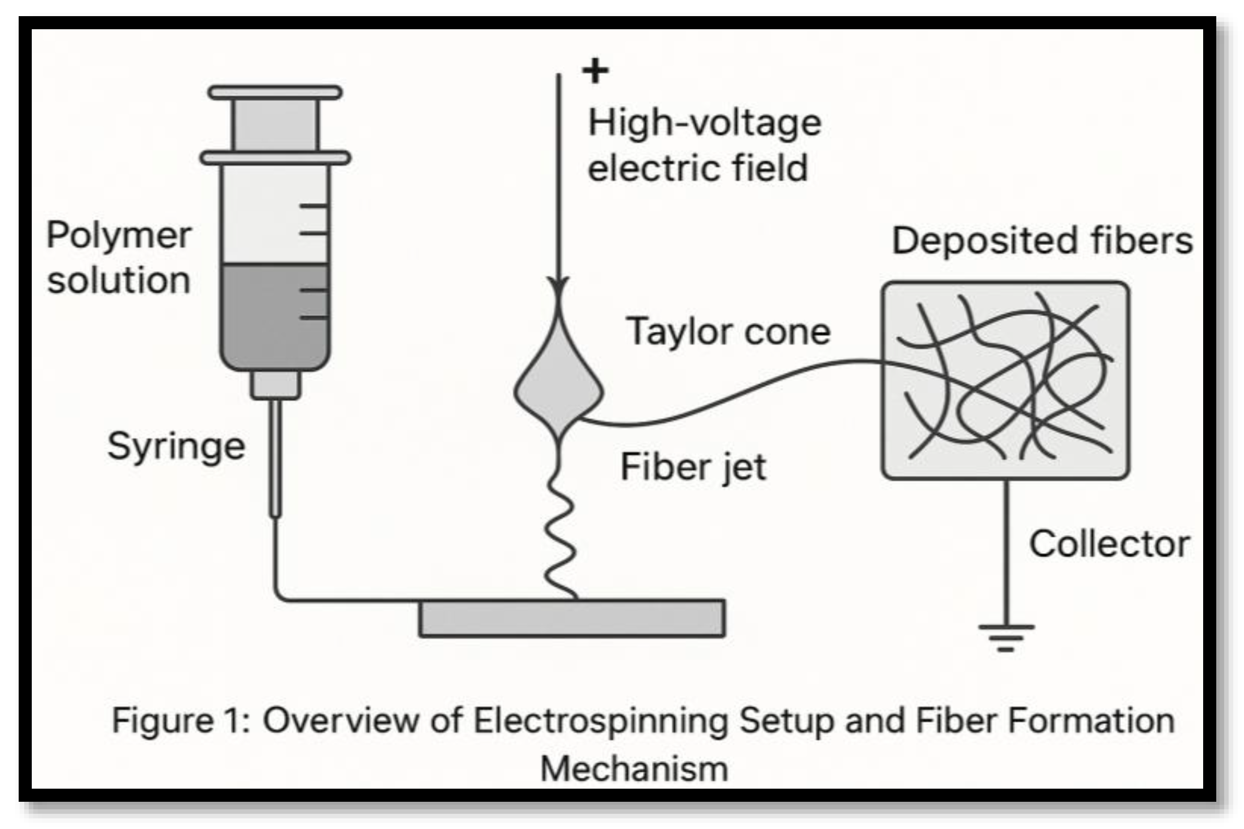
Figure 2.
Drug molecules are encapsulated within or coated onto fibers, and release kinetics vary depending on polymer type, fiber architecture, and external triggers.
Figure 2.
Drug molecules are encapsulated within or coated onto fibers, and release kinetics vary depending on polymer type, fiber architecture, and external triggers.
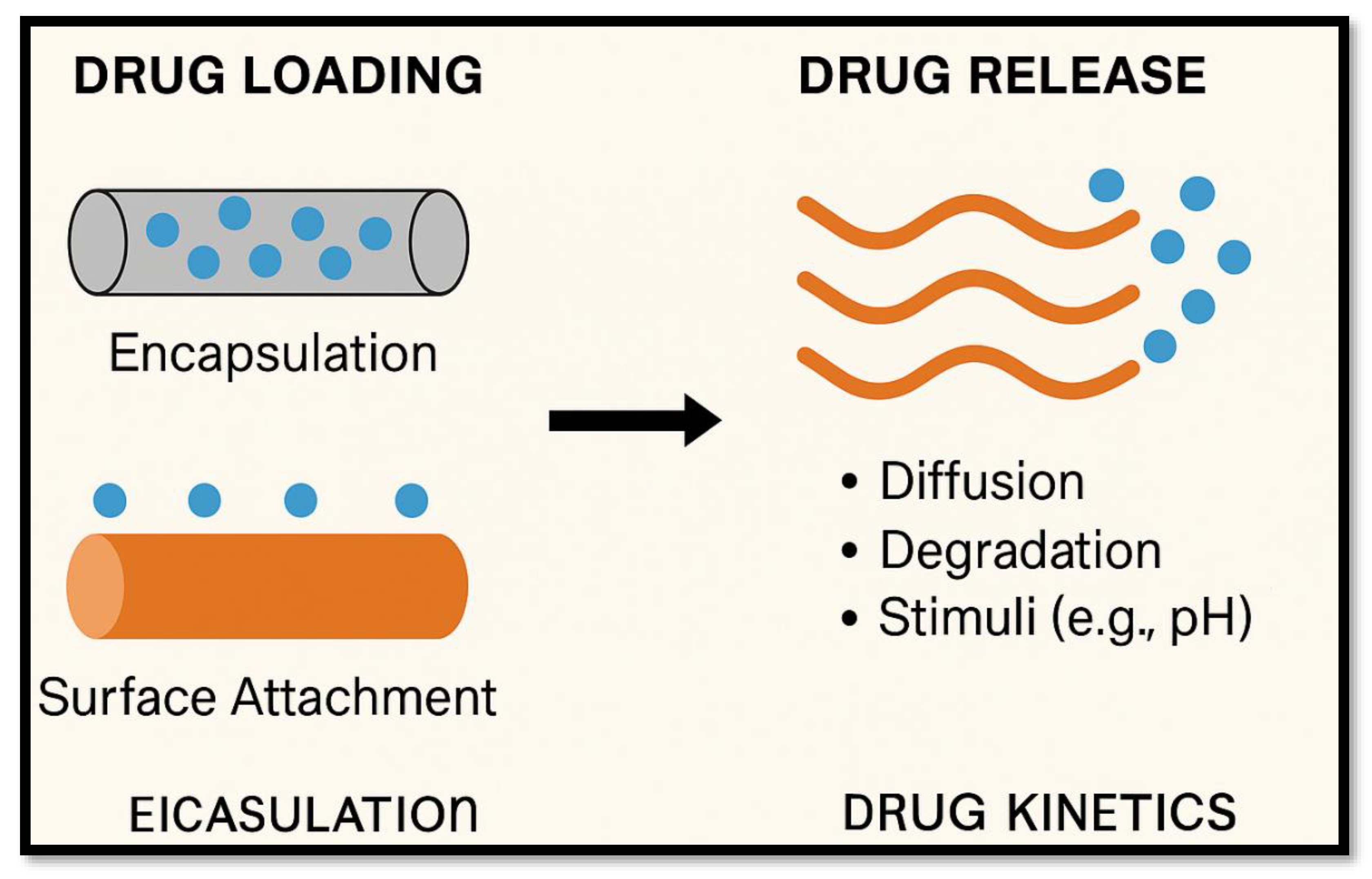
Figure 3.
Types of Nanofiber-Based Applications in Pharmaceutics.
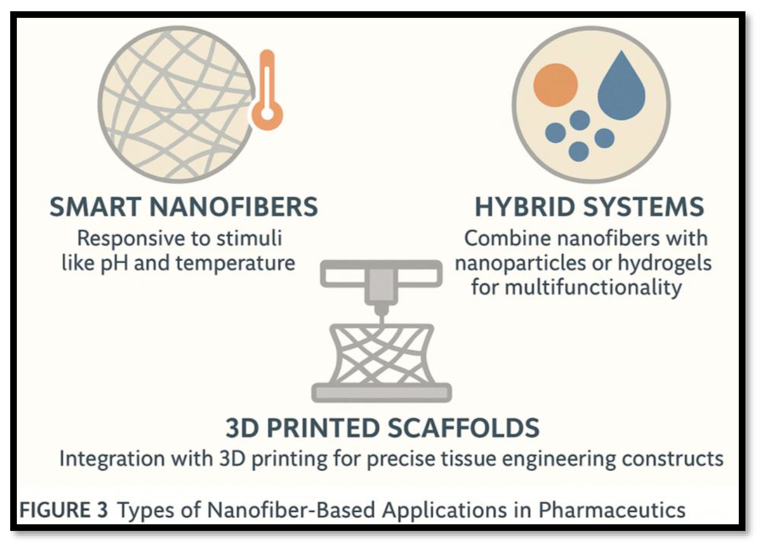

Table 1.
Electrospinning Parameters and Their Effects.
| S. NO | Parameter | Effect |
|---|---|---|
| 1 | Voltage | Affects fiber diameter and uniformity. |
| 2 | Polymer concentration | Controls solution viscosity and spinnability, impacting fiber formation. |
| 3 | Flow rate | Influences fiber morphology and diameter consistency. |
| 4 | The distance between the needle and the collector | Impacts the solvent evaporation rate and fiber formation. |
Table 2.
Common Polymers Used in Nanofibers.
| S. NO | Polymer | Type | Application |
|---|---|---|---|
| 1 | Polycaprolactone (PCL) | Biodegradable | Sustained drug delivery |
| 2 | Polylactic acid (PLA) | Biodegradable | Tissue engineering |
| 3 | Polyvinyl alcohol (PVA) | Water-soluble | Wound healing |
| 4 | Chitosan | Natural | Antibacterial dressing |
Disclaimer/Publisher’s Note: The statements, opinions and data contained in all publications are solely those of the individual author(s) and contributor(s) and not of MDPI and/or the editor(s). MDPI and/or the editor(s) disclaim responsibility for any injury to people or property resulting from any ideas, methods, instructions or products referred to in the content. |
© 2025 by the authors. Licensee MDPI, Basel, Switzerland. This article is an open access article distributed under the terms and conditions of the Creative Commons Attribution (CC BY) license (http://creativecommons.org/licenses/by/4.0/).
Copyright: This open access article is published under a Creative Commons CC BY 4.0 license, which permit the free download, distribution, and reuse, provided that the author and preprint are cited in any reuse.


