Submitted:
11 July 2025
Posted:
14 July 2025
You are already at the latest version
Abstract
Background: Gold nanoparticles (GNPs) are increasingly studied for their potential to enhance wound healing, but their overall efficacy remains uncertain. Methods: We conducted a systematic meta-analysis (search date: May 14th, 2025) across five databases. Included were randomized animal studies comparing GNPs to placebo, reporting wound closure percentages and relevant variance measures. Risk of bias was assessed using Cochrane and CAMARADES tools. Cohen’s d was used to estimate effect size under a random-effects model. Results: Thirty-one studies met the inclusion criteria. The pooled effect size was d = 4.52 (95% CI: 3.61 to 5.43; z = 9.73; p < 0.001), indicating a significant benefit of GNP. Although heterogeneity was moderate to high, results consistently favored GNPs. Conclusion: GNPs significantly accelerate wound healing in animal models, supporting their potential as therapeutic agents.
Keywords:
Gold nanoparticles
; wound healing
; animal study
; meta-analysis
1. Introduction
Wound healing restores tissue integrity and function through a complex interplay between cells, growth factors, and the extracellular matrix [1,2]. Key aspects of wound healing include physiological, nutritional, and management factors that are essential for optimal recovery [3,4,5,6]. The global advanced wound care market is expected to grow significantly, reaching approximately 19 billion dollars by 2027 [7]. Despite advancements in medical research, effectively treating chronic and acute wounds remains a major therapeutic challenge [7,8]. In recent decades, there has been a growing interest in the use of nanoparticles in medical therapies. Gold nanoparticles (GNPs) have received particular attention as delivery systems for therapeutic agents. Numerous studies have demonstrated their efficacy in promoting wound closure [9,10]. GNPs have emerged as a promising therapeutic option due to their distinct physicochemical properties and biological activity, proving effective across various models and applications [11,12].
Chronic wounds, or non-healing wounds, present a significant global health challenge, primarily due to infections caused by bacteria and fungi [13]. Among these, diabetic foot and leg ulcers are particularly burdensome, with a prevalence estimated at 2.21 per 1,000 people [7,14]. These wounds are associated with substantial morbidity and have a notable impact on both patient quality of life and health-care systems [14]. The incidence of non-healing wounds is increasing, driven by changing lifestyle factors and an ageing population [15,16].
Diabetic ulcers are becoming increasingly common as the population ages and diabetes prevalence increases. These ulcers lead to significant patient disability, higher mortality rates, and considerable economic costs [17,18]. Factors contributing to their development include neuropathy, limited range of motion, pressure, ischemia, and infection. Approximately 15% of individuals with diabetes may develop an ulcer at some point in their lives, with a high likelihood of recurrence [19,20]. Additionally, more than half of foot ulcers become infected, and infections are the leading cause of lower extremity amputations [21]. Despite all the available diagnostic and therapeutic tools, the treatment for diabetic foot ulcers is the rate and progression of healing [22,23].
Pressure injuries, also known as pressure ulcers, are localized damage to skin and underlying tissue, typically over a bony prominence. These injuries result from prolonged pressure or a combination of pressure and shear forces [24]. Risk factors include advanced age, malnutrition, and certain medical conditions [25]. Pressure injuries represent a significant global challenge in healthcare settings, particularly in hospitals [26]. The pooled estimate of pressure injury incidence is approximately 12%, with Europe reporting the highest prevalence and incidence rates [26,27].
Venous leg ulcers are open wounds on the lower leg, typically between the ankle and mid-calf, that persist for over six weeks. They are caused by venous blood flow, which leads to increased venous pressure in the attached area [28]. Venous ulcers make up approximately 70% of chronic leg ulcers, leading to a significant economic burden due to lengthy treatment times and high recurrence rates [29,30,31].
In this review we focus on covering all forms of cutaneous wounds, as they present significant challenges in wound healing, particularly in relation to severe conditions, as mentioned above. We address both excisional procedures, which are critical in the management and treatment of these types of wounds. The management of full-thickness wounds, characterized by the loss of both the epidermal and dermal layers, is critically important due to the significant impact that an impaired skin barrier can have on overall health [32]. In contrast, superficial wounds typically involve damage confined to the epidermis [33]. In current clinical practice for addressing severe burns and chronic wounds, surgical intervention is often recommended as a primary treatment strategy [34].
Gold nanoparticles have been shown to inhibit transforming growth factors, inflammatory markers, and angiogenesis markers [35,36]. Additionally, these nanoparticles exhibit antibacterial and antioxidant effects [37,38]. These combined properties are essential for promoting wound healing and preventing wound infection [39,40]. Recent studies indicate that gold microparticles show promise in providing pain relief and modulating inflammation in the management of knee- and hip osteoarthritis [41,42,43,44]. These gold nano- or microparticles appear to influence cellular signaling pathways involved in tissue repair and regeneration [43,45,46,47,48]. Additionally, gold nanoparticles have been shown to enhance collagen formation and organization, leading to improved wound closure and less scarring [49,50].
GNPs come in a variety of shapes and sizes, each affecting their efficacy in wound healing [50,51]. The most common shapes include spherical, rod-shaped, and caged nanoparticles. Spherical GNPs are known for their excellent biocompatibility and cellular uptake, while rod-shaped GNPs provide superior tissue penetration [52,53]. Caged GNPs have a higher drug-loading capacity, making them advantageous for targeted delivery, particularly in photothermal ablation compared to larger GNPs [54]. Triangular GNPs exhibit strong antibacterial properties, and star-shaped GNPs demonstrate high photothermal conversion efficiency [55,56].
GNPs can be administered to wounds using a variety of methods. One effective approach is the use of gold nanoparticle-infused hydrogels, which possess numerous bioactive properties and create a microenvironment that enhances wound healing [36,48]. Injectable formulations, such as thermosensitive hydrogels, provide improved tissue penetration [57]. Biocompatible scaffolds, like electrospun nanofiber materials infused with GNPs, not only offer structural support but also facilitate the distribution of nanoparticles. Electrospinning can deliver specific physical, chemical, and biological signals that accelerate the wound healing process [58,59]. Two innovative strategies include microneedle patches, which enhance skin penetration [60,61,62] and spray-based systems for uniform distribution across large wound areas [63,64]. The choice of delivery mechanism is determined by the wound's characteristics and the desired therapeutic outcomes.
Despite promising results with gold nanoparticles in cell culture studies and some animal trials, there is still a significant lack of information regarding systematic evaluation of their efficacy in wound healing. Most studies have been limited to in vitro models or short-term in vivo experiments. This highlights the need for comprehensive meta-analyses that can aggregate data from multiple studies to provide a clearer understanding of the effectiveness of GNPs in wound healing [12]. To our knowledge, there has been no systematic meta-analysis comparing the clinical efficacy of gold nanoparticle therapy on wound healing in randomized controlled trials involving animals.
This systematic review aimed to address the central research question: “What is the overall effect of gold nanoparticles on wound healing in animal models?”. Additionally, we aimed to discuss the implications of our findings for clinical applications and future research. We hypothesized that gold nanoparticles significantly enhance the wound healing process in animal models by promoting healing.
2. Materials and Methods
Reporting and registration
This report follows the PRISMA guidelines [65]. The protocol was registered in PROSPERO, the international register of systematic reviews (registration number: 622000), on December 3, 2024, and approved April 9, 2025 (PROSPERO 2025 CRD420250622000). The completed PRISMA checklist is provided in the Supplementary Materials (S1)
Information Sources and Search Strategy
This study identified randomized controlled trials that investigated the effect of gold nanoparticles compared to a placebo control in the treatment of wound healing in animals. A systematic search was performed in the following databases: PubMed (MEDLINE), Embase, Cochrane, Scopus and Web of Science, without restriction on language or date of publication. Three categories were used: wound healing, wound closure and gold nanoparticle treatment. The search was conducted by SK and SR, on May 14th, 2025. Detailed search strategies for each database are presented in Appendix A (Table A1).
Study selection
Duplicate entries were manually identified and removed by two reviewer (SK and SR) and independently performed the screening of eligible studies in two steps based on the PRISMA guidelines [65]. In the first step, all titles and abstracts were screened according to the predefined inclusion and exclusion criteria. The inclusion criteria consisted of animal randomized controlled studies that compared the effects of gold nanoparticle administration on wound healing outcomes to a placebo, with quantifiable data (SD or SEM) required for each group. In case of conflicts, both reviewers (SK and SR) re-evaluated the title and abstract and came to a mutual consensus after discussion.
In the second step, the two reviewers individually read all full-text articles included in the previous step. In addition to the formerly mentioned inclusion criteria, the final inclusion criteria were animal studies investigating the effects of gold nanoparticle administration on wound healing, compared to a placebo, specifically for measuring wound closure. Only studies reporting outcomes after three days were included to ensure that the initial inflammatory response had resolved and excluded after a maximum of 15 days to allow for a more focused evaluation of the early to mid-stages of wound healing in the proliferative phase [3,66,67]. Reasons for exclusion were pre-specified as the following: 1) no in-vivo experiment, 2) no extractable or available data, 3) no placebo or control, 4) incorrect randomization or wrong study design, 5) not gold nanoparticles and 6) use of non-excisional wound models (e.g. burn, incisional, pressure ulcers, or chemically induced wounds) To reduce heterogeneity and ensure comparability, only studies using full-thickness excisional wound models were included. Other wound types (e.g., incisional, partial-thickness, ischemic, or infected wounds) were excluded. For statistical purposes all data were converted into means and standard deviations when possible. If this was not possible, the study was excluded as no available data.
Quality of assessment
The quality of the included articles was assessed for bias by the Cochrane Collaboration’s tool [68], which includes an assessment of bias in: Random sequence generation, allocation concealment, blinding of participants and personnel, blinding of outcome assessment, incomplete outcome data, selective reporting, and other biases. The quality assessment was conducted by both reviewers (SK and SR) and any disagreements were resolved by discussion.
The Collaborative Approach to Meta-Analysis and Review of Animal Data from Experimental Studies (CAMARADES) checklist was used to evaluate the methodological quality of each individual study [69]. The tool includes 14 questions designed to identify potential biases in the study design. Scores range from 0 to 14, with higher scores indicating greater methodological quality [70].
Data extraction
For each eligible study the following study characteristics was retrieved; year of publication, number of participants, type of gold nanoparticle used, type of control used, effect of gold nanoparticle, wound closure percentage and more than 3 days of treatment. If essential data were missing from the original article, the corresponding author was contacted. If there were more than two treatment groups in one study, only data from the relevant groups was extracted. In studies where data was not clearly stated, SK used a ruler to directly measure wound closure percentages from available images or diagrams. Wound closure was measured with a ruler in ten studies [23,71,72,73,74,75,76,77,78,79]. In studies where the wound closure percentage was not calculated, a formula (w₀ − wₜ) / w₀ × 100 was applied to convert wound healing data from absolute wound areas to percentages. We extracted wound closure data from the time point where the gold nanoparticle intervention showed the largest effect compared to placebo.
Statistical analysis
To perform the analyses the statistical software Stata/MP 18.0 was used. Statistical heterogeneity was tested by including Tau-squared (τ²), H-squared (H²), and I-squared (I²). This allows for a more comprehensive understanding of the variability among the results, helping to determine whether differences in study outcomes are likely due to true differences in study variables, rather than random variability. Pooled standard mean difference (SMD) with 95% confidence interval (CI) was calculated.
Data presentation
Results of individual studies were summarized in structured tables or figures. Forest plots were generated for the meta-analysis to visually display effect sizes and confidence intervals. Heterogeneity was assessed and visually interpreted using these plots. Funnel plots were used to assess publication bias.
3. Results
3.1. Study selection
The study selection process is illustrated in the PRISMA flow diagram (Figure 1).
In the search conducted on May 14th a total of 514 studies were found after removal of duplicates. Of these, 383 were excluded based on irrelevance from title and abstract and 31 studies were included in the final meta-analysis after the assessment of full-text articles (1-31).
3.2. Study Characteristics
All 31 included studies used full-thickness excisional wound models. Some studies used diabetic animal models, induced local infection, or applied antibacterial therapies, but the primary wound type was consistently full-thickness excision. Table 1 presents the included study characteristics. The studies were published between 2012 and 2025. The sample size of each study ranged from 3 to 18 animals (mice, rats or rabbits). GNPs were used in all wound healing studies.
A wide range of GNP formulations and delivery strategies were employed. One study utilized a wound dressing and gel incorporating gold nanoparticles [79]. In four studies, multifunctional wound dressings were developed, enhancing antimicrobial activity [78,80,81,82]. Topical nanocomposite ointment therapy was employed in one study [83], while a multicomponent electrospun nanofiber wound dressing therapy was introduced in another [77]. A topical nanoantibiotic wound dressing therapy was explored in one study [75]. Antibacterial therapies were investigated in four studies [76,84,85,86] and Keratinocyte Growth Factor (KGF) conjugated with gold nanoparticles were utilized in two studies [74,87]. Photobiomodulation therapy with gold nanoparticles was examined in one study [72]. IL-4 modified gold nanozyme therapy was explored in two studies [88,89]. Nanocomposite wound healing therapy was emphasized for its application in diabetic foot ulcers in another study [23]. Antioxidant gold nanoparticles were examined in two studies [90,91], while photothermal and oxidative therapy with nanorods was investigated in four studies [81,92,93,94]. Herbal gold nanoparticle therapy was explored in three studies [95,96,97], and antimicrobial peptide-gold nanoparticle therapy was utilized in two studies [73,98]. Lastly, the topical application of green synthesized gold nanoparticles was addressed in four studies [71,96,97,99].
Only studies reporting outcomes after three days were included to ensure that the initial inflammatory response had resolved [16,66,100]. This provided a clearer understanding of how the proliferative phase began without interference from acute inflammation. Measuring after this phase offers more reliable assessments of wound healing progression and treatment effects, by reducing variability in inflammatory responses [66,100]. Several studies observed peak differences at day 7, indicating a common timeframe where the therapeutic effects of gold nanoparticles are most pronounced [9,73,77,80,83,84,89,90,94,97,98]. Four studies noted peak differences on day 9 [71,81,95,99], while another four studies observed it at day 10 [23,74,75,92]. Three studies found the earliest peak effect at day 3 [78,86,101] and 7 studies found the latest peak effect between day 12-15 [79,82,85,88,91,93,102].
3.3 Study Quality and Publication Bias
All 31 studies (1-31) were quality assessed using the Cochrane Collaboration’s tool for assessing risk of bias [68] (Table 2). All studies had a low risk of bias for random sequence generation, while allocation concealment was unclear in every case. A high risk of bias was identified for blinding and outcome assessment across all studies. Incomplete outcome data and selective reporting were consistently rated as low risk of bias. No studies were rated as having other significant sources of bias.
The overall quality score based on the CAMARADES tool ranged from 5 to 10, out of 14 with a median score of 8 (Table 3). All studies (n = 31) were published in peer-reviewed journals using mice, rats or rabbits as animal models. All studies reported randomization, though none implemented allocation concealment or blinding of outcome assessment—two key measures often underreported in preclinical animal research. A statement of temperature control was present in 13 studies (41.9%), while avoidance of anesthetics with intrinsic neuroprotective properties was not explicitly addressed in any case. Only 6 studies (19.4%) used animals with relevant comorbidities such as diabetes or hypertension, which may limit clinical translatability. Sample size calculations were reported in 2 studies (6.5%), potentially reducing statistical power. All studies reported compliance with animal welfare regulations, and 28 (90.3%) disclosed potential conflicts of interest. Clinically relevant animal models were used in 19 studies (61.3%), and 17 (54.8%) addressed treatment timing in relation to the clinical scenario. Dose–response investigations were conducted in 11 studies (35.5%), and 23 (74.2%) described appropriate statistical methods.
3.4. Dealing with Missing Data
Seven studies did not provide data on both mean and SD for each group regarding wound closure percentage and were contacted by e-mail with no responses (22, 24, 26, 62, 69, 76, 98).
3.5. Statistical Analysis and Pooling
In total, 31 studies were included in the meta-analysis. The results are presented in a forest plot illustrating effect size of each study and the estimated overall effect size regarding gold nanoparticle treatment versus placebo, with wound closure percentage as the measured outcome (Figure 2). The effect size measure used was Cohen's d and weights for the studies were calculated using the inverse-variance method, accounting for both within- and between-study variance. Additionally, a funnel plot was generated to assess publication bias among the studies included (Figure 3). The plot visually represented the effect sizes (Cohen's d) on the x-axis and the standard error on the y-axis. The resulting funnel plot exhibited some degree of asymmetry, indicating potential publication bias.
3.6. Meta-Analysis Results
A total of 31 studies reporting wound closure percentages were included in a meta-analysis using a random-effects model. The forest plot (Figure 2) illustrates the individual and pooled effect sizes. The pooled standardized mean difference (Cohen’s d) was 4.52 (95% CI: 3.61 to 5.43), indicating a large effect size. The overall test of effect was statistically significant (p < 0.001).
The funnel plot (Figure 3) displays effect sizes (Cohen's d) on the x-axis and standard errors on the y-axis for the studies included in the meta-analysis. The plot shows visual asymmetry, with a noticeable lack of studies in the bottom left quadrant, suggesting potential publication bias due to small studies with low precision and smaller effect sizes.
4. Discussion
This is the first systematic review to evaluate the effect of gold nanoparticle on wound healing outcomes in animal trials. The meta-analysis showed a significant effect of gold nanoparticles on wound closure in animals compared to placebo.
The meta-analysis showed substantial heterogeneity, which may reflect differences in measurement time points, GNP formulations, and animal models. Although biological differences between wound models—such as excisional, incisional, or infected wounds—can influence healing outcomes[2,103], all included studies in this review employed full-thickness excisional wounds. Other wound types (e.g., incisional, partial-thickness, ischemic, or infected wounds) were excluded due to potential differences in healing mechanisms and outcome trajectories, which could confound the interpretation of pooled treatment effects. Some studies included animals with infection or diabetes, or evaluated antibacterial therapies, but the underlying model remained a full-thickness excisional wound. To account for variation in follow-up times across studies, we extracted wound closure data from the time point where the GNP intervention showed the largest effect compared to placebo. This approach was chosen to capture the peak efficacy of the treatment within each study and to avoid underestimation due to differing healing timelines. Peak effects were identified at various time points across studies, ranging from day 3 to 15. Despite minor variations in treatment protocols and formulations, the consistently positive outcomes suggest that GNPs may be an effective strategy for enhancing wound healing in full-thickness excisional injuries [79].
Most studies included in the meta-analysis reported the largest differences in wound closure between days 7 and 10. This likely reflects the transition from early-phase variability, driven by inflammation, into the proliferative phase characterized by re-epithelialization, angiogenesis, and collagen deposition [2,104]. By this time, acute inflammation has typically subsided, offering a more stable baseline to observe treatment effects [67,105].
All included studies used GNPs as a core component of their wound-healing strategy. Notably, most studies showing peak effects between days 7 and 10 involved topical application of GNP formulations. This highlights a potential advantage of GNP-based therapies, as topical delivery can reduce systemic side effects compared to systemic administration [106]. Despite differences in specific formulations and mechanisms, nearly all studies reported effects on cell proliferation, migration, and/or angiogenesis. This convergence suggests that GNPs may broadly modulate key biological processes involved in wound repair [41,106].
All included studies reported improved wound healing with GNP-based treatments compared to controls. However, nanoparticle shapes and sizes varied widely—a critical factor, as these properties strongly influence biological activity [41,43,44] . These variations in nanoparticle shapes and sizes across the studies underscore the crucial role of these parameters in determining biological activity. While spherical nanoparticles seem to be the most common, several of the included studies utilized GNPs with less defined morphologies (rods, prisms, irregular shapes) [77,84,90]. This lack of standardization limits conclusions about structure–activity relationships. Exploring how size, shape, and dose influence efficacy remains an important direction for future research.
The preclinical exploration of GNPs exhibits promising potential in enhancing wound healing therapies [80,105]. However, several challenges must be addressed to translate these findings into effective clinical applications. Critical factors such as size, shape, and surface chemistry significantly influence the biocompatibility of GNPs, and their long-term safety is not fully understood [51,52,107]. These physical attributes affect cellular uptake, biodistribution, and toxicity, all of which are crucial parameters in wound healing [100,108]. Variations in synthesis methods lead to differences in biological activity and potential clinical efficacy [12]. Regulatory agencies require extensive safety and efficacy data, which are currently lacking for GNPs. The use of nanomaterials in humans raises ethical concerns regarding long-term exposure, environmental impact, and unforeseen adverse effects [109]. However, a factor to consider is the size of nanoparticles, which can be optimized to facilitate their recognition and clearance by the body's immune system, specifically by macrophages [41,42]. While their small size facilitates systemic circulation, achieving targeted delivery to inflamed tissues or immune cells remains challenging [41]. Further research is needed to compare the efficacy and safety of GNPs under standardized experimental conditions, including long-term studies to assess chronic exposure effects, accumulation risks, and biodegradation over time [110]. Additionally, exploring encapsulation and controlled release through the integration GNPs with hydrogel matrices or liposomes may enhance drug delivery strategies [111]. The development of targeted therapies utilizing stimuli-responsive particles that activate specifically in diseased tissues could further improve therapeutic outcomes. Collaborative translational research among academia, industry, and regulatory bodies is essential to accelerate clinical development and establish safety standards by defining acceptable dosages, exposure limits, and safety protocols [41].
Gold nanoparticles show great potential for the treatment of chronic wounds, but their use in clinical settings is still developing. It is crucial to tackle issues related to biocompatibility, delivery methods, and regulatory approval through dedicated research and innovation to bring these therapies into practice. Future efforts should concentrate on optimizing particle design, performing comparative studies, and creating clear regulations to support safe and effective clinical applications [41].
5. Conclusions
Gold nanoparticles significantly improved wound healing outcomes in animal models compared to controls, with a large pooled effect size and strong statistical support. Despite substantial heterogeneity, the use of a random-effects model allowed for a meaningful synthesis of results. These findings support the therapeutic potential of gold nanoparticles and underscore the need for further clinical investigation.
Supplementary Materials
The following supporting information can be downloaded at the website of this paper posted on Preprints.org.
Author Contributions
Conceptualization: Stephen Gunaratnam Klavsen, Sten Rasmussen; Methodology: Stephen Gunaratnam Klavsen, Sten Rasmussen; Formal analysis: Stephen Gunaratnam Klavsen; Investigation: Stephen Gunaratnam Klavsen, Sten Rasmussen; Writing – original draft: Stephen Gunaratnam Klavsen, Sten Rasmussen; Writing – review & editing: Sten Rasmussen; Supervision: Sten Rasmussen.
Funding
This study received no external funding.
Data Availability Statement
The data supporting the findings of this study are available from the corresponding author upon reasonable request.
Conflicts of Interest
The authors declare no conflict of interest.
Abbreviations
The following abbreviations are used in this manuscript:
GNPs – Gold Nanoparticles
ROS – Reactive Oxygen Species
IL-4 – Interleukin 4
SD – Standard Deviation
SE – Standard Error
CI – Confidence Interval
PRISMA – Preferred Reporting Items for Systematic Reviews and Meta-Analyses
CAMARADES – Collaborative Approach to Meta-Analysis and Review of Animal Data from Experimental Studies
Appendix A
Appendix A.1. Search Strategy
Table A1 shows the detailed search strategies used for each database included in this systematic review and meta-analysis.

Table A1.
Search strategies used for the included databases.
| Source | Thesaurus headings/free text and truncation | Results | Date of search |
| Pubmed | "gold nanoparticle*"[Text Word] "gold nanoparticles"[Text Word] "gold nanorod*"[Text Word] "gold nanoshell*"[Text Word] "AuNPs"[Text Word] "GNPs"[Text Word] "gold nanostructure"[Text Word] "gold nanomaterials"[Text Word] |
39,136 | 14-05-2025 |
| "wound*"[Text Word] "wound healing"[Text Word] "wound repair"[Text Word] "wound treatment"[Text Word] "tissue regeneration"[Text Word] "tissue repair"[Text Word] "skin regeneration"[Text Word] "skin repair"[Text Word] "Wound Healing/drug effects"[MeSH] "Re-Epithelialization/drug effects"[MeSH] |
514,331 | ||
| "randomized controlled trial"[Text Word] "randomised controlled trial"[Text Word] "RCT"[Text Word] "randomized clinical trial"[Text Word] |
723,707 | ||
| (("gold nanoparticle*" OR "gold nanoparticles" OR "gold nanorod*" OR "gold nanoshell*" OR "AuNPs" OR "GNPs" OR "gold nanostructure" OR "gold nanomaterials") AND (wound* OR "wound healing" OR "wound repair" OR "wound treatment" OR "tissue regeneration" OR "tissue repair" OR "skin regeneration" OR "skin repair" OR "Wound Healing/drug effects" OR "Re-Epithelialization/drug effects" OR "randomized controlled trial" OR "randomised controlled trial" OR "RCT" OR "randomized clinical trial")) | 519 | ||
| Embase | (gold nanoparticle* OR gold nanorod* OR gold nanoshell* OR "AuNPs" OR "GNPs") | 15,897 | 14-05-2025 |
| (randomized controlled trial* OR randomised controlled trial* OR random allocation OR random* OR "RCT") | 2,431,698 | ||
| (wound healing OR wound repair OR wound* OR tissue regeneration OR tissue repair OR skin regeneration OR skin repair) | 614,077 | ||
| (gold nanoparticle* OR gold nanorod* OR gold nanoshell* OR "AuNPs" OR "GNPs") AND (randomized controlled trial* OR randomised controlled trial* OR random allocation OR random* OR "RCT") AND (wound healing OR wound repair OR wound* OR tissue regeneration OR tissue repair OR skin regeneration OR skin repair) | 9 | ||
| Cochrane | (("gold" NEXT nanoparticle*) OR ("gold" NEXT nanorod*) OR ("gold" NEXT nanoshell*) OR "AuNPs" OR "GNPs") | 35 | 14-05-2025 |
| (randomized controlled trial OR randomised controlled trial OR randomized OR randomised OR RCT) | 8942 | ||
| (wound NEXT healing OR wound NEXT repair OR wound* OR tissue NEXT regeneration OR tissue NEXT repair OR skin NEXT regeneration OR skin NEXT repair) | 494 | ||
| (("gold" NEXT nanoparticle*) OR ("gold" NEXT nanorod*) OR ("gold" NEXT nanoshell*) OR "AuNPs" OR "GNPs") AND (randomized controlled trial OR randomised controlled trial OR randomized OR randomised OR RCT) AND (wound NEXT healing OR wound NEXT repair OR wound* OR tissue NEXT regeneration OR tissue NEXT repair OR skin NEXT regeneration OR skin NEXT repair) |
|||
| Scopus | (TITLE-ABS-KEY("gold nanoparticle*" OR "gold nanorod*" OR "gold nanoshell*" OR "AuNPs" OR "GNPs")) | 101,410 | 14-05-2025 |
| (TITLE-ABS-KEY("randomized controlled trial" OR "randomised controlled trial" OR "randomized" OR "randomised" OR "RCT")) | 1,465,658 | ||
| (TITLE-ABS-KEY("wound healing" OR "wound repair" OR "wound*" OR "tissue regeneration" OR "tissue repair" OR "skin regeneration" OR "skin repair")) | 750,089 | ||
| (TITLE-ABS-KEY("gold nanoparticle*" OR "gold nanorod*" OR "gold nanoshell*" OR "AuNPs" OR "GNPs")) AND (TITLE-ABS-KEY("randomized controlled trial" OR "randomised controlled trial" OR "randomized" OR "randomised" OR "RCT")) AND (TITLE-ABS-KEY("wound healing" OR "wound repair" OR "wound*" OR "tissue regeneration" OR "tissue repair" OR "skin regeneration" OR "skin repair")) |
|||
| Web of Science | TS=("gold nanoparticle*" OR "gold nanorod*" OR "gold nanoshell*" OR "AuNPs" OR "GNPs") |
127,319 | 14-05-2025 |
| (TS=("randomized controlled trial" OR "randomised controlled trial" OR "randomized" OR "randomised" OR "RCT")) | 1,153,119 | ||
| TS=("wound healing" OR "wound repair" OR "wound*" OR "tissue regeneration" OR "tissue repair" OR "skin regeneration" OR "skin repair") |
341,754 | ||
| (TS=("gold nanoparticle*" OR "gold nanorod*" OR "gold nanoshell*" OR "AuNPs" OR "GNPs")) AND (TS=("randomized controlled trial" OR "randomised controlled trial" OR "randomized" OR "randomised" OR "RCT")) AND (TS=("wound healing" OR "wound repair" OR "wound*" OR "tissue regeneration" OR "tissue repair" OR "skin regeneration" OR "skin repair")) |
5 |
References
- Raziyeva, K.; Kim, Y.; Zharkinbekov, Z.; Kassymbek, K.; Jimi, S.; Saparov, A. Immunology of Acute and Chronic Wound Healing. Biomolecules 2021, 11, 700. [CrossRef]
- Choudhary, V.; Choudhary, M.; Bollag, W.B. Exploring Skin Wound Healing Models and the Impact of Natural Lipids on the Healing Process. International Journal of Molecular Sciences 2024, 25, 3790. [CrossRef]
- Landén, N.X.; Li, D.; Ståhle, M. Transition from inflammation to proliferation: a critical step during wound healing. Cell Mol Life Sci 2016, 73, 3861. [CrossRef]
- Atkin, L. Chronic wounds: The challenges of appropriate management. Br J Community Nurs 2019, 24, S26. [CrossRef]
- Chan, B.C.L. Hypericin and Pheophorbide a Mediated Photodynamic Therapy Fighting MRSA Wound Infections: A Translational Study from In Vitro to In Vivo. Pharmaceutics 2021, 13, 1399. [CrossRef]
- Bui, U.T. Assessment, management and prevention of chronic wounds in the Australian context: a scoping review. Wound Practice and Research 2023, 31, 120. [CrossRef]
- Sen, C.K. Human Wound and Its Burden: Updated 2020 Compendium of Estimates. Adv Wound Care (New Rochelle) 2021, 10, 281. [CrossRef]
- Frykberg, R.G.; Banks, J. Challenges in the Treatment of Chronic Wounds. Adv Wound Care (New Rochelle) 2015, 4, 560. [CrossRef]
- Lau, P.S. Influence of gold nanoparticles on wound healing treatment in rat model: Photobiomodulation therapy. Lasers Surg Med 2017, 49, 380. [CrossRef]
- Korani, S. Evaluation of Antimicrobial and Wound Healing Effects of Gold Nanoparticles Containing Abelmoschus esculentus (L.) Aqueous Extract. Bioinorg Chem Appl 2021, 2021, 7019130. [CrossRef]
- Verma, R.; Gupta, P.P.; Satapathy, T.; Roy, A. A review of wound healing activity on different wound models. Journal of Applied Pharmaceutical Research 2019, 7, 1. [CrossRef]
- Nukaly, H.Y.; Ansari, S.A. An Insight Into the Physicochemical Properties of Gold Nanoparticles in Relation to Their Clinical and Diagnostic Applications. Cureus 2023, 15. [CrossRef]
- Bessa, L.J.; Fazii, P.; Di Giulio, M.; Cellini, L. Bacterial isolates from infected wounds and their antibiotic susceptibility pattern: some remarks about wound infection. Int Wound J 2015, 12, 47. [CrossRef]
- Martinengo, L. et al. Prevalence of chronic wounds in the general population: systematic review and meta-analysis of observational studies. Ann Epidemiol 2019, 29, 8. [CrossRef]
- Gould, L. et al. Chronic Wound Repair and Healing in Older Adults: Current Status and Future Research. Wound Repair Regen 2015, 23, 1. [CrossRef]
- Gethin, G.; Van Netten, J.J.; Probst, S.; Care, W. The impact of patient health and lifestyle factors on wound healing, part 2. Journal of Wound Management 2022.
- Mustoe, T.A.; O’Shaughnessy, K.; Kloeters, O. Chronic wound pathogenesis and current treatment strategies: A unifying hypothesis. Plast Reconstr Surg 2006, 117. [CrossRef]
- Batool, Z. et al. Hydrogel assisted synthesis of gold nanoparticles with enhanced microbicidal and in vivo wound healing potential. Sci Rep 2022, 12. [CrossRef]
- Apelqvist, J. Diagnostics and treatment of the diabetic foot. Endocrine 2012, 41, 384. [CrossRef]
- Lipsky, B.A. et al. Guidelines on the diagnosis and treatment of foot infection in persons with diabetes (IWGDF 2019 update). Diabetes Metab Res Rev 2020, 36. [CrossRef]
- Uçkay, I.; Gariani, K.; Pataky, Z.; Lipsky, B.A. Diabetic foot infections: state-of-the-art. Diabetes Obes Metab 2014, 16, 305. [CrossRef]
- Mariadoss, A.V.A.; Sivakumar, A.S.; Lee, C.H.; Kim, S.J. Diabetes mellitus and diabetic foot ulcer: Etiology, biochemical and molecular based treatment strategies via gene and nanotherapy. Biomedicine & Pharmacotherapy 2022, 151, 113134. [CrossRef]
- Martínez, S.P.H. et al. A novel gold calreticulin nanocomposite based on chitosan for wound healing in a diabetic mice model. Nanomaterials 2019, 9. [CrossRef]
- Edsberg, L.E.; Black, J.M.; Goldberg, M.; McNichol, L.; Moore, L.; Sieggreen, M. Revised National Pressure Ulcer Advisory Panel Pressure Injury Staging System. J Wound Ostomy Continence Nurs 2016, 43, 585. [CrossRef]
- Coleman, S. et al. Patient risk factors for pressure ulcer development: Systematic review. Int J Nurs Stud 2013, 50, 974. [CrossRef]
- Li, Z.; Lin, F.; Thalib, L.; Chaboyer, W. Global prevalence and incidence of pressure injuries in hospitalised adult patients: A systematic review and meta-analysis. Int J Nurs Stud 2020, 105, 103546. [CrossRef]
- Borojeny, L.; Albatineh, A.; Dehkordi, A.; Gheshlagh, R. The incidence of pressure ulcers and its associations in different wards of the hospital: A systematic review and meta-analysis. Int J Prev Med 2020, 11, 171. [CrossRef]
- Bai, Z.; Wang, H.; Sun, H.; Cui, L. Effect of hyperbaric oxygen therapy on the patients with venous leg ulcer: A systematic review and meta-analysis. Asian J Surg 2023, 46, 4131. [CrossRef]
- Nelson, E.A.; Adderley, U. Venous leg ulcers. BMJ Clin Evid 2016. [PMCID: PMC4714578].
- Neumann, H.A.M. et al. Evidence-based (S3) guidelines for diagnostics and treatment of venous leg ulcers. J Eur Acad Dermatol Venereol 2016, 30, 1843. [CrossRef]
- Probst, S.; et al. Prevalence and incidence of venous leg ulcers—A systematic review and meta-analysis. Int Wound J 2023, 20, 3906–3921. [CrossRef]
- Rittié, L. Cellular mechanisms of skin repair in humans and other mammals. J Cell Commun Signal 2016, 10, 103–120. [CrossRef]
- Dabiri, G.; Damstetter, E.; Phillips, T. Choosing a Wound Dressing Based on Common Wound Characteristics. Adv Wound Care (New Rochelle) 2016, 5, 32–41. [CrossRef]
- Tan, S.H.; Ngo, Z.H.; Sci, D.B.; Leavesley, D.; Liang, K. Recent Advances in the Design of Three-Dimensional and Bioprinted Scaffolds for Full-Thickness Wound Healing. Tissue Eng Part B Rev 2022, 28, 160–181. [CrossRef]
- Cherng, J.H.; et al. Hemostasis and Anti-Inflammatory Abilities of AuNPs-Coated Chitosan Dressing for Burn Wounds. J Pers Med 2022, 12, 1089. [CrossRef]
- Zhang, W.; et al. Hydrogel-based dressings designed to facilitate wound healing. Mater Adv 2024, 5, 1364–1394. [CrossRef]
- Chen, W.; et al. A novel wound dressing based on a gold nanoparticle self-assembled hydrogel to promote wound healing. Mater Adv 2023, 4, 2918–2925. [CrossRef]
- Dong, H.; et al. Ultrasmall gold nanoparticles/carboxymethyl chitosan composite hydrogel: Tough, restorable, biocompatible antimicrobial dressing for wound healing. Appl Mater Today 2024, 38, 102206. [CrossRef]
- Korani, S.; et al. Evaluation of Antimicrobial and Wound Healing Effects of Gold Nanoparticles Containing Abelmoschus esculentus (L.) Aqueous Extract. Bioinorg Chem Appl 2021, 2021, 7019130. [CrossRef]
- Chen, W.; et al. A novel wound dressing based on a gold nanoparticle self-assembled hydrogel to promote wound healing. Mater Adv 2023, 4, 2918–2925. [CrossRef]
- Danscher, G.; Rasmussen, S. nanoGold and µGold inhibit autoimmune inflammation: a review. Histochem. Cell Biol. 2023, 159, 225–232. [CrossRef]
- Rasmussen, S.; Frederickson, C.; Danscher, G. Inhibition of Local Inflammation by Implanted Gold: A Narrative Review of the History and Use of Gold. J. Rheumatol. 2023, 50, 704–705. [CrossRef]
- Rasmussen, S.; et al. Intra-articular injection of gold micro-particles with hyaluronic acid for painful knee osteoarthritis. BMC Musculoskelet. Disord. 2024, 25, 1–9. [CrossRef]
- Rasmussen, S.; Skjoldemose, E.; Jørgensen, N.K. Intraarticular gold microparticles using hyaluronic acid as the carrier for hip osteoarthritis. A 2-year follow-up pilot study. Sci. Rep. 2024, 14, 1–7. [CrossRef]
- Fu, C.; Jiang, Y.; Yang, X.; Wang, Y.; Ji, W.; Jia, G. Mussel-inspired gold nanoparticle and PLGA/L-lysine-g-graphene oxide composite scaffolds for bone defect repair. Int. J. Nanomedicine 2021, 16, 6693–6718. [CrossRef]
- Mostafa, A.A.; El-Sayed, M.M.H.; Emam, A.N.; Abd-Rabou, A.A.; Dawood, R.M.; Oudadesse, H. Bioactive glass doped with noble metal nanoparticles for bone regeneration: in vitro kinetics and proliferative impact on human bone cell line. RSC Adv. 2021, 11, 25628–25638. [CrossRef]
- Wang, T.; et al. Multifunctional gold clusterzymes with distinct glucose depletion and macrophage reprogramming capability towards regulating the regeneration cascade. Chem. Eng. J. 2024, 482, 149068. [CrossRef]
- Wang, Y.; Zhang, M.; Yan, Z.; Ji, S.; Xiao, S.; Gao, J. Metal nanoparticle hybrid hydrogels: the state-of-the-art of combining hard and soft materials to promote wound healing. Theranostics 2024, 14, 1534. [CrossRef]
- Zhou, S.; Xie, M.; Su, J.; Cai, B.; Li, J.; Zhang, K. New insights into balancing wound healing and scarless skin repair. J. Tissue Eng. 2023, 14, Article ID. [CrossRef]
- Li, Y.Y.; Ji, S.F.; Fu, X.B.; Jiang, Y.F.; Sun, X.Y. Biomaterial-based mechanical regulation facilitates scarless wound healing with functional skin appendage regeneration. Mil. Med. Res. 2024, 11, 1–24. [CrossRef]
- Y. Liu, J. Tan, A. Thomas, D. Ou-Yang, and V. R. Muzykantov. The shape of things to come: importance of design in nanotechnology for drug delivery. Ther Deliv 2012, 3, 181. [CrossRef]
- L. Zhang et al. Tumor Chemo-Radiotherapy with Rod-Shaped and Spherical Gold Nano Probes: Shape and Active Targeting Both Matter. Theranostics 2019, 9, 1893. [CrossRef]
- M. R. Kumalasari, R. Alfanaar, and A. S. Andreani. Gold nanoparticles (AuNPs): A versatile material for biosensor application. Talanta Open 2024, 9, 100327. [CrossRef]
- S. Siddique and J. C. L. Chow. Gold Nanoparticles for Drug Delivery and Cancer Therapy. Appl. Sci. 2020, 10, 3824. [CrossRef]
- W. Yang et al. Shape effects of gold nanoparticles in photothermal cancer therapy. Mater. Today Sustain. 2021, 13, 100078. [CrossRef]
- T. Muthukumar, Sudhakumari, B. Sambandam, A. Aravinthan, T. P. Sastry, and J. H. Kim. Green synthesis of gold nanoparticles and their enhanced synergistic antitumor activity using HepG2 and MCF7 cells and its antibacterial effects. Process Biochem. 2016, 51, 384–391. [CrossRef]
- P. Baei, S. Jalili-Firoozinezhad, S. Rajabi-Zeleti, M. Tafazzoli-Shadpour, H. Baharvand, and N. Aghdami. Electrically conductive gold nanoparticle-chitosan thermosensitive hydrogels for cardiac tissue engineering. Mater. Sci. Eng. C 2016, 63, 131–141. [CrossRef]
- K. F. Bruggeman, R. J. Williams, and D. R. Nisbet. Dynamic and Responsive Growth Factor Delivery from Electrospun and Hydrogel Tissue Engineering Materials. Adv. Healthc. Mater. 2018, 7, 1700836. [CrossRef]
- Z. Wang, W. Hu, W. Wang, Y. Xiao, Y. Chen, and X. Wang. Antibacterial Electrospun Nanofibrous Materials for Wound Healing. Adv. Fiber Mater. 2022, 5, 107–129. [CrossRef]
- P. Pan et al. Recent Advances in Multifunctional Microneedle Patches for Wound Healing and Health Monitoring. Adv. Nanobiomed Res. 2023, 3, 2200126. [CrossRef]
- Priya, S.; Tomar, Y.; Desai, V.M.; Singhvi, G. Enhanced skin drug delivery using dissolving microneedles: a potential approach for the management of skin disorders. Expert Opin Drug Deliv 2023, 20, 721–738. [CrossRef]
- Guillot, A.J.; Martínez-Navarrete, M.; Zinchuk-Mironova, V.; Melero, A. Microneedle-assisted transdermal delivery of nanoparticles: Recent insights and prospects. Wiley Interdiscip Rev Nanomed Nanobiotechnol 2023, 15, e1884. [CrossRef]
- Paula, A.; Nozaki, M.; Helena, M.; Lima, M.; Ângela, ·; Moraes, M. Sprayable Bioactive Dressings for Skin Wounds: Recent Developments and Future Prospects. Biomed Mater Devices 2022, 1, 569–586. [CrossRef]
- Patel, V.N.; et al. Comprehensive developmental investigation on simvastatin enriched bioactive film forming spray using the quality by design paradigm: a prospective strategy for improved wound healing. J Drug Target 2024. [CrossRef]
- Page, M.J.; et al. The PRISMA 2020 statement: An updated guideline for reporting systematic reviews. J Clin Epidemiol 2021, 134, 178–189. [CrossRef]
- Jiang, Y.; Xu, X.; Xiao, L.; Wang, L.; Qiang, S. The Role of microRNA in the Inflammatory Response of Wound Healing. Front Immunol 2022, 13, 852419. [CrossRef]
- Hannoodee, S.; Nasuruddin, D.N. Acute Inflammatory Response. Nature 2024, 206, 20. [CrossRef]
- Ryan, R.; Hill, S.; Prictor, M.; McKenzie, J. Study Quality Guide; Cochrane Consumers and Communication Review Group: Melbourne, Australia, May 2013. Available online: https://cccrg.cochrane.org/sites/cccrg.cochrane.org/files/uploads/StudyQualityGuide_May%202013.pdf.
- Higgins, J.P.T.; et al. Cochrane Handbook for Systematic Reviews of Interventions | Cochrane Training. Cochrane. Available online: https://training.cochrane.org/handbook (accessed on 7 December 2024).
- Macleod, M.R.; O’Collins, T.; Howells, D.W.; Donnan, G.A. Pooling of animal experimental data reveals influence of study design and publication bias. Stroke 2004, 35, 1203–1208. [CrossRef]
- Naraginti, S.; Kumari, P.L.; Das, R.K.; Sivakumar, A.; Patil, S.H.; Andhalkar, V.V. Amelioration of excision wounds by topical application of green synthesized, formulated silver and gold nanoparticles in albino Wistar rats. Mater. Sci. Eng. C 2016, 62, 293–300. [CrossRef]
- Lau, P.S.; et al. Influence of gold nanoparticles on wound healing treatment in rat model: Photobiomodulation therapy. Lasers Surg. Med. 2017, 49, 380–386. [CrossRef]
- Comune, M.; et al. Antimicrobial peptide-gold nanoscale therapeutic formulation with high skin regenerative potential. J. Control. Release 2017, 262, 58–71. [CrossRef]
- Pan, A.; et al. Topical Application of Keratinocyte Growth Factor Conjugated Gold Nanoparticles Accelerate Wound Healing. Nanomedicine 2018, 14, 1619–1628. [CrossRef]
- Wang, L.; et al. Mercaptophenylboronic Acid-Activated Gold Nanoparticles as Nanoantibiotics against Multidrug-Resistant Bacteria. ACS Appl. Mater. Interfaces 2020, 12, 51148–51159. [CrossRef]
- Hu, W.C.; Younis, M.R.; Zhou, Y.; Wang, C.; Xia, X.H. In Situ Fabrication of Ultrasmall Gold Nanoparticles/2D MOFs Hybrid as Nanozyme for Antibacterial Therapy. Small 2020, 16, 2000553. [CrossRef]
- Al-Musawi, S.; et al. Antibacterial activity of honey/chitosan nanofibers loaded with capsaicin and gold nanoparticles for wound dressing. Molecules 2020, 25, 4770. [CrossRef]
- Nor Azlan, A.Y.H.; Katas, H.; Mohamad Zin, N.; Fauzi, M.B. Dual action gels containing DsiRNA loaded gold nanoparticles: Augmenting diabetic wound healing by promoting angiogenesis and inhibiting infection. Eur. J. Pharm. Biopharm. 2021, 169, 78–90. [CrossRef]
- Chen, W.; et al. A novel wound dressing based on a gold nanoparticle self-assembled hydrogel to promote wound healing. Mater. Adv. 2023, 4, 2918–2925. [CrossRef]
- Wang, L.; Zheng, W.; Hou, Q.; Zhong, L.; Li, Q.; Jiang, X. Breathable and Stretchable Dressings for Accelerating Healing of Infected Wounds. Adv. Healthc. Mater. 2022, 11, 2201053. [CrossRef]
- Du, N.; et al. Upcycling of Expanded Polystyrene Waste into Multifunctional Antibacterial Platforms for Microbial Control. ACS Appl. Mater. Interfaces 2025. [CrossRef]
- Luo, J.; et al. Piezoelectric dual-network tough hydrogel with on-demand thermal contraction and sonopiezoelectric effect for promoting infected-joint-skin-wound healing via FAK and AKT signaling pathways. Natl. Sci. Rev. 2025, 12. [CrossRef]
- Choodari Gharehpapagh, A.; Farahpour, M.R.; Jafarirad, S. The biological synthesis of gold/perlite nanocomposite using Urtica dioica extract and its chitosan-capped derivative for healing wounds infected with methicillin-resistant Staphylococcus aureus. Int. J. Biol. Macromol. 2021, 183, 447–456. [CrossRef]
- Sun, Z.; et al. Albumin Broadens the Antibacterial Capabilities of Nonantibiotic Small Molecule-Capped Gold Nanoparticles. ACS Appl. Mater. Interfaces 2019, 11, 45381–45389. [CrossRef]
- Peng, J.; et al. Construction of multifunctional hydrogel containing pH-responsive gold nanozyme for bacteria-infected wound healing. Int. J. Biol. Macromol. 2024, 283. [CrossRef]
- Zhou, Y.; et al. Flexible PDMS-SERS platform for culture-free diagnosis of bacterial infections in clinical wound care. Talanta 2025, 293. [CrossRef]
- Li, S.; et al. Improved stability of KGF by conjugation with gold nanoparticles for diabetic wound therapy. Nanomedicine 2019, 14, 2909–2923. [CrossRef]
- Yao, M.Y.; et al. 白细胞介素4修饰的金纳米酶对糖尿病小鼠全层皮肤缺损的作用. 中华烧伤与创面修复杂志 2023, 39, 15–24. [CrossRef]
- Chao, F.; et al. Sprayable Hydrogel for pH-Responsive Nanozyme-Derived Bacteria-Infected Wound Healing. ACS Appl. Mater. Interfaces 2025. [CrossRef]
- Leu, J.G.; et al. The effects of gold nanoparticles in wound healing with antioxidant epigallocatechin gallate and α-lipoic acid. Nanomedicine 2012, 8, 767–775. [CrossRef]
- Jiang, N.; Liu, X.; Sui, B.; Wang, J.; Liu, X.; Zhang, Z. Using Hybrid MnO₂-Au Nanoflowers to Accelerate ROS Scavenging and Wound Healing in Diabetes. Pharmaceutics 2024, 16, 1244. [CrossRef]
- Xu, X.; et al. Controlled-temperature photothermal and oxidative bacteria killing and acceleration of wound healing by polydopamine-assisted Au-hydroxyapatite nanorods. Acta Biomater. 2018, 77, 352–364. [CrossRef]
- Li, W.; et al. Development of an Antiswelling Hydrogel System Incorporating M2-Exosomes and Photothermal Effect for Diabetic Wound Healing. ACS Nano 2023, 17, 22106–22120. [CrossRef]
- Gerile, S.; Wu, X.; Kang, J.; Qi, Y.; Dong, A. Thiol-terminated N-halamine ligands to photothermal gold nanorods for synergistically combating antibiotic-resistant bacteria. Soft Matter 2024. [CrossRef]
- Raghuwanshi, N.; et al. Synergistic effects of Woodfordia fruticosa gold nanoparticles in preventing microbial adhesion and accelerating wound healing in Wistar albino rats in vivo. Mater. Sci. Eng. C 2017, 80, 252–262. [CrossRef]
- Cai, R.; et al. Sericin-Assisted Green Synthesis of Gold Nanoparticles as Broad-Spectrum Antimicrobial and Biofilm-Disrupting Agents for Therapy of Bacterial Infection. Int. J. Nanomedicine 2025, 20, 3559–3574. [CrossRef]
- Biswal, A.; et al. Nano CaCO₃ mediated in vitro and in vivo wound healing characteristics of chitosan films without added drugs. Int. J. Biol. Macromol. 2025, 307. [CrossRef]
- Ma, P.; et al. Injectable Light-Responsive Hydrogel Dressing Promotes Diabetic Wound Healing by Enhancing Wound Angiogenesis and Inhibiting Inflammation. Polymers (Basel) 2025, 17, 607. [CrossRef]
- Salama, A.; et al. Curcumin-loaded gold nanoparticles with enhanced antibacterial efficacy and wound healing properties in diabetic rats. Int. J. Pharm. 2024, 666, 124761. [CrossRef]
- Yang, F.; Bai, X.; Dai, X.; Li, Y. The Biological Processes During Wound Healing. Regen. Med. 2021, 16, 373–390. [CrossRef]
- Li, S.; et al. Improved stability of KGF by conjugation with gold nanoparticles for diabetic wound therapy. Nanomedicine 2019, 14, 2909–2923. [CrossRef]
- Jiang, M.; Nie, R.; Kang, J.; Li, P.; Dong, A. Mild Phototherapy Strategies for Preventing Pathogen Infection and Enhancing Cell Proliferation in Diabetic Wound. Adv. Healthc. Mater. 2025. [CrossRef]
- Hammami, I.; Alabdallah, N.M.; Al jomaa, A.; Kamoun, M. Gold nanoparticles: Synthesis properties and applications. J. King Saud Univ. Sci. 2021, 33, 101560. [CrossRef]
- Rodríguez-León, E.; et al. Synthesis of Gold Nanoparticles Using Mimosa tenuiflora Extract, Assessments of Cytotoxicity, Cellular Uptake, and Catalysis. Nanoscale Res. Lett. 2019, 14, 1–16. [CrossRef]
- Mahmoud, N.N.; et al. Investigating Inflammatory Markers in Wound Healing: Understanding Implications and Identifying Artifacts. ACS Pharmacol. Transl. Sci. 2024, 7, 18–27. [CrossRef]
- Zhao, L.; et al. Topical drug delivery strategies for enhancing drug effectiveness by skin barriers, drug delivery systems and individualized dosing. Front. Pharmacol. 2024, 14, 1333986. [CrossRef]
- Yang, W.; et al. Shape effects of gold nanoparticles in photothermal cancer therapy. Mater. Today Sustain. 2021, 13, 100078. [CrossRef]
- Diller, R.B.; Tabor, A.J. The Role of the Extracellular Matrix (ECM) in Wound Healing: A Review. Biomimetics 2022, 7, 87. [CrossRef]
- Inbathamizh, L.; Varthan, M.K.H.; Kumar, R.S.R.; Rohinth, M.; Tawfeeq Ahmed, Z.H. Nanotechnology: Ethical Impacts, Health Issues, and Safety Issues. In Modern Nanotechnology: Volume 2: Green Synthesis, Sustainable Energy and Impacts; Springer: Cham, Switzerland, 2023; pp. 455–477. [CrossRef]
- Jakic, K.; et al. Long-Term Accumulation, Biological Effects and Toxicity of BSA-Coated Gold Nanoparticles in the Mouse Liver, Spleen, and Kidneys. Int. J. Nanomedicine 2024, 19, 4103. [CrossRef]
- Li, Q.; Li, X.; Zhao, C. Strategies to Obtain Encapsulation and Controlled Release of Small Hydrophilic Molecules. Front. Bioeng. Biotechnol. 2020, 8, 437. [CrossRef]
Figure 1.
PRISMA flowchart of study selection.
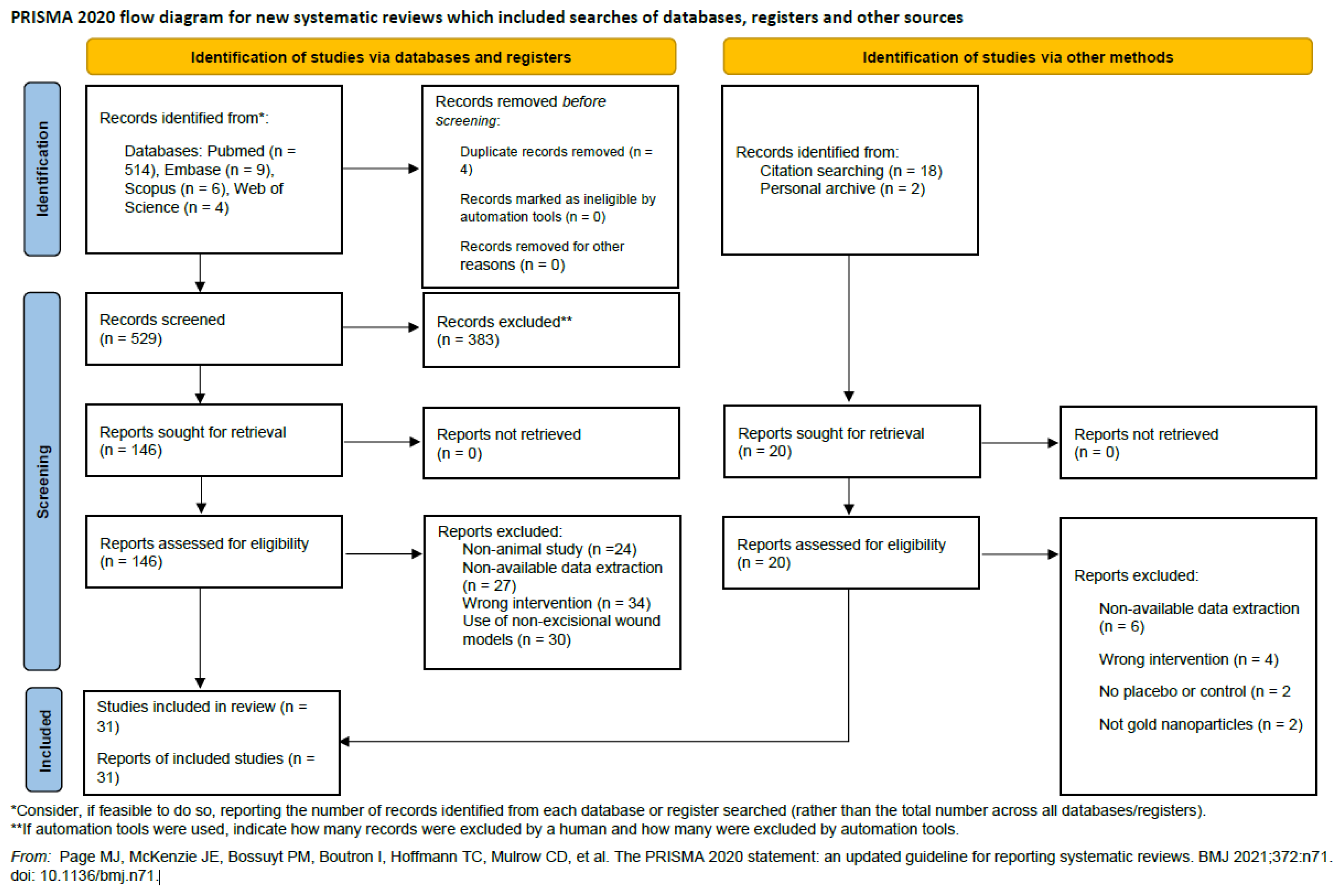
Figure 2.
Forest plot of pooled effect sizes for wound closure.
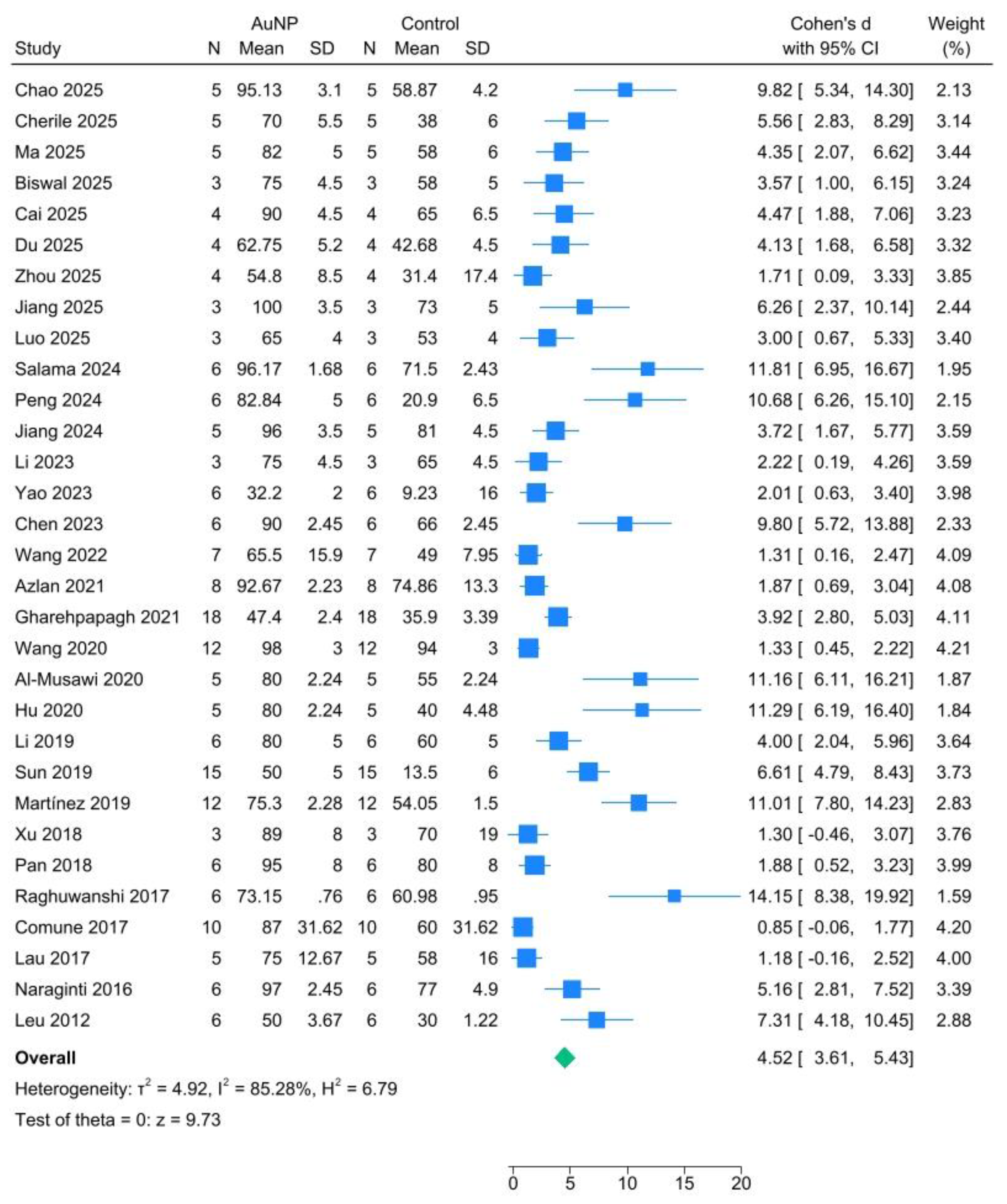
Figure 3.
Funnel plot assessing publication bias.
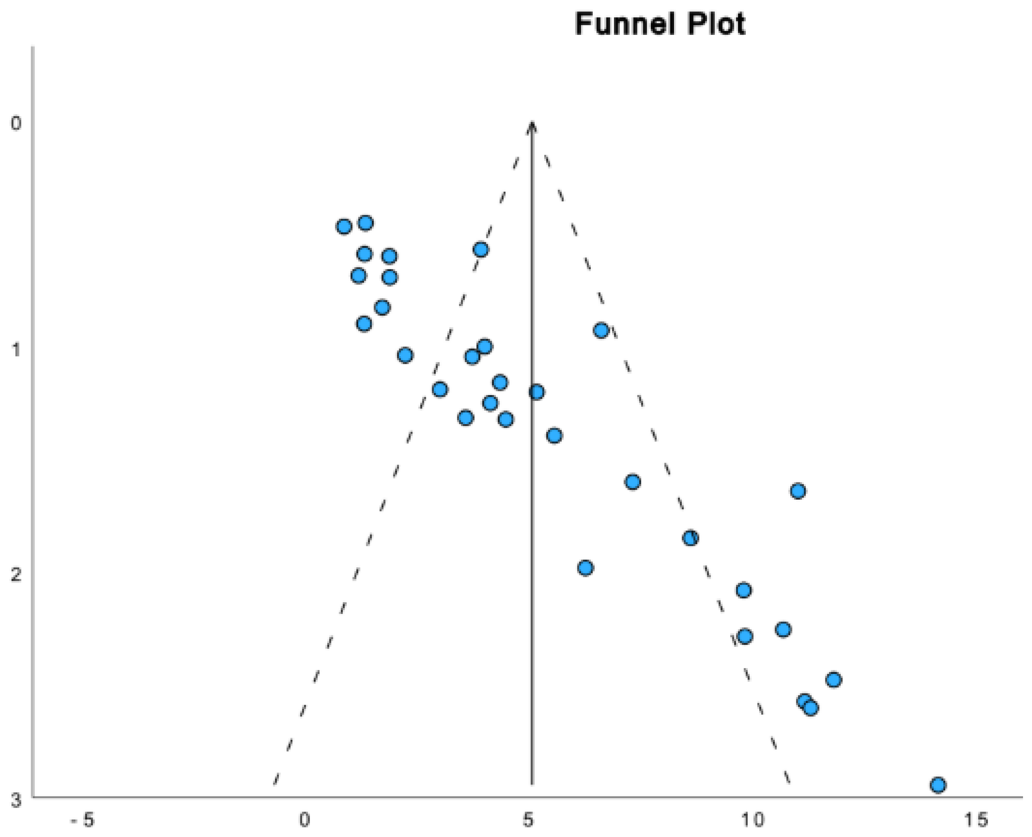
Table 1.
Study Characteristics of the Included Studies.
| Study | Intervention group, n | Control group, n | Wound type | Animal model | Day of peak effect | Study quality |
| 1. Chao et al., 2025 | 5 | 5 | Full thickness | Mice | Day 7 | 7/14 |
| 2. Gerile et al., 2025 | 5 | 5 | Full thickness | Mice | Day 7 | 7/14 |
| 3. Ma et al., 2025 | 5 | 5 | Full thickness | Mice | Day 7 | 6/14 |
| 4. Biswal et al., 2025 | 3 | 3 | Full thickness | Rats | Day 7 | 6/14 |
| 5. Cai et al., 2025 | 4 | 4 | Full thickness | Rats | Day 8 | 8/14 |
| 6. Du et al., 2025 | 4 | 4 | Full thickness | Mice | Day 9 | 7/14 |
| 7. Zhou et al., 2025 | 4 | 4 | Full thickness | Mice | Day 3 | 5/14 |
| 8. Jiang et al., 2025 | 3 | 3 | Full thickness | Mice | Day 13 | 7/14 |
| 9. Luo et al., 2025 | 3 | 3 | Full thickness | Mice | Day 12 | 7/14 |
| 10.Salama et al., 2024 | 6 | 6 | Full thickness | Rats | Day 9 | 7/14 |
| 11. Peng et al., 2024 | 6 | 6 | Full thickness | Rats | Day 12 | 7/14 |
| 12. Jiang et al., 2024 | 5 | 5 | Full thickness | Mice | Day 12 | 10/14 |
| 13. Li et al., 2023 | 3 | 3 | Full thickness | Mice | Day 14 | 10/14 |
| 14. Yao et al., 2023 | 6 | 6 | Full thickness | Mice | Day 15 |
7/14 |
| 15. Chen et al., 2023 | 6 | 6 | Full thickness | Rats | Day 13 |
8/14 |
| 16.Wang et al., 2022 | 7 | 7 | Full thickness | Rats | Day 7 |
10/14 |
| 17.Azlan et al., 2021 | 8 | 8 | Full thickness | Rats | Day 3 |
8/14 |
| 18.Gharehpapagh et al., 2021 | 18 | 18 | Full thickness | Mice | Day 7 |
9/14 |
| 19.Wang et al., 2020 | 12 | 12 | Full thickness | Rats | Day 10 |
10/14 |
| 20.Al-Musawi et al., 2020 | 5 | 5 | Full thickness | Rabbits | Day 7 |
7/14 |
| 21. Hu et al., 2020 | 5 | 5 | Full thickness | Mice | Day 4 |
7/14 |
| 22. Li et al., 2019 | 6 | 6 | Full thickness | Rats | Day 3 |
9/14 |
| 23. Sun et al., 2019 | 15 | 15 | Full-thickness | Mice | Day 7 | 7/14 |
| 24. Martínez et al., 2019 | 12 | 12 | Full thickness | Mice | Day 10 |
10/14 |
| 25. Xu et al., 2018 | 3 | 3 | Full thickness | Rats | Day 10 |
10/14 |
| 26. Pan et al., 2018 | 6 | 6 | Full thickness | Rats | Day 10 |
10/14 |
| 27. Raghuwanshi et al., 2017 | 6 | 6 | Full thickness | Rats | Day 9 |
10/14 |
| 28. Comune et al., 2017 | 10 | 10 | Full thickness | Mice | Day 7 | 10/14 |
| 29. Lau et al., 2017 | 5 | 5 | Full thickness | Rats | Day 7 | 9/14 |
| 30. Naraginti et al., 2016 | 6 | 6 | Full thickness | Rats | Day 9 | 10/14 |
| 31. Leu et al., 2012 | 6 | 6 | Full thickness | Mice | Day 7 | 10/14 |
Table 2.
Risk of Bias Assessment of the Included Studies Based on the Cochrane Collaboration’s Tool.
Table 2.
Risk of Bias Assessment of the Included Studies Based on the Cochrane Collaboration’s Tool.
| Id | Randomization | Allocation | Blinding | Assessment | Data | Reporting | Other |
| 1. Chao 2025 | Low | Unclear | High | High | Low | Low | Low |
| 2. Cherile 2025 | Low | Unclear | High | High | Low | Low | Low |
| 3. Ma 2025 | Low | Unclear | High | High | Low | Low | Low |
| 4. Biswal 2025 | Low | Unclear | High | High | Low | Low | Low |
| 5. Cai 2025 | Low | Unclear | High | High | Low | Low | Low |
| 6. Du 2025 | Low | Unclear | High | High | Low | Low | Low |
| 7. Zhou 2025 | Low | Unclear | High | High | Low | Low | Low |
| 8. Jiang 2025 | Low | Unclear | High | High | Low | Low | Low |
| 9. Luo 2025 | Low | Unclear | High | High | Low | Low | Low |
| 10. Salama 2024 | Low | Unclear | High | High | Low | Low | Low |
| 11. Peng 2024 | Low | Unclear | High | High | Low | Low | Low |
| 12. Jiang 2024 | Low | Unclear | High | High | Low | Low | Low |
| 13. Li 2024 | Low | Unclear | High | High | Low | Low | Low |
| 14. Yao 2023 | Low | Unclear | High | High | Low | Low | Low |
| 15. Chen 2023 | Low | Unclear | High | High | Low | Low | Low |
| 16. Wang 2022 | Low | Unclear | High | High | Low | Low | Low |
| 17. Azlan 2021 | Low | Unclear | High | High | Low | Low | Low |
| 18.Gharehpapagh 2021 | Low | Unclear | High | High | Low | Low | Low |
| 19. Wang 2020 | Low | Unclear | High | High | Low | Low | Low |
| 20.Al-Musawi 2020 | Low | Unclear | High | High | Low | Low | Low |
| 21. Hu 2020 | Low | Unclear | High | High | Low | Low | Low |
| 22. Li 2019 | Low | Unclear | High | High | Low | Low | Low |
| 23. Sun 2019 | Low | Unclear | High | High | Low | Low | Low |
| 24.Martínez 2019 | Low | Unclear | High | High | Low | Low | Low |
| 25. Xu 2018 | Low | Unclear | High | High | Low | Low | Low |
| 26. Pan 2018 | Low | Unclear | High | High | Low | Low | Low |
| 27.Raghuwanshi 2017 | Low | Unclear | High | High | Low | Low | Low |
| 28.Comune 2017 | Low | Unclear | High | High | Low | Low | Low |
| 29. Lau 2017 | Low | Unclear | High | High | Low | Low | Low |
| 30.Naraginti 2016 | Low | Unclear | High | High | Low | Low | Low |
| 31. Leu 2012 | Low | Unclear | High | High | Low | Low | Low |
Risk of bias was assessed across seven domains: random sequence generation (randomization), allocation concealment, blinding of participants and personnel, blinding of outcome assessment, incomplete outcome data, selective reporting, and other bias.
Table 3.
Characteristics of Included Studies.
| Publication | Year | (1) | (2) | (3) | (4) | (5) | (6) | (7) | (8) | (9) | (10) | (11) | (12) | (13) | (14) | Score |
|---|---|---|---|---|---|---|---|---|---|---|---|---|---|---|---|---|
| Chao | 2025 | X | X | X | X | X | X | X | 7 | |||||||
| Cherile | 2025 | X | X | X | X | X | X | X | 7 | |||||||
| Ma | 2025 | X | X | X | X | X | X | 6 | ||||||||
| Biswal | 2025 | X | X | X | X | X | X | 6 | ||||||||
| Cai | 2025 | X | X | X | X | X | X | X | X | 8 | ||||||
| Du | 2025 | X | X | X | X | X | X | X | 7 | |||||||
| Zhou | 2025 | X | X | X | X | X | 5 | |||||||||
| Jiang | 2025 | X | X | X | X | X | X | X | 7 | |||||||
| Luo | 2025 | X | X | X | X | X | X | X | 7 | |||||||
| Salama | 2024 | X | X | X | X | X | X | X | 7 | |||||||
| Peng | 2024 | X | X | X | X | X | X | X | 7 | |||||||
| Jiang | 2024 | X | X | X | X | X | X | X | X | X | X | 10 | ||||
| Li | 2023 | X | X | X | X | X | X | X | X | X | X | 10 | ||||
| Yao | 2023 | X | X | X | X | X | X | X | 7 | |||||||
| Chen | 2023 | X | X | X | X | X | X | X | X | 8 | ||||||
| Wang | 2022 | X | X | X | X | X | X | X | X | X | X | 10 | ||||
| Azlan | 2021 | X | X | X | X | X | X | X | X | 8 | ||||||
| Gharehpapagh | 2021 | X | X | X | X | X | X | X | X | X | 9 | |||||
| Wang | 2020 | X | X | X | X | X | X | X | X | X | X | 10 | ||||
| Al-Musawi | 2020 | X | X | X | X | X | X | X | 7 | |||||||
| Hu | 2020 | X | X | X | X | X | X | X | 7 | |||||||
| Li | 2019 | X | X | X | X | X | X | X | X | X | 9 | |||||
| Sun | 2019 | X | X | X | X | X | X | X | 7 | |||||||
| Martínez | 2019 | X | X | X | X | X | X | X | X | X | X | 10 | ||||
| Xu | 2018 | X | X | X | X | X | X | X | X | X | X | 10 | ||||
| Pan | 2018 | X | X | X | X | X | X | X | X | X | X | 10 | ||||
| Raghuwanshi | 2017 | X | X | X | X | X | X | X | X | X | X | 10 | ||||
| Comune | 2017 | X | X | X | X | X | X | X | X | X | X | 10 | ||||
| Lau | 2017 | X | X | X | X | X | X | X | X | X | 9 | |||||
| Naraginti | 2016 | X | X | X | X | X | X | X | X | X | X | 10 | ||||
| Leu | 2012 | X | X | X | X | X | X | X | X | X | X | 10 |
Studies fulfilling the criteria of: (1) peer-reviewed publication; (2) control of temperature; (3) random allocation to treatment or control; (4) blinded induction of model; (5) blinded assessment of outcome; (6) use of anesthetic without significant intrinsic neuroprotective activity; (7) use of animals with relevant comorbidities (e.g., aged, diabetic, hypertensive); (8) sample size calculation; (9) compliance with animal welfare regulations; (10) statement of potential conflicts of interest; (11) use of an appropriate animal model; (12) investigation of dose–response relationship; (13) timing of treatment relevant to clinical scenario; and (14) appropriate and clearly described statistical methods.
Disclaimer/Publisher’s Note: The statements, opinions and data contained in all publications are solely those of the individual author(s) and contributor(s) and not of MDPI and/or the editor(s). MDPI and/or the editor(s) disclaim responsibility for any injury to people or property resulting from any ideas, methods, instructions or products referred to in the content. |
© 2025 by the authors. Licensee MDPI, Basel, Switzerland. This article is an open access article distributed under the terms and conditions of the Creative Commons Attribution (CC BY) license (http://creativecommons.org/licenses/by/4.0/).
Copyright: This open access article is published under a Creative Commons CC BY 4.0 license, which permit the free download, distribution, and reuse, provided that the author and preprint are cited in any reuse.


