Submitted:
22 June 2025
Posted:
24 June 2025
Read the latest preprint version here
Abstract
This article covers a rare, benign tumor called hamartoma of the urinary bladder, made up of disorganized but normal bladder tissues like urothelium, fibrous stroma, smooth muscle, and fat. It lacks malignant features such as cellular atypia or necrosis. Patients often present with lower urinary tract symptoms (LUTS) and hematuria. Bladder hamartomas have been linked to syndromes like Peutz-Jeghers. Treatment typically involves transurethral resection, with rare recurrence; partial cystectomy is occasionally needed. This case report details the first known case in Germany and the 16th globally, involving a young patient, and includes a thorough literature review.
Keywords:
hamartoma
; urinary bladder
; gross hematuria
; uropathology
Introduction
Hamartomas are abnormally arranged growths of normal cells Indigenous to the organ, resulting in the formation of a mass or tumor [1,2]. They can manifest in various body parts, including the lungs, intestines, urinary bladder, skin, heart, brain, and breasts [1,3,4].
Although most hamartomas are asymptomatic, some can cause symptoms like painless hematuria, irritative voiding symptoms, or urinary retention if they grow sufficiently large [5]. The cause of hamartoma is still unknown, research has suggested that it may be associated with certain genetic disorders. [4,6,7,8]. The literature indicates that the tumor is rare, with only 15 published cases to date [4]. Notably, the rarity of this condition can lead to misdiagnosis; however, appropriate testing and diagnosis can result in proper treatment with positive outcomes. Here, we report the case of a 22-year-old male with bladder hamartoma presenting with LUTS. In addition, we provide a comprehensive review of the existing literature on this uncommon condition.
Case Presentation
A 22-year-old man was admitted to our urology department with LUTS persisting for two months, significantly reducing his quality of life due to nocturia (more than three to four times per night) and pollakiuria during the day. A normal urinalysis with U-Stix ruled out urinary infection.
Ultrasonography of the bladder revealed an intravesical growth. Despite the absence of clinical risk factors such as smoking, exposure to carcinogens, or advanced age, the lesion was initially suspected to be urothelial carcinoma. However, urine cytology revealed no neoplastic cells.
Cystoscopy demonstrated a large mass between the trigone and the posterior bladder wall, closely localized to the ureteral ostia. Photodynamic diagnosis (PDD) with hexaminolevulinate was negative (Figure 1). A biopsy was performed in the initial session to exclude malignancy.
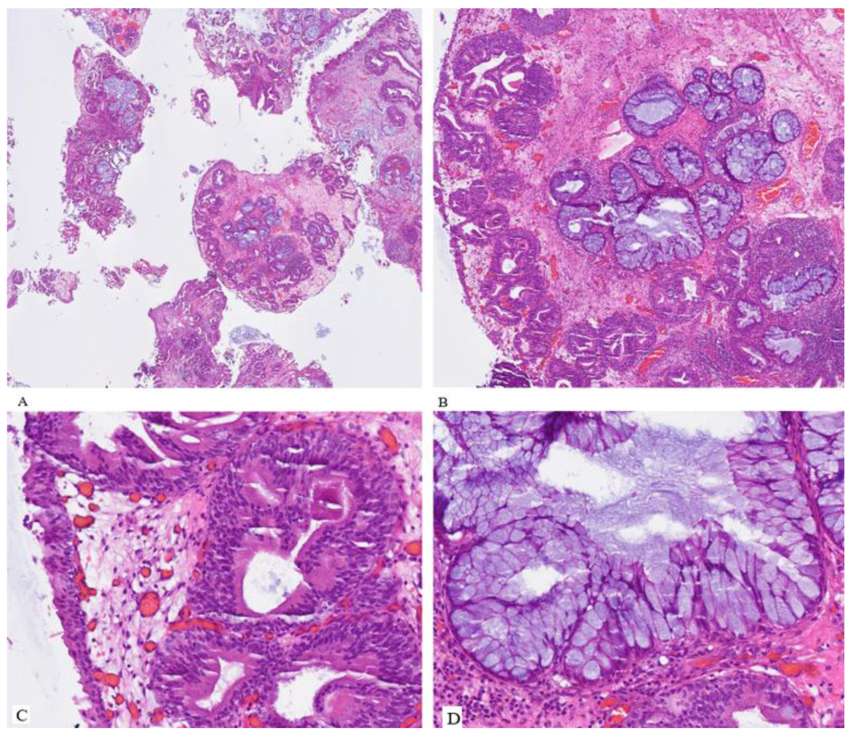
Histopathological examination revealed that the tumor was a hamartoma of the urinary bladder with pronounced urocystitis cystica and glandularis of the intestinal type, in addition to extensive intestinal metaplasia, partially abundant in goblet cells, and focal mucin extravasation. No evidence of mitosis, necrosis, or atypical features was detected in either the epithelium or the stroma (Figure 2).
Based on this finding, the patient was diagnosed with urinary bladder hamartoma was therefore made. We partially excised the hamartoma by transurethral resection in the second session to achieve locally controlled status and to minimize the hamartoma mass at the bladder outlet, which led to lower urinary tract symptoms. Complete resection was impossible because of both sides' close localization to the ureteral ostia.
The patient experienced rapid relief of symptoms after resection, with no nycturia on the first day after removal of the foley catheter. Human genetic testing revealed no evidence of any associated genetic diseases. The residual mass of the hamartoma was stable on control cystoscopy after 2 months, without new symptoms or mass increase.
Discussion and Literature Review
A comprehensive literature evaluation about urinary bladder hamartoma was conducted by searching reputable academic databases such as PubMed, Google Scholar, and the Cochrane Library.
The present study incorporated the terminology urinary bladder hamartoma, hamartoma of the urinary tract, and bladder hamartoma. Our search showed 92 results. After Screening and evaluating the abstracts of all papers, only 15 Case reports with urinary bladder hamartoma were found and could be included; therefore, 77 studies had to be excluded.
The first urinary bladder hamartoma was reported by Lathan et al. in 1963, with gross hematuria and pyuria as the leading clinical manifestations [9]. In the reported cases of hamartomas, including the present case, male patients were affected in 11 of 16 instances (67%), and the mean age at diagnosis was 25 years [4].
The leading clinical presentation was irritative lower urinary tract symptoms (LUTS) with or without gross hematuria (9/16, 56%). Gross hematuria was observed in 25% of the patients (4/16) [4]. Remarkably, 31,2% of lesions (5/16) appeared on the background of specific syndromes, such as Peutz-Jeghers syndrome [6,10], Beckwith-Wiedemann syndrome [8], Goldenhar syndrome [7], and Loeys-Dietz syndrome (LDS) [4]. Other rare findings were associated with schistosomiasis (1/16) [11], prenatal detection via ultrasound (US) (1/16) [12], and incidental findings on abdominal imaging (1/16) [4].
The posterior bladder wall was the most commonly affected area (8/16, 50%), followed by the bladder neck (4/16, 25%), trigone (3/16, 19%), anterior wall (1/16, 6.2%), left lateral wall (1/16, 6.2%), and the bladder dome (1/16, 6.2%) [4].
Ota et al. and Pescia et al. examined urine cytology without significant alterations or dysplasias [3,4].
Histopathological examination revealed that most lesions had a typical morphologic appearance: a lobulated mass composed of a mixed proliferation of tubular-glandular, nested, or even papillary epithelial components intermingled with smooth muscle bundles; fibromyxoid stroma with plump fibroblasts; and sometimes adipose tissue. [6]. Intestinal metaplasia and hypervascularity were other potential features observed in these lesions [3,5,6].
All patients underwent successful treatment with complete excision via transurethral resection, partial cystectomy (3/16) [1,3,10], or transvaginal excision (1/16) [6]. Intravesical mitomycin was administered before histological examination because of the suspicion of malignancy [6].No disease recurrence was reported, even after follow-up periods of up to 60 months via cystoscopy and ultrasound [4].
Possible differential diagnoses for urinary bladder hamartoma include cystitis cystica et glandularis, von Brunn hyperplasia, nested/microcystic variant of urothelial carcinoma, inverted urothelial papilloma, and nephrogenic adenoma, particularly in cases with unclear intravesical growth [13].
However, its rarity and benign pathological features, which resemble cystitis cystica et glandularis or von Brunn nest hyperplasia, may mask its identification, potentially leading to underdiagnosis or misdiagnosis.
From a clinical perspective, most reported cases in literature have been successfully treated with transurethral resection. However, in selected cases where complete resection is not feasible, close surveillance is essential to monitor potential progression or symptom recurrence. Given the benign nature of these lesions, aggressive interventions such as radical cystectomy should be avoided unless malignant transformation is confirmed.
Conclusions
This report presents the first documented case of urinary bladder hamartoma in Germany and the 16th worldwide. While complete transurethral resection is the preferred treatment, partial resection may be a viable alternative for patients with challenging tumor locations, preventing the need for partial cystectomy. Recognizing the benign nature of bladder hamartomas is crucial to avoid overtreatment and ensure appropriate patient management.
Funding
The authors received no financial support for this article's research, authorship, and/or publication.
Competing Interests
The authors have no relevant financial or non-financial interests to disclose.
Ethics Approval
This study was conducted in line with the ethical standards outlined in the Declaration of Helsinki and the research integrity policies of Hanover Medical School.
Consent to Participate
Informed consent was obtained from all individual participants included in the study.
Conflicts of Interest
The authors declared no potential conflicts of interest concerning this article's research, authorship, and/or publication.
References
- Brancatelli G, et al.: Hamartoma of the urinary bladder: case report and review of the literature. European radiology. 9. pp. 42–44 (1999). [CrossRef]
- Chinyama CN: Hamartoma. In: A. Sapino ,J. Kulka (eds.): Breast Pathology. pp. 135–143. Springer International Publishing. Cham (2020). 978-3-319-62538-6.
- Ota T, et al.: Hamartoma of the urinary bladder. International journal of urology : official journal of the Japanese Urological Association. 6. pp. 211–214 (1999). [CrossRef]
- Pescia C, et al.: A Rare Case of Urinary Bladder Hamartoma Clinically Mimicking an Urothelial Carcinoma: A Case Report and Review of the Literature. International journal of surgical pathology. 31. pp. 1572–1579 (2023). [CrossRef]
- Murray C; Marchan, Jennifer; Özel, Bora; Özel, Begüm: Bladder wall hamartoma: an unusual cause of urinary urgency and frequency. Female pelvic medicine & reconstructive surgery. 21. pp. e8-e10 (2015). [CrossRef]
- Kumar J; Albeerdy, Mohammad Irfaan; Shaikh, Nadeem Ahmed; Qureshi, Abdul Hafeez: Bladder hamartoma in Peutz-Jeghers syndrome: a rare case report. Afr J Urol. 27 (2021). [CrossRef]
- Adam A; Gayaparsad, Keshree; Engelbrecht, Matthys J.; Moshokoa, Evelyn M.: Bladder hamartoma: a unique cause of urinary retention in a child with Goldenhar syndrome. Saudi journal of kidney diseases and transplantation : an official publication of the Saudi Center for Organ Transplantation, Saudi Arabia. 24. pp. 89–92 (2013). [CrossRef]
- Williams MP; Ibrahim, S. K.; Rickwood, A. M.: Hamartoma of the urinary bladder in an infant with Beckwith-Wiedemann syndrome. British journal of urology. 65. pp. 106–107 (1990). [CrossRef]
- Moose LT; Garvey, Fred K.: Hamartoma of the Bladder. Journal of Urology. 89. pp. 185–187 (1963). [CrossRef]
- Keating MA; Young, R. H.; Lillehei, C. W.; Retik, A. B.: Hamartoma of the bladder in a 4-year-old girl with hamartomatous polyps of the gastrointestinal tract. Journal of Urology. 138. pp. 366–369 (1987). [CrossRef]
- Duvenage GF; Dreyer, L.; Reif, S.; Bornman, M. S.; Steinmann, C. F.: Bladder hamartoma. British journal of urology. 79. pp. 133–134 (1997). [CrossRef]
- Pieretti A; Wu, Chin-Lee; Pieretti, Rafael V.: Bladder Hamartoma in a Fetus: Case Report. Urology case reports. 2. pp. 154–155 (2014). [CrossRef]
Figure 1.
Transurethral images of the polypoid lesion in the urinary bladder under white light (A-C) and fluorescent cystoscopy with hexaaminoläuvlinat with a negative signal (D).
Figure 1.
Transurethral images of the polypoid lesion in the urinary bladder under white light (A-C) and fluorescent cystoscopy with hexaaminoläuvlinat with a negative signal (D).
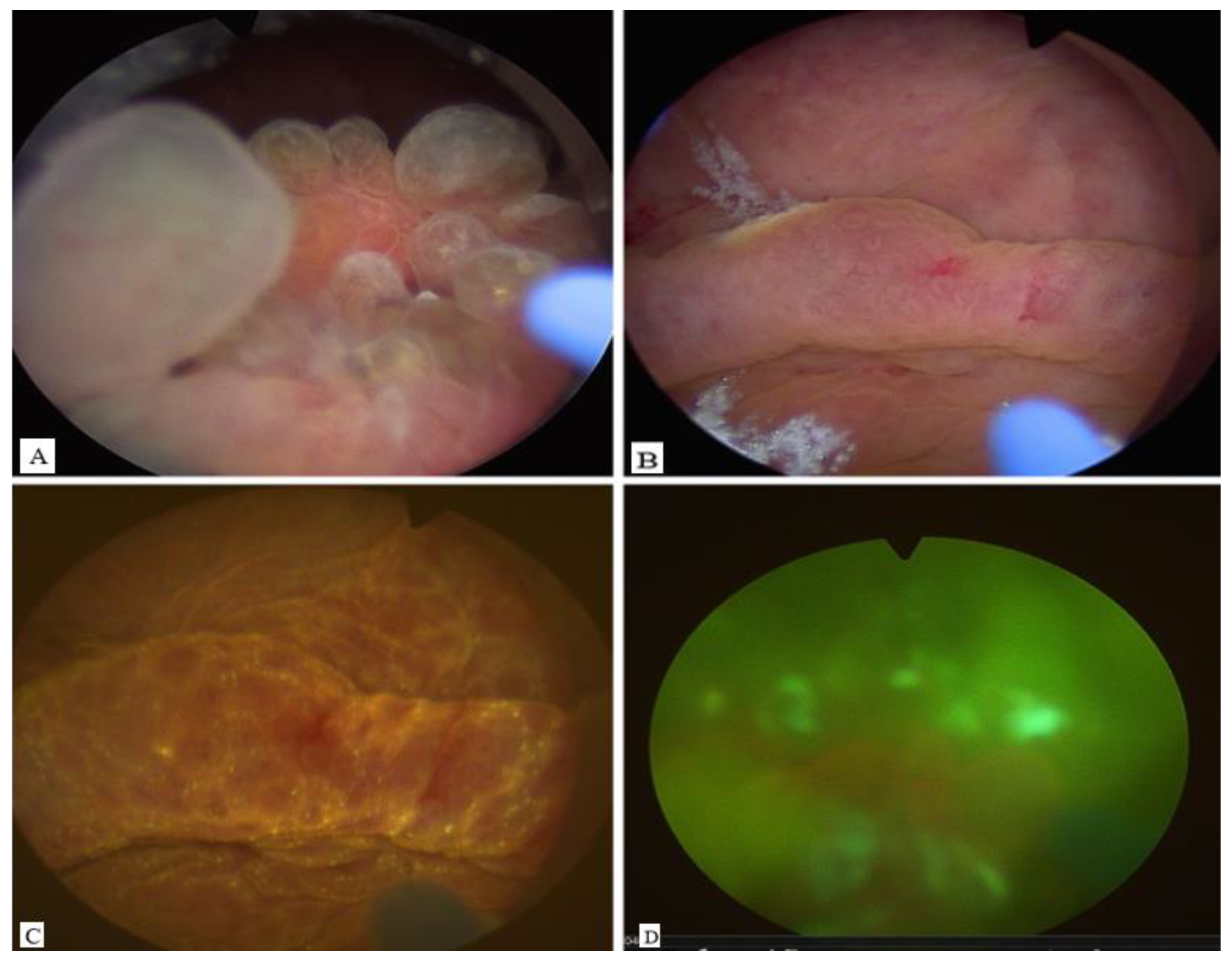
Figure 2.
The lesion showed multiple fragmented tissue sections with irregularly shaped lobulesglandular structures within a fibro-muscular stroma (A, H&E stain, 4 × magnification) (B, H&E stain, 10 × magnification) (C, H&E stain, 20 × magnification) (D, H&E stain, 40 × magnification). Abbreviations: H&E, hematoxylin and eosin.
Disclaimer/Publisher’s Note: The statements, opinions and data contained in all publications are solely those of the individual author(s) and contributor(s) and not of MDPI and/or the editor(s). MDPI and/or the editor(s) disclaim responsibility for any injury to people or property resulting from any ideas, methods, instructions or products referred to in the content. |
© 2025 by the authors. Licensee MDPI, Basel, Switzerland. This article is an open access article distributed under the terms and conditions of the Creative Commons Attribution (CC BY) license (http://creativecommons.org/licenses/by/4.0/).
Copyright: This open access article is published under a Creative Commons CC BY 4.0 license, which permit the free download, distribution, and reuse, provided that the author and preprint are cited in any reuse.


