Submitted:
23 May 2025
Posted:
26 May 2025
You are already at the latest version
Abstract
Background: Cardiovascular disease (CVD) is very spread in countries with a Western style diet, representing one of the major causes of morbidity. Genetic factors, obesity, diabetes, dyslipidemia, smoking, and ageing are risk factors for CVD outcome. From a pathogenic point of view, the condition of low-grade inflammation of the arteries leads to endothelial damage, and atherosclerosis development. Nowadays, a broad range of drugs is available to treat CVD, but many of them are associated with side effects. Therefore, alternative therapeutic remedies need to be discovered even in combination with conventional drugs. A balanced diet rich in fruits and vegetables, e.g., the Mediterranean diet, has been shown to lower the incidence of CVD. Plant-derived polyphenols are ingested with food, and these compounds can exert beneficial effects on human health, such as antioxidant, and anti-inflammatory activities. Objective: In the present review, the cellular and molecular bases of the beneficial effects of polyphenols on the prevention and treatment of CVD will be pointed out. Methods: This review has been accomplished on the basis of literature review spanning mainly in the last 2 decades. Results: We found in this respect, that an increased dietary intake of polyphenols is associated with a parallel decrease in chronic disease incidence, even including CVD. Conclusion: Despite a plethora of preclinical studies, more clinical trials are needed for a more appropriate treatment of CVD with polyphenols.
Keywords:
Cytokines
; Dendritic Cells
; Immunotherapy
; Macrophages
; Myocardial Infarction
; T Lymphocytes
Introduction
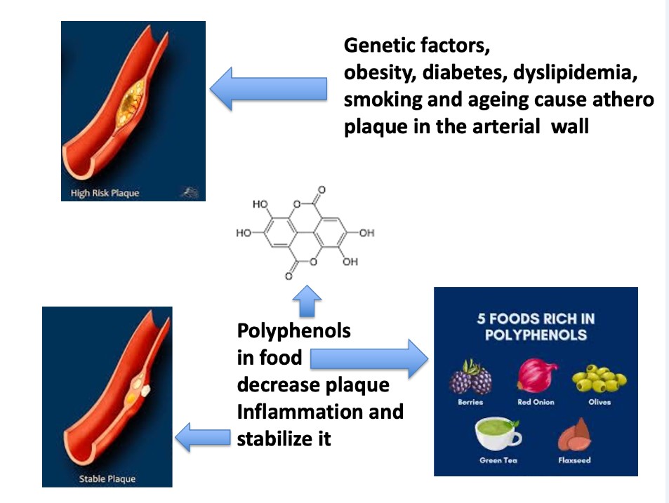
Cardiovascular disease represents one of the major causes of morbidity in countries adopting Western lifestyles with an annual expectation of deaths by 2030 that exceeds 23.6 million [1]. The term CVD encompasses a variety of conditions, such as coronary artery disease (CAD), stroke, peripheral artery disease, hypertension, cerebrovascular disease, and heart failure (HF). Among risk factors of CVD, genetic factors, obesity, diabetes, dyslipidemia, smoking, and ageing account for the occurrence of CVD [2,3]. The above conditions lead to endothelial cell dysfunction, oxidative stress, proliferation of smooth muscle cells, and fibroblasts, with conversion of macrophages to foam cells within the artery walls [4]. Furthermore, the condition of vascular low-grade inflammation promotes atherosclerotic plaque formation, ultimately, causing HF [5] . Therapeutically, a broad range of drugs is available for the treatment of CVD, i.e., statins, angiotensin- converting enzyme inhibitors, angiotensin receptor blockers, calcium channel blockers, fibrates, and beta-blockers, however, many of them are associated with side effects[6]. Therefore, there is a need to discover and apply innovative therapies even in combination with conventional ones for a more appropriate management of CVD [7,8]. It is known that a balanced diet is beneficial for preventing CVD. In fact, consumption of fruits and vegetables has been shown to decrease the incidence of CVD. In this respect, Mediterranean diet (MD) decreases inflammatory biomarkers, e.g., interleukin (IL)-1 beta, IL-5 and C-Reactive Protein (CRP), thus, preventing chronic disease outcome[7,9,10,11]. In this framework. PREDIMED study demonstrated that MD based on the high consumption of fruits, vegetables, whole grains, and extra virgin olive oil (EVOO) was associated with a reduced risk of CVD[12]. Of note, dietary interventions aimed at reducing low-grade inflammation, have led to divergent results due to differences in tested dietary compounds and chosen inflammatory markers [13,14]. Plant-derived compounds contained in food possess beneficial effects to human health. Among these natural products, polyphenols can be found in fruits, vegetables, seeds, nuts, as well as in red wine, tea, coffee, extra virgin EVOO, and chocolate [15,16,17,18,19]. Nowadays, the human population is more aware about the beneficial effects of polyphenols, and their dietary intake has increased, with a parallel decrease in chronic disease, even including CVD[20]. In the present review, classification, pharmacological activities, and main mechanisms of action of polyphenols will be described. Experimental and clinical evidence of the beneficial effects of these natural compounds on CVD will be discussed..
Classification and General Properties of Polyphenols
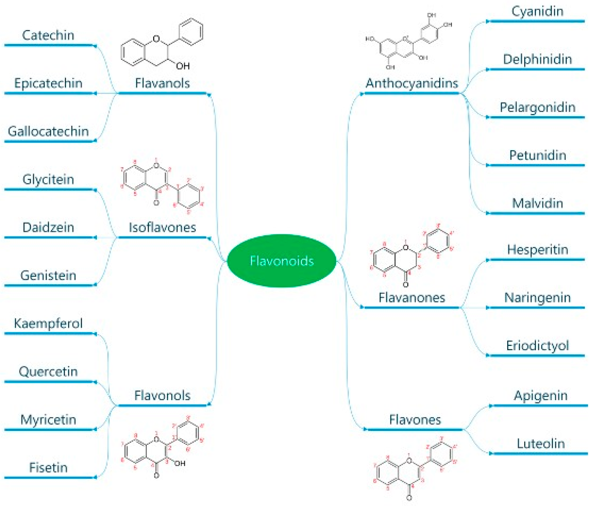
Polyphenols are classified according to the number of phenolic rings, and the structural elements they bind [16]. They can be divided into four main classes: flavonoids; non-flavonoids stilbenes; phenolic acids; and lignans (Error! Reference source not found.) [21]. Flavonoids are naturally occurring compounds, which encompass six categories: flavanones, flavones, flavanols, isoflavones, flavan-3ols, and anthocyanidins (Error! Reference source not found.) [22] . Structurally, they possess two aromatic rings and a heterocyclic ring with a C6-C3-C6 configuration (Error! Reference source not found.). They are contained in plants as glycoside and non-glycosylated conjugate compounds, and their type of structure influences bioavailability[23]. Stilbenes, e.g., resveratrol (RES), are composed of two phenyl residues linked by a two-carbon methylene bridge, which can be glycosylated, methylated, or prenylated by specific enzymes (Error! Reference source not found.) [24]. Among flavonoids, flavonols, and flavan-3- ols have been object of intensive research. The flavonol quercetin exhibits antihypertensive effects by acting on the contraction of smooth muscles in renal blood vessels, producing vasodilation[25]. Among flavan-3-ols, epigallocatechin-3-gallate (EGCG) is mostly present in green tea, and is endowed with antioxidant, anti-inflammatory, and antiatherogenic properties[26]. Among stilbenes, RES is the most studied compound for its anti-inflammatory, antioxidant, anti-proliferative, anti-apoptotic, and mitochondrial protective effects[27].
Figure 1.
Classification of polyphenols. Polyphenols are natural compounds found in plant-based foods and beverages. Their classification into different subclasses like phenolic acids, flavonoids, stilbenes and lignans is reported. The chemical formula of these molecules is also reported. Reproduced with permission from Caiati et al. [7].
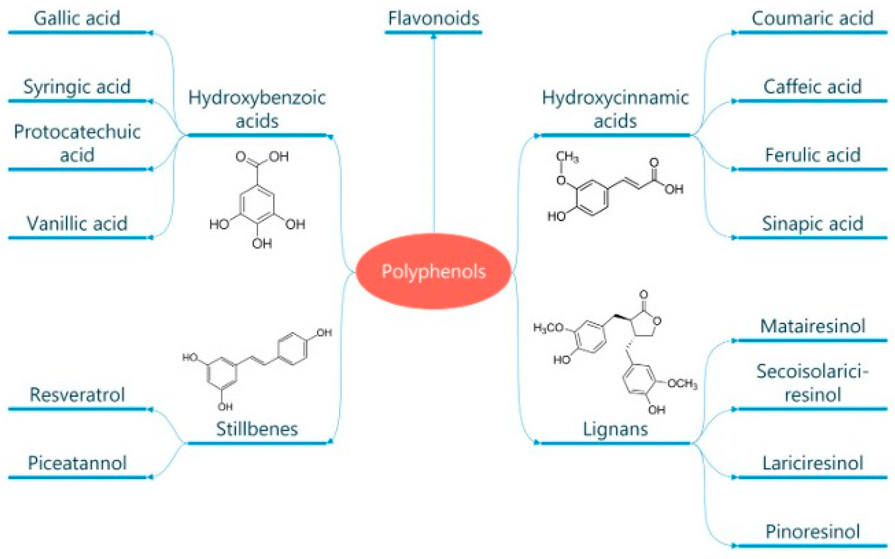
Figure 2.
Classification of flavonoids. Flavonoids are a subclass of polyphenols and can be classified into flavonols, flavones, isoflavones, flavanones, antho-cyanidins and flavanols based on their ring structure as here illustrated. Flavonoids have diverse biological activities and potential health benefits, including antioxidant and anti-inflammatory effects. Reproduced with permission from Caiati et al. [7].
Absorption of Polyphenols
In the stomach, polyphenols are digested by pepsin, and peristaltic movements in the presence of a low pH into particles, even less than 500 microns in diameter [28]. The passage of polyphenols from the stomach to the small intestine occurs at a pH around 7, and then, pancreatic and biliary enzymes become activated[29]. Polyphenols under the form of aglycons enter the intestinal epithelium according to different modalities. For instance, polyphenols with low molecular weight, i.e., phenolic acids, flavonoid aglycon, tea polyphenols, and cocoa polyphenols (epicatechin, procyanidin B2, and catechin) are absorbed by passive diffusion[30]. Another way of polyphenol absorption is the sodium-glucose transport through the sodium-glucose-linked transporter 1 (SGLT1)[31]. Accordingly, glycosides may be absorbed by SGTL1 in small amounts, and then, re-secreted into the digestive system, or they may be further digested by a cytosolic glucosidase[32]. Thus, polyphenols can undergo transepithelial transport through a monocarboxylic acid transporter, as in the case of caffeic acid, and ferulic acid (Error! Reference source not found.) [33]. Most polyphenols are absorbed in the large intestine, where they are digested by bacteria of the gut microbiota via glycosylation, hydroxylation, demethylation, deconjugation, ring cleavage, hydrolysis, epimerization, and chain shortening processes[34]. Polyphenols, once absorbed into the enterocytes of the small intestine, and before entering circulation, undergo the phase II of enzymatic detoxification with production of sulfates, glucuronides, and methylated derivatives [35]. Polyphenol bioavailability and accumulation in tissues depend on the multidrug resistance associated proteins, which are ATP-dependent efflux transporters, and referred to as phase III metabolism[36]. Then, polyphenols reach the blood stream mostly coupled to proteins, and the liver via the portal circulation, where they are conjugated to O-sulphate or O-glucuronide forms (a second phase metabolism), and finally are eliminated through kidneys[37]. (Table 1)
Antioxidant Properties of Polyphenols
Polyphenols behave as potent antioxidant agents thanks to catechol groups, and hydroxylation patterns, such as the 3-hydroxyl group in flavanols or electron shortage in anthocyanins[38]. Using the ferric reducing ability power, it has been demonstrated that the presence of a catechol ring in the structure of polyphenols is associated with their antioxidant activity[39]. Reactive oxygen species (ROS), i.e.,superoxide, hydrogen peroxide, and hypochlorous acid, are scavenged by quercetin, and catechin through the phenolic core, acting as buffer or collecting electrons [40]. Furthermore, polyphenols have been shown to inhibit enzymes that generate ROS, such as xanthine oxidase, and nicotinamide adenine dinucleotide phosphatase[41]. Among polyphenols, quercetin has the best capacity to chelate metal ions due to its low redox potential, thus preventing the production of ROS [27]. Scavenging activity of polyphenols is connected to their ability to donate an electron or H atom from an aromatic hydroxyl group to a free radical, thus abrogating its effect [42]. The antioxidant capacity of polyphenols in vivo is lower than in vitro, since it can be mimicked by other compounds[43]. For instance, in vivo, the polyphenol-mediated antioxidant activity exerted by apple consumption is mostly due to the metabolic effect of fructose on urate.
Effects of Polyphenols on the Vascular Endothelium
The major function of endothelial cells (ECS) is to regulate the vascular tone[44]. The endothelial (e) nitric oxide (NO) synthase (eNOS) generates NO from L- arginine, that, in turn, acts on the vascular smooth muscles, thus, triggering guanyl cyclase, with accumulation of cyclic guanosine monophosphate, which activates the protein kinase G, thus leading to vasorelaxation. Furthermore, the endothelium-derived hyperpolarizing factor causes vasorelaxation, targeting the K+ channels in the vasculature. Also, prostacyclin I2 (PGI2), generated during the cyclooxygenase (COX) pathway, leads to vasodilation. On the other hand, endothelial products, such as angiotensin II (ANG II), endothelin-1 (ET-1), and thromboxane (TXA) A2 play vasoconstrictive effects[45]. NO generation accounts for the main effects of polyphenols on the endothelium[46]. In this respect, red wine polyphenols are a potent inducer of serum NO in healthy subjects after 30 min from ingestion[47]. In vitro studies have demonstrated that healthy human peripheral blood monocytes are additional source of NO, thus contributing to the vasodilation after ingestion of red wine[16]. In this regard, short term oral treatment of normotensive rats with red wine polyphenols decreased blood pressure[48]. Such an effect depends on the induction of the gene responsible for inducible NO synthase, and COX-2 in the arteries, as well as on the calcium ion -dependent pathway[49]. In this last respect, RES and quercetin have been shown to induce increase in calcium concentration by opening the potassium channels or inhibiting Ca2+ ATP-ase within the endoplasmic reticulum of ECs [50]. Evidence has been provided that red wine polyphenols enhance endothelial NO production via the redox-responsive PI3/Akt channel, the increase in intracellular protein-Ca2+, and tyrosine phosphorylation with activation of eNOS[51,52]. Apart from NO generation, polyphenols exhibit other effects of the endothelium via increased release of PGI2 [53]. In fact, in vitro and in vivo studies, using cocoa extracts rich in procyanidins, demonstrated that the ratio leukotrienes to PGI2 was reduced. Moreover, polyphenols can increase endothelial NO by decreasing levels of phosphodiesterases (PDE)-2 and PDE-4[54]. (Table 2)
Anti-Inflammatory Activity of Polyphenols
Inflammation is a response of the body to various stimuli, even including pathogens, mechanical insults, and damaged tissue. Pro-inflammatory cytokines,e.g., interleukin (IL)-1 beta, IL-6, IL-8, and tumor necrosis factor (TNF)-alpha, as well as various enzymes, such as COX, lipooxigenase (LOX), and protein kinase are responsible for the inflammatory response . With special reference to the role of polyphenols, there is evidence that red wine polyphenols can in vitro reduce the production of pro-inflammatory cytokines, blocking the activation of the NF-kB pathway[55]. Moreover, red wine polyphenols can interfere with endotoxin binding to Toll-Like Receptor (TLR)-4, thus, abrogating the nuclear factor kappa-light chain enhancer of activated B cells (NF-kB) pathway with interruption of pro-inflammatory cytokine release[56]. Also, polyphenols contained in the fermented grape marc (FGM) induce activation of Foxp3+ T regulatory cells, with release of the anti-inflammatory cytokine, IL-10[57]. In addition, FGM reduces the respiratory burst of neutrophils and basophils in in vitro experiments, playing an antioxidant and anti- inflammatory activity[58]. Quercetin has been found to dampen the generation of prostaglandins, leukotrienes, and TAXs, abrogating production of COX and LOX[59,60]. In fact, both COX and LOX mediate the formation of arachidonic acid, which, in turn, fuels inflammation via release of IL-1 beta, and IL-8. The nucleotide-binding domain and leucine-rich repeat containing receptors (NLRs) belong to the family of pattern recognition receptors (PRR), triggering inflammatory responses upon danger and cell damage signals. Among them, NLRP3 inflammasome is a multiprotein complex, which activates the inflammatory caspase-1 [61]. Caspase-1 cleaves and maturates the pro-inflammatory cytokines, IL-1 beta and IL-18, as well as the protein gasodermin, contributing to the release of the above mediators, thus, initiating the cell death pyroptosis [62] . Activation of NLRP3 inflammasome is involved in CVD even including atherosclerosis, myocardial infarction, and cardiac remodeling[63]. In this framework, in the middle cerebral artery occlusion/reperfusion model, supplementation of various polyphenols decreased levels of NLRP3 [64]. This event is associated with the downregulation of IL-1 beta, and IL-18 in the serum or brain tissue[65]. In the myocardial ischemia (MI)/reperfusion model, certain polyphenols, i.e., RES and flavone in vivo reduced levels of caspase-1 and IL-1 beta in the myocardial tissue[66,67]. In all these studies, the decrease in NLRP3 levels was associated with improvement of clinical markers[64]. Clinically, aged male subjects at high cardiovascular risk underwent acute administration of aged wine with decrease in Tlr2, Nlrp3, and Il1receptor genes[68]. (Table 3)
Anti-Atherogenic Effects of Polyphenols
Atherosclerosis represents a pathogenic common denominator of various diseases, including CAD, ischemic stroke, and peripheral artery disease [69]. This disease stems from the endothelial damage provoked by several offending factors that then drive augmentation of ROS in the blood. Those offending factors have been recently reported [7,70]: in brief, they are largely man-made like stress, pollutants of all sorts (especially those contained in the food like farming chemicals, fertilizers, pesticides and herbicides like glyphosate), drugs, processed food, tabacco smoking, air pollution, alcohol, cosmetic and cleaning products, heavy metal, chronic infections, electromagnetic radiation (cellular phone, cell-tower emitting radiation), ionizing radiation (in particular those medically derived like computed tomography scan and angiography), intravascular prosthesis like arterial stents. Diabetes per se induces tissue damage and it terribly enhances the damaging effects of the previously mentioned atherogenic factors so dramatically enhancing formation of ROS [7]. From a pathogenic point of view, increased levels of ROS further enhance endothelial damage, with the intervention of neutrophils, macrophages, and platelets [71]. In fact, prolonged contact of ECs with hydrogen peroxide, peroxynitrite, and oxidized low density lipoproteins (ox-LDL), leads to severe damage of the endothelium[7,69,72]. One of the initial consequence of coronary endothelial dysfunction is the reduction of NO production and the ensuing microvascular vasoconstriction at rest. This kind of derangement can be spotted with positron emission tomography (PET) since it causes myocardial dishomogeneous perfusion at rest [73] and is the mechanism that explains the angiographic slow coronary phenomenon as recently demonstrated [74].
Then such a strong oxidative drive involve LDL microparticles that get oxidated too. This causes the first step of atherosclerotic plaque formation, that is the generation of oxidized LDLs, which pass through the endothelial barrier, eliciting cytotoxic effects and the inflammatory response[75], since ox-LDL microparticles are modified substances that elicit strong immunologic reaction. Focusing on the molecular biology details, activated ECs express adhesins, i.e., vascular cell adhesion-1 (VCAM-1), intercellular adhesion molecule 1 (ICAM-1), and E- selectins, which allow transmigration of monocytes and T cells into the arterial wall [76]. Particularly, monocytes engulf ox-LDL, becoming foam cells, which accumulate as fatty bands in the artery walls[77]. Then, a stabilized plaque is formed, which can break under prolonged inflammation [78]; this happens only in case of prolonged and unstopped exposure to those factors generating ROS with consequent escalating concentration of ox-LDL. Ruptured plaques cause thrombosis, which may lead to heart attacks, ischemic strokes, and peripheral ischemia [79]. There is evidence that polyphenols can exert beneficial effects on atherosclerosis. In cholesterol-fed rabbits, administration of red wine polyphenols decreased neointimal growth, lipid accumulation, and inflammation in the iliac arteries[80]. In hamsters, red wine supplementation reduced neointimal hyperplasia, inhibiting the entry of monocytes into the arterial wall [81]. Clinically, there is evidence that purple grape juice reduced the levels of plasmatic ox-LDL in patients with CAD[82]. Such an effect has been shown to depend on the production of NO by polyphenols, as also supported by others[83,84] (Table 4). However the main radical approach that can stop progression and start regression of atherosclerosis is the elimination of those damaging factors (mentioned before) that create ROS and cause chronic endothelium inflammation and ox-LDL [85].
Focus on the Cardiovascular Effects of Relevant Polyphenols
Flavan-3-Ols
Flavan-3 Ols (Error! Reference source not found.) represent the most abundant polyphenols in fruits, vegetables, red wine, green tea, and cocoa [86]. They encompass monomeric, oligomeric, and polymeric compounds. Monomeric forms include catechin, epicatechin, gallocatechin, epigallocatechin, epicatechin-3-O-gallate, and EGCG (Error! Reference source not found.). Oligomers or polymers are known as proanthocyanidins, while polymers composed of epicatechin or catechin are termed procyanidins. The antioxidant activity of flavan-3-ols is based on their ability to donate an electron or to chelate metal ions, thus, stopping, ROS production[87]. At the same time, flavan-3-ols maintain mitochondrial activity, while enhancing antioxidative enzymes involved in ROS scavenging [88,89]. The anti-inflammatory activity of flavan-3 -ols depends on the regulation of gene expression involved in cardiometabolic health. Particularly, they act on endothelial transcription factor GATA-2, the NF-kB p105 subunit, forkhead box C1, and peroxisome proliferator-activated receptor gamma[90]. In addition, flan-3-ols target different miRNA, regulating cellular pathways involved in cell adhesion, cellular signaling, and immune response[91]. Of note, cocoa flavan-3-ols metabolites enhance ApOAI expression, which represents the major component of high-density lipoproteins, thus exerting antiatherogenic properties[92]. The cardioprotective effects of flavan-3-ols have been attributed to two major microbial-derived metabolites, namely, the hydroxy-phenyl-gamma-valerolactones, and their derived hydroxy- phenyl valeric acids [93]. These metabolites have in vivo shown hypotensive activity in rats, and in vitro abrogation of monocyte adhesion to ECs treated with tumor necrosis factor (TNF)-alpha[94]. Another flavan-3-ol metabolite, the protocatechuic acid, diminished diabetic cardiomyopathy in rats, stimulating glucose metabolism, improving oxidative stress, and reducing inflammation[95]. Flavan-3-ols have been shown to act on gut dysbiosis, improving cardiac function. In fact, a metabolite from the gut microbiota, trimethylamine N-oxide (TMAO) has been associated with CVD pathogenesis in terms of increased cholesterol levels, and higher risk of atherosclerosis[96]. In this respect, the intake of cocoa and red berry flavan-3- ols reduces TMAO levels, improving cardiovascular markers in healthy aging adults[97]. Various clinical trials have been conducted, using cocoa flavan-3-ols in patients with CVD. In hypertensive individuals, consumption of dark chocolate led to a reduction of systolic blood pressure (SBP), and diastolic blood pressure (DBP) in comparison to baseline[98]. In another study, in essential hypertensive subjects, with impaired glucose tolerance, receiving 100g/day chocolate, a reduction of both SBP, and DBP, and an increase in flood- mediated dilation (FMD) were observed[99]. In patients with CAD receiving dietary high-flavan-3-ol intervention, an increase in brachial artery FMD, and a reduction in SBP were recorded[100]. In patients with congestive HF, intake of flavan-3-ol-rich chocolate improved FMD[101]. Other studies have been conducted with green tea catechins in both healthy, and unhealthy individuals. In healthy volunteers aged between 21 and 70 years, receiving two capsules of Camelia sinensis for 3 months, a reduction of SBP was observed[102]. In another group of healthy adult men aged 18-35 years, administration of 450 mg sour tea led to a reduction of SBP and DPB[103]. In healthy postmenopausal women, acute ingestion of catechin-rich green tea improved postprandial glucose status, while increasing serum thioredoxin levels, but no changes in cardiovascular risk factors were observed[104]. In overweight women aged 19-57 years, receiving low-calorie diet along with 3 capsules of green-tea or placebo capsules, a decrease in SBP, and DPB were observed in both groups[105]. Another trial conducted in healthy male volunteers, supplemented with an aqueous green tea extract, showed no alterations of cardiac risk factors[106]. Also, minor effects on cardiovascular risk markers were observed following tea catechin administration to active older people[107]. Taken together, the above data suggests that studies with polyphenols conducted in both healthy and unhealthy individuals has led to contrasting results.
Resveratrol
Stilbenes, and, particularly, RES (Error! Reference source not found.) despite a low bioavailability possess a strong antioxidant activity in vitro. RES protects cardiomyocytes, and ECS against ROS effects, inhibiting NADPH oxidases, while increasing the mitochondrial respiratory chain enzymes[108]. RES acts upregulating SIRT1, that, in turn, induces deacetylation of NF-kB, and enhancement of superoxide dismutase (SOD), catalase and glutathione peroxidase 1[109,110]. Furthermore, RES can reverse eNOS uncoupling, upregulating GCH1 expression in apolipoprotein E knockout mice[111]. Also, RES activates Nrf-2, which, in turn, increases cellular antioxidant content in placenta of sows and piglets[112]. As a potent anti-inflamamtory agent, RES can inhibit the expression of pro-inflammatory cytokines, downregulating TLR4 expression, and silencing NF-kB activity[113,114]. Moreover, RES can inhibit VCAM-1, ICAM-1, and E-selectin, suppressing the TNF-alpha-induced NF-kB activation[115]. RES inhibits COX-1 and COX-2 enzymes via SIRT1 activation, thus, decreasing PGE2 and TAX2, and consequently inflammation[116]. In patients with systolic HF, RES administration improved clinical conditions by inhibiting oxidative phosphorylation in leukocytes, gene expression encoding B cell receptors, and leukocyte extravasation signal[117]. ROS-mediated overexpression of (MAPKs) is involved in cardiac hypertrophy and remodeling[118]. RES can stimulate MKP-1 and downregulate mTOR, thus dampening mitogen-activated protein kinase (MAPK) activity, with reduction of cardiac and endothelial hypertrophy[119,120]. With special reference to cardiac fibrosis, it has been reported that RES can mitigate in rats cardia fibroblast activity, downregulating the transforming-growth factor-beta/Smad 2/3 signaling pathway via overexpression of the Smad 7 inhibitor protein and silencing miR-17 gene[121]. RES can modulate endothelial function, inhibiting overproduction of the vasoconstrictive agent, ET-1, enhancing eNOS phosphorylation, with increase in No production[122]. There is evidence that upregulation of ET-, and decrease in NO are involved in the pathogenesis of atherosclerosis[123]. The effects of RES on mitochondrial biogenesis have been documented. In fact, RES activates the AMPK/SIRT1/PGC1 alpha pathway, with enhancement of Nrf-1 and Nrf-2 transcription factors, thus, attenuating high-glucose oxidative stress, and cardiomyocyte apoptosis in diabetic mice[124]. As far as clinical trials are concerned, the effects of RES have been studied in patients with hypertension. In one study, long-term administration of RES could reduce hypertension along with standard medical therapy[125]. In a meta- analysis, in hypertensive subjects daily RES consumption reduced SBP, but not DBP[126]. Conversely, in other two studies the hypotensive effects of RES were not confirmed[117,127]. With special reference to vascular protection, RES long-term administration improved the FMD of the brachial artery in overweight and hypertensive individuals, stable CAD patients, and patients with metabolic syndrome, respectively[128,129,130]. In another study, acute RES administration to hypertensive patients improved FMD without changes of SBP[131]. A systematic review and meta-analysis have provided evidence that RES can modify lipid profile, diabetes and inflammation associated with atherosclerosis in metabolic syndrome patients[132,133,134]. A few clinical trials have been conducted in patients with HF. In post-MI patients, administration of 10 mg/day RES for 3 months improved the diastolic function[129]. In patients with angina pectoris, RES supplementation at 20 mg/day for 2 months reduced serum levels of the N-terminal prohormone brain natriuretic peptide (NT-proBNP) [135]. In patients with symptomatic systolic HF, 100 mg/day RES supplementation improved systolic and diastolic function, as well as serum biomarkers, such as NT-proBNP and IL-1 and Il-6 levels[117].
Curcumin
Curcumin (diferuloyl methane) is a natural polyphenol extracted from the rhizomes of the thurmeric plant (Curcuma longa L.) [136]. Structurally, curcumin possesses a constitutional double bond, thus behaving as an electron donor, which mitigates ROS effects[137]. Furthermore, curcumin exerts anti- inflammatory effects, as well as modulation of lipid metabolism, and of the immune system [138]. In cadmium-induced hypertensive rats, curcumin administration normalized vascular dysfunction and blood pressure[139]. Similar results were achieved in Sprague rats with lead acetate and cadmium chlorate-induced hypertension[140]. Furthermore, in spontaneous hypertensive rats, curcumin administration attenuated the coronary artery damage[141]. Also, in ANG-II-induced hypertensive rat model curcumin administration reduced the ANG-II type-I receptor-mediated vasoconstriction, thus preventing hypertension[142]. A few clinical trials have been conducted in hypertensive patients using curcumin. A group of 14 men and 24 women with an average blood pressure of 121-140/81-90 mm Hg received cavacurmin (500 mg), eicosapentaenoil acid, astaxanthin, and gamma linolenic acid for 4 weeks[143]. A significant decrease of SBP was observed only in women. In refractory or relapsing lupus nephritis patients, administration of curcumin (500 mg) for 3 months led to a significant decrease of SBP[144]. Moreover, a combination of curcumin and galactomannan (500 mg) was administered to obese subjects, with a declining trend in blood pressure, and aortic stiffness, and an increase in anti-inflammatory cytokines [145]. Conversely, in another study a 12-week treatment of healthy middle-aged and older adults with 200 mg curcumin did not modify blood pressure despite a reduction of oxidative stress and improvement of endothelial function[146]. Previously, evidence has been provided thar RES and curcumin in combination could lower oxidative stress, inflammation, and tumor growth[147]. Such a combination improved endothelial function, inhibiting the gene regulatory activity of TNF-alpha, and abrogating the NF-kB pathway.
Extra Virgin Olive Oil Polyphenols
EVOO represents a food supply endowed with antioxidant and anti-inflammatory activities[148]. EVOO is mainly composed of monosaturated fatty acids, alpha- tocopherol, and polyphenols[149]. The phenylethanoid derivatives, hydroxytyrosol (HT), and thyrosol are the major polyphenols contained in EVOO[150]. HT is the most studied EVOO polyphenol in terms of anti-inflammatory activity and CVD prevention. In healthy male Wistar rats, HT administration inhibited collagen-induced platelet aggregation in whole blood[151]. This effect has been attributed to the inhibition of platelet synthesis of TxB2, production of vascular PGI2, and increase in vascular NO. Also, HT alkyl ether derivatives exerted similar effects, thus acting as anti-aggregating agents at the endothelial level [152]. In human clinical trials, HT has been studied for its capacity to attenuate the pathogenesis of atherosclerosis. In 30 hypercholesterolemic volunteers (aged 20-70 years), administration of HT derived from Coratina olives led to a normalization of SBP and lipid profile[153]. Similar results were achieved through supplementation of Body Lipid, containing HT, berberine, coenzyme Q10, and monacolin K to hypercholesteremic individuals[154]. In another study, administration of HT and punicalagin to adult population improved dyslipidemia, and decreased SBP and DBP in an adult population [155]. HT and punicalagin increased endothelial capacity and reduced ox-LDL. Furthermore, in 40 healthy volunteers administration of HT (15 mg/day for 3 weeks) increased in blood samples antioxidant activity, oxidation biomarkers (thiols) and SOD1, while malonedialdehyde (MDA) and NO metabolites were decreased[156]. Conversely, in another study administration of HT to human volunteers with mild hyperlipidemia did not influence CVD biomarkers, while levels of vitamin C increased[157]. In this framework, a very recent study based on the supplementation of 15 mg HT/day to patients 24 h after stroke for 45 consecutive days led to encouraging results[158]. In fact, a decrease in glycated hemoglobin and DBP and a modulation of the expression of gene encoding for apolipoproteins were recorded. A limitation of these studies is the possible co-presence of other compounds that can also contribute to the efficacy of the treatment. This is the case of trials conducted with dietary supplementation of EVOO, where the effects of polyphenols cannot be distinguished from that of other components, such as unsaturated fatty acids.
Cardiovascular Effects of Wheat Polyphenols
Wheat (Triticum sp.) is largely used all over the wor s evidence that 2-3 servings/ day of whole wheat grains reduce the risk of CVDs[160]. Among phenolic acids, ferulic acid is the major component of wheat, and the number of hydroxyl groups correlates with its antioxidant potential[161]. Experimentally, extracts enriched in ferulic, synaptic, and p-coumaric acids downregulated pro-inflammatory cytokines, and chemokine/interferon-gamma-inducible protein 10 kDa[162]. Furthermore, fermented wheat germ polyphenols could reduce lipid metabolism in hyperlipidemic rats, activating the AMPK pathway. Clinically, wheat aleurone improved redox status in overweight/obese individuals at higher risk of CVD [163]. Ferulic acid could lower total cholesterol, triglycerides,MAD, C-RP in hyperlipidemic individuals, thus preventing atherosclerosis outcome[164]. The role of quercetin, a flavonol, contained in whole wheat grain, has preclinically been investigated. Its athero-protective effects have been ascribed to the suppression of inflammation and apoptosis [165] . Quercetin derivatives can induce regression of atheromatous plaques, triggering autophagy, and inhibiting the breakdown of elastin, macrophage infiltration, and production of both matrix-metallo-proteinase 9, and adhesion molecules[166,167]. Also, quercetin could prevent cardiac/ischemia and/or reperfusion injury through regulation of the PI3K/Akt pathway[168]. (Table 5).
Adverse Efeects of Polyphenols
A few side effects attributed to polyphenol administration have been recorded. For instance, RES administration to humans may lead to emesis, mild hepatic dysfunction, and diarrhea[169,170]. In rats, high oral doses of RES (3g/Kg/day) provoked nephrotoxicity[171]. Also, flavonoids can cause mild gastrointestinal symptoms, insomnia, headache, palpitations and increase in serum transaminases[172,173]. Other side effects of polyphenol ingestion are represented by a reduced gastrointestinal transport of folic acid, thiamine, and iron[174].
Conclusions and Future Trends
There is a large body of evidence that polyphenols exert antioxidant, and anti-inflammatory activities, thus, regulating major pathways involved in cellular activation , and metabolism. In this respect, polyphenols exert beneficial effects on CVD, such as stroke, hypertension, and HF. For example, MD is a balanced diet, which promotes human health, even including prevention of CVD. However, dietary foods contain many compounds, e.g., vitamins, minerals, polyphenols and unsaturated fatty acids, all endowed with protective effects in the host. Therefore, in this review emphasis has been placed on the cardioprotective effects of single polyphenols alone or a combination between them, to rule out potential effects of other dietary compounds. Undoubtedly, preclinical studies conducted with a variety of polyphenols suggest their beneficial effects on CVD. On the other hand, clinical trials are still a few and, sometimes, based on a low number of participants. Therefore, the actual effects of polyphenol intake on human healthy and unhealthy population need a more robust confirmation with more clinical trials.
Author Contributions
The authors confirm their contribution to the paper: study conception and design: E. J. author; data collection: E.J. author ; analysis and interpretation of results: E.J. author and C.C. author; draft manuscript: E.J. author and C.C. author. All authors reviewed the results and approved the final version of the manuscript.
Funding
This research received no external funding.
Abbreviations
| ANG II | Angiotensin II |
| CAD | Coronary Artery Disease |
| COX | Cyclooxygenase |
| CRP | C-Reactive Protein |
| CVD | Cardiovascular Disease |
| DBP | Diastolic Blood Pressure |
| ECs | Endothelial Cells |
| EGCG | Epigallo-Catechin-Gallate |
| ENOS | Endothelial Nitric Oxide Synthase |
| ET-1 | Endothelin-1 |
| EVO | Extra Virgin Olive Oil |
| FGM | Fermented Grape Marc |
| FMD | Flood-Mediated Dilation |
| HF | Heart Failure |
| ICAM | Intercellular Adhesion Molecule-1 |
| IL | Interleukin |
| LOX | Lipoxigenase |
| MAD | Malondialdehyde |
| MAPK | Mitogen-Activated Protein Kinase |
| MD | Mediterranean Diet |
| MI | Myocardial Ischemia |
| NF-kB | Nuclear Factor Kappa-Light Chain Enhancer of Activated B cells |
| NLRs | Nucleotide-Binding Domain and Leucine-Rich Repeat Containing Receptors |
| NO | Nitric Oxide |
| oxLDL | Oxidized Lipoproteins |
| Phosphodiesterase (PDE) | |
| PG | Prostaglandin |
| PGI2 | Prostacyclin-I 2 |
| PRR | Pattern Recognition Receptors |
| PVAs | Hydroxy-Phenyl-Valeric Acids |
| PVLs | Hydroxy-Phenyl-Gamma-Valerolactones |
| RES | Resveratrol |
| ROS | Reactive Oxygen Species |
| SBP | Systolic Blood Pressure |
| SGLT1 | Sodium-Glucose-Linked Transporter 1 |
| SOD | Superoxide Dismutase |
| TXA | Thromboxane |
| TMAO | Trimethyl-Amine-Oxide |
| TNF | Tumor Necrosis Factor-alpha |
| VCM | Vascular Cell Adhesion-1 |
References
- Laslett, L.J.; Alagona, P., Jr.; Clark, B.A., 3rd; Drozda, J.P., Jr.; Saldivar, F.; Wilson, S.R.; Poe, C.; Hart, M. The worldwide environment of cardiovascular disease: prevalence, diagnosis, therapy, and policy issues: a report from the American College of Cardiology. J Am Coll Cardiol 2012, 60, S1–S49. [Google Scholar] [CrossRef] [PubMed]
- Force, U.S.P.S.T.; Curry, S.J.; Krist, A.H.; Owens, D.K.; Barry, M.J.; Caughey, A.B.; Davidson, K.W.; Doubeni, C.A.; Epling, J.W., Jr.; Kemper, A.R.; et al. Risk Assessment for Cardiovascular Disease With Nontraditional Risk Factors: US Preventive Services Task Force Recommendation Statement. JAMA 2018, 320, 272–280. [Google Scholar] [CrossRef]
- Knowles, J.W.; Ashley, E.A. Cardiovascular disease: The rise of the genetic risk score. PLoS Med 2018, 15, e1002546. [Google Scholar] [CrossRef]
- Chen, J.; Zhang, X.; Millican, R.; Sherwood, J.; Martin, S.; Jo, H.; Yoon, Y.S.; Brott, B.C.; Jun, H.W. Recent advances in nanomaterials for therapy and diagnosis for atherosclerosis. Adv Drug Deliv Rev 2021, 170, 142–199. [Google Scholar] [CrossRef] [PubMed]
- Steven, S.; Frenis, K.; Oelze, M.; Kalinovic, S.; Kuntic, M.; Bayo Jimenez, M.T.; Vujacic-Mirski, K.; Helmstadter, J.; Kroller-Schon, S.; Munzel, T.; et al. Vascular Inflammation and Oxidative Stress: Major Triggers for Cardiovascular Disease. Oxid Med Cell Longev 2019, 2019, 7092151. [Google Scholar] [CrossRef]
- Li, J.; Liao, R.; Zhang, S.; Weng, H.; Liu, Y.; Tao, T.; Yu, F.; Li, G.; Wu, J. Promising remedies for cardiovascular disease: Natural polyphenol ellagic acid and its metabolite urolithins. Phytomedicine 2023, 116, 154867. [Google Scholar] [CrossRef]
- Caiati, C.; Stanca, A.; Lepera, M.E. Free Radicals and Obesity-Related Chronic Inflammation Contrasted by Antioxidants: A New Perspective in Coronary Artery Disease. Metabolites 2023, 13, 712. [Google Scholar] [CrossRef]
- Iqbal, I.; Wilairatana, P.; Saqib, F.; Nasir, B.; Wahid, M.; Latif, M.F.; Iqbal, A.; Naz, R.; Mubarak, M.S. Plant Polyphenols and Their Potential Benefits on Cardiovascular Health: A Review. Molecules 2023, 28. [Google Scholar] [CrossRef]
- Koelman, L.; Egea Rodrigues, C.; Aleksandrova, K. Effects of Dietary Patterns on Biomarkers of Inflammation and Immune Responses: A Systematic Review and Meta-Analysis of Randomized Controlled Trials. Adv Nutr 2022, 13, 101–115. [Google Scholar] [CrossRef]
- Lisco, G.; Giagulli, V.A.; De Pergola, G.; Guastamacchia, E.; Jirillo, E.; Triggiani, V. Pancreatic Macrophages and their Diabetogenic Effects: Highlight on Several Metabolic Scenarios and Dietary Approach. Endocr Metab Immune Disord Drug Targets 2023, 23, 304–315. [Google Scholar] [CrossRef]
- Soldati, L.; Di Renzo, L.; Jirillo, E.; Ascierto, P.A.; Marincola, F.M.; De Lorenzo, A. The influence of diet on anti-cancer immune responsiveness. J Transl Med 2018, 16, 75. [Google Scholar] [CrossRef] [PubMed]
- Garcia-Arellano, A.; Ramallal, R.; Ruiz-Canela, M.; Salas-Salvado, J.; Corella, D.; Shivappa, N.; Schroder, H.; Hebert, J.R.; Ros, E.; Gomez-Garcia, E.; et al. Dietary Inflammatory Index and Incidence of Cardiovascular Disease in the PREDIMED Study. Nutrients 2015, 7, 4124–4138. [Google Scholar] [CrossRef]
- Rochlani, Y.; Pothineni, N.V.; Kovelamudi, S.; Mehta, J.L. Metabolic syndrome: pathophysiology, management, and modulation by natural compounds. Ther Adv Cardiovasc Dis 2017, 11, 215–225. [Google Scholar] [CrossRef]
- van den Brink, W.; van Bilsen, J.; Salic, K.; Hoevenaars, F.P.M.; Verschuren, L.; Kleemann, R.; Bouwman, J.; Ronnett, G.V.; van Ommen, B.; Wopereis, S. Current and Future Nutritional Strategies to Modulate Inflammatory Dynamics in Metabolic Disorders. Front Nutr 2019, 6, 129. [Google Scholar] [CrossRef] [PubMed]
- Quideau, S.; Deffieux, D.; Douat-Casassus, C.; Pouysegu, L. Plant polyphenols: chemical properties, biological activities, and synthesis. Angew Chem Int Ed Engl 2011, 50, 586–621. [Google Scholar] [CrossRef]
- Magrone, T.; Magrone, M.; Russo, M.A.; Jirillo, E. Recent Advances on the Anti-Inflammatory and Antioxidant Properties of Red Grape Polyphenols: In Vitro and In Vivo Studies. Antioxidants (Basel) 2019, 9. [Google Scholar] [CrossRef] [PubMed]
- Serreli, G.; Boronat, A.; De la Torre, R.; Rodriguez-Morato, J.; Deiana, M. Cardiovascular and Metabolic Benefits of Extra Virgin Olive Oil Phenolic Compounds: Mechanistic Insights from In Vivo Studies. Cells 2024, 13. [Google Scholar] [CrossRef]
- Briones-Valdivieso, C.; Briones, F.; Orellana-Urzua, S.; Chichiarelli, S.; Saso, L.; Rodrigo, R. Novel Multi-Antioxidant Approach for Ischemic Stroke Therapy Targeting the Role of Oxidative Stress. Biomedicines 2024, 12. [Google Scholar] [CrossRef]
- Godos, J.; Ferri, R.; Lanza, G.; Caraci, F.; Vistorte, A.O.R.; Yelamos Torres, V.; Grosso, G.; Castellano, S. Mediterranean Diet and Sleep Features: A Systematic Review of Current Evidence. Nutrients 2024, 16. [Google Scholar] [CrossRef]
- Andriantsitohaina, R.; Auger, C.; Chataigneau, T.; Etienne-Selloum, N.; Li, H.; Martinez, M.C.; Schini-Kerth, V.B.; Laher, I. Molecular mechanisms of the cardiovascular protective effects of polyphenols. Br J Nutr 2012, 108, 1532–1549. [Google Scholar] [CrossRef]
- Manach, C.; Williamson, G.; Morand, C.; Scalbert, A.; Remesy, C. Bioavailability and bioefficacy of polyphenols in humans. I. Review of 97 bioavailability studies. Am J Clin Nutr 2005, 81, 230S–242S. [Google Scholar] [CrossRef] [PubMed]
- Clark, J.L.; Zahradka, P.; Taylor, C.G. Efficacy of flavonoids in the management of high blood pressure. Nutr Rev 2015, 73, 799–822. [Google Scholar] [CrossRef] [PubMed]
- Crozier, A.; Del Rio, D.; Clifford, M.N. Bioavailability of dietary flavonoids and phenolic compounds. Mol Aspects Med 2010, 31, 446–467. [Google Scholar] [CrossRef]
- Dubrovina, A.S.; Kiselev, K.V. Regulation of stilbene biosynthesis in plants. Planta 2017, 246, 597–623. [Google Scholar] [CrossRef]
- Oyagbemi, A.A.; Omobowale, T.O.; Ola-Davies, O.E.; Asenuga, E.R.; Ajibade, T.O.; Adejumobi, O.A.; Arojojoye, O.A.; Afolabi, J.M.; Ogunpolu, B.S.; Falayi, O.O.; et al. Quercetin attenuates hypertension induced by sodium fluoride via reduction in oxidative stress and modulation of HSP 70/ERK/PPARgamma signaling pathways. Biofactors 2018, 44, 465–479. [Google Scholar] [CrossRef]
- Mahmoud, A.M.; Hernandez Bautista, R.J.; Sandhu, M.A.; Hussein, O.E. Beneficial Effects of Citrus Flavonoids on Cardiovascular and Metabolic Health. Oxid Med Cell Longev 2019, 2019, 5484138. [Google Scholar] [CrossRef] [PubMed]
- Gal, R.; Halmosi, R.; Gallyas, F., Jr.; Tschida, M.; Mutirangura, P.; Toth, K.; Alexy, T.; Czopf, L. Resveratrol and beyond: The Effect of Natural Polyphenols on the Cardiovascular System: A Narrative Review. Biomedicines 2023, 11. [Google Scholar] [CrossRef]
- Bohn, T. Bioavailability of Non-Provitamin A Carotenoids. Curr Nutr Food Sci 2008, 240–258. [Google Scholar] [CrossRef]
- Day, A.J.; Canada, F.J.; Diaz, J.C.; Kroon, P.A.; McLauchlan, R.; Faulds, C.B.; Plumb, G.W.; Morgan, M.R.; Williamson, G. Dietary flavonoid and isoflavone glycosides are hydrolysed by the lactase site of lactase phlorizin hydrolase. FEBS Lett 2000, 468, 166–170. [Google Scholar] [CrossRef]
- Kosinska, A.; Andlauer, W. Cocoa polyphenols are absorbed in Caco-2 cell model of intestinal epithelium. Food Chem 2012, 135, 999–1005. [Google Scholar] [CrossRef]
- Henry-Vitrac, C.; Desmouliere, A.; Girard, D.; Merillon, J.M.; Krisa, S. Transport, deglycosylation, and metabolism of trans-piceid by small intestinal epithelial cells. Eur J Nutr 2006, 45, 376–382. [Google Scholar] [CrossRef] [PubMed]
- Rahman, B.; Schneider, H.P.; Broer, A.; Deitmer, J.W.; Broer, S. Helix 8 and helix 10 are involved in substrate recognition in the rat monocarboxylate transporter MCT1. Biochemistry 1999, 38, 11577–11584. [Google Scholar] [CrossRef] [PubMed]
- Konishi, Y.; Kobayashi, S.; Shimizu, M. Transepithelial transport of p-coumaric acid and gallic acid in Caco-2 cell monolayers. Biosci Biotechnol Biochem 2003, 67, 2317–2324. [Google Scholar] [CrossRef] [PubMed]
- Selma, M.V.; Espin, J.C.; Tomas-Barberan, F.A. Interaction between phenolics and gut microbiota: role in human health. J Agric Food Chem 2009, 57, 6485–6501. [Google Scholar] [CrossRef]
- Leonarduzzi, G.; Testa, G.; Sottero, B.; Gamba, P.; Poli, G. Design and development of nanovehicle-based delivery systems for preventive or therapeutic supplementation with flavonoids. Curr Med Chem 2010, 17, 74–95. [Google Scholar] [CrossRef]
- Lambert, J.D.; Sang, S.; Yang, C.S. Biotransformation of green tea polyphenols and the biological activities of those metabolites. Mol Pharm 2007, 4, 819–825. [Google Scholar] [CrossRef]
- Bohn, T.; Blackwood, M.; Francis, D.; Tian, Q.; Schwartz, S.J.; Clinton, S.K. Bioavailability of phytochemical constituents from a novel soy fortified lycopene rich tomato juice developed for targeted cancer prevention trials. Nutr Cancer 2013, 65, 919–929. [Google Scholar] [CrossRef]
- Kahkonen, M.P.; Heinonen, M. Antioxidant activity of anthocyanins and their aglycons. J Agric Food Chem 2003, 51, 628–633. [Google Scholar] [CrossRef]
- Sharma, P.; Jha, A.B.; Dubey, R.S.; Pessarakli, M. Reactive Oxygen Species, Oxidative Damage, and Antioxidative Defense Mechanism in Plants under Stressful Conditions. Journal of Botany 2012, 2012, 217037. [Google Scholar] [CrossRef]
- Krinsky, N.I. Mechanism of action of biological antioxidants. Proc Soc Exp Biol Med 1992, 200, 248–254. [Google Scholar] [CrossRef]
- O'Reilly, J.D.; Mallet, A.I.; McAnlis, G.T.; Young, I.S.; Halliwell, B.; Sanders, T.A.; Wiseman, H. Consumption of flavonoids in onions and black tea: lack of effect on F2-isoprostanes and autoantibodies to oxidized LDL in healthy humans. Am J Clin Nutr 2001, 73, 1040–1044. [Google Scholar] [CrossRef] [PubMed]
- Pollard, S.E.; Kuhnle, G.G.; Vauzour, D.; Vafeiadou, K.; Tzounis, X.; Whiteman, M.; Rice-Evans, C.; Spencer, J.P. The reaction of flavonoid metabolites with peroxynitrite. Biochem Biophys Res Commun 2006, 350, 960–968. [Google Scholar] [CrossRef] [PubMed]
- Lotito, S.B.; Frei, B. The increase in human plasma antioxidant capacity after apple consumption is due to the metabolic effect of fructose on urate, not apple-derived antioxidant flavonoids. Free Radic Biol Med 2004, 37, 251–258. [Google Scholar] [CrossRef] [PubMed]
- Cines, D.B.; Pollak, E.S.; Buck, C.A.; Loscalzo, J.; Zimmerman, G.A.; McEver, R.P.; Pober, J.S.; Wick, T.M.; Konkle, B.A.; Schwartz, B.S.; et al. Endothelial cells in physiology and in the pathophysiology of vascular disorders. Blood 1998, 91, 3527–3561. [Google Scholar]
- Sanches-Silva, A.; Testai, L.; Nabavi, S.F.; Battino, M.; Pandima Devi, K.; Tejada, S.; Sureda, A.; Xu, S.; Yousefi, B.; Majidinia, M.; et al. Therapeutic potential of polyphenols in cardiovascular diseases: Regulation of mTOR signaling pathway. Pharmacol Res 2020, 152, 104626. [Google Scholar] [CrossRef]
- Duarte, J.; Andriambeloson, E.; Diebolt, M.; Andriantsitohaina, R. Wine polyphenols stimulate superoxide anion production to promote calcium signaling and endothelial-dependent vasodilatation. Physiol Res 2004, 53, 595–602. [Google Scholar] [CrossRef]
- Matsuo, S.; Nakamura, Y.; Takahashi, M.; Ouchi, Y.; Hosoda, K.; Nozawa, M.; Kinoshita, M. Effect of red wine and ethanol on production of nitric oxide in healthy subjects. The American journal of cardiology 2001, 87, 1029–1031. [Google Scholar] [CrossRef]
- Li, H.F.; Chen, S.A.; Wu, S.N. Evidence for the stimulatory effect of resveratrol on Ca(2+)-activated K+ current in vascular endothelial cells. Cardiovasc Res 2000, 45, 1035–1045. [Google Scholar] [CrossRef]
- Andriambeloson, E.; Stoclet, J.C.; Andriantsitohaina, R. Mechanism of endothelial nitric oxide-dependent vasorelaxation induced by wine polyphenols in rat thoracic aorta. Journal of cardiovascular pharmacology 1999, 33, 248–254. [Google Scholar] [CrossRef]
- Martin, S.; Andriambeloson, E.; Takeda, K.; Andriantsitohaina, R. Red wine polyphenols increase calcium in bovine aortic endothelial cells: a basis to elucidate signalling pathways leading to nitric oxide production. Br J Pharmacol 2002, 135, 1579–1587. [Google Scholar] [CrossRef]
- Schramm, D.D.; Wang, J.F.; Holt, R.R.; Ensunsa, J.L.; Gonsalves, J.L.; Lazarus, S.A.; Schmitz, H.H.; German, J.B.; Keen, C.L. Chocolate procyanidins decrease the leukotriene-prostacyclin ratio in humans and human aortic endothelial cells. Am J Clin Nutr 2001, 73, 36–40. [Google Scholar] [CrossRef] [PubMed]
- Ndiaye, M.; Chataigneau, M.; Lobysheva, I.; Chataigneau, T.; Schini-Kerth, V.B. Red wine polyphenol-induced, endothelium-dependent NO-mediated relaxation is due to the redox-sensitive PI3-kinase/Akt-dependent phosphorylation of endothelial NO-synthase in the isolated porcine coronary artery. FASEB journal : official publication of the Federation of American Societies for Experimental Biology 2005, 19, 455–457. [Google Scholar] [CrossRef] [PubMed]
- Fu, W.; Conklin, B.S.; Lin, P.H.; Lumsden, A.B.; Yao, Q.; Chen, C. Red wine prevents homocysteine-induced endothelial dysfunction in porcine coronary arteries. J Surg Res 2003, 115, 82–91. [Google Scholar] [CrossRef]
- Lugnier, C.; Schini, V.B. Characterization of cyclic nucleotide phosphodiesterases from cultured bovine aortic endothelial cells. Biochem Pharmacol 1990, 39, 75–84. [Google Scholar] [CrossRef] [PubMed]
- Magrone, T.; Jirillo, E. Potential application of dietary polyphenols from red wine to attaining healthy ageing. Curr Top Med Chem 2011, 11, 1780–1796. [Google Scholar] [CrossRef]
- Magrone, T.; Jirillo, E. Polyphenols from red wine are potent modulators of innate and adaptive immune responsiveness. Proc Nutr Soc 2010, 69, 279–285. [Google Scholar] [CrossRef]
- Marzulli, G.; Magrone, T.; Kawaguchi, K.; Kumazawa, Y.; Jirillo, E. Fermented grape marc (FGM): immunomodulating properties and its potential exploitation in the treatment of neurodegenerative diseases. Curr Pharm Des 2012, 18, 43–50. [Google Scholar] [CrossRef]
- Marzulli, G.; Magrone, T.; Vonghia, L.; Kaneko, M.; Takimoto, H.; Kumazawa, Y.; Jirillo, E. Immunomodulating and anti-allergic effects of Negroamaro and Koshu Vitis vinifera fermented grape marc (FGM). Curr Pharm Des 2014, 20, 864–868. [Google Scholar] [CrossRef]
- Al-Khayri, J.M.; Sahana, G.R.; Nagella, P.; Joseph, B.V.; Alessa, F.M.; Al-Mssallem, M.Q. Flavonoids as Potential Anti-Inflammatory Molecules: A Review. Molecules 2022, 27. [Google Scholar] [CrossRef]
- Krauth, V.; Bruno, F.; Pace, S.; Jordan, P.M.; Temml, V.; Preziosa Romano, M.; Khan, H.; Schuster, D.; Rossi, A.; Filosa, R.; et al. Highly potent and selective 5-lipoxygenase inhibition by new, simple heteroaryl-substituted catechols for treatment of inflammation. Biochem Pharmacol 2023, 208, 115385. [Google Scholar] [CrossRef]
- Martinon, F.; Burns, K.; Tschopp, J. The inflammasome: a molecular platform triggering activation of inflammatory caspases and processing of proIL-beta. Mol Cell 2002, 10, 417–426. [Google Scholar] [CrossRef]
- Gaidt, M.M.; Hornung, V. Pore formation by GSDMD is the effector mechanism of pyroptosis. EMBO J 2016, 35, 2167–2169. [Google Scholar] [CrossRef] [PubMed]
- Liu, D.; Zeng, X.; Li, X.; Mehta, J.L.; Wang, X. Role of NLRP3 inflammasome in the pathogenesis of cardiovascular diseases. Basic Res Cardiol 2018, 113, 5. [Google Scholar] [CrossRef] [PubMed]
- Villalva, M.; Martinez-Garcia, J.J.; Jaime, L.; Santoyo, S.; Pelegrin, P.; Perez-Jimenez, J. Polyphenols as NLRP3 inflammasome modulators in cardiometabolic diseases: a review of in vivo studies. Food Funct 2023, 14, 9534–9553. [Google Scholar] [CrossRef]
- Jiang, J.; Gu, X.; Wang, H.; Ding, S. Resveratrol improves cardiac function and left ventricular fibrosis after myocardial infarction in rats by inhibiting NLRP3 inflammasome activity and the TGF-beta1/SMAD2 signaling pathway. PeerJ 2021, 9, e11501. [Google Scholar] [CrossRef] [PubMed]
- Feng, H.; Mou, S.Q.; Li, W.J.; Zhang, N.; Zhou, Z.Y.; Ding, W.; Bian, Z.Y.; Liao, H.H. Resveratrol Inhibits Ischemia-Induced Myocardial Senescence Signals and NLRP3 Inflammasome Activation. Oxid Med Cell Longev 2020, 2020, 2647807. [Google Scholar] [CrossRef]
- Lv, D.; Cheng, X.; Tang, L.; Jiang, M. The cardioprotective effect of total flavonoids on myocardial ischemia/reperfusion in rats. Biomed Pharmacother 2017, 88, 277–284. [Google Scholar] [CrossRef] [PubMed]
- Zhang, Y.; RuXian, G. Didymin, a natural flavonoid, relieves the progression of myocardial infarction via inhibiting the NLR family pyrin domain containing 3 inflammasome. Pharm Biol 2022, 60, 2319–2327. [Google Scholar] [CrossRef]
- Falk, E. Pathogenesis of atherosclerosis. J Am Coll Cardiol 2006, 47, C7–C12. [Google Scholar] [CrossRef]
- Caiati, C.; Pollice, P.; Favale, S.; Lepera, M.E. The Herbicide Glyphosate and Its Apparently Controversial Effect on Human Health: An Updated Clinical Perspective. Endocr Metab Immune Disord Drug Targets 2020, 20, 489–505. [Google Scholar] [CrossRef]
- Hopkins, P.N. Molecular biology of atherosclerosis. Physiol Rev 2013, 93, 1317–1542. [Google Scholar] [CrossRef] [PubMed]
- Caiati, C. Contrast-Enhanced Ultrasound Reveals That Lipoprotein Apheresis Improves Myocardial But Not Skeletal Muscle Perfusion. JACC Cardiovasc Imaging 2019, 12, 1441–1443. [Google Scholar] [CrossRef] [PubMed]
- Gould, K.L.; Ornish, D.; Scherwitz, L.; Brown, S.; Edens, R.P.; Hess, M.J.; Mullani, N.; Bolomey, L.; Dobbs, F.; Armstrong, W.T.; et al. Changes in myocardial perfusion abnormalities by positron emission tomography after long-term, intense risk factor modification. JAMA : the journal of the American Medical Association 1995, 274, 894–901. [Google Scholar] [CrossRef]
- Caiati, C.; Iacovelli, F.; Mancini, G.; Lepera, M.E. Hidden Coronary Atherosclerosis Assessment but Not Coronary Flow Reserve Helps to Explain the Slow Coronary Flow Phenomenon in Patients with Angiographically Normal Coronary Arteries. Diagnostics (Basel) 2022, 12, 2173. [Google Scholar] [CrossRef]
- Hansson, G.K.; Libby, P. The immune response in atherosclerosis: a double-edged sword. Nat Rev Immunol 2006, 6, 508–519. [Google Scholar] [CrossRef] [PubMed]
- Singh, R.B.; Mengi, S.A.; Xu, Y.J.; Arneja, A.S.; Dhalla, N.S. Pathogenesis of atherosclerosis: A multifactorial process. Exp Clin Cardiol 2002, 7, 40–53. [Google Scholar]
- Cai, H.; Harrison, D.G. Endothelial dysfunction in cardiovascular diseases: the role of oxidant stress. Circ Res 2000, 87, 840–844. [Google Scholar] [CrossRef]
- Jennings, L.K. Mechanisms of platelet activation: need for new strategies to protect against platelet-mediated atherothrombosis. Thromb Haemost 2009, 102, 248–257. [Google Scholar] [CrossRef]
- Furie, B.; Furie, B.C. Mechanisms of thrombus formation. N Engl J Med 2008, 359, 938–949. [Google Scholar] [CrossRef]
- Feng, A.N.; Chen, Y.L.; Chen, Y.T.; Ding, Y.Z.; Lin, S.J. Red wine inhibits monocyte chemotactic protein-1 expression and modestly reduces neointimal hyperplasia after balloon injury in cholesterol-Fed rabbits. Circulation 1999, 100, 2254–2259. [Google Scholar] [CrossRef]
- Rolnik, A.; Żuchowski, J.; Stochmal, A.; Olas, B. Quercetin and kaempferol derivatives isolated from aerial parts of Lens culinaris Medik as modulators of blood platelet functions. Industrial Crops and Products 2020, 152, 112536. [Google Scholar] [CrossRef]
- Stein, J.H.; Keevil, J.G.; Wiebe, D.A.; Aeschlimann, S.; Folts, J.D. Purple grape juice improves endothelial function and reduces the susceptibility of LDL cholesterol to oxidation in patients with coronary artery disease. Circulation 1999, 100, 1050–1055. [Google Scholar] [CrossRef] [PubMed]
- Fuhrman, B.; Lavy, A.; Aviram, M. Consumption of red wine with meals reduces the susceptibility of human plasma and low-density lipoprotein to lipid peroxidation. Am J Clin Nutr 1995, 61, 549–554. [Google Scholar] [CrossRef]
- Ciumarnean, L.; Milaciu, M.V.; Runcan, O.; Vesa, S.C.; Rachisan, A.L.; Negrean, V.; Perne, M.G.; Donca, V.I.; Alexescu, T.G.; Para, I.; et al. The Effects of Flavonoids in Cardiovascular Diseases. Molecules 2020, 25. [Google Scholar] [CrossRef]
- Ornish, D.; Brown, S.E.; Scherwitz, L.W.; Billings, J.H.; Armstrong, W.T.; Ports, T.A.; McLanahan, S.M.; Kirkeeide, R.L.; Brand, R.J.; Gould, K.L. Can lifestyle changes reverse coronary heart disease? The Lifestyle Heart Trial. Lancet 1990, 336, 129–133. [Google Scholar] [CrossRef] [PubMed]
- Pinto, P.; Santos, C.N. Worldwide (poly)phenol intake: assessment methods and identified gaps. Eur J Nutr 2017, 56, 1393–1408. [Google Scholar] [CrossRef]
- Fan, F.Y.; Sang, L.X.; Jiang, M. Catechins and Their Therapeutic Benefits to Inflammatory Bowel Disease. Molecules 2017, 22. [Google Scholar] [CrossRef]
- Rodriguez-Ramiro, I.; Martin, M.A.; Ramos, S.; Bravo, L.; Goya, L. Comparative effects of dietary flavanols on antioxidant defences and their response to oxidant-induced stress on Caco2 cells. Eur J Nutr 2011, 50, 313–322. [Google Scholar] [CrossRef]
- Khan, S.G.; Katiyar, S.K.; Agarwal, R.; Mukhtar, H. Enhancement of antioxidant and phase II enzymes by oral feeding of green tea polyphenols in drinking water to SKH-1 hairless mice: possible role in cancer chemoprevention. Cancer Res 1992, 52, 4050–4052. [Google Scholar]
- Ruskovska, T.; Massaro, M.; Carluccio, M.A.; Arola-Arnal, A.; Muguerza, B.; Vanden Berghe, W.; Declerck, K.; Bravo, F.I.; Calabriso, N.; Combet, E.; et al. Systematic bioinformatic analysis of nutrigenomic data of flavanols in cell models of cardiometabolic disease. Food Funct 2020, 11, 5040–5064. [Google Scholar] [CrossRef]
- Godos, J.; Vitale, M.; Micek, A.; Ray, S.; Martini, D.; Del Rio, D.; Riccardi, G.; Galvano, F.; Grosso, G. Dietary Polyphenol Intake, Blood Pressure, and Hypertension: A Systematic Review and Meta-Analysis of Observational Studies. Antioxidants (Basel) 2019, 8. [Google Scholar] [CrossRef] [PubMed]
- Oleaga, C.; Ciudad, C.J.; Izquierdo-Pulido, M.; Noe, V. Cocoa flavanol metabolites activate HNF-3beta, Sp1, and NFY-mediated transcription of apolipoprotein AI in human cells. Mol Nutr Food Res 2013, 57, 986–995. [Google Scholar] [CrossRef] [PubMed]
- Mena, P.; Bresciani, L.; Brindani, N.; Ludwig, I.A.; Pereira-Caro, G.; Angelino, D.; Llorach, R.; Calani, L.; Brighenti, F.; Clifford, M.N.; et al. Phenyl-gamma-valerolactones and phenylvaleric acids, the main colonic metabolites of flavan-3-ols: synthesis, analysis, bioavailability, and bioactivity. Nat Prod Rep 2019, 36, 714–752. [Google Scholar] [CrossRef] [PubMed]
- Lee, C.C.; Kim, J.H.; Kim, J.S.; Oh, Y.S.; Han, S.M.; Park, J.H.Y.; Lee, K.W.; Lee, C.Y. 5-(3',4'-Dihydroxyphenyl-gamma-valerolactone), a Major Microbial Metabolite of Proanthocyanidin, Attenuates THP-1 Monocyte-Endothelial Adhesion. International journal of molecular sciences 2017, 18. [Google Scholar] [CrossRef]
- Bhattacharjee, N.; Dua, T.K.; Khanra, R.; Joardar, S.; Nandy, A.; Saha, A.; De Feo, V.; Dewanjee, S. Protocatechuic Acid, a Phenolic from Sansevieria roxburghiana Leaves, Suppresses Diabetic Cardiomyopathy via Stimulating Glucose Metabolism, Ameliorating Oxidative Stress, and Inhibiting Inflammation. Front Pharmacol 2017, 8, 251. [Google Scholar] [CrossRef]
- Rahman, M.M.; Islam, F.; Or-Rashid, M.H.; Mamun, A.A.; Rahaman, M.S.; Islam, M.M.; Meem, A.F.K.; Sutradhar, P.R.; Mitra, S.; Mimi, A.A.; et al. The Gut Microbiota (Microbiome) in Cardiovascular Disease and Its Therapeutic Regulation. Front Cell Infect Microbiol 2022, 12, 903570. [Google Scholar] [CrossRef]
- Garcia-Cordero, J.; Mateos, R.; Gonzalez-Ramila, S.; Seguido, M.A.; Sierra-Cinos, J.L.; Sarria, B.; Bravo, L. Dietary Supplements Containing Oat Beta-Glucan and/or Green Coffee (Poly)phenols Showed Limited Effect in Modulating Cardiometabolic Risk Biomarkers in Overweight/Obese Patients without a Lifestyle Intervention. Nutrients 2023, 15. [Google Scholar] [CrossRef]
- Grassi, D.; Necozione, S.; Lippi, C.; Croce, G.; Valeri, L.; Pasqualetti, P.; Desideri, G.; Blumberg, J.B.; Ferri, C. Cocoa reduces blood pressure and insulin resistance and improves endothelium-dependent vasodilation in hypertensives. Hypertension 2005, 46, 398–405. [Google Scholar] [CrossRef]
- Grassi, D.; Desideri, G.; Necozione, S.; Lippi, C.; Casale, R.; Properzi, G.; Blumberg, J.B.; Ferri, C. Blood pressure is reduced and insulin sensitivity increased in glucose-intolerant, hypertensive subjects after 15 days of consuming high-polyphenol dark chocolate. J Nutr 2008, 138, 1671–1676. [Google Scholar] [CrossRef]
- Heiss, C.; Jahn, S.; Taylor, M.; Real, W.M.; Angeli, F.S.; Wong, M.L.; Amabile, N.; Prasad, M.; Rassaf, T.; Ottaviani, J.I.; et al. Improvement of endothelial function with dietary flavanols is associated with mobilization of circulating angiogenic cells in patients with coronary artery disease. J Am Coll Cardiol 2010, 56, 218–224. [Google Scholar] [CrossRef]
- Flammer, A.J.; Sudano, I.; Wolfrum, M.; Thomas, R.; Enseleit, F.; Periat, D.; Kaiser, P.; Hirt, A.; Hermann, M.; Serafini, M.; et al. Cardiovascular effects of flavanol-rich chocolate in patients with heart failure. Eur Heart J 2012, 33, 2172–2180. [Google Scholar] [CrossRef] [PubMed]
- Nantz, M.P.; Rowe, C.A.; Bukowski, J.F.; Percival, S.S. Standardized capsule of Camellia sinensis lowers cardiovascular risk factors in a randomized, double-blind, placebo-controlled study. Nutrition 2009, 25, 147–154. [Google Scholar] [CrossRef] [PubMed]
- Kafeshani, M.; Entezari, M.H.; Karimian, J.; Pourmasoumi, M.; Maracy, M.R.; Amini, M.R.; Hadi, A. A comparative study of the effect of green tea and sour tea on blood pressure and lipid profile in healthy adult men. ARYA Atheroscler 2017, 13, 109–116. [Google Scholar]
- Takahashi, M.; Miyashita, M.; Suzuki, K.; Bae, S.R.; Kim, H.K.; Wakisaka, T.; Matsui, Y.; Takeshita, M.; Yasunaga, K. Acute ingestion of catechin-rich green tea improves postprandial glucose status and increases serum thioredoxin concentrations in postmenopausal women. Br J Nutr 2014, 112, 1542–1550. [Google Scholar] [CrossRef] [PubMed]
- Diepvens, K.; Kovacs, E.M.; Nijs, I.M.; Vogels, N.; Westerterp-Plantenga, M.S. Effect of green tea on resting energy expenditure and substrate oxidation during weight loss in overweight females. Br J Nutr 2005, 94, 1026–1034. [Google Scholar] [CrossRef]
- Frank, J.; George, T.W.; Lodge, J.K.; Rodriguez-Mateos, A.M.; Spencer, J.P.; Minihane, A.M.; Rimbach, G. Daily consumption of an aqueous green tea extract supplement does not impair liver function or alter cardiovascular disease risk biomarkers in healthy men. J Nutr 2009, 139, 58–62. [Google Scholar] [CrossRef]
- Miyazaki, R.; Kotani, K.; Ayabe, M.; Tsuzaki, K.; Shimada, J.; Sakane, N.; Takase, H.; Ichikawa, H.; Yonei, Y.; Ishii, K. Minor effects of green tea catechin supplementation on cardiovascular risk markers in active older people: a randomized controlled trial. Geriatr Gerontol Int 2013, 13, 622–629. [Google Scholar] [CrossRef]
- Xia, N.; Daiber, A.; Forstermann, U.; Li, H. Antioxidant effects of resveratrol in the cardiovascular system. Br J Pharmacol 2017, 174, 1633–1646. [Google Scholar] [CrossRef]
- Bagul, P.K.; Deepthi, N.; Sultana, R.; Banerjee, S.K. Resveratrol ameliorates cardiac oxidative stress in diabetes through deacetylation of NFkB-p65 and histone 3. J Nutr Biochem 2015, 26, 1298–1307. [Google Scholar] [CrossRef]
- Spanier, G.; Xu, H.; Xia, N.; Tobias, S.; Deng, S.; Wojnowski, L.; Forstermann, U.; Li, H. Resveratrol reduces endothelial oxidative stress by modulating the gene expression of superoxide dismutase 1 (SOD1), glutathione peroxidase 1 (GPx1) and NADPH oxidase subunit (Nox4). J Physiol Pharmacol 2009, 60 Suppl 4, 111–116. [Google Scholar]
- Xia, N.; Daiber, A.; Habermeier, A.; Closs, E.I.; Thum, T.; Spanier, G.; Lu, Q.; Oelze, M.; Torzewski, M.; Lackner, K.J.; et al. Resveratrol reverses endothelial nitric-oxide synthase uncoupling in apolipoprotein E knockout mice. J Pharmacol Exp Ther 2010, 335, 149–154. [Google Scholar] [CrossRef] [PubMed]
- Meng, Q.; Guo, T.; Li, G.; Sun, S.; He, S.; Cheng, B.; Shi, B.; Shan, A. Dietary resveratrol improves antioxidant status of sows and piglets and regulates antioxidant gene expression in placenta by Keap1-Nrf2 pathway and Sirt1. Journal of Animal Science and Biotechnology 2018, 9, 34. [Google Scholar] [CrossRef] [PubMed]
- Yeung, F.; Hoberg, J.E.; Ramsey, C.S.; Keller, M.D.; Jones, D.R.; Frye, R.A.; Mayo, M.W. Modulation of NF-kappaB-dependent transcription and cell survival by the SIRT1 deacetylase. EMBO J 2004, 23, 2369–2380. [Google Scholar] [CrossRef]
- Yang, Y.; Li, S.; Yang, Q.; Shi, Y.; Zheng, M.; Liu, Y.; Chen, F.; Song, G.; Xu, H.; Wan, T.; et al. Resveratrol reduces the proinflammatory effects and lipopolysaccharide- induced expression of HMGB1 and TLR4 in RAW264.7 cells. Cellular physiology and biochemistry : international journal of experimental cellular physiology, biochemistry, and pharmacology 2014, 33, 1283–1292. [Google Scholar] [CrossRef]
- Zhang, Y.; Liu, H.; Tang, W.; Qiu, Q.; Peng, J. Resveratrol prevents TNF-alpha-induced VCAM-1 and ICAM-1 upregulation in endothelial progenitor cells via reduction of NF-kappaB activation. J Int Med Res 2020, 48, 300060520945131. [Google Scholar] [CrossRef]
- Meng, T.; Xiao, D.; Muhammed, A.; Deng, J.; Chen, L.; He, J. Anti-Inflammatory Action and Mechanisms of Resveratrol. Molecules 2021, 26. [Google Scholar] [CrossRef]
- Gal, R.; Deres, L.; Horvath, O.; Eros, K.; Sandor, B.; Urban, P.; Soos, S.; Marton, Z.; Sumegi, B.; Toth, K.; et al. Resveratrol Improves Heart Function by Moderating Inflammatory Processes in Patients with Systolic Heart Failure. Antioxidants (Basel) 2020, 9. [Google Scholar] [CrossRef]
- Muslin, A.J. MAPK signalling in cardiovascular health and disease: molecular mechanisms and therapeutic targets. Clin Sci (Lond) 2008, 115, 203–218. [Google Scholar] [CrossRef] [PubMed]
- Kondoh, K.; Nishida, E. Regulation of MAP kinases by MAP kinase phosphatases. Biochim Biophys Acta 2007, 1773, 1227–1237. [Google Scholar] [CrossRef]
- Song, J.; Huang, Y.; Zheng, W.; Yan, J.; Cheng, M.; Zhao, R.; Chen, L.; Hu, C.; Jia, W. Resveratrol reduces intracellular reactive oxygen species levels by inducing autophagy through the AMPK-mTOR pathway. Front Med 2018, 12, 697–706. [Google Scholar] [CrossRef]
- Zhang, Y.; Lu, Y.; Ong'achwa, M.J.; Ge, L.; Qian, Y.; Chen, L.; Hu, X.; Li, F.; Wei, H.; Zhang, C.; et al. Resveratrol Inhibits the TGF-beta1-Induced Proliferation of Cardiac Fibroblasts and Collagen Secretion by Downregulating miR-17 in Rat. Biomed Res Int 2018, 2018, 8730593. [Google Scholar] [CrossRef] [PubMed]
- Parsamanesh, N.; Asghari, A.; Sardari, S.; Tasbandi, A.; Jamialahmadi, T.; Xu, S.; Sahebkar, A. Resveratrol and endothelial function: A literature review. Pharmacol Res 2021, 170, 105725. [Google Scholar] [CrossRef] [PubMed]
- Forstermann, U.; Xia, N.; Li, H. Roles of Vascular Oxidative Stress and Nitric Oxide in the Pathogenesis of Atherosclerosis. Circ Res 2017, 120, 713–735. [Google Scholar] [CrossRef]
- Ma, S.; Feng, J.; Zhang, R.; Chen, J.; Han, D.; Li, X.; Yang, B.; Li, X.; Fan, M.; Li, C.; et al. SIRT1 Activation by Resveratrol Alleviates Cardiac Dysfunction via Mitochondrial Regulation in Diabetic Cardiomyopathy Mice. Oxid Med Cell Longev 2017, 2017, 4602715. [Google Scholar] [CrossRef]
- Theodotou, M.; Fokianos, K.; Mouzouridou, A.; Konstantinou, C.; Aristotelous, A.; Prodromou, D.; Chrysikou, A. The effect of resveratrol on hypertension: A clinical trial. Exp Ther Med 2017, 13, 295–301. [Google Scholar] [CrossRef]
- Liu, Y.; Ma, W.; Zhang, P.; He, S.; Huang, D. Effect of resveratrol on blood pressure: a meta-analysis of randomized controlled trials. Clin Nutr 2015, 34, 27–34. [Google Scholar] [CrossRef]
- Walker, J.M.; Eckardt, P.; Aleman, J.O.; da Rosa, J.C.; Liang, Y.; Iizumi, T.; Etheve, S.; Blaser, M.J.; J, L.B.; Holt, P.R. The effects of trans-resveratrol on insulin resistance, inflammation, and microbiota in men with the metabolic syndrome: A pilot randomized, placebo-controlled clinical trial. J Clin Transl Res 2019, 4, 122–135. [Google Scholar]
- Wong, R.H.; Howe, P.R.; Buckley, J.D.; Coates, A.M.; Kunz, I.; Berry, N.M. Acute resveratrol supplementation improves flow-mediated dilatation in overweight/obese individuals with mildly elevated blood pressure. Nutr Metab Cardiovasc Dis 2011, 21, 851–856. [Google Scholar] [CrossRef] [PubMed]
- Magyar, K.; Halmosi, R.; Palfi, A.; Feher, G.; Czopf, L.; Fulop, A.; Battyany, I.; Sumegi, B.; Toth, K.; Szabados, E. Cardioprotection by resveratrol: A human clinical trial in patients with stable coronary artery disease. Clin Hemorheol Microcirc 2012, 50, 179–187. [Google Scholar] [CrossRef]
- Fujitaka, K.; Otani, H.; Jo, F.; Jo, H.; Nomura, E.; Iwasaki, M.; Nishikawa, M.; Iwasaka, T.; Das, D.K. Modified resveratrol Longevinex improves endothelial function in adults with metabolic syndrome receiving standard treatment. Nutr Res 2011, 31, 842–847. [Google Scholar] [CrossRef]
- Marques, B.; Trindade, M.; Aquino, J.C.F.; Cunha, A.R.; Gismondi, R.O.; Neves, M.F.; Oigman, W. Beneficial effects of acute trans-resveratrol supplementation in treated hypertensive patients with endothelial dysfunction. Clin Exp Hypertens 2018, 40, 218–223. [Google Scholar] [CrossRef] [PubMed]
- Asgary, S.; Karimi, R.; Momtaz, S.; Naseri, R.; Farzaei, M.H. Effect of resveratrol on metabolic syndrome components: A systematic review and meta-analysis. Rev Endocr Metab Disord 2019, 20, 173–186. [Google Scholar] [CrossRef]
- Hoseini, A.; Namazi, G.; Farrokhian, A.; Reiner, Z.; Aghadavod, E.; Bahmani, F.; Asemi, Z. The effects of resveratrol on metabolic status in patients with type 2 diabetes mellitus and coronary heart disease. Food Funct 2019, 10, 6042–6051. [Google Scholar] [CrossRef]
- Simental-Mendia, L.E.; Guerrero-Romero, F. Effect of resveratrol supplementation on lipid profile in subjects with dyslipidemia: A randomized double-blind, placebo-controlled trial. Nutrition 2019, 58, 7–10. [Google Scholar] [CrossRef] [PubMed]
- Militaru, C.; Donoiu, I.; Craciun, A.; Scorei, I.D.; Bulearca, A.M.; Scorei, R.I. Oral resveratrol and calcium fructoborate supplementation in subjects with stable angina pectoris: effects on lipid profiles, inflammation markers, and quality of life. Nutrition 2013, 29, 178–183. [Google Scholar] [CrossRef] [PubMed]
- Goel, A.; Kunnumakkara, A.B.; Aggarwal, B.B. Curcumin as "Curecumin": from kitchen to clinic. Biochem Pharmacol 2008, 75, 787–809. [Google Scholar] [CrossRef]
- Mansuri, M.L.; Parihar, P.; Solanki, I.; Parihar, M.S. Flavonoids in modulation of cell survival signalling pathways. Genes Nutr 2014, 9, 400. [Google Scholar] [CrossRef]
- Hewlings, S.J.; Kalman, D.S. Curcumin: A Review of Its Effects on Human Health. Foods 2017, 6. [Google Scholar] [CrossRef]
- Kukongviriyapan, U.; Pannangpetch, P.; Kukongviriyapan, V.; Donpunha, W.; Sompamit, K.; Surawattanawan, P. Curcumin protects against cadmium-induced vascular dysfunction, hypertension and tissue cadmium accumulation in mice. Nutrients 2014, 6, 1194–1208. [Google Scholar] [CrossRef]
- Tubsakul, A.; Sangartit, W.; Pakdeechote, P.; Kukongviriyapan, V.; Apaijit, K.; Kukongviriyapan, U. Curcumin Mitigates Hypertension, Endothelial Dysfunction and Oxidative Stress in Rats with Chronic Exposure to Lead and Cadmium. Tohoku J Exp Med 2021, 253, 69–76. [Google Scholar] [CrossRef]
- Hu, J.; Shen, T.; Xie, J.; Wang, S.; He, Y.; Zhu, F. Curcumin modulates covalent histone modification and TIMP1 gene activation to protect against vascular injury in a hypertension rat model. Exp Ther Med 2017, 14, 5896–5902. [Google Scholar] [CrossRef] [PubMed]
- Yao, Y.; Wang, W.; Li, M.; Ren, H.; Chen, C.; Wang, J.; Wang, W.E.; Yang, J.; Zeng, C. Curcumin Exerts its Anti-hypertensive Effect by Down-regulating the AT1 Receptor in Vascular Smooth Muscle Cells. Sci Rep 2016, 6, 25579. [Google Scholar] [CrossRef] [PubMed]
- Birudaraju, D.; Cherukuri, L.; Kinninger, A.; Chaganti, B.T.; Shaikh, K.; Hamal, S.; Flores, F.; Roy, S.K.; Budoff, M.J. A combined effect of Cavacurcumin, Eicosapentaenoic acid (Omega-3s), Astaxanthin and Gamma -linoleic acid (Omega-6) (CEAG) in healthy volunteers- a randomized, double-blind, placebo-controlled study. Clin Nutr ESPEN 2020, 35, 174–179. [Google Scholar] [CrossRef]
- Khajehdehi, P.; Zanjaninejad, B.; Aflaki, E.; Nazarinia, M.; Azad, F.; Malekmakan, L.; Dehghanzadeh, G.R. Oral supplementation of turmeric decreases proteinuria, hematuria, and systolic blood pressure in patients suffering from relapsing or refractory lupus nephritis: a randomized and placebo-controlled study. J Ren Nutr 2012, 22, 50–57. [Google Scholar] [CrossRef]
- Campbell, M.S.; Fleenor, B.S. The emerging role of curcumin for improving vascular dysfunction: A review. Crit Rev Food Sci Nutr 2018, 58, 2790–2799. [Google Scholar] [CrossRef] [PubMed]
- Santos-Parker, J.R.; Strahler, T.R.; Bassett, C.J.; Bispham, N.Z.; Chonchol, M.B.; Seals, D.R. Curcumin supplementation improves vascular endothelial function in healthy middle-aged and older adults by increasing nitric oxide bioavailability and reducing oxidative stress. Aging (Albany NY) 2017, 9, 187–208. [Google Scholar] [CrossRef]
- Zhang, L.; Wang, X.; Si, H. Synergistic anti-inflammatory effects and mechanisms of the combination of resveratrol and curcumin in human vascular endothelial cells and rodent aorta. J Nutr Biochem 2022, 108, 109083. [Google Scholar] [CrossRef]
- Jimenez-Lopez, C.; Carpena, M.; Lourenco-Lopes, C.; Gallardo-Gomez, M.; Lorenzo, J.M.; Barba, F.J.; Prieto, M.A.; Simal-Gandara, J. Bioactive Compounds and Quality of Extra Virgin Olive Oil. Foods 2020, 9. [Google Scholar] [CrossRef] [PubMed]
- Cicerale, S.; Conlan, X.A.; Sinclair, A.J.; Keast, R.S. Chemistry and health of olive oil phenolics. Crit Rev Food Sci Nutr 2009, 49, 218–236. [Google Scholar] [CrossRef]
- Finicelli, M.; Squillaro, T.; Galderisi, U.; Peluso, G. Polyphenols, the Healthy Brand of Olive Oil: Insights and Perspectives. Nutrients 2021, 13. [Google Scholar] [CrossRef]
- Gonzalez-Correa, J.A.; Navas, M.D.; Munoz-Marin, J.; Trujillo, M.; Fernandez-Bolanos, J.; de la Cruz, J.P. Effects of hydroxytyrosol and hydroxytyrosol acetate administration to rats on platelet function compared to acetylsalicylic acid. J Agric Food Chem 2008, 56, 7872–7876. [Google Scholar] [CrossRef] [PubMed]
- Munoz-Marin, J.; De la Cruz, J.P.; Reyes, J.J.; Lopez-Villodres, J.A.; Guerrero, A.; Lopez-Leiva, I.; Espartero, J.L.; Labajos, M.T.; Gonzalez-Correa, J.A. Hydroxytyrosyl alkyl ether derivatives inhibit platelet activation after oral administration to rats. Food Chem Toxicol 2013, 58, 295–300. [Google Scholar] [CrossRef] [PubMed]
- Cicero, A.F.G.; Fogacci, F.; Di Micoli, A.; Veronesi, M.; Grandi, E.; Borghi, C. Hydroxytyrosol-Rich Olive Extract for Plasma Cholesterol Control. Applied Sciences 2022, 12. [Google Scholar] [CrossRef]
- D'Addato, S.; Scandiani, L.; Mombelli, G.; Focanti, F.; Pelacchi, F.; Salvatori, E.; Di Loreto, G.; Comandini, A.; Maffioli, P.; Derosa, G. Effect of a food supplement containing berberine, monacolin K, hydroxytyrosol and coenzyme Q(10) on lipid levels: a randomized, double-blind, placebo controlled study. Drug Des Devel Ther 2017, 11, 1585–1592. [Google Scholar] [CrossRef]
- Quiros-Fernandez, R.; Lopez-Plaza, B.; Bermejo, L.M.; Palma Milla, S.; Zangara, A.; Candela, C.G. Oral Supplement Containing Hydroxytyrosol and Punicalagin Improves Dyslipidemia in an Adult Population without Co-Adjuvant Treatment: A Randomized, Double-Blind, Controlled and Crossover Trial. Nutrients 2022, 14. [Google Scholar] [CrossRef]
- Colica, C.; Di Renzo, L.; Trombetta, D.; Smeriglio, A.; Bernardini, S.; Cioccoloni, G.; Costa de Miranda, R.; Gualtieri, P.; Sinibaldi Salimei, P.; De Lorenzo, A. Antioxidant Effects of a Hydroxytyrosol-Based Pharmaceutical Formulation on Body Composition, Metabolic State, and Gene Expression: A Randomized Double-Blinded, Placebo-Controlled Crossover Trial. Oxid Med Cell Longev 2017, 2017, 2473495. [Google Scholar] [CrossRef]
- Lopez-Huertas, E.; Fonolla, J. Hydroxytyrosol supplementation increases vitamin C levels in vivo. A human volunteer trial. Redox Biol 2017, 11, 384–389. [Google Scholar] [CrossRef] [PubMed]
- Naranjo, A.; Alvarez-Soria, M.J.; Aranda-Villalobos, P.; Martinez-Rodriguez, A.M.; Martinez-Lara, E.; Siles, E. Hydroxytyrosol, a Promising Supplement in the Management of Human Stroke: An Exploratory Study. International journal of molecular sciences 2024, 25. [Google Scholar] [CrossRef]
- Alam, M.; Baenziger, P.S.; Frels, K. Emerging Trends in Wheat (Triticum spp.) Breeding: Implications for the Future. Front Biosci (Elite Ed). 2024, 16, 2. [Google Scholar] [CrossRef]
- Laddomada, B.; Caretto, S.; Mita, G. Wheat Bran Phenolic Acids: Bioavailability and Stability in Whole Wheat-Based Foods. Molecules 2015, 20, 15666–15685. [Google Scholar] [CrossRef]
- Li, W.; Sun, X.Y.; Mariga, A.M.; Yang, Q.; Fang, Y.; Hu, Q.H.; Pei, F. Effects of ferulic acid on the polymerization behavior of gluten protein and its components. Food Hydrocolloids 2024, 147. [Google Scholar] [CrossRef]
- Calabriso, N.; Massaro, M.; Scoditti, E.; Pasqualone, A.; Laddomada, B.; Carluccio, M.A. Phenolic extracts from whole wheat biofortified bread dampen overwhelming inflammatory response in human endothelial cells and monocytes: major role of VCAM-1 and CXCL-10. Eur J Nutr 2020, 59, 2603–2615. [Google Scholar] [CrossRef] [PubMed]
- Costabile, G.; Vitale, M.; Della Pepa, G.; Cipriano, P.; Vetrani, C.; Testa, R.; Mena, P.; Bresciani, L.; Tassotti, M.; Calani, L.; et al. A wheat aleurone-rich diet improves oxidative stress but does not influence glucose metabolism in overweight/obese individuals: Results from a randomized controlled trial. Nutr Metab Cardiovasc Dis 2022, 32, 715–726. [Google Scholar] [CrossRef] [PubMed]
- Salazar-Lopez, N.J.; Astiazaran-Garcia, H.; Gonzalez-Aguilar, G.A.; Loarca-Pina, G.; Ezquerra-Brauer, J.M.; Dominguez Avila, J.A.; Robles-Sanchez, M. Ferulic Acid on Glucose Dysregulation, Dyslipidemia, and Inflammation in Diet-Induced Obese Rats: An Integrated Study. Nutrients 2017, 9. [Google Scholar] [CrossRef] [PubMed]
- Lu, X.L.; Zhao, C.H.; Yao, X.L.; Zhang, H. Quercetin attenuates high fructose feeding-induced atherosclerosis by suppressing inflammation and apoptosis via ROS-regulated PI3K/AKT signaling pathway. Biomed Pharmacother 2017, 85, 658–671. [Google Scholar] [CrossRef]
- Cao, H.; Jia, Q.; Shen, D.; Yan, L.; Chen, C.; Xing, S. Quercetin has a protective effect on atherosclerosis via enhancement of autophagy in ApoE(-/-) mice. Exp Ther Med 2019, 18, 2451–2458. [Google Scholar] [CrossRef]
- Kondo, M.; Izawa-Ishizawa, Y.; Goda, M.; Hosooka, M.; Kagimoto, Y.; Saito, N.; Matsuoka, R.; Zamami, Y.; Chuma, M.; Yagi, K.; et al. Preventive Effects of Quercetin against the Onset of Atherosclerosis-Related Acute Aortic Syndromes in Mice. International journal of molecular sciences 2020, 21. [Google Scholar] [CrossRef]
- Ferenczyova, K.; Kalocayova, B.; Bartekova, M. Potential Implications of Quercetin and its Derivatives in Cardioprotection. International journal of molecular sciences 2020, 21. [Google Scholar] [CrossRef]
- Brown, V.A.; Patel, K.R.; Viskaduraki, M.; Crowell, J.A.; Perloff, M.; Booth, T.D.; Vasilinin, G.; Sen, A.; Schinas, A.M.; Piccirilli, G.; et al. Repeat dose study of the cancer chemopreventive agent resveratrol in healthy volunteers: safety, pharmacokinetics, and effect on the insulin-like growth factor axis. Cancer Res 2010, 70, 9003–9011. [Google Scholar] [CrossRef]
- la Porte, C.; Voduc, N.; Zhang, G.; Seguin, I.; Tardiff, D.; Singhal, N.; Cameron, D.W. Steady-State pharmacokinetics and tolerability of trans-resveratrol 2000 mg twice daily with food, quercetin and alcohol (ethanol) in healthy human subjects. Clin Pharmacokinet 2010, 49, 449–454. [Google Scholar] [CrossRef]
- Crowell, J.A.; Korytko, P.J.; Morrissey, R.L.; Booth, T.D.; Levine, B.S. Resveratrol-associated renal toxicity. Toxicol Sci 2004, 82, 614–619. [Google Scholar] [CrossRef] [PubMed]
- Hu, J.; Webster, D.; Cao, J.; Shao, A. The safety of green tea and green tea extract consumption in adults - Results of a systematic review. Regul Toxicol Pharmacol 2018, 95, 412–433. [Google Scholar] [CrossRef] [PubMed]
- Cruz-Correa, M.; Shoskes, D.A.; Sanchez, P.; Zhao, R.; Hylind, L.M.; Wexner, S.D.; Giardiello, F.M. Combination treatment with curcumin and quercetin of adenomas in familial adenomatous polyposis. Clin Gastroenterol Hepatol 2006, 4, 1035–1038. [Google Scholar] [CrossRef]
- Hurrell, R.; Egli, I. Iron bioavailability and dietary reference values. Am J Clin Nutr 2010, 91, 1461S–1467S. [Google Scholar] [CrossRef] [PubMed]

Table 1.
Antioxidant Activity of Polyphenols.
| 1.1 Scavenging activity depends on the donation of an electron or H atom from a hydroxyl group to afree radical [42] |
| 1.2 A catechol group in the structure of polyphenols is associated with antioxidant activity[39] |
| 1.3 The phenolic core of quercetin and catechin scavenges reactive oxygen species (ROS), acting as a buffer or collecting electrons[40] |
| 1.4 Polyphenols inhibit enzymes, such as xanthine oxidase and nicotinamide adenine dinucleotide phosphatase, thus, reducing the generation of ROS [41] |
| 1.5 Quercetin exhibits the best capacity to chelate metal ions[117] |
Table 2.
Effects of Polyphenols on the Vascular Endothelium.
| 2.1 Polyphenol-induced nitric oxide (NO) generation from endothelial cells and monocytes contributes to artery vasodilation[16,46,47] |
| 2.2 In rats, ingestion of red wine polyphenols generates hypotension through activation of inducible NO synthase, cyclooxygenase-2, and calcium ion-dependent pathway in the arteries[49,50] |
| 2.3 Red wine polyphenols trigger endothelial NO production via the PI3/Akt pathway, the increase in intracellular protein-Ca2+, and tyrosine phosphorylation[51,52] |
| 2.4 Cocoa extracts rich in procyanidins cause vasodilation via increased release of prostacyclin I2[53] |
| 2.5 Polyphenols increase endothelial NO by decreasing phosphodiesterase (PDE)-2, and PDE-4[54] |
Table 3.
Anti-Inflammatory Activity of Polyphenols.
| 3.1 Red wine polyphenols reduce the production of pro-inflammatory cytokines, inhibiting the NF-kB pathway, and/or activating T regulatory cells, with release of the anti-inflammatory cytokine, interleukin (IL)-10[16,57] |
| 3.2 Fermented grape marc reduces the respiratory burst of human neutrophils, and basophils[58] |
| 3.3 Quercetin decreases the release of IL-1 beta, and IL-8, abrogating the generation of cyclooxygenase and lipoxygenase[59,60] |
| 3.4 Polyphenols dampen the activity of the inflammasome NLRP3, with downregulation of caspase1, IL-1 beta, and IL18[63,64,65,66] |
| 3.5 Reduction of NLRP3 is associated with improvement of clinical markers, as seen in aged male subject at high cardiovascular risk following acute administration of red wine[64,68] |
Table 4.
Anti-Atherogenic Effects of Polyphenols.
| 4.1 In cholesterol-fed rabbits and in hamsters administration of red wine polyphenols decreases neo-intimal growth, lipid accumulation, and entry of monocytes in the iliac arteries[80,81] |
| 4.2 In patients with coronary artery disease, supplementation of purple grape juice reduces levels of oxidized lipoproteins through generation of nitric oxide[82,83,84] |
Table 5.
Cardiovascular Effects of Polyphenols.
| 5.1 Flavan-3-Ols |
| 5.1a-Flavan-3-ols metabolites, hydroxy-phenyl-gamma-valerolactones, hydroxy-phenyl valeric acid, and protocatechuic acid exhibit hypotensive activity in rats and decrease diabetic cardiomyopathy, with reduction of inflammatory biomarkers[93,94,95] |
| 5.1b- Cocoa flavan-3-ols supplementation reduces trimethylamine-N oxide in healthy individuals, systolic blood pressure (SBP), and diastolic blood pressure (DBP) in hypertensive individuals, and in patients with coronary artery disease, while increasing flood-mediated dilation (FMD)[98,99,101] |
| 5.1c- Administration of green tea catechins to healthy volunteers decreased SBP, and DBP, and improvedpostprandial glucose status, while lowering serum thioredoxin levels[102,103,104] |
| 5.1d- No effects of green tea catechin supplementation were observed in healthy male volunteers, activeolder people, and overweight women[105,106,107] |
| 5.2 Resveratrol (RES) |
| 5.2a- In rodents, RES mitigates cardiac, endothelial hypertrophy, and cardiac fibrosis, dampening MAPK activity and transforming-growth factor-beta/Smad 2/3 signaling pathway[119,120,121] |
| 5.2b- RES inhibits endothelin-1, with production of nitric oxide, and prevention of atherosclerosis [123] |
| 5.2c- In diabetic mice, RES attenuated high-glucose oxidative stress, and cardiomyocyte apoptosis through enhancement of Nrf-1, and Nrf-2 transcription factors[124] |
| 5.2d- In patients with hypertension, RES administration reduced hypertension[125,126], while in other two studies such an effect was not confirmed[117,127,142] |
| 5.2e- In hypertensive patients, stable coronary artery disease patients, and patients with metabolic syndrome, long term RES administration improved the FMD of the brachial artery[128,129,130,131] |
| 5.2f- RES administration can modify the lipid profile, diabetes, and inflammation in patients with atherosclerosis[132,133,134] |
| 5.2g- In patients with heart failure, RES administration improved both systolic and diastolic function,reducing the serum levels of the N-terminal prohormone brain natriuretic peptide[117,129,135] |
| 5.3 Curcumin |
| 5.3a- In hypertensive rat models, curcumin administration normalized vascular function, attenuating coronary artery damage[139,140,141,142] |
| 5.3b- In hypertensive patients, refractory or relapsing lupus nephritis patients and obese subjects curcumin reduced blood pressure, with an increase in anti-inflammatory cytokines[143,144,145] |
| 5.3c- In another study, curcumin did not modify blood pressure in healthy middle-aged and older adults[146] |
| 5.4 Extra Virgin Olive Oil (EVOO) |
| 5.4a- Hydroxytyrosol (HT) inhibited platelet aggregation in rats, decreasing thromboxane B2, and prostacyclin, while increasing nitric oxide [151,152] |
| 5.4b- In hypercholesterolemic individuals, HT administration normalized the lipid profile, with reduction of SBP, and DBP study, HT [153,154,155]. In another administration did not modify lipid profile and cardiovascular biomarkers[157] |
| 5.4c- In patients with stroke, administration of HT 24 h after stroke decreased glycated hemoglobin and DPB[158] |
Disclaimer/Publisher’s Note: The statements, opinions and data contained in all publications are solely those of the individual author(s) and contributor(s) and not of MDPI and/or the editor(s). MDPI and/or the editor(s) disclaim responsibility for any injury to people or property resulting from any ideas, methods, instructions or products referred to in the content. |
© 2025 by the authors. Licensee MDPI, Basel, Switzerland. This article is an open access article distributed under the terms and conditions of the Creative Commons Attribution (CC BY) license (http://creativecommons.org/licenses/by/4.0/).
Copyright: This open access article is published under a Creative Commons CC BY 4.0 license, which permit the free download, distribution, and reuse, provided that the author and preprint are cited in any reuse.


