Submitted:
19 May 2025
Posted:
20 May 2025
You are already at the latest version
Abstract
Biosensors have emerged as highly sensitive, rapid, and specific tools for detecting food safety hazards, particularly in perishable products such as fish, meat, and poultry. These products are susceptible to microbial contamination and often contain additives intended to improve shelf life and flavor, which may pose health risks to consumers. Recent advances in biosensor technologies integrated with smartphones, artificial sensing systems, 3D printing, and the Internet of Things (IoT) offer promising solutions for real-time monitoring. This review explores the types, mechanisms, standardization approaches, and validation processes of biosensors used to detect contaminants and additives in animal-based food products. Furthermore, the paper highlights current challenges, technical limitations, and future perspectives regarding the broader implementation of biosensors in modern food safety monitoring systems.
Keywords:
biosensor
; fish
; meat
; quality
; monitoring
; standardization
1. Introduction
Many types of contaminants interns of physical, allergenic, environmental, chemical and biological components present in fish, meat, poultry and related products. At present, about 600 million due to foodborne illnesses and 420,000 annual food-related deaths are reported by World Health Organization [1]. Quality and safety deteriorated fish, meat, poultry and related product could cause foodborne illnesses on consumers. Chemical and biological contaminants such as antibiotics residues, toxic chemicals, additives, heavy metals, pathogens and pesticides present in food products have been reported. For instance, nitrate in meat sample [2], chloramphenicol (antibiotics residue) in beef and pork meat samples [3], Salmonella enterica, Listeria monocytogenes, and Escherichia coli in ready-to-eat beef, chicken and turkey breast meat [4], adulteration of donkey meat in cooked sausages [5], fish spoilege bioamines like hypoxanthine in fish samples [6] etc. have been reported. Rapidly and accurate detection of these food contaminants that solve the traditional (culture-based techniques) are getting emphasis to the current food quality and safety adminstration system. Labor-intensiveness, expensive (required cost of chemicals), time-consuming, requirement of trained personnel, and very limited to onsite detection (laboratory based) are constraints of culture-based, antibody-based immunoassays, fatty acid and protein profiling, chromatographic separations, and spectroscopic techniques of food contaminant detection [1]. Target analytes in meat and fish samples types of bioreceptors and means of measurement is illustrated in Figure 1.
Biosensors are analytic devices employed to analyse, record, and transform biochemical information by controlling the interaction of immobilized bioreceptors and chemical components from pathogenic or naturally produced, or additives used in foods [7,8]. Food biosensors applications in intelligent packaging like labeling, microbial spoilage, time-temperature integrators, biosensors, nanosensors, Radio Frequency Identification tags, barcodes, etc. are being familiar at industrial or commercial levels [9]. Based on their measurement techniques and type of transducers use during real-time monitoring, biosensors are classified as physical (measure changes in mass as pressure, strain or fors), electrical (measure changes in electric distribution), calorimetric (measure changes in heat), optical (measure changes in light), magnetic (measure changes in magnetic field). and ion channel switch (measure changes in functional molecular interaction) [1]. Other classifies biosensors based on the type of biorecognition element employ to detect target analytes in fish, meat and poultry- related products quality and safety monitoring. Enzyme-based biosensors, immunosensors and DNA-based biosensor are known in these type as described by Nami, et al. [7]. Readout mechanisms of biosensors are also means of classification into acoustic wave, surface plasmon resonance-SPR, and mass spectrometry, and label-based like fluorescence and chemiluminescence bioreceptors as designated by Nanda et al. [9].
Standardization and validation of biosensors in fish, meat, poultry and related product quality and safety monitoring for better reliability, reproducibility, and regulatory acceptance is mandatory at commercial and industrial levels. Complexity of food matrices and interference of environmental and biochemical changes could reduce reliability and reproducibility of biosensors. Hence, calibration and standardization should be regular approaches to establish For instance, calibrating heat-transfer biosensor employed to detect trace levels of chemical additives in dairy for assurance of consistent sensitivity and reproducibility was conducted [10]. Although many types of biosensors to monitor fish and meat samples are being developed to date, due to standardization and validation limitations incorporating into regulatory and commercial systems, such as HACCP, ISO 22000:2018, or Codex Alimentarius frameworks are not yet realized. This review is intended to share insights on biosensing strategies of biosensors to monitor contaminants and additives in fish, meat, poultry and related products. Moreover, a brief discussion on standardization and validation of biosensors in real-time quality analysis as well as current challenges, technical limitations, and future perspectives of biosensor utilization have been addressed.
2. Biosensors and Monitoring Strategies of Fish, Meat, Poultry and Related Product Quality Parameters
2.1. Biosensor Development Strategies and Mechanism of Sensing
Biosensors are analytic devices employed to analyse, record, and transform biochemical information by controlling the interaction of immobilized bioreceptors and chemical components from pathogenic or naturally produced, or additives used in foods [7]. Bioreceptors are incorporated into biosensor as reversible and irreversible immobilization strategies [1]. The presence of chemical components such as xanthine, histamine in fish and fishery products, pathogens like Salmonella species in poultry products and Echerichia coli (E. coli) in ground beef are mainly sources of food-borne diseases [7]. Hence, monitoring their availability and standardizing their permissible limit is very crucial. Bioreceptors developed by reversible immobilization employing proteins and enzymes are applicable for generating biorecognition elements. These biorecognition elements easily detach from the sensing surface so that to be linking and binding agents during reuse of biosensor [1,11]. However, the biorecognition element (bioreceptors) made by irreversible immobilization has strong crosslinking, entrapment, and covalent bonding mechanisms [11,12]. Irreversible immobilization of bioreceptors help to develop highly stable biorecognition element though have significant limitations such as loss of enzyme activity, toxicity of linkers used, and demand of high purity of enzymes [1]. Figure 2 shows detection of biogenic amines in meat samples with different measurement techniques. Moreover, Figure 3 illustrates the strategies of glucose biosensor preparation process and electrochemical measurement glucose reduction in fresh meat.
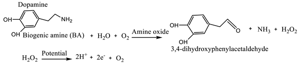
Biosensors contain a bioreceptor and a transducer as two major parts for accurate detection of chemical components and transform biochemical information into electrical or optical signals. These bioreceptors are immobilized with nucleic acids, antigens, or hormones, or enzymes, molecularly imprinted polymers (MIP), or chemoresponsive dyes such as chemical/natural pH dyes, conjugated polymers, colorimetric sensor arrays, and fluorophores for recognizing and identifying each target element. Whereas, the transducer helps to transform the biochemical information into electrical or optical signals later measured employing colorimetric or electroanalytical devices [7]. The sensing mechanism of these biosensors are based on the reaction of active site with the bioreceptor (immobilized biorecognition) as biological or organic material) and the substrate from the tested food material. The electrons produced due to the chemical reaction create a medium of electron flow on the surface of the electrode so that the transducer transform them as response signals. A typical sensing mechanism of biogenic amine to monitor the quality and safety of meat is presented by Nami, et al. [7]. First, biogenic amine present in a meat product is oxidized into hydrogen peroxide (H2O2), NH 3, and aldehyde in the presence of oxygen and water using amine oxides as a catalyst (Equation 1). Next, by applying a high potential, the produced hydrogen peroxide (H2O2) is dissociated into 2 hydrogen ions, oxygen creating and 2 electrons. Then, 2 electrons are formed which use as electron flow providing response signals by the surface of the electrode.
 (1)
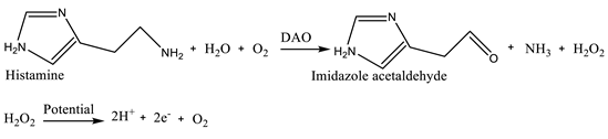
(1)Similar sensing mechanism of histamine to monitor the histamine in the fish spoilage is presented in Equation 2 [13]. First, histamine is oxidized into imidazole acetaldehyde, NH3, and H2O2 using diamine oxidase (DAO). Then, the produced hydrogen peroxide ((H2O2) is dissociated into 2 hydrogen ions, oxygen and creates 2 electrons which use as electron flow providing response signals by the surface of the electrode.
 (2)
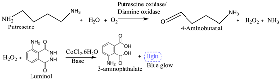
(2)Omanovic-Miklicanin and Valzacchi [14] developed chemiluminescence biosensors to detect the presence of putrescine (Put) and cadaverine in beef, pork, chicken, turkey and fish meat samples. They used putrescine oxidase or diamine oxidase as biorecognition elements (bioreceptors) and microplate luminometer as detection device. The enzymatic reaction and biosensing mechanism are explained in Equation 3. Since the putrescine does not show chemiluminescence characteristic its concentration in meat samples cannot be determined directly during the chemiluminescence reaction. However, it can be measured indirectly by measuring H2O2. Hence, first, the putrescine is oxidized into H2O2, 4-aminobutanal and NH3 in the presence of oxygen and water and using putrescine oxidase or diamine oxidase enzymes (Equation 3). Then, H2O2 is reacted with luminol in an alkaline solution and using cobalt (II) chloride hexahydrate as chemiluminescence catalyst. At last, this catalyzed chemiluminescence reaction of luminol with hydrogen peroxide create 3-aminophthalate with light and the created light is measured employing microplate luminometer (Equation 3). The produced light intensity due to this reaction is proportional to the concentration of hydrogen peroxide.
 (3)
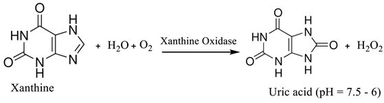
(3)Availability of xanthine in chicken meat can be sensed using an optical biosensor developed employing guanine deaminase and xanthine oxidase as bioreceptors [15]. Briefly, xanthine oxidase and dye phenol red indicator were co-immobilized into sol-gel based circular plastic discs to develop the biosensor. The mechanism for this biosensing is: first, xanthine is converted into uric acid and H2O2 in the presence H2O and oxygen and xanthine oxidase as an enzymatic catalyst (Equation 4). The produced uric acid change pH range of the medium in range of 7.5 - 6. Then, using phenol red as absorptive dye the change in colour can be visualized.
 (4)
(4)2.2. Types of Biosensors Employed to Monitor Fish, Meat, Poultry and Related Product Quality Parameters
Several biosensors have been developed and introduced to the current world. These biosensors have different applications based on the target analyte intended to be determined. These have been employed for fish, meat, poultry and related product quality and safety monitoring. Nami, et al. [7] classified particular biosensors based on the type of bioreceptor (biorecognition Element) utilize into enzyme-based biosensors, immunosensors and DNA-based biosensor. Enzyme-based biosensors employ enzymes to create contact with sample analyte and produce signal for measurement. Immunosensors use antibodies as a bioreceptor which are applicable for the detection of pathogens or toxins in meat samples. These antibodies contact with pathogen or toxin and create signal for measurement. However, DNA-based biosensors are applicable to detect particular DNA of meat samples using DNA as biorecognition element. Here, the bioreceptor DNA interact with the target DNA in the particular analyte. Some of the enzyme-based biosensors, immunosensors and DNA-based biosensor and their bioreceptor employed for monitoring fish, meat, poultry and related product quality and safety are summarized in Table 1.
Nanda, et al. [9] on the other hand classified biosensors based on the immobilization of biorecognition elements, types of transducers use, and detection techniques apply as label-free and label-based. Bioreceptor based biosensors are developed by immobilizing enzyme, aptamer, whole cell, nano, immunosensors, and antibody. Electrochemical, optical, mass-based/gravimetric biosensors are based on their transducers use during real-time analysis of target analytes. Some transducers and electrode employed with biosensors for fish and meat quality monitoring are presented in Table 1. On the other hand, label-free such as acoustic wave, surface plasmon resonance-SPR, and mass spectrometry, and label-based like fluorescence and chemiluminescence bioreceptors are grouped based on their detection techniques.
Emerging biosensor technologies such as smartphones, artificial sensing, 3D printing, and Internet of Things (IOT) are being applicable for detection techniques of fish, meat, poultry and related product quality and safety monitoring. Using machine learning models are applicable in optical sensor-based methods considering color changes and water loss to predict beef quality [17].
3. Applications of Biosensors in Real-Time Food Quality Monitoring In Fish, Meat and Meat Products
3.1. Biosensor-Based Detection of Freshness Indicators in Fish, Meat and Meat Products
In order to measure the qualitative and quantitative characteristics of meat and meat products freshness, the visual appearance, pH, and meaty aroma are the major ones. In this line, metabolites, synthesized by chemical oxidation and microbial proliferation, could modify the quality of muscle foods freshness. Throughout storage, metabolites produced by from microbial development and chemical oxidations change the muscle foods quality and freshness.
3.1.1. Hypoxanthine
In the fish/meat industry, the xanthine (XA) is exploited as a freshness indicator because of its accumulation in the tissues after died. In order to monitor the freshness of pork, a TiO2 and graphene composite has been established [6]. The biosensor assesses the oxidation activity of xanthine oxidase (XOD) and hypoxanthine (Hx) during seven days at refrigerated condition [6]. The Hx existence provoked a sour taste that is facile to detect the fish and meat sample degradation. Pierini, et al. [23] developed an electroanalytical tool (edge plane pyrolytic graphite electrode (EPPGE)) to determine the Hx, Xa and UA content in Argentinian fish samples. Similarly, to control the Hx content in pork meat freshness at several post-mortem periods, Guo, et al. [24] developed an enzyme sensor by joining O2 electrode and XOD. These authors reported that the produced biosensor displayed heightened sensitivity to Hx compared to HPLC analysis. Through grafting reduced expanded graphene oxide (REGO) with Fe3O4 nanoparticles, Dervisevic, et al. [25] produced a new amperometric xanthine biosensor and applied it to control fish freshness till 20 days. The xanthine concentration was detected at a range between 2 and 36 mM, at 3 seconds at detection limit equal to 0.17 mA/M. Interestingly, after 25 days of old fish samples, biosensor held 70% of its activity. In order to assess the fish freshness with color marking by the unaided eye, XOD was employed in enzyme-mediated AuNRs oxidation [26]. In this study, the color of the sensing system has a good link with Hx level at a range between 0.05 and 0.63 mM. Chen, et al. [27] proposed a fluorescence sensor derived from platinum nanoparticles (Pt NPs) to perceive Hx in aquatic products. At a [Hx] ranged between 8-2500 μM, the new biomaterial possesses a linear connection and detection limit at 2.88 μM. In meat samples, a μPAD biosensor was developed to Hx detection. The detection and quantitative limits were registered at 1.8 and 6.1 mg/L, respectively. The proposed assay exhibited a linear dynamic in the range of 5–40 mg/L. The analysis time was 5 min for triplicate measurement [28]. To assess the meat freshness by Hx detection, Görgülü, et al. [29] fabricated multi-enzyme biosensors. In this study, Polypyrrole–polyvinyl sulphonate (PPy–PTS) films were synthesized on the platinum electrode surface by electropolymerization. The indicator enzymes, XOD and uricase, were immobilized within the polymer matrix. The registered amperometric response, at a potential of +400 mV, was attributed to the current resulting from the enzymatic oxidation of H2O2. The established biosensor displayed a minimum detection limit of 2.5 µM, a concentration range with a linear response of 2.5 to 10 µM. After 33 days of storage, the biosensor maintained 65% of its initial performance, demonstrating acceptable long-term stability for practical applications. For the evaluation of HX in beef, chicken fish, and pork meat, Devi, et al. [30] evolved a biosensor designed with Au/Fe nanoparticules, and XOD was covalently grafted onto the electrode surface. At an optimal response within 3 s at pH 7.2 and 30°C, the biosensor showed linearity in the range of 0.05 µM to 150 µM for Hx, with a detection limit of 0.05 µM. By employing an absorption transmission approach, Garg and Verma [15] fabricated an optical biosensor to detect Hx. The assay operates on the enzymatic reaction catalyzed by XO, which transforms xanthine into uric acid and H₂O₂. Owing to the uric acid formation, this reaction provokes a pH reduction, characteristically from 7.5 to 6.0. These investigated the xanthine content in chicken meat during 5 days of storage. As projected, xanthine concentrations augmented eventually, representing deteriorating of meat quality, from ~5 µM on day 1 to 44 µM on day 5. The method's reliability was established within spiked samples; display a recovery % between 94.2 and 96.5%. Zhang, et al. [31] developed a new tool named electrochemiluminescence (ECL) of CdS quantum dots (QDs), combining electrochemistry and chemiluminescence. This technique indicated that electrical energy was used to launch a chemical reaction that generates light. These authors assembled and synthesized the new material onto poly (diallyldimethylammonium chloride)-functionalized carbon nanospheres (PFCNSs) and leading to an increase of ECL intensity by dissolved O2 as a coreactant. The sensor established a fast response with a linear range from 2.5 × 10⁻⁸ -1.4 × 10⁻⁵ M and a detection limit of 5 nM (S/N = 3), and the obtained findings from fish sample analysis were closely matched to those from standard amperometric methods.
3.1.2. Biogenic Amines and Volatile Amines
Biogenic amines, small organic compounds comprising one or more amino groups, are categorized into aliphatic, aromatic, and heterocyclic amines. These amines are mainly synthesized by enzymatic decarboxylation of free amino acids or by amination and transamination of aldehydes and ketones [32].
The most prevalent biogenic amines present in aquatic and meat products are tyramine, cadaverine, putrescine, histamine, and trimethylamine [33]. Throughout the muscle food deterioration, the formation of histamine, putrescine, and cadaverine are generally used as freshness indicators and could be monitored. Zhai, et al. [34] created an amine-responsive bilayer films by using agar (AG), anthocyanins (AN), gellan gum (GG), and TiO₂ nanoparticles for visual monitoring of meat spoilage. The AG-AN layer served as the detecting layer for volatile amines, while tThe AG-AN/GG-2%TiO₂ film noticed trimethylamine (TMA) at a limit of 0.018 mM, a typical gas from meat spoilage. During silver carp and pork spoilage, the film exhibited a range color variation between the rose-red and green, stress its possible use in intelligent food packaging.
Based on the peroxidase-like activity of (Fe and Co) codoped-CDs, Li, et al. [35] developed a colorimetric tool to detect the cadaverine and the putrescine. With the enzymatic hydrolysis by diamine oxidase, biogenic amines were disintegrated to generate H2O2 which reacts with tetramethyl-benzidine with the catalysis of (Fe,Co)-codoped CDs. The colorimetric method was used to perceive of cadaverine and the putrescine in various fish samples with a limit of 0.06 mg/kg. Checked by HPLC, the recoveries of the colorimetric method were confirmed by standards, signifying that the established colorimetric method was sensitive and accurate.
In order to monitor biogenic amines (BAs), Luo, et al. [36] developed a hydrogel composed of β-d-glucose pentaacetate (β-D-GP), silver ions, and agarose. Under alkaline conditions, in contact with BAs, β-D-GP could be hydrolyzed to form β-d-glucose, which decreases silver ions to silver nanoparticles, and generate visible color variations. These changes can be analyzed with the naked eye or quantified using smartphone-based RGB (red/green/blue) analysis in fish samples. Polyaniline (PANI) synthesized via in situ chemical oxidative polymerization was spray-coated onto flexible interdigitated electrodes (IDEs) for noticing ammonia gas and have been employed for checking food quality. The sensor's electrical response augmented linearly with increasing ammonia concentrations. It confirmed a constant linear response in the 50–150 ppm range and effectively evaluated the meat and sheep liver freshness in real-time[37]. Chang, et al. [38] produced a detecting system provided with an ultrasensitive amine gas sensor to perceive volatile amines from raw fish. Remarkably, the sensor offers an electrical response within 1 min that meticulously links with TVB-N values. The sensor’s ppb-level sensitivity and integrated humidity control enable fast and accurate detection. These findings support the development of real-time, on-site freshness monitoring in fish processing environments. The amine gas sensor is able to detect ammonia, dimethylamine (DMA), and trimethylamine (TMA) at ppb levels, permitting it to monitor volatile compounds released from raw fish and indicate spoilage. The method truthfully releases the effects of storage temperature and a fish portions viz. ventral, dorsal, and lateral on spoilage development. For beltfish and mackerel, the sensors displayed robust correlation with TVB-N values.
Through in-situ polymerization, Shi, et al. [39] deposited the TiO2-PANI- into Silk Fibroin Fiber (SFF). The novel composite (TiO2-PANI/SFF) played the role of an excellent micro sensor exhibiting sensing capability, with a response value equal to 0.82 and a response time of 10 seconds to 100 μg/L of NH3. In pork samples, when utilized to evaluate freshness, the sensor's was powerfully interrelated with TVB-N levels (R² = 0.99). In order to measure the TVB-N in pork by combing 2 non-destructive sensing methods: colorimetric sensors and HIS: hyperspectral imaging [40]. For data fusion and modeling, these authors proposed a BP-AdaBoost which corresponds to an effective backpropagation adaptive boosting algorithm. The performance of model was examined relative to PCA-BPANN: backpropagation artificial neural network model. Test results revealed that the data fusion model outdid the single-sensor models, with BP-AdaBoost proposing superior capability in handling complex data fusion compared to PCA-BPANN. In pork meat, this investigation revealed the possible integrating of HIS and colorimetric sensors, and the BP-AdaBoost algorithm for non-destructive TVB-N.
3.2. Biosensor-Based Detection of Microbial Hazards in Fish, Meat and Meat Products
The occurrence of pathogenic microorganisms in food caused significant dangers to general health safety and can also affect the environment. The biosensors expansion has importantly improved food safety [41]. Conventional microbiological methods characteristically comprise enrichment, filtration, and incubation phases, requiring a time frame of from 2 - 10 days to obtain [42]. Contrarily, modern biosensor-based tools proposed earlier and more precise detection, with the further advantage of on-site pertinence. For pathogens, their toxins, and metabolites, their low detection limits highpoint the importance of highly sensitive analytical tools for guaranteeing the fish, meat and meat products safety.
Across different optical sensing methods, colorimetric, fluorescence, Chemiluminescence Surface Plasmon Resonance (SPR) and Localized Surface Plasmon Resonance (LSPR), are usually employed [43]. In the SPR-based biosensing, normally engages reflectance spectroscopy for the detection of target pathogens, the bioreceptors are fixed to a metal transducer surface. Where specific wavelengths of electromagnetic radiation act towards the metal's electron and generate resonance. When bacterial cells attach to this surface, they induce quantifiable variations in the refractive index [41]. In order to detect pathogenic microorganisms in different meat and meat products, optical biosensors were employed. As an illustration, a fiber-optic immunosensor, fortified with immunomagnetic separation, certainly perceived Listeria monocytogenes in meat at levels as < 3 × 10² CFU/mL [44]. Another method employed an aptamer-based fiber-optic biosensor to select L. monocytogenes in artificially infected ready-to-eat (RTE) meat, effectively identifying it specifically among other microbial strains [45]. Oh, et al. [46] engaged LSPR to detect Salmonella Typhimurium in pork at 4 log CFU/mL within 30 min. To synchronize detect the E. coli O157:H7, Salmonella enteritidis, and Listeria monocytogenes, Zhang, et al. [47] established an SPR biosensor combined to an enrichment broth. In order to simplify selective recognition, polyclonal antibodies which are special for each pathogen were anchored on separate channels of SPR chip. After an enrichment step, chicken meat were analyzed using the SPR system, efficaciously perceiving target microorganisms at 14, 6, and 28 CFU/25 g for E. coli O157:H7, Salmonella enteritidis, and Listeria monocytogenes, respectively. Liang, et al. [48] produced a smartphone-based biosensor to detect microbial spoilage on ground beef. In this study, le lower limit of detection was between 10 and 100 CFU of Escherichia coli K12. Morant-Miñana and Elizalde [49] produced an electrochemical genosensor for the Campylobacter spp detection. This new material, developed from thin-film gold electrodes dropped onto Cyclo Olefin Polymer (COP), displayed high sensitivity, robust linear response observed for Campylobacter spp, and positively authenticated by real poultry meat samples. It displayed similar findings to those obtained with purified PCR products with a concentration ranged between 1 - 25 nM, and a LOD equal to 90 pM. Ohk and Bhunia [4] developed and optimized a multiplex fiber optic sensor able to detect simultnesouly L. monocytogenes, E. coli O157:H7, and S. enterica in food samples. Streptavidin-coated optical sensors were equipped with biotinylated polyclonal antibodies and treated with bacterial suspensions or supplemented food samples for 2 hours. In this study, turkey, ready-to-eat beef, and chicken samples were inoculated with ~102 CFU of each pathogen /25 g was enriched during 18 hours in a selective enrichment medium SEL broth and tested by the biosensor. The sensor positively recognized each pathogen individually or in combination and the detection limit was 10³ CFU/mL for all three pathogens. This new approach, multiplex fiber optic biosensor, could be proper to detect simultaneously Listeria, E. coli, and Salmonella in food, decreasing the necessity for separate single-pathogen detection systems. By virtue of its excellent characteristics like ultra-rapid electron transfer aptitude, great surface/volume ratio, suitability for biological applications, and its single connections with DNA bases of the aptamer, Muniandy, et al. [50] fabricated an rGO-azophloxine nanocomposite (rGO-AP) aptasensor to detect foodborne pathogens. The contact of the label-free single-stranded deoxyribonucleic acid (ssDNA) aptamer with S. Typhimurium was examined by variance pulse voltammetry exploration, and this aptasensor indicated high selectivity and sensitivity for the detection of intact bacterial cells. rGO-AP revealed a linear detection range between 10 to 108 CFU/mL and a good linearity (R2 = 98%). Furthermore, rGO-AP could detect, bacterial concentration ranging from 10-104 CFU/g in the inoculated chicken sample with S. Typhimurium. Rasooly [51] evaluated the potential of Surface plasmon resonance (SPR) biosensors to detect staphylococcal enterotoxin B (SEB), engaging 2 antibodies, in foods. A capturing antibody, covalently enclosed to the biosensor chip surface was performed to the initial binding of the antigen and a second antibody sticks to the captured antigen. Initially, the whole assessment cycle taked 5 minutes when using a single antibody or 8 minutes when two antibodies are employed. Interestingly, SPR biosensor could detect SEB in meat at 10 ng/ml, with initial binding < 2 min. On another study conducted byLiu, et al. [52], a fast detection of Salmonella serotypes B and D in ready-to-eat (RTE) turkey has been explored. These authors proved that a concentration of Salmonella < 3×10² cells/mL at1 hour was attained. Additionally, the findings displayed that the sensor is able to distinguish low concentration of live Salmonella cells from high levels of dead Salmonella cells.
3.3. Biosensor-Based Detection of Contaminants, Antibiotics, and Drug Residues in Fish, Meat And Meat Products
Food quality valuation includes perceiving impurities viz. drug residues, pesticides, toxins and heavy metals. Conventional tools like mass spectrometry and capillary electrophoresis are costly and taking considerable time. To guarantee consumer security, biosensors offer a closer and a gainful alternative with adequate perception. For instance, for heavy metals as As, Cd, Hg, biosensors employed enzymes (e.g. glucose oxidase, urease, cholinesterase, alkaline phosphatase) and genetically modified microorganisms [53]. By developing a chemiluminescence sensor called MIP (molecularly imprinted polymer (MIP), Cai, et al. [54] recognized eight benzimidazoles in beef and mutton, establishing ultrafast sensitivity. In fact, these authors confirmed that the detection limits were ranged between 1.5 and 21 pg/mL, with 18 minutes, and High recovery efficiency (66-91%). In order to identify or fungal or bacterial toxins existing in meat products, electrochemical biosensors are used. As an example, trichothecene (T-2 toxin) was detected inn swine meat [55]. By employing an electrochemical and SPR biosensors, Staphylococcal enterotoxin B were sensed in pork [56], in potted meat [51], respectively. By amperometric biosensor, Dinçkaya, et al. [2] appraised the nitrate concentrations in meat and confirmed that LOD was 2.2 × 10⁻⁹ M with a response time equal to 10 s. On the other hand, some studies employed SPR as biosensors to identify drug residues. In several meat species like pork, beef, and chicken, SPR technique was able to detect sulphonamides and chloramphenicol have been quantified [57,58,59]. In order to detect the SDM: sulfadimethoxine in beef and chicken meat, Mohammad-Razdari, et al. [60] established electrochemical biosensor based on pencil graphite electrode (PGE) and adapted with a reduced graphene oxide (RGO) and Au nanoparticles for sulfadimethoxine (SDM). In the best-performing trials, the proposed biosensor showed a linear range from 10− 10−5 M, LOD at 3.7 × 10⁻¹⁶ M towards SDM. For meat sample applications, the aptasensor was applied to fish, chicken, and beef and showed acceptable recovery rates across the tested concentration range, demonstrating dependable performance and accuracy in analytic quantification between 92 and 103%. For the label-free detection of ceftiofur residues in meat trials, Stevenson, et al. [61] developed an affinity-based electrochemical biosensor. These authors validated a platform that can detect ceftiofur within 15 min of using the sample at levels down to 0.01 ng/mL in phosphate-buffered saline and 10 ng/mL in 220 mg ground turkey meat samples. The Table 2 summarizes some examples of biosensors to monitor the quality and safety of fish, meat and meat products.
4. Standardization and Validation of Biosensors in Real-Time Food Quality Monitoring
The standardization and validation of biosensors are indispensable processes for ensuring their reliability, reproducibility, and regulatory acceptance in the food industry. Unlike conventional chemical assays, biosensors often demonstrate significant variability due to differences in biological recognition elements, sensor fabrication, and susceptibility to environmental factors such as temperature, pH, and matrix complexity. This variability makes necessary robust calibration and method validation protocols to ensure consistent performance across food matrices and operational environments [76,77].
Calibration and standardization are foundational steps for establishing accuracy and consistency in biosensor output. The calibration process typically involves the use of matrix-matched reference standards, ideally certified, to reflect real-world food conditions in terms of composition, viscosity, and potential interferents [78]. Biosensors must exhibit predictable and linear responses across a defined concentration range of the target analyte. For example, a heat-transfer biosensor used for detecting trace levels of chemical additives in dairy was calibrated using milk samples with varying fat contents to ensure consistent sensitivity and reproducibility [10].
4.1. Validation
Although harmonized guidelines for validation of biosensor-based methods do not exist, a valid text is represented by the International Council for Harmonisation (ICH) “Bioanalytical Method Validation and Study Sample Analysis – M10 guideline, 2022”. This document was adopted also by the European Medicines Agency (EMA) and the Food and Drug Administration (FDA). Method validation, as defined in the ICH M10 guideline, requires comprehensive evaluation of analytical performance. Key parameters include accuracy, precision (repeatability and intermediate precision), selectivity, sensitivity, linearity, limit of detection (LOD), limit of quantitation (LOQ), carryover, and analyte stability (EMA - European Medicines Agency, 2024; ICH Expert Working Group, 2022; U.S. Department of Health and Human Services Food and Drug Administration, 2022).
4.1.1. Specificity and Cross-Reactivity Challenges
In biosensor-based ligand binding assays (LBA), specificity refers to the sensor's ability to detect only the target analyte without interference from structurally similar compounds, such as analogues, metabolites, or co-formulated substances. This becomes critical when detecting contaminants like veterinary drug residues or pesticide metabolites. Specificity is typically evaluated by spiking blank matrix samples with structurally related compounds at their expected maximal concentrations. A well-validated biosensor should show negligible response to these analogues and maintain accuracy for the primary analyte within ±25% at the extremes of its dynamic range. In cases where specificity is compromised, adjusting the quantification range or employing alternative recognition elements (e.g., more selective antibodies or aptamers) may be necessary.
4.1.2. Selectivity in Complex Food Matrices
Selectivity addresses the biosensor’s performance in distinguishing the analyte from endogenous matrix components that may interfere with detection. This is especially challenging in samples like milk, eggs, or processed foods, where proteins, fats, and enzymes can cause non-specific binding or signal suppression. To ensure selectivity, the assay must be tested in at least 10 different blank food matrix samples, with analyte spiked at both low and high concentrations. The signal from unspiked samples should fall below the lower limit of quantification (LLOQ) in at least 80% of the matrices tested. Selectivity testing should also consider lipemic and hemolyzed conditions, as well as matrices derived from diseased or stressed animal populations when relevant.
4.1.3. Calibration Curve and Reportable Range
Accurate quantification with biosensors depends on the establishment of a calibration curve, relating analyte concentration to signal response. The curve should span from the LLOQ to the upper limit of quantification (ULOQ), ideally covering at least six concentration points plus a blank. Many biosensor platforms use a logistic fit (4- or 5-parameter models) to accommodate non-linear signal responses, especially near saturation zones. A robust calibration curve requires consistency across multiple runs (minimum of six), with at least 75% of calibration points meeting accuracy criteria (±25% at LLOQ/ULOQ; ±20% at other levels).
4.1.4. Accuracy and Precision Requirements
Validation of accuracy (closeness to the true value) and precision (repeatability) is conducted using quality control (QC) samples at multiple concentration levels, typically LLOQ, low, medium, high, and ULOQ. Within-run and between-run performance should be assessed over at least six analytical runs using independently prepared QCs. Acceptable accuracy and precision limits are ±20% (±25% for LLOQ and ULOQ). A total error (sum of bias and variability) threshold of ≤30% (≤40% at extremes) is often applied as an overall acceptance criterion.
4.1.5. Dilution Linearity and High-Dose Hook Effect
Due to the limited dynamic range of many biosensors, dilution of samples with high analyte concentrations is necessary. Dilution linearity must be verified to ensure that sample dilution does not introduce bias. This is also critical for identifying the hook effect, a phenomenon where excessive analyte saturates binding sites, leading to signal suppression. Dilution series should be tested in at least three independent preparations, demonstrating linearity across the measured range, with ≤20% deviation from expected values.
4.1.6. Stability Under Analytical Conditions
Stability testing ensures that storage, processing, and handling conditions do not compromise the biosensor's performance. This includes assessments of freeze-thaw stability, bench-top stability, and long-term storage. For each condition, QCs at low and high concentrations should be evaluated, and analyte recovery should remain within ±20% of nominal values. This step is particularly important for biosensors using biologically active components (e.g., enzymes or antibodies), which are prone to degradation under suboptimal storage.
These criteria ensure the biosensor ability to generate reliable results for target contaminants such as pesticides, preservatives, or industrial pollutants [79]. These validation criteria should be tailored depending on whether the biosensor detects contaminants, chemical additives, toxins, or other analytes. A critical point is also food matrices, in fact the ICH M10 Guideline underlines that other pivotal parameters are matrix effects, incurred sample reanalysis (ISR), and inter-batch reproducibility; all of which are particularly relevant for biosensors deployed in complex food matrices like oils, processed meats, lipid-reach sea foods (e.g., shellfish) [80,81]. In Table 3 a summary of these parameters as well as a brief description is reported.
Despite innovative sensor designs, regulatory approval remains a time-intensive process. In fact, often, apart from a validation study, a comparison study with validated chemical reference methods, such as HPLC or mass spectrometry, is preferred. These comparative assessments are crucial for establishing biosensor equivalence in terms of sensitivity, selectivity, and reproducibility. Without this level of validation, biosensors face challenges in gaining acceptance for routine food safety monitoring, despite offering advantages such as portability and real-time readouts [82,83].
Biosensor integration into quality control systems presents operational challenges, including interoperability with digital traceability platforms, training personnel in sensor operation, and upgrading existing laboratories or processing infrastructure. In large-scale manufacturing environments, biosensor data must seamlessly interface with automated decision-support systems for tasks such as batch release or contamination alerts [84,85].
Furthermore, data harmonization is critical. Standardized biosensor outputs must be structured and formatted for compatibility with central databases that consolidate information from inspections, internal audits, and supply chain feedback. As highlighted by Wijayanti, et al. [86], biosensors are increasingly incorporated into the digitalization of food quality frameworks, but effective deployment requires unified validation standards and interoperable data formats to enable real-time risk assessment and traceability [86].
4.2. Limits and Challenges for Biosensors Application in in Real-Time Food Quality Monitoring
Achieving high sensitivity and specificity remains a central challenge in the development of biosensors for detecting food additives and contaminants. These parameters determine the biosensor’s ability to detect target analytes at trace levels and to discriminate them from structurally similar compounds. In complex food matrices, such as milk or cereals, matrix components can interact with sensor surfaces or recognition elements, leading to background signal noise or false positives [87].
For instance, certain immunoassays for mycotoxins have demonstrated cross-reactivity with masked or metabolized toxin forms, undermining their selectivity. Similarly, surface-enhanced Raman scattering (SERS)-based lateral flow biosensors developed for detecting colistin in milk have shown matrix interference from milk proteins, which reduced analytical clarity despite fast detection times. Such cases highlight the need for advanced recognition elements and sample pre-treatment strategies to mitigate matrix effects and improve signal fidelity. Moreover, the operational stability of biosensors, especially those incorporating biological recognition elements like enzymes or antibodies, is a persistent issue limiting their shelf-life. Enzyme-based biosensors are particularly susceptible to denaturation or leaching during storage, which reduces signal reproducibility and overall reliability [86,88].
Efforts to improve stability have focused on immobilization techniques such as cross-linking, encapsulation in polymeric matrices, or covalent bonding to support materials. These approaches aim to preserve the functional conformation of the biomolecules and enhance resilience to environmental stressors during storage and use. However, long-term validation of such methods under varied food storage conditions remains limited and is critical for regulatory and industrial acceptance. Another critical issue may be represented by environmental factors, including temperature, humidity, and pH, that have a significant impact on biosensor performance. Temperature fluctuations can alter enzyme kinetics, signal generation rates, or the refractive index in optical systems. For example, enzymatic biosensors may show exaggerated signals at elevated temperatures or delayed responses in colder environments. Similarly, pH instability affects the electrochemical response of sensors, especially those incorporating carbon nanomaterials for detecting heavy metals or preservatives [89].
Humidity can degrade sensitive components, particularly in optical biosensors, where uncontrolled moisture introduces signal noise or damages light-sensitive dyes. Moreover, food matrices with variable composition further complicate biosensor operation, reinforcing the need for robust calibration and compensation mechanisms to ensure consistent performance [90].
Some examples of issues in biosensors validation and application for the analysis of food additives and contaminants, along with the study strategies developed for their resolution, are proposed in Table 4.
5. Challenges, Limitations, and Future Perspectives in Biosensor Applications for Fish, Meat, Poultry, and Related Products Safety Monitoring
Addressing the challenges and limitations in ensuring the safety of fish, meat, poultry, and related products is a fundamental pillar of modern food systems. However, biosensors—despite their transformative potential—still face multifaceted limitations that restrict their scalability and real-world implementation. These challenges span across biological, technical, regulatory, and economic domains, especially in resource-limited settings or small-to-medium-scale enterprises.
One of the most fundamental limitations stems from the complexity of food matrices, which vary widely in moisture content, fat and protein composition, and microbial load. These intrinsic properties can interfere with biosensor readings, especially in systems relying on electrochemical or optical signals [87,96,97,98]. High-fat samples like beef or lamb may cause signal drift or fouling of the sensing surface, while the high-water activity in fish products may lead to enzymatic degradation or dilution of target analytes [87,96]. Additionally, meat and poultry tissues can contain a mixture of endogenous enzymes and oxidation byproducts that further complicate signal stability [97,98]. In seafood, detection is further complicated by the presence of marine-specific hazards such as tetrodotoxin, okadaic acid, or domoic acid, which require ultra-sensitive detection limits and matrix-adapted recognition elements [96,99,100]. In poultry, early-stage detection of infection is difficult due to low biomarker concentrations during the asymptomatic phases of disease progression, which often fall below the limit of detection (LOD) of many conventional biosensors [97,101].
Cross-reactivity and specificity pose another technical barrier. Biosensors must be able to differentiate between highly similar microbial species or strains, such as Campylobacter jejuni versus C. coli, or between pathogenic and non-pathogenic E. coli strains, which often share structural markers [87,101,102,103]. The inability of many biosensors to discriminate between viable and non-viable cells may lead to false positives, especially in post-sanitization environments [102,104]. Moreover, many detection platforms still struggle with achieving the necessary selectivity in mixed microbial environments, particularly in raw or minimally processed products [96,105,106].
Furthermore, detection of residues such as tetracyclines or aflatoxins—particularly in trace amounts across different feed types, tissues, or products—requires extremely sensitive and consistently calibrated platforms [106,107,108,109,110,111]. Small deviations in temperature, pH, or sample handling can cause shifts in biosensor response, making reproducibility a serious concern for both researchers and industry practitioners [87,90,112].
Operational challenges are also significant. Electrochemical biosensors often require external power sources and supporting instruments (e.g., potentiostats), which hinder their portability and real-time usability in field inspections [113,114,115,116]. Similarly, colorimetric biosensors, despite their visual simplicity, tend to require multi-step sample preparation and are vulnerable to variations in ambient lighting or subjective interpretation, especially in environments lacking standardized conditions [100,117,118]. Smartphone-based visual readers are being tested to mitigate these issues, but their precision and user-friendliness still vary widely [119,120]. In contrast, SERS-based biosensors, while capable of ultra-sensitive detection, are technically demanding due to their reliance on precision optical components (e.g., Raman lasers, detectors) and the need for specialized substrates such as gold or MOF-coated nanoparticles [100,121,122,123,124,125,126]. Additionally, there is no universal SERS substrate that can accommodate all analyte types, necessitating tailored fabrication for each application [121,125,126].
Moreover, optical and surface plasmon resonance (SPR) technologies provide label-free, real-time monitoring capabilities that are especially suited to packaging and food processing environments [127,128], but they require precise optical setups, which limit portability.
From a regulatory and commercial perspective, biosensor platforms are not yet widely incorporated into formal food safety systems such as HACCP, ISO 22000, or Codex Alimentarius frameworks. Validation against gold-standard methods (e.g., culture-based enumeration, ELISA, or qPCR) is still lacking for many sensor formats, which affects their credibility in audits, certifications, and trade compliance [87,112,129,130,131]. The absence of harmonized validation protocols makes it difficult to compare results across borders or industries, leading to skepticism among food producers and regulatory bodies alike [90,112].
On the economic front, biosensors incorporating nanomaterials, CRISPR technology, or microfluidics often have high development and production costs, especially when coupled with surface functionalization and antibody/aptamer design [97,129,132,133,134,135,136]. This restricts their use in lower-income regions or small-scale food businesses. Scaling up from laboratory prototypes to commercial-grade devices often requires substantial investment in cleanroom facilities, testing, and certification [116,137]. Moreover, concerns regarding the long-term environmental and human health impacts of nanomaterials (e.g., silver nanoparticles, graphene oxide) continue to raise regulatory red flags, necessitating the shift toward green synthesis and biodegradable materials [138,139,140]. The need for non-toxic, disposable sensor platforms is gaining traction in global sustainability goals [138].
Biosensors also have an emerging role in monitoring cultured (cell-based) meat, a sector with specific challenges related to contamination control during cell cultivation, the composition of growth media, and the use of biochemical additives [141,142].
Finally, while point-of-care (POC) and intelligent packaging biosensors are increasingly being designed, real-world uptake is still slow. High costs, difficulty in integrating sensor data into existing software ecosystems, and energy requirements for continuous operation (especially for cold chains or remote sites) further hinder long-term monitoring applications [104,143,144]. In addition, data security and interoperability challenges persist, particularly when transferring biosensor data to cloud-based regulatory or logistics systems [130,131].
Despite the broad range of challenges, biosensor development is progressing rapidly, supported by innovations in nanotechnology, synthetic biology, electronics, and digital infrastructure. The next generation of biosensors is being engineered to meet not only technical performance benchmarks but also criteria for usability, affordability, and sustainability.
A key design philosophy is alignment with the REASSURED framework—Real-time connectivity, Ease of sample collection, Affordable, Sensitive, Specific, User-friendly, Rapid, Robust, Equipment-free, and Deliverable to end users [5,6]. Recent advancements in lab-on-a-chip (LOC) systems and wearable biosensors now allow continuous monitoring of animal stress biomarkers (e.g., cortisol, IL-6), meat spoilage indicators (e.g., biogenic amines), or microbial contamination in real-world environments [120,145,146,147,148]. LOC devices can be used directly in slaughterhouses, packaging lines, or distribution centers, reducing delays between contamination and detection.
Integration of biosensor data with IoT platforms, blockchain-enabled traceability, and AI-driven analytics is revolutionizing food safety by enabling predictive diagnostics and real-time response. For example, blockchain can secure biosensor data logs for traceable certification, while AI algorithms can analyze spectral or electrical patterns to detect anomalies or mixed contaminations [149-154
[149,150,151,152,153,154]. AI-based decision-support systems can also be trained in biosensor outputs to guide preventive measures in processing plants or farms. Smartphone-enabled biosensors also bridge accessibility gaps by allowing frontline inspectors and small producers to capture and transmit results immediately, often with GPS and timestamp metadata
[119,152]. These solutions support decentralized decision-making and democratize food safety monitoring [85,155,156].
The frontiers of biosensor technology are also being expanded through multiplexing and advanced signal amplification. CRISPR-Cas systems offer unparalleled specificity at attomolar levels, enabling detection of pathogens like Listeria monocytogenes, E. coli O157:H7, or Salmonella enterica in complex matrices [132,133,157,158]. Meanwhile, nanozyme-based colorimetric sensors provide robust alternatives to enzyme-based assays, maintaining stability under diverse environmental conditions and simplifying fabrication [159,160,161,162]. Nanozymes also eliminate cold-chain dependence for sensor reagents [161].
Multi-analyte aptasensors are being designed to simultaneously detect microbial pathogens, spoilage indicators, and chemical toxins in a single run—dramatically improving throughput and cost-effectiveness [96,105,163]. Similarly, molecularly imprinted polymer (MIP)-based sensors are showing high selectivity for volatile markers like histamine or trimethylamine, offering practical applications for seafood spoilage detection [164,165]. Such developments are particularly useful in import-exports where spoilage needs to be evaluated rapidly at ports or distribution hubs.
In the realm of packaging, intelligent sensors are now integrated directly into films, labels, or coatings to detect changes in gas composition (e.g., CO₂, NH₃), humidity, or microbial growth. These include hypoxanthine-sensing films for fish freshness and polymyxin B-aptamer platforms for endotoxin detection in poultry products [166,167,168,169,170,171]. Emerging solid-state SERS substrates (e.g., paper, elastomers, AuNS-glass composites) also offer durability and reusability in smart packaging applications [170,172,173]. Such features make them attractive for both consumers and regulatory audits.
To overcome energy and maintenance concerns, self-powered biosensors using biofuel cells or photoelectrochemical modules are being tested for autonomous deployment in storage environments with minimal infrastructure [144]. These devices align well with sustainability goals and reduce the carbon footprint of food monitoring
Ultimately, the successful integration of biosensor technologies into food safety systems will rely not only on overcoming technical and operational barriers but also on establishing robust regulatory frameworks and fostering international standardization. It is essential that biosensor data be recognized as legally valid and interoperable across digital platforms used in global supply chains. Interdisciplinary collaboration among scientists, technologists, policymakers, and industry stakeholders will be key to accelerating the transition from research prototypes to field-deployable, validated tools.
By addressing current limitations in sensitivity, matrix interference, cost-effectiveness, and data integration, biosensors can be positioned as core components of intelligent, sustainable, and resilient food safety systems for the fish, meat, and poultry industries.
6. Conclusions
Biosensors have emerged as transformative tools for ensuring the safety and quality of fish, meat, poultry, and related food products. Their capacity to rapidly and sensitively detect contaminants, pathogens, spoilage markers, and drug residues positions them as viable and often superior alternatives to conventional laboratory-based methods. Recent advancements including the integration of nanomaterials, lab-on-a-chip platforms, smartphone interfaces, and IoT connectivity have significantly enhanced their portability, usability, and real-time monitoring capabilities.
Nevertheless, several critical challenges remain. The complexity of food matrices, environmental variability (such as pH, humidity, and temperature), and the inherent instability of biological recognition elements can affect performance and limit reproducibility. Regulatory acceptance is further constrained by the absence of harmonized validation standards and insufficient comparative assessments with gold-standard analytical techniques. Additionally, high development and implementation costs hinder widespread adoption, particularly in resource-limited settings.
Looking forward, the development of biosensors should prioritize robustness, affordability, and compliance with international regulatory frameworks. Embracing the REASSURED criteria ensuring devices are real-time, easy to use, affordable, sensitive, specific, user-friendly, rapid, robust, equipment-free, and deliverable to end users will be key to broader deployment. Integration with AI-powered analytics and blockchain-based traceability systems can also unlock new opportunities for predictive diagnostics and transparent supply chain management. With continued interdisciplinary collaboration and innovation, biosensors are well-positioned to become cornerstone technologies in next-generation food safety and quality assurance systems.
Author Contributions
S.S.: writing—review and editing, writing—original draft, validation, resources, investigation, formal analysis, data curation, conceptualization. T.V.: writing—review and editing, writing—original draft, visualization, validation, supervision, project administration, methodology, investigation, formal analysis, data curation, conceptualization. E.H.: writing—original draft, writing—review and editing, validation, formal analysis, data curation, investigation, visualization. Z.T.T.: writing—review and editing, writing—original draft, visualization, validation, methodology, investigation, data curation. . T.DM.: writing—review and editing, writing—original draft, visualization, validation, methodology, investigation, data curation. All authors have read and agreed to the published version of the manuscript.
Funding
This research received no external funding
Institutional Review Board Statement
Not applicable.
Informed Consent Statement
Not applicable.
Data Availability Statement
No new data were created or analyzed in this study. Data sharing is not applicable to this article.
Conflicts of Interest
The authors declare no conflicts of interest.
References
- Daramola, O.B.; Omole, R.; Akinsanola, B.A. Emerging applications of biorecognition elements-based optical biosensors for food safety monitoring. Discover Sensors 2025. [Google Scholar] [CrossRef]
- Dinçkaya, E.; Akyilmaz, E.; Sezgintürk, M.K.; Ertaş, F.N. Sensitive nitrate determination in water and meat samples by amperometric biosensor. Prep. Biochem. Biotechnol. 2010, 40, 119–128. [Google Scholar] [CrossRef] [PubMed]
- Zhang, N.; Xiao, F.; Bai, J.; Lai, Y.; Hou, J.; Xian, Y.; Jin, L. Label-free immunoassay for chloramphenicol based on hollow gold nanospheres/chitosan composite. Talanta 2011, 87, 100–105. [Google Scholar] [CrossRef] [PubMed]
- Ohk, S.-H.; Bhunia, A.K. Multiplex fiber optic biosensor for detection of Listeria monocytogenes, Escherichia coli O157:H7 and Salmonella enterica from ready-to-eat meat samples. Food Microbiol. 2013, 33, 166–171. [Google Scholar] [CrossRef]
- Mansouri, M.; Fathi, F.; Jalili, R.; Shoeibie, S.; Dastmalchi, S.; Khataee, A.; Rashidi, M.-R. SPR enhanced DNA biosensor for sensitive detection of donkey meat adulteration. Food Chem. 2020, 331, 127163. [Google Scholar] [CrossRef]
- Albelda, J.A.; Uzunoglu, A.; Santos, G.N.C.; Stanciu, L.A. Graphene-titanium dioxide nanocomposite based hypoxanthine sensor for assessment of meat freshness. Biosens. Bioelectron. 2017, 89, 518–524. [Google Scholar] [CrossRef]
- Nami, M.; Taheri, M.; Siddiqui, J.; Deen, I.A.; Packirisamy, M.; Deen, M.J. Recent Progress in Intelligent Packaging for Seafood and Meat Quality Monitoring. Adv. Mater. Technol. 2024, 9. [Google Scholar] [CrossRef]
- Tsegay, Z.T.; Hosseini, E.; Varzakas, T.; Smaoui, S. The latest research progress on polysaccharides-based biosensors for food packaging: A review. Int. J. Biol. Macromol. 2024, 282, 136959. [Google Scholar] [CrossRef]
- Nanda, P.K.; Bhattacharya, D.; Das, J.K.; Bandyopadhyay, S.; Ekhlas, D.; Lorenzo, J.M.; Dandapat, P.; Alessandroni, L.; Das, A.K.; Gagaoua, M. Emerging Role of Biosensors and Chemical Indicators to Monitor the Quality and Safety of Meat and Meat Products. Chemosensors 2022, 10, 322. [Google Scholar] [CrossRef]
- Nastasijevic, I.; Kundacina, I.; Jaric, S.; Pavlovic, Z.; Radovic, M.; Radonic, V. Recent Advances in Biosensor Technologies for Meat Production Chain. Foods 2025, 14, 744. [Google Scholar] [CrossRef]
- Fritea, L.; Tertis, M.; Sandulescu, R.; Cristea, C. Chapter Eleven - Enzyme–Graphene Platforms for Electrochemical Biosensor Design With Biomedical Applications, in Methods in Enzymology, C.V. Kumar, Editor 2018), Academic Press. pp. 293-333. [CrossRef]
- Sun, G.; Wei, X.; Zhang, D.; Huang, L.; Liu, H.; Fang, H. Immobilization of Enzyme Electrochemical Biosensors and Their Application to Food Bioprocess Monitoring. Biosensors 2023, 13, 886. [Google Scholar] [CrossRef] [PubMed]
- Torre, R.; Costa-Rama, E.; Lopes, P.; Nouws, H.P.A.; Delerue-Matos, C. Amperometric enzyme sensor for the rapid determination of histamine. Anal. Methods 2019, 11, 1264–1269. [Google Scholar] [CrossRef]
- Omanovic-Miklicanin, E.; Valzacchi, S. Development of new chemiluminescence biosensors for determination of biogenic amines in meat. Food Chem. 2017, 235, 98–103. [Google Scholar] [CrossRef] [PubMed]
- Garg, D.; Verma, N. Fibre-optic biosensor for the detection of xanthine for the evaluation of meat freshness. Journal of Physics: Conference Series. 2020, 1531, 012098. [Google Scholar] [CrossRef]
- Uwimbabazi, E.; Mukasekuru, M.R.; Sun, X. Glucose Biosensor Based on a Glassy Carbon Electrode Modified with Multi-Walled Carbon Nanotubes-Chitosan for the Determination of Beef Freshness. Food Anal. Methods 2017, 10, 2667–2676. [Google Scholar] [CrossRef]
- Lee, I.-H.; Ma, L. Integrating machine learning, optical sensors, and robotics for advanced food quality assessment and food processing. Food Innov. Adv. 2025, 4, 65–72. [Google Scholar] [CrossRef]
- Koçoğlu, I.O.; Erden, P.E.; Kılıç, E. Disposable biogenic amine biosensors for histamine determination in fish. Anal. Methods 2020, 12, 3802–3812. [Google Scholar] [CrossRef]
- Zór, K.; Dymek, K.; Ortiz, R.; Faure, A.M.; Saatci, E.; Gorton, L.; Bardsley, R.; Nistor, M. Indirect, non-competitive amperometric immunoassay for accurate quantification of calpastatin, a meat tenderness marker, in bovine muscle. Food Chem. 2012, 133, 598–603. [Google Scholar] [CrossRef]
- Pikkemaat, M.; Rapallini, M.; Karp, M.; Elferink, J. Application of a luminescent bacterial biosensor for the detection of tetracyclines in routine analysis of poultry muscle samples. Food Addit. Contam. Part A 2010, 27, 1112–1117. [Google Scholar] [CrossRef]
- Swaidan, A.; Barras, A.; Addad, A.; Tahon, J.-F.; Toufaily, J.; Hamieh, T.; Szunerits, S.; Boukherroub, R. Colorimetric sensing of dopamine in beef meat using copper sulfide encapsulated within bovine serum albumin functionalized with copper phosphate (CuS-BSA-Cu3(PO4)2) nanoparticles. J. Colloid Interface Sci. 2021, 582, 732–740. [Google Scholar] [CrossRef]
- Vizzini, P.; Manzano, M.; Farre, C.; Meylheuc, T.; Chaix, C.; Ramarao, N.; Vidic, J. Highly sensitive detection of Campylobacter spp. In chicken meat using a silica nanoparticle enhanced dot blot DNA biosensor. Biosens. Bioelectron. 2021, 171, 112689. [Google Scholar] [CrossRef] [PubMed]
- Pierini, G.D.; Robledo, S.N.; Zon, M.A.; Di Nezio, M.S.; Granero, A.M.; Fernández, H. Development of an electroanalytical method to control quality in fish samples based on an edge plane pyrolytic graphite electrode. Simultaneous determination of hypoxanthine, xanthine and uric acid. Microchem. J. 2018, 138, 58–64. [Google Scholar] [CrossRef]
- Guo, C.; You, S.; Li, C.; Chen, T.; Wang, X. One-Step and Colorimetric Detection of Fish Freshness Indicator Hypoxanthine Based on the Peroxidase Activity of Xanthine Oxidase Grade I Ammonium Sulfate Suspension. Front. Microbiol. 2021, 12. [Google Scholar] [CrossRef]
- Dervisevic, M.; Custiuc, E.; Çevik, E.; Durmus, Z.; Şenel, M.; Durmus, A. Electrochemical biosensor based on REGO/Fe3O4 bionanocomposite interface for xanthine detection in fish sample. Food Control. 2015, 57, 402–410. [Google Scholar] [CrossRef]
- Chen, Z.; Lin, Y.; Ma, X.; Guo, L.; Qiu, B.; Chen, G.; Lin, Z. Multicolor biosensor for fish freshness assessment with the naked eye. Sensors Actuators B: Chem. 2017, 252, 201–208. [Google Scholar] [CrossRef]
- Chen, J.; Lu, Y.; Yan, F.; Wu, Y.; Huang, D.; Weng, Z. A fluorescent biosensor based on catalytic activity of platinum nanoparticles for freshness evaluation of aquatic products. Food Chem. 2020, 310, 125922. [Google Scholar] [CrossRef]
- Mooltongchun, M.; Teepoo, S. A Simple and Cost-effective Microfluidic Paper-Based Biosensor Analytical Device and its Application for Hypoxanthine Detection in Meat Samples. Food Anal. Methods 2019, 12, 2690–2698. [Google Scholar] [CrossRef]
- Görgülü, M.; Çete, S.; Arslan, H.; Yaşar, A. Preparing a new biosensor for hypoxanthine determination by immobilization of xanthine oxidase and uricase in polypyrrole-polyvinyl sulphonate film. Artif. Cells, Nanomedicine, Biotechnol. 2013, 41, 327–331. [Google Scholar] [CrossRef]
- Devi, R.; Yadav, S.; Nehra, R.; Yadav, S.; Pundir, C. Electrochemical biosensor based on gold coated iron nanoparticles/chitosan composite bound xanthine oxidase for detection of xanthine in fish meat. J. Food Eng. 2013, 115, 207–214. [Google Scholar] [CrossRef]
- Zhang, Y.; Deng, S.; Lei, J.; Xu, Q.; Ju, H. Carbon nanospheres enhanced electrochemiluminescence of CdS quantum dots for biosensing of hypoxanthine. Talanta 2011, 85, 2154–2158. [Google Scholar] [CrossRef]
- Martuscelli, M.; Esposito, L.; Mastrocola, D. Biogenic Amines’ Content in Safe and Quality Food. Foods 2021, 10, 100. [Google Scholar] [CrossRef] [PubMed]
- Schirone, M.; Esposito, L.; D’onofrio, F.; Visciano, P.; Martuscelli, M.; Mastrocola, D.; Paparella, A. Biogenic Amines in Meat and Meat Products: A Review of the Science and Future Perspectives. Foods 2022, 11, 788. [Google Scholar] [CrossRef]
- Zhai, X.; Zou, X.; Shi, J.; Huang, X.; Sun, Z.; Li, Z.; Sun, Y.; Li, Y.; Wang, X.; Holmes, M.; et al. Amine-responsive bilayer films with improved illumination stability and electrochemical writing property for visual monitoring of meat spoilage. Sensors Actuators B: Chem. 2020, 302. [Google Scholar] [CrossRef]
- Li, Y.-F.; Lin, Z.-Z.; Hong, C.-Y.; Huang, Z.-Y. Colorimetric detection of putrescine and cadaverine in aquatic products based on the mimic enzyme of (Fe,Co) codoped carbon dots. J. Food Meas. Charact. 2021, 15, 1747–1753. [Google Scholar] [CrossRef]
- Luo, Q.; Zhang, Y.; Zhou, Y.; Liu, S.G.; Gao, W.; Shi, X. Portable functional hydrogels based on silver metallization for visual monitoring of fish freshness. Food Control. 2021, 123. [Google Scholar] [CrossRef]
- Matindoust, S.; Farzi, A.; Nejad, M.B.; Abadi, M.H.S.; Zou, Z.; Zheng, L.-R. Ammonia gas sensor based on flexible polyaniline films for rapid detection of spoilage in protein-rich foods. J. Mater. Sci. Mater. Electron. 2017, 28, 7760–7768. [Google Scholar] [CrossRef]
- Chang, L.-Y.; Chuang, M.-Y.; Zan, H.-W.; Meng, H.-F.; Lu, C.-J.; Yeh, P.-H.; Chen, J.-N. One-Minute Fish Freshness Evaluation by Testing the Volatile Amine Gas with an Ultrasensitive Porous-Electrode-Capped Organic Gas Sensor System. ACS Sensors 2017, 2, 531–539. [Google Scholar] [CrossRef]
- Shi, Y.; Li, Z.; Shi, J.; Zhang, F.; Zhou, X.; Li, Y.; Holmes, M.; Zhang, W.; Zou, X. Titanium dioxide-polyaniline/silk fibroin microfiber sensor for pork freshness evaluation. Sensors Actuators B: Chem. 2018, 260, 465–474. [Google Scholar] [CrossRef]
- Li, H.; Chen, Q.; Zhao, J.; Wu, M. Nondestructive detection of total volatile basic nitrogen (TVB-N) content in pork meat by integrating hyperspectral imaging and colorimetric sensor combined with a nonlinear data fusion. LWT 2015, 63, 268–274. [Google Scholar] [CrossRef]
- Ali, A.A.; Altemimi, A.B.; Alhelfi, N.; Ibrahim, S.A. Application of Biosensors for Detection of Pathogenic Food Bacteria: A Review. Biosensors 2020, 10, 58. [Google Scholar] [CrossRef]
- Ito, T.; Sekizuka, T.; Kishi, N.; Yamashita, A.; Kuroda, M. Conventional culture methods with commercially available media unveil the presence of novel culturable bacteria. Gut Microbes 2018, 10, 77–91. [Google Scholar] [CrossRef]
- Li, Z.; Lin, H.; Wang, L.; Cao, L.; Sui, J.; Wang, K. Optical sensing techniques for rapid detection of agrochemicals: Strategies, challenges, and perspectives. Sci. Total. Environ. 2022, 838, 156515. [Google Scholar] [CrossRef] [PubMed]
- Mendonça, M.; Conrad, N.L.; Conceição, F.R.; Moreira, Â.N.; da Silva, W.P.; Aleixo, J.A.; Bhunia, A.K. Highly specific fiber optic immunosensor coupled with immunomagnetic separation for detection of low levels of Listeria monocytogenes and L. ivanovii. BMC Microbiol. 2012, 12, 275–275. [Google Scholar] [CrossRef] [PubMed]
- Alhogail, S.; Suaifan, G.A.; Zourob, M. Rapid colorimetric sensing platform for the detection of Listeria monocytogenes foodborne pathogen. Biosens. Bioelectron. 2016, 86, 1061–1066. [Google Scholar] [CrossRef] [PubMed]
- Oh, S.Y.; Heo, N.S.; Shukla, S.; Cho, H.J.; Vilian, A.E.; Kim, J.; Lee, S.Y.; Han, Y.-K.; Yoo, S.M.; Huh, Y.S. Development of gold nanoparticle-aptamer-based LSPR sensing chips for the rapid detection of Salmonella typhimurium in pork meat. Sci. Rep. 2017, 7, 10130. [Google Scholar] [CrossRef]
- Zhang, H.; Ma, X.; Liu, Y.; Duan, N.; Wu, S.; Wang, Z.; Xu, B. Gold nanoparticles enhanced SERS aptasensor for the simultaneous detection of Salmonella typhimurium and Staphylococcus aureus. Biosens. Bioelectron. 2015, 74, 872–877. [Google Scholar] [CrossRef]
- Liang, P.-S.; Park, T.S.; Yoon, J.-Y. Rapid and reagentless detection of microbial contamination within meat utilizing a smartphone-based biosensor. Sci. Rep. 2014, 4, 5953. [Google Scholar] [CrossRef]
- Morant-Miñana, M.C.; Elizalde, J. Microscale electrodes integrated on COP for real sample Campylobacter spp. detection. Biosens. Bioelectron. 2015, 70, 491–497. [Google Scholar] [CrossRef]
- Muniandy, S.; Dinshaw, I.J.; Teh, S.J.; Lai, C.W.; Ibrahim, F.; Thong, K.L.; Leo, B.F. Graphene-based label-free electrochemical aptasensor for rapid and sensitive detection of foodborne pathogen. Anal. Bioanal. Chem. 2017, 409, 6893–6905. [Google Scholar] [CrossRef]
- Rasooly, A. Surface Plasmon Resonance Analysis of Staphylococcal Enterotoxin B in Food. J. Food Prot. 2001, 64, 37–43. [Google Scholar] [CrossRef]
- Liu, J.; Jasim, I.; Shen, Z.; Zhao, L.; Dweik, M.; Zhang, S.; Almasri, M. A microfluidic based biosensor for rapid detection of Salmonella in food products. PLOS ONE 2019, 14, e0216873. [Google Scholar] [CrossRef] [PubMed]
- Turdean, G.L. Design and Development of Biosensors for the Detection of Heavy Metal Toxicity. Int. J. Electrochem. 2011, 2011, 1–15. [Google Scholar] [CrossRef]
- Cai, Y.; He, X.; Cui, P.L.; Liu, J.; Bin Li, Z.; Jia, B.J.; Zhang, T.; Wang, J.P.; Yuan, W.Z. Preparation of a chemiluminescence sensor for multi-detection of benzimidazoles in meat based on molecularly imprinted polymer. Food Chem. 2019, 280, 103–109. [Google Scholar] [CrossRef]
- Wang, Y.; Zhang, L.; Peng, D.; Xie, S.; Chen, D.; Pan, Y.; Tao, Y.; Yuan, Z. Construction of Electrochemical Immunosensor Based on Gold-Nanoparticles/Carbon Nanotubes/Chitosan for Sensitive Determination of T-2 Toxin in Feed and Swine Meat. Int. J. Mol. Sci. 2018, 19, 3895. [Google Scholar] [CrossRef]
- Tang, D.; Tang, J.; Su, B.; Chen, G. Ultrasensitive Electrochemical Immunoassay of Staphylococcal Enterotoxin B in Food Using Enzyme-Nanosilica-Doped Carbon Nanotubes for Signal Amplification. J. Agric. Food Chem. 2010, 58, 10824–10830. [Google Scholar] [CrossRef]
- Gao, D.; Guan, C.; Wen, Y.; Zhong, X.; Yuan, L. Multi-hole fiber based surface plasmon resonance sensor operated at near-infrared wavelengths. Opt. Commun. 2014, 313, 94–98. [Google Scholar] [CrossRef]
- McGrath, T.; Baxter, A.; Ferguson, J.; Haughey, S.; Bjurling, P. Multi sulfonamide screening in porcine muscle using a surface plasmon resonance biosensor. Anal. Chim. Acta 2005, 529, 123–127. [Google Scholar] [CrossRef]
- Yuan, J.; Oliver, R.; Aguilar, M.-I.; Wu, Y. Surface Plasmon Resonance Assay for Chloramphenicol. Anal. Chem. 2008, 80, 8329–8333. [Google Scholar] [CrossRef] [PubMed]
- Mohammad-Razdari, A.; Ghasemi-Varnamkhasti, M.; Izadi, Z.; Rostami, S.; Ensafi, A.A.; Siadat, M.; Losson, E. Detection of sulfadimethoxine in meat samples using a novel electrochemical biosensor as a rapid analysis method. J. Food Compos. Anal. 2019, 82. [Google Scholar] [CrossRef]
- Stevenson, H.S.; Shetty, S.S.; Thomas, N.J.; Dhamu, V.N.; Bhide, A.; Prasad, S. Ultrasensitive and Rapid-Response Sensor for the Electrochemical Detection of Antibiotic Residues within Meat Samples. ACS Omega 2019, 4, 6324–6330. [Google Scholar] [CrossRef]
- Baş, S.Z.; Gülce, H.; Yildiz, S. Hypoxanthine Biosensor Based on Immobilization of Xanthine Oxidase on Modified Pt Electrode and Its Application for Fish Meat. Int. J. Polym. Mater. Polym. Biomater. 2014, 63, 476–485. [Google Scholar] [CrossRef]
- Zhang, Y.; Xin, Y.; Yang, H.; Zhang, L.; Xia, X.; Tong, Y.; Chen, Y.; Ma, L.; Wang, W. Novel affinity purification of xanthine oxidase from Arthrobacter M3. J. Chromatogr. B 2012, 906, 19–24. [Google Scholar] [CrossRef] [PubMed]
- Erol, E.; Yildirim, E.; Cete, S. Construction of biosensor for hypoxanthine determination by immobilization of xanthine oxidase and uricase in polypyrrole-paratoluenesulfonate film. J. Solid State Electrochem. 2020, 24, 1695–1707. [Google Scholar] [CrossRef]
- Torres, A.C.; Ghica, M.E.; Brett, C.M.A. Design of a new hypoxanthine biosensor: xanthine oxidase modified carbon film and multi-walled carbon nanotube/carbon film electrodes. Anal. Bioanal. Chem. 2012, 405, 3813–3822. [Google Scholar] [CrossRef]
- Mustafa, F.; Andreescu, S. Paper-Based Enzyme Biosensor for One-Step Detection of Hypoxanthine in Fresh and Degraded Fish. ACS Sensors 2020, 5, 4092–4100. [Google Scholar] [CrossRef]
- Bratcher, C.; Grant, S.; Vassalli, J.; Lorenzen, C. Enhanced efficiency of a capillary-based biosensor over an optical fiber biosensor for detecting calpastatin. Biosens. Bioelectron. 2008, 23, 1674–1679. [Google Scholar] [CrossRef]
- Costa, C.A.B.; Grazhdan, D.; Fiutowski, J.; Nebling, E.; Blohm, L.; Lofink, F.; Rubahn, H.-G.; Hansen, R.d.O. Meat and fish freshness evaluation by functionalized cantilever-based biosensors. Microsyst. Technol. 2019, 26, 867–871. [Google Scholar] [CrossRef]
- Zór, K.; Castellarnau, M.; Pascual, D.; Pich, S.; Plasencia, C.; Bardsley, R.; Nistor, M. Design, development and application of a bioelectrochemical detection system for meat tenderness prediction. Biosens. Bioelectron. 2011, 26, 4283–4288. [Google Scholar] [CrossRef]
- Liu, G. , Chai, C., & Yao, B., Rapid evaluation of Salmonella pullorum contamination in chicken based on a portable amperometric sensor. J. Biosens. Bioelectron. 2013, 137, 2. [Google Scholar]
- Helali, S.; Alatawi, A.S.E.; Abdelghani, A. Pathogenic Escherichia coli biosensor detection on chicken food samples. J. Food Saf. 2018, 38. [Google Scholar] [CrossRef]
- Banerjee, P.; Bhunia, A.K. Cell-based biosensor for rapid screening of pathogens and toxins. Biosens. Bioelectron. 2010, 26, 99–106. [Google Scholar] [CrossRef] [PubMed]
- Ferguson, J.; Baxter, A.; Young, P.; Kennedy, G.; Elliott, C.; Weigel, S.; Gatermann, R.; Ashwin, H.; Stead, S.; Sharman, M. Detection of chloramphenicol and chloramphenicol glucuronide residues in poultry muscle, honey, prawn and milk using a surface plasmon resonance biosensor and Qflex® kit chloramphenicol. Anal. Chim. Acta 2005, 529, 109–113. [Google Scholar] [CrossRef]
- Wu, Y.-Y.; Huang, P.; Wu, F.-Y. A label-free colorimetric aptasensor based on controllable aggregation of AuNPs for the detection of multiplex antibiotics. Food Chem. 2020, 304, 125377. [Google Scholar] [CrossRef] [PubMed]
- Lu, X.; Zheng, H.; Li, X.-Q.; Yuan, X.-X.; Li, H.; Deng, L.-G.; Zhang, H.; Wang, W.-Z.; Yang, G.-S.; Meng, M.; et al. Detection of ractopamine residues in pork by surface plasmon resonance-based biosensor inhibition immunoassay. Food Chem. 2012, 130, 1061–1065. [Google Scholar] [CrossRef]
- Chen, X.; Yao, C.; Li, Z. Microarray-based chemical sensors and biosensors: Fundamentals and food safety applications. TrAC Trends Anal. Chem. 2022, 158. [Google Scholar] [CrossRef]
- Romanholo, P.V.; Sgobbi, L.F. Chapter 4 - Validation of biosensors, in Biosensors in Precision Medicine, L.C. Brazaca and J.R. Sempionatto, Editors(2024), Elsevier. pp. 105-131. [CrossRef]
- Zhang, J.; Huang, H.; Song, G.; Huang, K.; Luo, Y.; Liu, Q.; He, X.; Cheng, N. Intelligent biosensing strategies for rapid detection in food safety: A review. Biosens. Bioelectron. 2022, 202, 114003. [Google Scholar] [CrossRef]
- Oliveira, B.D.Á.; Gomes, R.S.; de Carvalho, A.M.; Lima, E.M.F.; Pinto, U.M.; da Cunha, L.R. Revolutionizing food safety with electrochemical biosensors for rapid and portable pathogen detection. Braz. J. Microbiol. 2024, 55, 2511–2525. [Google Scholar] [CrossRef]
- Ozkan, S.A.; Uslu, B.; Sezgintürk, M.K. Biosensors; Taylor & Francis: London, United Kingdom, 2022. [Google Scholar] [CrossRef]
- Nath, S. Advancements in food quality monitoring: integrating biosensors for precision detection. Sustain. Food Technol. 2024, 2, 976–992. [Google Scholar] [CrossRef]
- Indyk, H.E.; Woollard, D.C. Single laboratory validation of an optical biosensor method for the determination of folate in foods. J. Food Compos. Anal. 2013, 29, 87–93. [Google Scholar] [CrossRef]
- Schulze, H.; Scherbaum, E.; Anastassiades, M.; Vorlová, S.; Schmid, R.D.; Bachmann, T.T. Development, validation, and application of an acetylcholinesterase-biosensor test for the direct detection of insecticide residues in infant food. Biosens. Bioelectron. 2002, 17, 1095–1105. [Google Scholar] [CrossRef]
- Thakur, M.S.; Ragavan, K.V. Biosensors in food processing. J. Food Sci. Technol. 2012, 50, 625–641. [Google Scholar] [CrossRef] [PubMed]
- Meliana, C.; Liu, J.; Show, P.L.; Low, S.S. Biosensor in smart food traceability system for food safety and security. Bioengineered 2024, 15, 2310908. [Google Scholar] [CrossRef] [PubMed]
- Wijayanti, S.D.; Tsvik, L.; Haltrich, D. Recent Advances in Electrochemical Enzyme-Based Biosensors for Food and Beverage Analysis. Foods 2023, 12, 3355. [Google Scholar] [CrossRef]
- Curulli, A. Electrochemical Biosensors in Food Safety: Challenges and Perspectives. Molecules 2021, 26, 2940. [Google Scholar] [CrossRef]
- Inês, A.; Cosme, F. Biosensors for Detecting Food Contaminants—An Overview. Processes 2025, 13, 380. [Google Scholar] [CrossRef]
- Lv, M.; Liu, Y.; Geng, J.; Kou, X.; Xin, Z.; Yang, D. Engineering nanomaterials-based biosensors for food safety detection. Biosens. Bioelectron. 2018, 106, 122–128. [Google Scholar] [CrossRef]
- Griesche, C.; Baeumner, A.J. Biosensors to support sustainable agriculture and food safety. TrAC Trends Anal. Chem. 2020, 128. [Google Scholar] [CrossRef]
- Ouyang, Q.; Wang, L.; Ahmad, W.; Rong, Y.; Li, H.; Hu, Y.; Chen, Q. A highly sensitive detection of carbendazim pesticide in food based on the upconversion-MnO2 luminescent resonance energy transfer biosensor. Food Chem. 2021, 349, 129157. [Google Scholar] [CrossRef] [PubMed]
- Yang, J.; Zhang, Y.; Luo, G.; Zheng, S.; Han, S.; Liang, S.; Lin, Y. Collaborative Light-off and Light-on Bacterial Luciferase Biosensors: Innovative Approach for Rapid Contaminant Detection. Biosens. Bioelectron. 2025, 280, 117369. [Google Scholar] [CrossRef]
- Jadán, F.; Aristoy, M.-C.; Toldrá, F. Biosensor Based on Immobilized Nitrate Reductase for the Quantification of Nitrate Ions in Dry-Cured Ham. Food Anal. Methods 2017, 10, 3481–3486. [Google Scholar] [CrossRef]
- Zhao, L.; Guo, H.; Chen, H.; Zou, B.; Yang, C.; Zhang, X.; Gao, Y.; Sun, M.; Wang, L. A Rapid and Sensitive Aptamer-Based Biosensor for Amnesic Shellfish Toxin Domoic Acid. Bioengineering 2022, 9, 684. [Google Scholar] [CrossRef] [PubMed]
- Fonfría, E.S.; Vilariño, N.; Campbell, K.; Elliott, C.; Haughey, S.A.; Ben-Gigirey, B.; Vieites, J.M.; Kawatsu, ✗. K.; Botana, L.M. Paralytic Shellfish Poisoning Detection by Surface Plasmon Resonance-Based Biosensors in Shellfish Matrixes. Anal. Chem. 2007, 79, 6303–6311. [Google Scholar] [CrossRef]
- Baskaran, N.; Sakthivel, R.; Karthik, C.S.; Lin, Y.-C.; Liu, X.; Wen, H.-W.; Yang, W.; Chung, R.-J. Polydopamine-modified 3D flower-like ZnMoO4 integrated MXene-based label-free electrochemical immunosensor for the food-borne pathogen Listeria monocytogenes detection in milk and seafood. Talanta 2024, 282. [Google Scholar] [CrossRef] [PubMed]
- Bahadır, E.B.; Sezgintürk, M.K. Applications of commercial biosensors in clinical, food, environmental, and biothreat/biowarfare analyses. Anal. Biochem. 2015, 478, 107–120. [Google Scholar] [CrossRef]
- Radhakrishnan, R.; Suni, I.I.; Bever, C.S.; Hammock, B.D. Impedance Biosensors: Applications to Sustainability and Remaining Technical Challenges. ACS Sustain. Chem. Eng. 2014, 2, 1649–1655. [Google Scholar] [CrossRef]
- Yibar, A.; Ajmi, N.; Duman, M. First report and genomic characterization of Escherichia coli O111:H12 serotype from raw mussels in Türkiye. BMC Genom. 2024, 25, 1–12. [Google Scholar] [CrossRef]
- Li, P.; Long, F.; Chen, W.; Chen, J.; Chu, P.K.; Wang, H. Fundamentals and applications of surface-enhanced Raman spectroscopy–based biosensors. Curr. Opin. Biomed. Eng. 2020, 13, 51–59. [Google Scholar] [CrossRef]
- Boodhoo, N.; Doost, J.S.; Sharif, S. Biosensors for Monitoring, Detecting, and Tracking Dissemination of Poultry-Borne Bacterial Pathogens Along the Poultry Value Chain: A Review. Animals 2024, 14, 3138. [Google Scholar] [CrossRef] [PubMed]
- Masdor, N.A.; Altintas, Z.; Tothill, I.E. Surface Plasmon Resonance Immunosensor for the Detection of Campylobacter jejuni. Chemosensors 2017, 5, 16. [Google Scholar] [CrossRef]
- Zhang, H.; Zhou, L.; Zhu, Z.; Yang, C. Recent Progress in Aptamer-Based Functional Probes for Bioanalysis and Biomedicine. Chem. – A Eur. J. 2016, 22, 9886–9900. [Google Scholar] [CrossRef]
- Mak, W.C.; Beni, V.; Turner, A.P. Lateral-flow technology: From visual to instrumental. TrAC Trends Anal. Chem. 2016, 79, 297–305. [Google Scholar] [CrossRef]
- Sharma, P.; Thakur, D.; Kumar, D. Novel Enzymatic Biosensor Utilizing a MoS2/MoO3 Nanohybrid for the Electrochemical Detection of Xanthine in Fish Meat. ACS Omega 2023, 8, 31962–31971. [Google Scholar] [CrossRef] [PubMed]
- Callaway, T.R.; Lillehoj, H.; Chuanchuen, R.; Gay, C.G. Alternatives to Antibiotics: A Symposium on the Challenges and Solutions for Animal Health and Production. Antibiotics 2021, 10, 471. [Google Scholar] [CrossRef]
- Laxminarayan, R.; Duse, A.; Wattal, C.; Zaidi, A.K.M.; Wertheim, H.F.L.; Sumpradit, N.; Vlieghe, E.; Hara, G.L.; Gould, I.M.; Goossens, H.; et al. Antibiotic resistance—the need for global solutions. Lancet Infect. Dis. 2013, 13, 1057–1098. [Google Scholar] [CrossRef]
- Carlet, J. The gut is the epicentre of antibiotic resistance. Antimicrob. Resist. Infect. Control. 2012, 1, 39–39. [Google Scholar] [CrossRef] [PubMed]
- Er, B.; Onurdağ, F.K.; Demirhan, B.; Özgacar, S.Ö.; Öktem, A.B.; Abbasoğlu, U. Screening of quinolone antibiotic residues in chicken meat and beef sold in the markets of Ankara, Turkey. Poult. Sci. 2013, 92, 2212–2215. [Google Scholar] [CrossRef]
- Zhai, S.; Zhu, Y.; Feng, P.; Li, M.; Wang, W.; Yang, L.; Yang, Y. Ochratoxin A: its impact on poultry gut health and microbiota, an overview. Poult. Sci. 2021, 100, 101037. [Google Scholar] [CrossRef]
- Lozano, M.; Diaz, G. Microsomal and cytosolic biotransformation of aflatoxin B1 in four poultry species. Br. Poult. Sci. 2006, 47, 734–741. [Google Scholar] [CrossRef]
- Manessis, G.; Gelasakis, A.I.; Bossis, I. Point-of-Care Diagnostics for Farm Animal Diseases: From Biosensors to Integrated Lab-on-Chip Devices. Biosensors 2022, 12, 455. [Google Scholar] [CrossRef]
- Trinh, T.N.D.; Nguyen, H.A.; Thi, N.P.A.; Nam, N.N.; Tran, N.K.S.; Trinh, K.T.L. Biosensors for Seafood Safety Control—A Review. Micromachines 2024, 15, 1509. [Google Scholar] [CrossRef]
- Jiang, H.; Sun, Z.; Guo, Q.; Weng, X. Microfluidic thread-based electrochemical aptasensor for rapid detection of Vibrio parahaemolyticus. Biosens. Bioelectron. 2021, 182, 113191. [Google Scholar] [CrossRef] [PubMed]
- Villalonga, A.; Sánchez, A.; Mayol, B.; Reviejo, J.; Villalonga, R. Electrochemical biosensors for food bioprocess monitoring. Curr. Opin. Food Sci. 2022, 43, 18–26. [Google Scholar] [CrossRef]
- López, M.; Moreno-Guzman, M.; Jurado, B.; Escarpa, A. Chapter 9 - Food Microfluidics Biosensors, in Comprehensive Analytical Chemistry, V. Scognamiglio, G. Rea, F. Arduini, and G. Palleschi, Editors(2016), Elsevier. pp. 273-312. [CrossRef]
- Yang, F.-Q.; Ge, L. Colorimetric Sensors: Methods and Applications. Sensors 2023, 23, 9887. [Google Scholar] [CrossRef] [PubMed]
- Cui, Y.; Zhao, J.; Li, H. Chromogenic Mechanisms of Colorimetric Sensors Based on Gold Nanoparticles. Biosensors 2023, 13, 801. [Google Scholar] [CrossRef]
- Chi, J.; Ju, P.; Bi, F.; Wen, S.; Xiang, Z.; Chen, J.; Qiu, M. A smartphone-assisted ultrasensitive colorimetric aptasensor based on DNA-encoded porous MXene nanozyme for visual detection of okadaic acid. Food Chem. 2024, 464, 141776. [Google Scholar] [CrossRef]
- Roda, A.; Michelini, E.; Zangheri, M.; Di Fusco, M.; Calabria, D.; Simoni, P. Smartphone-based biosensors: A critical review and perspectives. TrAC Trends Anal. Chem. 2016, 79, 317–325. [Google Scholar] [CrossRef]
- Pan, Y.; Xu, S.; Wang, Z.; Jiang, C.; Ma, X. Sensitive SERS aptasensor for histamine detection based on Au/Ag nanorods and IRMOF-3@Au based flexible PDMS membrane. Anal. Chim. Acta 2023, 1288, 342147. [Google Scholar] [CrossRef]
- Lee, S.; Dang, H.; Moon, J.-I.; Kim, K.; Joung, Y.; Park, S.; Yu, Q.; Chen, J.; Lu, M.; Chen, L.; et al. SERS-based microdevices for use as in vitro diagnostic biosensors. Chem. Soc. Rev. 2024, 53, 5394–5427. [Google Scholar] [CrossRef] [PubMed]
- Chauhan, N.; Saxena, K.; Rawal, R.; Yadav, L.; Jain, U. Advances in surface-enhanced Raman spectroscopy-based sensors for detection of various biomarkers. Prog. Biophys. Mol. Biol. 2023, 184, 32–41. [Google Scholar] [CrossRef]
- Usman, F.; Ghazali, K.H.; Fen, Y.W.; Meriaudeau, F.; Jose, R. Biosensing through surface enhanced Raman spectroscopy: A review on the role of plasmonic nanoparticle-polymer composites. Eur. Polym. J. 2023, 195. [Google Scholar] [CrossRef]
- Zhou, J.-Y.; Zhu, J.; Weng, G.-J.; Li, J.-J.; Zhao, J.-W. Fabrication of SERS composite substrates using Ag nanotriangles-modified SiO2 photonic crystal and the application of malachite green detection. Spectrochim. Acta Part A: Mol. Biomol. Spectrosc. 2024, 318, 124472. [Google Scholar] [CrossRef] [PubMed]
- Li, K.; Li, H.; Yin, M.; Yang, D.; Xiao, F.; Tammina, S.K.; Yang, Y. Fluorescence-SERS dual-mode for sensing histamine on specific binding histamine-derivative and gold nanoparticles. Spectrochim. Acta Part A: Mol. Biomol. Spectrosc. 2022, 273, 121047. [Google Scholar] [CrossRef] [PubMed]
- Ravindran, N.; Kumar, S.; M, Y.; S, R.; A, M.C.; S, N.T.; K, S.C. Recent advances in Surface Plasmon Resonance (SPR) biosensors for food analysis: a review. Crit. Rev. Food Sci. Nutr. 2021, 63, 1055–1077. [Google Scholar] [CrossRef]
- Zhou, J.; Qi, Q.; Wang, C.; Qian, Y.; Liu, G.; Wang, Y.; Fu, L. Surface plasmon resonance (SPR) biosensors for food allergen detection in food matrices. Biosens. Bioelectron. 2019, 142, 111449. [Google Scholar] [CrossRef]
- Ramezani, M.; Esmaelpourfarkhani, M.; Taghdisi, S.M.; Abnous, K.; Alibolandi, M. Chapter 22 -Application of nanosensors for food safety, in Nanosensors for Smart Cities, B. Han, V.K. Tomer, T.A. Nguyen, A. Farmani, and P. Kumar Singh, Editors(2020), Elsevier. pp. 369-386. [CrossRef]
- Prodromidis, M.I. Impedimetric immunosensors—A review. Electrochimica Acta 2010, 55, 4227–4233. [Google Scholar] [CrossRef]
- Bosona, T.; Gebresenbet, G. Food traceability as an integral part of logistics management in food and agricultural supply chain. Food Control 2013, 33, 32–48. [Google Scholar] [CrossRef]
- Arshad, F.; Abdillah, A.N.; Shivanand, P.; Ahmed, M.U. CeO2 nanozyme mediated RPA/CRISPR-Cas12a dual-mode biosensor for detection of invA gene in Salmonella. Biosens. Bioelectron. 2023, 247, 115940. [Google Scholar] [CrossRef]
- Zhu, L.; Liang, Z.; Xu, Y.; Chen, Z.; Wang, J.; Zhou, L. Ultrasensitive and Rapid Visual Detection of Escherichia coli O157:H7 Based on RAA-CRISPR/Cas12a System. Biosensors 2023, 13, 659. [Google Scholar] [CrossRef] [PubMed]
- Wang, J.Y.; Tuck, O.T.; Skopintsev, P.; Soczek, K.M.; Li, G.; Al-Shayeb, B.; Zhou, J.; Doudna, J.A. Genome expansion by a CRISPR trimmer-integrase. Nature 2023, 618, 855–861. [Google Scholar] [CrossRef]
- Mohtar, L.G.; Aranda, P.; Messina, G.A.; Nazareno, M.A.; Pereira, S.V.; Raba, J.; Bertolino, F.A. Amperometric biosensor based on laccase immobilized onto a nanostructured screen-printed electrode for determination of polyphenols in propolis. Microchem. J. 2019, 144, 13–18. [Google Scholar] [CrossRef]
- Ye, Y.; Ji, J.; Sun, Z.; Shen, P.; Sun, X. Recent advances in electrochemical biosensors for antioxidant analysis in foodstuff. TrAC Trends Anal. Chem. 2020, 122. [Google Scholar] [CrossRef]
- Weng, X.; Neethirajan, S. Ensuring food safety: Quality monitoring using microfluidics. Trends Food Sci. Technol. 2017, 65, 10–22. [Google Scholar] [CrossRef]
- Sonu; Chaudhary, V. A Paradigm of Internet-of-Nano-Things Inspired Intelligent Plant Pathogen-Diagnostic Biosensors. ECS Sensors Plus 2022, 1, 031401. [Google Scholar] [CrossRef]
- Ariyarathna, I.R.; Rajakaruna, R.; Karunaratne, D.N. The rise of inorganic nanomaterial implementation in food applications. Food Control. 2017, 77, 251–259. [Google Scholar] [CrossRef]
- Verma, N.; Kaur, G. Chapter 2 - Trends on Biosensing Systems for Heavy Metal Detection, in Comprehensive Analytical Chemistry, V. Scognamiglio, G. Rea, F. Arduini, and G. Palleschi, Editors(2016), Elsevier. pp. 33-71. [CrossRef]
- Pawar, D.; Presti, D.L.; Silvestri, S.; Schena, E.; Massaroni, C. Current and future technologies for monitoring cultured meat: A review. Food Res. Int. 2023, 173, 113464. [Google Scholar] [CrossRef]
- FAO, Food Safety Aspects of Cell-Based Food—Background Document Two: Generic Production Process, FAO, Editor.(2022), FAO: Rome.
- Dudefoi, W.; Villares, A.; Peyron, S.; Moreau, C.; Ropers, M.-H.; Gontard, N.; Cathala, B. Nanoscience and nanotechnologies for biobased materials, packaging and food applications: New opportunities and concerns. Innov. Food Sci. Emerg. Technol. 2018, 46, 107–121. [Google Scholar] [CrossRef]
- Hao, S.; Sun, X.; Zhang, H.; Zhai, J.; Dong, S. Recent development of biofuel cell based self-powered biosensors. J. Mater. Chem. B 2020, 8, 3393–3407. [Google Scholar] [CrossRef] [PubMed]
- Zhang, D.; Liu, Q. Biosensors and bioelectronics on smartphone for portable biochemical detection. Biosens. Bioelectron. 2016, 75, 273–284. [Google Scholar] [CrossRef]
- Kim, J.; Imani, S.; de Araujo, W.R.; Warchall, J.; Valdés-Ramírez, G.; Paixao, T.R.L.C.; Mercier, P.P.; Wang, J. Wearable salivary uric acid mouthguard biosensor with integrated wireless electronics. Biosens. Bioelectron. 2015, 74, 1061–1068. [Google Scholar] [CrossRef]
- Zhou, Q.; Kannan, P.; Natarajan, B.; Maiyalagan, T.; Subramanian, P.; Jiang, Z.; Mao, S. MnO2 cacti-like nanostructured platform powers the enhanced electrochemical immunobiosensing of cortisol. Sensors Actuators B: Chem. 2020, 317. [Google Scholar] [CrossRef]
- Piñeiro, M.; Morales, J.; Vizcaíno, E.; Murillo, J.A.; Klauke, T.; Petersen, B.; Piñeiro, C. The use of acute phase proteins for monitoring animal health and welfare in the pig production chain: The validation of an immunochromatographic method for the detection of elevated levels of pig-MAP. Meat Sci. 2013, 95, 712–718. [Google Scholar] [CrossRef]
- Neethirajan, S. Recent advances in wearable sensors for animal health management. Sens. Bio-Sens. Res. 2017, 12, 15–29. [Google Scholar] [CrossRef]
- Land, K.J.; Boeras, D.I.; Chen, X.-S.; Ramsay, A.R.; Peeling, R.W. REASSURED diagnostics to inform disease control strategies, strengthen health systems and improve patient outcomes. Nat. Microbiol. 2018, 4, 46–54. [Google Scholar] [CrossRef]
- Lin, K.; Chavalarias, D.; Panahi, M.; Yeh, T.; Takimoto, K.; Mizoguchi, M. Mobile-based traceability system for sustainable food supply networks. Nat. Food 2020, 1, 673–679. [Google Scholar] [CrossRef] [PubMed]
- Abdelbasset, W.K.; Savina, S.V.; Mavaluru, D.; Shichiyakh, R.A.; Bokov, D.O.; Mustafa, Y.F. Smartphone based aptasensors as intelligent biodevice for food contamination detection in food and soil samples: Recent advances. Talanta 2022, 252, 123769. [Google Scholar] [CrossRef]
- Zhang, Y.; Lin, T.; Shen, Y.; Li, H. A High-Performance Self-Supporting Electrochemical Biosensor to Detect Aflatoxin B1. Biosensors 2022, 12, 897. [Google Scholar] [CrossRef] [PubMed]
- Xie, Y.; Lu, L.; Gao, F.; He, S.-J.; Zhao, H.-J.; Fang, Y.; Yang, J.-M.; An, Y.; Ye, Z.-W.; Dong, Z. Integration of Artificial Intelligence, Blockchain, and Wearable Technology for Chronic Disease Management: A New Paradigm in Smart Healthcare. Curr. Med Sci. 2021, 41, 1123–1133. [Google Scholar] [CrossRef]
- WHO, Food safety.(2022, May 19), WHO.
- Chen, S.; Brahma, S.; Mackay, J.; Cao, C.; Aliakbarian, B. The role of smart packaging system in food supply chain. J. Food Sci. 2020, 85, 517–525. [Google Scholar] [CrossRef]
- Li, F.; Ye, Q.; Chen, M.; Zhou, B.; Zhang, J.; Pang, R.; Xue, L.; Wang, J.; Zeng, H.; Wu, S.; et al. An ultrasensitive CRISPR/Cas12a based electrochemical biosensor for Listeria monocytogenes detection. Biosens. Bioelectron. 2021, 179, 113073. [Google Scholar] [CrossRef]
- Ma, L.; Peng, L.; Yin, L.; Liu, G.; Man, S. CRISPR-Cas12a-Powered Dual-Mode Biosensor for Ultrasensitive and Cross-validating Detection of Pathogenic Bacteria. ACS Sensors 2021, 6, 2920–2927. [Google Scholar] [CrossRef]
- Yang, L.; Xu, X.; Song, Y.; Huang, J.; Xu, H. Research progress of nanozymes in colorimetric biosensing: Classification, activity and application. Chem. Eng. J. 2024, 487. [Google Scholar] [CrossRef]
- Sun, Z.; Zhang, B.; Tu, H.; Pan, C.; Chai, Y.; Chen, W. Advances in colorimetric biosensors of exosomes: novel approaches based on natural enzymes and nanozymes. Nanoscale 2023, 16, 1005–1024. [Google Scholar] [CrossRef] [PubMed]
- Zhao, F.; Wang, L.; Li, M.; Wang, M.; Liu, G.; Ping, J. Nanozyme-based biosensor for organophosphorus pesticide monitoring: Functional design, biosensing strategy, and detection application. TrAC Trends Anal. Chem. 2023, 165. [Google Scholar] [CrossRef]
- Zhang, Z.; Liu, D.; Zhang, X.; Luo, X.; Lin, W.; Li, Z.; Huang, J. Silver nanoparticles deposited carbon microspheres nanozyme with enhanced peroxidase-like catalysis for colorimetric detection of Hg2+ in seafood. Microchim. Acta 2023, 190, 1–11. [Google Scholar] [CrossRef] [PubMed]
- Li, J.; Lin, X.; Wu, J.; Ying, D.; Duan, N.; Wang, Z.; Wu, S. Multifunctional magnetic composite nanomaterial for Colorimetric-SERS Dual-Mode detection and photothermal sterilization of Vibrio parahaemolyticus. Chem. Eng. J. 2023, 477. [Google Scholar] [CrossRef]
- Ayerdurai, V.; Cieplak, M.; Kutner, W. Molecularly imprinted polymer-based electrochemical sensors for food contaminants determination. TrAC Trends Anal. Chem. 2022, 158. [Google Scholar] [CrossRef]
- Munir, M.A.; Rahmawati, F.; Jamal, J.A.; Ibrahim, S.; Said, M.M.; Ahmad, M.S. Inspecting Histamine Isolated from Fish through a Highly Selective Molecularly Imprinted Electrochemical Sensor Approach. ACS Omega 2023, 8, 13352–13361. [Google Scholar] [CrossRef]
- Ding, S.-J.; Chang, B.-W.; Wu, C.-C.; Chen, C.-J.; Chang, H.-C. A new method for detection of endotoxin on polymyxin B-immobilized gold electrodes. Electrochem. Commun. 2007, 9, 1206–1211. [Google Scholar] [CrossRef]
- Su, W.; Lin, M.; Lee, H.; Cho, M.; Choe, W.-S.; Lee, Y. Determination of endotoxin through an aptamer-based impedance biosensor. Biosens. Bioelectron. 2012, 32, 32–36. [Google Scholar] [CrossRef]
- Kubicek-Sutherland, J.Z.; Vu, D.M.; Noormohamed, A.; Mendez, H.M.; Stromberg, L.R.; Pedersen, C.A.; Hengartner, A.C.; Klosterman, K.E.; Bridgewater, H.A.; Otieno, V.; et al. Direct detection of bacteremia by exploiting host-pathogen interactions of lipoteichoic acid and lipopolysaccharide. Sci. Rep. 2019, 9, 1–14. [Google Scholar] [CrossRef]
- Wang, L.; He, Y.; Wu, Z. Design of a Blockchain-Enabled Traceability System Framework for Food Supply Chains. Foods 2022, 11, 744. [Google Scholar] [CrossRef] [PubMed]
- Mustafa, F.; Othman, A.; Andreescu, S. Cerium oxide-based hypoxanthine biosensor for Fish spoilage monitoring. Sensors Actuators B: Chem. 2021, 332. [Google Scholar] [CrossRef]
- Cheng, X.; Liu, W.; Wang, Z.; Yang, R.; Yu, L.; Du, Q.; Ge, A.; Liu, C.; Chi, Z. Improved triple-module fluorescent biosensor for the rapid and ultrasensitive detection of Campylobacter jejuni in livestock and dairy. Food Control. 2022, 137. [Google Scholar] [CrossRef]
- Das, A.; Terry, L.R.; Guo, H. A surface-enhanced Raman spectroscopy based smart Petri dish for sensitive and rapid detection of bacterial contamination in shrimp. Food Chem. Adv. 2023, 2. [Google Scholar] [CrossRef]
- Guo, H.; Li, Y.; Pi, F. Sensitive and reproducible gold nanostar@metal–organic framework-based SERS membranes for the online monitoring of the freshness of shrimps. Anal. 2023, 148, 2081–2091. [Google Scholar] [CrossRef]
Figure 1.
Schematic representation of meat and fish sample target analytes, biorecognition elements and an analytical method.
Figure 1.
Schematic representation of meat and fish sample target analytes, biorecognition elements and an analytical method.
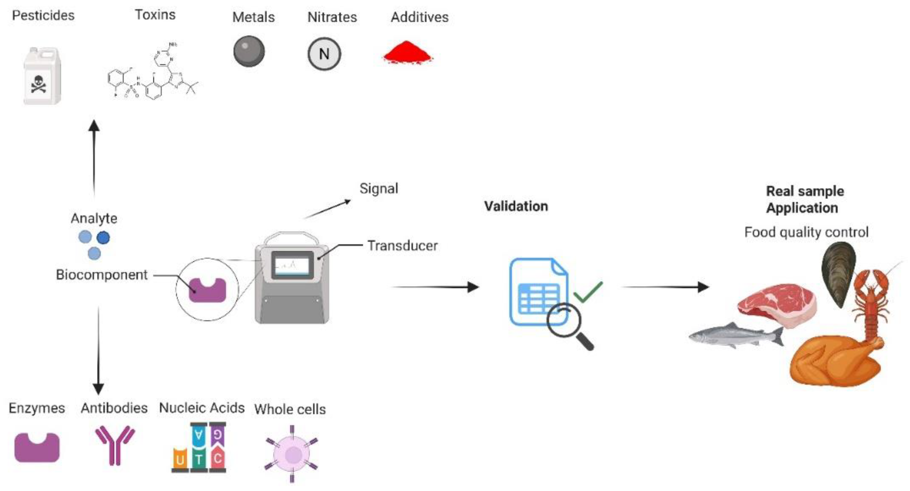
Figure 2.
Diagram illustrating the detection of biogenic amines in meat samples.
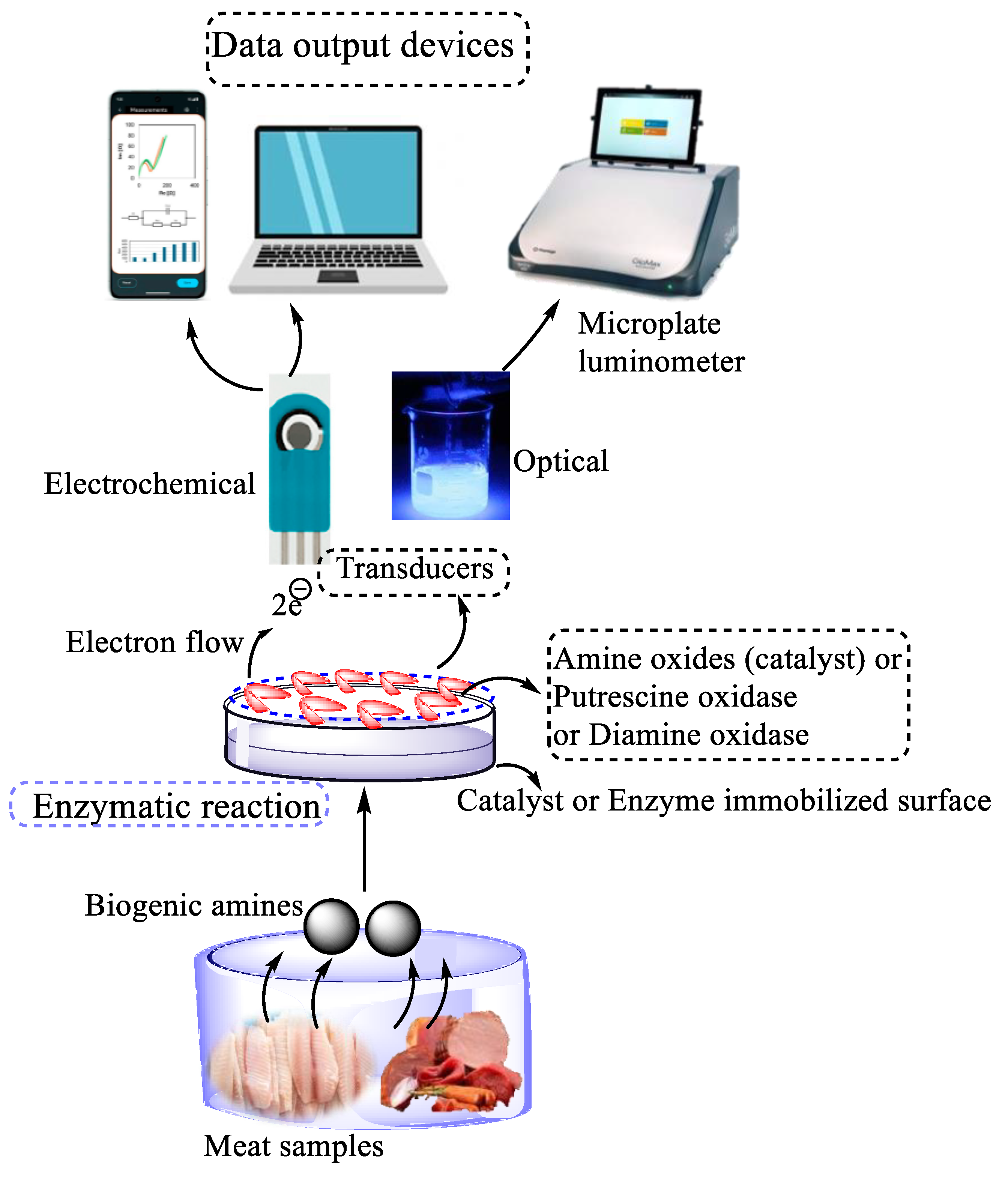
Figure 3.
Illustration of stepwise glucose biosensor preparation process and electrochemical measurement of glucose reduction in fresh meat (reproduced from Uwimbabazi, et al. [16] with permission from the Journal of Food Analytical Methods, copyright 2017).
Figure 3.
Illustration of stepwise glucose biosensor preparation process and electrochemical measurement of glucose reduction in fresh meat (reproduced from Uwimbabazi, et al. [16] with permission from the Journal of Food Analytical Methods, copyright 2017).
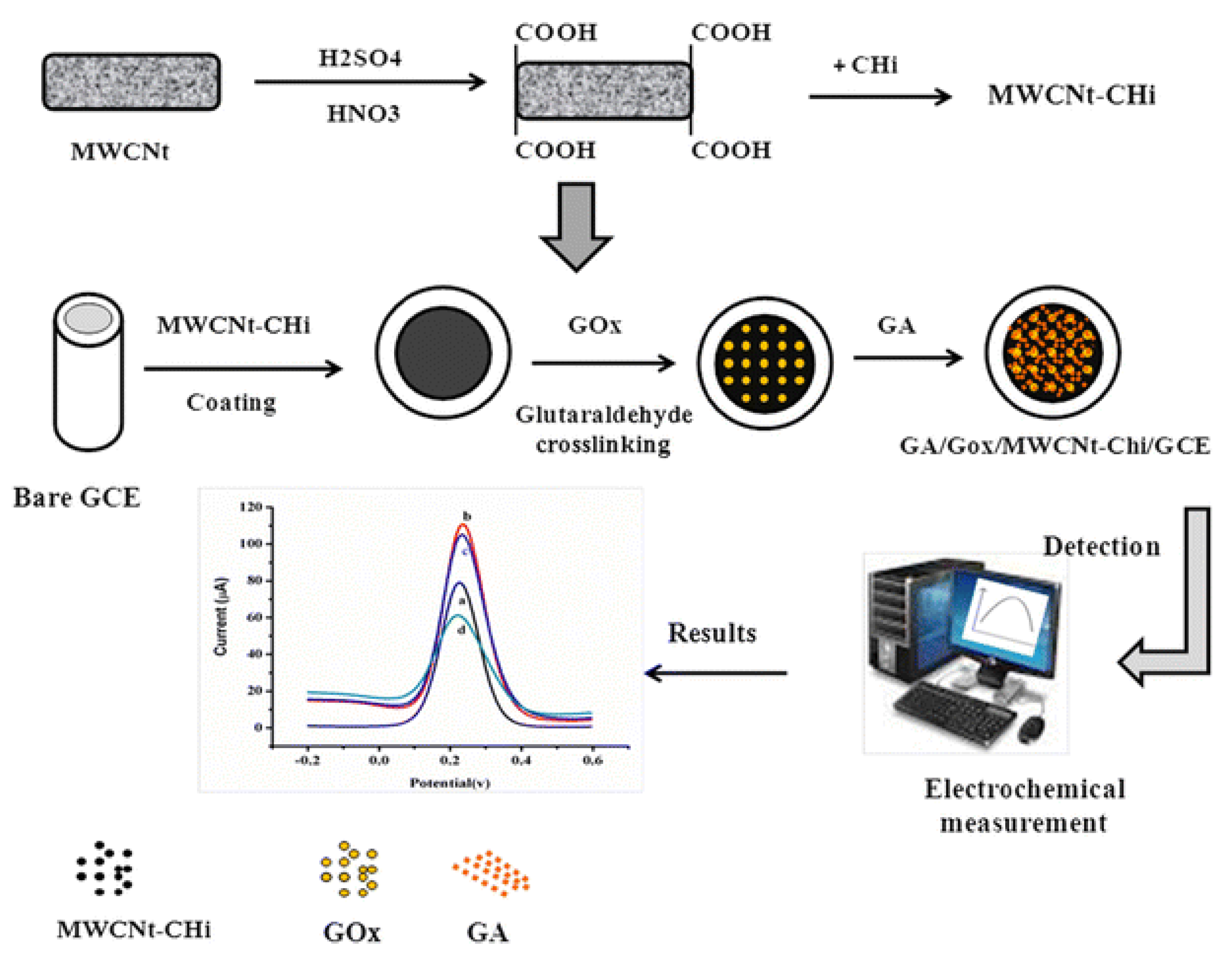

Table 1.
Type of biosensor employed for fish, meat, poultry and related product quality and safety monitoring.
Table 1.
Type of biosensor employed for fish, meat, poultry and related product quality and safety monitoring.
| Type of biosensor | Target Analyte | Source of analyte | Bioreceptor | Transducer/ Electrode | Data visualization device | Measured value | Reference |
| Enzyme-based biosensors | Histamine | Fish | Diamine oxidase (DAO) or monoamine oxidase (MAO) enzymes | Electrochemical Screen printed carbon electrodes |
cyclic voltammetry (CV), chronoamperometry and electrochemical impedance spectroscopy (EIS) | 8.957x10⁻² mM | [18] |
| Hypoxanthine | Pork meat | Xanthine oxidase | Electrochemical Platinum wire as counter electrode |
Thermogravimetric Analysis |
111.3 µM at 7 days | [6] | |
| Putrescine | Beef, pork, chicken, turkey and fish meat | Putrescine oxidase |
Electrochemical |
Chemiluminescence Microplate luminometer |
0.8 – 2 mg/L | [14] | |
| Xanthine | Chicken meat | Guanine deaminase and Xanthine oxidase | Electrochemical with fibre optic probe |
Spectrometer (OceanOptics) | 44 µM at 5 days | [15] | |
| Nitrate | Meat sample | Nitrate reductase | Ag/AgCl reference electrode and platinum auxiliary electrode and working electrode glassy carbon (GCE) | Voltammetric analysis | Detection limit: 2.2 × 10⁻⁹ M and limit of quantification: 5.79 x10⁻⁹ M | [2] | |
| Glucose reduction | Glucose oxidase | Beef meat | Glassy carbon electrode modified with multi-walled carbon nanotubes and chitosan | Cyclic voltammetry (CV), differential pulse voltammetry (DPV), and electrochemical impedance spectroscopy (EIS) | Reduction in glucose from 0.01-0.06 mmol/L | [16] | |
| Immunosensors | Chloramphenicol | Beef and pork meat samples | Monoclonal antibody to CAP (anti-CAP) | Electrochemical immunosensor; Electrochemical impedance spectroscopy technique | Electrochemical impedance spectroscopy (EIS | Detection limit: 0.06 ng/mL | [3] |
| Salmonella enterica, Listeria monocytogenes, and Escherichia coli | Ready-to-eat beef, chicken and turkey breast meat | Alexa Fluor 647-labeled monoclonal antibodies | Streptavidin coated optical waveguides | Fluorometer | Detection limit: 103 CFU/mL | [4] | |
| Calpastatin | Beef meat | Primary anti-calpastain antibody and secondary enzyme-labelled antibody | Potentiostat–galvanostat with Gold working (W.E.) and counter (C.E.) electrodes silver pseudo-reference electrode |
Amperometric detection |
481 ng/mL | [19] | |
| Tetracycline | Poultry muscle samples | Lyophilized reconstituted sensor cells | Cell-biosensor | Bioluminescence with SynergyTM HT Multi-detection Microplate Reader | Sensitivity: 10 µg/kg | [20] | |
| DNA-based biosensors | DNA (Donkey meat) | Donkey adulteration in cooked sausages | DNA | Multi-parameter SPR device with gold chips | Surface plasmon resonance (SPR) | Detection limit: 1.0 nM | [5] |
| Dopamine Adulterant | Beef meat | Anti-dopamine substance | - | Colorimetric sensor | Detection limit: 0.13 mM | [21] | |
| Bacteria (Campylobacter spp.) | Chicken meat | Genomic Campylobacter DNA |
Paper membrane, and biotinylated-DNA detection probe | Chemiluminescent read-out | 3 pg/µL of DNA Detection limit | [22] |
Table 2.
Biosensors to monitor the quality and safety of fish, meat and meat Products.
| Analyte | Employed electrode and material of detection | Immobilization Technique | Food product | LOD | Sensitivity/linear range/ detection time | References |
| Some examples for biosensors detecting freshness of fish, meat and meat products. | ||||||
| Hx | XOD within a Nafion matrix on a graphene–titanium dioxide | Entrapment | Pork | 9.5 μM | Sensitivity: 4.1 nA/μM Linear range 20–512 μM |
[6] |
| XOD and horseradish peroxidase | Adsorption | Raw and treated meat samples | 1.8 mg/L | Quantitative limit: 6.1 mg L−1 | [28] | |
| XOD and polyvinylferrocenium perchlorate matrix on a platinum | Adsorption | Fish | 0.6 μM, | Linear range 2.1–103 μM | [62] | |
| XOD and platinum electrode with single-walled carbon nanohorns (SWCsNH) and gold nanoparticles (AuNP) | Covalent | Fish | 0.61 | Linear range 1.5–35.4 | [63] | |
| XOD and uricase within a polypyrrole-paratoluenesulfonate composite film | Entrapment | Fish | 5 | 5-500 Linear range | [64] | |
| XOD on carbon film electrodes and carbon nanotube | Cross-linking | Fish | 0.77 | 10-130 | [65] | |
| XOD onto a modified platinum electrode surface. | Entrapment | Seafood | 0.0023 | 0.01–10 | [64] | |
| XOD onto paper substrate | Adsorption | Fish | 4.1 | 4–35 | [66] | |
| Calpastatin | Capillary and optical fiber biosensor | Covalent | Longissimus muscle from beef | Calpastatin activity (R2 = 0.6058) | [67] | |
| Cadaverine | Receptor molecules onto the surface of thiol-gold | Covalent | Beef, chicken, or pork | [68] | ||
| Putrescine | Casein onto the electrode surface using glutaraldehyde | Covalent | Beef, pork, chicken, turkey meat samples | LOD: 0.8 mg/L–1.3 mg/L | Linearity range: 1–2 mg/L | [69] |
| TVB-N | pre-fabricated responsive dyes, embedded onto a paper or polymer film | Adsorption | Pork meat | Correlation coefficient (R2 = 0.932) | [40] | |
| Some examples for biosensors detecting pathogen microorganisms and toxins in meat and meat products | ||||||
| Campylobacter spp. | Amino-modified DNA probes onto a nylon membrane | Covalent | Chicken meat | LOD: 3 pg/μL of DNA | - | [22] |
| Salmonella enterica, Listeria monocytogenes, and Escherichia coli O157:H7 | antibodies onto the optical fiber surface using carbodiimide | Covalent | Beef, turkey breast and chicken | LOD: 103 CFU/mL | - | [4] |
|
SalmonellaTyphimurium Staphyloccocus aureus |
Thiol-modified aptamers onto gold nanoparticles | Non-covalent | Pork | 15 CFU/mL 35 CFU/mL |
Recovery rate: 94.12%–108.33% | [47] |
| S. enterica serovar Typhimurium | Amine-terminated DNA aptamers onto a carboxyl-functionalized graphene-modified electrode employing carbodiimide | Covalent | Chicken meat | LOD: 1 CFU/mL | Linear range (detection): 1–8 log CFU/mL | [50] |
| Salmonella pullorum | specific antibodies onto the electrode surface using glutaraldehyde | Covalent | Chicken meat | LOD: 100 CFU/mL | Detection time: 1.5 to 2 h | [70] |
| E coli K-12 | specific antibodies onto the gold electrode surface. | Adsorption | Chicken meat | LOD: 3 log CFU/mL | - | [71] |
|
Listeria monocytogenes |
thiol-modified DNA aptamers onto gold nanoparticles | Covalent | Meat samples | LOD: 2 log CFU/g | Linear detection range : From 10² to 10⁷ CFU/m Detection time <30 minutes |
[45] |
|
L. monocytogenes toxin S. aureus enterotoxin B |
live mammalian cells onto the surface of gold interdigitated microelectrodes | Adsorption | Salami | 10⁴ CFU/mL 100 ng/mL | Detection time <1 hour | [72] |
| Staphylococcal enterotoxin B | anti-SEB antibodies onto a gold-coated SPR | Covalent | Meat | 0.5 ng/mL | 0.5 ng/mL to 20 ng/mL Detection time <20min |
[51] |
| TrichotheceneT-2 toxin | Anti-T-2 toxin antibodies onto a modified electrode surface using glutaraldehyde | Covalent | Swine meat | 0.04 ng/mL | 0.05 – 20 ng/mL Detection time = 30 min |
[55] |
| Some examples for biosensors detecting antibiotics, drug residues, and additives in meat products | ||||||
| Tetracyclines | E. colicells in agarose gel on the surface of microplates or membrane | Entrapment | Poultry muscle samples | 2–5 µg/kg | 2 to 100 µg/kg. Detection time = 3Hours |
[20] |
| Chloramphenicol (CAP) | CAP–protein conjugate onto the SPR sensor chip | Covalent | Poultry muscle | 100 ng/kg | to 1 µg/kg detection time <30 min |
[73] |
|
Oxytetracycline (OTC) Kanamycin (KAN) Ampicillin (AMP) |
aptamers onto citrate-stabilized gold nanoparticles | Adsorption | Chicken |
0.42 ng/mL 0.31 ng/mL0.28 ng/mL |
1–100 ng/mL 1–80 ng/mL 1–60 ng/mL Detection time = 15 min |
[74] |
| Ractopamine | ractopamine–BSA conjugate onto a carboxymethylated dextran chip | Covalent | Pork | 0.09 ng/mL | 0.1 – 10 ng/mL Detection time = 10 min |
[75] |
Table 3.
Summary of Key Validation Parameters for Biosensor-Based Methods according to ICH M10 guideline.
Table 3.
Summary of Key Validation Parameters for Biosensor-Based Methods according to ICH M10 guideline.
| Parameter | Definition | Regulatory Expectation | Biosensor-Specific Considerations |
|---|---|---|---|
| Specificity | Ability to detect only the target analyte, not structurally similar compounds | Interference from related compounds should result in <LLOQ signal; accuracy ±25% at extremes | Biosensors using antibodies/aptamers must be screened against analogs, metabolites, and additives |
| Selectivity | Differentiation of analyte in presence of matrix components | ≥80% of blank matrices should show <LLOQ signal; accuracy within ±25% at LLOQ | Must account for interference from fats, enzymes, or proteins common in food matrices |
| LOD (Limit of Detection) | Lowest concentration distinguishable from blank with confidence | typically signal/noise (S/N) ≥3 | Important for contaminant detection; impacted by sensor noise and baseline stability |
| LOQ (Limit of Quantification) | Lowest concentration quantifiable with acceptable accuracy & precision | S/N typically ≥10 | Defines lower end of calibration; matrix effects often limit LOQ in real food samples |
| Calibration Curve | Relationship between analyte concentration and sensor response | ≥6 levels + blank; logistic fit often used; 75% points within ±20–25% of nominal value | Non-linear response at low/high ranges often requires 4/5-parameter modeling |
| Accuracy | Closeness of measured value to true value | Within ±20% (±25% at LLOQ/ULOQ); evaluated within- and between-runs | Challenging when sensor drift or matrix effects occur; needs robust QC planning |
| Precision | Repeatability of results under same conditions | CV ≤20% (≤25% at LLOQ/ULOQ); across ≥6 runs and 5 QC levels | Signal variability from biorecognition elements (e.g. enzyme-based biosensors) must be managed |
| Total Error | Sum of bias (accuracy) and variability (precision) | Should not exceed 30% (40% at LLOQ/ULOQ) | A helpful global indicator of biosensor method performance |
| Dilution Linearity | Consistency of measurement across diluted samples | Mean ±20% of expected after correction; ≥3 dilutions tested | Needed for samples exceeding range; verifies absence of hook effect |
| Hook Effect | Signal suppression at high analyte concentrations | No signal drop-off in undiluted samples expected above ULOQ | Particularly relevant in immunoassay-based biosensors |
| Carry-over | Residual analyte signal from prior sample influencing subsequent results | Signal in blank after ULOQ standard must be <LLOQ | Typically minimal in biosensors; confirm with blank after high calibrator |
| Stability | Analyte remains unchanged during storage, preparation, and analysis | Mean ±20% at low/high QC; validated over actual storage conditions | Biosensor reagents (e.g. enzymes, aptamers) and analyte stability must both be validated |
Table 4.
Biosensor applications for food additives and contaminants: key challenges and mitigation strategies.
Table 4.
Biosensor applications for food additives and contaminants: key challenges and mitigation strategies.
| Analyte | Biosensor Type | Matrix | LOD/ LOQ | Key Challenges | Mitigation Strategies | Study |
|---|---|---|---|---|---|---|
| Carbendazim | upconversion-MnO2 luminescent resonance energy transfer | food | LOD: 0.05 ng·mL−1 | specificity | aptamer integration and high fluorescence quenching capability of MnO2 nanosheets | [91] |
| cadmium (Cd), lead (Pb) and mercury (Hg) | luciferase-based biosensors | food | LOD:Cd: 0.01 μM Pb: 0.025 nM Hg: 2 nM | decrease of sensitivity | expression of Pb importers or nonspecific modifications | [92] |
| Nitrate | Immobilized Nitrate Reductase | dry-cured ham | - | comparison with HPLC | good agreement with standard HPLC method: R 2 = 0.971 | [93] |
| amnesic shellfish toxins: domoic acid | Aptamer-Based Biosensor | - | LOD: 13.7 nM | specificity | identification and truncation optimization | [94] |
| Paralytic Shellfish Poisoning Toxins | Surface Plasmon Resonance-Based Biosensors | shellfish | - | interferences | comparison of several extraction methods | [95] |
Disclaimer/Publisher’s Note: The statements, opinions and data contained in all publications are solely those of the individual author(s) and contributor(s) and not of MDPI and/or the editor(s). MDPI and/or the editor(s) disclaim responsibility for any injury to people or property resulting from any ideas, methods, instructions or products referred to in the content. |
© 2025 by the authors. Licensee MDPI, Basel, Switzerland. This article is an open access article distributed under the terms and conditions of the Creative Commons Attribution (CC BY) license (http://creativecommons.org/licenses/by/4.0/).
Copyright: This open access article is published under a Creative Commons CC BY 4.0 license, which permit the free download, distribution, and reuse, provided that the author and preprint are cited in any reuse.


