Submitted:
23 April 2025
Posted:
24 April 2025
You are already at the latest version
Abstract
Ischemia/reperfusion (I/R) damage remains a major problem in surgery, primarily based on high oxidative stress generated during the reperfusion process.
Mitochondria are significantly affected, their metabolic and energetic processes
are impaired and the redox system is out of balance.
Regulation and restoration of the redox balance by oxidative preconditioning with ozone is being investigated worldwide in cell and animal models. Selected preclinical trials and their results, with a focus on cardiological and neuronal I/R damage, are presented and discussed.
We regularly find an upregulation of antioxidants, demonstrated in SOD (superoxide dismutase) and GSH (glutathione, reduced form, and a decrease in oxidative stress as a result, shown here using the typical stress parameters MDA (malondialdehyde) and TBARS (thiobarbituric acid reactive substances).
Mitochondrial biogenesis, comparable to moderate physical activity, is induced by ozone oxidative preconditioning in an I/R model in rats and reviewed in this paper.
Keywords:
redox balance
; oxidative stress
; low-dose ozone
; redox bioregulation
; Ischemia/reperfusion damage
; mitochondrial biogenesis
; ozone therapy
1. Introduction
Great expectations can be placed on secondary prevention during surgical interventions to prevent or minimize ischemia/reperfusion (I/R) injury through oxidative preconditioning by applying low-dose medical ozone: High oxidative stress is significantly reduced, the redox balance is maintained or largely restored: Mitochondria remain intact and can fulfill their metabolic and energetic obligations.
I/R damage due to a sudden oxygen flow during reperfusion is a major problem in surgery e.g. in cardiology (heart attack), neurology (stroke), vascular surgery and, last but not least, in transplant surgery [1]. This results in an excess of reactive oxygen species (ROS) such as hydrogen peroxide, superoxide radicals and hydroxyl radicals, which, together with oxygen, form lipoperoxides (LPO) and initiate radical chain reactions. Mitochondria are damaged and lose their metabolic functions significantly.
2. I/R Damage and the Regulation by Oxidative Preconditioning Through Low-Dose Medical Ozone
Our focus here is on myocardial and cerebral I/R damage, which is probably the most common type worldwide that occurs after a heart attack or apoplexy. Fortunately, there are an increasing number of preclinical studies with promising results, which will certainly soon be followed by clinical studies. We here refer only to those preclinical trials, in which typical redox parameters were measured to underline the redox regulation by ozone, and -for the sake of clarity- by very few but significant and reliable, clearly reproducible ones: They are viewed either from the oxidative stress side and from the antioxidants side of the redox balance (see Table 1 and Figure 1).
Fundamental results of redox bioregulation by ozone are manifested in a regulation of the redox balance by ozone preconditioning to protect against I/R damage in surgery; shown in Figure 1 as a selection of preclinical studies in cell and animal models in which meaningful parameters were measured. We regularly find an upregulation of antioxidants, demonstrated here in SOD and GSH, and a decrease in oxidative stress as a result, shown here using the typical stress parameters MDA and TBARS. Starting this idea in 1998 by the León research group it is today of worldwide interest with good prospects of establishing a method which, very easy to handle and due to its mechanism of action, enables general application, e.g. in brain and heart surgery, in order to significantly reduce I/R damage [2]. Clinical studies should follow and are obviously in progress. A short survey on the mechanism of action is displayed in Figure 2.
The values measured for the I/R groups are taken as baseline and the antioxidants represent the increase (green) after ozone preconditioning while the oxidative stress is characterized by a decrease (orange). Each bar represents the prevention of ROS induced injury in different organs as known from ischemia during surgery and subsequent reperfusion. Upregulating the cellular and mitochondrial antioxidants seems to be the main mechanism of systemically administered ozone at low concentrations and doses: Oxidative stress decreases and the redox balance is restored.
2.1. Mechanism of Action and the Ozone Effect
To start a surgical procedure, the blood flow is interrupted generating ischemia over a certain period of time, at the end of which reperfusion is started, whereby an excess of oxygen floods ischemic areas not being metabolized by the overstrained mitochondria. Reactive oxygen compounds are formed, such as superoxide radicals, hydrogen peroxide, OH-radicals, lipoperoxides and others with several cell and organ destructions as a consequence.
Ozone does not pass through the cell membrane (polar molecular structure), as is often assumed and repeatedly reported, nor does it decompose into oxygen and oxygen radicals, it has an extremely short half-life in blood and biological fluids and does not react directly with cell organelles or compartments. It follows an indirect reaction mechanism interacting with signaling pathways (such as the Nrf2 or NFkB pathways) as is the case with several other drugs [19]. By ozone oxidative preconditioning ozone activates -by its immediately formed (cell membrane), short-chain peroxides and the reduction via the glutathione system- the Nrf2 pathway initiating antioxidant production to restore the redox balance, described in detail in [20], see Figure 2.
2.2. Heart Protection Against I/R Damage Through Ozone-Mediated Mitochondrial Biogenesis
Mitochondrial biogenesis: To maintain their specific functions, all organ cells (except RBCs) depend on intact mitochondria as energy suppliers. Perfect regulatory processes take place between the cells and their mitochondria: If the cell has a high energy requirement, mitochondrial activity increases, the metabolic rate is intensified and ATP production improves. Moderate physical activity leads to optimal intercommunication and signal transmission, to an improved oxygen supply. The mitochondria are also increasingly stimulated to multiply, i.e. divide (fission), when energy requirements increase, and the mitochondrial density in the cells increases as a consequence. Mitochondrial fusion, in balance with mitochondrial fission, serves, among other things, to repair defective or insufficient mitochondria.
This means that the number of mitochondria in an organ cell adapts to the energy requirement; thus improving cell protection and reducing the risk of oxidative stress.
In organs with a high energy requirement such as muscle cells, hepatocytes or the myocardial cells, there are from 1,000 to several 100,000 (egg cells) mitochondria with a volume share of up to 25% of the cell volume [23].
Impaired regulation in the mitochondria determines the fate of the cell and results in a variety of diseases, including neurodegenerative diseases, chronic inflammations, such as rheumatoid arthritis, vascular diseases, insulin resistance and diabetes type 2, aging and age-related diseases but also cancer [23,24].
The regulation of oxidative stress by ozone in ROS-induced mitochondriopathies was demonstrated in cases of Rheumatoid arthritis, osteoarthritis, diabetes type 2 and aging described in more detail elsewhere [25].
Can Medical Ozone Induce Mitochondrial Fission and Increase the Mitochondrial Density Through Preventive Intraperitoneal Ozone Administration?
This chapter of the present paper focuses on oxidative preconditioning by ozone for the prevention of myocardial reperfusion injury in animal models. In long-lasting ischemia (> 15min) severe myocardial damage can occur followed by a broad spectrum of corresponding problems, such as destruction of myocytes due to muscle paralysis, vascular damage and irreversible cell damage induced by the enormous increase in reactive oxygen species (ROS) during reperfusion [26].
Surprisingly, histological studies revealed an increase in mitochondrial density after preventive intraperitoneal ozone administration according to the low-dose concept. However, this study attracted little interest and was probably forgotten due to language problems, but is highly relevant today with the increasing importance of mitochondrial medicine [13].
Animal model. In several preclinical studies oxidative preconditioning with ozone proved to be an effective method of regulating redox balance in order to prevent reperfusion damage (see Table 1).
On this basis, the effectiveness of ozone was tested in an ischemia/reperfusion model of the myocardium in animals: Thirty male albino rats weighing 100-150 g were divided into 3 groups, each with n=10: group 1 served as control without pretreatment. The animals in group 2 received a preventive i. p. ozone application 2x per week for 2 months and group 3 for 3 months (ozone concentration: 4 μg /mL; dose 28 μg per 100g rat corresponding to 400 μg per 100 mL blood), followed by I/R injury by 30 min ischemia and 30 min reperfusion. For detailed information, please refer to the original publication [13].
Result. After 2 months oxidative preconditioning with ozone, myocytes after I/R injury showed an increase in mitochondrial density by 33.9 % compared to control (p<0.05), while mitochondrial density decreased again to 16.8 % after 3 months of preventive ozone administration compared to control (p<0.05), see Figure 3.
A similar effect is known from moderate physical activity, e.g. in cardiac and skeletal muscle cells: the increased energy demand boosts mitochondrial activity combined with an increase in fission and autophagy processes. It can even compensate for the loss of mitochondria due to obesity as shown in an animal trial [27].
The induction of mitochondrial biogenesis by ozone application was first described in the literature by Barakat 2006 as shown here.
The main contribution of ozone seems to be the interaction within the redox balance: maintaining or restoring the balance protecting the cells and organelles from dysfunctions by an excess of reactive oxygen species. Mitochondria can continue to fulfill their cellular obligations in metabolic regulation. Though the oxidative stressor MDA remains still high compared to the control, but the antioxidant capacity in the tissue increased.
2.3. Protection of Mitochondria in the Heart Muscle Against I/R Damage
Coronary artery bypass surgery is frequently associated with I/R damage which can be significantly reduced by oxidative preconditioning through i.p. ozone application at low doses, here shown in an animal model [14].
Procedure: 30 Adult male Sprague-Dawley rats (200–250 g), 5 groups (sham group, I/R group, ozone+I/R and 2 control groups) operated with 6 animals each; the “ozone group” animals received 100 μg/kg daily for 5 days (e.g. 25 μg per rat in 2 mL volume) in prevention to the I/R process (30 min of ischemia followed by 2h of reperfusion). Among the critical protection parameters the antioxidative enzymes SOD 1 (Cu, Zn SOD) and SOD 2, the manganese-dependent SOD (Mn-SOD) in the mitochondria increased in a statistically significant manner with p<0,05 compared to the I/R animal group, see Figure 1.
The transcription factor Nrf2 induces this process of corresponding protein production in order to control finally the redox balance. We must be aware that Nrf2 itself is a crucial factor which must be kept in a balance, not to initiate metastasis in cancer patients. That means ozone concentrations and doses should follow the low-dose concept [28,29].
In this study, Nrf2 mRNA decreased by 62% as a result of the I/R process compared to the sham-operated (control) group, whereas ozone oxidative preconditioning considerably attenuated this process (ozone + I/R group): Nrf2 was only 11% below the sham-operated group. The mitochondria remained largely intact, and only mild myocardial apoptosis was observed [14].
Protection of the myocardium from I/R injury by preventive low-dose ozone application (here 20 μg/mL, 2 mL per animal)) is reversed by blocking Nrf2 and its pathway, as shown in cell and animal models. Oxidative stress and repair parameters behave accordingly (see Figure 1). As a result of restoring the redox balance, mitochondria damage and feroptosis (Fe-dependent apoptosis) is kept low and the infarct size small [17]. Good results are also reported at higher ozone concentrations but oxidative stress remained at a high level and could only be downregulated by drastically reducing the ozone concentration, while maintaining the dose (see Table 1 and Figure 1) [16].
3. Protection Against Brain I/R Injury
3.1. Cell Model: Protection of Neuronal Cells
The SH-SY5Y cell line is widely used in in-vitro trials to study neuronal viability and functions as basis for neurodegenerative diseases [30].
As an ozone concentration of 20 μg/mL showed the best viability and activity of the neuronal cells compared to 40 μg/mL and 80 μg/mL this was the concentration of choice for ozone oxidative preconditioning in this I/R cell model in order to protect the mitochondria from losing their functions and the cells from damage and apoptosis.
Again, stress parameters were reduced, here MDA representing oxidative stress by 42 % and SOD representing the antioxidants as repair enzymes, increased by 200 % compared to the I/R group and shown in Figure 1. The redox balance shifts towards the norm, mitochondria are protected, cytochrome C-release as a measure for permeability is attenuated, apoptosis increased by the I/R process to 37,1 % of the cells, whereas ozone preconditioning decreased it to 28,4 %. Control: apoptosis in 13 % of the cells [15]. These results demonstrate protection against cerebral I/R injury by ozone at the cellular level, confirmed using an animal model.
3.2. Cerebral I/R Injury Animal Model
The protective effect of ozone in I/R injury by maintaining the redox balance of the cell, protecting mitochondrial damage and reducing cell apoptosis (here ferroptosis as iron-dependent apoptosis) was demonstrated in an in vitro model using SH-SY5Y cells, a cell line derived from human bone marrow. As the cerebral I/R injury is usually followed by neuronal death or loss with all the well-known consequences, ozone preconditioning with its protective effect opens up new aspects and treatment strategies for cerebral I/R injuries and neuronal disorders in prevention and therapy.
Middle cerebral artery occlusion/reperfusion (MCAO/R) as a standard animal model is widely used to gain basic knowledge on focal cerebral ischemia in humans.
Procedure. 112 rats (260-300 g) were randomly divided into seven groups, 16 animals in each group; we here compare only 3 groups to emphasize the ozone effect and ozone mechanism: sham group as control, MCAO-group: middle artery occlusion for 120 min, followed by surgery and reperfusion. Ozone + MCAO: Intraperitoneal pretreatment with Ozone (oxidative preconditioning) before MCAO; ozone concentration: 40 μg/mL with 2,5 mL/kg/d for 5 days.
Result. Ozone preconditioning protected the brain from cerebral reperfusion injury, neuronal damage and the infarction area was significantly reduced compared to the MCAO-group. By activating the Nrf2 pathway, ozone preconditioning leads to a powerful antioxidant capacity reducing the critical reactive oxidant species and, among other factors, the crucial parameters such as LPO, MDA, GSH in rat brain are kept close to the healthy range, displayed in Figure 4, as well as the reduction of the infarct area (by 62.3 %) and the number of intact neurons compared to the MCAO-group showing an increase by 83.2% [18].
4. Conclusion and Future Perspective
Ischemia reperfusion damage remains a major problem in surgery, in cardiology (heart attack), neurology (stroke), vascular surgery and, last but not least, in transplant surgery, primarily based on high oxidative stress generated during the reperfusion process:. At low concentrations and low doses, ozone oxidative preconditioning provides a new treatment concept to restore the redox balance, to protect the mitochondria and maintain the cell integrity. Basic mechanisms, treatment procedures concentrations and doses in the non-toxic ranges are well known, guidelines have been published [31]. This knowledge, gained in a series of preliminary studies in different research groups with the same results, is presented here in myocardium and brain I/R ischemia/reperfusion models and could directly be transferred to clinical studies.
References
- Zhang, M.; Liu, Q.; Meng, H.; Duan, H.; Liu, X.; Wu, J; Gao, F; Wang, S.; Tan, R.; Yuan, J. Ischemia-reperfusion injury: molecular mechanisms and therapeutic targets. Signal Transduct Target Ther. 2024, 9, 12. [CrossRef]
- Léon, O.S.; Menéndez, S.; Merino, N.; Castillo, R.; Sam, S.; Pérez, L.; Cruz, E.; Bocci, V. Ozone oxidative preconditioning: A protection against cellular damage by free radicals. Mediat. Inflamm. 1998, 7, 289–294. [CrossRef]
- Ajamieh, H.; Merino, N.; Candelario-Jalil, E.; Menendez, S.; Martinez-Sanchez, G.; Re, L.; Giuliani, A.; Leon, O.S. Similar protective effect of ischemic and ozone oxidative preconditioning in liver/ischemia/reperfusion injury. Pharmacological Research. 2002, 45, 333-339. [CrossRef]
- Ajamieh, H.; Menendez, S.; Martınez-Sanchez, G.; Candelario-Jalil, E.; Re, L.; Giuliani, A.; Leon Fernandez. O. Effects of ozone oxidative preconditioning on nitric oxide generation and cellular redox balance in a rat model of hepatic ischaemia–reperfusion. Liver International. 2004, 24, 55–62. [CrossRef]
- Ajamieh, H.; Berlanga, J.; Merino, N.; Martinez Sanchez, G.; Carmona, A.; Menendez Cepero, S.; Giuliani, A.; Re, L.; Leon, O.S. Role of protein synthesis in the protection conferred by ozone-oxidative-preconditioning in hepatic ischaemia/reperfusion. Transpl. Int. 2005, 18, 604–612. [CrossRef]
- Calunga, J.; Zamora, Z.; Borrego, A.; Del Rıo, S.; Barber, E.; Menendez, S.; Hernandez, F.; Montero, T.; Taboada, D. Ozone Therapy on Rats Submitted to Subtotal Nephrectomy: Role of Antioxidant System. Mediators of Inflammation. 2005, 4, 221–227. [CrossRef]
- Zamora, Z.; Borrego, A.; Lopez, O., Delgado, T.; Gonzalez, R.; Menedez, S.; Hernandez, F.; Schulz, S. Effects of ozone oxidative preconditioning on TNF-alpha release and intracellular antioxidant-prooxidant balance in mice during endotoxic shock. Mediators Inflamm., 2005, Feb 24, 16-22. [CrossRef]
- Santana-Rodríguez, N.; Llontop, P.; Clavo, B.; Fiuza-Perez, M.; Zerecero, K.; Ayub, A.; Alshehri, K.; Yordi, N.; Re, L.; Raad, W.; Fernandez-Perez, L.; García-Herrera, R.; Huang, C.; Bhora, F. FACS. Ozone Therapy Protects Against Rejection in a Lung Transplantation Model: A NewTreatment? Ann Thorac Srg. 2017, 104, 258-464. https://www.annalsthoracicsurgery.org/article/S0003-4975(17)30360-0/fulltext.
- Clavo, B.; Catalá, L.; Pérez, J.; Rodríguez, V.; Robaina, F. Ozone Therapy on Cerebral Blood Flow: A Preliminary Report. Evid Based Complement Alternat Med. 2004, 1, 315-319. [CrossRef]
- Zhu, F.; Ding, S.; Liu, Y.; Wang, X.; Wu, Z. Ozone-mediated cerebral protection: Unraveling the mechanism through ferroptosis and the NRF2/SLC7A11/GPX4 signaling pathway. Journal of Chemical Neuroanatomy. 2024, 136, 102387. [CrossRef]
- Wang, L.; Chen, Z.; Liu, Y.; Du, Y.; Liu, X. Ozone oxidative postconditioning inhibits oxidative stress and apoptosis in renal ischemia and reperfusion injury through inhibition of MAPK signaling pathway. Drug Des. Devel Ther. 2018, 12, 1293–1301. [CrossRef]
- Wang, P.; Cui, Y.; Ren, Q., Yan, B.; Zhao, Y.; Yu, P.; Gao, G.; Shi, H.; Chang, S.; Chang, Y.; Mitochondrial ferritin attenuates cerebral ischaemia/reperfusion injury by inhibiting ferroptosis. Cell Death Dis. 2021, ,12, 447. [CrossRef]
- Barakat, S.; Saleh, N.; Thabet, S.; El Missiri, A.; Badawy, M. Ischämie/Reperfusions-Modell am Herzen nach oxidativer Konditionierung durch Ozon. [Ozone Oxidative Preconditioning in Ischemia/Reperfusion Model on the Myocardium]. Ozone-Textbook, Basics-Prevention-Therapy. Ecomed Publisher Landsberg, Germany. 2006, (Viebahn-Hänsler, R.; Knoch, H.G., Eds.).
- Meng, W.; Xu, Y.; Lic, D.; Zhua, E.; Deng, L.; Liu, Z.; Zhang, G.; Liu, H. Ozone protects rat heart against ischemia-reperfusion injury: A role for oxidative preconditioning in attenuating mitochondrial injury. Biomedicine & Pharmacotherapy. 2017, 88,1090-1097. [CrossRef]
- Cai, H.; Xi T.; Zheng, L.; Huang, L.; Peng, Y.; Liao, R.; Zhu, Y. Ozone alleviates ischemia/reperfusion injury by inhibiting mitochondrion-mediated apoptosis pathway in SH-SY5Y cells. Cell Biol Int. 2020, 44, 975–984. [CrossRef]
- Wang, R.; Liu, F.; Huang, P.; Zhang.; He, J.; Pang, X.; Zhang, D.; Guan,Y. Ozone preconditioning protects rabbit heart against global ischemia-reperfusion injury in vitro by up-regulating HIF-1. Biomedicine & Pharmacotherapy. 2022, 150, 113033. https://www.sciencedirect.com/science/article/pii/S075333222200422X?via%3Dihub.
- Ding, S.; Duanmu, X.; Xu, L.; Zhu, L.; Zhouquan, Wu, Z . Ozone pretreatment alleviates ischemia–reperfusion injury-induced myocardial ferroptosis by activating the Nrf2/Slc7a11/Gpx4. Biomedicine & Pharmacotherapy. 2023,165, 115185. [CrossRef]
- Zhu, F.; Ding, S .; Liu, Y.; Wang, X.; Wu, Z. Ozone-mediated cerebral protection: Unraveling the mechanism through ferroptosis and the NRF2/SLC7A11/GPX4 signaling pathway. Journal of Chemical Neuroanatomy. 2024, 136, 102387. [CrossRef]
- Dash, U.; Nayak, V.; Navani, S.; Samal, R.; Agrawal, P.; Singh, A.; Majhi, S.; Mogaref, D.G.; Duttaroy, A.; Jena, A.B. Understanding the molecular bridges between the drugs and immune cell. Pharmacology & Therapeutics. 2025, 267,108805.
- Viebahn-Haensler, R.; León Fernández, O. Ozone in Medicine. The Low-Dose Ozone Concept. The Redox-Bioregulatory Effect as Prominent Biochemical Mechanism and the Role of Glutathione. Ozone Sci. Eng. 2024, 46, 267–279. [CrossRef]
- Sagai, M.; Bocci, V. Mechanisms of Action Involved in Ozone Therapy: Is Healing Induced via a Mild Oxidative Stress? Medical Gas Research. 2011, 1, 29. [CrossRef]
- Togi, M.; Nagashima, S.; Kitay, Y.; Muromoto, R.; Kashiwakura, J.; Miura, T.; Matsuda, T. Implication of NF-kB Activation on Ozone-Induced HO-1 Activation. BPB Reports 2021, 4, 59-63. doi.org/10.1248/bpbreports.4.2_59.
- Zong, Y.; Li, H.; Liao, P.; Chen, L.; Pan, Y.; Zheng, Y.; Zhang, C.; Liu, D.; Zheng, M.; Gao, J.; Mitochondrial dysfunction: mechanisms and advances in Therapy. Sig Transduct Target Ther. 2024, 9, 124. [CrossRef]
- Bhatti, J.S.; Bhatti, G.K.; Reddy, P.H. Mitochondrial dysfunction and oxidative stress in metabolic disorders—A step towards mitochondria based therapeutic strategies. Biochim. Biophys. Acta. 2017, 1863, 1066–1077. [CrossRef]
- Viebahn-Haensler, R.; León Fernández, O.S. Mitochondrial Dysfunction, Its Oxidative Stress-Induced Pathologies and Redox Bioregulation through Low-Dose Medical Ozone: A Systematic Review. Molecules. 2024, 29, 2738. [CrossRef]
- Ambrosio, G.; Tritto, I. Reperfusion injury: Experimental evidence and clinical implications. Am Heart J. 1999, 138, 69-75. [CrossRef]
- Jun, L.; Knight, E.; Broderick, T.L.; Al-Nakkash, L.; Tobin, B.; Geetha, T.; Babu, J.R. Moderate-Intensity Exercise Enhances Mitochondrial Biogenesis Markers in the Skeletal Muscle of a Mouse Model Affected by Diet-Induced Obesity. Nutrients. 2024, 16, 1836. [CrossRef]
- Galié, M.; Covi, V.; Tabaracci, G.; Malatesta, M. The Role of Nrf2 in the Antioxidant Cellular Response to Medical Ozone Exposure. Int. J. Mol. Sci. 2019, 16, 4009. [CrossRef]
- .
- Carvajal-Oliveros, A.; Uriostegui-Arcos, M.; Zurita, M.; Melchy-Perez, E.; Narváez-Padilla, V.; Reynaud, E. The BE (2)-M17 cell line has a better dopaminergic phenotype than the traditionally used for Parkinson´s research SH-SY5Y, which is mostly serotonergic. IBRO Neurosci Rep. 2022, 13, 543-551. [CrossRef]
- Viebahn-Haensler, R.; León Fernández, O.S.; Fahmy, Z. Ozone in medicine: The low-dose ozone concept. Guidelines and treatment strategies. Ozone Sci. Eng. 2012, 34, 408–424. [CrossRef]
Figure 1.
Ischemia/reperfusion injury and its protection by regulation of the redox balance through ozone preconditioning. Repair parameter (SOD or GSH) versus oxidative stress parameter (MDA or TBARS) in preclinical studies as described in Table 1. .
Figure 1.
Ischemia/reperfusion injury and its protection by regulation of the redox balance through ozone preconditioning. Repair parameter (SOD or GSH) versus oxidative stress parameter (MDA or TBARS) in preclinical studies as described in Table 1. .
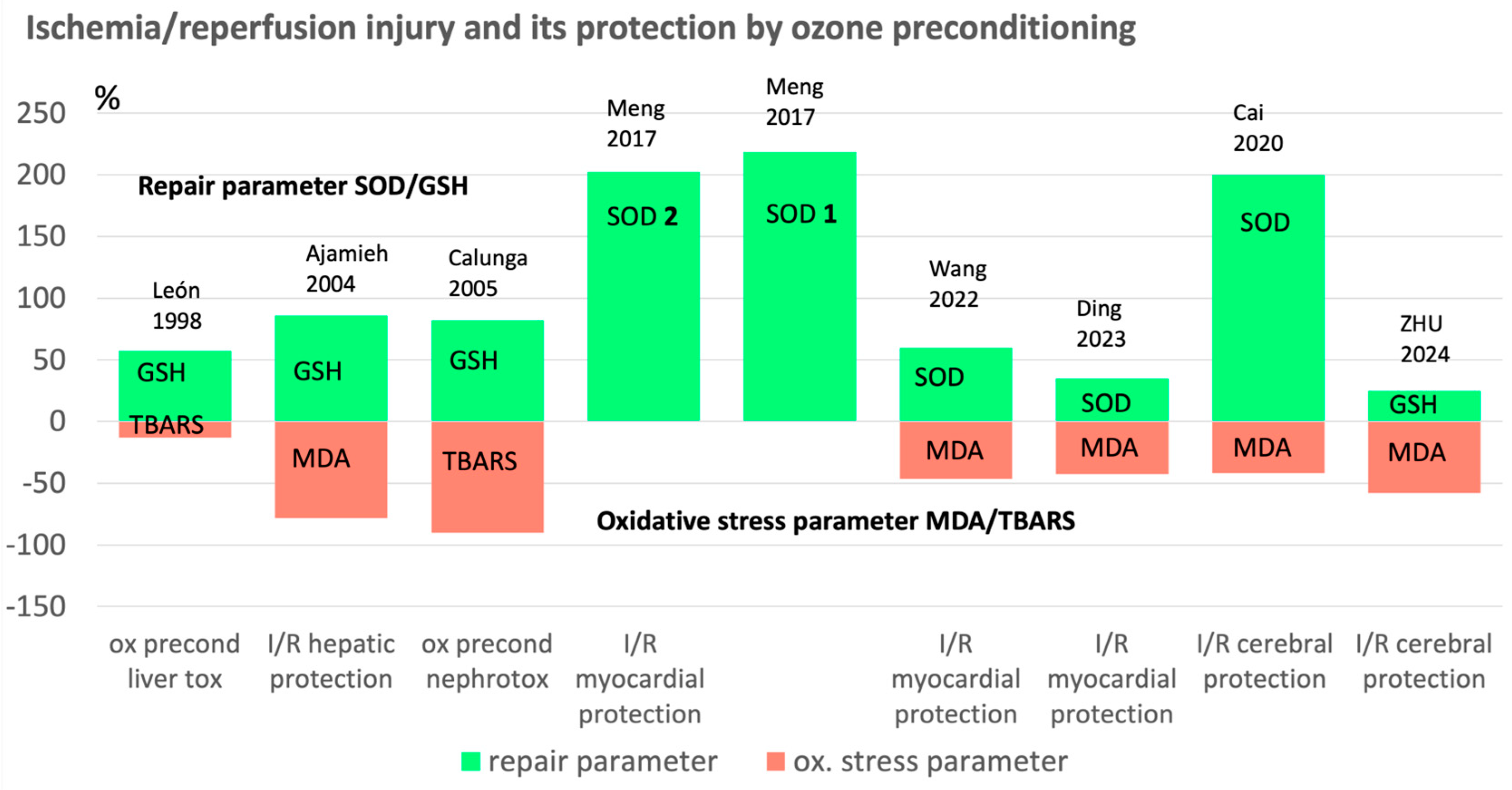
Figure 2.
Ischemia/reperfusion: oxidative stress and regulation by medical ozone. Ozone immediately reacts with isolated double bonds (cell membranes) to form short chain “ozone peroxides”, which are reduced by the glutathione system (GSH/GSSG) and pass on the information to the cell nucleus via the NFkB and Nrf2 pathway; finally, antioxidants are produced. The reactive oxygen species ROS decrease and the redox system can be balanced. GSHox: Glutathione peroxidase, GSred: Glutathione reductase; GGT: γ- glutamyl transferase, CAT: Catalase, SOD: Superoxide dismutase, NFkB and Nrf2: Nuclear factors, MDA: malondialdehyde [20,21,22].

Figure 3.
Mitochondrial density in the myocardium after oxidative preconditioning by ozone during 2 and 3 months. The number of mitochondria plays a special role for the myocardium with its high energy requirements. Mitochondrial density measured as % of the myocard areal in an Ischemia/reperfusion model in animals. Each group with 10 animals (rats). Control: no pretreatment, Ischemia 30 min, reperfusion 30 min. “2 months”: these 10 rats were pretreated with i.p. ozone applications for 2 months twice per week. “3 months”: same procedure as the “2 months group” for 3 months. Ozone concentration: 4 μg /mL; dose 28 μg per 100 g rat corresponding to 400 μg per 100 mL blood, followed by I/R injury by 30 min ischemia and 30 min reperfusion [13].
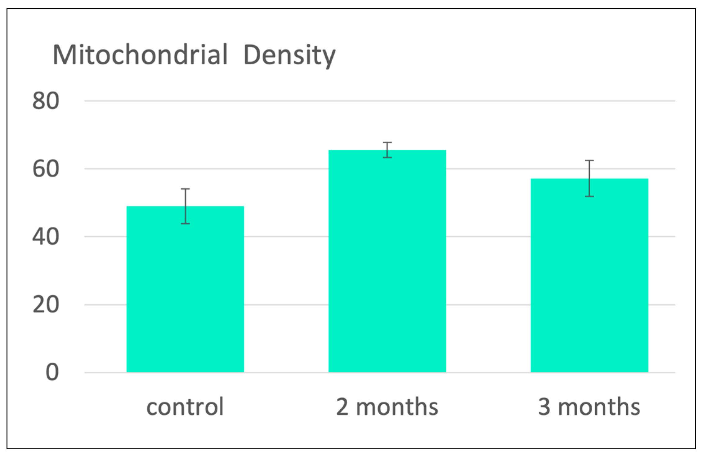
Figure 4.
Protection against brain injury in an ischemia/reperfusion model in rats. A. Critical parameters backing the underlying ozone mechanism. LPO, MDA in μmol/g protein; GSH in mg/g protein. All values are mean ± SD. Statistical results, LPO: sham vs MCAO, p<0.001; MCAO vs ozone+MCAO, p<0.01. MDA: sham vs MCAO, p<0.0001; MCAO vs ozone+MCAO, p<0.001. GSH: sham vs MCAO, p<0.0001, MCAO vs ozone+MCAO, p<0.05. B. Reduction of infarct volume (ratio in %); number of neurons per mm2. Statistical results, Infarct volume: (sham=O) MCAO vs ozone+MCAO, p<0.1. number of neurons: sham vs MCAO, p<0.0001; MCAO vs ozone+MCAO, p<0.0001 [18].
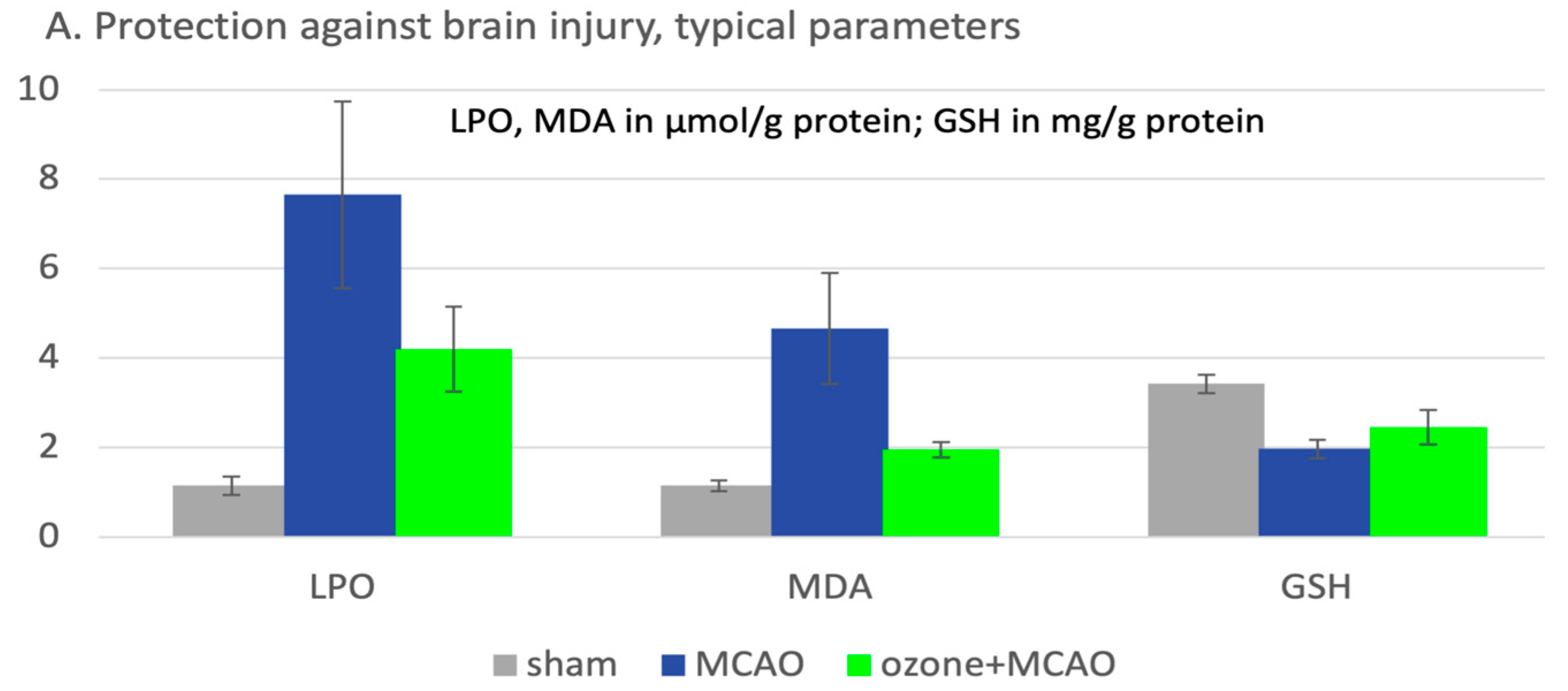
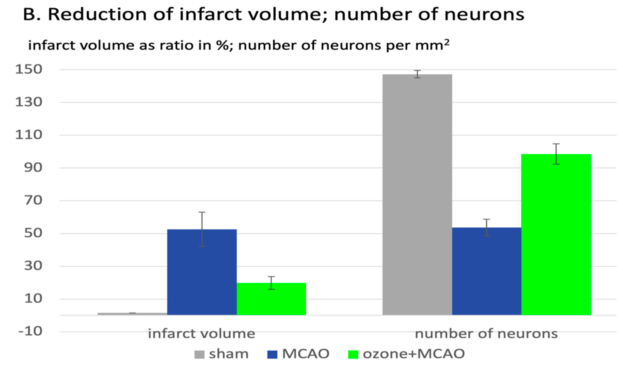

Table 1.
Preclinical studies on the prevention of reperfusion injuries and intoxication by ozone oxidative preconditioning (without any claim to completeness).
Table 1.
Preclinical studies on the prevention of reperfusion injuries and intoxication by ozone oxidative preconditioning (without any claim to completeness).
| Subject, type of study | Procedure | Antioxidants / oxidative stress parameters relevant for ozone effect | References |
|---|---|---|---|
| Preclinical trial in rats. Ozone oxidative preconditioning: A protection against cellular damage by free radicals. |
6 animal groups: adult female Sprague-Dawley rats, 220–250 g. 10 animals per group.4 control groups Toxicity-producing ROS: by CCl4 solution; ozone preconditioning: 15 O3 treatments as rectal insufflation (1mg/kg), 4,5-5 mL, 50 µg/mL. |
Only a few redox parameters, relevant to ozone effect here discussed: SOD in u/g; GSH in mmol/g: TBARS in nmol/g protein. | León et al. 1998 [2]. |
| Preclinical trial in rats. Similar protective effect of ischemic and ozone oxidative preconditioning in liver/ischemia/reperfusion injury. |
Adult male Wistar rats (250–300 g). n=32, hepatic ischaemia I/R (n = 8): 90 min hepatic ischaemia 90 min reperfusion (n=8). ozone preconditioning: 15 O3 treatments as rectal insufflation (n=8) (1mg/kg), 5–5.5 mL, 50 μg/mL. |
MDA+4-hydroxynonenal in mmol/g; total SH-groups in mol/mg protein. | Ajamieh et al. 2002 [3]. |
| Preclinical trial in rats. Effects of ozone oxidative preconditioning on nitric oxide generation and cellular redox balance in a rat model of hepatic ischemia/reperfusion. |
60 adult male Wistar rats (250–300 g), 10 per group. hepatic ischemia I/R 90 min ischemia, 90 min reperfusion; ozone preconditioning: 15 O3 treatments as rectal insufflation (1mg/kg), 5–5.5 mL, 50 μg/mL. | GSH in μg/g tissue, MDA+4 hydroxynonenal in mmol/g. |
Ajamieh et al. 2004 [4]. |
| Preclinical trial in rats. Role of protein synthesis in protection conferred by ozone-oxidative-preconditioning in hepatic ischemia/reperfusion. |
Adult male Wistar rats (10 per group, 250–275 g) hepatic ischemia 90 min ischemia, ozone preconditioning: 15 O3 treatments as rectal insufflation (1mg/kg), 5–5.5 mL, 50 μg/mL. | Mn-SOD in U/g tissue (SOD 2 mitochondrial SOD); Cu/Zn-SOD in U/g tissue (SOD 1 cytosol), MDA+4 hydroxynonenal in mmol/g. | Ajamieh et al. 2005 [5]. |
| Preclinical trial in rats. Ozone Therapy on Rats Submitted to Subtotal Nephrectomy: Role of Antioxidant System. |
30 female Wistar rats (180–200 g), 10 per group: 15 treatments 2.5−2.6mL, conc. 50 μg/mL rectal insufflation 1x/day, partial nephrectomy. | GSH (nmol/mg protein) TBARS in nmol/g protein. |
Calunga et al. 2005, [6]. |
| Preclinical trial in rats. Ischemia/reperfusion animal model on rat myocard after ozone oxidative preconditioning. [Ischämie/Reperfusions-Modell am Herzen nach oxidativer Konditionierung durch Ozon]. |
30 male albino rats (100-150 g), 3 groups, each with n=10, preventive ozone i.p. 2x per week for 2 months or for 3 months, conc.: 4 μg /mL; 28 μg per 100 g rat (400 μg per 100 mL blood), followed by by 30 min ischemia and 30 min reperfusion. | SOD, MDA, Mitochondrial biogenesis. |
Barakat et al. 2006 [13]. |
| Preclinical trial in rats. Ozone Therapy Protects Against Rejection in a Lung Transplantation Model: A New Treatment? |
Male Sprague-Dawley rats, n=36, rectal O3 daily for 2 weeks prior to lung transplantation (20-50 μg per animal) and 50 μg/dose 3x/week up to 3 months. |
Ozone pre- and postconditioning significantly decreased the expression of all genes related to oxidative stress and chronic rejection. | Santana-Rodríguez et al. 2017 [7]. |
| Preclinical trial in rats. Ozone protects rat heart against ischemia-reperfusion injury: A role for oxidative preconditioning in attenuating mitochondrial injury. |
Adult male Sprague-Dawley rats (200–250 g) OzoneOP 2 mL ozone 100 μg/kg/day for 5 days, 30 min of cardiac ischemia followed by 2 h reperfusion. | SOD 1 and SOD 2 in u/mg protein | Meng et al. 2017 [14]. |
| Preclinical trial in cell culture. Ozone alleviates ischemia/reperfusion injury by inhibiting mitochondrion-mediated apoptosis pathway in SH-SY5Y cells. |
SH-SY5Y cells as model for neuronal function tests; ozone oxidative preconditioning via incubation with 40 μg/mL and cultured for 2, 6;12, and 24 hrs. | SOD in u/mg protein, MDA in nmol/mg protein. | Cai et al. 2020 [15]. |
| Preclinical trial in rabbits. Ozone preconditioning protects rabbit heart against global ischemia-reperfusion injury in vitro by up-regulating HIF-1. |
Adult rabbits (2.50–2.75 kg), ozone oxidative preconditioning: i.p. injections 10 mL daily for 5 days. 3 conc´s: 20; 40, 80 μg/mL followed by 20 min ischemia, 60 min reperfusion. | SOD in u/mg protein, MDA in mmol/mg protein | Wang et al. 2022 [16]. |
| Preclinical trial in cell culture and in an animal model (mice). Ozone pretreatment alleviates ischemia–reperfusion injury-induced myocardial ferroptosis by activating the Nrf2/Slc7a11/Gpx4 axis. |
1.H9c2 cardiomyocytes; 2. Male C57 mice, 7 weeks of age, 22 -24 g. n=36, 12 per group, ozone preconditioning, 25 μg/mL i.p. injections, 2 mL per day for 5 days. 30 min. ischemia, 2 hrs reperfusion. | SOD activity in % of CTL (cytotoxic T lymphocyte activity), MDA in μmol/g protein | Ding et al. 2023 [17]. |
| Preclinical trial in rats. Ozone-mediated cerebral protection: Unraveling the mechanism through ferroptosis and the NRF2/SLC7A11/GPX4 signaling pathway. |
Sprague-Dawley (SD) rats (260–300 g), middle artery occlusion for 120 min, followed by surgery and reperfusion. Ozone oxidative preconditioning, i.p. injections (intra peritoneal), 20 μg/mL with 2,5 mL/kg/d, 5 days. | GSH in mg/g protein, LPO (lipoperoxides), MDA in μmol/g protein. | Zhu et al 2024 [18]. |
Disclaimer/Publisher’s Note: The statements, opinions and data contained in all publications are solely those of the individual author(s) and contributor(s) and not of MDPI and/or the editor(s). MDPI and/or the editor(s) disclaim responsibility for any injury to people or property resulting from any ideas, methods, instructions or products referred to in the content. |
© 2025 by the authors. Licensee MDPI, Basel, Switzerland. This article is an open access article distributed under the terms and conditions of the Creative Commons Attribution (CC BY) license (http://creativecommons.org/licenses/by/4.0/).
Copyright: This open access article is published under a Creative Commons CC BY 4.0 license, which permit the free download, distribution, and reuse, provided that the author and preprint are cited in any reuse.


