

Submitted:
21 April 2025
Posted:
22 April 2025
You are already at the latest version
Abstract
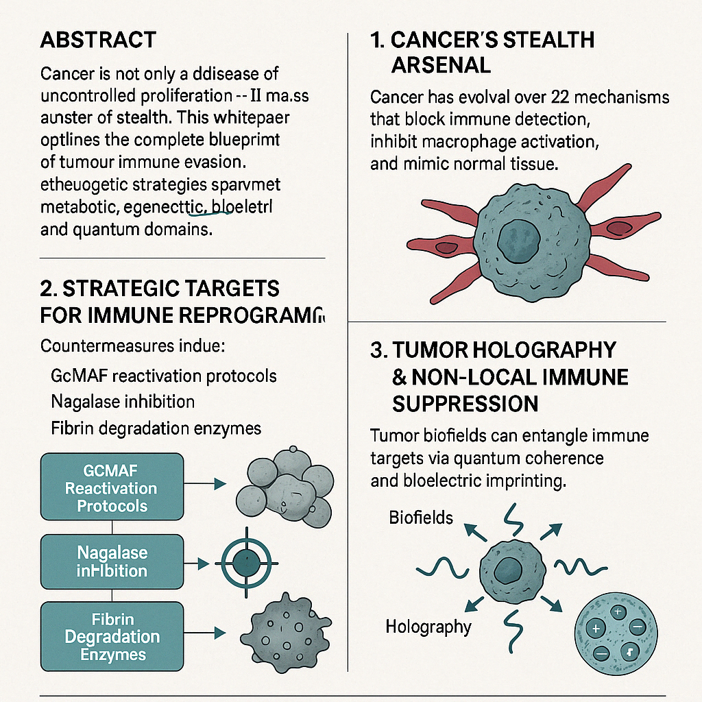
Cancer is not only a disease of uncontrolled proliferation — it is a master of stealth. This white paper outlines the complete blueprint of tumour immune evasion, with a focus on Nagalase-mediated GcMAF suppression, fibrin-based shielding, exosome manipulation, and advanced quantum biofield cloaking. The objective is clear: full-spectrum disruption of cancer’s stealth systems, restoration of immune visibility, and deployment of advanced therapeutic strategies across metabolic, epigenetic, bioelectric, and quantum layers.
Keywords:
GcMAF
; Nagalase
; Immune Evasion
; Cancer Stealth
; Tumour Microenvironment
; Exosomes
; TRPV1
; Bioelectric Medicine
; PEMF
; ECS
; Scalar Fields
; Quantum Oncology
; EZ Water
; Fourth Phase Water
; Structured Water
; Tumour Holography
; Redox Medicine
; Immune Reprogramming
; Clinical Protocols
; Oncology
; Immunotherapy
; Systems Biology
; Clinical Integrative Medicine
; AI in Healthcare
; Molecular
; Immunology
; Biofield Science
; ECS Therapeutics
1. Introduction
The therapeutic silence around Nagalase, fibrin, and immune suppression has allowed cancer’s stealth technologies to evolve unchecked. GcMAF (Gc protein-derived Macrophage Activating Factor), once heralded for its immune-activating potential, was suppressed both biologically — via Nagalase — and commercially. This white paper reconstructs the forgotten power of GcMAF, aligns it with modern metabolic, ECS, and quantum therapeutic frameworks, and redefines the field of immune reactivation therapy. Our goal is to create a deployable, modular therapeutic strategy that brings tumour invisibility back into biological focus.
2. Cancer’s Stealth Arsenal: Mechanisms of Immune Evasion
Cancer has evolved over 22 stealth mechanisms that block detection, inhibit immune activation, or mimic normal tissue. These include:
- Nagalase-mediated GcMAF inhibition: Nagalase cleaves Gc protein, preventing conversion to GcMAF — thereby disabling macrophage activation (Yamamoto et al., 2008).
- Fibrin shielding: Tumours encase themselves in fibrin, reducing immune cell infiltration and impeding drug penetration.
- Tumour-derived exosomes: Loaded with suppressive proteins and miRNA to downregulate immune surveillance (Whiteside, 2016).
- Immune checkpoint hijacking: PD-L1/PD-1 expression prevents T-cell cytotoxicity.
- Cancer stem cell (CSC) evasion: Dormant subpopulations resist immune activation and radiotherapy.
- Metabolic switching: Warburg effect and glutamine addiction create hostile, hypoxic microenvironments.
- Tumour microbiome signalling: Pathogen-associated molecular pattern (PAMP) mimicry interferes with immune pattern recognition receptors (PRRs).
- Ferroptosis/necroptosis suppression: Resistance to iron-dependent death pathways.
- Glycan cloaking: Glycosylation of surface proteins hinders immune binding.
- Epigenetic silencing: DNA methylation suppresses MHC and other antigen-presenting genes.
- Holographic field imprinting: Tumours can project biofield interference patterns to surrounding tissues.
3. Strategic Targets for Immune Reprogramming
This section provides direct countermeasures to each stealth mechanism:
- GcMAF Protocol Regeneration: Using oral colostrum macrophage activators, chondroitin sulfate-binding modulators, and transdermal adjuncts like PEA (palmitoylethanolamide).
- Nagalase Inhibition: Agents such as MGN-3 (Biobran), mistletoe lectins, and immuno-regenerative mycological compounds (e.g., Trametes versicolor).
- Fibrin Degradation: Systemic enzymes (e.g., serrapeptase, nattokinase), magnesium, and bioavailable bromelain.
- Exosome Disruption: Milk exosome blockers (lactadherin analogs), plant exosome competitors, and ECS-linked glycoprotein modulators.
- miRNA Modulation: miR-21, miR-155, and miR-146a targeting using polyphenols (e.g., curcumin, apigenin).
- Quantum/Electromagnetic Therapies: PEMF, scalar field disruption, Tesla harmonics to break biofield synchronization.
4. Tumour Holographic Imprinting & Non-Local Immune Suppression
Tumour biofields can entangle immune targets through non-local coherence.
Disruption Protocol:
- PEMF coils tuned to 7.83 Hz (Schumann resonance)
- Scalar + longitudinal wave emission from bifilar Tesla coils
- Ormus (monatomic elements) structured with bio geometric coherence fields
- Red/NIR light therapy (660–880 nm) with water structuring overlay
5. Fourth-Phase Water Structuring and Bioelectric Weaponization
EZ (exclusion zone) water formed along hydrophilic surfaces contributes to tumour coherence, intracellular protection, and electrostatic resilience.
Therapeutic Disruption:
- Shilajit, fulvic acid, molecular hydrogen water to de-structure EZ water
- Biophoton pulsed fields
- Cold plasma and negative ion therapy to alter water charge boundaries
Key Reference: Pollack, G. H. (2013). The Fourth Phase of Water: Beyond Solid, Liquid, and Vapor.
6. Quantum Entanglement and Cancer’s Remote Signalling
Cancer cell populations demonstrate intercellular coordination that mimics quantum entanglement — including signal synchronization at distance and field harmonics.
Suggested Tools:
- PEMF-modulated coherence breakers
- Photonic shields (Faraday mesh, phase-cancelling LED panels)
- Quantum biophysical inhibitors (ECS–GPCR–TRPV1 interfaces)
Key Reference: Cifra, M., Fields, J. Z., & Farhadi, A. (2011). Electromagnetic cellular interactions. Prog Biophys Mol Biol, 105(3), 223–246.
7. Dataset Overview
Dataset Overview
- A structured, 23-row × 40+ category dataset accompanies this paper. Each row represents a unique stealth disruption target, cross-mapped with:
- Therapeutic agents (natural & synthetic)
- ECS and GPCR involvement
- miRNA regulation profiles
- Nagalase inhibitors & macrophage activators
- Bioelectric and redox resonance disruptors
- Structured water destabilizers
- Field-level immune recalibraters
 This dataset is formatted for AI deployment, clinical protocol generation, and integration into the Master Control Room OS.
8. Clinical Translation Box
Target Use Cases:
- Immune evasion reversal
- Macrophage reactivation therapy
- Microdose Immunotherapeutics
- Tumour field coherence disruption
- Cancer cell “de-cloaking” protocols
Clinicians may use this framework to generate protocol outputs via AI systems or SaaS platforms (e.g., Neuro Stack / Quantum Oncology OS).
9. Licensing and Contact
This white paper is part of the Quantum Oncology OS. Licensing, API access, and protocol customization available.
 Contact: ecssignalling@gmail.com
🔗 GitHub: https://github.com/TeamMohamed
 License: CC BY-NC 4.0
References
- Yamamoto, N., M. Urade, and N. Yamamoto. 2008. Immunotherapy of metastatic colorectal cancer with Gc protein-derived macrophage-activating factor. J Med Food 11, 4: 755–762. [Google Scholar]
- Whiteside, T. L. 2016. Tumor-derived exosomes and their role in cancer progression. Adv Clin Chem 74: 103–141. [Google Scholar] [PubMed]
- Rubik, B. 2002. Biofield hypothesis: role of energy fields in medicine. J Altern Complement Med 8, 6: 703–717. [Google Scholar] [CrossRef] [PubMed]
- Pollack, G. H. 2013. The Fourth Phase of Water: Beyond Solid, Liquid, and Vapor. [Google Scholar]
- Cifra, M., J. Z. Fields, and A. Farhadi. 2011. Electromagnetic cellular interactions. Prog Biophys Mol Biol 105, 3: 223–246. [Google Scholar] [CrossRef] [PubMed]
Disclaimer/Publisher’s Note: The statements, opinions and data contained in all publications are solely those of the individual author(s) and contributor(s) and not of MDPI and/or the editor(s). MDPI and/or the editor(s) disclaim responsibility for any injury to people or property resulting from any ideas, methods, instructions or products referred to in the content. |
© 2025 by the authors. Licensee MDPI, Basel, Switzerland. This article is an open access article distributed under the terms and conditions of the Creative Commons Attribution (CC BY) license (http://creativecommons.org/licenses/by/4.0/).
Copyright: This open access article is published under a Creative Commons CC BY 4.0 license, which permit the free download, distribution, and reuse, provided that the author and preprint are cited in any reuse.