

Submitted:
11 April 2025
Posted:
14 April 2025
You are already at the latest version
Abstract
Background/Objectives: Stroke survivors often develop asymmetric gait patterns that may lead to abnormal knee joint loading and potentially increased risk of osteoarthri-tis. This study aimed to investigate differences in knee joint loading between paretic and non-paretic limbs during walking in individuals post-stroke. Methods: Twen-ty-one chronic stroke survivors underwent three-dimensional gait analysis. A modified musculoskeletal model with a specialized knee mechanism was used to estimate medi-al and lateral tibiofemoral contact forces during the stance phase. Statistical Paramet-ric Mapping was used to identify significant differences in joint kinematics, kinetics, and contact forces between limbs. Stepwise regression analyses examined relationships between knee moments and compartmental contact forces. Results: Significant dif-ferences in knee loading were observed between limbs, with the non-paretic limb expe-riencing higher medial compartment forces during early stance (6.7-15.1%, p=0.001; 21.9-30.7%, p=0.001) and late stance (72.3-93.7%, p< 0.001), and higher lateral com-partment forces during pre-swing (86.2-99.0%, p< 0.001). In the non-paretic limb, knee extensor moment was the primary predictor of first peak medial contact force (R²=0.573), while knee abductor moment was the primary predictor in the paretic limb (R²=0.559). Conclusions: Musculoskeletal modeling revealed distinct asymmetries in knee joint loading between paretic and non-paretic limbs post-stroke, with the non-paretic limb experiencing consistently higher loads, particularly during late stance. These findings suggest that rehabilitation strategies should address not only paretic limb function but also potentially harmful compensatory mechanisms in the non-paretic limb to prevent long-term joint degeneration
Keywords:
stroke rehabilitation
; knee joint loading
; musculoskeletal modeling
; gait biomechanics
1. Introduction
Stroke remains one of the leading causes of long-term disability worldwide, resulting in significant impairments in sensory, motor, cognitive, and visual functions that substantially impact daily activities [1], [2]. A particularly challenging consequence of stroke is asymmetric gait patterns, where survivors predominantly rely on their unaffected limb for locomotion, leading to what is known as hemiplegic gait [3]. While, this compensatory strategy enables mobility and allow patients to retain steady walking state, it may introduce biomechanical alterations in both lower limbs, such as abnormal knee loading. Despite achievements in rehabilitation techniques [4], [5], stroke survivors still encounter significant abnormalities in their walking patterns, such as altered joint mobility, spatiotemporal asymmetries, uneven weight distribution on the lower limbs, and modifications in muscle activation patterns [6].
Abnormal walking pattern in unilateral conditions and the repetitive high loads imposed on joint structures can lead to stress-related joint injuries and disorders in the unaffected limb or disease progression in the affected limb. A similar pattern has been observed in individuals with unilateral osteoarthritis (OA) or lower-limb amputation, who are at higher risk of developing OA in the contralateral knee joint (i.e. the knee of the opposite limb from the affected side). This supports the belief that asymmetric mechanical loading plays an important role in the development of knee OA [7], [8], [9]. However, these findings are based on external measures as proxies for actual joint loading, such as external knee joint moments [10]. Extensive research on knee loading during gait of knee OA patients has revealed knee adduction and flexion moments (KAM and KFM respectively) only relate to first peak of knee contact forces [11] whereas no statistical relationship was found between the second knee contact force (KCF) peak and external joint moments. Moreover, a case-study using instrumented knee prosthesis showed that a reduction in first peak KAM does not guarantee a reduction in medial contact load [12], or indicate changes in the relative distribution of the loading between medial and lateral compartments. Last, some studies in patients with early stages of knee OA suggest that altered KAM and KFM are not risk factors in the initial development of knee OA [13].
The characteristic impairments of post-stroke gait, including reduced walking speed, altered knee joint range of motion [14], abnormal muscle co-activation patterns [15], and asymmetrical knee joint kinetics between paretic and non-paretic limbs [16], could increase joint loading and contribute to cartilage degeneration [17]. Knee cartilage thinning has been reported in hemiparetic knees [18], [19], particularly on the lateral side [20], and is linked to knee pain and knee OA onset or progression. Knee OA prevalence among stroke patients can reach up to 21% [21], which can negatively affect rehabilitation outcome [22] and quality of life. A previous study [23] in stroke survivors, showed that there is likely to be an increase in loading either in the non-paretic limb or in the paretic limb during gait compared to healthy individuals, while in another study [24] no different knee loading was found between lower limbs. Again, these studies rely on knee external joint moments, which correlate only with the first peak of the medial KCF [25], leaving other knee loading parameters – such as complete waveforms and peak lateral KCF – unexplored. Furthermore, the relationship between external moments and peak knee loading in stroke gait remains unclear.
Musculoskeletal (MSK) modeling offers a more sophisticated approach to estimating internal joint forces, accounting for both external loads and muscle contributions, thus providing more accurate insights into the actual cartilage loading conditions experienced during gait [26]. It is primarily based on accurate recorded motion from 3D gait analysis labs using retroreflective markers and devices recording ground reaction forces, as inputs to inverse dynamics algorithms that calculate muscle and joint reaction forces utilizing a wealth of MSK models [27], [28]. However, despite its potential and its rather limited use in examining post stroke gait [29], there is currently no research applying musculoskeletal modeling to examine knee joint loading in stroke survivors. Nevertheless, it has been extensively used to calculate knee contact forces in numerous scenarios [30], [31], in particular knee OA and total knee replacement patients, and validated against actual forces recorded from a limited number of total joint replacement devices [32].
Therefore, the purpose of this study was to investigate the differences in knee joint loading between paretic and non-paretic limbs during walking in individuals post-stroke using musculoskeletal modeling. We hypothesized that the non-paretic limb would experience greater knee joint forces compared to the paretic limb, reflecting the compensatory strategies commonly adopted by stroke survivors during gait. As a secondary aim, we explored the relationship between external joint moments and knee joint loading and tried to validate findings of previous literature on knee OA patients and healthy individuals. These insights could inform training protocols to optimize stroke recovery.
2. Materials and Methods
Participants
Twenty-one individuals with chronic stroke participated in this study. Participants were recruited from the outpatient Neurological Rehabilitation Unit at the University Hospital of Alexandroupolis, Greece and participated in two separate investigations: eleven participants from a previous repeatability study [33] and ten from a subsequent investigation. The participants' affected side was distributed as follows: eleven with left hemiparesis and ten with right hemiparesis. Mean age was 62.8 ± 4.7 years and mean body mass index 28.9 ± 4.14 kg. All participants provided written informed consent, and the study protocols were approved by the institutional ethics committee of the Democritus University of Thrace - Greece. Participants were included if they met the following criteria: (1) chronic phase of stroke (at least 6 months post-stroke), (2) age above 18 years, (3) walking speed above 0.2 m/s with no upper limit, (4) ability to walk without assistance, and (5) diagnosed hemi-paresis with observable motor impairment in the affected limb at the time of enrolment, as confirmed by a licensed healthcare professional (6). Participants with only non-motor stroke symptoms (such as isolated facial weakness, dysarthria, or sensory deficits) were not eligible for the study. According to the National Institutes of Health Stroke Scale [32], almost all patients had a score between 1 and 4 (minor stroke) with one patient scoring 7 (moderate stroke).
Data Collection
Three-dimensional gait analysis was performed using two different motion capture setups. For the first cohort, three-dimensional marker coordinates were recorded using six infrared cameras (Vicon MX 0306012, Oxford, UK) sampling at 100 Hz. For the second cohort, a 10-camera Vicon (Vicon Motion Systems Ltd, Oxford, UK) system was employed, also sampling at 100 Hz. In both setups, ground reaction forces were recorded using two force plates (type 9281B11 and 9281CA, Kistler Instruments AG, Winterthur, Switzerland) embedded in the middle of a 10-meter walkway, sampling at 1000 Hz.
Retroreflective markers were placed on anatomical landmarks following two protocols. For the first cohort, markers were placed according to the Vicon Plug-in-Gait lower-body protocol, while the second cohort utilized a full-body marker according to the Conventional Gait Model set comprising 57 markers, although only the markers corresponding to the PiG model were used for the analysis. All participants walked barefoot along the walkway at their self-selected speed. Marker trajectories and ground reaction forces were processed using Vicon Nexus software and filtered using a low-pass filter with a 6 Hz cutoff frequency. A minimum of five successful trials were collected for each participant, where a successful trial was defined as clean force plate contact with the affected limb.
Musculoskeletal Modeling
A modified version of the Lerner model [34] was used to estimate joint kinematics, kinetics, and knee contact forces. The original model was adapted by condensing the torso segments to the pelvis due to marker protocol restrictions in the lower limb. The model included a ball-and-socket joint between the third and fourth lumbar vertebra, three translations and three rotations of the pelvis, ball-and-socket joints at the hips, and hinge ankle and subtalar joints.
The knee mechanism was specifically designed to resolve medial and lateral tibiofemoral contact forces. While sagittal plane rotation and translations of the tibia and patella relative to the femur followed Delp's specifications [35], the tibiofemoral mechanism was augmented with a distal femoral component and a tibial plateau body. These components enabled configuration of frontal-plane alignment through orientation parameters in both femur and tibia. The tibiofemoral articulation was modeled using a series of joints: a primary knee joint defining sagittal-plane rotations and translations, and two hinge joints connecting the sagittal articulation frame to medial and lateral tibiofemoral compartments. These hinge joints, with axes perpendicular to the frontal-plane, were welded at the anterioposterior mid-point of the tibial plateaus, maintaining fixed positions relative to the tibia while articulating with the femoral component during flexion-extension. While individual hinge joints could not resist frontal-plane moments, their parallel arrangement allowed load sharing between medial and lateral compartments, resolving the net reaction forces and frontal-plane moments across the tibiofemoral joint. The knee remained a single degree-of-freedom joint with motion restricted to the sagittal plane.
Marker trajectories and ground reaction forces were used as input in OpenSim 3.3. The model was scaled to each participant's anthropometry using markers from a recorded static trial prior to recordings. For each successful walking trial, joint angles were calculated using inverse kinematics, solving for the generalized coordinates that minimized the weighted sum of squared differences between experimental and model marker positions. Joint moments were computed using inverse dynamics and normalized to body weight. Static optimization was then performed to estimate muscle forces, using a cost function that minimized the sum of squared muscle activations. Finally, joint reaction analysis was conducted to calculate the vertical medial and lateral knee contact forces during stance phase (heel strike to toe off), which were normalized to body weight.
Statistical Analysis
Joint kinematics, kinetics, and vertical knee contact forces were time-normalized to 100% of the stance phase. Group differences between paretic and non-paretic limbs were assessed using Statistical Parametric Mapping (SPM), enabling statistical analysis and comparison of the waveform data across the entire stance phase by conducting point-by-point hypothesis testing while controlling for multiple comparisons [36].
For kinematic analysis, we examined hip flexion-extension, hip abduction-adduction, hip internal-external rotation, knee flexion-extension, and ankle dorsi-plantarflexion angles. Kinetic variables included the corresponding joint moments, with the addition of the knee abductor moment. For knee contact forces, both medial and lateral compartment forces were analyzed.
Statistical significance was set at α = 0.05. The SPM analysis generated statistical parametric maps (SPM{t}) to identify regions of significant differences between limbs. Significance clusters were defined as temporal regions where the SPM{t} statistic exceeded the critical threshold. The temporal location, duration, and p-value are reported for each significant cluster.
Relationships between knee contact forces and joint moments were examined through a stepwise regression analysis. For each trial, two peaks were identified in both medial and lateral compartment forces: the first peak during early stance (5-50%) and the second peak during late stance (51-100%). At each peak time point, the corresponding knee extensor and knee abductor moments were extracted. The stepwise regression first identified the strongest predictor (either knee extensor or abductor moment) based on adjusted R² values. The second predictor was then added to assess the incremental contribution to the model fit. For each model, we calculated the initial R², the R² change (ΔR²) after adding the second predictor, and the statistical significance of the R² change through an F-test. The latter was calculated as ((R²change)/(1))/((1-R²full)/(n-3)), where R²change is the improvement in R² after adding the second predictor, R²full is the R² of the complete model, and n is the sample size. This analysis was performed separately for paretic and non-paretic limbs. All contact forces were normalized to body weight, and moments were expressed in Nm/kg. Statistical significance was set at α = 0.05. All statistical analyses were performed using custom Python (v3.8) scripts using the open-source spm1d package [36] and statsmodels packages [37].
3. Results
Significant differences in knee joint contact forces were observed between paretic and non-paretic limbs during the stance phase. In the medial compartment, distinct periods of significant differences occurred during early and late stance. The lateral compartment exhibited differences only during late stance. Overall, medial compartment forces were substantially higher than lateral compartment forces in both limbs (Table 1).
Statistical parametric mapping analysis revealed significant between-limb differences in knee contact forces during specific stance phases (see Figure 1). The medial compartment forces exhibited two distinct periods of significant differences. During early stance, differences were observed from 6.7% to 15.1% of stance (p=0.001and from 21.9% to 30.7% (p=0.001). The most pronounced difference in medial compartment forces occurred during terminal stance and pre-swing, spanning from 72.3% to 93.7% of stance (p<0.001). For the lateral compartment, a single period of significant difference was identified during pre-swing, extending from 86.2% to 99.0% of stance (p<0.001).
Kinematics and Kinetics
Joint kinematics analysis revealed consistent asymmetrical patterns between limbs throughout stance. At the hip, flexion angle showed two distinct periods of differences: during early stance (0-36.9%, p=0.002) and late stance (59.2-91.2%, p=0.004), with higher angles on the non-paretic side. Higher non-paretic knee flexion angles were found during mid-stance (10.6-56.7%, p<0.001) and terminal stance (84.4-99%, p=0.029). Non-paretic ankle plantarflexion was significantly larger at initial contact (0-5.8%, p=0.046) and ipsilateral dorsiflexion during pre-swing (90.1-99%, p=0.042).
Joint kinetics analysis revealed distinct bilateral asymmetries across all joints, being higher mainly on the non-paretic side. Joint moments are named according to the counteracting muscle group around the respective joint. Hip extensor moment was higher on the non-paretic side during loading response (11.7-19.8%, p<0.001) and non-paretic hip abductor moment exhibited higher values during early stance (25.1-38.6%, p<0.001) and pre-swing (81.9-90.6%, p<0.001). Hip internal rotator moment demonstrated sustained differences through early and mid-stance (13-52%, p<0.001), with larger values on the non-paretic side. Non-paretic knee extensor moment was higher during mid-stance (29.7-39.4%, p<0.001) and terminal stance (86.5-96.3%, p<0.001), while non-paretic knee abductor moment showed significant larger values during early stance (24.5-38.3%, p<0.001) and pre-swing (84-91.2%, p=0.002). The ankle plantarflexor moment exhibited differences during loading response (8.6-15%, p=0.012) and a prolonged period of bilateral asymmetry during terminal stance and pre-swing (65.2-95.3%, p<0.001), with higher values on the non-paretic side.
Regression analysis
The relationships between knee moments and contact forces showed distinct patterns between limbs. In the paretic limb, the first peak medial contact force was predominantly explained by the knee abductor moment (R²adj = 0.559, p < 0.001), with knee extensor moment providing additional explanatory power (ΔR²adj = 0.079, p < 0.001), yielding a final R²adj of 0.638. The second peak medial contact force demonstrated a weaker association with the knee abductor moment (R²adj = 0.167, p < 0.001), with no significant contribution from the knee extensor moment (ΔR²adj = 0.003, p = 0.516).
Lateral compartment forces in the paretic limb showed that the first peak was primarily associated with the knee abductor moment (R²adj = 0.142, p < 0.001), with knee extensor moment contributing modestly (ΔR²adj = 0.039, p = 0.028). The second peak exhibited an inverse pattern, with knee extensor moment as the primary predictor (R²adj = 0.166, p < 0.001) and knee abductor moment adding minor explanatory power (ΔR²adj = 0.035, p = 0.036). However, all associations between lateral contact forces and joint moments remained weak.
In the non-paretic limb, the first peak medial contact force was primarily associated with the knee extensor moment (R²adj = 0.573, p < 0.001), further strengthened by the addition of knee abductor moment (ΔR²adj = 0.099, p < 0.001), achieving a final R²adj of 0.671. The second peak showed weaker associations, with knee abductor moment as the primary predictor (R²adj = 0.109, p < 0.001) and knee extensor moment providing minimal additional explanation (ΔR²adj = 0.041, p = 0.027).
Lateral compartment forces in the non-paretic limb were predominantly associated with knee extensor moments. The first peak showed a moderate relationship with knee extensor moment (R²adj = 0.228, p < 0.001), with no significant contribution from knee abductor moment (ΔR²adj = -0.005, p = 1.000). Similarly, the second peak was primarily explained by knee extensor moment (R²adj = 0.227, p < 0.001), with knee abductor moment showing a non-significant contribution (ΔR²adj = 0.020, p = 0.099).
4. Discussion
To our knowledge, this is the first study reporting compartmental knee joint forces during post stroke gait. Our findings reveal distinct asymmetries in knee joint loading between paretic and non-paretic limbs, with the latter experiencing consistently higher loads, particularly during late stance. The increased loading in the non-paretic limb appears to be driven by compensatory mechanisms, as evidenced by the significant kinematic and kinetic differences observed between limbs.
Our musculoskeletal modeling analysis reveals that actual joint contact forces show distinct asymmetries between legs during multiple phases of stance. The non-paretic medial compartment exhibited significant higher loading during early and late stance than the paretic side, along with the corresponding lateral compartment which also showed higher loading during terminal stance. Higher non-paretic joint moments (see Figure 2) seen in early to mid and late stance phase may explain this finding, since internal muscle moments must be produced to counteract the external ones for each motion frame. From the motion strategy perspective, higher non-paretic hip flexion/extension and knee flexion angles during most part of the stance phase could explain the higher moments on the sagittal plane due to the increased lever arm of the GRF, hence the elevated muscle forces, especially from knee extensors/hip flexors. Liu et al [38] found that in particular vastii and rectus femoris muscles contributed the most on medial joint forces of knee OA patients during walking.
The finding of increased loading in the non-paretic limb aligns with studies of other populations with unilateral impairments. For instance, in lower-limb amputees, the intact limb consistently shows higher loading than the prosthetic limb [39], with studies reporting twice the prevalence of knee pain and osteoarthritis in the intact knee compared to the general population [9]. Similarly, individuals with unilateral hip osteoarthritis demonstrate increased loading in their contralateral knee [7], suggesting this may be a common compensatory mechanism in unilateral lower extremity pathologies. On the contrary, Marrocco et al. [40] argued that stroke patients load their knees evenly, since they found no significant differences in knee abductor moment between paretic and non-paretic limbs in chronic stroke survivors. However, some participants showed increased loading on the paretic side, others on the non-paretic side, and some exhibited bilateral increases, when compared to healthy. This discrepancy between joint moments and internal joint forces showed that individual stroke survivors may adopt different loading strategies between legs, something that was evident in our study. Our findings provide direct evidence of joint loading patterns that could not be fully captured by external moment analysis alone, highlighting the value of musculoskeletal modeling in understanding post-stroke gait adaptations.
Our regression analysis reveals important distinctions in how joint moments relate to knee contact forces between paretic and non-paretic limbs. Before discussing the results, we have to underline the differential effect of the knee moments on knee loading at frontal and fore-aft planes of motion. Knee abductor moment primarily loads the medial compartment through direct mechanical effect, by pulling femur and tibia closer from the medial side, although passive elements like lateral collateral ligament and muscles like tensor fasciae latae (through the iliotibial band) can also abduct the tibia. On the contrary, knee extensor moment indirectly affects overall knee loading, via the muscle force output used to counteract it. In the non-paretic limb, the first peak medial contact force showed a strong relationship with knee extensor moment (R²=0.573), enhanced by the addition of knee abductor moment (final R²=0.671). This finding differs from previous studies in knee osteoarthritis patients, where the knee abductor moment typically shows the strongest correlation with medial compartment loading [11]. Increased step width seen in stroke patients [41], [42] to augment base of support during walking, could place the GRF vector closer to the knee joint centre in the frontal plane, thus decreasing knee abductor moment and possibly its role to the medial knee joint force, as seen in OA patients walking during increased step width [11]. Interestingly, the paretic limb demonstrated an opposite pattern, with knee abductor moment being the primary predictor of first peak medial contact force (R²=0.559). The latter more closely resembles the relationships reported in osteoarthritis literature [10], suggesting that less flexed paretic knee allowed the corresponding moment to have a direct effect on medial loading. The weaker correlations observed for second peak forces in both limbs (R²=0.167 paretic, R²=0.109 non-paretic) align with previous research showing poor predictive value of joint moments for late stance loading [11], suggesting that muscle forces in multiple planes of motion relate to late stance knee loading.
The distinct moment-force relationships between limbs have important clinical implications. Traditional gait analysis focusing solely on inverse dynamics-based joint moments may not adequately capture the actual joint loading conditions in stroke survivors, particularly in the non-paretic limb where knee extensor moment plays a more prominent role than typically assumed. Furthermore, the increased loading in the non-paretic limb, combined with altered movement patterns, may explain the higher risk of developing knee osteoarthritis in the non-paretic limb reported in longitudinal studies [28]. These findings suggest that rehabilitation strategies should consider not only restoring paretic limb function but also addressing the potentially harmful compensatory mechanisms in the non-paretic limb. Future interventions might benefit from incorporating targeted approaches to optimize load distribution between limbs while maintaining functional mobility.
Several limitations should be considered when interpreting our findings. First, the musculoskeletal model used in this study, while modified appropriately for knee contact force estimation, has not been directly validated against instrumented knee implant data in stroke patients. The model's accuracy in predicting knee contact forces depends on numerous assumptions regarding muscle parameters, joint kinematics, and load distribution mechanisms that may differ in post-stroke gait compared to healthy or osteoarthritic individuals. Second, muscle activation patterns, which significantly influence joint contact forces, were estimated through static optimization that minimizes muscle activations without accounting for the altered neuromuscular control and potential muscle co-contractions commonly observed in stroke survivors. Third, our model used generic rather than subject-specific parameters for muscle properties and joint alignments, which may impact force predictions, particularly in a population with potential muscle adaptations following stroke. Finally, the medial-lateral force distribution relies on simplified tibiofemoral geometry that cannot fully capture individual anatomical variations or potential joint deformities. Nevertheless, knee joint forces reported in this study align well with data from other modelling studies [43], [44] and in vivo data from instrumented prostheses [45], [46], indirectly validating our findings. These limitations highlight the need for future validation studies using instrumented implants or alternative measurement techniques in stroke populations to improve the accuracy of knee loading estimates.
Author Contributions
Conceptualization, G.G., S.F. and N.A.; methodology, G.G. and M.M.; software, G.G.; validation, G.G., E.M. and E.G.; formal analysis, G.G.; resources, N.A. and K.V.; data curation, G.G., M.M., E.M. and S.F.; writing—original draft preparation, G.G. and E.G.; writing—review and editing,G.G., M.M., S.F. and E.M.; visualization, G.G.; supervision, N.A., K.V. and J.L.; project administration, N.A., E.G. and J.L.; funding acquisition, N.A. and K.V.. All authors have read and agreed to the published version of the manuscript.
Funding
This work was funded by the project “Study of the interrelationships between neuroimaging, neurophysiological and biomechanical biomarkers in stroke rehabilitation” (MIS 5047286), which was implemented under the Action “Support for Regional Excellence” and funded by the Opera-tional Program “Competitiveness, Entrepreneurship and Innovation” (NSRF 2014–2020) and co-financed by Greece and the European Union (European Regional Development Fund).
Institutional Review Board Statement
This study received ethical approval from the Research Ethics Committee of the Democritus Uni-versity of Thrace (DUTH/EHDE/28061/165, 20 January 2023) and was conducted in accordance with international ethical rules.
Informed Consent Statement
Informed consent was obtained from all subjects involved in the study
Data Availability Statement
The data presented in this study are available upon request from the corresponding author. The data are not publicly available due to privacy reasons.
Conflicts of Interest
The authors declare no conflicts of interest
References
- Wafa, H.A.; Wolfe, C.D.; Emmett, E.; Roth, G.A.; Johnson, C.O.; Wang, Y. Burden of Stroke in Europe: Thirty-Year Projections of Incidence, Prevalence, Deaths, and Disability-Adjusted Life Years. Stroke 2020, 51, 2418–2427. [Google Scholar] [CrossRef] [PubMed]
- Wallin, M.T.; Culpepper, W.J.; Nichols, E.; Bhutta, Z.A.; Gebrehiwot, T.T.; Hay, S.I.; Khalil, I.A.; Krohn, K.J.; Liang, X.; Naghavi, M.; et al. Global, regional, and national burden of multiple sclerosis 1990–2016: A systematic analysis for the Global Burden of Disease Study 2016. Lancet Neurol. 2019, 18, 269–285. [Google Scholar] [CrossRef] [PubMed]
- J.-M. Belda-Lois, S. M. Horno, I. Bermejo-bosch, J. C. Moreno, J. L. Pons, D. Farina, M. Iosa, M. Molinari, F. Tamburella, A. Ramos, A. Caria, T. Solis-escalante, C. Brunner, and M. Rea, “Rehabilitation of gait after stroke:a top down approach,” J. Neuroeng. Rehabil., vol. 66, no. December, 2011.
- Raghavan, P. Emerging Therapies for Spastic Movement Disorders. Phys. Med. Rehabilitation Clin. North Am. 2018, 29, 633–644. [Google Scholar] [CrossRef] [PubMed]
- Luque-Moreno, C.; Ferragut-Garcías, A.; Rodríguez-Blanco, C.; Heredia-Rizo, A.M.; Oliva-Pascual-Vaca, J.; Kiper, P.; Oliva-Pascual-Vaca, Á. A Decade of Progress Using Virtual Reality for Poststroke Lower Extremity Rehabilitation: Systematic Review of the Intervention Methods. BioMed Res. Int. 2015, 2015, 1–7. [Google Scholar] [CrossRef]
- Cruz, T.H.; Lewek, M.D.; Dhaher, Y.Y. Biomechanical impairments and gait adaptations post-stroke: Multi-factorial associations. J. Biomech. 2009, 42, 1673–1677. [Google Scholar] [CrossRef]
- Shakoor, N.; Foucher, K.C.; A Wimmer, M.; A Mikolaitis-Preuss, R.; Fogg, L.F.; A Block, J. Asymmetries and relationships between dynamic loading, muscle strength, and proprioceptive acuity at the knees in symptomatic unilateral hip osteoarthritis. Arthritis Res. Ther. 2014, 16, 455. [Google Scholar] [CrossRef]
- Andriacchi, T.P.; Mündermann, A. The role of ambulatory mechanics in the initiation and progression of knee osteoarthritis. Curr. Opin. Rheumatol. 2006, 18, 514–518. [Google Scholar] [CrossRef]
- Robbins, C.B.; Vreeman, D.J.; Sothmann, M.S.; Wilson, S.L.; Oldridge, N.B. A Review of the Long-Term Health Outcomes Associated With War-Related Amputation. Mil. Med. 2009, 174, 588–592. [Google Scholar] [CrossRef]
- Holder, J.; van Drongelen, S.; Uhlrich, S.D.; Herrmann, E.; Meurer, A.; Stief, F. Peak knee joint moments accurately predict medial and lateral knee contact forces in patients with valgus malalignment. Sci. Rep. 2023, 13, 1–11. [Google Scholar] [CrossRef]
- Richards, R.E.; Andersen, M.S.; Harlaar, J.; Van Den Noort, J.C. Relationship between knee joint contact forces and external knee joint moments in patients with medial knee osteoarthritis: effects of gait modifications. Osteoarthr. Cartil. 2018, 26, 1203–1214. [Google Scholar] [CrossRef]
- Walter, J.P.; D'Lima, D.D.; Colwell, C.W., Jr.; Fregly, B.J. Decreased knee adduction moment does not guarantee decreased medial contact force during gait. J. Orthop. Res. 2010, 28, 1348–1354. [Google Scholar] [CrossRef] [PubMed]
- Baert, I.A.; Jonkers, I.; Staes, F.; Luyten, F.P.; Truijen, S.; Verschueren, S.M. Gait characteristics and lower limb muscle strength in women with early and established knee osteoarthritis. Clin. Biomech. 2013, 28, 40–47. [Google Scholar] [CrossRef] [PubMed]
- Chen, G.; Patten, C.; Kothari, D.H.; Zajac, F.E. Gait differences between individuals with post-stroke hemiparesis and non-disabled controls at matched speeds. Gait Posture 2005, 22, 51–56. [Google Scholar] [CrossRef] [PubMed]
- Buurke, J.H.; Nene, A.V.; Kwakkel, G.; Erren-Wolters, V.; Ijzerman, M.J.; Hermens, H.J. Recovery of Gait After Stroke: What Changes? Neurorehabilit. Neural Repair 2008, 22, 676–683. [Google Scholar] [CrossRef]
- Allen, J.L.; Kautz, S.A.; Neptune, R.R. Forward propulsion asymmetry is indicative of changes in plantarflexor coordination during walking in individuals with post-stroke hemiparesis. Clin. Biomech. 2014, 29, 780–786. [Google Scholar] [CrossRef]
- Robbins, S.M.; Maly, M.R. The effect of gait speed on the knee adduction moment depends on waveform summary measures. Gait Posture 2009, 30, 543–546. [Google Scholar] [CrossRef]
- Yang, C.; Lee, C.; Chen, T.; Lee, S.; Weng, M.; Huang, M. Ultrasonographic Findings in Hemiplegic Knees of Stroke Patients. Kaohsiung J. Med Sci. 2005, 21, 70–77. [Google Scholar] [CrossRef]
- Aderibigbe, A.S.; Famurewa, O.C.; Komolafe, M.A.; Omisore, A.D.; Adetiloye, V.A. Sonographic soft tissue arthritic changes associated with post-stroke hemiplegic knee pain: utility of musculoskeletal ultrasound in a resource-limited setting. Pol. J. Radiol. 2020, 85, 45–52. [Google Scholar] [CrossRef]
- Tunç, H.; Öken, Ö.; Kara, M.; Tiftik, T.; Doğu, B.; Ünlü, Z.; Özçakar, L. Ultrasonographic measurement of the femoral cartilage thickness in hemiparetic patients after stroke. Int. J. Rehabilitation Res. 2012, 35, 203–207. [Google Scholar] [CrossRef] [PubMed]
- Jeong, K.-Y.; Lee, H.J. Prevalence of Knee Osteoarthritis and Health-Related Quality of Life in Stroke Patients over 60 Years Old: A Cross-Sectional Study Using Korean National Health and Nutrition Examination Survey V. Ann. Geriatr. Med. Res. 2021, 25, 178–186. [Google Scholar] [CrossRef]
- J. Fournier, H. Finestone, J. Lauzon, and T. M. Campbell, “Prevalence, Impact, and Treatment of Co-Occurring Osteoarthritis in Patients With Stroke Undergoing Rehabilitation: A Review,” Stroke, vol. 52, no. 10, pp. E618–E621, Oct. 2021. [CrossRef]
- Marrocco, S.; Crosby, L.D.; Jones, I.C.; Moyer, R.F.; Birmingham, T.B.; Patterson, K.K. Knee loading patterns of the non-paretic and paretic legs during post-stroke gait. Gait Posture 2016, 49, 297–302. [Google Scholar] [CrossRef] [PubMed]
- Shen, K.-H.; Prajapati, S.K.; Borrelli, J.; Gray, V.L.; Westlake, K.P.; Rogers, M.W.; Hsiao, H.-Y. Neuromechanical control of impact absorption during induced lower limb loading in individuals post-stroke. Sci. Rep. 2022, 12, 1–12. [Google Scholar] [CrossRef] [PubMed]
- Holder, J.; Trinler, U.; Meurer, A.; Stief, F. A Systematic Review of the Associations Between Inverse Dynamics and Musculoskeletal Modeling to Investigate Joint Loading in a Clinical Environment. Front. Bioeng. Biotechnol. 2020, 8, 603907. [Google Scholar] [CrossRef] [PubMed]
- Mohout, I.; Elahi, S.A.; Esrafilian, A.; Killen, B.A.; Korhonen, R.K.; Verschueren, S.; Jonkers, I. Signatures of disease progression in knee osteoarthritis: insights from an integrated multi-scale modeling approach, a proof of concept. Front. Bioeng. Biotechnol. 2023, 11, 1214693. [Google Scholar] [CrossRef]
- Asghari, M.; Behzadipour, S.; Taghizadeh, G. A planar neuro-musculoskeletal arm model in post-stroke patients. Biol. Cybern. 2018, 112, 483–494. [Google Scholar] [CrossRef]
- M. S. Andersen, “Introduction to musculoskeletal modelling,” Comput. Model. Biomech. Biotribology Musculoskelet. Syst. Biomater. Tissues, pp. 41–80, Jan. 2021. [CrossRef]
- Giarmatzis, G.; Fotiadou, S.; Giannakou, E.; Kokkotis, C.; Fanaradelli, T.; Kordosi, S.; Vadikolias, K.; Aggelousis, N. Understanding Post-Stroke Movement by Means of Motion Capture and Musculoskeletal Modeling: A Scoping Review of Methods and Practices. Biomed 2022, 2, 409–421. [Google Scholar] [CrossRef]
- Curreli, C.; Di Puccio, F.; Davico, G.; Modenese, L.; Viceconti, M. Using Musculoskeletal Models to Estimate in vivo Total Knee Replacement Kinematics and Loads: Effect of Differences Between Models. Front. Bioeng. Biotechnol. 2021, 9. [Google Scholar] [CrossRef]
- Tomasi, M.; Artoni, A.; Mattei, L.; Di Puccio, F. On the estimation of hip joint loads through musculoskeletal modeling. Biomech. Model. Mechanobiol. 2022, 22, 379–400. [Google Scholar] [CrossRef]
- D. D. D’Lima, B. J. Fregly, S. Patil, N. Steklov, and C. W. Colwell, “Knee joint forces: prediction, measurement, and significance,” http://dx.doi.org/10.1177/0954411911433372, vol. 226, no. 2, pp. 95–102, Jan. 2012. [CrossRef]
- Giarmatzis, G.; Fotiadou, S.; Giannakou, E.; Karakasis, E.; Vadikolias, K.; Aggelousis, N. Evaluating the Repeatability of Musculoskeletal Modelling Force Outcomes in Gait among Chronic Stroke Survivors: Implications for Contemporary Clinical Practice. Biomechanics 2024, 4, 333–345. [Google Scholar] [CrossRef]
- Lerner, Z.F.; DeMers, M.S.; Delp, S.L.; Browning, R.C. How tibiofemoral alignment and contact locations affect predictions of medial and lateral tibiofemoral contact forces. J. Biomech. 2015, 48, 644–650. [Google Scholar] [CrossRef]
- Delp, S.L.; Anderson, F.C.; Arnold, A.S.; Loan, P.; Habib, A.; John, C.T.; Guendelman, E.; Thelen, D.G. OpenSim: Open-Source Software to Create and Analyze Dynamic Simulations of Movement. IEEE Trans. Biomed. Eng. 2007, 54, 1940–1950. [Google Scholar] [CrossRef] [PubMed]
- Pataky, T.C. One-dimensional statistical parametric mapping in Python. Comput. Methods Biomech. Biomed. Eng. 2012, 15, 295–301. [Google Scholar] [CrossRef] [PubMed]
- Seabold, S.; Perktold, J. Statsmodels: Econometric and Statistical Modeling with Python. In Proceedings of the 9th Python in Science Conference, Austin, TX, USA, 28 June-3 July 2010; pp. 92–96. [Google Scholar] [CrossRef]
- Liu, T.; Xie, H.; Yan, S.; Zeng, J.; Zhang, K. The Effect of Thigh Muscle Forces on Knee Contact Force in Female Patients with Severe Knee Osteoarthritis. Bioengineering 2024, 11, 1299. [Google Scholar] [CrossRef] [PubMed]
- Miller, R.H.; Krupenevich, R.L.; Pruziner, A.L.; Wolf, E.J.; Schnall, B.L. Medial knee joint contact force in the intact limb during walking in recently ambulatory service members with unilateral limb loss: a cross-sectional study. PeerJ 2017, 5, e2960. [Google Scholar] [CrossRef]
- Marrocco, S.; Crosby, L.D.; Jones, I.C.; Moyer, R.F.; Birmingham, T.B.; Patterson, K.K. Knee loading patterns of the non-paretic and paretic legs during post-stroke gait. Gait Posture 2016, 49, 297–302. [Google Scholar] [CrossRef]
- Hak, L.; Houdijk, H.; van der Wurff, P.; Prins, M.R.; Mert, A.; Beek, P.J.; van Dieën, J.H. Stepping strategies used by post-stroke individuals to maintain margins of stability during walking. Clin. Biomech. 2013, 28, 1041–1048. [Google Scholar] [CrossRef]
- Hak, L.; Blokland, I.; Houdijk, H. How do people after stroke adapt step parameters and margins of stability at different walking speeds? Gait Posture 2020, 81. [Google Scholar] [CrossRef]
- Kumar, D.; Manal, K.; Rudolph, K. Knee joint loading during gait in healthy controls and individuals with knee osteoarthritis. Osteoarthr. Cartil. 2013, 21, 298–305. [Google Scholar] [CrossRef]
- Giarmatzis, G.; Zacharaki, E.I.; Moustakas, K. Real-Time Prediction of Joint Forces by Motion Capture and Machine Learning. Sensors 2020, 20, 6933. [Google Scholar] [CrossRef]
- Kutzner, I.; Heinlein, B.; Graichen, F.; Bender, A.; Rohlmann, A.; Halder, A.; Beier, A.; Bergmann, G. Loading of the knee joint during activities of daily living measured in vivo in five subjects. J. Biomech. 2010, 43, 2164–2173. [Google Scholar] [CrossRef]
- D'Lima, D.D.; Patil, S.; Steklov, N.; Slamin, J.E.; Colwell, C.W. Tibial Forces Measured In Vivo After Total Knee Arthroplasty. J. Arthroplast. 2006, 21, 255–262. [Google Scholar] [CrossRef] [PubMed]
Figure 1.
Knee joint contact forces throughout stance phase for paretic (solid blue line) and non-paretic (dashed red line) limbs. Left panel shows medial compartment forces and right panel shows lateral compartment forces. Forces are normalized to body weight (BW).
Figure 1.
Knee joint contact forces throughout stance phase for paretic (solid blue line) and non-paretic (dashed red line) limbs. Left panel shows medial compartment forces and right panel shows lateral compartment forces. Forces are normalized to body weight (BW).
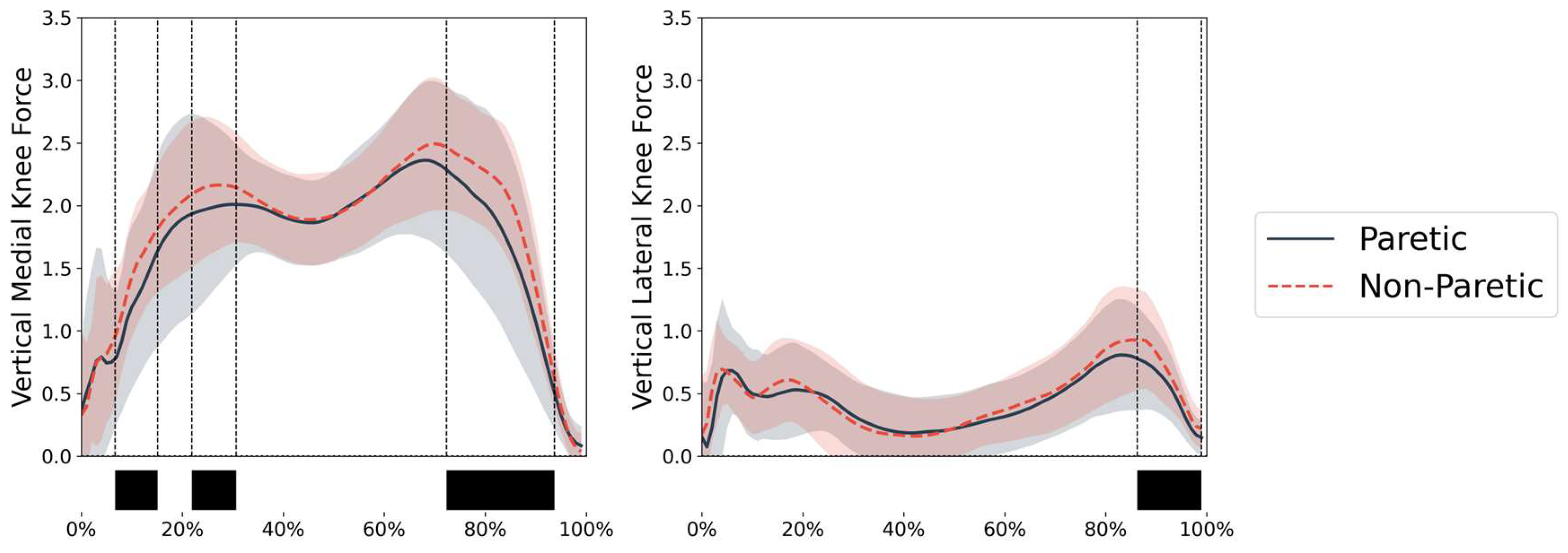
Figure 2.
Lower limb kinematics and kinetics during stance phase for paretic (solid blue line) and non-paretic (dashed red line) limbs. Top panel shows joint angles for hip (flexion-extension, abduction-adduction, internal-external rotation), knee (flexion-extension), and ankle (dorsi-plantarflexion). Bottom panel shows corresponding joint moments. Angles are in degrees and moments are normalized to body mass (Nm/kg). Shaded areas represent ±1 standard deviation. Black bars indicate periods of significant differences between limbs (p<0.05).
Figure 2.
Lower limb kinematics and kinetics during stance phase for paretic (solid blue line) and non-paretic (dashed red line) limbs. Top panel shows joint angles for hip (flexion-extension, abduction-adduction, internal-external rotation), knee (flexion-extension), and ankle (dorsi-plantarflexion). Bottom panel shows corresponding joint moments. Angles are in degrees and moments are normalized to body mass (Nm/kg). Shaded areas represent ±1 standard deviation. Black bars indicate periods of significant differences between limbs (p<0.05).
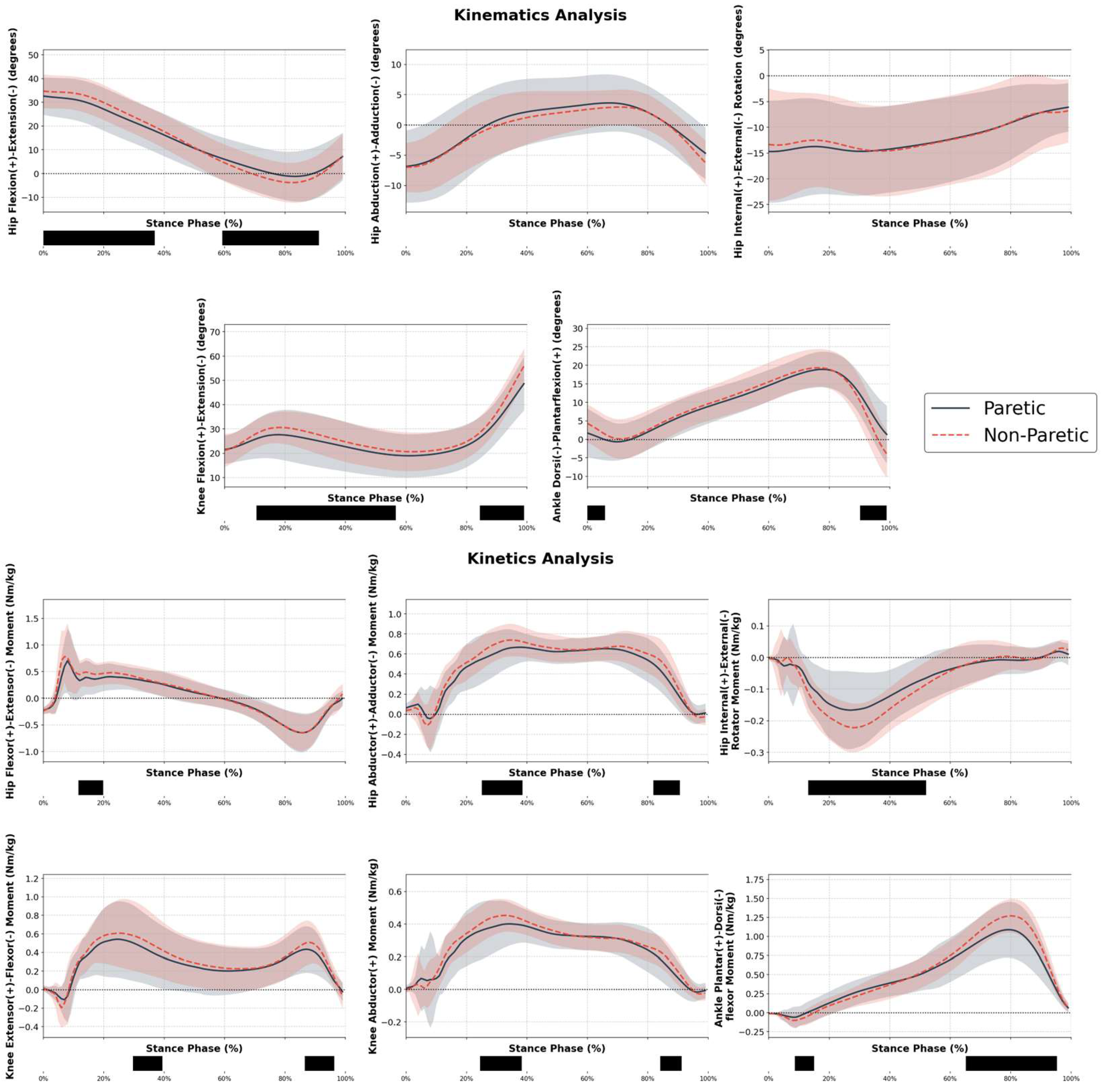

Table 1.
Peak knee contact forces and corresponding joint moments during stance phase. Values are presented as mean ± standard deviation for both medial and lateral compartments of paretic and non-paretic limbs. Contact forces are normalized to body weight (BW).
Table 1.
Peak knee contact forces and corresponding joint moments during stance phase. Values are presented as mean ± standard deviation for both medial and lateral compartments of paretic and non-paretic limbs. Contact forces are normalized to body weight (BW).
| Side | Variable | Contact Force (BW) | Knee extensor Moment (Nm/kg) | Knee Abductor Moment (Nm/kg) |
| Paretic | First Medial Peak | 2.37 ± 0.51 | 0.57 ± 0.34 | 0.42 ± 0.11 |
| Paretic | Second Medial Peak | 2.58 ± 0.60 | 0.30 ± 0.22 | 0.34 ± 0.10 |
| Paretic | First Lateral Peak | 0.97 ± 0.41 | 0.39 ± 0.36 | 0.26 ± 0.28 |
| Paretic | Second Lateral Peak | 0.91 ± 0.41 | 0.41 ± 0.24 | 0.17 ± 0.13 |
| Non-Paretic | First Medial Peak | 2.42 ± 0.43 | 0.63 ± 0.34 | 0.44 ± 0.11 |
| Non-Paretic | Second Medial Peak | 2.72 ± 0.47 | 0.33 ± 0.23 | 0.32 ± 0.10 |
| Non-Paretic | First Lateral Peak | 0.88 ± 0.29 | 0.42 ± 0.33 | 0.21 ± 0.17 |
| Non-Paretic | Second Lateral Peak | 1.03 ± 0.40 | 0.48 ± 0.26 | 0.20 ± 0.10 |
Disclaimer/Publisher’s Note: The statements, opinions and data contained in all publications are solely those of the individual author(s) and contributor(s) and not of MDPI and/or the editor(s). MDPI and/or the editor(s) disclaim responsibility for any injury to people or property resulting from any ideas, methods, instructions or products referred to in the content. |
© 2025 by the authors. Licensee MDPI, Basel, Switzerland. This article is an open access article distributed under the terms and conditions of the Creative Commons Attribution (CC BY) license (https://creativecommons.org/licenses/by/4.0/).
Copyright: This open access article is published under a Creative Commons CC BY 4.0 license, which permit the free download, distribution, and reuse, provided that the author and preprint are cited in any reuse.