Submitted:
22 February 2025
Posted:
28 February 2025
You are already at the latest version
Abstract
Background/Objectives: Hamstring strain injuries are common in elite athletes and affect return-to-sport timeline. Although magnetic resonance imaging (MRI) is the gold standard for assessing injury severity, ultrasonography (US) is a more accessible and cost-effective alternative. This study aimed to evaluate the agreement between US and MRI in the diagnosis of hamstring injuries and their prognostic value in predicting recovery. Methods: This retrospective study included elite athletes with acute first-time hamstring strains who underwent both MRI and US within five days after injury. The injuries were classified according to location (muscle belly, musculotendi-nous junction, or tendon) and severity (modified Peetrons classification). The agree-ment between imaging findings and return-to-sport timelines was analyzed. Results: US demonstrated 70% agreement with MRI in identifying injury locations, with the highest concordance for muscle belly (90%) and musculotendinous junction (80%) in-juries but lower accuracy for tendons (60%). Recovery times differed significantly by location and severity (p < 0.01), with tendon grade 3 injuries requiring the longest re-covery (383 days), while muscle belly injuries had the shortest recovery (16 days). Musculotendinous junction grade 2, tendon grade 1, and tendon grade 2 injuries had similar recovery durations (57–65 days). Conclusions: High-resolution US is a reliable diagnostic tool for muscle belly and musculotendinous junction injuries; however, MRI remains essential for high-grade tendon injuries. US serves as a first-line imaging mo-dality, with MRI reserved for cases that require a detailed prognostic assessment. These findings provide guidance for optimizing imaging strategies for hamstring inju-ry management.
Keywords:
1. Introduction
2. Materials and Methods
Study Design and Ethical Considerations
Study Population
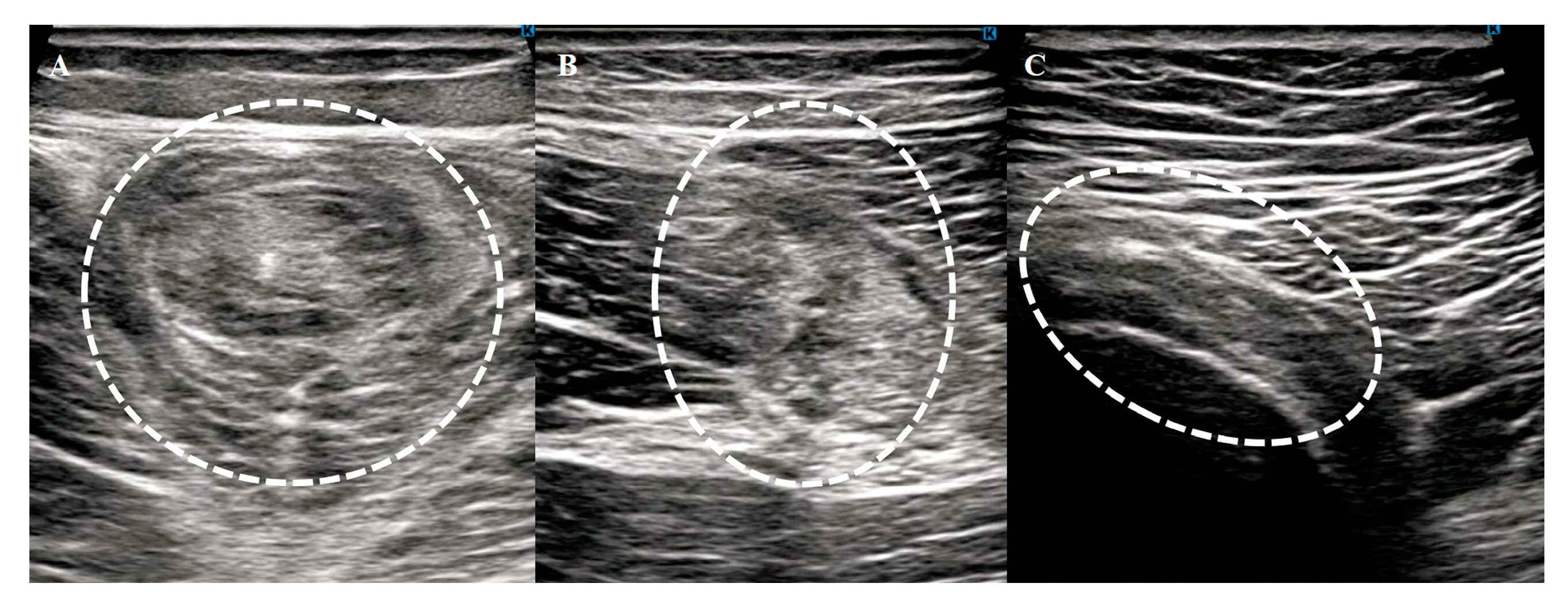
Hamstring Muscle Diagnosis Using Ultrasonography
- Changes in echogenicity or fiber disruption within the muscle.
- Edema or hemorrhage, identified as areas of increased echogenicity with or without visible fiber disruption in orthogonal planes.
- Hypoechoic fluid tracking along the fascial layer surrounding the muscle, indicative of intermuscular hematoma.
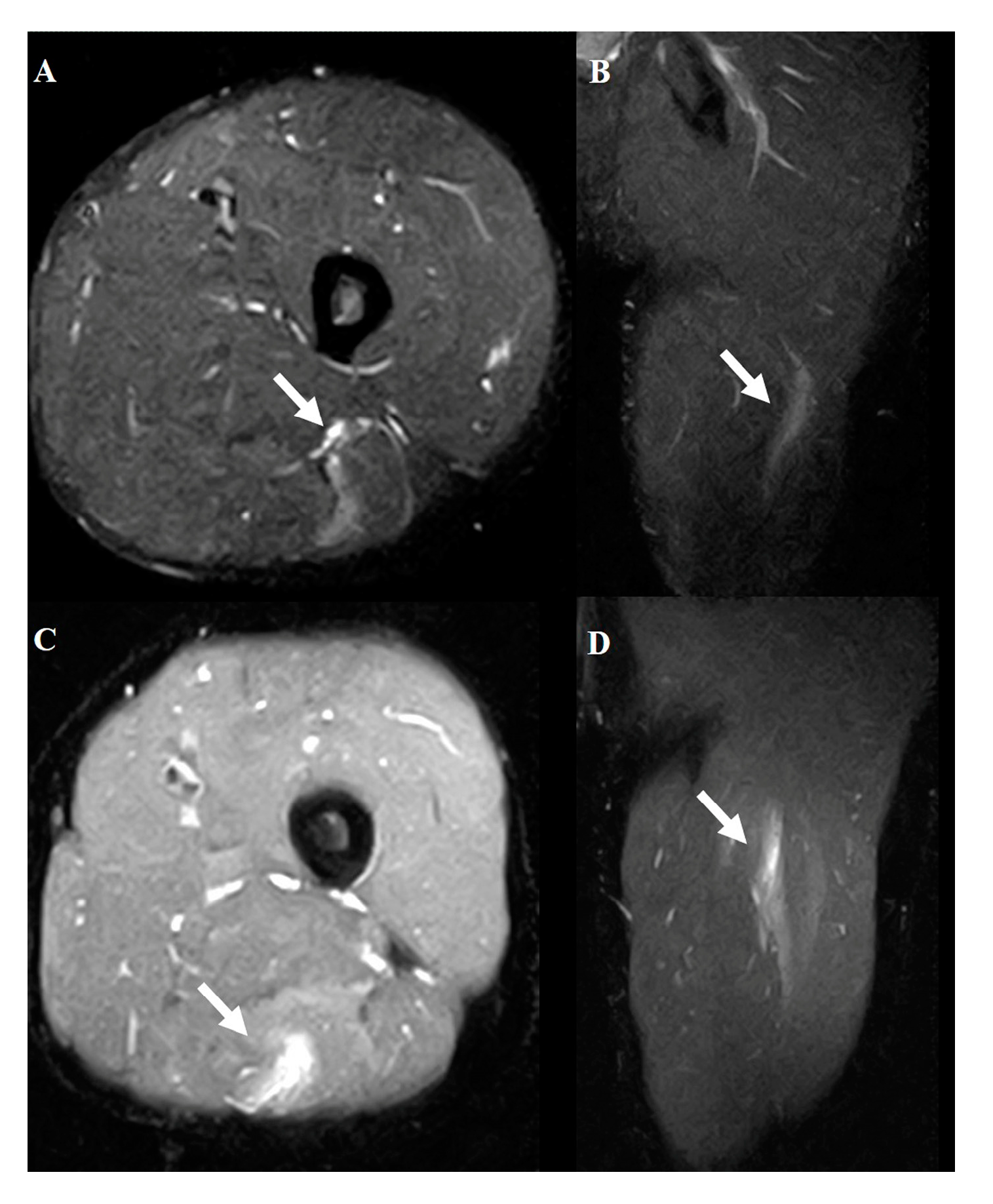

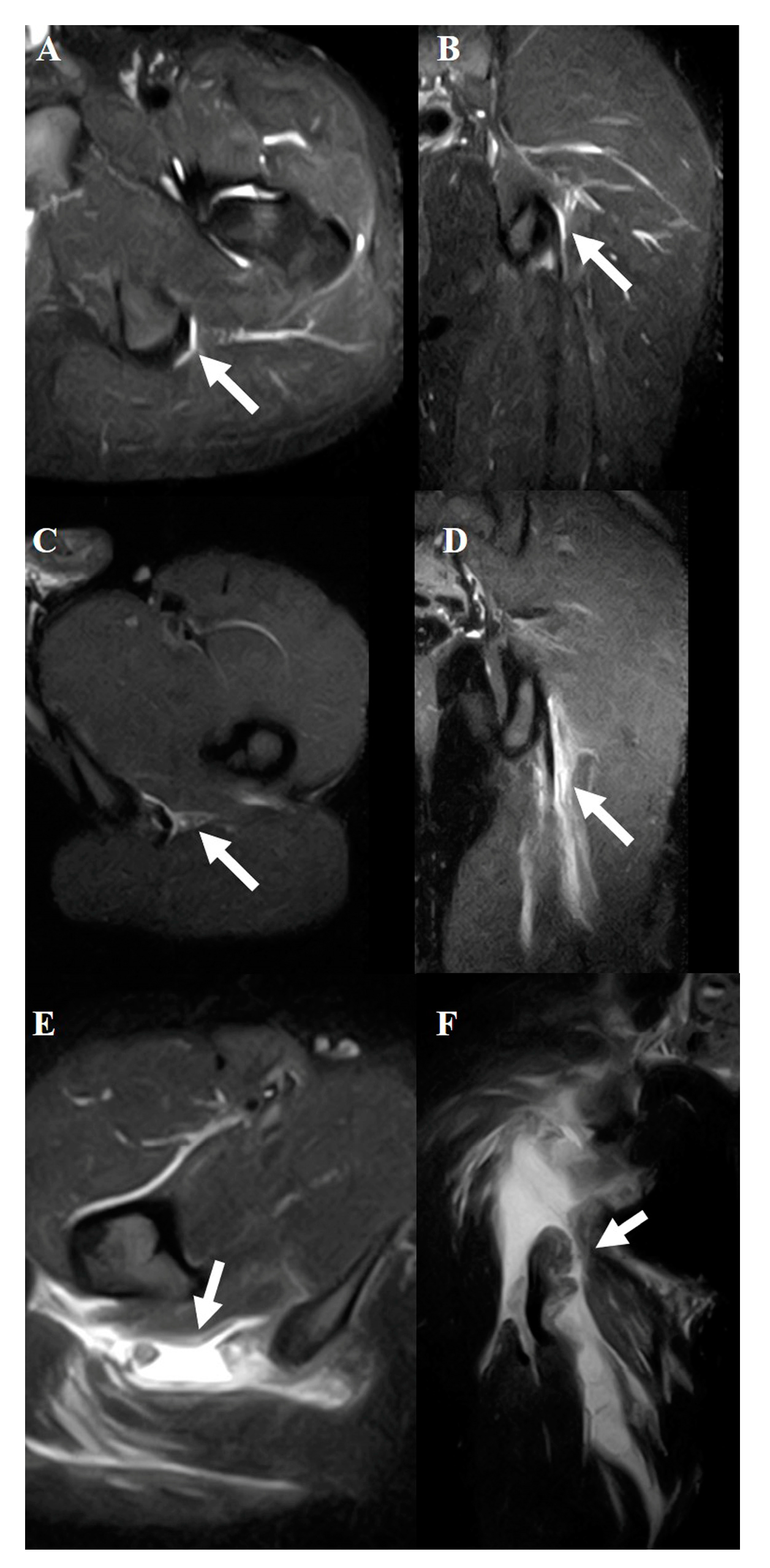
Muscle Diagnosis Using MRI
Evaluation Criteria
Statistical Analysis
3. Results
Ultrasonography’s Diagnostic Accuracy as Part of Agreement Analysis
Patient Demographics and Clinical Characteristics
Agreement Between Ultrasonography and MRI, and Clinical Relevance of Injury Location
Impact of Injury Severity on Return to Sport Based on MRI Analysis
Combined Analysis of Injury Location and Severity
- Tendon grade 3 injuries required the longest recovery time, averaging 383 days, and differed significantly from all the other groups (p < 0.001).
- Musculotendinous junction grade 3 injuries had a recovery time of 100 days, which was significantly longer than that of most of the other groups, except tendon grade 3 (p < 0.01).
- Groups with musculotendinous junction grade 2 and tendon grades 1 and 2 showed similar recovery times, ranging from 57 to 65 days.
- Muscle grade 1 injuries had the shortest recovery time, averaging 16 days.
4. Discussion
5. Conclusions
Author Contributions
Funding
Institutional Review Board Statement
Informed Consent Statement
Data Availability Statement
Acknowledgments
Conflicts of Interest
Abbreviations
| MRI | magnetic resonance imaging |
References
- Heer, S.T.; Callander, J.W.; Kraeutler, M.J.; Mei-Dan, O.; Mulcahey, M.K. Hamstring injuries: Risk Factors, Treatment, and Rehabilitation. J. Bone Joint Surg. Am. 2019, 101, 843–853. [Google Scholar] [CrossRef] [PubMed]
- Kerkhoffs, G.M.M.J.; van Es, N.; Wieldraaijer, T.; Sierevelt, I.N.; Ekstrand, J.; van Dijk, C.N. Diagnosis and prognosis of acute hamstring injuries in athletes. Knee Surg. Sports Traumatol. Arthrosc. 2013, 21, 500–509. [Google Scholar] [CrossRef]
- Arner, J.W.; McClincy, M.P.; Bradley, J.P. Hamstring injuries in athletes: evidence-based treatment. J. Am. Acad. Orthop. Surg. 2019, 27, 868–877. [Google Scholar] [CrossRef] [PubMed]
- Heiderscheit, B.C.; Sherry, M.A.; Silder, A.; Chumanov, E.S.; Thelen, D.G. Hamstring strain injuries: recommendations for diagnosis, rehabilitation, and injury prevention. J. Orthop. Sports Phys. Ther. 2010, 40, 67–81. [Google Scholar] [CrossRef]
- Greenky, M.; Cohen, S.B. Magnetic resonance imaging for assessing hamstring injuries: clinical benefits and pitfalls – a review of the current literature. Open Access J. Sports Med. 2017, 8, 167–170. [Google Scholar] [CrossRef]
- Connell, D.A.; Schneider-Kolsky, M.E.; Hoving, J.L.; Malara, F.; Buchbinder, R.; Koulouris, G.; Burke, F.; Bass, C. Longitudinal study comparing sonographic and MRI assessments of acute and healing hamstring injuries. A.J.R. Am. J. Roentgenol. 2004, 183, 975–984. [Google Scholar] [CrossRef]
- van Beek, E.J.R.; Kuhl, C.; Anzai, Y.; Desmond, P.; Ehman, R.L.; Gong, Q.; Gold, G.; Gulani, V.; Hall-Craggs, M.; Leiner, T.; Lim, C.C.T.; Pipe, J.G.; Reeder, S.; Reinhold, C.; Smits, M.; Sodickson, D.K.; Tempany, C.; Vargas, H.A.; Wang, M. Value of MRI in medicine: more than just another test? J. Magn. Reson. Imaging 2019, 49, e14–25. [Google Scholar] [CrossRef] [PubMed]
- Petersen, J.; Thorborg, K.; Nielsen, M.B.; Skjødt, T.; Bolvig, L.; Bang, N.; Hölmich, P. The diagnostic and prognostic value of ultrasonography in soccer players with acute hamstring injuries. Am. J. Sports Med. 2014, 42, 399–404. [Google Scholar] [CrossRef]
- Hendawi, T.K.; Rendos, N.K.; Warrell, C.S.; Hackel, J.G.; Jordan, S.E.; Andrews, J.R.; Ostrander, R.V. Medial elbow stability assessment after ultrasound-guided ulnar collateral ligament transection in a cadaveric model: ultrasound versus stress radiography. J. Shoulder Elbow Surg. 2019, 28, 1154–1158. [Google Scholar] [CrossRef]
- Chiang, Y.-P.; Wang, T.-G.; Hsieh, S.-F. Application of ultrasound in sports injury. J. Med. Ultrasound 2013, 21, 1–8. [Google Scholar] [CrossRef]
- Fournier-Farley, C.; Lamontagne, M.; Gendron, P.; Gagnon, D.H. Determinants of return to play after the nonoperative management of hamstring injuries in athletes: A systematic review. Am. J. Sports Med. 2016, 44, 2166–2172. [Google Scholar] [CrossRef] [PubMed]
- Kuske, B.; Hamilton, D.F.; Pattle, S.B.; Simpson, A.H.R.W. Patterns of hamstring muscle tears in the general population: A systematic review. PLOS One 2016, 11, e0152855. [Google Scholar] [CrossRef]
- van der Horst, N.; van de Hoef, S.; Reurink, G.; Huisstede, B.; Backx, F. Return to play after hamstring injuries: A qualitative systematic review of definitions and criteria. Sports Med. 2016, 46, 899–912. [Google Scholar] [CrossRef]
- Peetrons, P. Ultrasound of muscles. Eur. Radiol. 2002, 12, 35–43. [Google Scholar] [CrossRef] [PubMed]
- Zein, M.I.; Mokkenstorm, M.J.K.; Cardinale, M.; Holtzhausen, L.; Whiteley, R.; Moen, M.H.; Reurink, G.; Tol, J.L.; Qatari and Dutch Hamstring Study Group. Baseline clinical and MRI risk factors for hamstring reinjury showing the value of performing baseline MRI and delaying return to play: a multicentre, prospective cohort of 330 acute hamstring injuries. Br. J. Sports Med. 2024, 58, 766–776. [Google Scholar] [CrossRef]
- Pedret, C. Hamstring muscle injuries: MRI and ultrasound for diagnosis and prognosis. J. Belg. Soc. Radiol. 2021, 105, 91. [Google Scholar] [CrossRef]
- Naqvi, G.A.; Jadaan, M.; Harrington, P. Accuracy of ultrasonography and magnetic resonance imaging for detection of full thickness rotator cuff tears. Int. J. Shoulder Surg. 2009, 3, 94–97. [Google Scholar] [CrossRef]
- Bhimani, R.; Lubberts, B.; DiGiovanni, C.W.; Tanaka, M.J. Dynamic ultrasound can accurately quantify severity of medial knee injury: A cadaveric study. Arthrosc. Sports Med. Rehabil. 2022, 4, e1777–e1787. [Google Scholar] [CrossRef]
- Ergün, T.; Peker, A.; Aybay, M.N.; Turan, K.; Muratoğlu, O.G.; Çabuk, H. Ultrasonography vıew for acute ankle ınjury: comparison of ultrasonography and magnetic resonance ımaging. Arch. Orthop. Trauma. Surg. 2023, 143, 1531–1536. [Google Scholar] [CrossRef]
- Koulouris, G.; Connell, D. Evaluation of the hamstring muscle complex following acute injury. Skelet. Radiol. 2003, 32, 582–589. [Google Scholar] [CrossRef]
- Ekstrand, J.; Lee, J.C.; Healy, J.C. MRI findings and return to play in football: a prospective analysis of 255 hamstring injuries in the UEFA Elite Club Injury Study. Br. J. Sports Med. 2016, 50, 738–743. [Google Scholar] [CrossRef] [PubMed]
- Cohen, S.B.; Towers, J.D.; Zoga, A.; Irrgang, J.J.; Makda, J.; Deluca, P.F.; Bradley, J.P. Hamstring injuries in professional football players: magnetic resonance imaging correlation with return to play. Sports Health 2011, 3, 423–430. [Google Scholar] [CrossRef] [PubMed]
- Askling, C.M.; Tengvar, M.; Saartok, T.; Thorstensson, A. Acute first-time hamstring strains during high-speed running: a longitudinal study including clinical and magnetic resonance imaging findings. Am. J. Sports Med. 2007, 35, 197–206. [Google Scholar] [CrossRef] [PubMed]

| Variable | Value |
| Number of patients | 71 |
| Sex (% male) | 61 (86%) |
| Age (yr, mean ± SD) | 21.0 ± 4.7 (18–48) |
| Injured side (%) | Right: 48, Left: 52 |
| Sports activity | Rugby: 49, Sprint: 14, Baseball: 13, Soccer: 11, Judo: 8, Long-distance running: 7, American football: 3, Field events: 2, Tennis: 1, Lacrosse: 1 |
| Time to imaging (hr) | US: 57.2 ± 59.3, MRI: 88.9 ± 83.1 |
| Injury Location | Agreement Rate (%) |
| Overall Agreement | 70 |
| By Muscle Group | |
| Semitendinosus | 100 |
| Biceps Femoris | 82 |
| Semimembranosus | 68 |
| By Specific Location | |
| Musculotendinous Junction | 80 |
| Muscle Belly | 90 |
| Tendon | 60 |
|
Musculotendinous junction |
Tendon | Muscle | P-value | |
| Proportion of Injuries (%) | 65 | 21 | 14 | |
| Time to return to sporting activity (Days) |
70±36 | 83±90 | 16±8 | <0.01 |
| Grade 1 | Grade 2 | Grade 3 | P-value | |
| Proportion of Injuries (%) | 37 | 32 | 31 | |
| The time to return to sporting activity (Days) |
35±28 | 56±28 | 111±67 | <0.01 |
| Injured Part | Grade | Number of Patients | Return to Sporting Activity (Days, Mean ± SD) |
| Muscle | 1 | 8 | 17 ± 8 |
| 2 | 1 | 8 ± 0 | |
| 3 | 0 | ||
| Musculotendinous Junction | 1 | 10 | 31 ± 13 |
| 2 | 17 | 57 ± 21 | |
| 3 | 20 | 100 ± 27 | |
| Tendon | 1 | 6 | 65 ± 39 |
| 2 | 8 | 59 ± 37 | |
| 3 | 1 | 383 ± 0 |
| Group | Muscle Grade 1 | Muscle Grade 2 | Musculotendinous Grade 1 | Musculotendinous Grade 2 | Musculotendinous Grade 3 | Tendon Grade 1 | Tendon Grade 2 | Tendon Grade 3 |
| Muscle Grade 2 | n.s. | |||||||
| Musculotendinous Grade 1 | n.s. | n.s. | ||||||
| Musculotendinous Grade 2 | <0.05 | n.s. | n.s. | |||||
| Musculotendinous Grade 3 | <0.001 | <0.05 | <0.001 | <0.001 | ||||
| Tendon Grade 1 | <0.05 | n.s. | n.s. | n.s. | n.s. | |||
| Tendon Grade 2 | <0.05 | n.s. | n.s. | n.s. | n.s. | <0.01 | ||
| Tendon Grade 3 | <0.001 | <0.001 | <0.001 | <0.001 | <0.001 | <0.001 | <0.001 |
Disclaimer/Publisher’s Note: The statements, opinions and data contained in all publications are solely those of the individual author(s) and contributor(s) and not of MDPI and/or the editor(s). MDPI and/or the editor(s) disclaim responsibility for any injury to people or property resulting from any ideas, methods, instructions or products referred to in the content. |
© 2025 by the authors. Licensee MDPI, Basel, Switzerland. This article is an open access article distributed under the terms and conditions of the Creative Commons Attribution (CC BY) license (http://creativecommons.org/licenses/by/4.0/).


