
Submitted:
20 February 2025
Posted:
21 February 2025
You are already at the latest version
Abstract
Background: Clarifying the etiology of mental disorders should lead to significant therapeutic and preventive measure improvements. Mental disorders are caused by genetic, epigenetic, and environmental factors. A valid assessment of environmental etiological factors is complicated due to a lack in methodologies. We aim to demonstrate how to optimally assess the environmental etiological factors with a special focus on stress. Methods: We searched the PubMed database up to December 2024 for relevant state-of-the-art references. Results: The most frequently used stress assessment method involves self-reporting questionnaires, but the usefulness of the answers may be diminished, for example by recall bias. Blood or salivary cortisol, heart rate, respiratory rate, plethysmography readings, and electrodermal activity are biological markers of acute stress. Biological markers such as hair and nail cortisol levels, telomere length, mitochondrial DNA copy number, and epigenetic markers are indicative of a history of stress. Conclusion: A quantitative prospective assessment of environmental etiological factors including their frequency, severity, total exposure length, timing, and possible accumulation is still needed. In addition, protective environmental factors of mental disorders should not be neglected. The computation of a polyenviromic risk/protective score is an innovative research approach into the etiology of psychiatric disorders.
Keywords:
mental disorders
; environmental etiological factors
; assessment
; stress
; questionnaires
; biological marker
Plain Language Summary
- Identifying the causes of mental disorders will aid in the discovery of optimal treatments
- The psychiatric diseases causes include interplays of genetic, epigenetic, and environmental factors
- Technical advances have facilitated the detection of genetic/epigenetic causes of mental diseases, decreasing even testing costs, but a valid assessment of environmental factors remains a challenge
- Stress is one of the most important environmental causative factors of mental disorders
- Assessing the history of stressful experiences in a patient´s life is a challenge because retrospective questionnaires are inaccurate and biased
- In this narrative review, we demonstrate how to assess an individual´s stress history by measuring hair and nail cortisol content, telomere length, and mitochondrial DNA copy number, as well as epigenetic markers
- We also suggest avenues for research: A quantitative prospective evaluation of environmental etiological factors including their frequency, severity, duration, and timing, and a similar assessment of protective variables are needed
1. Introduction
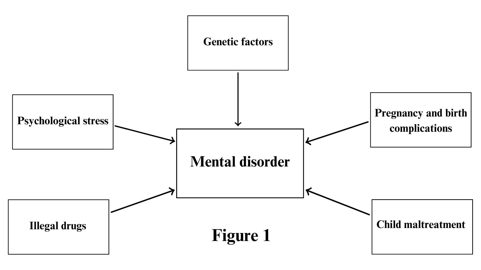
One third of individuals in the population will develop a mental disorder over the course of their life. Mental disorders are mostly chronic, and frequently life-long. Apart from suicide, delirium tremens, and anorexia nervosa, psychiatric disorders usually do not result in a patient´s death. However, they may significantly deteriorate their quality of life. Moreover, their negative impact on a country´s economy is also apparent. In spite of all the medical breakthroughs, the treatment of mental disorders remains unsatisfactory. Symptoms can be treated, but curing psychiatric diseases is uncommon. This lack of treatment success is due in part to lack of knowledge of the exact causes of mental disorders. The progressive elucidation of the etiological factors of mental disorders will lead to the improvement of therapeutic and preventive measures improving the life of patients [1].
In the past, identifying genetic causes of mental disorders was impossible. The human genome was not fully sequenced until 2003, when the National Institutes of Health and the Celera company successfully completed the task [2]. From then on, full genome sequencing has become cheaper. The Human Genome Project took 13 years and cost $2.7 billion, by contrast recent technologies have reduced the cost of sequencing to $200 per human genome [3]. However, identifying environmental causes of mental disorders remains a challenge, due to a lack of effective methodological approaches. These environmental etiological factors can be physical, chemical, biological, psychological, social, ecological, economic, or religious/spiritual in nature, and all of them require their own particular assessment tests.
The European Network of National Schizophrenia Networks Studying Gene-Environment Interactions (EU-GEI) studywas the largest and most extensive incidence and case-control study assessing multiple genetic and environmental risk factors and their interactions in the development of schizophrenia [4]. From 2010 till 2015, 2774 incident cases of psychotic disorders were identified across 17 sites in 6 countries (The Netherlands, UK, France, Italy, Spain and Brazil). The study involved analyzing data from 1497 healthy controls where researchers collected the following data: demographic, clinical, social, cognitive, and biological exposures, including premorbid adjustment, childhood and adult adversities, cannabis use, migration, and/or discrimination. Additionally, DNA examinations were taken via blood samples or cheek swabs. The cost of the project reached €15 million. The EU-GEI study findings included several interesting results regarding the environmental etiological factors of psychoses.
Based on the data from the EU-GEI study, Tarricone et al compared the social disadvantages in pre- and post-migration phases, migration adversities, and the mismatch between achievements and expectations between first-generation migrants with first-episode psychosis against healthy first-generation migrants [5]. The authors assessed 249 cases and 219 controls. Pre-migration (OR, 1.61; 95% CI, 1.06–2.44; P = 0.027) and post-migration social disadvantages (OR, 1.89; 95% CI, 1.02–3.51; P = 0.044), along with expectations/achievements mismatches (OR, 1.14; 95% CI, 1.03–1.26; P = 0.014) were all significantly associated with psychosis with a dose response effect between the number of adversities across all phases and odds of psychosis (⩾6: OR, 14.09; 95% CI, 2.06–96.47; P = 0.007). The study results were independent of ethnicity or length of stay in the country of arrival.
Jongsma et al analyzed the records of 1130 cases and 1497 population-based controls from the EU-GEI study [6]. Crude excess odds of psychosis were found in participants from ethnic minorities (OR, 2.03; 95% CI, 1.69-2.43), a finding persisting after adjustment for confounders (OR, 1.61; 95% CI, 1.31-1.98).
Sideli et al assessed 829 patients with a first-episode of psychosis and 1283 community controls from 16 EU-GEI sites for child maltreatment, education attainment, and IQ [7]. Childhood maltreatment was associated with lower educational attainment (P = 0.001) and childhood neglect with lower attainment and IQ values in both cohorts (P < 0.001). Childhood abuse was associated with IQ only in the control group (P < 0.001).
Di Forti et al obtained data from 901 patients with first-episode psychosis across 11 sites and 1237 population controls from the same sites [8]. Increased odds of developing psychotic disorders were found in daily cannabis users compared to never users (adjusted OR, 3.2; 95% CI, 2.2–4.1), with the odds increasing with daily use of high-potency types of cannabis (tetrahydrocannabinol ≥10%) (adjusted OR, 4.8; 95% CI, 2.5–6.3).
1.1. Polyenviromic Risk/Protective Score
After completion of the EU-GEI study, Padmanabhan et al introduced the concept of polyenviromic risk score (PERS), which represents a summary assessment of environmental risk exposures in an individual [9]. The calculation of PERS involves binarizing the odds ratio for each environmental risk factor obtained from the literature. The presence or absence of each environmental risk factor is determined for the individual, where the log of the odds ratio for each environmental risk factor is multiplied by either 1 (risk factor present in the individual) or 0 (risk factor absent). The results are added together, and the sum is divided by the total number of risk factors assessed.
Risk factor present in the individual (p) = log(odds ratio) x 1
Risk factor absent in the individual (a) = log(odds ratio) x 0
p + a
______________________________
total number of risk factors assessed
In this way, environmental risk factors are aggregated. The PERS calculation adapts a formula used for the calculation of polygenic risk scores [10]. The polyenviromic protective score summarizing environmental protective factors can be calculated in the same way as the PERS.
Padmanabhan et al conducted the first polyenviromic risk score study [9]. The authors investigated whether an aggregate score of exposure to nine environmental risk factors could predict conversion to psychosis in a pilot longitudinal sample of 83 young participants with a high familial risk of schizophrenia. A high PERS was significantly associated with conversion to psychosis (OR, 1.97; P = 0.009). An environmental risk aggregate index was concluded to help predict conversion to psychosis in individuals with a high familial schizophrenia risk.
In our study, we administered the internationally accepted questionnaire from the EU-GEI study (enriched with mood and anxiety disorder-relevant measures) to patients at two large university hospitals in the Czech Republic where we compared environmental risk and protective factors in patients with psychosis or a mood disorder (PSYCH+MOOD), and those with an anxiety disorder (ANX) [11]. We enrolled 94 PSYCH+MOOD patients (average age, 42.5 years; 46 men) and 52 ANX patients (average age, 47.2 years; 17 men). Both the PERS and the polyenviromic protective score were similar in both the PSYCH+MOOD and ANX groups (P = 0.149; P = 0.466, respectively). We concluded that the polyenviromic risk/protective score scales still require scientific validation in large psychiatric, and ideally prospective, studies.
1.2. Pregnancy and Birth Complications in the Etiology of Mental Disorders
Based upon Stilo and Murray´s review of literature regarding risk factors in schizophrenia, the etiology of schizophrenia is multifactorial, related to interactions between genetic background and environmental contributors [12]. The most important environmental risk factors (alone or in combination with others) involve pregnancy and birth complications, the birth season (winter or spring), severe maternal malnutrition or hypovitaminosis, and maternal influenza.
Al-Haddad et al extensively described the impact of infections and inflammation during pregnancy on the developing fetal brain [13]. Accumulating evidence suggests that the fetal brain development can be dependent on a wide variety of viral and bacterial infections, leading to development of neuropsychiatric diseases later in life. Neuroinflammation in children can instigate schizophrenia, autism spectrum disorder, and depression. Animal studies suggest that both pregnancy infections and inflammation can result in direct injury to neurons and their progenitor cells, or in indirect injury via activation of cytokine production and oxidative stress by microglia and astrocytes.
In the context of heavy metal exposure and neurotoxicity, Vorvolakos et al argued against the existence of a safe threshold for lead exposure [14]. Lead has been proven to alter physiological neurotransmissions by affecting cholinergic, dopaminergic, and glutamatergic systems. The subsequent consequences include apoptosis and excitotoxicity in the brain. From a clinical perspective, constant cognitive impairment and difficulties in concentration can occur. Prenatal exposure to lead is associated with antisocial behavior and schizophrenia.
According to Van den Bergh et al, prenatal exposure to maternal stress increases the risk for mental health problems later in life [15]. A systematic analysis was performed by the authors in available human studies to identify harmful stressors and biological correlates between maternal stress and offspring outcomes. For example, maternal stress can affect the offspring neurodevelopment, resulting in anxiety disorders, depression, schizophrenia, or attention deficit hyperactivity disorder. Damaged brain connectivities were identified in the neurobiological background of affected individuals, involving the amygdala and pre-frontal cortex or by changes in the hypothalamo-pituitary-adrenal axis and the autonomous nervous systems.
In the Czech Republic, data on maternal infections, intoxications, malnutrition, stress, somatic or mental disorders, medication, substance abuse, social and economic problems and other important factors are comprehensively collected in pregnancy record books or medical birth records. However, getting hold of relevant records for the assessment of middle-aged or older individuals may be complicated if their mothers or their gynecologists are already deceased. A medical electronic database comprising important data on patients and their relatives would be a valuable tool for the health system to maintain.
1.3. Environmental Risk Factors in Mental Disorders in Adulthood
According to the review by Stilo and Murray, the evidence highlights an important etiological role for cannabis use in schizophrenia [12]. Prospective epidemiological studies have consistently reported an association between cannabis use and schizophrenia with an estimated 2- to 3-fold increased risk. For a valid assessment of cannabis use, the following variables need to be considered: The individual dose, the time span and frequency of use, the age at which cannabis use began, and whether high potency tetrahydrocannabinol was used.
Vassos et al metaanalyzed four studies which state the well-established association between urbanicity and risk of schizophrenia [16]. Schizophrenia´s incidence has increased in line with rising levels of urbanicity, as measured by population size or density. This association is indicated as an incidence rate ratio. The authors observed a linear association between the logarithm of the odds of risk for schizophrenia and urbanicity. Compared to most rural environments, the risk for schizophrenia in the most urban environments is estimated to be 2.37 times higher, with the same effect present for nonaffective psychosis.
1.4. How to Assess a Stressful Experience?
The effect size of individual environmental etiological factors in mental disorders varies. Some are more important than others, depending on the specific psychiatric disease. Some risk/protective factors may be non-specific and are associated with several mental disorders, whereas other only have a specific role in certain diagnosis or in its subgroup. The most important and non-specific environmental risk factor in psychiatry is stress [1].
The most frequently used assessment method uses self-reporting questionnaires. Their application is relatively quick and cheap, even in the case of comprehensive evaluations. However, their value may be diminished due to recall bias, the respondent´s cognitive impairment, or a delusional interpretation of reality. The following are examples of self-reporting questionnaires:
- Stress Questionnaire [17]: This questionnaire detects acute perceived stress. The questions aim to discover whether individuals continue working at night once they arrive home, have low self-confidence or guilty feelings, think about problems when they are supposed to be relaxing, present mood swings or difficulty making decisions, etc. Each of 25 questions can be answered as "yes“ or "no“ resulting in a score of either 1 or 0, respectively. The total score is added and an appropriate recommendation is offered to the respondent.
- Perceived Stress Scale [18]: This scale detects stress perceived during the preceding month. The questions inquire whether the respondent is upset due to unexpected events, whether they feel nervous, stressed, irritated, and/or angry. Each of the ten questions may get a score from "0“ (never) to "4“ (very often), and the total is the sum of all answers.
- Perceived Stress Questionnaire [19]: This questionnaire detects stress experienced during the preceding month. The questions ask whether the respondent feels calm, safe, tired, and/or frustrated, whether they have trouble relaxing, are worried, lack time for themselves, feel under pressure from other people, etc. Each of the 30 questions may be scored from "1“ (almost never) to "4“ (usually). The results of the test correlate highly with anxiety traits.
A semi-structured interview with relevant open-ended questions performed by an experienced researcher is another stress assessment method. In contrast to the structured interviews, a semi-structured interview is open, allowing the respondent to express their thoughts more freely. Semi-structured interviews achieve more accurate assessments than self-reporting questionnaires. However, semi-structured interviews are staff- and cost-demanding. Semi-structured interviews target specific traits; for example, they can be used to detect and assess a post-traumatic stress disorder (PTSD) in children from 0 to 7 years of age [20].
1.5. Biological Markers of Perceived Acute Stress
Cortisol is a glucocorticoid steroid and a stress hormone which is released in diurnal cycle, but can also be triggered by stress and low blood-glucose concentrations. This hormone functions to increase blood sugar through gluconeogenesis activation, it suppresses the immune system, and aids in the metabolism of fat, protein, and carbohydrates [21]. Additionally, cortisol contributes to the pathogenesis and clinical expression of psychotic disorders, major depressive disorder, anxiety disorders, and PTSD [22,23,24,25].
Salivary cortisol is one of the biomarkers of acute psychological stress. Blood and urine sample cortisol detection are other viable ways of measurement, however, they do not show a linear correlation with salivary cortisol levels [26].
Immanuel et al concluded that heart rate variability is a valid measure of psychological stress response. Studies (n = 15) were reviewed on the associations between heart rate variability and psychological stress in individuals exposed to either real-life or experimental stressors [27]. Amongst the heart rate variability metrics, the root-mean-square deviation, the high frequency power, and the ratio of low frequency to high frequency were most reported.
Additionally, according to Nicolo et al´s review, respiratory rate is a fundamental vital sign sensitive to different stressors including emotional stress [28]. The authors presented a multidisciplinary approach to respiratory monitoring using suitable sensors and techniques. Respiratory rate sensitivity to stressful conditions stands superior compared to other vital signs.
Plethysmography is another method to detect acute mental stress. The study by Barki and Chung with 14 participants presented an ear-mounted photoplethysmography system, which achieved a high level of accuracy (92.04%) for detecting acute stress [29].
Electrodermal activity is a bio-signal to detect a perceived stress. The review by Klimek et al based on 74 studies published between 2012 and 2022 found that electrodermal activity is most often measured on the wrist in healthy individuals under laboratory conditions [30]. Studies using machine learning to predict stress via electrodermal activity have increased in number. According to Klimek et al, future studies on relevant populations should focus on measuring electrodermal activity in real-life stress situations.
1.6. Hair and Nail Cortisol
A notable marker of hypothalamic-pituitary-adrenal axis activity over a period of several months is the cortisol content of hair. A meta-analysis by Khoury et al included 28 studies investigating the strength of the association between adverse experiences and hair cortisol levels [31]. Results of multilevel modeling analyses indicated that the overal effect size was small but significant (d = 0.213; 95% CI, 0.034 – 0.397). According to moderator analyses, factors such as features of the adversity exposure (eg, type of adversity, timing of adversity), characteristics of the samples (eg, clinical status, racial distribution), and features of the publication (eg, publication type, geographic region of the study) moderated the strength and direction of association.
As stated in the systematic review of 22 articles by Koumantarou Malisiova et al, scalp hair cortisol analysis proves to be a consistent tool for measuring long-term stress exposure [32]. Patients with depression showed a general trend for higher hair cortisol concentrations than controls, whereas patients with PTSD tended to have lower hair cortisol concentrations. However, little is known about hair cortisol content in other mental disorders. Differences among study cohorts and the timing of cortisol sampling seem to explain the result discrepancies. The correlations of hair cortisol concentrations with self-reported measures of stress were decidedly inconclusive. The authors suggested that further research is needed to obtain specific hair cortisol profiles for individual psychiatric disorders.
A growing number of studies have tested for associations between indicators of social adversity (stress) and hair cortisol content in children. In a systematic review of 44 published studies in children aged from birth to 8 years (n = 8370), Bryson et al examined socioeconomic factors (eg, low parental education, low income, and unemployment), psychosocial factors (eg, parental stress, poor mental health, and family violence), and children´s direct exposure to maltreatment, abuse, and stressful events [33]. However, there was inconsistent evidence of associations; 24% of the associations between adversity and higher hair cortisol levels were positive, 6% were negative, and 70% were null. According to the authors, the evidence for the association between social adversity and hair cortisol levels in young children was inconclusive.
Botschek et al identified 14 publications involving 1916 participants investigating the hair cortisol content after various psychological and neuropsychiatric interventions [34]. Associations between the hair cortisol content and PTSD, depressive disorders, and ongoing social and family stress were documented. Differing results were seen in the effect of relaxation techniques, mental training or cognitive behavioral therapy on the cortisol content. Some studies reported a reduction in hair cortisol levels following treatment, whereas others did not observe a significant effect. The baseline level of hair cortisol appears to be particularly important. In some studies, high baseline levels are linked to a stronger treatment response, suggesting that hair cortisol may have a predictive value.The authors suggest that hair cortisol levels hold potential as a biological marker to assess the effectiveness of interventions for mental disorders.
In addition to hair cortisol, human nail cortisol could serve as a retrospective biomarker for chronic stress, according to a systematic review of 18 studies by Philips et al [35]. Mass spectrometry and immunoassays are the primary methods used for analysis. In some studies, nail cortisol levels were found to correlate with saliva and hair levels. Elevated nail cortisol has been associated with depression, and its accumulation in nails may reflect a long timespan. However, the validation of this measure is limited by variables such as age, sex, ethnicity, and individual nail characteristics. Phillips et al emphasize the need for further research to confirm the utility of nail cortisol as a chronic stress biomarker across the human lifespan.
1.7. Telomere Length and Mitochondrial DNA Copy Number
According to Lu et al, maintenance of the linear chromosome ends, otherwise known as telomeres, in mammals requires accurate replication of their genetic material and protection against DNA damage [36]. Such functions are performed by the telomerase holoenzyme and a unique nucleoprotein structure, where a group of telomere-associated proteins bind to telomeric DNA, forming specialized protein/DNA complexes, consisting of telomeric reverse transcriptase, telomeric RNA component, and other assisting factors, extending telomeric repeats at the ends of chromosomes. Without proper telomere maintenance, telomere gradually shorten with each round of DNA replication due to 'end replication problem'.
Aubert and Lansdorp reviewed the role of telomeres and telomerase in human ageing and ageing-associated diseases [37]. Their findings suggest that the loss of telomere repeats in stem cells and lymphocytes contributes to human aging, even though the average telomere lengths vary among individuals of the same age. The proliferation of increasing numbers of cells may decline in healthy individuals as a result of gradual telomere shortening over time.
In a narrative review, Lin and Epel stated that the research from in vitro and longitudinal human studies suggests that stress can induce telomere damage [38]. Glucocorticoids, reactive oxygen species, mitochondria, and inflammation are key mediators linking psychological stress and telomere maintenance. The initial telomere setting at birth plays a critical role in determining the lifelong trajectory, and stress influences this setting. Stress effects on telomeres can be intergenerational, with prenatal stress affecting telomere development during fetal growth, and through "telotype transmission" - the direct inheritance of short telomeres from parental germline.
Schutte and Malouff conducted a meta-analysis of eight studies involving 1,143 participants, finding a significant association between high levels of perceived stress and short telomere length, with an effect size of r = -0.25 (P < 0.001) [39].
Oliveira et al reviewed 18 studies of healthy or diseased adults and children through MEDLINE, EMBASE, Cochrane Central and grey literature databases to examine the link between chronic social stress (like poverty, violence, or family caregiving) and telomere length across lifespan of individuals [40]. Of the studies, 16 were cross-sectional and 2 were longitudinal. The studies differed in type of stress exposure, methods to measure telomere lengths, and sampled cell types. Although the data were insufficient for a meta-analysis, their narrative review showed that chronic social stress accompanies telomere shortening in both early and adult exposures, with most studies showing a significant association.
Stress induced by adversity is probably a cause of accelerated telomere shortening from an early age. Coimbra et al systematically reviewed 11 studies focused on the association between stress and telomere length in children aged 3 - 15 years [41]. The authors concluded that adversities in childhood (eg, violence, low socioeconomic status, maternal depression, family disruption, and institutionalization) negatively affect telomere length. This suggests that exposed individuals show signs of accelerated erosion of telomeric ends from an early age.
Darrow et al´s meta-analysis involving 14,827 individuals explored the potential relation between leukocyte telomere length (LTL) and psychiatric disorders in adults and controls [42]. They found a significant overall effect size showing LTL shortening across all psychiatric disorders (P < 0.001). Although not significantly different, PTSD, anxiety disorders, and depressive disorders had comparatively larger effect sizes (-1.27, -0.53, and -0.55), and psychotic and bipolar disorders had comparatively smaller ones (-0.23 and -0.26).
Monroy-Jaramillo et al conducted a non-systematic literature search of telomere length studies published until June 2016 (n = 104) [43]. Their findings revealed that patients with major depressive disorder and anxiety appeared to have shorter leukocyte telomeres than controls. However, the results for other psychiatric disorders and substance-use disorders were inconclusive, potentially due to variability in medication treatments and responses. The findings suggested that some psychotropic medications may modulate telomere length, leading the authors to propose that telomeres may serve functions beyond just ageing markers.
Li et al performed a meta-analysis to estimate the relationship between telomere length and PTSD [44]. Five studies containing 3,851 participants were included. The analysis presented with a significant association between short telomere lengths and PTSD, with a mean difference of -0.19 (95% CI, -0.27, -0.01; P < 0.001). This association was observed across all gender groups, and short telomere lengths were significantly linked to histories of sexual assault and childhood trauma, though not to combat exposure.
Ridout et al reviewed the evidence supporting the use of telomere length and mitochondrial DNA copy number (mtDNAcn) as biomarkers for detecting the biological impact of adverse childhood experiences (ACEs), including abuse or neglect [45]. They proposed that ACEs may affect telomere length and mtDNAcn through various mechanisms, such as hypothalamic-pituitary-adrenal axis disruption, altered cortisol and glucocorticoid signaling, increased expression of stress-related genes, relocation of telomerase to the mitochondria, and hightened exposure to reactive oxygen species. These alterations may exacerbate mental disorders through inflammation or apoptosis and contribute to the intergenerational transmission of psychological trauma. The authors concluded that understanding these mechanisms could provide insights into the pathophysiology of stress-related diseases and inform future therapeutic strategies.
Tyrka et al investigated the association between mitochondrial DNA copy number and telomere length in a cohort of healthy adults (n = 392) [46]. Participants were categorized into four groups based on the presence or absence of early life adversities (such as childhood parental loss and maltreatment) and lifetime psychopathology (including depression, anxiety, and substance use disorders): no adversity/no disorder (n = 136); adversity/no disorder (n = 91); no adversity/disorder (n = 46); and adversity/disorder (n = 119). A positive correlation between mtDNAcn (high mtDNA copy numbers) and telomere length (short telomeres) was observed in the overall sample (r = 0.120; P ≤ 0.001), as well as within each group (r = 0.279-0.558; P ≤ 0.001 - 0.007). These correlations remained significant after adjusting for confounding variables such as age, smoking, and body mass index. The findings suggest a co-regulation of telomere length and mitochondrial function, with potential implications for the pathophysiology of stress-related mental disorders.
In a large-scale genomic study involving 11,670 women, Cai et al identified highly significant associations between major depression, a stress-related disorder, and both mtDNAcn (P = 9.00 × 10-42) and telomere length (P = 2.84 × 10⁻¹⁴) [47]. Conditional regression analyses revealed that both mtDNAcn and telomere length were associated with adverse life events, though the molecular alterations were dependent on the presence of depression. The authors further found, through experiments in mice, that these stress-induced molecular changes are partially reversible.
Picard and McEwen conducted a systematic review of the impact of psychological stress on mitochondrial function [48]. The review insluded 23 experimental studies using male laboratory animals, which found that both acute and chronic stressors affected specific aspects of mitochondrial function, especially in the brain. Of these, 19 studies reported significant detrimental effects of psychological stress on mitochondria, while four found increases in mitochondrial function or size following stress exposure. In humans, only six observational, non-experimental studies were available. Overall, the evidence supports the theory that acute and chronic stressors alter various facets of mitochondrial biology, including morphology, energy production, and mitochondrial DNA copy number in whole blood. Chronic stress, in particular, may lead to molecular and functional recalibrations within mitochondria. The authors highlighted the need for prospective studies employing more sensitive measures of specific mitochondrial outcomes to deepen our understanding of these stress-induced changes.
1.8. Epigenetic Markers
The field of epigenetics encompasses heritable or de-novo molecular changes that influence gene activity and expression, without altering the primary DNA sequence [1]. These epigenetic effects may arise from environmental factors or physiological developmental processes and can affect both germinal and non-germinal cells. Three main mechanisms of epigenetic regulation have been identified: DNA methylation, histone modification, and non-coding RNAs (such as microRNAs, miRNAs; long non-coding RNAs, lncRNAs). These mechanisms interact in a reciprocal manner, creating a feedback loop; (eg, miRNAs can be regulated via DNA methylation or histone modifications, and vice versa). DNA methylation and histone modifications participate in an 'epigenetic conversation' to regulate transcription.
Childhood maltreatment is associated with an elevated risk for several psychiatric disorders. Epigenetic mechanisms represent a form of molecular memory that may lead to long-lasting alterations in brain function. Lutz et al reviewed human studies indicating that DNA methylation plays a critical role in mediating the neurobiological consequences of childhood maltreatment across the lifespan, thereby contributing to the development of maladaptive behavioral patterns and psychopathology [49]. Key genes involved in this process include those encoding the glucocorticoid receptor, monoamine oxidase A, serotonin transporter, and brain-derived neurotrophic factor.
Daskalakis et al reviewed eight epigenome-wide studies exploring DNA CpG islands in individuals with PTSD [50]. Their findings suggest that epigenetic changes associated with PTSD involve synaptic plasticity, cholinergic and oxytocin signaling, and inflammatory responses. However, most of these studies were underpowered, and all used DNA derived from blood samples, potentially overlooking epigenetic signals specific to the brain.
In their review, Fabrizio et al investigated the role of histone methylation in transcriptional memory in Saccharomyces cerevisiae, where this mechanism allows cells to retain memory of environmental stress across mitotic divisions [51]. The evidence from Caenorhabditis elegans further implicated histone methylation in the transgenerational inheritance of stress responses, providing insights potentially relevant to human anxiety disorders such as PTSD.
Environmental factors play an important role in the onset of depression, leading to significant epigenetic modifications. Poon et al reviewed recent findings from both preclinical and clinical studies, discussing how stress-induced depression is associated with differential epigenetic regulation of specific brain-derived neurotrophic factor exons, which may underlie the pathophysiology of depression [52].
Bonnaud et al reviewed the molecular mechanisms of histone acetylation, focusing on its dysregulation in neuropsychiatric disorders [53]. The authors highlighted that environmental factors, including stress, drugs, and infections, are associated with impaired histone acetylation, which may contribute to the onset and progression of these disorders.
2. Discussion
The aim of our review was to present the current knowledge on environmental etiological factors of mental disorders and their assessment in order to facilitate further research. We mentioned the European Network of National Schizophrenia Networks Studying Gene-Environment Interactions study, the calculation of the polyenviromic risk/protective score, pregnancy and birth complications in short, but we paid a special attention to psychological stress. It is possible to measure its magnitude using self-reporting questionnaires, but the correctness of the answers can be decreased by a recall bias. That is why the effort to find and validate biological markers of perceived stress is imperative. Telomere length, mitochondrial DNA copy number and epigenetic findings have a potential to become reliable biological markers of stress perceived in the past. The main limitation of the current review is that we covered a vast area of environmental etiological factors of mental disorders, so the application of the PRISMA guidelines in all etiological factors in a sole article would be impossible. Therefore, our review is not systematic but only a narrative one. Nevertheless, we consider the topic of environmental etiological factors in mental disorders in its complexity as weighty and worth mentioning in one summarizing article.
3. Conclusions
- Surprisingly, obtaining a valid assessment of environmental etiological factors of mental disorders seems to be more complicated than obtaining an evaluation of epigenetic/genetic variables, due to methodological drawbacks.
- Environmental factors have mostly been studied non-systematically, using different methodologies and in isolation, polyenvironmental risk or protective scores have not been widely used.
- Research into environmental protective factors has been neglected.
- Measures of external etiological factors have not been unified/standardized.
- Striving to create a valid assessment method for external etiological factors should lead to improved mental disease prevention and personalized treatment strategies.
4. Future Directions
- A quantitative prospective assessment of environmental factors is more reliable than a qualitative/dichotomic retrospective one.
- Evaluating environmental etiological factors in detail – their frequency and severity as well as the total exposure length – is crucial.
- The assessment of timing is also important, the same environmental etiological factor can be deleterious in a certain period of life, but neutral or protective in another one.
- Simultaneous or subsequent accumulations of environmental factors should be studied.
- Biological environmental etiological factors as well as biological markers of disorders under study should not be neglected.
- Protective environmental factors (like an individual´s favorable social or economic situations) should also be considered.
Author Contributions
All authors made a significant contribution to the work reported, whether that is in the conception, study design, execution, acquisition of data, analysis and interpretation, or in all these areas; took part in drafting, revising or critically reviewing the article; gave final approval of the version to be published; have agreed on the journal to which the article has been submitted; and agree to be accountable for all aspects of the work. Conceptualization, all authors; Methodology, L.H., D.K. and K.L.; Writing – Original Draft Preparation, L.H.; Writing – Review & Editing, all authors; Supervision, J.H. and K.L.; Funding Acquisition, L.H.
Funding
This research was funded by the University Hospital Hradec Kralove [MH CZ – DRO, UHHK; No. 00179906]. The sponsor had no role in the design, execution, interpretation, or writing of the manuscript.
Institutional Review Board Statement
Not applicable.
Informed Consent Statement
Not applicable.
Data Availability Statement
The data supporting the conclusions of this article can be made available by the corresponding author upon request.
Acknowledgments
Baria Ahmad contributed to the preparation of this manuscript.
Conflicts of Interest
The authors declare no conflict of interest.
Abbreviations
ACEs, adverse childhood experiences; ANX, anxiety disorder; DNA, deoxyribonucleic acid; EU-GEI, The European Network of National Schizophrenia Networks Studying Gene-Environment Interactions; lncRNAs, long non-coding RNAs; LTLs, leukocyte telomere lengths; miRNAs, microRNAs; MOOD, mood disorder; mtDNAcn, mitochondrial DNA copy number; PERS, polyenviromic risk score; PRISMA, The Preferred Reporting Items for Systematic Reviews and Meta-Analysis; PSYCH, psychosis; PTSD, post-traumatic stress disorder; RNA, ribonucleic acid.
References
- Hosak, L; Malekirad, M. ; Latalova, K. The Etiology of Mental Disorders. Concise, Clear and Synoptical., 1st ed.; Charles University, Karolinum Press: Prague, Czech Republic, 2022; p. 9. [Google Scholar]
- Rood, J.E.; Regev, A. The legacy of the Human Genome Project. Science. 2021, 373(6562), 1442–1443. [Google Scholar] [CrossRef] [PubMed]
- Mullin, E. The Era of Fast, Cheap Genome Sequencing Is Here. Science. /: from: https, 7 November 2023. [Google Scholar]
- Gayer-Anderson, C.; Jongsma, H.E.; Di Forti, M.; et al. The EUropean Network of National Schizophrenia Networks Studying Gene-Environment Interactions (EU-GEI): Incidence and First-Episode Case-Control Programme. Soc Psychiatry Psychiatr Epidemiol. 2020, 55(5), 645–657. [Google Scholar] [CrossRef]
- Tarricone, I.; D´Andrea, G.; Jongsma, H.E.; et al. Migration history and risk of psychosis: results from the multinational EU-GEI study. Psychol Med. 2022, 52(14), 2972–2984. [Google Scholar] [CrossRef]
- Jongsma, H.E.; Gayer-Anderson, C.; Tarricone, I.; et al. Social disadvantage, linguistic distance, ethnic minority status and first-episode psychosis: results from the EU-GEI case-control study. Psychol Med. 2021, 51(9), 1536–1548. [Google Scholar] [CrossRef] [PubMed]
- Sideli, L.; Schimmenti, A.; La Barbera, D.; et al. Choldhood Maltreatment, Educational Attainment, and IQ: Findings From a Multicentric Case-control Study of First-episode Psychosis (EU-GEI). Schizophr Bull. 2022, 48(3), 575–589. [Google Scholar] [CrossRef] [PubMed]
- Di Forti, M.; Quattrone, D.; Freeman, T.P.; et al. The contribution of cannabis use to variation in the incidence of psychotic disorder across Europe (EU-GEI): a multicentre case-control study. Lancet Psychiatry. 2019, 6(5), 427–436. [Google Scholar] [CrossRef]
- Padmanabhan, J.L.; Shah, J.L.; Tandon, N.; Keshavan, M.S. The “polyenviromic risk score”: aggregating environmental risk factors predicts conversion to psychosis in familial high-risk subjects. Schizophr Res. 2017, 181, 17–22. [Google Scholar] [CrossRef]
- International Schizophrenia Consortium. Common polygenic variation contributes to risk of schizophrenia and bipolar disorder. Nature. 2009, 460(7256), 748–752. [Google Scholar] [CrossRef]
- Hosak, L.; Hosakova, K.; Malekirad, M.; Kamaradova Koncelikova, D.; Zapletalova, J.; Latalova, K. Environmental Factors in the Etiology of Mental Disorders in the Czech Republic. Neuropsychiatr Dis Treat. 2023, 19, 349–359. [Google Scholar] [CrossRef]
- Stilo, S.A.; Murray, R.M. Non-Genetic Factors in Schizophrenia. Curr Psychiatry Rep. 2019, 21(10), 100. [Google Scholar] [CrossRef]
- Al-Haddad, B.J.S.; Oler, E.; Armistead, B.; et al. The fetal origins of mental illness. Am J Obstet Gynecol. 2019, 221(6), 549–562. [Google Scholar] [CrossRef] [PubMed]
- Vorvolakos, T.; Arseniou, S.; Samakouri, M. There is no safe threshold for lead exposure: A literature review. Psychiatriki. 2016, 27(3), 204–214. [Google Scholar] [CrossRef]
- Van den Bergh, B.R.H.; van den Heuvel, M.I.; Lahti, M.; et al. Prenatal developmental origins of behavior and mental health: The influence of maternal stress in pregnancy. Neurosci Biobehav Rev. 2020, 117, 26–64. [Google Scholar] [CrossRef] [PubMed]
- Vassos, E.; Pedersen, C.B.; Murray, R.M.; Collier, D.A.; Lewis, C.M. Meta-Analysis of the Association of Urbanicity With Schizophrenia. Schizophr Bull. 2012, 38(6), 1118–1123. [Google Scholar] [CrossRef]
- Stress Questionnaire [homepage on the Internet]. ISMA. Available from: https://isma.org.uk/sites/default/files/clients/413/Stress-Questionnaire-F2.pdf. Accessed , 2023. 15 November.
- Perceived Stress Scale [homepage on the Internet] State of New Hampshire Employee Assistance Program. Available from: https://www.das.nh.gov/wellness/docs/percieved%20stress%20scale.pdf. Accessed , 2023. 15 November.
- Shahid, A.; Wilkinson, K.; Marcu, S.; Shapiro, C.M. (Eds.) STOP, THAT and One Hundred Other Sleep Scales. 1st ed.; Springer; New York, United States of America, 2012; pp. 273-274.
- Sheeringa, M.S.; Zeanah, C.H. PTSD Semi-Structured Interview and Observation Record for Infants and Young Children., 1st ed.; Department of Psychiatry and Neurology, Tulane University Health Sciences Center: New Orleans, United States of America, 1994; pp. 1–5. [Google Scholar]
- Lightman, S.L.; Birnie, M.T.; Conway-Campbell, B.L. Dynamics of ACTH and Cortisol Secretion and Implications for Disease. Endocrine Rev. 2020, 41(3), 1–21. [Google Scholar] [CrossRef] [PubMed]
- Labad, J. The role of cortisol and prolactin in the pathogenesis and clinical expression of psychotic disorders. Psychoneuroendocrinology. 2019, 102, 24–36. [Google Scholar] [CrossRef]
- Kennis, M,.; Gerritsen, L.; van Dalen, M.; Williams, A.; Cuijpers, P.; Bockting, C. Prospective biomarkers of major depressive disorder: a systematic review and meta-analysis. Mol Psychiatry. [CrossRef]
- Juruena, M.F.; Eror, F.; Cleare, A.J.; Young, A.H. The Role of Early Life Stress in HPA Axis and Anxiety. Adv Exp Med Biol. 2020, 1191, 141–153. [Google Scholar] [CrossRef]
- Schindler-Gmelch, L.; Capito, K.; Steudte-Schmiedgen, S.; Kirschbaum, C.; Berking, M. Hair Cortisol Research in Posttraumatic Stress Disorder - 10 Years of Insights and Open Questions. A Systematic Review. Curr Neuropharmacol. [CrossRef]
- Hellhammer, D.H.; Wüst, S.; Kudielka, B.M. Salivary cortisol as a biomarker in stress research. Psychoneuroendocrinology. 2009, 34(2), 163–171. [Google Scholar] [CrossRef]
- Immanuel, S.; Teferra, M.N.; Baumert, M.; Bidargaddi, N. Heart Rate Variability for Evaluating Psychological Stress Changes in Healthy Adults: A Scoping Review. Neuropsychobiology. 2023, 82(4), 187–202. [Google Scholar] [CrossRef]
- Nicolo, A.; Massaroni, C.; Schena, E.; Sacchetti, M. The Importance of Respiratory Rate Monitoring: From Healthcare to Sport and Exercise. Sensors (Basel). 2020, 20(21), 6396. [Google Scholar] [CrossRef]
- Barki, H.; Chung, W.Y. Mental Stress Detection Using a Wearable In-Ear Plethysmography. Biosensors (Basel). 2023, 13(3), 397. [Google Scholar] [CrossRef] [PubMed]
- Klimek, A.; Mannheim, I.; Schouten, G.; Wouters, E.J.M.; Peeters, M.W.H. Wearables measuring electrodermal activity to assess perceived stress in care: a scoping review. Acta Neuropsychiatr. [CrossRef]
- Khoury, J.E.; Enlow, M.B.; Plamondon, A.; Lyons-Ruth, K. The association between adversity and hair cortisol levels in humans: A meta-analysis. Psychoneuroendocrinology. 2019, 103, 104–117. [Google Scholar] [CrossRef]
- Koumantarou Malisiova, E.; Mourikis, I.; Darviri, C.; et al. Hair cortisol concentrations in mental disorders: A systematic review. Physiol Behav. 2021, 229, 113244. [Google Scholar] [CrossRef] [PubMed]
- Bryson, H.E.; Price, A.M.; Goldfeld, S.; Mensah, F. Associations between social adversity and young children´s hair cortisol. A systematic review. Psychoneuroendocrinology. 2021, 127, 105176. [Google Scholar] [CrossRef]
- Botschek, T.; Husslein, V.; Peters, E.M.J.; Brosig, B. Hair cortisol as outcome parameter for psychological and neuropsychiatric interventions – a literature review. Front Psychiatry. 2023, 14, 1227153. [Google Scholar] [CrossRef]
- Phillips, R.; Kraeuter, A.K.; McDermott, B.; Lupien, S.; Sarnyai, Z. Human nail cortisol as a retrospective biomarker of chronic stress: A systematic review. Psychoneuroendocrinology. 2021, 123, 104903. [Google Scholar] [CrossRef] [PubMed]
- Lu, W.; Liu, Y.Z.D.; Songyand, Z.; Wan, M. Telomeres-structure, function, and regulation. Exp Cell Res. 2013, 319(2), 133–141. [Google Scholar] [CrossRef]
- Aubert, G.; Lansdorp, P.M. Telomeres and aging. Physiol Rev. 2008, 88(2), 557–579. [Google Scholar] [CrossRef]
- Lin, J.; Epel, E. Stress and telomere shortening: Insights from cellular mechanisms. Ageing Res Rev. 2022, 73, 101507. [Google Scholar] [CrossRef]
- Schutte, N.S.; Malouff, J.M. The Relationship Between Perceived Stress and Telomere Length: A Meta-analysis. Stress Health. 2016, 32(4), 313–319. [Google Scholar] [CrossRef]
- Oliveira. B.S.; Zunzunegui, M.V.; Quinlan, J.; Fahmi, H.; Tu, M.T.; Guerra, R.O. Systematic review of the association between chronic social stress and telomere length: A life course perspective. Ageing Res Rev. 2016, 26, 37–52. [Google Scholar] [CrossRef] [PubMed]
- Coimbra, B.M.; Carvalho, C.M.; Moretti, P.N.; Mello, M.F.; Belangero, S.I. Stress-related telomere length in children: A systematic review. J Psychiatr Res. 2017, 92, 47–54. [Google Scholar] [CrossRef] [PubMed]
- Darrow, S.M.; Verhoeven, J.E.; Revesz, D.; et al. The association between psychiatric disorders and telomere length: A Meta-analysis involving 14,827 persons. Psychosom Med. 2016, 78(7), 776–787. [Google Scholar] [CrossRef]
- Monroy-Jaramillo, N.; Dyukova, E.; Walss-Bass, C. Telomere length in psychiatric disorders: Is it more than an ageing marker? World J Biol Psychiatry. 2018, 19(sup2), S2–S20. [Google Scholar] [CrossRef]
- Li, X.; Wang, J.; Zhou, J.; Huang, P.; Li, J. The association between post-traumatic stress disorder and shorter telomere length: A systematic review and meta-analysis. J Affect Disord. 2017, 218, 322–326. [Google Scholar] [CrossRef] [PubMed]
- Ridout, K.K.; Khan, M.; Ridout, S.J. Adverse Childhood Experiences Run Deep: Toxic Early Life Stress, Telomeres, and Mitochondrial DNA Copy Number, the Biological Markers of Cumulative Stress. Bioessays. 2018, 40(9), e1800077. [Google Scholar] [CrossRef] [PubMed]
- Tyrka, A.R.; Carpenter, L.L.; Kao, H.T.; et al. Association of telomere length and mitochondrial DNA copy number in a community sample of healthy adults. Exp Gerontol. 2015, 66, 17–20. [Google Scholar] [CrossRef]
- Cai, N.; Chang, S.; Li, Y.; et al. Molecular signatures of major depression. Curr Biol. 2015, 25(9), 1146–1156. [Google Scholar] [CrossRef]
- Picard, M.; McEwen, B.S. Psychological Stress and Mitochondria: A Systematic Review. Psychosom Med. 2018, 80(2), 141–153. [Google Scholar] [CrossRef]
- Lutz, P.E.; Almeida, D.; Fiori, L.M.; Turecki, G. Childhood maltreatment and stress-related psychopathology: the epigenetic memory hypothesis. Curr Pharm Des. 2015, 21(11), 1413–1417. [Google Scholar] [CrossRef]
- Daskalakis, N.P.; Rijal, C.M.; King, C.; Huckins, L.M.; Ressler, K.J. Recent Genetics and Epigenetics Approaches to PTSD. Curr Psychiatry Rep. 1007. [Google Scholar]
- Fabrizio, P.; Garvis, S.; Palladino, F. Histone Methylation and Memory of Environmental Stress. Cells. 2019, 8(4), 339. [Google Scholar] [CrossRef] [PubMed]
- Poon, C.H.; Heng, B.C.; Lim, L.W. New insights on brain-derived neurotrophic factor epigenetics: from depression to memory extinction. Ann N Y Acad Sci. 2021, 1484(1), 9–31. [Google Scholar] [CrossRef] [PubMed]
- Bonnaud, E.M.; Suberbielle, E.; Malnou, C.E. Histone acetylation in neuronal (dys)function. Biomol Concepts. 2016, 7(2), 103–116. [Google Scholar] [CrossRef] [PubMed]
Disclaimer/Publisher’s Note: The statements, opinions and data contained in all publications are solely those of the individual author(s) and contributor(s) and not of MDPI and/or the editor(s). MDPI and/or the editor(s) disclaim responsibility for any injury to people or property resulting from any ideas, methods, instructions or products referred to in the content. |
© 2025 by the authors. Licensee MDPI, Basel, Switzerland. This article is an open access article distributed under the terms and conditions of the Creative Commons Attribution (CC BY) license (http://creativecommons.org/licenses/by/4.0/).
Copyright: This open access article is published under a Creative Commons CC BY 4.0 license, which permit the free download, distribution, and reuse, provided that the author and preprint are cited in any reuse.


