Preprint
Article

This version is not peer-reviewed.

An Advanced Pneumatic Compression Therapy System Improves Leg Volume and Fluid, Adipose Tissue Thickness, Symptoms, Quality of Life, and Reduces Risk of Lymphedema in Women with Lipedema

A peer-reviewed article of this preprint also exists.

Submitted:

18 February 2025

Posted:

19 February 2025

You are already at the latest version

Abstract

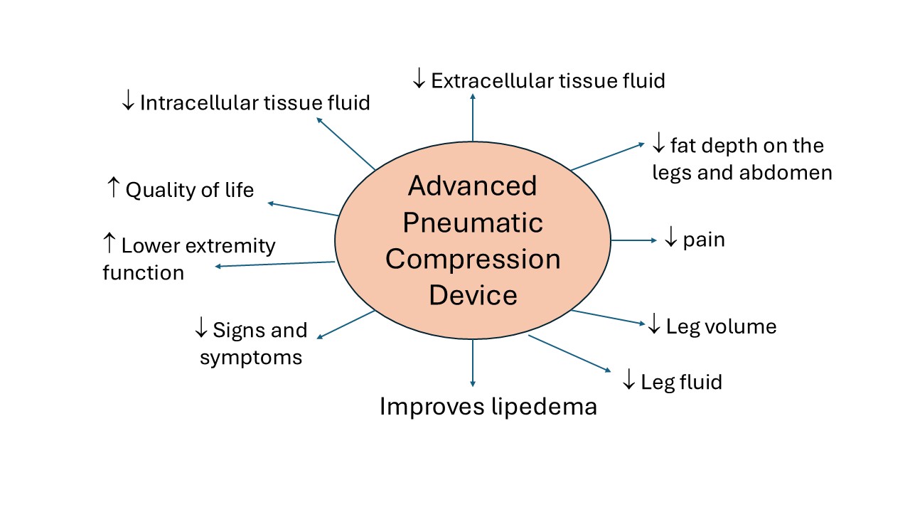
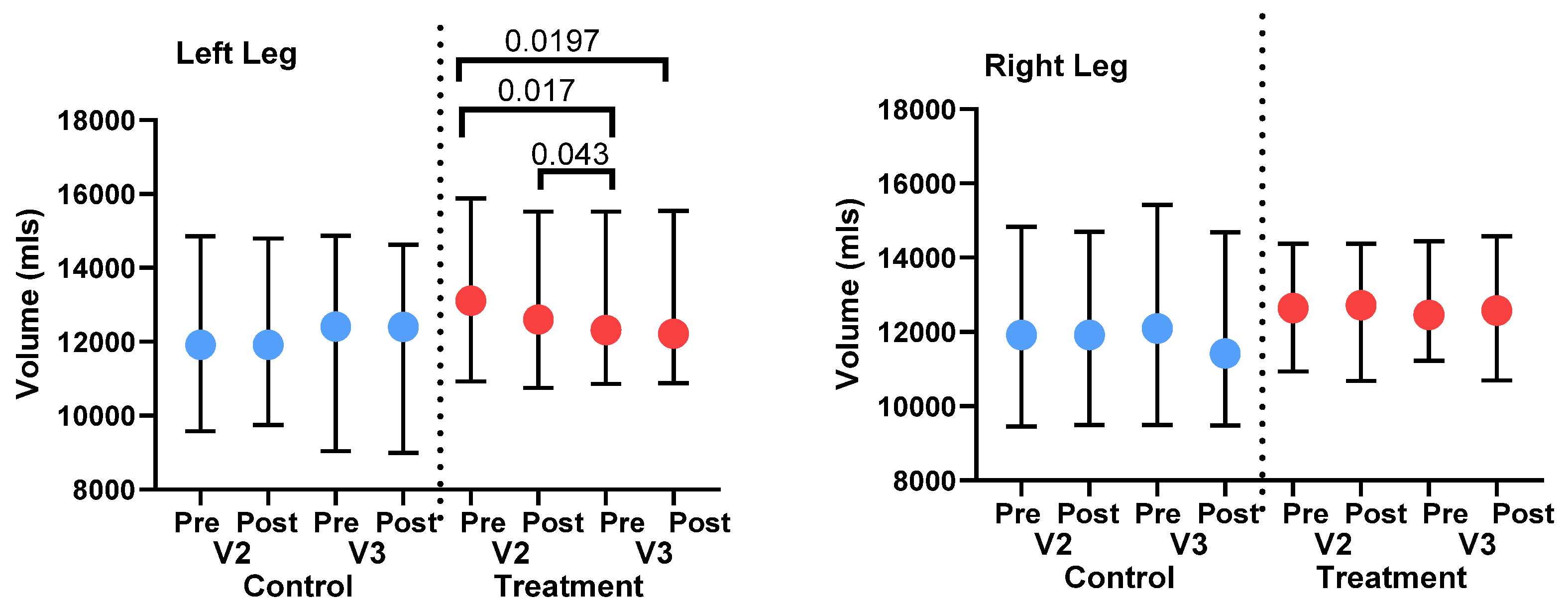
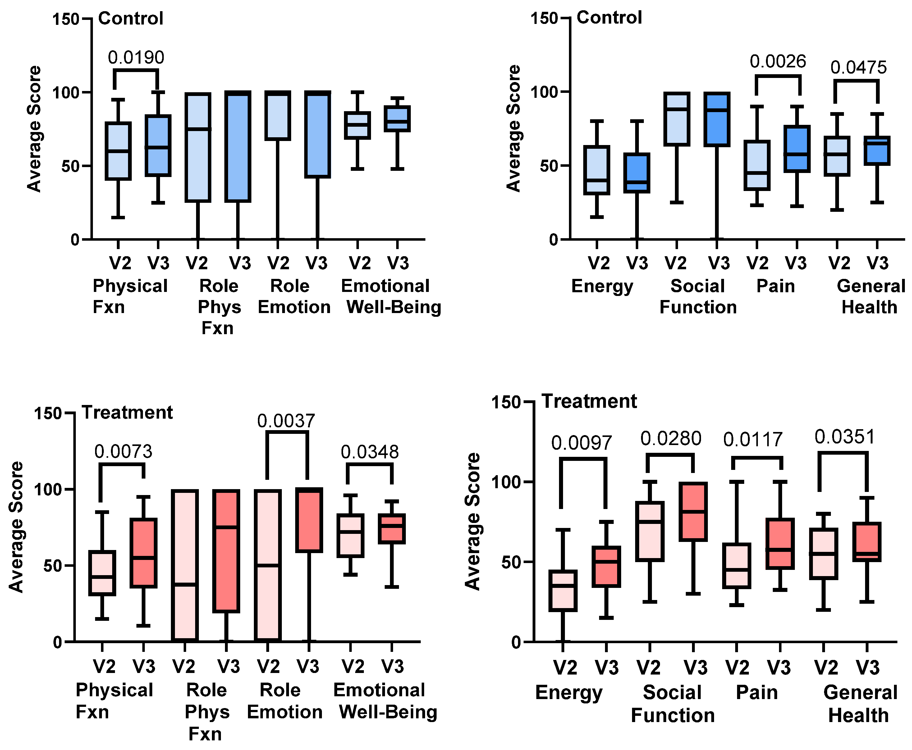
Lipedema is a painful disease of subcutaneous adipose tissue (SAT) in women. This study determined whether an advanced pneumatic compression device (APCD) improved lipedema SAT depth, swelling and pain. Women with lipedema started 20-30 mm Hg compression leggings then were randomized to an APCD (Lympha Press Optimal Plus) for 30 days (Treatment; n=22) or no APCD (Control; n=24). APCD Treatment significantly reduced left leg volume (3D imaging, LymphaTech; P<0.043) and fluid in the left (P=0.0018) and right legs (P=0.0476; SOZO, bioimpedance spectroscopy); Controls showed no change. Treatment significantly decreased extracellular fluid (ECF) and intracellular fluid (ICF) in left (P=0.0077; P=0.0060) and right legs (P=0.0476; P≤0.025), respectively. Only ECF decreased significantly in the left (P<0.0183) and right legs (P=0.0009) in Controls. SAT depth decreased significantly by ultrasound after Treatment at the anterior (P≤0.0234) and medial thigh (P≤0.0052), medial knee (P≤0.0002) and posterior calf (P≤0.0118) but not in Controls. All signs and symptoms of lipedema improved in the Treatment group including swelling (P=0.0005) and tenderness (pain) of right (P=0.0003) and left legs (P<0.0001); only swelling improved in Controls (P=0.0377). 87.5% of SF-36 quality of life improved after Treatment (P≤0.0351) compared to 37.5% in Controls (P≤0.0475). APCDs are effective treatment for lipedema.

Keywords: 
;  ;  ;  ;  ;  ;  ;  

1. Introduction

Lipedema is a disease of connective tissue in women [1,2]. A main feature of lipedema is increased fibrotic subcutaneous adipose tissue (SAT) deposited in the legs, pelvis, abdomen [3] and/or arms of women that is often painful, feels heavy, and can grow to such an extent that it affects mobility [4].
Women with lipedema present as stages: Stage 1, Smooth skin over fibrotic nodular tissue, Stage 2 dimpling or mattress pattern of the skin over larger fibrotic nodules, and Stage 3, lobules of fibrotic nodular lipedema tissue. Lipedema tissue is difficult to decrease by diet, exercise, or even bariatric surgery [5,6,7] in part due to the high amounts of tissue fibrosis present.
Lymphedema and swelling are notable co-morbidities with lipedema. The risk of lymphedema increases with stage as does body mass index (BMI) [8]. The association of lymphedema with lipedema is likely due to increased endothelial cell permeability in micro-vessels from an inflammatory insult [9,10,11], followed by a higher rate of lymphatic vessel pumping rates even in the tissue of women with early-stage lipedema [12]. Other co-morbidities associated with lipedema include allergies [13], attention-deficit / hyperactivity disorder [14], hypermobile joints [15], and vein disease [16].
Conservative management options for lipedema focus on improving lymphatic flow and controlling edema (swelling) with compression garments and manual lymphatic drainage and reducing pain or discomfort by deep tissue therapy to reduce fibrosis [17,18]. Pneumatic compression device therapy is effective in treating symptoms of lipedema, including swelling, easy bruising and pain [19,20,21,22,23]. An advanced pneumatic compression device (APCD), Lympha Press Optimal Plus, has been used extensively for treatment of lipedema and includes timings, modes and garments that can be employed to treat the lipedematous areas effectively.
The goal of this study is to determine if in-home use of an APCD can reduce lipedema tissue depth, tissue volume and fluid content, and improve quality of life, pain, and other signs and symptoms in women with primarily Stage 2 lipedema without evidence of lymphedema or vein disease.

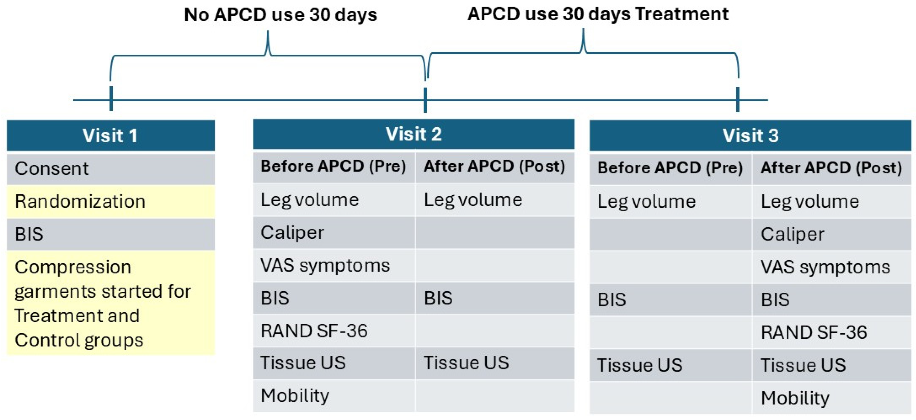
2. Materials and Methods

Patient recruitment: The study was conducted according to the guidelines of the Declaration of Helsinki and approved by the WCG institutional review board (protocol code 1356101; date of approval 06/29/2023). Informed consent was obtained from all subjects involved in the study prior to commencement of study activities. Patients seen at the clinic between 2021 and 2023 who had a diagnosis of lipedema and had expressed interest in research were recruited by phone call. Fifty-eight patients were invited to the clinic and presented for duplex scanning to rule out venous disease; 46 returned for the screening visit and signed the consent form. All 46 women completed the study.
Lipedema Criteria: Women enrolled in the study met criteria of being a biologic female, having the upper torso spared of abnormal SAT on physical exam, a mismatch between the amount of SAT on the limbs versus the upper torso, symmetrically affected SAT, and hands and feet without evidence of lymphedema per the criteria of Wold et al. [24].
Inclusion criteria:
  • Ambulatory females, age 18 - 70 years.
  • Stage 2-3 Type II-III lipedema.
  • Pain score with or without pressure in any lipedema area of 3 or more out of 11-point Likert visual analogue scale.
  • Able to maintain a consistent eating plan and exercise regimen for the 60-day study with weight stability (within 4.5 kg or usual weight fluctuation per patient) over three months.
  • Willing to wear compression garments during the study.
  • Agreement to wash off manual therapy of any kind including massage, physical therapy, occupational therapy, instrument assisted soft tissue therapy or other deep tissue therapy and decongestive therapy methods (manual lymph drainage, pneumatic compression use, and compression garment other than provided) over 30 days prior to Visit 2.
Exclusion Criteria
1.
Inability to understand the purpose of the study and complete consent.
2.
Bed bound, preventing assessment of activities of daily living.
3.
Contraindications to APCD use:
a)
serious arterial insufficiency measured as a monophasic pulse wave by Doppler (Terason uSmart 3300 - 15L4 uSmart Linear Array Transducer; Burlington, Massachusetts, USA) without arterial disease
b)
edema due to decompensated congestive heart failure (CHF) by history or physical exam
c)
active phlebitis by physical exam
d)
active deep vein thrombosis by history or physical exam
e)
localized wound infection by physical exam
f)
cellulitis by physical exam
4.
Positive Stemmer sign on the feet.
5.
Lymphedema with minimal to no lipedema.
6.
Weight >170 kg due to weight restriction on bioimpedance spectroscopy device.
7.
Undergoing surgery during the time of the study.
8.
Weight loss surgery within the past 18 months.
9.
Use of diuretic medication.
10.
Participation in other research at the time of the study.
11.
Use of immunosuppressant medications including Gleevec, diosmin, methotrexate, corticosteroids, Plaquenil or other.
12.
Medical illness deemed significant by the Principal Investigator.
13.
Waist to hip ratio > 0.85 suggestive of obesity with lipedema.
Women in the study had a history of chronic venous insufficiency (CVI) that had been treated previously except two in the Treatment group who did not have CVI and therefore did not have CVI treatment. All women were confirmed to have lack of venous reflux (effective treatment) by venous duplex ultrasound.
Randomization: Randomized by Research Randomizer [25] at Visit 1 (V1) to treatment with the APCD or Control.
Pneumatic compression: The APCD device used was Lympha Press® Optimal Plus (LymphaPress, Chadds Ford, PA) (Figure 1). The device is a calibrated gradient compression therapy system allowing a variety of adjustments, including pressure selection and adjustable timings and treatment modes. The compression appliance used (LymphaPants®) contains 24 inflatable chambers and treats bilateral legs, hips, buttocks and abdomen. Pressure used was 45 mmHg, with modes Sequential or Wave chosen according to the patient’s comfort level, with Pretherapy, a proximal decongestive mode, added prior to the main treatment mode. Each APCD includes compliance software that records device usage and settings.
Compression: All women received a static compression garment (Absolute Support leggings 20-30 mm Hg; discountsurgical.com) at V1 and wore them through V3.
Study Protocol: The women with lipedema underwent a 30-day washout period starting at Baseline (V1) and were asked not to use any decongestive therapies (manual therapies, pneumatic compression, compression garments other than the leggings provided for study participants at V1) over the next 30 days; all women were provided compression garments at V1. At Visit 2 (V2) 22 women randomized to the Treatment Group began the use of the APCD at home for 1-2 hours per day during the following 30 days. Women in the Treatment Group also used the APCD in clinic for a one-hour session during V2 and Visit 3 (V3), the latter after 30 days of APCD use; 24 women randomized to the Control group did not use the APCD pump at home or during any visit. During the time (60 minutes) allotted for treatment with the APCD pump, the Control subjects reclined in the same position (legs elevated) as used for the Treatment group during the APCD pumping sessions and maintained that position for the full 60 minutes.
Mobility: Women with lipedema often have mobility limitations [26]. Mobility was assessed during V2 and V3 by:
  • The “Timed up and Go (TUG)” test [27].
  • Quantitative assessment of walking several times across a special mat (GAITRite) that records and analyzes the pattern of footsteps [28]. Measurements included right toe location, ambulatory time or velocity, number of steps or cadence, left heel-to-heel or right heel-to-heel base support.
  • Lower extremity function scale (LEFS): a questionnaire containing 20 questions about a person’s ability to perform everyday tasks [29].
Caliper Measurements: Under the umbilicus, on the anterior thigh and medial thigh at V2 and V3 (Lange, NutriActiva, Minneapolis, MN, USA) as previously measured in women with lipedema [30].

Leg Volume

Tape measurement: At V2 and V3 with the back of the leg against a measuring grid. Measurements began at the ankle around the malleoli at 10 cm and continued every 10 cm to the upper thigh (70 cm maximum) with on average, seven measurements total for lower and upper leg. Volume of the leg was estimated using the formula for the volume of the frustum of a cone [31]. While consistent measurement by the same research staff at each visit is important, due to staff schedules, subjects were measured by different staff at each visit.
3D Imaging: Leg volume was measured at V2 and V3 using a 3D imaging system (LymphaTech, Atlanta, GA, USA) [32]. Measurement time, Pre, was before, and Post, was after treatment (Treatment Group) or no treatment (Control Group). Volume of the leg was estimated using the formula for the volume of the frustum of a cone [33].
Bioimpedance spectroscopy (BIS): Fluid/water of the whole body and the legs, and the intracellular and extracellular fluid in the legs were assessed at V1-V3 (SOZO® applications, Impedimed, Brisbane, Australia). Bilateral L-Dex® Analysis was used to measure tissue fluid content of the legs and represents the ratio of measured impedance between the arms and legs, with the legs being the at-risk limbs and the arms the lower risk limbs, as compared to an equivalent healthy population, with the normal range -10 to 10.
RAND Short Form 36-Item (SF-36) Survey (Version 1.0): Administered during V2 and V3 and scored as previously published [34].
Visual Analogue Scales (VAS): During V2 and V3 women filled in an 11-point Likert visual analogue scale (range 0-10) to assess the following symptoms in the legs: Tenderness, aching or throbbing, burning or stinging, tired or heavy, swelling or tightness of the legs, difficulty walking and easy bruising.
Ultrasound assessment of adipose tissue depth: Adipose tissue depth was assessed during V2 and V3 using a Terason uSmart 3300 ultrasound unit (Burlington, Massachusetts, USA). A 15L4 uSmart Linear Array transducer was placed perpendicular to the skin in a transverse plane. Six locations were assessed in areas typically affected by lipedema by the same individual throughout the study: Anterior thigh midpoint between the groin and the superior aspect of the patella, lateral thigh measured directly over the head of the femur, medial thigh measured directly down from the midpoint measurement on the anterior thigh, medial knee, fat pad under the knee measured from the inferior patella, and posterior calf at the largest point with the measurement logged as distance from the popliteal space and the medial malleolus.
Statistics: Data are presented as mean±standard deviation (SD). The differences between measures within a visit or between two visits for the same group were by paired t-tests (between two visits or pre and post an intervention, treatment or rest) or by Friedman’s test followed by Dunn’s multiple comparison for non-parametric data (between 3-4 evaluations), and by Friedman’s ANOVA with Tukey’s multiple comparison test for parametric data (between 3-4 evaluations). Significance of data was set as P<0.05.

3. Results

Starting at Baseline (V1) all women in the study underwent an initial 30-day washout period where they wore 20-30 mm Hg leggings and did not use an APCD (Figure 2). After 30 days, starting at V2, 22 women were randomized to use an APCD at home during the following 30 days in addition to a one-hour session (Treatment Group) at the start of V2 and V3; the remainder of the women were in the Control group (n=24) and did not use an APCD pump at home or during any visit.

3.1. Compliance

Twenty of twenty-two women from the Treatment group had data that could be downloaded from the APCD compliance software. Sixteen were 100% compliant (daily use), two were 75-89% compliant (five-six days/week treatment), and two were 50-74% compliant.

3.2. Demographics

Forty-six women were randomized to the Treatment Group (APCD for 30 days) or the Control Group (Table 1). The groups were well matched without significant differences in variables (Table 1). Notable were the high numbers of women with anxiety and birth control use.

Quantitative Measures

3.3. Leg volume

3.3.1. Tape Measure

There was no significant change in mean tape measure volume from V2 to V3 for the Treatment Group left (5.8±1.6 to 5.9±1.6 L) or right leg (5.7±1.4 to 5.9±1.6 L), or for the Control Group left leg (5.5±1.7 L to 5.7±1.8) or right leg (5.6±1.7 L to 5.7±1.5 L).

3.3.2. 3D Leg Imaging

There was a significant decrease in left leg volume by 3D imaging for the Treatment group Pre-V2 to Post-V2 (after use of the APCD for one hour during the visit), and from Pre-V2 or Post-V2, to Pre-V3 (after 30 days of the APCD). There was no significant change in volume of the right legs in the Treatment group over the course of the study (Figure 3). There was a significant increase in mean left leg volume for the Control group Post-V2 to Pre-V3 (Figure 3), over the 30 days the Control Group did not use an APCD, and despite the use of compression garments. There were no other significant changes in volume for the left or right legs in the Control group (Figure 3).

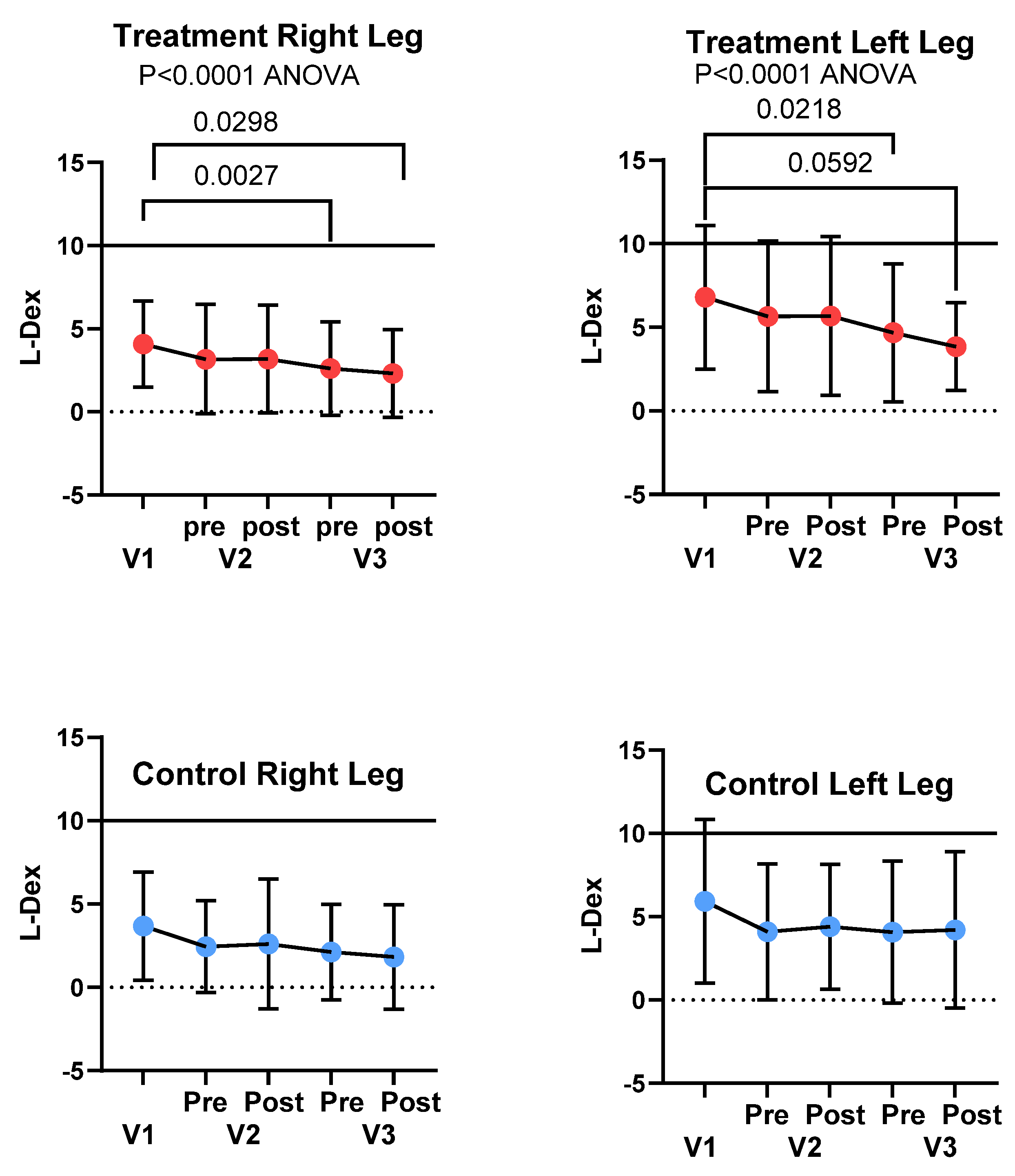
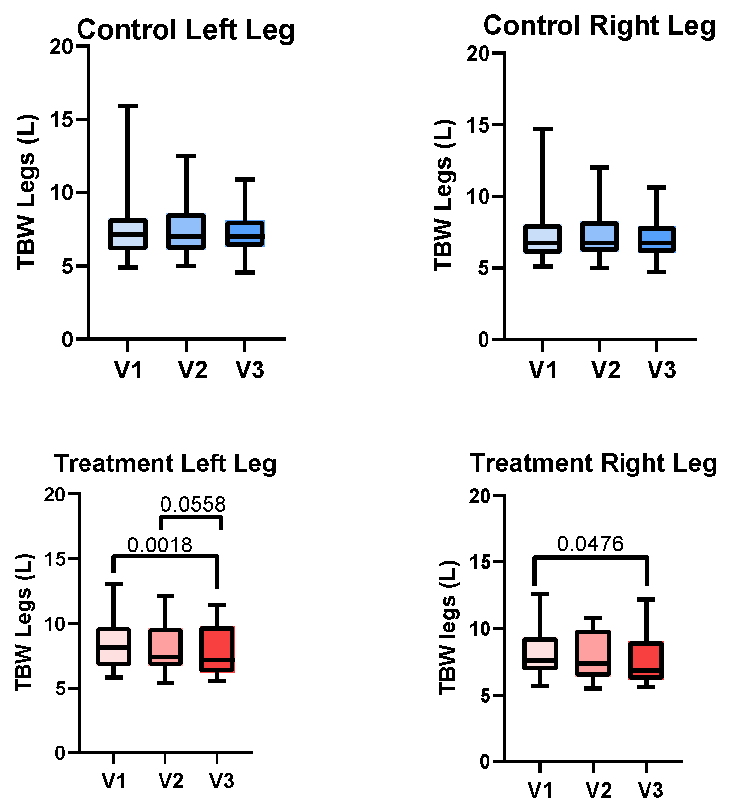
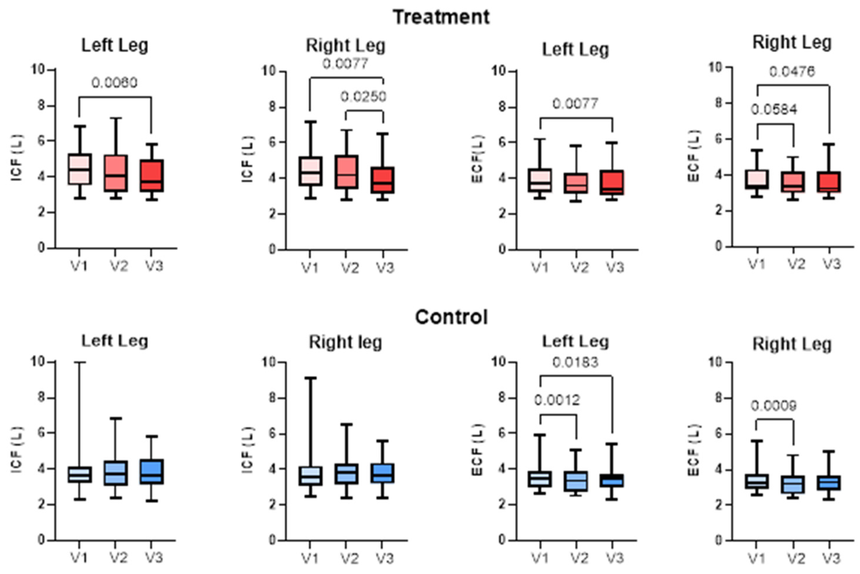
3.4. Bioimpedance Spectroscopy

3.4.1. There was a significant decrease in the potential risk of lymphedema (L-Dex Score; SOZO) in the Treatment group for the right and left legs but not the Control group between V1 and V3 (Figure 4). L-Dex scores for the right and left legs were significantly different at V1 for the Treatment group (4.1±2.6 and 6.8±4.3; P<0.0001) and Control group (3.7±3.2 and 5.9±4.9; P<0.0001), respectively.
3.4.2. Combining all left legs (n=46) and all right legs (n=46) in this study, the mean total body water (TBW) of the left legs was 8.1±2.2 L compared to the right, 7.8±2.1 L (P=0.0077). TBW significantly decreased in the right and left legs between V1 and V3 for the Treatment group but not the Control group (Figure 5).
3.4.3. There was no significant difference in TBW of the legs between V2 and V3 for the Treatment group, or Control group for the right and left legs, though the decrease from V2 to V3 just missed significance for the left leg in the Treatment Group (Figure 5). There was a significant reduction in TBW of the right and left legs between V1 and V3 in the Treatment group but not the Control group (Figure 5).
3.4.4. Extracellular fluid (ECF) significantly decreased in the Treatment group in the left leg from V1 to V3 and in the right leg from V1 to V2 and V2 to V3. Intracellular fluid (ICF) significantly decreased in the Treatment Group left leg from V1 to V3 and in the right leg from V1 to V3 and from V2 to V3. ECF decreased in the left leg of the Control Group from V1 to V2 and V1 to V3 and in the right leg from V1 to V2 (Figure 6). There was no significant change in ICF in either leg of the Control group.

3.5. Caliper

There were no differences between caliper measurements on the right and left anterior and medial legs of women with lipedema in the Treatment or Control Groups between visits (Table 2). The caliper measurements of the abdomen significantly decreased for the Treatment Group and not the Control Group.

3.6. Timed Up and Go

There was no difference in the Timed Up and Go test for the Treatment Group between V2 (12.32±2.033 sec) and V3 (12.73±2.028 sec) or the Control Group between V2 (11.75±3.110 sec) and V3 (11.58±3.243 sec).

3.7. Gait-Rite

There was no difference in left or right toe location, ambulatory time or velocity, number of steps or cadence, or Left heel-to-heel or Right heel-to-heel base support in V2 or V3 in either the Treatment or Control groups (data not shown).

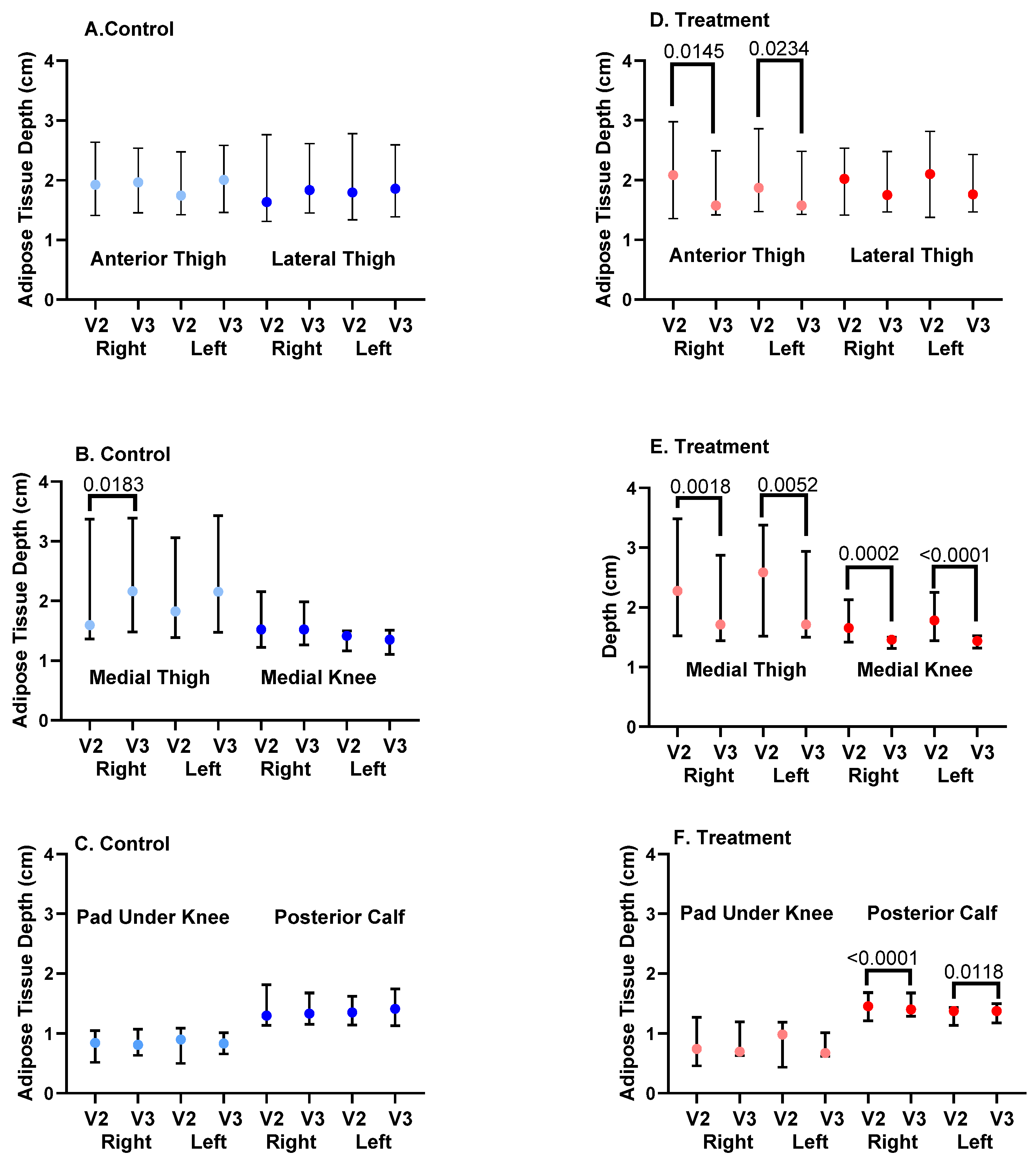
3.8. Ultrasound Depth of SAT

3.8.1. SAT depth significantly decreased from V2 to V3 for the Treatment group at the right and left anterior thighs, medial thighs, medial knees and posterior calves (Figure 7). There was no significant change in SAT depth from V2 to V3 for Control subjects at the anterior or lateral thigh, medial knee, or pad under the knee or posterior calf. SAT depth increased for the left and right medial thighs in the Control group (Figure 7).
3.8.2. To assess if the APCD had acute effects, we examined SAT depth before and after one hour of APCD use at V2 and V3 and compared these values to the Control group who received no intervention during the visits. There were no significant acute changes in SAT depth at six locations on the right and left legs for the Control group (Table 3).
There was an acute and significant reduction in SAT depth for the anterior thigh on the right and left legs for the Treatment group, with sustained decrease during V3 so that acute changes were not found during V3 (Pre- and Post-APCD; Table 3; Figure 7). There were significant acute reductions in the size of the pad under the knee and the posterior calf for the right leg of the Treatment Group during V2 whose decrease was sustained for the posterior calf during V3. The medial thigh and medial knee pad for the right leg in the Treatment group during V3 also decreased significantly and acutely (Table 3).

Qualitative Measures

3.9. Visual Analogue Scales

Symptoms of aching or throbbing, burning or stinging, tired or heavy, swelling or tightness of the legs, difficulty walking, and easy bruising significantly reduced in the Treatment Group from V2 to V3. There were no significant changes in any of the leg symptoms in the Control Group from V2 to V3 except for swelling tightness of the legs (Table 4).

3.10. RAND-SF-36

In the Treatment Group, there were significant improvements in physical function, emotional well-being and emotional role, energy, social function, pain, and general health from V2 to V3. There was a significant improvement in physical function, pain and general health between V2 and V3 in the Control Group (Figure 8).

3.11. LEFS

There was a significant increase (improvement) in the value of the LEFS for the Treatment group from V2 to V3 (61.3±14.4 to 70.1±16.2; P=0.0009) but not in the Control group (72.5±14.6 to 72.9±15.2; P=NS). There was a significant difference in the baseline LEFS scores for the Control and Treatment groups (P=0.0129), with the Treatment Group starting at a lower level than the Control Group.

4. Discussion

This study aimed to evaluate the benefits of an APCD in women with lipedema, primarily Stage 2, without clinically evident lymphedema or active venous disease. There is controversy on whether there is edema in lipedema, with some authors in support [2,35,36,37,38,39,40] and some against [41,42]. The data from this paper supports the presence of edema in lipedema tissue that is improved by an APCD.
To evaluate leg volume as one measure of leg edema, we utilized 3D imaging technology. There was no change in volume in the left or right leg in the Control group and no change in volume of the right leg in the Treatment group, yet there was a significant reduction in mean volume of the left leg in the Treatment group. In a study of 440 people with leg swelling, 52 of which had lipedema with lymphedema, the left leg was preferentially affected in unilateral and bilateral lymphedema [16]. The authors state that the left-sided lymphedema proclivity could be explained by chronic left iliac vein compression by the overlying right common iliac artery or rarely, by the ipsilateral internal iliac artery (May-Thurner syndrome), accentuated by abdominal obesity, which is consistent with our data. Indeed, when combining all the left legs and all the right legs in this study, the mean TBW of the left legs was significantly higher when compared to the right legs. L-Dex measurements were also higher in the left legs than the right legs in both the Treatment and Control groups in support of an increased risk of lymphedema due to increased fluid in the left legs over the right legs.
Tape measurements of the legs to calculate leg volume showed no change in this study for either the Treatment or Control groups. This may be due to different staff taking measurements during each visit. Even with rigorous training, user error contributes to a significant discrepancy in tape measurement data for leg volume. In addition, measuring leg circumference in serial measurements using a tape measure is tedious and labor-intensive [43]. In this study, tape measurements were made every 10 cm from the ankle to the thigh; measurements every 4 cm may have improved accuracy but could have also contributed further to error.
To more accurately measure fluid changes in the legs, we employed the use of BIS to measure TBW of the legs and risk of lymphedema. The use of an APCD significantly reduced TBW in the legs in the Treatment group and not the Control group; in fact, there was an increase in volume of the left leg in Controls. The APCD also reduced the risk of lymphedema in the legs in the Treatment group and not the Control group by L-Dex measurement which compares fluid in the legs to fluid in the arms, where the arms tend to be less affected than the legs in lipedema. L-Dex correlates with limb volume by perometry, degree of pitting edema, lymphoscintigraphy, and lymphedema staging using indocyanine green (ICG) lymphography [44], therefore, the L-Dex measurement provides a strong link between lipedema and lymphedema in this study.
We also investigated whether there was a change in the location of fluid in the leg specifically in the intracellular and extracellular spaces. The extracellular space includes the interstitial space between cells and outside vascular walls, and also plasma inside blood vessels [45]. About 60-80% by weight of adipose tissue is lipid, 5-30% is water and the remaining 2-3% is protein [46]. The fluid in the interstitial space is free flowing but there are also proteoglycan filaments which bind water and salt forming a “tissue gel [2],” or ground substance. Extracellular fluid in the legs decreased after compression garment use in the Control group confirming that compression wear is important in the treatment of lipedema. The fluid within cells, the ICF, did not change in the Control group. With APCD use, there was a decrease in both ICF and ECF. When body water loss is minimal, the water deficit comes primarily from the extracellular space; as more body water is lost, a proportionately greater percentage of the water deficit comes from the intracellular space [47,48], consistent with the reduction in ECF by compression garments alone and reduction in ECF+ICF by the addition of the APCD.
The ground substance accounts for only a few percent of the volume fraction in adipose tissue and free fluid is negligible [49]. On subjecting adipose tissue to a uniaxial compressive strain of 50%, the volume of liquid expressed from adipose tissue was less than 1% of the overall volume [50]. However, the interstitial space is larger in lipedema tissue [51,52], tissue salt, which binds proteoglycans, is higher in adipose and muscle tissue in the legs of women with lipedema [53], and lymphatic vessels pump at higher rates in women with early lipedema [12] suggestive of increased interstitial fluid. Future research could include assessments of compressive strain studies to better understand fluid in lipedema tissue.
For our study, the question becomes which cells reduced their intracellular fluid? Cells maintain their shape and water content owing to the difference in solute concentration, where the cytoplasm of a cell has higher osmotic and hydraulic pressure than the immediate surroundings [54]. Cells are also sensitive to changes in hydraulic and osmotic pressure around them. For example, when hydrostatic pressure was increased by 70 mm Hg around leukocytes (immune cells) for 2 hours, the radius of the cells and intracellular sodium and potassium ion concentrations decreased causing cell shrinkage [54]. Ion movement should induce osmosis-driven water flow across the membrane, which is likely the reason for the observed volumetric deformation of the immune cells. Since water can move freely across the cell membrane between the intracellular and interstitial space, the APCD in our study likely changed the osmotic gradient, which is required for fluid movement between cells in the leg tissue. Changing the osmotic gradient across cell membranes may allow for longer periods of time of fluid reduction after treatment with an APCD versus compression alone. Future studies should consider long-term evaluations of fluid reduction after APCD use in women with lipedema.
The leg grossly contains skin, skeletal muscles, connective tissue (including bone, tendons and ligaments), nerves, blood and lymphatic vessels, and cells including adipocytes, fibroblasts, myocytes, immune cells or other cells [55]. Each adipocyte comprises a single lipid vacuole and a nucleus within a phospholipid bilayer, in a collagen mesh. Due to the high intracellular lipid droplet content, adipose tissue functions as an incompressible inviscid fluid [50]. Skeletal muscle on the other hand is comprised of 75% water [47,48]. Women with lipedema have poor muscle function of their legs [56], but no ultrastructural studies of muscles from women with lipedema are available. The reduction in ICF in the leg of women with lipedema may therefore have resulted from reduction of water from adipocytes, myocytes and/or immune cells amongst others. A focus on muscle architecture in treatment studies may provide additional insight into the pathophysiology of lipedema.
The APCD may have also reduced intravascular fluid as well. This is consistent with the APCD pump significantly reducing leg volume by 3D imaging which thereby reshaped the limbs. Reducing excess fluid in in the extracellular space is important to reduce the feeling of heaviness in the legs, but reduction of fluid in the vessels may reduce fluid available to leak through abnormally permeable endothelial cells into lipedema tissue [9,10,11]. These data highly suggest that there is edema in the tissue of women with lipedema and this edema can be effectively reduced in multiple compartments using an APCD. In addition, use of an APCD might reduce the risk of developing lymphedema in women with lipedema.
Lipedema is considered primarily to be a disease of subcutaneous adipose connective tissue. In addition to fluid reduction, we wanted to determine if an APCD could reduce lipedema SAT depth. Use of the APCD was associated with reduction in SAT depth near the umbilicus by caliper measurements and SAT depth on the anterior and medial thigh, medial knee and posterior calves by ultrasound. In the women who did not use an APCD, there was an increase in medial thigh SAT depth. That abdomen SAT reduced by caliper measurements supports the use of APCD garments that treat the torso along with the legs in women with lipedema who have affected abdomens along with the legs.
It is unclear if the reduction in SAT depth after use of an APCD is a direct reduction in adipose tissue or other components of the tissue (such as fibrosis) or simply reflects the decrease in tissue fluid after the use of the APCD. Under a hypertonic pressure of 400 mosmol, half of adipocytes lose visible lipid droplets and the remaining adipocytes also experience a decrease in lipid droplet size and adopt a spindle-like shape consistent with de-differentiation into mesenchymal stem cells [57]. The reduction in SAT depth could therefore reflect a change in the adipocytes themselves. Tissue biopsies were taken during this study; evaluation of cell size and morphology in these samples is ongoing.
Lipedema tissue is painful, and it has been proposed that this is neuropathic pain due to inflammation [58]. Use of the APCD reduced many symptoms of pain in the legs whereas a similar reduction was not found in the Control group, suggesting an APCD can improve symptoms of lipedema associated with pain over that of compression alone. While there was a reduction in body pain on the SF-36 in both the Treatment and Control groups, the VAS specifically targeted tenderness (pain) in the legs and therefore is a better representation of lipedema pain in this study, where lipedema pain was reduced by the use of an APCD.
Reduction in fluid and SAT in women with lipedema is important but it needs to translate to an improvement in quality of life and function. There was no change in the TUG functional test for the women in this study. It should be noted, however, that the time it took the women to complete the test was close to the mean for 80–84-year-old women in the Norwegian Tromsø Study, and similar to a meta-analysis of TUG data [59]. These data suggest that women with lipedema with average age of 50 years are likely functionally impaired by their lipedema. It may require more than 3 months of APCD use to translate into improved function. Unfortunately for the gait-rite data, we did not have good normative data for comparison.
While there were no significant changes in objective functional measures in this study after APCD treatment, there was a significant improvement in quality-of-life measures by the RAND SF-36 in the Control group, specifically physical function, pain and general health, likely reflecting improvements due to compression garments, and in the Treatment group in physical function, emotional wellbeing, role of emotion, energy, social function, pain and general health, reflecting improvement due to the use of compression garments and the APCD. That women’s social and emotional function improved after use of an APCD suggests improvement in mental health in addition to physical health, which was not seen with compression garments alone. In addition, there was a significant improvement in the LEFS, with questions about daily life tasks and leg symptoms, in the Treatment Group but not the Control group. The APCD therefore significantly improved quality of life women living with lipedema over that of compression garments alone. There was a significant difference in the starting value of the LEFS at V2 between the Treatment and Control groups, but this is likely due to variance within the population.
The WHR of the women in this study was below the threshold set for central obesity in women of greater than 0.8 [60], despite the mean BMI exceeding criteria for obesity (>30 kg/m2). The mean WHtR was just under the cutoff of 0.6 for a reasonable future risk of death and cardiovascular events set for women aged 50 years or older in a study of 5,956 women from Germany [61]. Other estimates state a WHtR 0.5-0.59 indicates increased health risks and that people with a WHtR of 0.6 or more have further increased health risks [62].
The study population in this paper had high levels of anxiety which is known to be a co-morbidity in the hypermobile joint syndrome population [63]. Also notable in this study population were high levels of allergies reaching almost 100% of participants, in agreement with published data [13]. Mast cell activation syndrome is linked to allergies[64] and may be linked to lipedema where investigators found high levels of histamine and its metabolites in tissue samples from women with lipedema compared to controls; histamine levels reduced after treatment with sodium cromoglycate, suggesting a reduction of mast cell activity [65]. These data suggest that lipedema is more likely than not, a systemic disease rather than a focal disease of the arms and legs.

5. Conclusions

The APCD used in this study for 30 days by women with lipedema who also wore 20-30 mm Hg compression leggings daily, significantly reduced tissue fluid, SAT depth, and improved quality of life, pain and leg symptoms over women who wore compression garments alone. Advanced pneumatic compression devices should be considered for the conservative care of women with lipedema who present with symptoms of leg heaviness, swelling and pain. Finally, this study lends support to the presence of edema in lipedema.

Author Contributions

Conceptualization, K.L.H. and L.M.; methodology, K.L.H., L.M and C.Z.; validation, L.M. and C.Z.; formal analysis, K.L.H., L.M. and C.Z.; investigation, L.M., C.Z., T.Z., M.S. resources, L.M.; data curation, L.M., C.Z. and T.Z.; writing—original draft preparation, K.L.H.; writing—review and editing, K.L.H., L.M. and C.Z.; project administration, L.M. and C.Z.; funding acquisition, L.M. All authors have read and agreed to the published version of the manuscript.

Funding

This research was funded by Lympha Press, and The Carolina Vein Center, and “The APCD was funded by Lympha Press”.

Data Availability Statement

The data presented in this study are available on request from the corresponding author due to due to proprietary nature of the information as it pertains to Lympha Press.

Acknowledgments

The authors acknowledge all staff at the Carolina Vein Center whose work and dedication made this study possible.

Conflicts of Interest

K.L.H. is an independent contractor with Lympha Press.

Abbreviations

The following abbreviations are used in this manuscript:
APCD Intermittent pneumatic compression device
BIS Bioimpedance spectroscopy
BMI Body mass index
COPD Chronic obstructive pulmonary disease
CVD Cardiovascular disease
CVI Chronic venous insufficiency
ECF Extracellular fluid
ICF Intracellular fluid
ICG Indocyanine green
HTN Hypertension
MCAS Mast cell activation syndrome
NS Non-significant
PCOS Polycystic ovarian syndrome
POTS Postural orthostatic tachycardia syndrome
SAT Subcutaneous adipose tissue
SF-36 RAND Short Form 36-Item Survey
US Ultrasound
VAS Visual analogue scales
WHR Waist to hip ratio
WHtR Waist to height ratio

References

  1. B. A. Jagtman, J. P. Kuiper, and A. J. Brakkee, “[Measurements of skin elasticity in patients with lipedema of the Moncorps “rusticanus” type],” Phlebologie., vol. 37, no. 3, pp. 315-9., Jul-Sep 1984.
  2. K. L. Herbst et al., “Standard of care for lipedema in the United States,” (in eng), Phlebology, Journal Article vol. 36, no. 10, pp. 779-796, Dec 2021.
  3. K. L. Herbst et al., “Standard of care for lipedema in the United States,” (in eng), Phlebology, Journal Article vol. 28, no. 2683555211015887, p. 2683555211015887, May 28 2021.
  4. T. Wright, M. Babula, J. Schwartz, C. Wright, N. Danesh, and K. Herbst, “Lipedema Reduction Surgery Improves Pain, Mobility, Physical Function, and Quality of Life: Case Series Report,” (in eng), Plast Reconstr Surg Glob Open, vol. 11, no. 11, p. e5436, Nov 2023. [CrossRef]
  5. J. H. Bast, L. Ahmed, and R. Engdahl, “Lipedema in patients after bariatric surgery,” Surg Obes Relat Dis., vol. 12, no. 5, pp. 1131-2. [CrossRef]
  6. S. Pouwels, S. Huisman, H. J. M. Smelt, M. Said, and J. F. Smulders, “Lipoedema in patients after bariatric surgery: report of two cases and review of literature,” Clin Obes., vol. 8, no. 2, pp. 147-150. [CrossRef]
  7. S. Pouwels, H. J. Smelt, M. Said, J. F. Smulders, and M. M. Hoogbergen, “Mobility Problems and Weight Regain by Misdiagnosed Lipoedema After Bariatric Surgery: Illustrating the Medical and Legal Aspects,” Cureus., Case Reports vol. 11, no. 8, p. e5388. [CrossRef]
  8. K. Herbst, L. Mirkovskaya, A. Bharhagava, Y. Chava, and C. H. Te, “Lipedema Fat and Signs and Symptoms of Illness, Increase with Advancing Stage,” Archives of Medicine, vol. 7, no. 4:10, pp. 1-8, 2015.
  9. S. AL-Ghadban et al., “Dilated Blood and Lymphatic Microvessels, Angiogenesis, Increased Macrophages, and Adipocyte Hypertrophy in Lipedema Thigh Skin and Fat Tissue,” Journal of Obesity, 2019.
  10. M. Allen, M. Schwartz, and K. L. Herbst, “Interstitial Fluid in Lipedema and Control Skin,” (in eng), Womens Health Rep (New Rochelle), vol. 1, no. 1, pp. 480-487, 2020. [CrossRef]
  11. K. Strohmeier et al., “Multi-Level Analysis of Adipose Tissue Reveals the Relevance of Perivascular Subpopulations and an Increased Endothelial Permeability in Early-Stage Lipedema,” Biomedicines, vol. 10, 1163, pp. 1-22, 2022. [CrossRef]
  12. J. C. Rasmussen, M. B. Aldrich, C. E. Fife, K. L. Herbst, and E. M. Sevick-Muraca, “Lymphatic function and anatomy in early stages of lipedema,” (in eng), Obesity (Silver Spring, Md.), Journal Article Research Support, Non-U.S. Gov’t vol. 30, no. 7, pp. 1391-1400, Jul 2022.
  13. M. Ghods, I. Georgiou, J. Schmidt, and P. Kruppa, “Disease progression and comorbidities in lipedema patients: A 10-year retrospective analysis,” (in eng), Dermatol Ther, Journal Article vol. 33, no. 6, p. e14534, Nov 2020.
  14. A. C. Amato, J. L. Amato, and D. A. Benitti, “The Association Between Lipedema and Attention-Deficit/Hyperactivity Disorder,” (in eng), Cureus, vol. 15, no. 2, p. e35570, Feb 2023. [CrossRef]
  15. K. Beltran and K. L. Herbst, “Differentiating lipedema and Dercum’s disease,” Int J Obes (Lond). vol. 41, no. 2, pp. 240-245., Feb 2017.
  16. S. M. Dean, E. Valenti, K. Hock, J. Leffler, A. Compston, and W. T. Abraham, “The clinical characteristics of lower extremity lymphedema in 440 patients,” J Vasc Surg Venous Lymphat Disord., vol. 8, no. 5, pp. 851-859. [CrossRef]
  17. K. L. Herbst, C. Ussery, and A. Eekema, “Pilot study: whole body manual subcutaneous adipose tissue (SAT) therapy improved pain and SAT structure in women with lipedema,” Horm Mol Biol Clin Investig., Clinical Trial vol. 33, no. 2, pp. /j/hmbci.2018.33.issue-2/hmbci-2017-0035/hmbci-2017-0035.xml. [CrossRef]
  18. P. M. C. Donahue et al., “Physical Therapy in Women with Early Stage Lipedema: Potential Impact of Multimodal Manual Therapy, Compression, Exercise, and Education Interventions,” (in eng), Lymphat Res Biol, Journal Article vol. 8, no. 10, Nov 8 2021.
  19. G. Szolnoky, B. Borsos, K. Barsony, M. Balogh, and L. Kemeny, “Complete decongestive physiotherapy with and without pneumatic compression for treatment of lipedema: a pilot study,” (in eng), Lymphology, vol. 41, no. 1, pp. 40-4, Mar 2008.
  20. G. Szolnoky, E. Varga, M. Varga, M. Tuczai, E. Dosa-Racz, and L. Kemeny, “Lymphedema treatment decreases pain intensity in lipedema,” Lymphology., Research Support, Non-U.S. Gov’t vol. 44, no. 4, pp. 178-82., Dec 2011.
  21. M. Volkan-Yazıcı, G. Yazici, and M. Esmer, “The Effects of Complex Decongestive Physiotherapy Applications on Lower Extremity Circumference and Volume in Patients with Lipedema,” (in eng), Lymphat Res Biol, Journal Article vol. 19, no. 1, pp. 111-114, Feb 2021.
  22. M. Volkan-Yazici and M. Esmer, “Reducing Circumference and Volume in Upper Extremity Lipedema: The Role of Complex Decongestive Physiotherapy,” (in eng), Lymphat Res Biol, Journal Article vol. 5, no. 10, Apr 5 2021.
  23. T. Atan and Y. Bahar-Özdemir, “The Effects of Complete Decongestive Therapy or Intermittent Pneumatic Compression Therapy or Exercise Only in the Treatment of Severe Lipedema: A Randomized Controlled Trial,” (in eng), Lymphat Res Biol, vol. 19, no. 1, pp. 86-95, Feb 2021.
  24. L. E. Wold, E. A. Hines, Jr., and E. V. Allen, “Lipedema of the legs; a syndrome characterized by fat legs and edema,” (in eng), Ann Intern Med, vol. 34, no. 5, pp. 1243-50, May 1951.
  25. Research Randomizer. (1997). Social Psychology Network. [Online]. Available: http://www.randomizer.org/about.htm.
  26. I. Forner-Cordero, M. V. Perez-Pomares, A. Forner, A. B. Ponce-Garrido, and J. Munoz-Langa, “Prevalence of clinical manifestations and orthopedic alterations in patients with lipedema: A prospective cohort study,” (in eng), Lymphology, Journal Article vol. 54, no. 4, pp. 170-181, 2021.
  27. A. Christopher, E. Kraft, H. Olenick, R. Kiesling, and A. Doty, “The reliability and validity of the Timed Up and Go as a clinical tool in individuals with and without disabilities across a lifespan: a systematic review,” (in eng), Disabil Rehabil, vol. 43, no. 13, pp. 1799-1813, Jun 2021. [CrossRef]
  28. B. Bilney, M. Morris, and K. Webster, “Concurrent related validity of the GAITRite walkway system for quantification of the spatial and temporal parameters of gait,” (in eng), Gait Posture, Journal Article vol. 17, no. 1, pp. 68-74, Feb 2003.
  29. J. M. Binkley, P. W. Stratford, S. A. Lott, and D. L. Riddle, “The Lower Extremity Functional Scale (LEFS): scale development, measurement properties, and clinical application. North American Orthopaedic Rehabilitation Research Network,” Phys Ther., vol. 79, no. 4, pp. 371-83., 1999.
  30. M. Ibarra, A. Eekema, C. Ussery, D. Neuhardt, K. Garby, and K. L. Herbst, “Subcutaneous adipose tissue therapy reduces fat by dual X-ray absorptiometry scan and improves tissue structure by ultrasound in women with lipoedema and Dercum disease,” Clin Obes., vol. 8, no. 6, pp. 398-406. [CrossRef]
  31. J. R. Casley-Smith, “Measuring and representing peripheral oedema and its alterations,” Lymphology, vol. 27, 2, pp. 56-70, 1994.
  32. C. Yahathugoda et al., “Use of a Novel Portable Three-Dimensional Imaging System to Measure Limb Volume and Circumference in Patients with Filarial Lymphedema,” (in eng), Am J Trop Med Hyg, vol. 97, no. 6, pp. 1836-1842, Dec 2017. [CrossRef]
  33. J. M. Binkley, M. J. Weiler, N. Frank, L. Bober, J. B. Dixon, and P. W. Stratford, “Assessing Arm Volume in People During and After Treatment for Breast Cancer: Reliability and Convergent Validity of the LymphaTech System,” (in eng), Phys Ther, vol. 100, no. 3, pp. 457-467, Mar 10 2020. [CrossRef]
  34. J. E. Ware, Jr. and C. D. Sherbourne, “The MOS 36-item short-form health survey (SF-36). I. Conceptual framework and item selection,” (in eng), Med Care, vol. 30, no. 6, pp. 473-83, Jun 1992.
  35. B. H. Duhon, T. T. Phan, S. L. Taylor, R. L. Crescenzi, and J. M. Rutkowski, “Current Mechanistic Understandings of Lymphedema and Lipedema: Tales of Fluid, Fat, and Fibrosis,” (in eng), Int J Mol Sci, vol. 23, no. 12, Jun 14 2022.
  36. L. M. Pereira de Godoy, H. J. Pereira de Godoy, P. Pereira de Godoy Capeletto, M. F. Guerreiro Godoy, and J. M. Pereira de Godoy, “Lipedema and the Evolution to Lymphedema With the Progression of Obesity,” (in eng), Cureus, vol. 12, no. 12, p. e11854, Dec 2 2020. [CrossRef]
  37. T. Coppel et al., “Best Practice Guidelines: The management of lipoedema.,” Wounds UK, vol. 13, no. 1. [Online]. Available: http://www.wounds-uk.com/best-practice-statements/best-practice-guidelines-the-management-of-lipoedema.
  38. I. Forner-Cordero, G. Szolnoky, A. Forner-Cordero, and L. Kemény, “Lipedema: an overview of its clinical manifestations, diagnosis and treatment of the disproportional fatty deposition syndrome - systematic review,” Clinical Obesity, vol. 2, no. 3-4, pp. 86-95, June 2012.
  39. J. M. Alcolea et al., “Documento de Consenso Lipedema 2018,” in 33rd National Congress of the Spanish Society of Aesthetic Medicine (SEME), Malaga, Spain, 2018, Barcelona, Spain: LITOGAMA S.L., 2018.
  40. M. Sandhofer et al., “Prevention of Progression of Lipedema With Liposuction Using Tumescent Local Anesthesia: Results of an International Consensus Conference,” Dermatol Surg., vol. 46, no. 2, pp. 220-228. [CrossRef]
  41. T. Bertsch et al., “Lipoedema – myths and facts, Part 5. European Best Practice of Lipoedema – Summary of the European Lipoedema Forum consensus,” Phlebologie, vol. 49, pp. 31-49, 2020.
  42. S. Reich-Schupke, P. Altmeyer, and M. Stücker, “Thick legs not always lipedema.,” J Dtsch Dermatol Ges., vol. 11, no. 3, pp. 225-233, 2013.
  43. S. Russo et al., “Standardization of lower extremity quantitative lymphedema measurements and associated patient-reported outcomes in gynecologic cancers,” (in eng), Gynecol Oncol, vol. 160, no. 2, pp. 625-632, Feb 2021. [CrossRef]
  44. C. J. Coroneos, F. C. Wong, S. M. DeSnyder, S. F. Shaitelman, and M. V. Schaverien, “Correlation of L-Dex Bioimpedance Spectroscopy with Limb Volume and Lymphatic Function in Lymphedema,” (in eng), Lymphat Res Biol, vol. 17, no. 3, pp. 301-307, Jun 2019. [CrossRef]
  45. M. Gillham and D. Sidebotham, “Chapter 32 - Water and Electrolyte Disturbances,” in Cardiothoracic Critical Care, D. Sidebotham, A. McKee, M. Gillham, and J. H. Levy Eds. Philadelphia: Butterworth-Heinemann, 2007, pp. 470-480.
  46. L. Weiss, Histology: Cell and Tissue Biology. Elsevier Biomedical, 1983.
  47. D. L. Costill, R. Coté, and W. Fink, “Muscle water and electrolytes following varied levels of dehydration in man,” (in eng), J Appl Physiol, vol. 40, no. 1, pp. 6-11, Jan 1976. [CrossRef]
  48. M. J. Durkot, O. Martinez, D. Brooks-McQuade, and R. Francesconi, “Simultaneous determination of fluid shifts during thermal stress in a small-animal model,” (in eng), J Appl Physiol (1985), vol. 61, no. 3, pp. 1031-4, Sep 1986. [CrossRef]
  49. E. Hvidberg, “Water-binding by connective tissue and the acid mucopolysaccharides of the ground substance,” Acta pharmacologica et toxicologica, vol. 17, pp. 267-76, 2009.
  50. K. Comley and N. Fleck, “The compressive response of porcine adipose tissue from low to high strain rate,” International Journal of Impact Engineering, vol. 46, pp. 1-10, 2012/08/01/ 2012. [CrossRef]
  51. H. Suga, J. Araki, N. Aoi, H. Kato, T. Higashino, and K. Yoshimura, “Adipose tissue remodeling in lipedema: adipocyte death and concurrent regeneration,” J Cutan Pathol, vol. 3, p. 3, 2009.
  52. G. Felmerer et al., “Adipose Tissue Hypertrophy, An Aberrant Biochemical Profile and Distinct Gene Expression in Lipedema,” J Surg Res., vol. 253:294-303., no. doi, p. 10.1016/j.jss.2020.03.055., May 11 2020.
  53. R. Crescenzi et al., “Upper and Lower Extremity Measurement of Tissue Sodium and Fat Content in Patients with Lipedema,” (in eng), Obesity (Silver Spring, Md.), vol. 28, no. 5, pp. 907-915, May 2020. [CrossRef]
  54. T. H. Hui, Z. L. Zhou, J. Qian, Y. Lin, A. H. Ngan, and H. Gao, “Volumetric deformation of live cells induced by pressure-activated cross-membrane ion transport,” (in eng), Phys Rev Lett, vol. 113, no. 11, p. 118101, Sep 12 2014. [CrossRef]
  55. A. Tobias, B. D. Ballard, and S. S. Mohiuddin, “Physiology, Water Balance. [Updated 2022 Oct 3],” StatPearls [Internet]Treasure Island (FL): StatPearls Publishing, 2024. [Online]. Available: https://www.ncbi.nlm.nih.gov/books/NBK541059/.
  56. J. van Esch-Smeenge, R. J. Damstra, and A. A. Hendrickx, “Muscle strength and functional exercise capacity in patients with lipoedema and obesity: a comparative study,” Journal of Lymphoedema, vol. 12, no. 1, pp. 27-31, 2017.
  57. Y. Li et al., “Compression-induced dedifferentiation of adipocytes promotes tumor progression,” (in eng), Sci Adv, vol. 6, no. 4, p. eaax5611, Jan 2020. [CrossRef]
  58. A. Chakraborty et al., “Indications of Peripheral Pain, Dermal Hypersensitivity, and Neurogenic Inflammation in Patients with Lipedema,” (in eng), Int J Mol Sci, Journal Article vol. 23, no. 18, Sep 7 2022.
  59. R. W. Bohannon, “Reference values for the timed up and go test: a descriptive meta-analysis,” (in eng), J Geriatr Phys Ther, vol. 29, no. 2, pp. 64-8, 2006. [CrossRef]
  60. V. A. Benites-Zapata et al., “High waist-to-hip ratio levels are associated with insulin resistance markers in normal-weight women,” (in eng), Diabetes Metab Syndr, vol. 13, no. 1, pp. 636-642, Jan-Feb 2019. [CrossRef]
  61. H. J. Schneider et al., “The predictive value of different measures of obesity for incident cardiovascular events and mortality,” (in eng), The Journal of clinical endocrinology and metabolism, vol. 95, no. 4, pp. 1777-85, Apr 2010. [CrossRef]
  62. E. Brenner, I. Forner-Cordero, G. Faerber, S. Rapprich, and M. Cornely, “Body mass index vs. waist-to-height-ratio in patients with lipohyperplasia dolorosa (vulgo lipedema),” (in eng), J Dtsch Dermatol Ges, vol. 21, no. 10, pp. 1179-1185, Oct 2023. [CrossRef]
  63. A. Bulbena, J. Gago, G. Pailhez, L. Sperry, M. A. Fullana, and O. Vilarroya, “Joint hypermobility syndrome is a risk factor trait for anxiety disorders: a 15-year follow-up cohort study,” Gen Hosp Psychiatry., vol. 33, no. 4, pp. 363-70. [CrossRef]
  64. K. Amin, “The role of mast cells in allergic inflammation,” Respiratory Medicine, vol. 106, no. 1, pp. 9-14, 2012/01/01/ 2012. [CrossRef]
  65. G. Bonetti et al., “Targeting Mast Cells: Sodium Cromoglycate as a Possible Treatment of Lipedema,” (in eng), Clin Ter, vol. 174, no. Suppl 2(6), pp. 256-262, Nov-Dec 2023. [CrossRef]
Figure 1. APCD device (Lympha Press Optimal Plus with LymphaPants, Lymphapress, Chadds Ford, PA.
Preprints 149801 g001
Figure 2. Study layout. Procedures during Visit 1 (Baseline), Visit 2 with time points Pre (before) APCD) and Post (after APCD), and Visit 3 with time points Pre and Post APCD. The Control group followed the same process, without home use of an APCD, and during visits, they reclined for 60 minutes in the same position as the Treatment group without performing the APCD therapy. BIS=bioimpedance spectroscopy; US=ultrasound; VAS=visual analogue scale.
Preprints 149801 g002
Figure 3. Leg volume measurements before and after Lympha Press Optimal advanced pneumatic compression device in the Treatment group or after no intervention in the Control group. Volumes (mean±SD) were assessed Pre and Post APCD (red) or no intervention (blue) in Visit 2 (V2) and Pre and Post APCD in Visit 3 (V3) by 3D imaging (LymphaTech, Atlanta, GA, USA).
Preprints 149801 g003
Figure 4. L-Dex measurement (SOZO, Impedimed, Brisbane, Australia) during Baseline Visit 1 (V1), Visit 2 (V2) before 30 days of advanced pneumatic compression device (APCD); Lymph Press Optimal with Lympha Pants, Glenn Mills, PA, USA) in the Treatment group, and Visit 3 (V3) after APCD use (Treatment group) or no APCD use (Controls). During V2 and V3, L-Dex measures were Pre and Post either resting (Control; blue) or use of the APCD for one hour (Treatment; red). Values below the line at an L-Dex score of 10 on the graph are within normal limits.
Preprints 149801 g004
Figure 5. Total body water (mean±SD) in the left and right legs of women with lipedema in the Control (blue) and Treatment (red) groups during Baseline Visit 1 (V1), Pre-Treatment with an advanced pneumatic compression device (APCD; Lymph Press Optimal with Lympha Pants, Glenn Mills, PA, USA), Visit 2 (V2), and Post-Treatment Visit 3 (V3), by bioimpedance spectroscopy (SOZO, Impedimed, Brisbane, Australia).
Preprints 149801 g005
Figure 6. Intracellular fluid (ICF) and extracellular fluid (ECF) in the right and left legs of women with lipedema after advanced pneumatic compression device (APCD; Lymph Press Optimal with Lympha Pants, Glenn Mills, PA, USA) use for 30 days (Treatment) or no treatment (Control). Fluid volumes (mean±SD) were evaluated by bioimpedance spectroscopy (SOZO, Impedimed, Brisbane, Australia) for the right and left legs of the Treatment (red) and Control (blue) groups during Baseline Visit 1 (V1), Pre-Treatment Visit 2 (V2), and Post-Treatment (APCD Treatment group only) Visit 3 (V3).
Preprints 149801 g006
Figure 7. Subcutaneous adipose tissue depth after advanced pneumatic compression device (APCD; Lymph Press Optimal with Lympha Pants, Glenn Mills, PA, USA) use in the Treatment group or no treatment (Control) group of women with lipedema. Depth measurements of adipose tissue in six locations on the right and left legs of women with lipedema were measured at Visit 2 (V2) and Visit 3 (V3) in the Treatment (red) and Control (blue) groups; V3 reflects changes after APCD use in the Treatment group.
Preprints 149801 g007
Figure 8. RAND SF-36 quality of life measurements after compression garment use alone (Control) or combined with use of an advanced pneumatic compression device (APCD; Lymph Press Optimal with Lympha Pants, Glenn Mills, PA, USA; Treatment). The RAND SF-36 was administered during Visit 2 (V2) and Visit 3 (V3). Eight health concepts are shown for the Control (blue) and Treatment (red) groups: physical functioning, role limitations due to physical health problems, role limitations due to personal or emotional problems, emotional well-being, energy (vitality), social functioning, bodily pain, and general health perceptions. Fxn=functioning.
Preprints 149801 g008
Table 1. Demographics of the study group.
Demographic Group (N) P-value* Total
Control (24) Treat (22)
Age 48.8±8.5 52±9.2 NS 50.3±8.9
Female/Male 24/0 22/0 NS 46
Race: White/Black 23/2 16/5 NS 39/7
Systolic BP (mmHg) 122.5±16 124.7±14 NS 123.5±15
Diastolic BP (mmHg) 79.3±8.5 80.5±8 NS 72.8±9
Heart rate (bpm) 73.5±8.3 72±10.4 NS 72.8±9
Height (cm) 161±7 161.6±6.6 NS 161.3±6.7
Weight (kg) 94.9±23 104.7±22 NS 99.4±22.7
Waist (cm) 92.4±13 98.4±11.4 NS 95.2±13
Hips (in) 121.1±17 130.4±15 NS 125.3±6.7
WHR 0.76±0.04 0.75±0.05 NS 0.76±0.05
WHtR 0.57±0.08 0.61±0.07 NS 0.59±0.07
BMI (kg/m2) 35.3±7 38.9±7.5 NS 37±7.4
Lipedema Stages Control (number) Treatment (number)
1 0 0
2 23 20
3 1 2
Medical History Control (%) Treatment (%)
Migraine +/- aura 20 / 24 33 / 14
Depression 24 48
Anxiety 40 52
Allergies 100 95
Hypertension 8 29
Prediabetes/Diabetes 20 / 12 14 / 9.5
Pregnant (ever) 84 86
Miscarriage 32 33.3
Birth Control 92 90
Menopause 40 62
Asthma COPD 16 28.5
Thyroid Disease 28 38
Control (Mean±SD) Treatment (Mean±SD)
Age 1st menstruation 12.4±1.9 12.2±1.3
Times pregnant 2.3±1.7 2.1±2.0
Age menopause 50.2±4.2 46.7±7
Surgical History Control (%) Treatment (%)
Bariatric 16 33.3
Gastrointestinal 64 52
Musculoskeletal 32 43
Reproductive 72 67
*NS=non-significant; BMI=body mass index; BP=blood pressure; COPD=chronic obstructive pulmonary disease; WHR=waist to hip ratio; WHtR=waist to height ratio.
Table 2. Caliper measurements (mean±SD; mm) at locations of lipedema tissue in Control and Treatment Groups during Visit 2 (before advanced pneumatic compression device [APCD] treatment) and Visit 3 (after APCD use for 30 days Treatment group only).
Location Visit 2 Visit 3 P-value
Control Group
Under Umbilicus 40.3±17.3 40.5±14.8 NS
Right Anterior Thigh 61.1±6.3 62.2±4.8 NS
Right medial Thigh 55.2±9.1 56.9±7 NS
Left Anterior Thigh 60.8±6.4 62.6±5 NS
Left Medial Thigh 55.5±9.3 57.9±7 NS
Treatment Group
Under Umbilicus 45.1±14.3 38.9±14.4 0.0279
Right Anterior Thigh 63.8±4.7 64.1±4.4 NS
Right medial Thigh 57.1±8 58.8±7 NS
Left Anterior Thigh 62.2±6.8 64.1±4 NS
Left Medial Thigh 58.1±7.5 59±9 NS
Table 3. Adipose tissue depth (cm) at six locations on the legs of women with lipedema during Visit 2 before any treatment except compression garment use in all women and Visit 3 after 30 days of treatment with an intermittent pneumatic compression device (APCD; Lymph Press Optimal with Lympha Pants, Glenn Mills, PA, USA). During each visit, adipose tissue depth was measured before (Pre) and after (Post) one hour of APCD treatment in the Treatment group or without intervention in the Control group.
Adipose Tissue Location Control (n=24)
Visit 2 P-value Visit 3 P-value
Pre Post Pre Post
Right Leg
Anterior thigh 2.2±1 2.1±1.0 NS 2.3±1.0 2.3±1.0 NS
Lateral thigh 2.1±1.1 2.1±1.0 NS 2.2±0.9 2.2±0.9 NS
Medial thigh 2.3±1.2 2.3±1.3 NS 2.5±1.1 2.5±1.2 NS
Medial knee 1.9±1.1 1.6±0.7 NS 1.5±0.5 1.4±0.5 NS
Pad under knee 0.84±0.4 0.8±0.4 NS 0.94±0.5 0.94±0.5 NS
Posterior calf 1.44±0.5 1.4±0.4 NS 1.4±0.4 1.4±0.4 NS
Left Leg
Anterior thigh 2.1±0.9 2.2±0.9 NS 2.2±1.0 2.2±1.1 NS
Lateral thigh 2.1±1.0 2.1±0.8 NS 2.2±0.9 2.2±0.9 NS
Medial thigh 2.3±1.2 2.3±1.2 NS 2.5±1.1 2.5±1.2 NS
Medial knee 1.7±0.7 1.6±0.7 NS 1.4±0.5 1.4±0.5 NS
Pad under knee 0.82±0.4 0.89±0.4 NS 1.0±0.5 0.95±0.5 NS
Posterior calf 1.5±0.5 1.4±0.4 NS 1.4±0.4 1.5±0.4 NS
Adipose Tissue Location Treatment (n=22)
Visit 2 P-value Visit 3 P-value
Pre Post Pre Post
Right Leg
Anterior thigh 2.3±1.0 2.1±0.9 0.009 2.1±0.8 2.0±0.8 NS
Lateral thigh 2.2±0.9 2.1±0.8 NS 2.1±0.6 2.0±0.6 NS
Medial thigh 2.6±1.2 2.5±0.9 NS 2.3±1.0 2.2±0.9 0.0097
Medial knee 1.9±0.7 1.7±0.5 NS 1.5±0.3 1.4±0.2 0.0097
Pad under knee 0.86±0.4 0.74±0.4 0.025 0.9±0.4 0.9±0.4 NS
Posterior calf 1.5±0.3 1.4±0.3 0.036 1.4±0.2 1.3±0.2 NS
Left Leg
Anterior thigh 2.2±1.0 2.1±0.9 0.007 2.1±0.8 2.0±0.8 NS
Lateral thigh 2.2±0.8 2.0±0.7 NS 2.0±0.6 1.9±0.6 NS
Medial thigh 2.6±1.2 2.3±1.0 NS 2.3±1.0 2.2±0.9 NS
Medial knee 1.9±0.6 1.8±0.5 NS 1.5±0.2 1.5±0.3 NS
Pad under knee 0.86±0.4 0.8±0.4 NS 1.0±0.7 0.8±0.3 NS
Posterior calf 1.5±0.3 1.4±0.3 NS 1.4±0.2 1.4±0.5 NS
Table 4. Subject visual analog scale ratings of signs and symptoms for the right and left leg during Visit 2 before any treatment except compression garment use in all subjects, and during Visit 3 after treatment with an intermittent pneumatic compression device (APCD) for 30 days in the Treatment group only.
Location Control (n=24) Treatment (n=22)
Visit 2 Visit 3 P-value* Visit 2 Visit 3 P-value*
Right Leg
Tender 4.7±2.2 3.7±1.9 NS 4.6±2.2 2.5±1.7 0.0003
Ache throb 4.1±2.3 2.9±2.4 NS 4.5±2.4 1.3±1.8 <0.0001
Burn Sting 2.2±2.4 1.7±1.9 NS 2.6±2.4 0.59±1.3 0.0016
Tired Heavy 5.0±2.3 4.0±2.5 NS 5.5±2.5 1.9±1.9 <0.0001
Swelling Tightness 4.8±2.5 3.7±2.4 0.0377 4.5±2.6 1.5±1.5 0.0005
Difficulty Walking 2.5±2.9 1.8±2.0 NS 4.1±2.8 0.7±1.3 <0.0001
Easy bruising 5.9±2.89 5.0±2.5 NS 5.9±3.0 3.3±3.2 0.0016
Left Leg
Tender 4.7±2.2 3.7±1.9 NS 5.0±2.0 2.5±1.7 <0.0001
Ache throb 4.1±2.3 2.9±2.4 NS 4.8±2.2 1.3±1.8 <0.0001
Burn Sting 2.2±2.4 1.7±1.9 NS 2.6±2.4 0.59±1.3 0.0008
Tired Heavy 5.0±2.3 4.0±2.5 NS 5.6±2.4 1.9±1.9 <0.0001
Swelling Tightness 4.8±2.5 3.7±2.4 NS 5.1±2.6 1.4±1.6 <0.0001
Difficulty Walking 2.5±2.9 1.8±2.0 NS 4.2±2.8 0.73±1.3 <0.0001
Easy Bruising 5.9±2.9 5.0±2.5 NS 5.9±3.0 3.3±3.2 0.0011
Disclaimer/Publisher’s Note: The statements, opinions and data contained in all publications are solely those of the individual author(s) and contributor(s) and not of MDPI and/or the editor(s). MDPI and/or the editor(s) disclaim responsibility for any injury to people or property resulting from any ideas, methods, instructions or products referred to in the content.
Copyright: This open access article is published under a Creative Commons CC BY 4.0 license, which permit the free download, distribution, and reuse, provided that the author and preprint are cited in any reuse.
Prerpints.org logo

Preprints.org is a free preprint server supported by MDPI in Basel, Switzerland.

Subscribe

Disclaimer

Terms of Use

Privacy Policy

Privacy Settings

© 2025 MDPI (Basel, Switzerland) unless otherwise stated