Submitted:
18 February 2025
Posted:
18 February 2025
You are already at the latest version
Abstract
Background: We address the application of artificial intelligence (AI) techniques in thyroid cytopathology, specifically for diagnosing papillary thyroid carcinoma (PTC), the most common type of thyroid cancer. Methods: Our research introduces deep learning frameworks that analyze cytological images from fine-needle aspiration cytology (FNAC), a key preoperative diagnostic method for PTC. The first framework is a patch-level classifier referrred as "TCS-CNN" based on a convolutional neural network (CNN) architecture, hardly predicting thyroid cancer based on the the Bethesda system (TBS) category. The second framework is an attention-based deep multiple instance learning (AD-MIL) model, which employs a feature extractor using TCS-CNN and an attention mechanism to aggregate features from smaller patch-level regions into predictions for larger patch-level regions, referred to as bag-level in this context. Results: The proposed frameworks achieve an accuracy of 97% and a recall of 96% across various patch-level prediction tasks, accurately capturing the local malignancy information and demonstrating their robustness and adaptability to different region sizes. Conclusions: The study provides a feasibility analysis for thyroid cytopathology classification and visual interpretability for AI diagnosis, suggesting potential improvements in patient outcomes and reductions in healthcare costs.
Keywords:
1. Introduction
2. Related Works
3. Data
3.1. Data Collection and Preprocessing
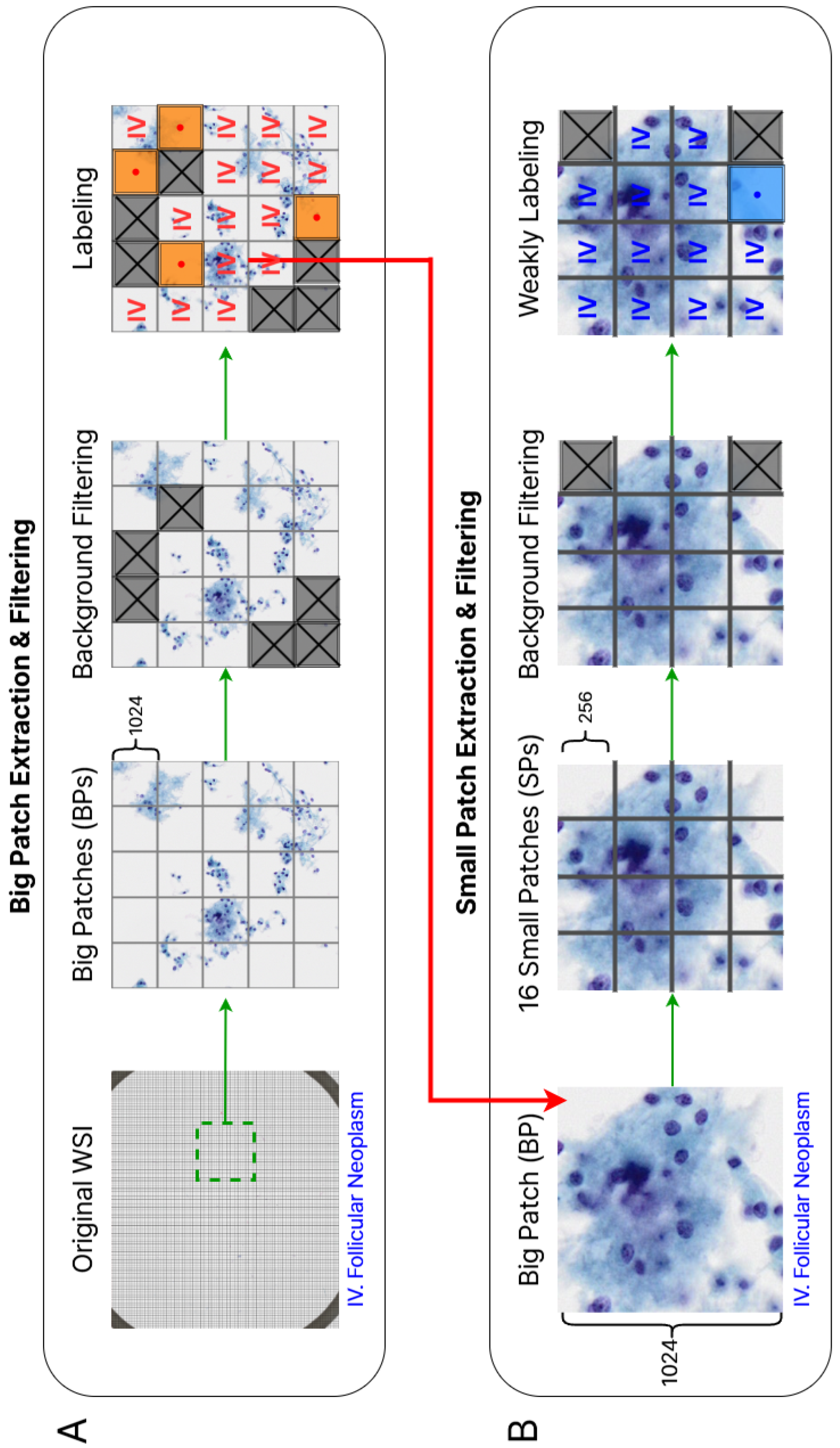
3.2. Patch Extraction and Filtering
3.3. Dataset Partitioning and Normalization
4. Methodologies
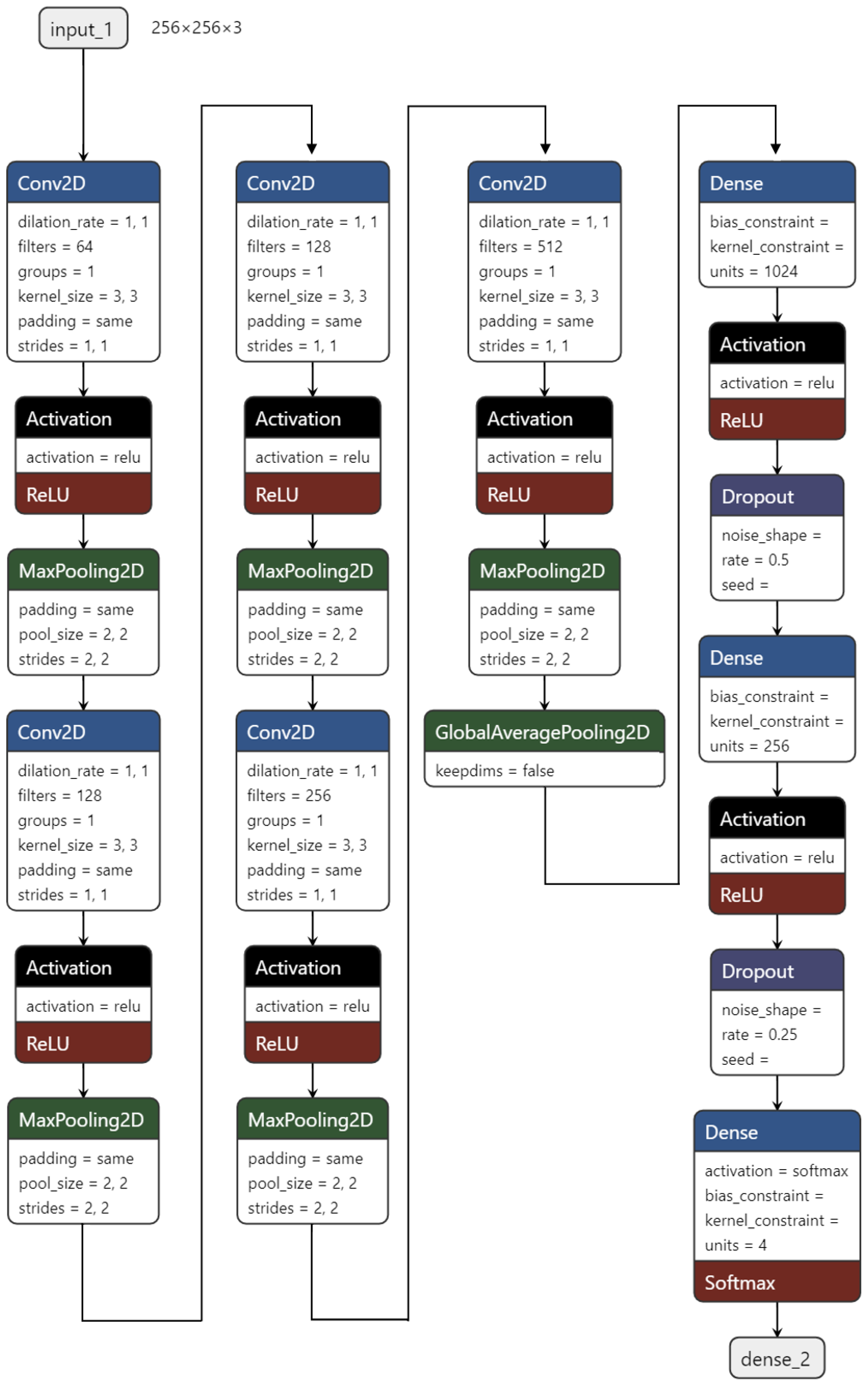
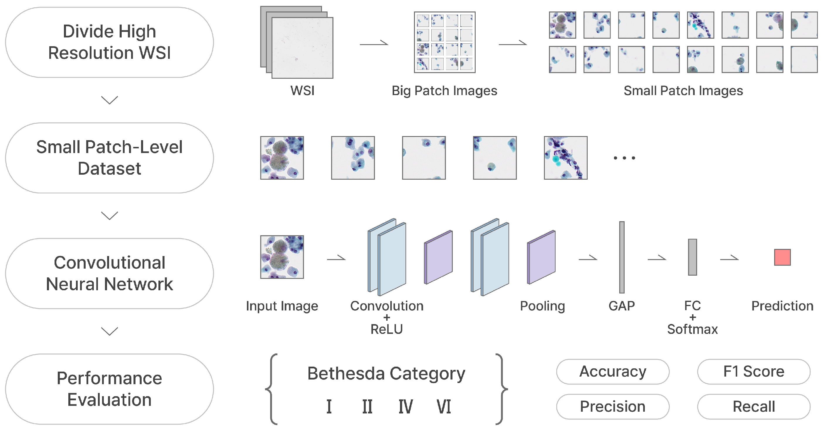
4.1. SP Classifier Using TCS-CNN Architecture
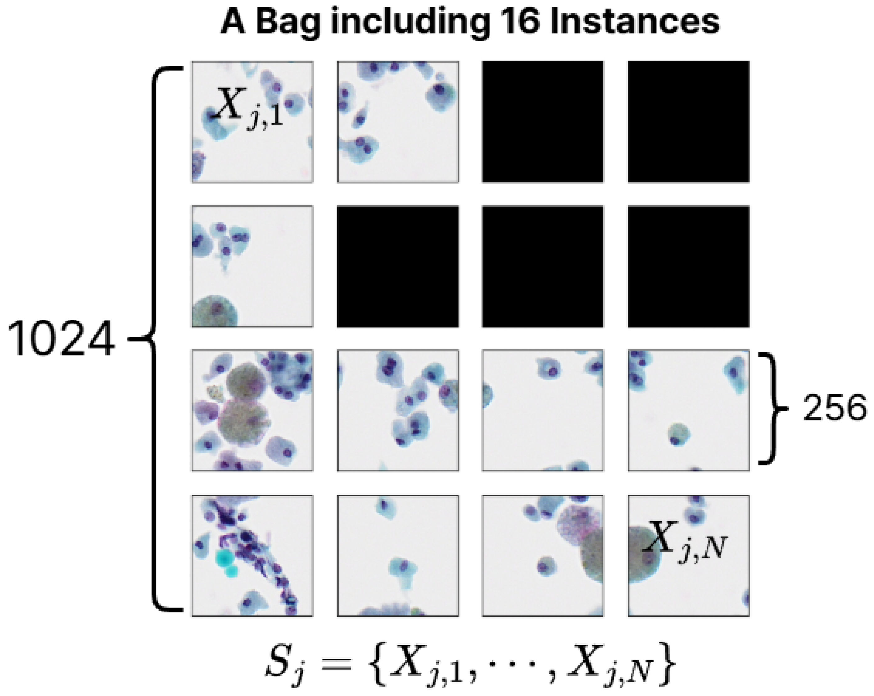
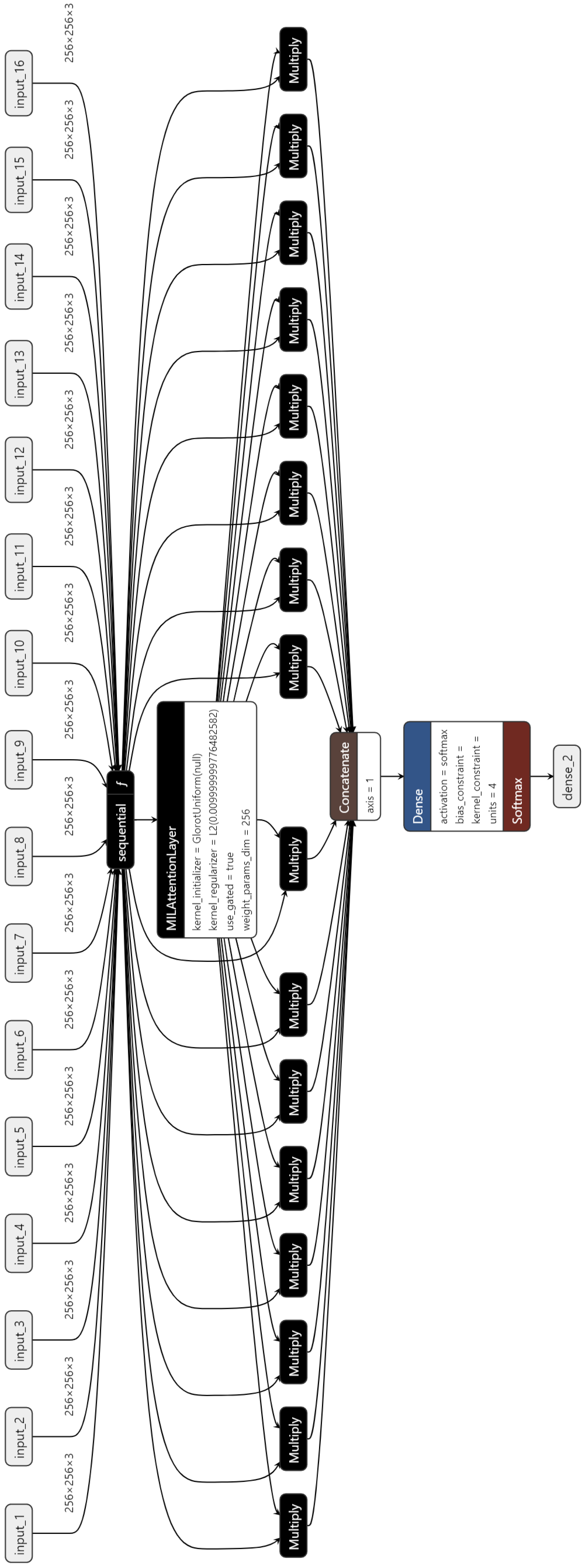
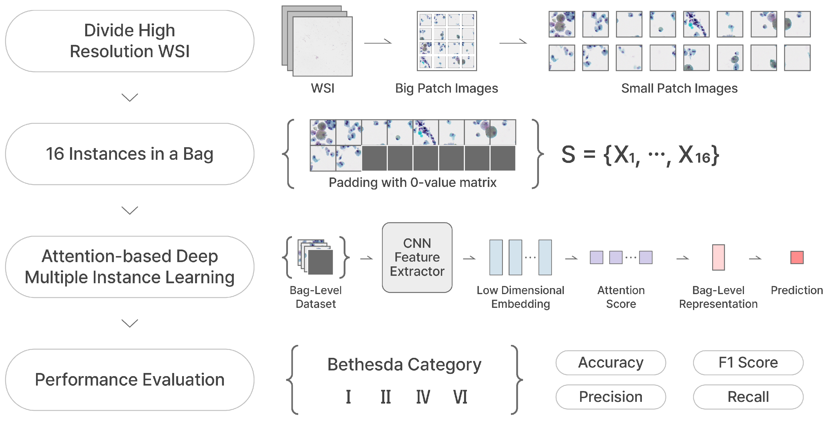
4.2. BP Classifier Using AD-MIL
5. Experiment
5.1. Training Procedure
5.2. Evaluation Metrics
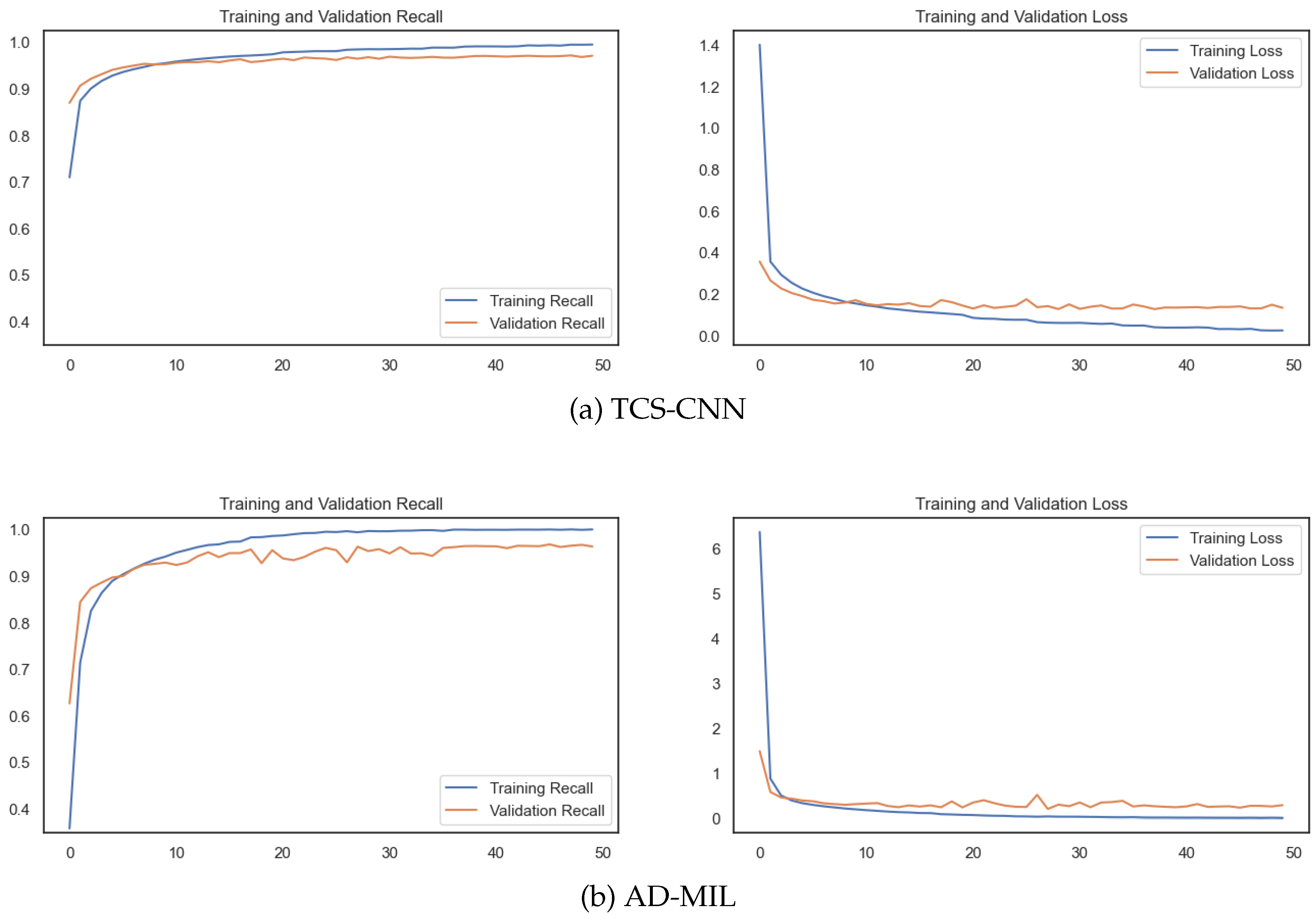
6. Results
6.1. Performance
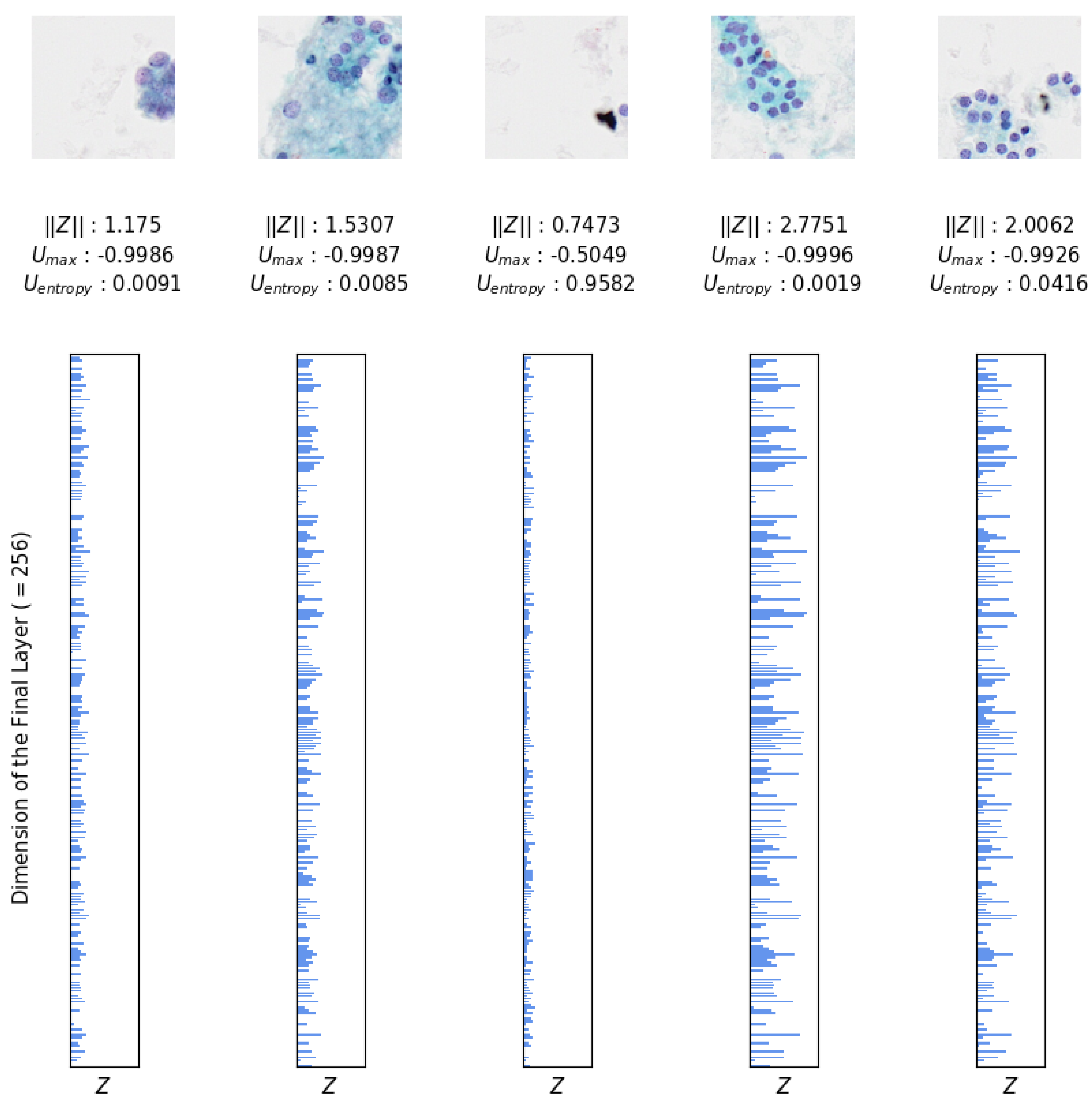
6.2. Uncertainty Analysis
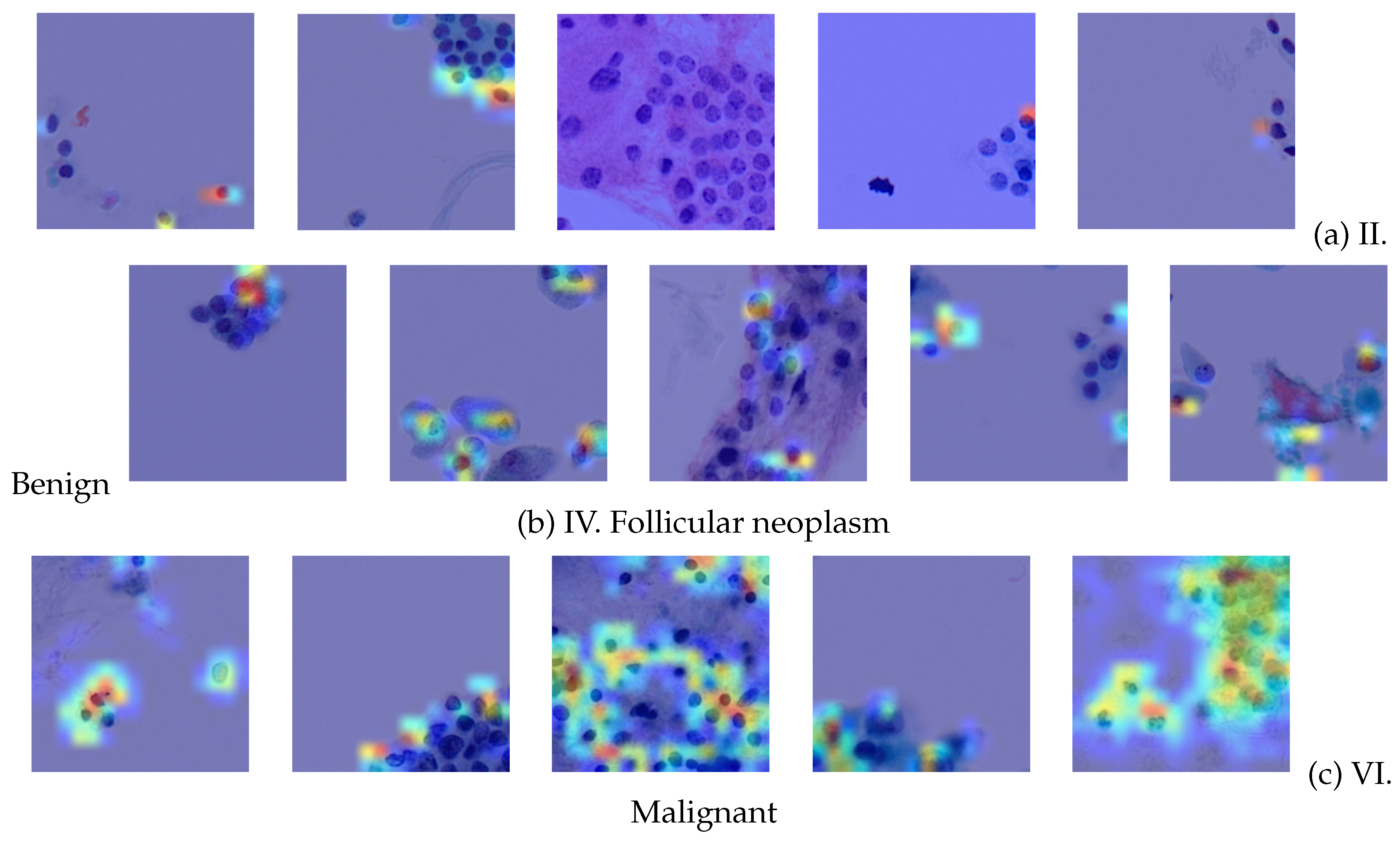
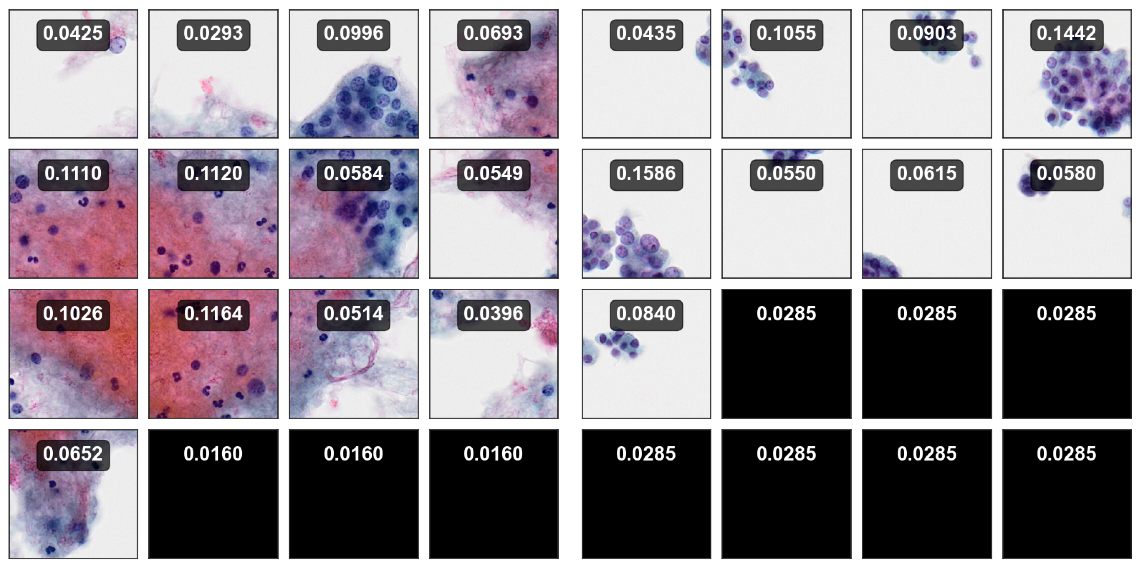
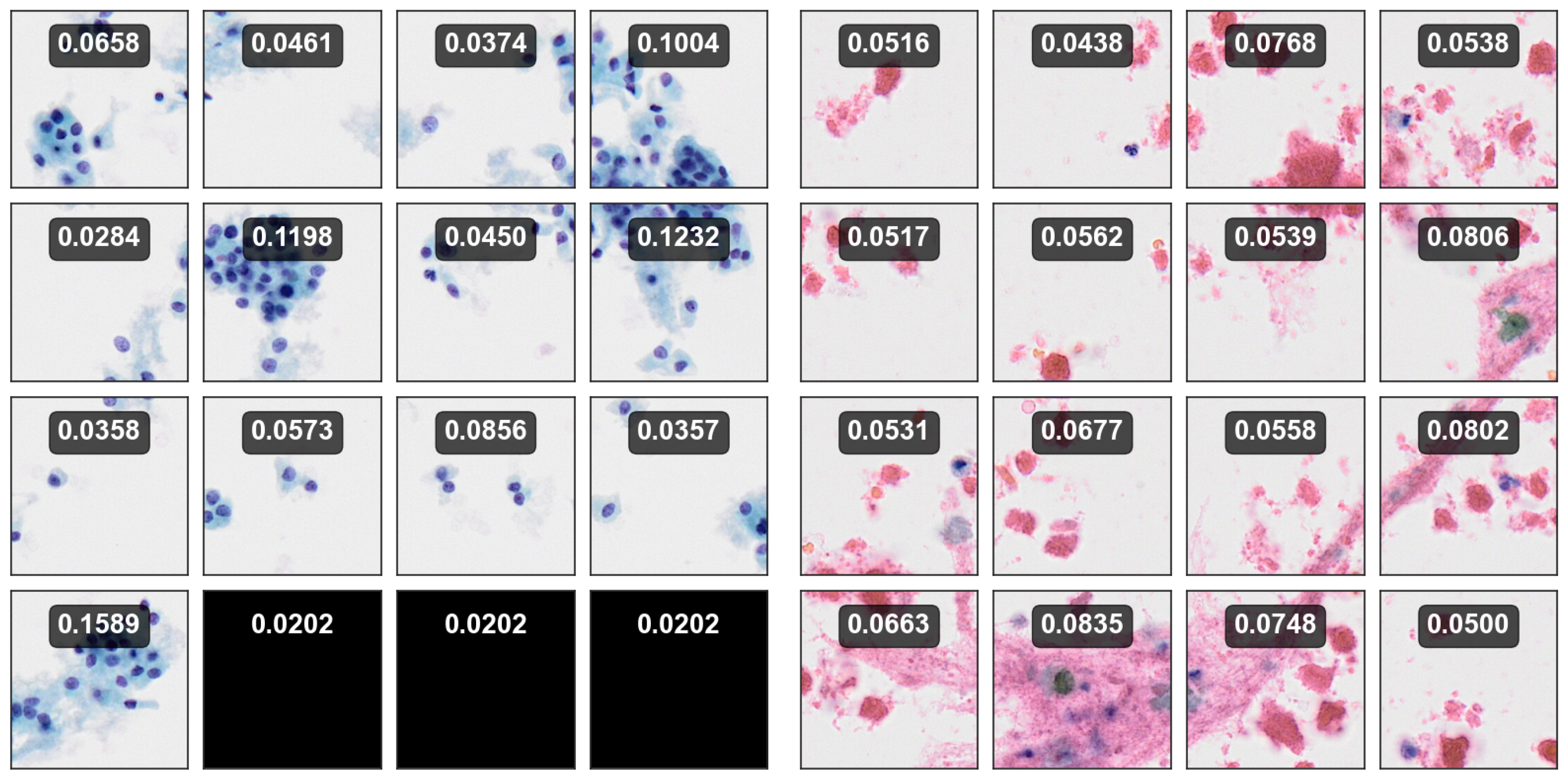
6.3. Visualization
7. Discussion
8. Conclusions
Author Contributions
Funding
Institutional Review Board Statement
Informed Consent Statement
Data Availability Statement
Conflicts of Interest
References
- Frasoldati, A.; Flora, M.; Pesenti, M.; Caroggio, A.; Valcavi, R. Computer-assisted cell morphometry and ploidy analysis in the assessment of thyroid follicular neoplasms. Thyroid 2001, 11, 941–946. [Google Scholar] [CrossRef]
- Gupta, N.; Sarkar, C.; Singh, R.; Karak, A.K. Evaluation of diagnostic efficiency of computerized image analysis based quantitative nuclear parameters in papillary and follicular thyroid tumors using paraffin-embedded tissue sections. Pathology Oncology Research 2001, 7, 46–55. [Google Scholar] [CrossRef] [PubMed]
- Murata, S.i.; Mochizuki, K.; Nakazawa, T.; Kondo, T.; Nakamura, N.; Yamashita, H.; Urata, Y.; Ashihara, T.; Katoh, R. Detection of underlying characteristics of nuclear chromatin patterns of thyroid tumor cells using texture and factor analyses. Cytometry: The Journal of the International Society for Analytical Cytology 2002, 49, 91–95. [Google Scholar] [CrossRef] [PubMed]
- Karslıoğlu, Y.; Celasun, B.; Günhan, Ö. Contribution of morphometry in the differential diagnosis of fine-needle thyroid aspirates. Cytometry Part B: Clinical Cytometry: The Journal of the International Society for Analytical Cytology 2005, 65, 22–28. [Google Scholar] [CrossRef]
- Aiad, H.; Abdou, A.; Bashandy, M.; Said, A.; Ezz-Elarab, S.; Zahran, A. Computerized nuclear morphometry in the diagnosis of thyroid lesions with predominant follicular pattern. Ecancermedicalscience 2009, 3. [Google Scholar] [CrossRef]
- Tsantis, S.; Dimitropoulos, N.; Cavouras, D.; Nikiforidis, G. Morphological and wavelet features towards sonographic thyroid nodules evaluation. Computerized Medical Imaging and Graphics 2009, 33, 91–99. [Google Scholar] [CrossRef] [PubMed]
- Ozolek, J.A.; Tosun, A.B.; Wang, W.; Chen, C.; Kolouri, S.; Basu, S.; Huang, H.; Rohde, G.K. Accurate diagnosis of thyroid follicular lesions from nuclear morphology using supervised learning. Medical image analysis 2014, 18, 772–780. [Google Scholar] [CrossRef] [PubMed]
- Daskalakisa, A.; Kostopoulosa, S.; Spyridonosa, P.; Glotsosa, D.; Ravazoulab, P.; Kardarib, M.; Kalatzisc, I.; Cavourasc, D.; Nikiforidisa, G. Design of a multi-classifier system for discriminating benign from malignant thyroid nodules using routinely H&E-stained cytological images 3. Computers in Biologyand Medicine 2008, 38, 196–203. [Google Scholar]
- Wang, W.; Ozolek, J.A.; Rohde, G.K. Detection and classification of thyroid follicular lesions based on nuclear structure from histopathology images. Cytometry Part A: The Journal of the International Society for Advancement of Cytometry 2010, 77, 485–494. [Google Scholar] [CrossRef]
- Gopinath, B.; Shanthi, N. Support Vector Machine based diagnostic system for thyroid cancer using statistical texture features. Asian Pacific Journal of Cancer Prevention 2013, 14, 97–102. [Google Scholar] [CrossRef] [PubMed]
- Margari, N.; Mastorakis, E.; Pouliakis, A.; Gouloumi, A.R.; Asimis, E.; Konstantoudakis, S.; Ieromonachou, P.; Panayiotides, I.G. Classification and regression trees for the evaluation of thyroid cytomorphological characteristics: a study based on liquid based cytology specimens from thyroid fine needle aspirations. Diagnostic Cytopathology 2018, 46, 670–681. [Google Scholar] [CrossRef] [PubMed]
- Maleki, S.; Zandvakili, A.; Gera, S.; Khutti, S.D.; Gersten, A.; Khader, S.N. Differentiating noninvasive follicular thyroid neoplasm with papillary-like nuclear features from classic papillary thyroid carcinoma: Analysis of cytomorphologic descriptions using a novel machine-learning approach. Journal of pathology informatics 2019, 10, 29. [Google Scholar] [CrossRef]
- LeCun, Y.; Bottou, L.; Bengio, Y.; Haffner, P. Gradient-based learning applied to document recognition. Proceedings of the IEEE 1998, 86, 2278–2324. [Google Scholar] [CrossRef]
- LeCun, Y.; Bengio, Y.; Hinton, G. Deep learning. nature 2015, 521, 436–444. [Google Scholar] [CrossRef]
- Simonyan, K.; Zisserman, A. Very deep convolutional networks for large-scale image recognition. arXiv 2014, arXiv:1409.1556 2014. [Google Scholar]
- He, K.; Zhang, X.; Ren, S.; Sun, J. Deep residual learning for image recognition. In Proceedings of the Proceedings of the IEEE conference on computer vision and pattern recognition, 2016, pp.
- Szegedy, C.; Vanhoucke, V.; Ioffe, S.; Shlens, J.; Wojna, Z. Rethinking the inception architecture for computer vision. In Proceedings of the Proceedings of the IEEE conference on computer vision and pattern recognition, 2016, pp.
- Kim, E.; Corte-Real, M.; Baloch, Z. A deep semantic mobile application for thyroid cytopathology. In Proceedings of the Medical Imaging 2016: PACS and Imaging Informatics: Next Generation and Innovations. SPIE, Vol. 9789; 2016; pp. 36–44. [Google Scholar]
- Sanyal, P.; Mukherjee, T.; Barui, S.; Das, A.; Gangopadhyay, P. Artificial intelligence in cytopathology: a neural network to identify papillary carcinoma on thyroid fine-needle aspiration cytology smears. Journal of pathology informatics 2018, 9, 43. [Google Scholar] [CrossRef] [PubMed]
- Guan, Q.; Wang, Y.; Ping, B.; Li, D.; Du, J.; Qin, Y.; Lu, H.; Wan, X.; Xiang, J. Deep convolutional neural network VGG-16 model for differential diagnosing of papillary thyroid carcinomas in cytological images: a pilot study. Journal of Cancer 2019, 10, 4876. [Google Scholar] [CrossRef] [PubMed]
- Wang, Y.; Guan, Q.; Lao, I.; Wang, L.; Wu, Y.; Li, D.; Ji, Q.; Wang, Y.; Zhu, Y.; Lu, H.; et al. Using deep convolutional neural networks for multi-classification of thyroid tumor by histopathology: a large-scale pilot study. Annals of translational medicine 2019, 7. [Google Scholar] [CrossRef] [PubMed]
- Elliott Range, D.D.; Dov, D.; Kovalsky, S.Z.; Henao, R.; Carin, L.; Cohen, J. Application of a machine learning algorithm to predict malignancy in thyroid cytopathology. Cancer cytopathology 2020, 128, 287–295. [Google Scholar] [CrossRef]
- Irshad, H.; Veillard, A.; Roux, L.; Racoceanu, D. Methods for nuclei detection, segmentation, and classification in digital histopathology: a review—current status and future potential. IEEE reviews in biomedical engineering 2013, 7, 97–114. [Google Scholar] [CrossRef]
- Nayak, N.; Chang, H.; Borowsky, A.; Spellman, P.; Parvin, B. Classification of tumor histopathology via sparse feature learning. In Proceedings of the 2013 IEEE 10th international symposium on biomedical imaging. IEEE; 2013; pp. 410–413. [Google Scholar]
- Hou, L.; Samaras, D.; Kurc, T.M.; Gao, Y.; Davis, J.E.; Saltz, J.H. Patch-based convolutional neural network for whole slide tissue image classification. In Proceedings of the Proceedings of the IEEE conference on computer vision and pattern recognition, 2016, pp.
- Durand, T.; Thome, N.; Cord, M. Weldon: Weakly supervised learning of deep convolutional neural networks. In Proceedings of the Proceedings of the IEEE conference on computer vision and pattern recognition, 2016, pp.
- Courtiol, P.; Tramel, E.W.; Sanselme, M.; Wainrib, G. Classification and disease localization in histopathology using only global labels: A weakly-supervised approach. arXiv 2018, arXiv:1802.02212 2018. [Google Scholar]
- Couture, H.D.; Marron, J.S.; Perou, C.M.; Troester, M.A.; Niethammer, M. Multiple instance learning for heterogeneous images: Training a cnn for histopathology. In Proceedings of the Medical Image Computing and Computer Assisted Intervention–MICCAI 2018: 21st International Conference, Granada, Spain, 2018, Proceedings, Part II 11. Springer, 2018, September 16-20; pp. 254–262.
- Ilse, M.; Tomczak, J.; Welling, M. Attention-based deep multiple instance learning. In Proceedings of the International conference on machine learning. PMLR; 2018; pp. 2127–2136. [Google Scholar]
- Wang, Z.; Poon, J.; Sun, S.; Poon, S. Attention-based multi-instance neural network for medical diagnosis from incomplete and low quality data. In Proceedings of the 2019 International joint conference on neural networks (IJCNN). IEEE; 2019; pp. 1–8. [Google Scholar]
- Wang, S.; Zhu, Y.; Yu, L.; Chen, H.; Lin, H.; Wan, X.; Fan, X.; Heng, P.A. RMDL: Recalibrated multi-instance deep learning for whole slide gastric image classification. Medical image analysis 2019, 58, 101549. [Google Scholar] [CrossRef] [PubMed]
- Dov, D.; Kovalsky, S.Z.; Cohen, J.; Range, D.E.; Henao, R.; Carin, L. Thyroid cancer malignancy prediction from whole slide cytopathology images. In Proceedings of the Machine Learning for Healthcare Conference. PMLR; 2019; pp. 553–570. [Google Scholar]
- Qiu, S.; Guo, Y.; Zhu, C.; Zhou, W.; Chen, H. Attention Based Multi-Instance Thyroid Cytopathological Diagnosis with Multi-Scale Feature Fusion. In Proceedings of the 2020 25th International Conference on Pattern Recognition (ICPR). IEEE; 2021; pp. 3536–3541. [Google Scholar]
- Dov, D.; Kovalsky, S.Z.; Assaad, S.; Cohen, J.; Range, D.E.; Pendse, A.A.; Henao, R.; Carin, L. Weakly supervised instance learning for thyroid malignancy prediction from whole slide cytopathology images. Medical image analysis 2021, 67, 101814. [Google Scholar] [CrossRef]
- Cibas, E.S.; Ali, S.Z. The 2017 Bethesda system for reporting thyroid cytopathology. Thyroid 2017, 27, 1341–1346. [Google Scholar] [CrossRef]
- He, K.; Zhang, X.; Ren, S.; Sun, J. Delving deep into rectifiers: Surpassing human-level performance on imagenet classification. In Proceedings of the Proceedings of the IEEE international conference on computer vision, 2015, pp.
- Pappas, N.; Popescu-Belis, A. Explaining the stars: Weighted multiple-instance learning for aspect-based sentiment analysis. In Proceedings of the Proceedings of the 2014 Conference on Empirical Methods In Natural Language Processing (EMNLP), 2014, pp.
- Pappas, N.; Popescu-Belis, A. Explicit document modeling through weighted multiple-instance learning. Journal of Artificial Intelligence Research 2017, 58, 591–626. [Google Scholar] [CrossRef]
- Deng, J.; Dong, W.; Socher, R.; Li, L.J.; Li, K.; Fei-Fei, L. Imagenet: A large-scale hierarchical image database. In Proceedings of the 2009 IEEE conference on computer vision and pattern recognition. Ieee; 2009; pp. 248–255. [Google Scholar]
- Howard, A.G.; Zhu, M.; Chen, B.; Kalenichenko, D.; Wang, W.; Weyand, T.; Andreetto, M.; Adam, H. Mobilenets: Efficient convolutional neural networks for mobile vision applications. arXiv 2017, arXiv:1704.04861 2017. [Google Scholar]
- Kingma, D.P.; Ba, J. Adam: A method for stochastic optimization. arXiv 2014, arXiv:1412.6980 2014. [Google Scholar]
- Guo, C.; Pleiss, G.; Sun, Y.; Weinberger, K.Q. On calibration of modern neural networks. In Proceedings of the International conference on machine learning. PMLR; 2017; pp. 1321–1330. [Google Scholar]
- Pearce, T.; Brintrup, A.; Zhu, J. Understanding softmax confidence and uncertainty. arXiv 2021, arXiv:2106.04972 2021. [Google Scholar]
- Selvaraju, R.R.; Cogswell, M.; Das, A.; Vedantam, R.; Parikh, D.; Batra, D. Grad-cam: Visual explanations from deep networks via gradient-based localization. In Proceedings of the Proceedings of the IEEE international conference on computer vision, 2017, pp.
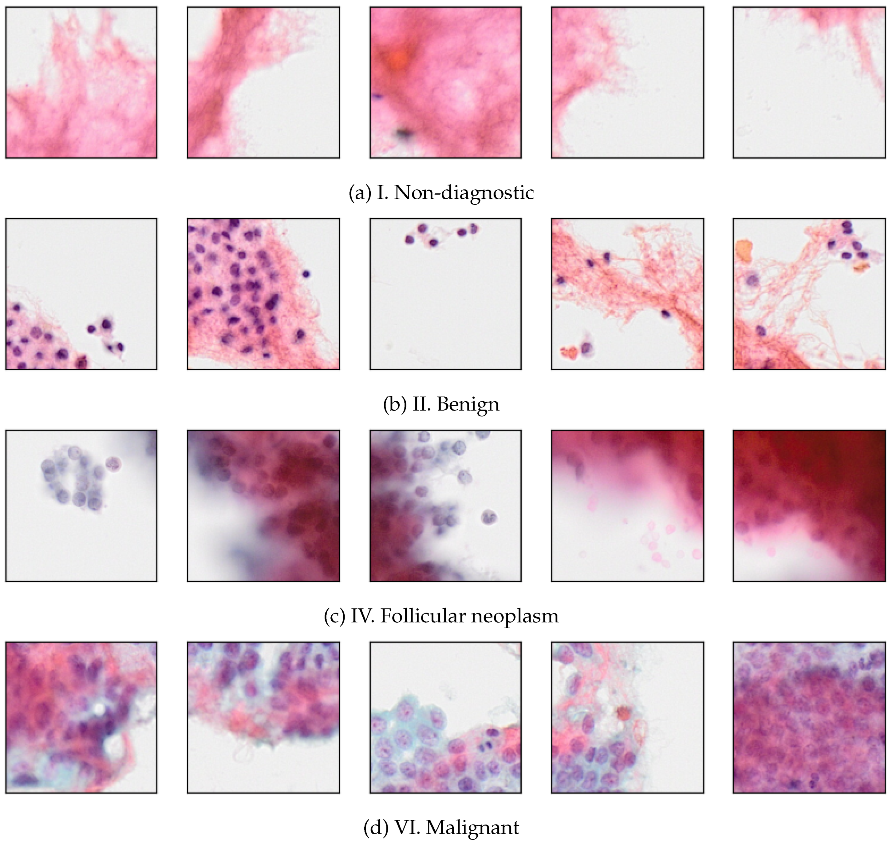

| Category | Number of images |
|---|---|
| I. Non-diagnostic | |
| II. Benign | |
| IV. Follicular neoplasm | |
| VI. Malignant | |
| Total |
| Category | Number of images |
|---|---|
| I. Non-diagnostic | |
| II. Benign | |
| IV. Follicular neoplasm | |
| VI. Malignant | |
| Total |
| Metrics | Split 1 | Split 2 | Split 3 | Split 4 | Split 5 |
|---|---|---|---|---|---|
| Precision | 0.9578 | 0.9599 | 0.9604 | 0.9622 | 0.9623 |
| Recall | 0.9597 | 0.9602 | 0.9568 | 0.9596 | 0.9558 |
| F1-score | 0.9587 | 0.9601 | 0.9585 | 0.9609 | 0.9589 |
| Accuracy | 0.9720 | 0.9727 | 0.9721 | 0.9732 | 0.9723 |
| Model | Precision | Recall | F1-score | Accuracy |
|---|---|---|---|---|
| VGG16 | 0.9324 | 0.9327 | 0.9326 | 0.9522 |
| Inception-v3 | 0.8587 | 0.8472 | 0.8527 | 0.8842 |
| Mobilenet | 0.9259 | 0.9267 | 0.9263 | 0.9461 |
| TCS-CNN | 0.9578 | 0.9597 | 0.9587 | 0.9720 |
| AD-MIL | 0.9616 | 0.9649 | 0.9631 | 0.9681 |
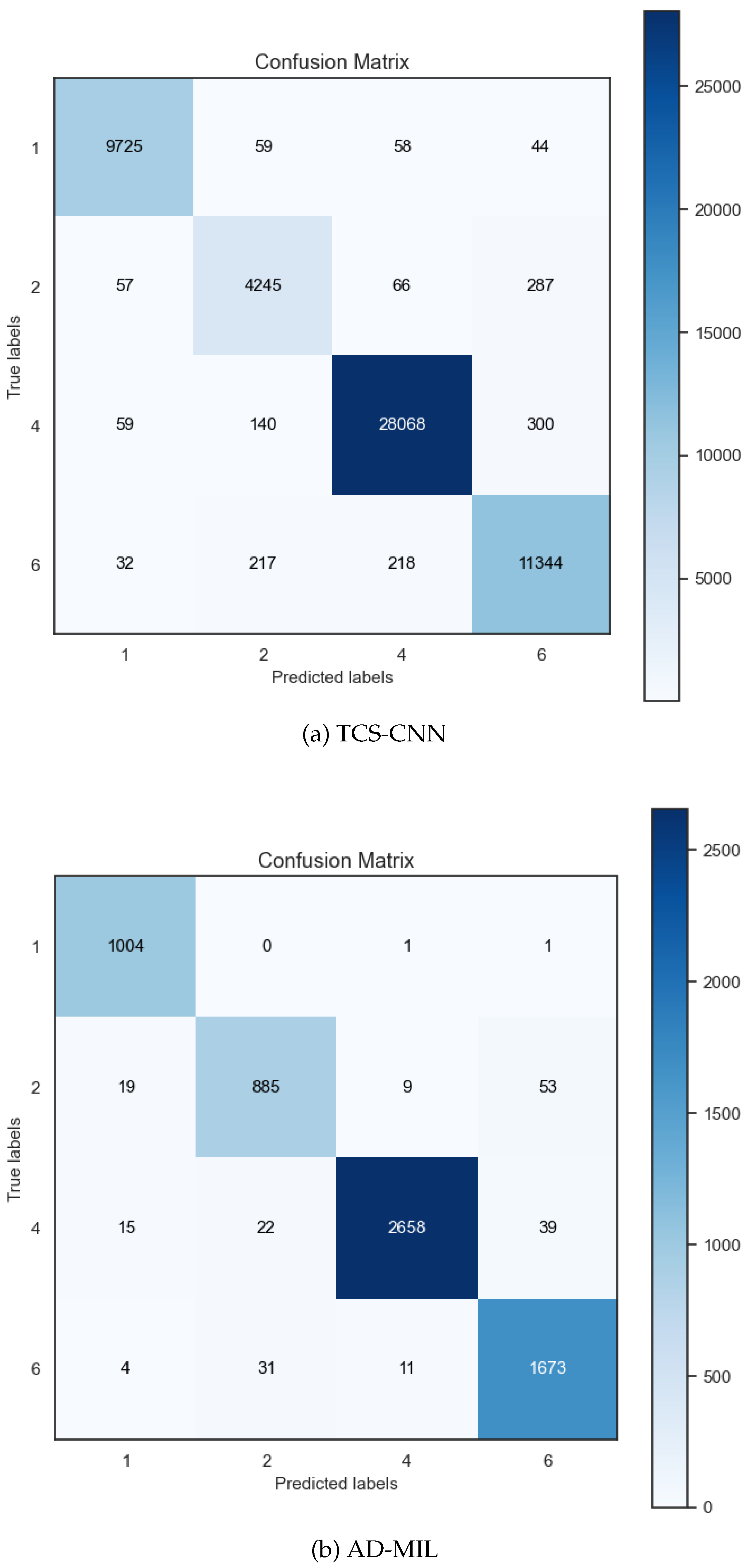
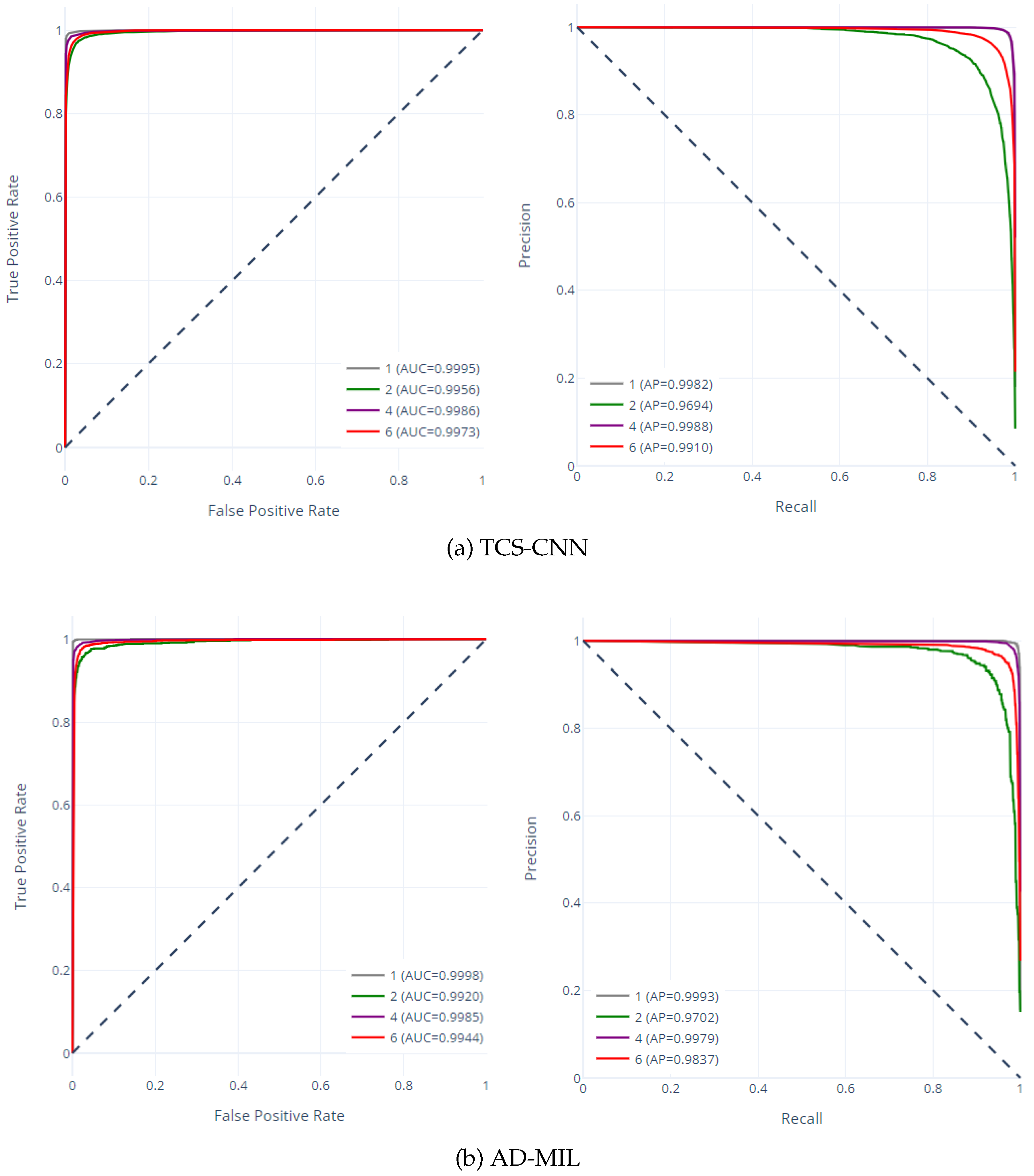
| Method | Cancer Types | Precision | Recall | F1-score | Support |
|---|---|---|---|---|---|
| ]5*TCS-CNN | I. Non-diagnostic | 0.9850 | 0.9837 | 0.9844 | 9886 |
| II. Benign | 0.9107 | 0.9119 | 0.9113 | 4655 | |
| IV. Follicular neoplasm | 0.9880 | 0.9825 | 0.9852 | 28567 | |
| VI. Malignant | 0.9473 | 0.9605 | 0.9538 | 11811 | |
| Average | 0.9578 | 0.9597 | 0.9587 | 54919 | |
| ]5*AD-MIL | I. Non-diagnostic | 0.9635 | 0.9980 | 0.9805 | 1006 |
| II. Benign | 0.9435 | 0.9161 | 0.9296 | 966 | |
| IV. Follicular neoplasm | 0.9922 | 0.9722 | 0.9821 | 2734 | |
| VI. Malignant | 0.9473 | 0.9732 | 0.9601 | 1719 | |
| Average | 0.9616 | 0.9649 | 0.9631 | 6425 |
Disclaimer/Publisher’s Note: The statements, opinions and data contained in all publications are solely those of the individual author(s) and contributor(s) and not of MDPI and/or the editor(s). MDPI and/or the editor(s) disclaim responsibility for any injury to people or property resulting from any ideas, methods, instructions or products referred to in the content. |
© 2025 by the authors. Licensee MDPI, Basel, Switzerland. This article is an open access article distributed under the terms and conditions of the Creative Commons Attribution (CC BY) license (http://creativecommons.org/licenses/by/4.0/).


