Submitted:
12 February 2025
Posted:
13 February 2025
You are already at the latest version
Abstract
Oropouche fever is caused by the Oropouche virus (OROV; Bunyaviridae, Orthobunyavirus), one of the most frequent arboviruses that infect humans in the Brazilian Amazon. This year, an OROV outbreak was identified in Brazil, and its vertical transmission was reported, which was associated with fetal death and microcephaly. We describe the clinical manifestations identified in three cases of congenital OROV infection with confirmed serology (OROV-IgM) in the mother-newborn binomial. One of the newborns died, and post-mortem molecular analysis using real-time RT-qPCR identified the OROV genome in several tissues. All three newborns were born in the Amazon region in Brazil, and the mothers reported fever, rash, headache, myalgia, and/or retroorbital pain during pregnancy. The newborns presented with severe microcephaly secondary to brain damage and arthrogryposis, suggestive of an embryo/fetal disruptive process at birth. Brain and spinal images identified overlapping sutures, cerebral atrophy, brain cysts, thinning of the spinal cord, corpus callosum, and posterior fossa abnormalities. Fundoscopic findings included macular chorioretinal scars, focal pigment mottling, and vascular attenuation. The clinical presentation of vertical OROV infection resembled congenital Zika syndrome to some extent but presents some distinctive features on brain imaging and in several aspects of its neurological presentation. A recognizable syndrome with severe brain damage, neurological alterations, arthrogryposis, and fundoscopic abnormalities can be associated with in-utero OROV infection.
Keywords:
1. Introduction
2. Materials and Methods
3. Case reports
4. Discussion
Author Contributions
Funding
Institutional Review Board Statement
Informed Consent Statement
Data Availability Statement
Acknowledgments
Conflicts of Interest
Abbreviations.
| CLIA | Chemiluminescent immunoassay |
| CHKV | Chikungunya virus |
| CMV | Cytomegalovirus |
| CSF | Cerebrospinal fluid |
| CT | Computed tomography |
| CZS | Congenital Zika syndrome |
| DENV | Dengue virus |
| Det | Detected |
| FBDS | Fetal brain disruption sequence |
| HIV | Human immunodeficiency virus |
| ELISA | Capture Immunoenzymatic Assay |
| MoH | Brazilian Ministry of Health |
| MRI | Magnetic resonance imaging |
| NA | Not available |
| NDet | Not detected |
| OD | Optical density |
| OROV | Oropouche virus |
| RT-qPCR | Real-time reverse quantitative transcription–polymerase chain reaction |
| Toxo | Toxoplasmosis |
| US | Ultrasound |
| WG | Weeks of gestation |
| ZIKV | Zika virus |
References
- Anderson, C.R.; Spence, L.; Downs, W.G.; Aitken, T.H. Oropouche virus: a new human disease agent from Trinidad, West Indies. Am J Trop Med Hyg. 1961, 10, 574–578. [Google Scholar] [CrossRef] [PubMed]
- Azevedo EAN, Silva AFD, Silva VGD, et al. Genomic and phenotypic characterization of the Oropouche virus strain implicated in the 2022-24 large-scale outbreak in Brazil. J Med Virol. 2024, 96, e70012. [Google Scholar] [CrossRef]
- Chiang JO, Azevedo RS, Justino MCA, et al. Neurological disease caused by Oropouche virus in northern Brazil: should it be included in the scope of clinical neurological diseases? J Neurovirol. 2021, 27, 626–630. [Google Scholar] [CrossRef] [PubMed]
- Brazil SECRETARIADEVIGILÂNCIAEMSAÚDEE, A.M.B.I.E.N.T.E. Recomendação para intensificação da vigilância de transmissão vertical do vírus Oropouche. Nota técnica Nº 15/2024 - SVSA/MS. BRASIL. MINISTÉRIO DA SAÚDE/ , 2024. Accessed August 5, 2024. https://www.gov.br/saude/pt-br/centrais-de-conteudo/publicacoes/notas-tecnicas/2024/nota-tecnica-no-15-2024-svsa-ms.pdf/view.
- PAN AMERICAN HEALTH ORGANIZATION/ WORLD HEALTH ORGANIZATION, Epidemiological Update Oropouche in the Americas Region - 6 September 2024. Washington D.C: 2024. Accessed September 9, 2024. https://www.paho.org/en/documents/epidemiological-update-oropouche-americas-region-6-september-2024.
- Martins FEN, Chiang JO, Nunes BRN et al. Newborns with microcephaly in Brazil and potential vertical transmission of Oropouche vírus: a case series. Lancet Infect Dis 2024. [CrossRef]
- Naveca, F.G.; Nascimento, V.A.D.; Souza, V.C.; Nunes, B.T.D.; Rodrigues, D.S.G.; Vasconcelos, P.F.D.C. Multiplexed reverse transcription real-time polymerase chain reaction for simultaneous detection of Mayaro, Oropouche, and Oropouche-like viruses. Mem Inst Oswaldo Cruz. 2017, 112, 510–513. [Google Scholar] [CrossRef] [PubMed]
- Di Donato N, Chiari S, Mirzaa GM, et al. Lissencephaly: Expanded imaging and clinical classification. Am J Med Genet A. 2017, 173, 1473–1488. [Google Scholar] [CrossRef]
- Del Campo M, Feitosa IM, Ribeiro EM, et al. The phenotypic spectrum of congenital Zika syndrome. Am J Med Genet A. 2017, 173, 841–857. [Google Scholar] [CrossRef]
- Moore, C.A.; Weaver, D.D.; Bull, M.J. Fetal brain disruption sequence. J Pediatr. 1990, 116, 383–6. [Google Scholar] [CrossRef]
- De Rose DU, Gallini F, Battaglia DI, et al. A novel homozygous variant in JAM3 gene causing hemorrhagic destruction of the brain, subependymal calcification, and congenital cataracts (HDBSCC) with neonatal onset. Neurol Sci. 2021, 42, 4759–4765. [Google Scholar] [CrossRef]
- Doobin, D.J.; Kemal, S.; Dantas, T.J.; Vallee, R.B. Severe NDE1-mediated microcephaly results from neural progenitor cell cycle arrests at multiple specific stages. Nat Commun. 2016, 7, 12551. [Google Scholar] [CrossRef]
- Thomas AX, Link N, Robak LA et al. ANKLE2-related microcephaly: A variable microcephaly syndrome resembling Zika infection. Ann Clin Transl Neurol. 2022, 9, 1276–1288. [Google Scholar] [CrossRef]
- Moore CA, Staples JE, Dobyns WB, et al. Characterizing the Pattern of Anomalies in Congenital Zika Syndrome for Pediatric Clinicians. JAMA Pediatr. 2017, 171, 288–295. [Google Scholar] [CrossRef]
- Barreto, A.R.F.; Carrasco, M.; Dabrowski, A.K.; Sun, L.R.; Tekes, A. Subpial Hemorrhage in Neonates: What Radiologists Need to Know. AJR Am J Roentgenol. 2021, 216, 1056–1065. [Google Scholar] [CrossRef]
- Zhuang, X. , Jin, K., Li, J. et al. Subpial hemorrhages in neonates: imaging features, clinical factors and outcomes. Sci Rep. 2023, 13, 3408. [Google Scholar] [CrossRef] [PubMed]
- Wesselmann KM, Postigo-Hidalgo I, Pezzi L, et al. Emergence of Oropouche fever in Latin America: a narrative review. Lancet Infect Dis. 2024, 24, e439–e452. [Google Scholar] [CrossRef]
- Cramer JA, Rassner UA, Hedlund GL. Limitations of T2*-Gradient Recalled-Echo and Susceptibility-Weighted Imaging in Characterizing Chronic Subdural Hemorrhage in Infant Survivors of Abusive Head Trauma. Am J Neuroradiol. 2016, 37, 1752–6. [Google Scholar] [CrossRef] [PubMed]
- van der Linden H Jr, Carvalho MD, van der Linden V, et al. Epilepsy Profile in Infants with Congenital Zika Virus Infection. N Engl J Med. 2018, 379, 891–892. [Google Scholar] [CrossRef] [PubMed]
- Pessoa A, van der Linden V, Yeargin-Allsopp M et al. Motor Abnormalities and Epilepsy in Infants and Children With Evidence of Congenital Zika Virus Infection. Pediatrics. 2018, 141, S167–S179. [Google Scholar] [CrossRef] [PubMed]
- Leal MC, van der Linden V, Bezerra TP, et al. Characteristics of Dysphagia in Infants with Microcephaly Caused by Congenital Zika Virus Infection, Brazil, 2015. Emerg Infect Dis. 2017, 23, 1253–1259. [Google Scholar] [CrossRef]
- Ventura CV, Zin A, Paula Freitas B, et al. Ophthalmological manifestations in congenital Zika syndrome in 469 Brazilian children. J AAPOS. 2021, 25, 158.e1–158.e8. [Google Scholar] [CrossRef]
- Singh, S.; Kumar, A. Ocular Manifestations of Emerging Flaviviruses and the Blood-Retinal Barrier [published correction appears in Viruses. 2019 May 24, 11, E475. https://doi.org/10.3390/v11050475]. Viruses. 2018, 10, 530. [CrossRef]
- da Hora LCD, Muniz LF, Venâncio LGA, et al. Cortical Auditory Evoked Potentials in Children with Prenatal Exposure to Zika Virus. Viruses. 2022, 14, 1923. [Google Scholar] [CrossRef]
- De Regge, N.; van den Berg, T.; Georges, L.; Cay, B. Diagnosis of Schmallenberg virus infection in malformed lambs and calves and first indications for virus clearance in the fetus. Vet Microbiol. 2013, 162, 595–600. [Google Scholar] [CrossRef] [PubMed]
- Peperkamp NH, Luttikholt SJ, Dijkman R, et al. Ovine and Bovine Congenital Abnormalities Associated With Intrauterine Infection With Schmallenberg Virus. Vet Pathol. 2015, 52, 1057–1066. [Google Scholar] [CrossRef]
- Hartley, W.J.; Wanner, R.A.; Della-Porta, A.J.; Snowdon, W.A. Serological evidence for the association of Akabane virus with epizootic bovine congenital arthrogryposis and hydranencephaly syndromes in New South Wales. Aust Vet J. 1975, 51, 103–104. [Google Scholar] [CrossRef]
- Agerholm, J.S.; Hewicker-Trautwein, M.; Peperkamp, K.; Windsor, P.A. Virus-induced congenital malformations in cattle. Acta Vet Scand. 2015, 57, 54. [Google Scholar] [CrossRef] [PubMed]
- Hughes, H.R.; Kenney, J.L.; Calvert, A.E. Cache Valley virus: an emerging arbovirus of public and veterinary health importance. J Med Entomol. 2023, 60, 1230–1241. [Google Scholar] [CrossRef]
- Edwards, J.F.; Karabatsos, N.; Collisson, E.W.; de la Concha Bermejillo, A. Ovine fetal malformations induced by in utero inoculation with Main Drain, San Angelo, and LaCrosse viruses. Am J Trop Med Hyg. 1997, 56, 171–176. [Google Scholar] [CrossRef]
- Calisher, C.H.; Sever, J.L. Are North American Bunyamwera serogroup viruses etiologic agents of human congenital defects of the central nervous system? Emerg Infect Dis. 1995, 1, 147–151. [Google Scholar] [CrossRef]
- Borborema CAT, Pinheiro FP, Albuquerque BC, et al. Primeiro registro de epidemias causadas pelo vírus Oropouche no estado do Amazonas. Rev. Inst. Med. Trop. São Paulo. 1982, 24, 132–139. [Google Scholar]
- Naveca FG, Almeida TAP, Souza V et al. Human outbreaks of a novel reassortant Oropouche virus in the Brazilian Amazon region. Nat Med. 2024. [Google Scholar] [CrossRef]

| Case 1 | Case 2 | Case 3 | |
|---|---|---|---|
| Maternal | |||
| Prenatal Oropouche Fever (WG*) |
21-22 | 8-9 | 8-9 |
| Maternal age (years) | 22 | 33 | 37 |
| Drugs/ Health conditions | No | No | Diabetes (from 33 WG) |
| Consanguinity | No | 2º degree | No |
| Previous pregnancies | 0 | 3 | 1 |
| Positive family history | No | No | No |
| Birth | |||
| Gestational age (weeks) | 38 | 36 | 37 |
| Head circumference (cm, Z-score**) |
30 (-2.66) | 26 (-4,40) | 28 |
| Weight (grams, Z-score) | 2,740 (-0.75) | 2,108 (-1.46) | 1,826 |
| Length (cm, Z-score) | 45 (-1.8) | 45 (-1.06) | 45 |
| Physical findings | |||
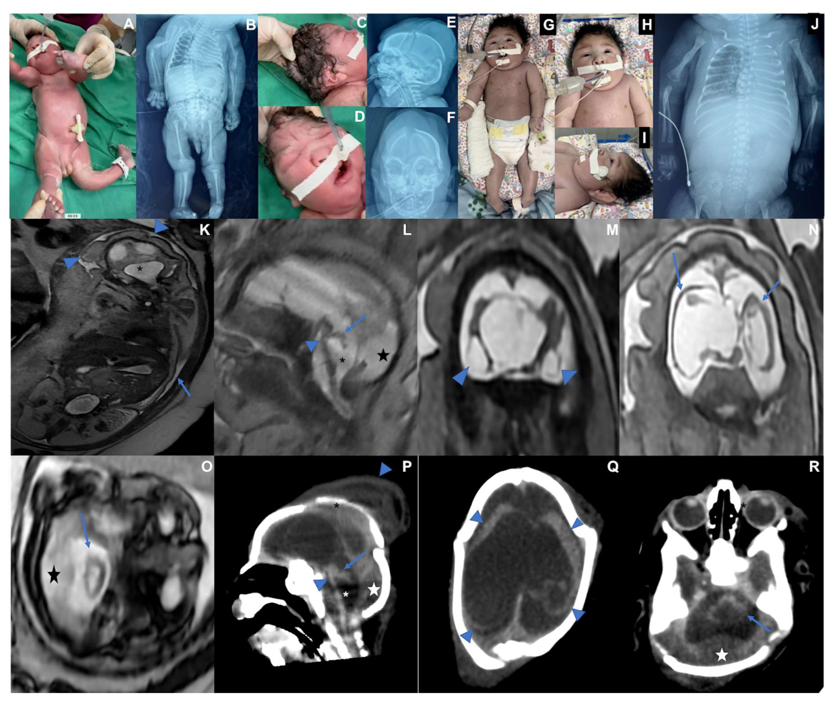
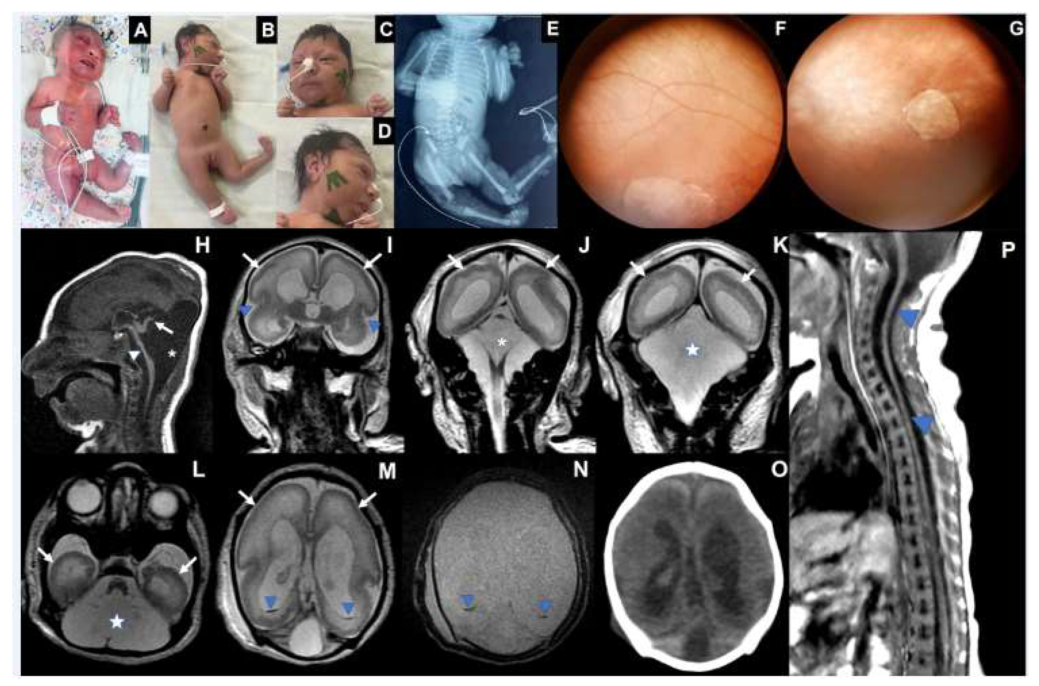
| Microcephaly / Craniofacial disproportion / Skull collapse / short neck | Yes | Yes | Yes |
| Arthrogryposis multiplex | No | Yes | Yes |
| Fetal hydrops | No | Yes | Yes |
| Neurologic findings | Hypotonia, deep tendon hyperreflexia, dystonia, and epileptic seizures | Severe hypotonia (flaccid paralysis), no primitive or deep tendon reflexes, lack of suction, slow swallowing | Hypotonia, no deep tendon reflexes, lack of suction, slow swallowing |
| Ophthalmological findings | Visual impairment and nystagmus at 3 months | Corneal edema, optic disc atrophy, and chorioretinal scar in OU. | Optic disc pallor and increased disc cupping (OS), chorioretinal macular scar and focal pigment mottling (OU), and vascular attenuation (OS). |
| Additional Exams | |||
| G-Banded Karyotype |
NA | 46,XY | 46,XX |
| Abdominal US*** | Normal | Pleural Effusion | Ascites |
| Echocardiogram | Patent oval foramen | Normal | Normal |
| Brainstem evoked response | Normal | NA | Normal |
| Electroneuromyography | Normal | NA | Normal |
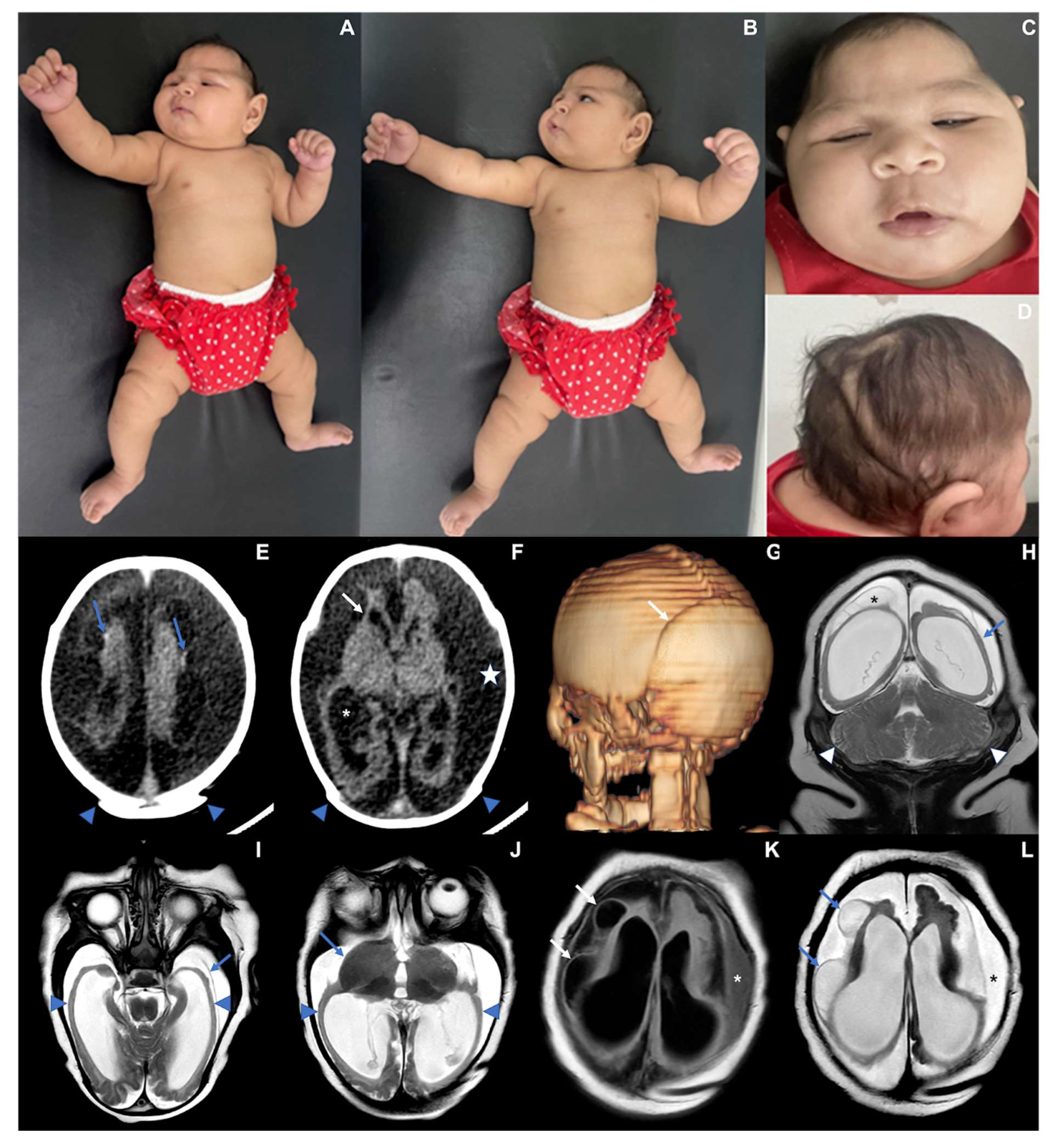
| Brain and Spinal Cord Images | Overlapping sutures, marked cerebral atrophy, partial lissencephaly, absent corpus callosum,brain cysts (cystic encephalomalacia and/or chronic subpial hemorrhages), subdural effusion, suspected striatal fusion, normal posterior fossa, normal spinal cord | Overlapping sutures, marked cerebral atrophy, absent corpus callosum, diffuse lissencephaly, severe brainstem and cerebellar hypoplasia, brain cysts (cystic encephalomalacia and/or chronic subpial hemorrhages), subdural effusion | Overlapping sutures, moderate cerebral atrophy, hypoplastic corpus callosum, diffuse lissencephaly, severe brainstem and cerebellar hypoplasia with Dandy-Walker-like features, intraventricular hemorrhage Global thinning of the spinal cord, mainly on the thoracic segment |
| *WG: weeks of gestation, **Z-score using Intergrowth 2st charts (https://intergrowth21.tghn.org/standards-tools/), ***US: ultrasound NA: not available, OS: left eye, OU: both eyes. | |||
| Biological Sample |
Time of sampling (postpartum days) |
RT-qPCR |
OROV ELISA IgM |
Titer (HI) |
Additional serological data |
|
|---|---|---|---|---|---|---|
| Case 1 | Serum | 2 | NDet | positive | 1:640 | Toxo (CLIA IgG/IgM): negative Rubella, CMV, Herpes ( CLIA IgG): positive Rubella, CMV, Herpes (CLIA IgM): negative DENV, CHKV, ZIKV (ELISA IgM): negative |
| Urine | 7 | NDet | NA | NA | NA |
|
| Mother Case 1 | Serum | 1 | NDet | positive | ≥1:1280 | **HIV, Syphilis, Hepatitis B and C: negative Toxo (CLIA IgG/IgM): negative Rubella, CMV, Herpes ( CLIA IgG): positive ZIKV (ELISA IgG): positive |
| Rubella, CMV, Herpes (CLIA IgM): negative DENV, CHKV, ZIKV (ELISA IgM): negative |
||||||
| Case 2 | Serum | 1 | NDet | positive | 1:80 | Toxo (CLIA IgG/IgM): negative Rubella, CMV, Herpes (CLIA IgG): positive Rubella, CMV, Herpes (CLIA IgM): negative DENV, CHKV, ZIKV (ELISA IgM and RT-qPCR): negative |
| Post-Mortem tissues |
CSF | 47 | Det | positive | DENV, CHKV, ZIKV (RT-qPCR): negative | |
| Pleural Fluid | Det | NA | DENV, CHKV, ZIKV (RT-qPCR): negative | |||
| Brain | Det | NA | DENV, CHKV, ZIKV (RT-q-PCR): negative | |||
| Lung | Det | NA | NA | |||
| Kidney | Det | NA | NA | |||
| Liver | NDet | NA | NA | |||
| Spleen | NDet | NA | NA | |||
| Heart | Inconclusive | NA | NA |
|||
| Mother Case 2 | Serum | 1 | NA | positive | 1:320 | **HIV, Syphilis, Hepatitis B and C: negative Toxo (CLIA IgG/IgM): negative Rubella, CMV, Herpes (CLIA IgG): positive Rubella, CMV, Herpes (CLIA IgM): negative DENV, CHKV, ZIKV (ELISA IgM): negative |
| Case 3 | Serum | 5 | NDet | positive | 1:160 | Toxo, Rubella, CMV, Herpes (CLIA IgG): positive Toxo, Rubella, CMV, Herpes (CLIA IgM): negative DENV, ZIKV, CHKV (ELISA IgM): negative |
| CSF | NDet | positive | DENV, ZIKV, CHKV (ELISA IgM): negative |
|||
| Mother Case 3 | Serum | * | NA | positive | 1:160 | **HIV, Syphilis, Hepatitis B and C: negative Toxo, CMV, Rubella (CLIA IgG): positive Toxo, CMV, Rubella (CLIA IgM): negative DENV, CHKV, ZIKV (ELISA IgM): negative |
Disclaimer/Publisher’s Note: The statements, opinions and data contained in all publications are solely those of the individual author(s) and contributor(s) and not of MDPI and/or the editor(s). MDPI and/or the editor(s) disclaim responsibility for any injury to people or property resulting from any ideas, methods, instructions or products referred to in the content. |
© 2025 by the authors. Licensee MDPI, Basel, Switzerland. This article is an open access article distributed under the terms and conditions of the Creative Commons Attribution (CC BY) license (http://creativecommons.org/licenses/by/4.0/).


