Submitted:
08 February 2025
Posted:
10 February 2025
You are already at the latest version
Abstract
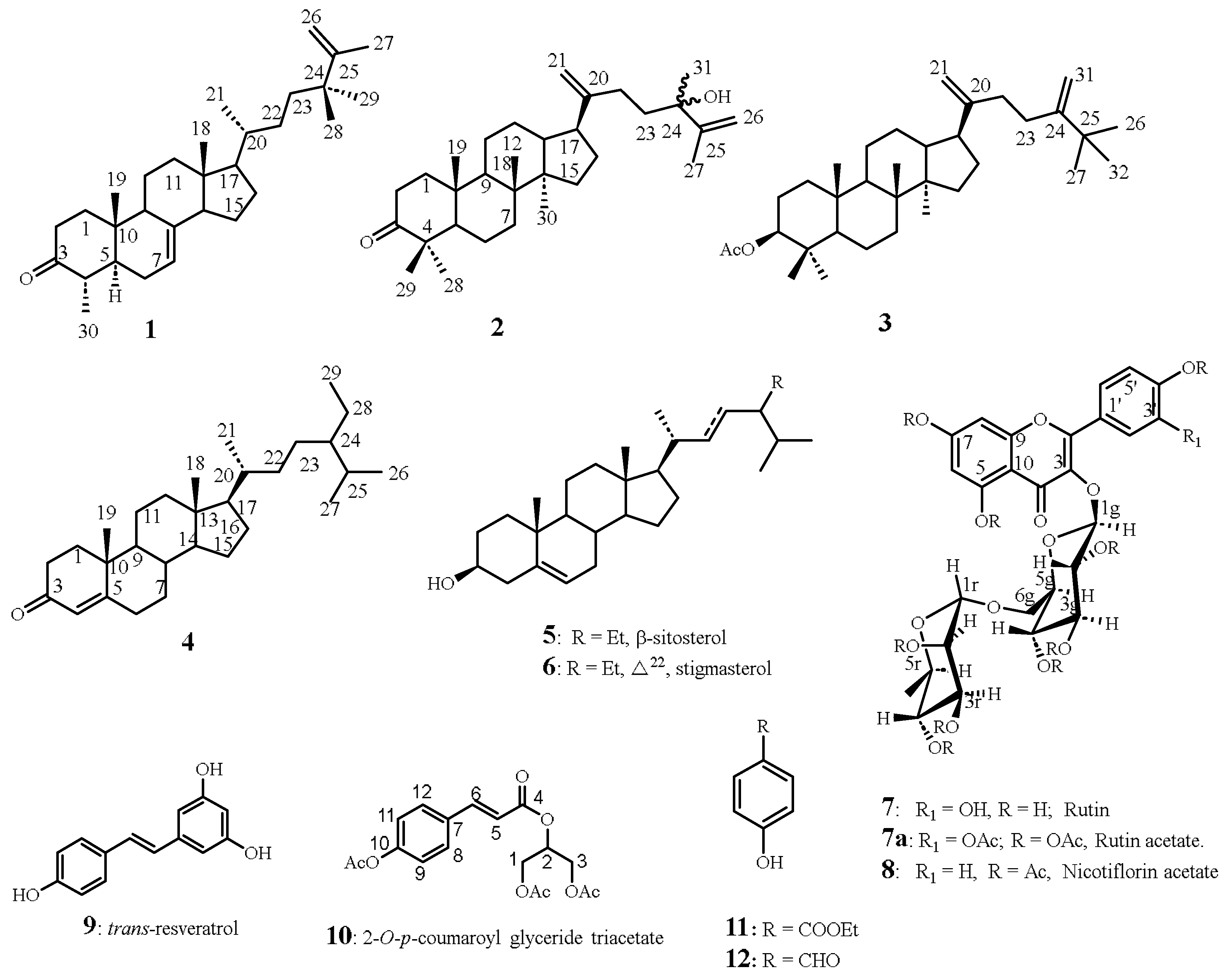
The aerial parts of Smilax canariensis Brouss., an endemic plant species of the Canary Islands and Madeira, were chemically investigated, resulting in the isolation of multiple known and novel compounds. These include three known flavonol glycosides: quercetin-3-O-rutinoside, rutin (7), quercetin-3-O-rutinoside decaacetate (7a), kaempferol-3-O-rutinoside nonaacetate, nicotiflorin acetate (8), 1-O-p-coumaroylglycerol triacetate (10) and trans-resveratrol (9). Additionally, a new sterol, 24,24- dimethy-5α-cholesta-7, 25-dien-3-one (1), and two novel dammarane–type triterpenes, 24-hydroxy-24-methyl-dammara-20,25-dien-3-one (2) and 3-acetyl-25-methyl-dammara-20,24-diene (3), were identified. In addition, stigmasterol, sitosterol and stigmast-4-en-3-one (4) were obtained. The structural elucidation of these compounds was achieved via 1D and 2D NMR spectroscopy, mass spectrometry, and comparison with literature data. This study provides the first phytochemical profile of S. canariensis and highlights its potential as a source of bioactive compounds for pharmacological applications.
Keywords:
Smilax canariensis Brouss.
; flavonol glycosides
; triterpenes
; sterols
1. Introduction
The genus Smilax (Liliaceae) comprises over 350 species distributed widely across tropical and temperate regions, with notable applications in traditional medicine, particularly in East Asia and North America [1]. Many of them have long been used as medicinal herbs, especially as traditional Chinese medicines in China [2], for their diuretic, laxative, depurative, and hypoglycemic properties, whereas only four species are found in Europe: Smilax aspera, located in the Mediterranean basin; Smilax azorica, native to the Azores Islands; Smilax excelsa, in the Black Sea and Caspian Sea region; and Smilax canariensis Brouss. ex Willd, endemic to the Canary Islands and Madeira, which is the subject of our study.
Morphologically, S. canariensis is characterized by leaves with 3 to 5 nerves and a simple umbel inflorescence. It is an evergreen, lauriform plant that is narrower at the ends of the older branches, and there are few or no thorns on the older stems. Notably, when a fruit ripens, it acquires a dark color.
Traditional medicine in the Canary Islands has extensively utilized endemic plants, including S. canariensis, commonly known as "zarzaparrilla sin espinas." This plant is well regarded for its diuretic, laxative, depurative, and hypoglycemic properties. Despite these traditional uses, the phytochemical composition of S. canariensis has not been extensively studied, presenting a significant opportunity to explore its chemical constituents and potential bioactivities [2]. This study aims to address this gap by isolating and characterizing the secondary metabolites present in the aerial parts of this species, thereby uncovering potential pharmacologically active compounds and contributing to the understanding of their chemical diversity. To the best of our knowledge, this plant has not previously been the subject of phytochemical analysis.
2. Results and Discussion
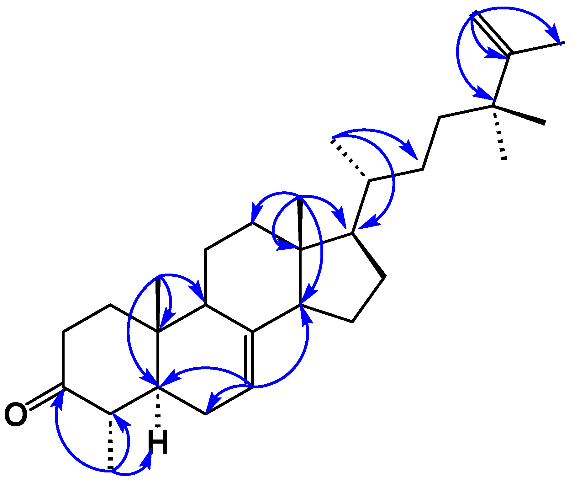
Compound 1 was obtained as colorless needles, mp 138–140 °C. Its molecular formula was determined to be C30H48O by its HRESIMS at m/z 447.3609 (calcd for [C30H48O+Na]+, 447.3603), together with its 13C NMR data. The1H NMR spectrum of 1 indicated two tertiary methyl protons at δH 0.59 and 0.80 ppm, one secondary methyl proton at δH 1.06 (d, J = 6.5 Hz) and an olefinic proton at δH 5.16 (br dd, J = 5.6 and 2.0 Hz), which are characteristic signals of H3-18, H3-19, H3-30 and H-7 of 1 having a lophenol ring system [3]. The carbon signals (Table 1) due to the ring systems of 1 were almost identical to those of 4 obtained after ketonization of 24(R)-ethyllophenol [4]. The presence of the ketone at C-3 was evident in the 13C NMR spectrum since it showed a carbonyl signal at δC 210.5, which presented cross-peaks between the protons at C-1, C-2, and C-4 and the methyl group at C-4 in the HMBC spectrum. The structure of 1 was further elucidated by analysis of its HMBC spectrum (Figure 2). The C-18 and C-19 methyl groups were positioned at the ring junctions C-13 and C-10 on the basis of the HMBC correlations of H3-18/C-12, C-13, C-14, and C-17 and H3-19/C-1, C-5, C-9, and C-10, respectively.
The HMBC correlations placed the side chain at C-17 of the lophenol skeleton (Figure 1). This was evidenced by the methine proton (δH 1.20, m, H-17) correlating with carbons at δC 28.6 (C-16), 31.3 (C-22), 43.9 (C-13), and 55.5 (C-14), along with δC 19.6 (C-21). Furthermore, the methyl protons H3-21 (δH 0.99, d, J = 6.5 Hz) were strongly correlated with δC 56.6 (C-17), 37.5 (C-20), and 31.3 (C-22), confirming the structure of the side chain. In addition, the 1H-NMR spectrum of 1 showed a vinyl methyl signal at δH 1.71 (3H, brs) and a terminal methylene proton at δH 4.85 and 4.88 (1H, each, brs, H-26a, b) due to an isopropenyl group and two tertiary methyl protons at δH 1.08 (6H, s, 28-H3, 29-H3). The cross peaks observed between the vinylic protons H-26 a, b and the carbon signals at δC 152.4 (C-25), 20.0 (C-27) and with the methyl protons at δH 1.08 (6H, s, 28-H3, 29-H3) and C-25, C-26 and C-23 were consistent with the placement of the extra two methyl groups at C-24. The NMR spectroscopic features of compound 1 were analogous to those of 4α,24,24-trimethyl-5α-cholesta-7,25-dien-3β-ol (24,24-dimethyl-25-dehydrolophenol) (1a), which was isolated as the acetyl derivative from the unsaponifiable lipid of Clerodendrum inerme [5], except that 1 had a keto carbon at δC 210.5, where 1a had 3β-acetate; hence, 1a was considered to have the structure 24,24- dimethy-5α-cholesta-7,25-dien-3-one.

Table 1.
1H and 13C NMR data of 24, 24-dimethyl-5α-cholesta-7, 25-dien-3-one –(1).
| Position | 1Ha | 1Hb | 13Ca | 13Cb |
| 1a | 2.05 m | 1.13 m |
39.8 |
39.8 |
| 1b | 1.25 m | 1,74 m | ||
| 2a | 2.48 td (14.3, 5.7) | 2.21 ddd (13.6, 3.9, 2.6) | 38.3 | 38.3 |
| 2b | 2.30 m | 2.09 td (13.6, 5.6) | ||
| 3 | 213.4 | 210.5 | ||
| 4 | 2.13 m | 1.88 m | 45.8 | 45.9 |
| 5 | 1.44 m | 1.73 m | 50.5 | 50.5 |
| 6a | 2.15 m | 1.92 m | 28.1 | 28.5 |
| 6b | 1.90 m | 1.54 m | ||
| 7 | 5.21 brs | 5.16 br dd (5.6, 2.0) | 117.4 | 118.4 |
| 8 | 139.4 | 139.3 | ||
| 9 | 1.75 m | 1.48 m | 49.5 | 49.8 |
| 10 | 35.4 | 35.5 | ||
| 11a | 1.61 m | 1.38 (2H, m) | 22.1 | 22.1 |
| 11b | 1.55 m | |||
| 12a | 2.16 m | 1.99 dt (12.5, 3.7) | 40.1 | 40.2 |
| 12b | 1.48 m | 1.13 m | ||
| 13 | 43.6 | 43.9 | ||
| 14 | 1.80 m | 1.73 m | 55.1 | 55.5 |
| 15a | 1.53 m | 1.55 m | 23.7 | 23.7 |
| 15b | 1.42 m | 1.43 m | ||
| 16a | 1.79 m | 1.90 m | 29.8 | 28.6 |
| 16b | 1.27 m | 1.28 m | ||
| 17 | 1.25 m | 1.20 m | 56.1 | 56.6 |
| 18 | 0.56 m | 0.59 s | 12.07 | 12.4 |
| 19 | 1.08 s | 0.80 s | 13.9 | 13.8 |
| 20 | 1.33 m | 1.33 m | 36.8 | 37.5 |
| 21 | 0.92 d (6.4) | 0.99 d (6.5) | 19.2 | 19.6 |
| 22a | 1.24 m | 1.33 m | 30.6 | 31.3 |
| 22b | 0.88 m | 0.98 m | ||
| 23a | 1.40 m | 1.45 m | 37.5 | 38.0 |
| 23b | 1.20 m | 1.23 m | ||
| 24 | 28.9 | 39.3 | ||
| 25 | 152.5 | 152.4 | ||
| 26a | 4.73 brs | 4.88 brs | 109.5 | 110.6 |
| 26b | 4.66 brs | 4.85brs | ||
| 27 | 1.69 brs | 1.71 brs | 19.6 | 20.0 |
| 28 | 1.01c | 1.06 d (6.5) | 11.6 | 12.5 |
| 29 | 1.01 s | 1.08 s | 27.7 | 27.8 |
| 30 | 1.02 s | 1.08 s | 27.4 | 28.1 |
| a CDCl3, 600, 150 MHz. b C6D6, 600, 150 MHz. c Partially hidden. | ||||
Figure 1.
Key HMBC correlations of compound 1.
Compound 2 was isolated as a viscous oil, and its molecular formula is C31H50O2, as deduced from its positive HRESIMS (found [M + Na]+ m/z 477.3706, calcd 477.3709) and 13C NMR data, which are indicative of seven indices of hydrogen deficiency. The 13C NMR (Table 2) data revealed 31 carbon resonances, which were classified by the HSQC spectrum as seven methyl, 12 methylene (two of them as terminal double bonds), four methine, and eight quaternary carbons, including two olefinic carbons at δC 153.8 (C-20) and 150.9 (C-25) and one carbonyl at δC 215.5 (C-3). Three of the implied six degrees of unsaturation were explained by multiple bonds, two by carbon‒carbon double bonds and one by a carbon‒oxygen double bond; consequently, 2 was a tetracyclic triterpene. Detailed analysis of the 13C NMR data confirmed that compound 2 belongs to the dammarane-type triterpene class, characterized by the presence of an additional methyl group. The 1H NMR spectrum exhibited seven distinct tertiary methyl signals at δH 0.70, 0.80, 1.64, 1.16, 0.99, 1.09, and 0.87 ppm (each integrating for three protons, singlets), which is consistent with this classification. These data corresponded to a dammarane triterpene skeleton similar to that of 24-methyldammara-20,25-dien-3-one isolated from Copernicia prunifera [6] and from Cissus quadrangularis [7]. The gross structure of 2 was deduced from its HMBC spectrum (Figure 2). The HMBC correlations between the tertiary methyl protons and their neighboring carbons were used to establish the dammarane backbone.
All expected correlations (Me-18→C-8, 7, 9, 14; Me-19→C-10, 1, 5, 9; and Me-30→C-14, 8, 13, 15) displayed strong cross-peaks. Furthermore, the HMBC spectrum placed the keto carbon at C-3 (Figure 2), from which the methyl proton at δH 0.99 (Me-29) correlated with the carbon at δC 28.7 (C-28), 47.6 (C-4), 55.5 (C-5) and 215.3 (C-3). The side chain was placed at C-17 of the main skeleton from the HMBC correlation of the methine proton at δH 2.31 (m, H-17), with the carbons at δC 29.9 (C-16), 29.5 (C-22), 46.2 (C-13), 40.9 (C-14), 108.1 (C-21) and 153.8 (C-20), and the terminal olefinic methylene protons at δH 4.97 brs and 4.93 brs (2H, H-21), with the carbons at δC 29.5 (C-22), 48.5 (C-17) and 153.8 (C-20). The HMBC experiments also confirmed the presence of a methyl group at δH 1.16 S, HSQC 28.7/28.68, and a methyl group on an oxygen-bearing carbon atom at C-24 (δC 75.38/75.32), since long-range couplings were observed between C-24 and the protons of the vinyl methyl group (H-27 at δH 1.651/1.639 brs), the terminal methylene (H-26a, δH 5.12 brd (1.3 Hz) and H-26b at δH 4.86 dd (2.7 and 1.3 Hz). The 13C NMR spectrum of compound 2 showed splitting of the signals of certain carbons (Table 2). The splitting of these carbon signals is consistent with the presence of a mixture of C-24 epimers [8]. On the basis of this combined evidence, the structure of 2 is defined as a 24-epimer mixture of 24-hydroxy-24-methyldammara-20,25-dien-3-one.
Figure 2.
Key HMBC correlations of compound 2.

Compound 3 was obtained as a white amorphous solid, mp (120–122 °C, hexanes). Its molecular formula was determined to be C34H56O2 on the basis of its HRESIMS peak at m/z 519.4174 [M+Na]+ (calcd for C34H56O2, 519.4178). Its 1H and 13C NMR (Table 2) spectra were very similar to those of 2. The structural difference between 3 and 2 was the functional group at C-3. The C-3 ketone in 2 was replaced by an acetoxy group in 3, which was evidenced by the lack of a carbonyl signal, while acetoxy group signals at δH 2.05 (3H, s) and δC 21.39, 171.12 were present in the NMR spectrum of 3. The 1H-1H COSY correlations between H-2 and H-3 (δH 4.49) and the HMBC correlations from H-3 to C-4 (δC 38.1) and the acetoxy carbonyl (δC 171.12) and from H3-28/H3-29 to C-3 (δC 80.9), C-4 (δC 38.1) and C-5 (δC 56.3) confirmed the above assignment (Figure 3). The large coupling constant of H-3 (dd, J =10.7, 3.0 Hz) suggested that the acetoxyl group at C-3 was β-oriented. The 1H NMR spectrum of 3 also showed side chain signals at δH 1.08 (9H, s, H-26, H-27 and H-32) and at δH 4.88 and 4.71 (each 1H, brs, H2-31). The side chain structure was corroborated through HMBC correlations, including signals from H-21a (δH 4.76, br s) and H-21b (δH 4.74, br s) with carbons at δC 17 and δC 20. The deshielded t-butyl singlet at δH 1.08 further indicated the presence of an additional methyl group attached to C-25 and linked to the double bond. These signals were consistent with a 24-methylene-25-methyl-side chain [9]. The combined data suggested that compound 3 was 25-methyldammara-20, 24-diene-3-β-yl-acetate.
Figure 3.
Key HMBC correlations of compound 3.
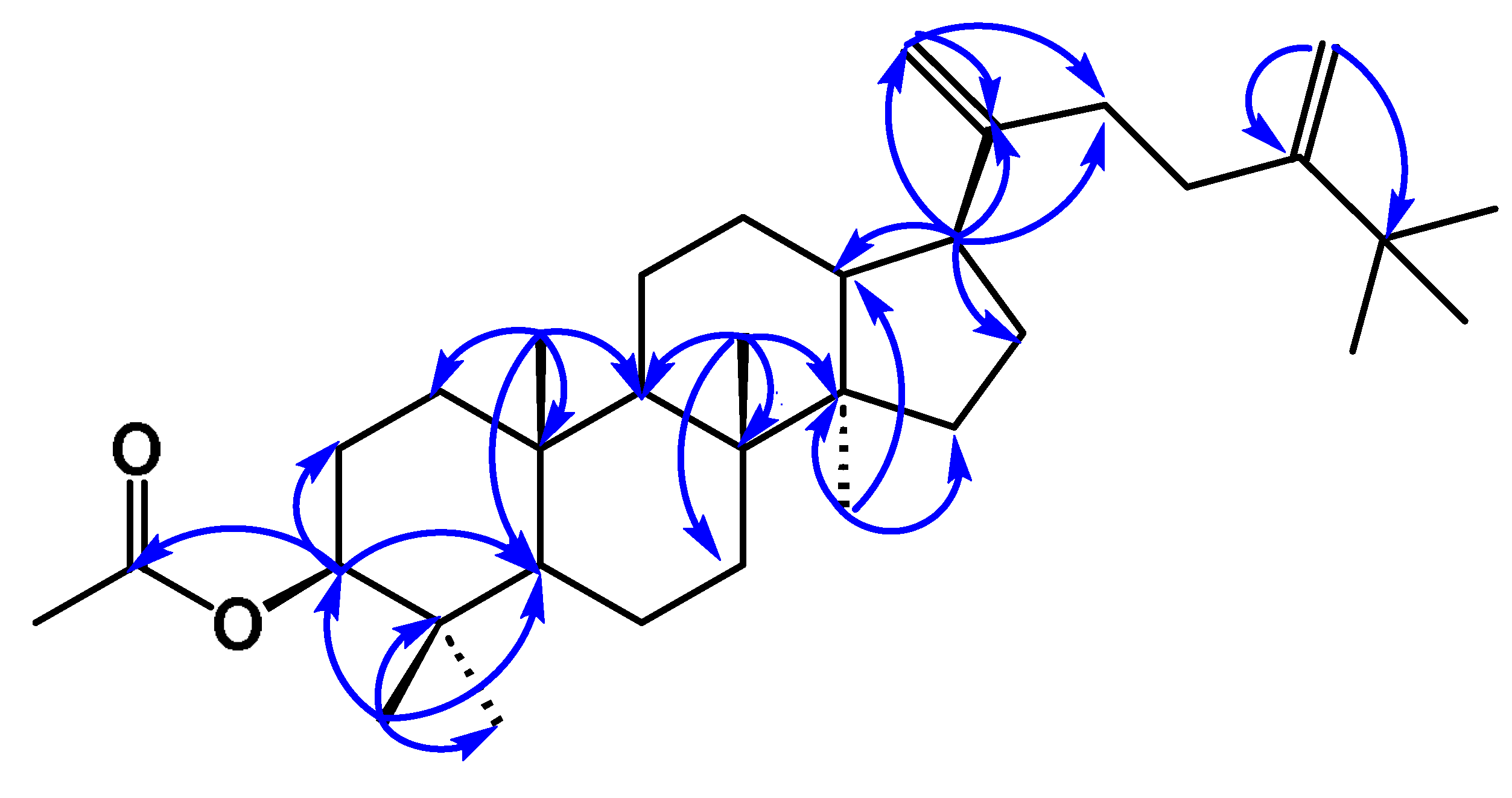
Table 2.
1H (600 MHz) and 13C (150 MHz) NMR data of 24-hydroxy-24-methyl-dammara-20, 25-dien-3-one 2 and 3-acetyl-25-methyl-dammara-20, 24-diene 3, in CDCl3.
Table 2.
1H (600 MHz) and 13C (150 MHz) NMR data of 24-hydroxy-24-methyl-dammara-20, 25-dien-3-one 2 and 3-acetyl-25-methyl-dammara-20, 24-diene 3, in CDCl3.
| δH (J values are given in Hz) δC | ||||
| atom/position | (2) | (3) | (2) | (3) |
| H-1a | 1.87 m | 1.71 m |
40.1 |
38.7 |
| H-1b | 0.95 m | 1.06 m | ||
| H-2a | 2.27 m | 1.64 m |
34.4 |
23.9 |
| H-2b | 2.23 m | 1.64 m | ||
| H-3 | C-3 | 4.49 d d (10.7, 5.5) | 215.5 | 80.9 |
| 47.6 | 38.1 | |||
| H-5 | 1.10 m | 0.85 m | 55.6 | 56.3 |
| H-6a | 1.31 m | 1.51 m |
20.1 |
18.1 |
| H-6b | 1.22 m | 1.46 t d (12.8, 3.0) | ||
| H-7a | 1.40 m | 1.59 m |
35.3 |
35.4 |
| H-7b | 1.14 m | 1.28 m | ||
| 40.7 | 40.5 | |||
| H-9 | 1.15 m | 1.33 dd (12.3, 3.0) | 50.7 | 50.9 |
| 37.2 | 37.1 | |||
| H-11a | 1.17 m | 1.52 m |
22.35/22.33a |
21.3 |
| H-11a | 1.05 m | 1.20 m | ||
| H-12a | 1.56 (2H, m) | 1.58 m |
25.6 |
25.0 |
| H-12b | 1.56 (2H, m) | 1.08 m | ||
| H-13 | 1.79 m | 1.67 m | 46.26/46.07a | 45.6 |
| 49.9 | 49.5 | |||
| H15a | 1.60 m | 1.60 m |
31.96/31.94a |
31.3 |
| H-15b | 1.08 m | 1.11 m | ||
| H-16a | 1.99 m | 1.93 m |
29.9/29.8a |
29.2 |
| H16b | 1.57 m | 1.41 m | ||
| H-17 | 2.31 m | 2.23 t d (10.6, 6.8) | 48.58/48.47a | 48.06 |
| Me 18 | 0.80 brs | 0.98 s | 16.3 | 15.82 |
| Me-19 | 0.70 s | 0.87 s | 16.4 | 16.3 |
| 153.86/153.81a | 153.7 | |||
| H-21a | 4.97 brs | 4.76 brs |
108.12/108.05a |
107.61 |
| H-21b | 4.93 m | 4.74 brs | ||
| H-22a | 2.23 m | 2.12 m |
29.56/29.55a |
34.1 |
| H-22b | 2.07 m | 2.12 m | ||
| H-23a | 1.77 (2H, m) | 2.18 m |
39.7/40.0a |
30.3 |
| H-23b | 1.77 (2H, m) | 2.18 m | ||
| 75.38/75.32a | 158.3 | |||
| 150.9 | 36.3 | |||
| H-26a | 5.12 brd (1.3) | 1.08 s |
110.4 |
29.5 |
| H-26b | 4.86 dd (2.7, 1.3) | |||
| Me-27 | 1.651/1.639 brs | 1.08 s | 19.95 | 29.5 |
| Me-28 | 1.16 s | 0.86 s | 28.71/28.68a | 28.2 |
| Me-29 | 0.99 s | 0.85 s | 21.5 | 16.5 |
| Me-30 | 1.09 s | 0.87 s | 27.1 | 15.9 |
| Me-31 | 1.16 s | 4.88 br s H31a | 28.7/28.68a |
106.14 |
| 4.71 brs H31b | ||||
| Me-32 | 1.08 s | 29.5 | ||
| Ac | 2.05 (3H, s) | 21.39, 171.12 | ||
a Value on the right corresponds to the minor isomer.
This study represents the first comprehensive phytochemical investigation of Smilax canariensis, leading to the identification of novel triterpenes and sterols, as well as several known compounds. These findings underscore the chemical and pharmacological potential of this endemic species, paving the way for further studies on its bioactive properties.
3. Experimental
3.1. General
Melting points were measured on a Reichert Thermovar apparatus without correction. Optical rotations were recorded on a Perkin Elmer 2H polarimeter equipped with a 1 dm cell. NMR spectra (1H and 13C) were obtained via Bruker Advance II 500 and Bruker Advance III 600 spectrometers in CDCl3, C6D6, CD3COCD3, and C5D5N, with residual solvent signals serving as internal references, (δH 7.26; δC 76.7), (δH 7.16; δC 128.39), (δH 2.05, δC 29.92), and (δH 8.74, 7.58, 7.22; δC 150.35, 135.91, 123.87), respectively. The pulse conditions for 1D and 2D NMR were as follows [10]. Mass spectrometry (ESI-TOF and EI-MS) was performed using a Micromass LCT Premier XE and a Micromass Autospec instrument at 70 eV, respectively. Column chromatography was performed on Amberlite XAD 2 (Supelco XAD -2-3019), Sephadex LH-20 Pharmacia (ref. 17-0090-01), silica gel (Merck 2300-400 mesh), octadecyl-functionalized silica gel (Aldrich 377635-1006) and analytical TLC Merck Kieselget 60 F254. HPLC separations were carried out on a JASCO Pu-980 series pumping system equipped with a JASCO UV-975 detector and a Waters Kromasil Si 5 mm (10 × 250 mm) column. A Mackerey-Nagel VP 250/10 nucleodur Sphinx RP 5 µm column was used for HPLC‒RP chromatography; chromatograms were visualized under UV light at 255 and 366 nm and/or sprayed with oleum followed by heating. All the solvents were distilled before use. For acetylations, dry phenolic material was dissolved in the minimum volume of pyridine. Two times the amount of acetic anhydride was added, and the mixture was left to stand overnight at ambient temperature. The mixture was then diluted with H2O and extracted three times with ethyl acetate. The organic phase was evaporated at reduced pressure, and the residue was further purified by HPLC (SiO2 column) using EtOAc-hexane as the eluent. NMR spectra are given in the Supplementary Material (Figures S1-S19).
3.2. Plant Material
The aerial parts of Smilax canariensis Brouss. Ex Willd were collected in April 2011 from Las Nieves (Velhoco) and Santa Cruz de La Palma (UTM coordinates: 228115/3177048). The plant was identified by Prof. Pedro Luis Pérez de Paz from the Department of Botany, Faculty of Pharmacy, University of La Laguna, where a voucher specimen (TFC: 49.941) has been deposited.
3.3. Extraction and Isolation of the Constituents
The fresh aerial parts of Smilax canariensis (7.5 kg) were finely divided and subjected to exhaustive extraction with ethanol at ambient temperature for two weeks. The resulting extract was filtered and concentrated under reduced pressure, yielding 240 g of brown residue. This residue was dissolved in two liters of distilled water and extracted with dichloromethane to yield 30.4 g of residue, followed by n-butanol extraction, which yielded 11 g of residue. The remaining aqueous layer was concentrated to yield 125 g of residue.
The dichloromethane extract (30.4 g) of S. canariensis was adsorbed onto silica gel and subjected to column chromatography on silica gel (Merck, 230–400 mesh, 300 g). The column was eluted with stepwise gradients of n-hexane and ethyl acetate, yielding 109 fractions (600 mL each). The evolution of this chromatography system was followed by the addition of CCF. Fractions SH16–17 provided a white solid (24,24-dimethyl-5α-cholesta-7,25-dien-3-one) upon recrystallization in methanol, whereas subfractions SH51–55 yielded a mixture of β-sitosterol and stigmasterol. Fractions SH1-15 (1.5 g) were eluted with pure n-hexane, which consists of a complex mixture of essential oils, and were not investigated further. Frs SH16-17 were purified by precipitation in MeOH, resulting in a white solid, which was identified as 24,24- dimethy-5α-cholesta-7,25-dien-3-one 1. Fractions SH39-41 (152 mg) were purified via column chromatography over Sephadex LH-20 (hex:CH2Cl2:MeOH; 2:1:1) and an SHT column. Subfraction SHT5-6 was rechromatographed over a SiO2 column and used as an eluent mixture of benzene–EtOAc, totaling 18 fractions, 10 mL each. Subfractions 8-9 yielded 17 mg of Stigmast-4-en-3-one 4, which showed significant hypoglycemic activity [12].
Subfraction SHT8-9 (28 mg) was further purified on Sephadex LH-20 (hex:CH2Cl2:MeOH; 2:1:1) to yield 12 mg of 25-methyldammara-20, 24-diene-3-β-yl-acetate 3. Fractions SH51-55 were eluted with a 7:3 hexane-EtOAc mixture, yielding 218 mg of colorless crystals with a melting point of 125 °C in acetone (lit. mp 130 °C) [11], identified as a 3:1 mixture (for the integral) of β-sitosterol 5 and stigmasterol 6, respectively.
Fras SH58-67 (235 mg) were chromatographed on Sephadex LH-20 (hex:CH2Cl2:MeOH; 2:1:1) columns, and fractions of 15 mL each were collected. A total of 35 fractions were collected, which, according to their behavior in the CCF, were regrouped into SHS11-12 (25 mg) and SHS20-22 (14 mg). Subsequent purification of SHS20-22 by HPLC (hexane:ethyl acetate 9:1) allowed us to obtain the majority of the products (Tr = 40 min, flow rate of 2 mL min-1), identified as ethyl p-hydroxybenzoate 11. Subfractions SHS11-12 were obtained after chromatography via high-performance liquid chromatography (HPLC) (SiO2, EtOAc-Hex 8:2 flow ratio of 2 mL min-1, Tr 22 min), 12 mg of 24-hydroxy-24-methyl-dammara-20, 25-dien-3-one (2).
Fras SH89-97 (337 mg) were chromatographed on Sephadex LH-20 (hex:CH2Cl2:MeOH; 2:1:1, SHY column), and Subfracs SHY20-23 yielded 34 mg of a substance with strong UV absorption (Rf = 0.32, hexane: ethyl acetate 20%) at the melting point (114–116 °C, MeOH). This compound was identified from its physical and spectroscopic data as p-hydroxybenzaldehyde (12).
A 5.5 g portion of the n-butanol extract was subjected to low-pressure chromatography on an RP C-18 column (7 × 40 cm) with a linear gradient of 40,500 ml H2O–MeOH mixtures beginning with H2O and ending with MeOH and an SM column. (Frs. SM20-24, 740 mg) were rechromographed on Sephadex LH-20 (CH2Cl2:MeOH; 1:1, Sph column). Frs SpH8-9, after precipitation in methanol/dichloromethane, yielded 16.5 mg of trans-resveratrol (9). Frs. SpH (22.5 mg) was purified by precipitation in a methanol‒ethyl acetate mixture to yield rutin (7) (7.1 mg), and the mother liquor, which was composed of a complex mixture of flavonoids, was acetylated with acetic anhydride in pyridine for 12 h. After elimination of the solvent in vacuo, 12.5 mg of the crude product, which was rechromatographyed via HPLC (SiO2, EtOAc‒Hex 7:3, flow 2 mL min-1), was obtained. Three pure products were obtained: quercetin-3-O-rutinose decaacetate (rutin acetate, 7a, 3.7 mg, Tr: 20.2 min), kaempferol-3-O-rutinose nonaacetate (nicotiflorin acetate, 8, 3.4 mg, Tr: 17.6 min) and 1-O-p-coumaroylglycerol triacetate (10), 2.8 mg (Tr: 9.76 min).
Chemical structures of all identified products are provided in Figure 4.
3.3.1. 24,24-Dimethyl-5α-cholesta-7,25-dien-3-one (1)
Colorless needles, mp 138–140 °C, MeOH. 1H 13C NMR (Table 1), HREI-MS, m/z 447.3610, (calcd for C30H48O, 447.3603) [M+Na]+ (100%).
3.3.2. 24-Hydroxy-24-methyl-dammara-20,25-dien-3-one (2)
Viscous oil, 1H 13C NMR (Table 2), HRESIMS, m/z 477.3706, (calcd for C31H50O2, 477.3709) [M+Na]+.
3.3.3. 25-Methyldammara-20,24-diene-3-β-yl-acetate (3)
Amorphous white solid, mp 120–122 °C, hexanes. 1H 13C NMR (Table 2), HRESIMS, m/z 519.4174, (calcd for C34H56O2, 519.4178) [M+Na]+.
3.3.4. Stigmast-4-en-3-one (4)
Amorphous solid, mp 77–80 °C (lit. mp 87–88 °C) [12], and for the 1H 13C NMR data, see Table S1 in the Supplementary Material. HREIMS mass spectrometry, showed the molecular ion peak at m/z 435.3592, (calcd for C29H48O, 435.3603) [M+Na]+.
3.3.5. Quercetin-3-O-rutinoside, Rutin (7)
Compound 7 was isolated as a yellow solid with a melting point of 240 °C and presented a molecular formula of C27H30O16, which was determined by high-resolution high-resolution high-efficiency high-efficiency high-efficiency high-energy high-energy mass spectrometry (HREIMS) mass spectrometry (HRMS), with a molecular ion peak at m/z 609.1460 (calcd for C27H29O16, 609.1456) [M-H]+. Peracetylation of 7 afforded the known rutin decaacetate 7a; both were identified by MS and 1H and 13C NMR spectrometry (Kazuma et al., 2003; de Alcantara et al., 2023) [13,14].
3.3.6. Kaempherol-3-O-rutinosidenonaacetate, nicotiflorin acetate (8).
The molecular formula of C45H48O24 was determined by high-resolution high-resolution high-energy ion mass spectrometry (HREIMS) mass spectrometry, which revealed a molecular ion peak at m/z 995.2460 (calcd for C45H48O24+Na, 995.2433) [M+Na]+. 1H and 13C NMR spectra were obtained (Jayasinghe et al., 2004) [15].
3.3.7. Trans-resveratrol (3,4′,5-trihydroxystilbene) (9).
Compound 9 was isolated as a yellow solid with a melting point of 254 °C (lit. mp 265–268 °C) [16]. The molecular formula C14H12O3 was determined by high-resolution high-resolution high-energy ion mass spectrometry (HREIMS) mass spectrometry, which revealed a molecular ion peak at m/z 227.0711 (100%) (calcd for C14H11O3, 227.0708) [M-H]+. 1H and 13C NMR spectra were obtained (Šmidrkal et al., 2010) [16].
3.3.8. 2-O-p-Coumaroylglycerol triacetate (Juncusyl ester B triacetate) (10).
The molecular formula C18H20O8 was determined by high-resolution high-resolution high-energy ion mass spectrometry (HREIMS) mass spectrometry, which revealed a molecular ion peak at m/z 387.1061 (100%) (calcd for C18H20O8+Na, 387.1056) [M+Na]+. 1H NMR (C6D6, 500 MHz) δ: 4.33 (1H, dd, J = 12.06, 4.02 Hz, H1a), 4.25 (1H, dd, J = 12.06, 4.16 Hz, H1b), 5.38 (1H, dddd, J = 6.03, 5.89, 4.16 and 4.02 Hz, H2), 4.20 (1H, dd, J = 11.92, 6.03 Hz, H3a), 4.05 (1H, dd, J = 11.92, 5.89 Hz, H3b), 6.39 (2H, d, J = 8.5 Hz, H8,12), 6.11 (2H, d, J = 8.5 Hz, H9,11), 1.70, 1.68, 1.62 (each 3H, s, Ac). 13C NMR (C6D6, 125 MHz) δ: 62.97 C-1, 69.97 C-2, 62.82 C-3, 166.4 C-4, 118.11 C-5, 144.96 C-6, 132.29 C-7, 129.82 C-8, 12, 122.64 C-9, 11, 152.9 C-10, (20.81, 168.41; 20.81, 170.15; 20.49, 169.99) 3Ac. The signal assignments of H-1 and C-1 can be interchanged with those of H-3 and C-3.
3.3.9. Ethyl p-hydroxybenzoate (11).
The molecular formula C9H9O3 was determined by high-resolution high-resolution high-energy ion mass spectrometry (HREIMS) mass spectrometry, which revealed a molecular ion peak at m/z 165.0548 (100%) (calcd for C9H9O3, 165.0552) [M]+. 1H and 13C NMR spectra were obtained (Fier et al., 2016) [17].
3.3.10. p-Hydroxybenzaldehyde (12).
Compound 12 was isolated as an amorphous solid with a melting point of 114–116 °C in MeOH (lit. mp 115–116 °C (from ethanol) [18]. The molecular formula C7H5O2 was determined by high-resolution high-resolution high-energy ion mass spectrometry (HREIMS) mass spectrometry, which revealed a molecular ion peak at m/z 121.0293 (100%) (calcd for C7H5O2, 121.0290) [M-H]+. 1H and 13C NMR spectra were obtained (Kashparova et al., 2017) [18].
References
- Shao, B., Guo, H.Z., Cui, Y.J., Ye, M., Han, J., Guo, D.A. Steroidal saponins from Smilax China and their anti-inflammatory activities. Phytochemistry, 2007, 68, 623–630. [CrossRef]
- Abdala, S., Martin-Herrera, D., Benjumea, D., Pérez-Paz, P. Diuretic activity of Smilax canariensis, an endemic Canary Island species. Journal of Ethnopharmacology, 2008, 119, 12–16. [CrossRef]
- Toshihiro Itoh, Tsutomu Tamura, Masakazu Sagawa, Toshitake Tamura, Taro Matsumoto, 1980. 4α-Methyl-5α-cholest-8(14)-en-3β-ol from the seeds of Capsicum annuum. Phytochemistry, 1983, 22, 11, 2621−2622. [CrossRef]
- Toshihiro Itoh, Tsutomu Tamura, Masakazu Sagawa, Toshitake Tamura, Taro Matsumoto. 4(R)-ethyllophenol from Solanum melongena seeds. Phytochemistry, 1980, 19, 11, 2491−2492. [CrossRef]
- Toshihiro Akihisa, Parthasarathi Ghosh, Swapnadip Thakur, Hiroshi Nagata, Toshitake Tamura, Taro Matsumoto. 24,24-Dimethyl-25-dehydrolophenol, a 4α-methylsterol from Clerodendrum inerme. Phytochemistry, 1990, 29, 5, 1639−1641. [CrossRef]
- Buana C. de Almeida, Bruno Q. Araújo, Elcio D. S. Barros,a Sâmya D. L. Freitas, Dayany S. A. Maciel,b Ari J. S. Ferreira, Rafael C. Guadagnin, Gerardo M. Vieira Júnior, João H. G. Lagob, and Mariana H. Chaves. Dammarane Triterpenoids from Carnauba, Copernicia prunifera (Miller) H. E. Moore (Arecaceae), Wax. Journal of the Brazilian Chemical Society, 2017, 28, 8, 1371−1376. [CrossRef]
- Thanika Pathomwichaiwat, Pannee Ochareon, Noppamas Soonthomchareonnon, Zulfiqar Ali, Ikhlas A. Khan, Sompop Prathanturarug. Alkaline phosphatase activity-guided isolation of active compounds and new dammarane-type triterpenes from Cissus quadrangularis hexane extract. Journal of Ethnopharmacology, 2015, 160, 52−60. [CrossRef]
- V. Anjaneyulu, G. Sambasiva Rao, J. D. Connolly. Occurrence of 24-epimers of cycloart-25-ene-3-β, 24-diols in the stems of Euphorbia trigona. Phytochemistry, 1985, 24, 1610−1612. [CrossRef]
- Seongki Kim, Toshihiroakihisa, Toshitaketamura, Taro Matsumoto,Takaoyokota, Nobutaka. 24-Methylene-25-methylcholesterol in Phaseolus vulgaris seed: structural relation to brassinosteroids. Phytochemistry, 1988, 27, 2, 629−631. [CrossRef]
- Jesús G. Díaz, Chemical composition of Hypericum Coadunatum Chr. from the Canary Islands. Journal of Molecular Structure, 2022, 1248, 15, 131447. [CrossRef]
- P.C. Kiprono, F. Kaberia, J.M. Keriko, J.N. Karanja. The in vitro anti-fungal and antibacterial activities of beta-sitosterol from Senecio lyratus (Asteraceae). Zeitschrift für Naturforschung, 2000, 55c, 485−488.
- Fathaiya Jamaluddin, Sohaila Mohamed and Md Nordin Lajisb. Hypoglycemic effect of Stigmast-4-en-3-one, from Parkia speciosa empty pods. Food Chemistry, 1995, 54, 1, 9−13. [CrossRef]
- Kohei Kazuma, Naonobu Noda, Masahiko Suzuki. Malonylated flavonol glycosides from the petals of Clitoria ternatea. Phytochemistry, 2003, 62, 229–237. [CrossRef]
- De Alcantara Pinto, Douglas Chaves; Pitasse-Santos, Paulo; de Souza, Gabriela Alves; Castro, Rosane Nora; Freire de Lima, Marco Edilson. Peracetylation of polyphenols under rapid and mild reaction conditions. Natural Product Research, 2023, 37, 2279−2284. [CrossRef]
- U.L.B. Jayasinghe, B.A.I.S. Balasooriya, A.G.D. Bandara & Y. Fujimoto, Glycosides from Grewia damine and Filicium decipiens. Natural Product Research: Formerly Natural Product Letters, 2004, 18, 6, 499−502. [CrossRef]
- Šmidrkal, J., Harmatha, J., Buděšínský, M., Vokáč, K., Zídek, Z., Kmoníčková, E., Merkl R., Filip, V. Modified approach for preparing (E)-stilbenes related to resveratrol and evaluating their potential immunobiological effects. Collection of Czechoslovak Chemical Communications, 2010, 75, 2, 175–186. [CrossRef]
- Fier, Patrick S.; Maloney, Kevin M. Direct conversion of haloarenes to phenols under mild, transition-metal-free conditions. Organic Letters, 2016, 18, 2244−2247.. [CrossRef]
- Kashparova, Vera P.; Klushin, Victor A.; Zhukova, Irina Yu.; Kashparov, Igor S.; Chernysheva, Daria V.; Il'chibaeva, Irina B.; Smirnova, Nina V.; Kagan, Efim Sh.; Chernyshev, Victor M. A TEMPO-like nitroxide combined with an alkyl-substituted pyridine: An efficient catalytic system for the selective oxidation of alcohols with iodine. Tetrahedron Letters, 2017, 58, 36, 3517−3521. [CrossRef]
Disclaimer/Publisher’s Note: The statements, opinions and data contained in all publications are solely those of the individual author(s) and contributor(s) and not of MDPI and/or the editor(s). MDPI and/or the editor(s) disclaim responsibility for any injury to people or property resulting from any ideas, methods, instructions or products referred to in the content. |
© 2025 by the authors. Licensee MDPI, Basel, Switzerland. This article is an open access article distributed under the terms and conditions of the Creative Commons Attribution (CC BY) license (http://creativecommons.org/licenses/by/4.0/).
Copyright: This open access article is published under a Creative Commons CC BY 4.0 license, which permit the free download, distribution, and reuse, provided that the author and preprint are cited in any reuse.


