Submitted:
22 January 2025
Posted:
22 January 2025
You are already at the latest version
Abstract
Hip dysplasia (HD) is a prevalent disease in medium to large-breed dogs, characterized by joint laxity and degenerative joint changes. Early diagnosis of HD poses significant challenges, as radiographic imaging often identifies the disease only in advanced stages. Conversely, ultrasonography, a non-invasive and cost-effective imaging modality, offers the potential for earlier detection by evaluating the surrounding soft tissues and synovial changes. This study aimed to assess the relationship between the ventral hip ultrasonographic findings, and hip joint laxity evaluated through stress radiographs on 22 young Estrela Mountain dogs (n=44 hips) aged 4 to 8 months. Key ultrasound measurements included synovial fluid in the cranial femoral neck recess (CFNR) and capsular-synovial fold thickness (CFT). Radiographic laxity was estimated by measuring the distraction index (DI). The mean ± standard deviation of the CFNR area, CFT, and DI was 45.58±25.40 mm², 3.21±0.90 mm, and 0.40±0.10, respectively. The Pearson correlation coefficient was statistically significant between all these variables (P<0.05). The ventral ultrasonographic approach to hip joint revealed potential, considering the early diagnosis of HD in dogs, by showing relationships between changes in periarticular soft tissues and joint laxity. Further studies are needed to associate ultrasonographic findings with radiographic signs of HD and related clinical signs in dogs.
Keywords:
1. Introduction
2. Materials and Methods
2.1. Animals
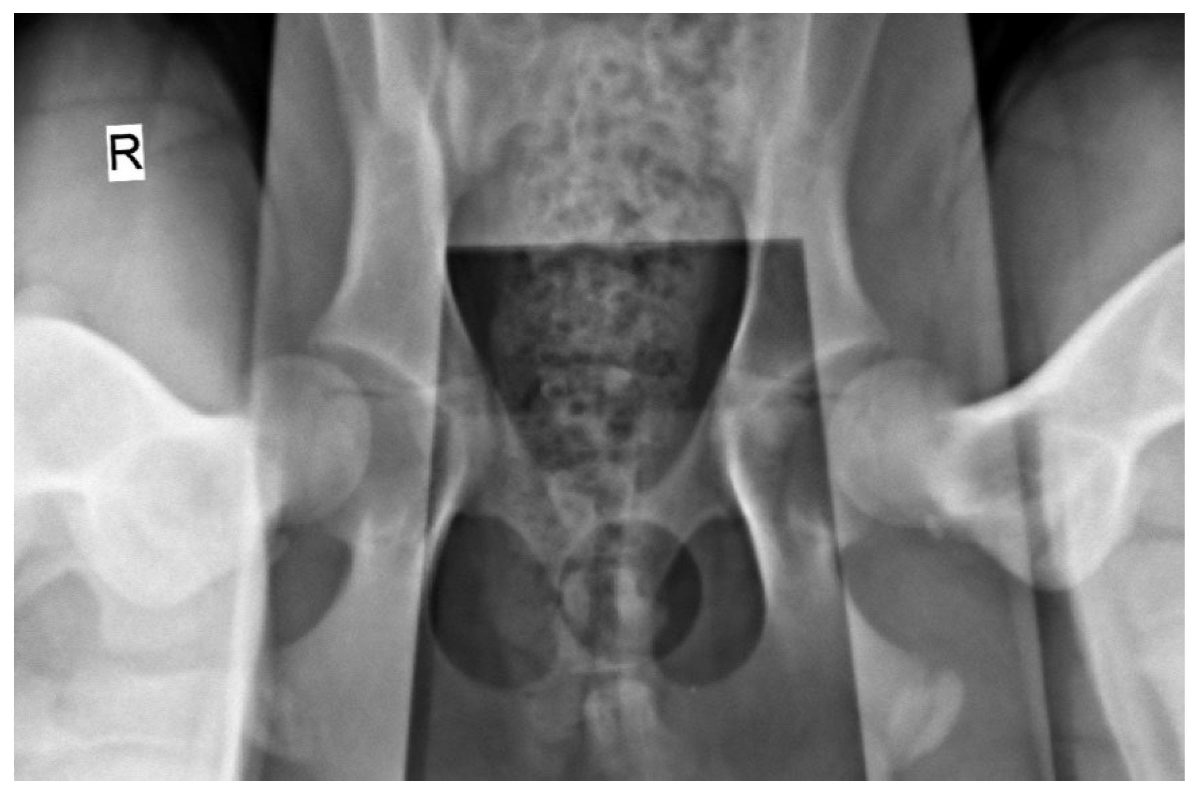
2.2. Radiographic Hip Stress View and Hip Laxity Measurement
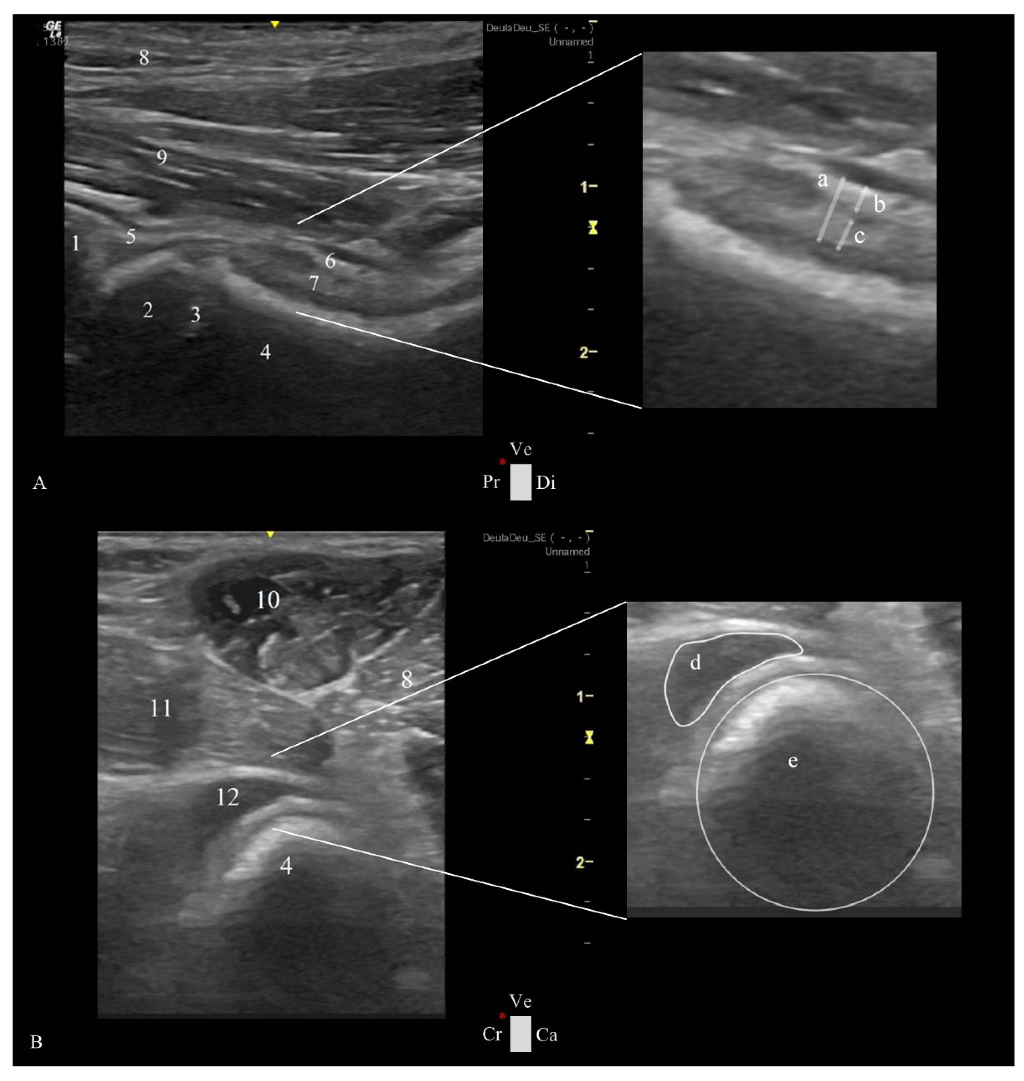
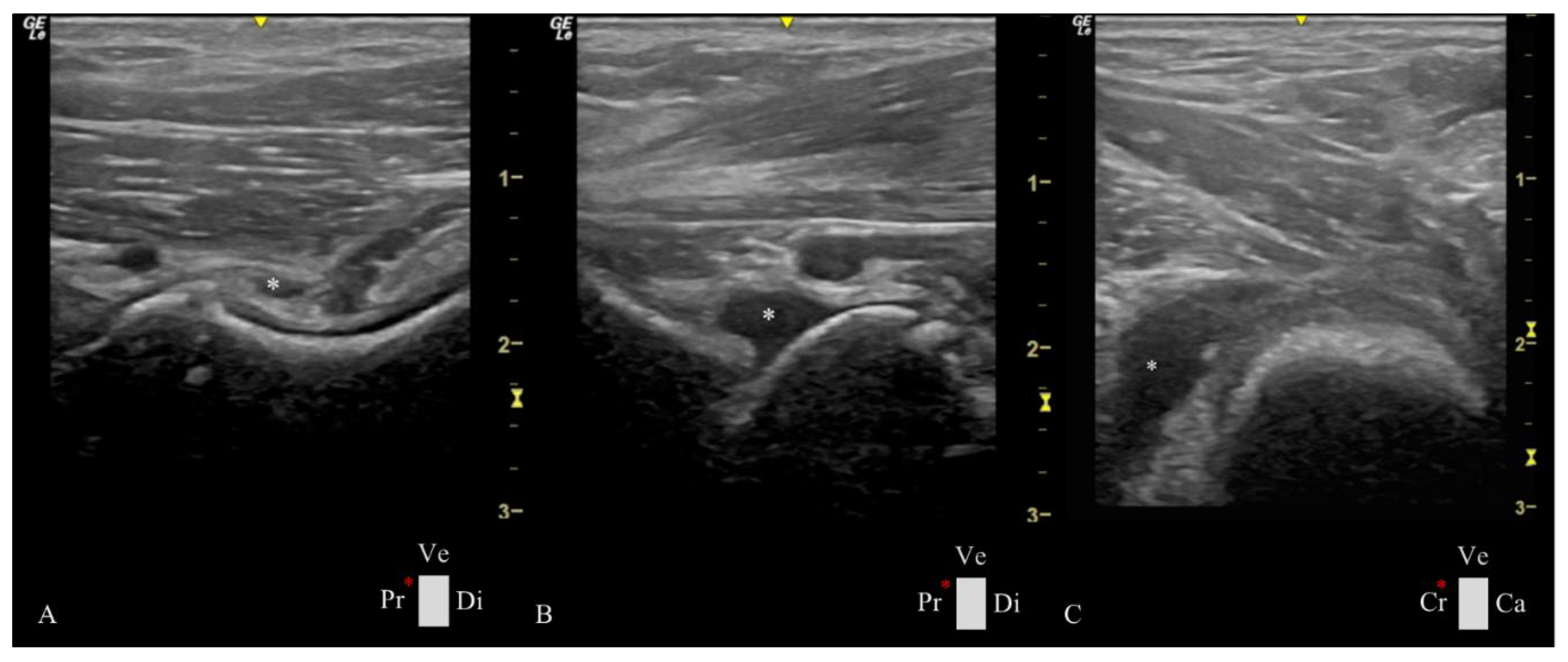
2.3. Ultrasonographic Ventral Hip Joint Approach
- − Longitudinal Femoral Head-Neck Plane
- − Transverse Femoral Head-Neck Plane
2.4. Ultrasonographic Hip Joint Measurements
2.5. Statistical Analysis
3. Results
4. Discussion
5. Conclusion
Author Contributions
Funding
Institutional Review Board Statement
Informed Consent Statement
Conflicts of Interest
Abbreviations
| HD | Hip Dysplasia |
| CFNR | Cranial Femoral Neck Recess |
| CFT | Capsular-Synovial Fold Thickness |
| DI | Distraction Index |
| SD | Standard Deviation |
| r | Pearson Correlation |
References
- Weigel, J.P.; Wasserman, J.F. Biomechanics of the Normal and Abnormal Hip Joint. Vet Clin North Am Small Anim Pract 1992, 22, 513–528. [Google Scholar] [CrossRef]
- Tomé, I.; Alves-Pimenta, S.; Sargo, R.; Pereira, J.; Colaço, B.; Brancal, H.; Costa, L.; Ginja, M. Mechanical Osteoarthritis of the Hip in a One Medicine Concept: A Narrative Review. BMC Vet Res 2023, 19. [Google Scholar] [CrossRef]
- Ginja, M.M.D.; Ferreira, A.J.; Jesus, S.S.; Melo-Pinto, P.; Bulas-Cruz, J.; Orden, M.A.; San-Roman, F.; Llorens-Pena, M.P.; Gonzalo-Orden, J.M. Comparison of Clinical, Radiographic, Computed Tomographic and Magnetic Resonance Imaging Methods for Early Prediction of Canine Hip Laxity and Dysplasia. Vet Radiol Ultrasound 2009, 50, 135–143. [Google Scholar] [CrossRef] [PubMed]
- Li, F.; Tang, Y.; Song, B.; Yu, M.; Li, Q.; Zhang, C.; Hou, J.; Yang, R. Nomenclature Clarification: Synovial Fibroblasts and Synovial Mesenchymal Stem Cells. Stem Cell Res Ther 2019, 10, 260. [Google Scholar] [CrossRef]
- Smith, G.K.; Biery, D.N.; Gregor, T.P. New Concepts of Coxofemoral Joint Stability and the Development of a Clinical Stress-Radiographic Method for Quantitating Hip Joint Laxity in the Dog. J Am Vet Med Assoc 1990, 196, 59–70. [Google Scholar] [CrossRef] [PubMed]
- Willemsen, K.; Möring, M.M.; Harlianto, N.I.; Tryfonidou, M.A.; van der Wal, B.C.H.; Weinans, H.; Meij, B.P.; Sakkers, R.J.B. Comparing Hip Dysplasia in Dogs and Humans: A Review. Front Vet Sci 2021, 8. [Google Scholar] [CrossRef] [PubMed]
- Roberts, T.; McGreevy, P.D. Selection for Breed-Specific Long-Bodied Phenotypes Is Associated with Increased Expression of Canine Hip Dysplasia. Vet J 2010, 183, 266–272. [Google Scholar] [CrossRef]
- Ohlerth, S.; Geiser, B.; Flückiger, M.; Geissbühler, U. Prevalence of Canine Hip Dysplasia in Switzerland Between 1995 and 2016—A Retrospective Study in 5 Common Large Breeds. Front Vet Sci 2019, 6. [Google Scholar] [CrossRef]
- Henrigson, B.; Norberg, I.; Olssons, S.-E. On the Etiology and Pathogenesis of Hip Dysplasia: A Comparative Review. J Small Anim Pract 1966, 7, 673–688. [Google Scholar] [CrossRef]
- Lopez, M.; Schachner, E. Diagnosis, Prevention, and Management of Canine Hip Dysplasia: A Review. Vet Med-Res Rep 2015, 181. [Google Scholar] [CrossRef]
- Riser, W. The Dysplastic Hip Joint: Radiologic and Histologic Development. Vet Pathol 1975, 12, 279–305. [Google Scholar]
- Ginja, M.M.D.; Silvestre, A.M.; Gonzalo-Orden, J.M.; Ferreira, A.J.A. Diagnosis, Genetic Control and Preventive Management of Canine Hip Dysplasia: A Review. Vet J 2010, 184, 269–276. [Google Scholar] [CrossRef]
- Flückiger, M. Scoring Radiographs for Canine Hip Dysplasia – The Big Three Organisations in the World. Eur J Companion Anim Pract 2007, 17, 135–140. [Google Scholar]
- Flückiger, M.A.; Friedrich, G.A.; Binder, H. A Radiographic Stress Technique for Evaluation of Coxofemoral Joint Laxity in Dogs. Vet Surg 1999, 28, 1–9. [Google Scholar] [CrossRef]
- Lust, G.; Beilman, W.T.; Rendano, V.T. A Relationship between Degree of Laxity and Synovial Fluid Volume in Coxofemoral Joints of Dogs Predisposed for Hip Dysplasia. Am J Vet Res 1980, 41, 55–60. [Google Scholar] [CrossRef]
- Farese, J.P.; Lust, G.; Williams, A.J.; Dykes, N.L.; Todhunter, R.J. Comparison of Measurements of Dorsolateral Subluxation of the Femoral Head and Maximal Passive Laxity for Evaluation of the Coxofemoral Joint in Dogs. Am J Vet Res 1999, 60, 1571–1576. [Google Scholar] [CrossRef]
- Cook, C.R. Ultrasound Imaging of the Musculoskeletal System. Vet Clin North Am Small Anim Pract 2016, 46, 355–371. [Google Scholar] [CrossRef] [PubMed]
- Todd-Donato, A.B.; VanDeventer, G.M.; Porter, I.R.; Krotscheck, U. Ultrasound Is an Accurate Imaging Modality for Diagnosing Hip Luxation in Dogs Presenting with Hind Limb Lameness. J Am Vet Med Assoc 2024, 262, 1379–1387. [Google Scholar] [CrossRef]
- Sudula, S. Imaging the Hip Joint in Osteoarthritis: A Place for Ultrasound? Ultrasound 2016, 24, 111–118. [Google Scholar] [CrossRef]
- Bergamino, C.; Etienne, A.-L.; Busoni, V. Developing a Technique for Ultrasound-Guided Injection of the Adult Canine Hip. Vet Radiol Ultrasound 2015, 56, 456–461. [Google Scholar] [CrossRef]
- Greshake, R.J.; Ackerman, N. Ultrasound Evaluation of the Coxofemoral Joints of the Canine Neonate. Vet Radiol 1993, 34, 99–104. [Google Scholar] [CrossRef]
- Sack, D.; Canapp, D.; Canapp, S.; Majeski, S.; Curry, J.; Sutton, A.; Cullen, R. Iliopsoas Strain Demographics, Concurrent Injuries, and Grade Determined by Musculoskeletal Ultrasound in 72 Agility Dogs. Can J Vet Res 2023, 87, 196–201. [Google Scholar]
- Entani, M.G.; Franini, A.; Dragone, L.; Barella, G.; De Rensis, F.; Spattini, G. Efficacy of Serial Ultrasonographic Examinations in Predicting Return to Play in Agility Dogs with Shoulder Lameness. Animals 2021, 12, 78. [Google Scholar] [CrossRef] [PubMed]
- Kern, T.; Manfredi, J.; Tomlinson, J. Ultrasonographic Appearance of Supraspinatus and Biceps Tendinopathy Improves in Dogs Treated with Low-Intensity Extracorporeal Shock Wave Therapy: A Retrospective Study. Front Vet Sci 2023, 10. [Google Scholar] [CrossRef]
- Jacqmin, M.; Livet, V.; Sonet, J.; Harel, M.; Viguier, E.; Moissonnier, P.H.; Cachon, T. Use of Ultrasonography in Diagnosis of Medial Compartment Disease of the Elbow in Dogs. Vet Comp Orthop Traumatol 2023, 36, 132–138. [Google Scholar] [CrossRef] [PubMed]
- Kwak, S.G.; Kim, J.H. Central Limit Theorem: The Cornerstone of Modern Statistics. Korean J Anesthesiol 2017, 70, 144. [Google Scholar] [CrossRef]
- Tomé, I.; Alves-Pimenta, S.; Costa, L.; Pereira, J.; Sargo, R.; Brancal, H.; Ginja, G.; Colaço, B. Establishment of an Ultrasound-Guided Protocol for the Assessment of Hip Joint Osteoarthritis in Rabbits – a Sonoanatomic Study. PLoS One 2023, 14. [Google Scholar] [CrossRef]
- Butler, J.R.; Gambino, J. Canine Hip Dysplasia. Vet Clin North Am Small Anim Pract 2017, 47, 777–793. [Google Scholar] [CrossRef]

| N (number of hips) |
Minimum | Maximum | Mean ± SD | |||
|---|---|---|---|---|---|---|
| Ultrasonographic Measurements | Longitudinal View | Capsular-Synovial Fold Thickness | 44 | 1.7 mm | 6.31 mm | 3.21 ± 0.90 mm |
| Outer Synovial Membrane Thickness | 44 | 0.50 mm | 2.50 mm | 1.37 ± 0.42 mm | ||
| Inner Synovial Membrane Thickness | 44 | 0.70 mm | 2.50 mm | 1.25 ± 0.39 mm | ||
| Transverse View | Femoral Neck Diameter | 44 | 13.00 mm | 22.00 mm | 17.13 ± 2.11 mm | |
| CFNR Area | 44 | 15.00 mm² | 135.00 mm² | 45.58 ± 25.40 mm² | ||
| CFNR Index | 44 | 0.88 | 6.43 | 2.67 ± 1.36 | ||
| Radiographic Measurements | Distraction Index | 44 | 0.20 | 0.65 | 0.40 ± 0.10 | |
| Variables in the study | Ultrasonographic Measurements | Radiographic Measurements | |||||
|---|---|---|---|---|---|---|---|
| Longitudinal femoral head-neck plane | Transverse femoral head-neck plane | ||||||
| Capsular-Synovial Fold Thickness | Outer Synovial Membrane Thickness | Internal Synovial Lining Thickness | Femoral Neck Diameter | CFNR Area | CFNR Index | Distraction Index | |
| Capsular-Synovial Fold Thickness | 0.58* | ||||||
| Outer Synovial Membrane Thickness | 0.90* | 0.55* | |||||
| Inner Synovial Membrane Thickness | 0.70* | 0.80* | 0.30* | ||||
| Femoral Neck Diameter | 0.60* | 0.92* | 0.29 | - 0.09 | |||
| CFNR Area | 0.75* | 0.71* | 0.63* | 0.22 | 0.77* | ||
| CFNR Index | 0.74* | 0.69* | 0.54* | - 0.03 | 0.96* | 0.85* | |
| Age | 0.46* | 0.47* | 0.69* | 0.59* | 0.39* | 0.24 | 0.14 |
Disclaimer/Publisher’s Note: The statements, opinions and data contained in all publications are solely those of the individual author(s) and contributor(s) and not of MDPI and/or the editor(s). MDPI and/or the editor(s) disclaim responsibility for any injury to people or property resulting from any ideas, methods, instructions or products referred to in the content. |
© 2025 by the authors. Licensee MDPI, Basel, Switzerland. This article is an open access article distributed under the terms and conditions of the Creative Commons Attribution (CC BY) license (http://creativecommons.org/licenses/by/4.0/).


