Submitted:
21 January 2025
Posted:
22 January 2025
You are already at the latest version
Abstract
The biological activities, including cell viability, oxidative stress, genotoxicity/antigenotoxicity, and antimicrobial activity, were evaluated for the visible-light-responsive TiO2-based ICT complex with dihydroquercetin (DHQ) and compared with the pristine TiO2, its inorganic component. Pristine TiO2 did not induce cytotoxicity in MRC-5 or HeLa cells within the tested concentration range (1-20 mg/mL), while TiO2/DHQ displayed a significant reduction in cell viability in both cell lines at higher concentrations (≥10 mg/mL). Analysis of reactive oxygen species (ROS) production revealed that TiO2/DHQ significantly reduced ROS levels in both cell types (MRC-5 and HeLa), with HeLa cells showing a more substantial reduction at lower concentrations. Genotoxicity assessment using the comet assay demonstrated that TiO2 induced DNA damage in MRC-5 cells, while TiO2/DHQ did not, indicating that DHQ mitigates the genotoxic potential of TiO2. Furthermore, TiO2/DHQ exhibited antigenotoxic effects by reducing H2O2-induced DNA damage in MRC-5 cells, supporting its protective role against oxidative stress. Preliminary antimicrobial tests revealed that TiO2/DHQ exhibits antimicrobial activity against E. coli under visible light excitation, while TiO2 does not. These findings suggest that TiO2-based ICT complex with DHQ with enhanced antioxidant properties can potentially serve as a safe, non-toxic biocide agent.
Keywords:
TiO2
; Dihydroquercetin
; Interfacial charge transfer complex
; Cytotoxicity
; Antigenotoxicity
; Antimicrobial activity
1. Introduction
Titanium dioxide (TiO2) is a widely studied wide bandgap (3.2 eV) photocatalyst [1] displaying antimicrobial activity when exposed to ultraviolet (UV) light. Photogenerated charge carriers (electrons and holes) lead to the formation of reactive oxygen species (superoxide anions and hydroxyl radicals, respectively) capable of effectively inactivating a broad range of microorganisms, including bacteria, viruses, and fungi [2]. Because of that, TiO2 is a promising candidate for a variety of eco- and bio-related applications, such as wastewater and air purification [1], self-cleaning surfaces [3], medical devices [4,5], and food packaging [6]. For example, TiO2 coatings can reduce the risk of healthcare-associated infections in hospitals [7], while wastewater treatment by TiO2-based materials leads to photocatalytic degradation of organic pollutants and inactivation of pathogens [8]. However, excitation by high-energy photons (UV light) limits any photo-driven application of TiO2, including antimicrobial. So, an extension of TiO2 response in the visible spectral range (Vis) is crucial from a technological point of view, particularly in indoor and low-light intensity environments. The most recent, promising strategy to obtain visible light-responsive TiO2-based composites is the formation of interfacial charge transfer (ICT) complexes upon coordination of organic molecules [9], preferably benzene derivatives, to TiO2 surface [10]. The functionalization of the TiO2 surface with properly chosen non-absorbing in Vis organic molecules leads to the redshifted TiO2 absorption, enabling its photoactivity under visible light excitation [11]. Recent studies have demonstrated the successful formation of TiO2-based ICT complexes with various ligand molecules (catechol [10,12,13,14,15,16], salicylic acid [10,16,17,18], and phenol [19,20,21,22] derivatives) with enhanced photocatalytic abilities [15,16,23,24,25,26].
However, TiO2 particulates can pass to the organism via oral, inhalation, and dermal exposure routes and accumulate in various tissues. Previous research on human lung cell systems (A549 epithelial cells, macrophages differentiated from THP-1, and A549/differentiated THP-1 cocultures) indicated that morphological and interface properties of TiO2 crucially affect their toxicity [27]. Also, exposure to TiO2 NPs in human embryonic lung cells resulted in cytotoxicity and genotoxicity after 24 h of exposure, suggesting that exposure to TiO2 NPs may lead to undesired health effects and bioaccumulation [28].
In the most recent studies, high biological value compounds originating from the plant, such as flavonoids, gained attention since, on one side, they can coordinate to the metal-oxide surfaces and modulate their optical properties and, on the other side, diminish the undesired toxic effect of inorganic particulates [29,30], which increases with the decrease of particle size. However, studies concerning the use of bioactive compounds to facilitate the formation of ICT complex and take advantage of their enhanced optical properties in photocatalytic bactericidal activity [31], as well as to evaluate toxicity using either in vitro [32,33] or in vivo [34,35] experiments upon particle functionalization, are still scarce.
Dihydroquercetin (DHQ), a naturally occurring flavonoid, is an efficient scavenger of reactive oxygen species (ROS) capable of protecting cells from oxidative stress, a crucial factor in various diseases and aging processes [36]. Because of that, DHQ has a wide range of therapeutic effects, including anticancer, anti-inflammatory, antigenotoxic, anti-Alzheimer, antiangiogenic, diuretic, diabetes prevention, and some cardiovascular diseases [37,38].
Very recently, we prepared a TiO2-based ICT complex with DHQ (TiO2/DHQ), performing thorough optical characterization supported with density functional theory (DFT) calculations and indirect electron paramagnetic resonance (EPR) spectroscopy techniques to detect and identify transient species upon photoexcitation [30]. As an extension, to follow the safer-by-design approach, we evaluated the cytotoxic and genotoxic potential of TiO2/DHQ in human fetal lung fibroblasts MRC-5 in vitro, a cell type often used in cytocompatibility studies [39]. Also, knowing that the TiO2/DHQ displays extended absorption in the Vis spectral range, the antimicrobial efficiency of TiO2/DHQ against E. coli was tested using solely Vis light to estimate the possibility of its safe applications in antimicrobial treatments.
2. Experimental
2.1. Synthesis and Optical Characterization of ICT Complex Between TiO2 and DHQ
Dihydroquarcetin (DHQ; commercial product of SIBPRIBOR OOO, Irkutsk, Russia, 97% purity) was obtained by the courtesy of Professor Lada Živković (Faculty of Pharmacy, University of Belgrade). TiO2 (Aeroxide® P25) was purchased from ACRŌS ORGANICS. All other used chemicals were high-grade (Sigma-Aldrich). Milli-Q deionized water with a resistivity of 18.2 MΩ cm-1 was used as a solvent.
The TiO2-based ICT complex with DHQ was prepared following the procedure described in our recent publication [30]. Briefly, 0.5 g of TiO2 powder was combined with 317 mg of DHQ dissolved in 250 mL of deionized water. The dispersion was vigorously stirred for 3 h at 40 °C and left without stirring at room temperature for another 48 h. The powder was almost immediately colored, indicating the formation of the ICT complex. Then, the solid was separated by centrifugation, thoroughly washed five times with 20 mL of water, and dried in the oven at 40 °C.
Reflection spectroscopy measurements of pristine TiO2 and TiO2/DHQ were carried out using a Shimadzu UV−Visible UV-2600 spectrophotometer equipped with an integrated sphere (ISR-2600 Plus). Detailed information concerning composition, based on thermogravimetric analysis, surface structure, i.e., coordination of DHQ to surface Ti atoms, based on infrared measurements, and energy alignment of TiO2/DHQ, based on DFT calculation, can be found in reference 30.
2.2. Biological Effects of ICT Complex Between TiO2 and DHQ
2.2.1. Treatments Preparation
TiO2 and TiO2/DHQ stock solutions (40 mg/mL) were prepared by suspending powders in a complete RPMI medium (RPMI 1640 medium (Biowest, Nuaillé, France), 10% fetal calf serum (FCS, Gibco, Waltham, MA, USA), and 1% antibiotic–antimycotic solution (Capricorn Scientific GmbH, Ebsdorfergrund Germany)). The suspensions were left for 24 h in a humified incubator with 5% CO2 at 37 °C to allow the release of any material attached to the TiO2 surface. After 24 h, the supernatant was separated from the solid by centrifugation at 3000 g for 10 min. Then, the supernatant was diluted with fresh RPMI medium in serial dilutions to reach final concentrations (1, 2, 5, 10, and 20 mg/mL) used in further experiments.
2.2.2. Cell Culture
Human fetal lung fibroblasts (MRC-5) and human cervical carcinoma cells (HeLa) were propagated in 25 cm2 tissue culture flasks in a humidified incubator with 5% CO2 at 37 °C. MRC-5 and HeLa were grown in a complete medium. After reaching 70% confluence, the cells were trypsinized (0.25% trypsin-EDTA solution, Institute for Virology, Vaccines, and Sera “Torlak”, Belgrade, Serbia), seeded in 96-well plates (1.5×10⁴ cells/well) and, left to attach to wells for 24 h at 37 °C (5% CO2)
2.2.3. Cytotoxicity Evaluation
The MRC-5 and HeLa cells were treated with TiO2 and TiO2/DHQ in the concentration range described in Section 2.2.1. (total volume of 100 µL per well). Following the incubation with the treatments or medium alone (control) at 37 °C for 24 h, we performed an MTT assay. MTT reagent at a working concentration of 0.5 mg/mL (thiazolyl blue tetrazolium bromide, Sigma Aldrich, St. Louis, MO, USA) was added (10 µL per well), and the cells were left for 2 h in the dark at 37 °C for the reaction to occur. Then, purple formazan crystals were dissolved with sodium dodecyl sulfate (10% SDS in 0.01 M HCl, Sigma Aldrich, St. Louis, MO, USA), and the absorbance was measured at 570 nm on a microplate reader (BioTek ELx800, VT, USA) after the complete solubilization of the crystals. Three independent experiments in triplicates were performed (n=9).
2.2.4. H2DCFDA Assay (2′,7′-Dichlorofluorescin Diacetate)
To evaluate the ROS production in MRC-5 and HeLa, medium in the cell cultures, prepared according to the procedure in Section 2.2.2., was exchanged by 100 μL/well treatments with various concentrations of TiO2 and TiO2/DHQ prepared as described in Section 2.2.1. After 24 h, treatments were removed, and cells were rinsed with PBS. Further, we followed the manufacturer’s instructions for the DCFDA assay. So, 5 μM of the cell-permeable oxidation-sensitive probe, H2DCFDA (Merck Millipore, 2ʹ,7ʹ-Dichlorofluorescin Diacetate - CAS 4091-99-0 - Calbiochem), using PBS as the diluent, was added to cells and left for 45 min in the dark. Next, the cells were washed with PBS and exposed to PBS alone (control) or the 200 μM H2O2 (positive control). After 1 h of incubation and the conversion of non-fluorescent H2DCFDA to the highly fluorescent 2′,7′-dichlorofluorescein (DCF), the intracellular ROS generation level in cells was determined by measuring the fluorescence at 535 nm upon excitation at 485 nm using a fluorescent plate reader (Wallac 1420 multilabel counter Victor 3V). Data were expressed as relative fluorescence intensity. Three independent experiments in triplicates were performed (n=9).
2.3. Comet Assay
Before processing for the alkaline comet assay, MRC-5 cells were incubated for 24 hours with TiO2 or TiO2/DHQ in a range of concentrations in a complete RPMI medium in 96-well plates for genotoxicity testing. For the antigenotoxic effect, after rinsing with PBS 24 h incubated cells with TiO2/DHQ, 50 µM H2O2 was added to a serum-free medium to induce DNA damage. After the incubation period, the treatments were rinsed with PBS. Then, cells were trypsinized with a 0.25% trypsin-EDTA solution and centrifuged at 300 g for 5 minutes. The cell pellets were resuspended in a complete medium to achieve a density of approximately 1×10⁴ cells.
The comet assay was performed according to Tice et al. [40]. Cells were suspended in 0.5% low-melting-point agarose (Sigma Aldrich, St. Louis, MO) and pipetted onto slides pre-coated with 1% normal-melting agarose (Sigma Aldrich, St. Louis, MO). The slides were covered with coverslips and stored at 4 °C for 5 min to solidify the agarose. After removing the coverslips, the slides were immersed in a pre-cooled lysis solution (2.5 M NaCl, 100 mM EDTA, 10 mM Tris, 1% Triton X-100, and 10% dimethyl sulfoxide; pH 10, adjusted by NaOH) and stored overnight at 4 °C.
The next day, the slides were placed in electrophoresis buffer in a horizontal tank, and electrophoresis was conducted at 4 °C for 30 min at a constant voltage (0.8 V/cm). After electrophoresis, the slides were neutralized and stained with ethidium bromide (20 µg/mL). The comets were examined under an Olympus BX 50 microscope (Olympus Optical Co., GmbH, Hamburg, Germany) equipped with a mercury lamp HBO (50 W, 516-560 nm, Zeiss). Comets were visually scored and classified into five categories (A, B, C, D, and E) based on the extent of DNA damage, as described by Anderson et al. [41]. The degree of DNA damage was expressed as the mean number of cells in comet classes B+C+D+E. Two replicate slides were prepared for each treatment, and 100 randomly selected cells per slide were analyzed. The entire experiment was repeated three times.
2.4. Antimicrobial Activity
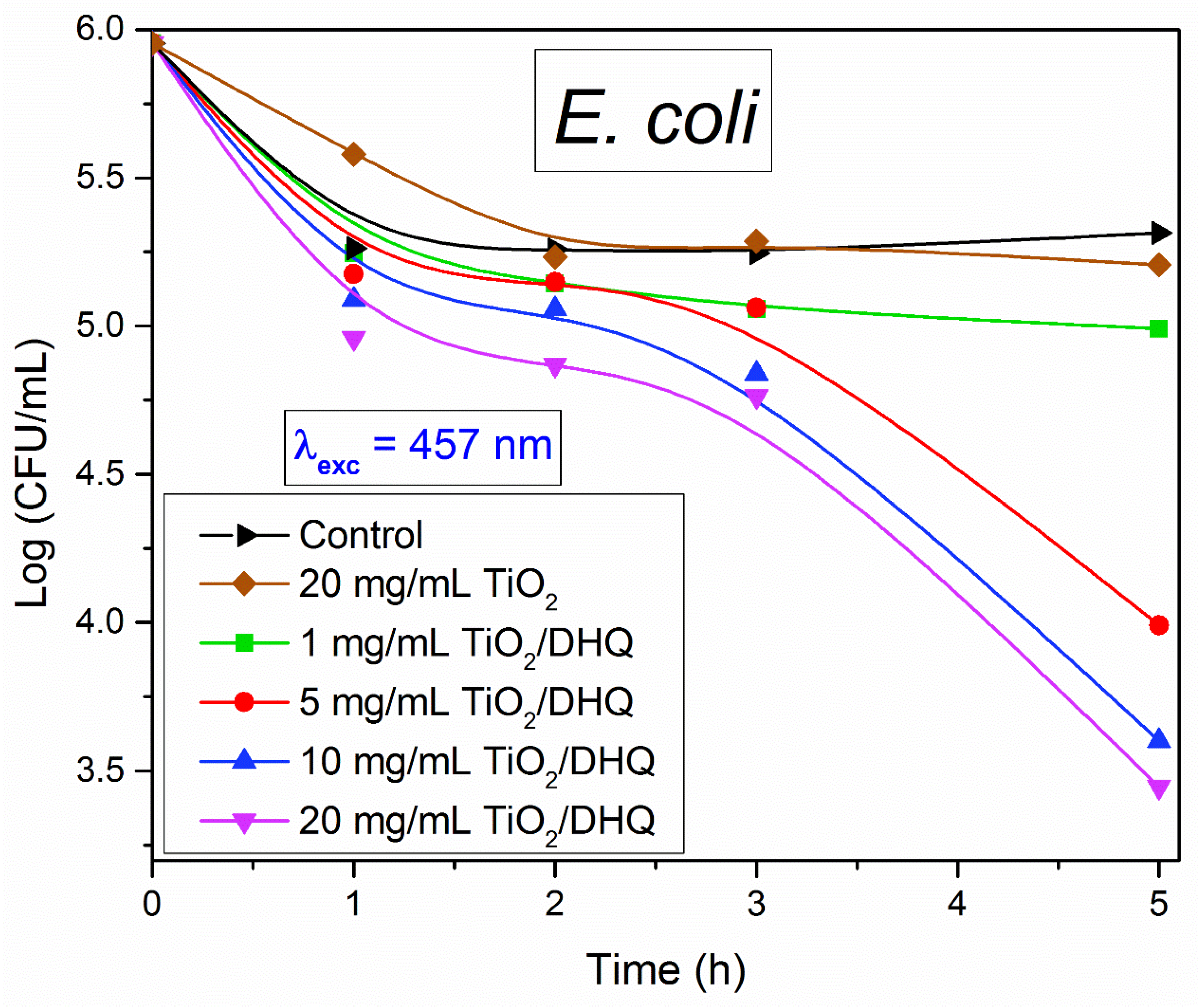
The photoinduced bactericidal activity of TiO2/DHQ was evaluated against Gram-negative bacteria E. coli (ATCC 25922) under nearly monochromatic Vis light illumination (457 nm, see Figure 1). The microbial inoculum was prepared by transferring the microorganisms into 3 mL of tryptone soy broth (TSB) supplemented with 0.6% yeast extract (TSB1Y) and incubating them overnight at 37 °C (~18 h). The incubated test cultures were diluted to a working concentration of approximately 105 CFU/mL.
Various amounts of the TiO2/DHQ samples (25, 50, and 100 mg) were placed in glass Petri Dishes containing 5 mL of the microbial suspension in saline solution (8.5% NaCl). The Petri Dishes were incubated under the light for 5 h. After a series of dilutions, a 0.1 mL aliquot of the solution was transferred into a sterile Petri dish and overlaid with tryptone soy agar (1.5% agar in TSB1Y). The inoculated plates were incubated at 37 °C for 24 h, and the surviving cells were counted (CFU/mL; CFU – colony-forming units).
To obtain unambiguous information about the photocatalytic antibacterial activity of TiO2/DHQ under exclusive Vis light excitations, we performed the following control experiments: the time-dependent reduction of E. coli under Vis light illumination in the absence of any photocatalyst, the time-dependent reduction of E. coli in the presence of TiO2/DHQ in the dark, and time-dependent reduction of E. coli in the presence of pristine TiO2 in the dark, as well as under Vis light illumination.
3. Results and Discussions
3.1. Basic Properties of ICT Complex Between TiO2 and DHQ
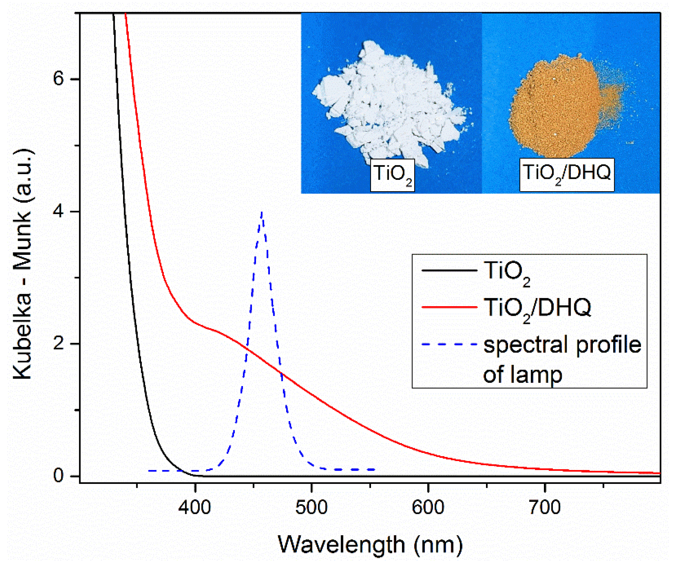
The prerequisite to understanding the toxicological and antimicrobial behavior of functionalized TiO2 powder with DHQ lies in its optical properties and surface structure, i.e., coordination of DHQ to TiO2 surface. Figure 1 shows the optical absorption of pristine TiO2 powder (Degussa P25) and the corresponding ICT complex with DHQ, measured by diffuse reflection spectroscopy. Also, images of pristine TiO2 and TiO2/DHQ powders and the spectral profile of excitation light from the light source used in antimicrobial testing are included in Figure 1. The significant red shift of the absorption onset in TiO2/DHQ (~600 nm) is a consequence of the ICT complex formation and is consistent with the reported literature data for TiO2-based ICT complexes with catecholate-type ligands [10,11,12,13,14,15,16].
The coordination of catechol to the TiO2 surface is almost intuitive since the formation of the ICT complex occurs by the condensation reaction between hydroxyl groups from the inorganic and organic components of the ICT complex. The remaining question after infrared spectroscopy analysis of the TiO2-catechol complex and its components, does catechol coordinates to the TiO2 surface by bridging or chelating coordination was solved by applying Job’s method of continuous variation [10], and the formation of bridging coordination was in addition supported by the density functional calculations (DFT) [13]. Of course, due to the presence of multiple hydroxyl groups in DHQ, it is mandatory to check how DHQ coordinates to the TiO2 surface, i.e., is it over neighboring hydroxyl groups at positions 3ʹ and 4ʹ (ring B), with hydroxyl groups at positions 5 and 7 (ring A) and 3 (ring C) remaining free. Infrared analysis of free and bound DHQ to TiO2, supported by the DFT calculation using [Ti18O31(OH)8]/DHQ cluster as a model system, undoubtedly proved the above-mentioned coordination of DHQ to the TiO2 surface [30]. For clarity, the formation mechanism of TiO2/DHQ, based on the condensation reaction, and surface structure of the ICT complex is presented in Scheme 1.
The binding way of DHQ to the TiO2 surface significantly affects the bioactivity of the ICT complex since hydroxyl groups on the phenolic ring display antioxidant activity. The presence of multiple hydroxyl groups increases the ability of the phenolic compound to donate hydrogen atoms to reactive free radicals, stabilizing them. Besides, the aromatic ring of phenolic compounds increases their antioxidant activity. So, on one side, although two hydroxyl groups at positions 3ʹ and 4ʹ (ring B) are consumed in the formation of Ti‒O‒C linkage between TiO2 and DHQ, the remaining three free hydroxyl groups can significantly reduce the toxicity of the TiO2. On the other side, although the TiO2-based ICT complex with DHQ should have increased photoinduced antimicrobial performance due to visible-light response, the ability of free hydroxyl groups to scavenge ROS can somewhat diminish its antimicrobial ability.
3.2. Cytotoxicity
Evaluation of effects of 24 h incubation with pristine TiO2 on cell viability in MRC-5 cells (Figure 2A) and HeLa cells (Figure 2B) showed that TiO2 in the investigated concentration range (1-20 mg/mL) did not induce cytotoxic effects. Moreover, a slight increase in the percentage of viable non-malignant MRC-5 cells was observed compared to the control sample, while in the case of HeLa cancer cells, there was no significant difference versus non-treated control. The concentration-dependent cell viability in MRC-5 and HeLa cells after 24 h incubation with TiO2/DHQ is presented in Figures 2C and 2D, respectively. The results indicate that exposure of MRC-5 cells to the released material from TiO2/DHQ leads to significant cytotoxic effects at concentrations above 10 mg/mL compared to the control (sample treated only with medium). However, the observed percentage reduction of viable cells after the treatment with 10 and 20 mg/mL TiO2/DHQ was quite small (7.0% and 21.6% viability reduction, respectively). Similarly, the exposure of HeLa cells to the released material from TiO2/DHQ during 24 h displayed concentration-dependent cytotoxicity (Figure 2D) and a significant reduction of viability at 10 mg/mL concentration or higher (compared to control). The most pronounced decrease in viability (25%) was observed for the highest concentration of TiO2/DHQ (20 mg/mL).
The functionalization of TiO2 with DHQ increased the cytotoxic potential of cervical cancer cells, consistent with previous findings of DHQ cytotoxicity in HeLa cells [42]. It should be mentioned that the inhibitory effect on cell viability was more pronounced in HeLa cancer cells than in normal MRC-5 cells, most likely a consequence of different DHQ effects in cancer versus normal cells. In a recent study, Mohammed et al. [43] indicated that DHQ and its metabolites have better inhibitory activity on the colon HCT-116 cancer cell line than HEK-293 normal cells in the tested concentration range. Further, a study by Chahardoli et al. [44] showed that biosynthesized TiO2 nanoparticles using quercetin had dual effects in normal and cancer cells with IC50 values below 100 and 50 μg/mL for human breast cancer cells of MCF-7 and melanoma cancer cells of A375, respectively. However, biosynthesized TiO2 nanoparticles did not significantly affect normal skin fibroblast cells up to 50 μg/mL.
Figure 3 shows the production of ROS in MRC-5 and HeLa cells as a function of the concentration of pristine TiO2 and TiO2-based ICT complex with DHQ after the 24-hour incubation period. Analysis of the obtained results indicated the decrease of the ROS production in MRC-5 non-malignant cells with the increase of the pristine TiO2 concentration compared to control (Figure 3A) with the pronounced effect for the higher TiO2 concentration (≥5 mg/mL). In the case of HeLa cancer cells, none of the applied TiO2 concentrations influenced ROS production compared to the control (Figure 3B). On the other hand, in both cell lines, MRC-5 and HeLa, the impact of TiO2/DHQ treatment on ROS levels is significant (Figure 3C and Figure 3D, respectively). Incubation of MRC-5 cells with TiO2/DHQ induced a significant reduction of ROS level when cells were exposed to TiO2/DHQ concentration equal to or higher than 5 mg/mL, while in the case of HeLa cells, a concentration-dependent reduction of ROS level started from the lowest TiO2/DHQ concentration (1 mg/mL).
In conclusion, the TiO2-based ICT complex with DHQ has increased antioxidative potential compared to the pristine TiO2 in both types of cells, and this observation is by related studies in accordance. Previous results indicate that DHQ inhibits intracellular ROS generation in human cells in a dose-dependent manner, the pronounced inhibition of ROS generation with an increase of the DHQ concentration from 10 to 100 µg/mL [45]. Also, DHQ displays a reduction in the activity of antioxidant enzymes (superoxide dismutase (SOD), catalase (CAT), and glutathione peroxidase (GPX) in pre-treated cells, along with a decreased intracellular ROS production [46]. Li et al. [47] observed that DHQ efficiently scavenge •OH, DPPH•, and ABTS•+ (hydroxyl, 2,2-diphenyl-1- picrylhydrazyl, and 2,2′-azino-bis(3-ethylbenzothiazoline-6-sulfonic acid) diammonium salt radicals, respectively), and to protect bone marrow-derived mesenchymal stem cells from •OH-induced cytotoxicity. Also, DHQ prevented oxidative damage, such as ROS generation, glutathione depletion, and single-strand break formation in human primary dermal fibroblasts (NHDF) and epidermal keratinocytes (NHEK) [48]. In addition, in a previous study, we showed that free hydroxyl groups in positions 3, 5, and 7 of the DHQ moiety in TiO2/DHQ enable the reduction of ABTS•+ radical cation, while exposure to UV light further enhances the reduction. In the presence of TiO2, photogenerated electrons reduce ABTS•+, whereas in the TiO2/DHQ, both photogenerated electrons and hydroxyl groups contribute to the decrease of ABTS•+, enabling the ICT complex to exhibit both photocatalytic and radical-scavenging properties. Also, TiO2 did not affect DPPH radical concentration, while the TiO2/DHQ induced a 35% decrease in DPPH relative concentration due to the radical-scavenging potential of the hydroxyl groups on the DHQ moiety attached to the TiO2 surface [30].
Based on the results of this study and the literature data, the TiO2-based ICT complex with DHQ has increased cytotoxicity and a pronounced antioxidant effect compared with pristine TiO2 due to DHQ’s antioxidant properties. This conclusion agrees with a report showing that titanium dioxide nanotubes loaded with quercetin are more effective anticancer and ROS-reducing agents than titanium dioxide nanotubes or quercetin themselves [49].
3.4. Genotoxicity and Antigenotoxicity
Besides their cytotoxic activity, we investigated the genotoxic potential of TiO2 and TiO2/DHQ and the antigenotoxic effect of TiO2/DHQ. The presented results in Figure 4A,B, assessed by the comet assay, indicate the genotoxic effect of TiO2 and TiO2/DHQ in MRC-5 cells. The pristine TiO2 caused significant DNA damage compared to the control in the investigated concentration range (1-20 mg/mL). As already shown, TiO2 nanoparticles can cause adverse reactions leading to cell damage, inflammation, and immune responses [50]. The comet assay, used in this study, is commonly used in genotoxicity testing of DNA damage and repair in various experimental models [51]. Several research groups have already indicated the potential risks of genotoxicity associated with exposure to TiO2 nanoparticles, showing that they can cause DNA and chromosome damage and gene mutations either in in vitro or in vivo experiments [52,53,54]. The form and degree of damage highly depend on the physical and chemical properties of TiO2, which govern its reactivity and bioavailability [50]. The genotoxic effects do not only depend on the morphology of TiO2 particulates (size and shape) but also on the test subjects (cells/animals) and the type, concentration, and duration of exposure [50,52,53,54]. We showed that the pristine TiO2 can cause significant DNA damage in MRC-5 cells. Although three mechanisms are recognized for the genotoxic effects of TiO2 particulates, ranging from nano-to micro sizes, the available data suggest that the oxidative stress, e.g., nanoparticle-induced ROS production, is a signal of other physiological effects, including cytotoxicity and genotoxicity [52,55].
However, the TiO2-based ICT complex with DHQ did not induce DNA damage in MRC-5 cells under the same experimental conditions (Figure 4B). These results are consistent with literature data obtained for ICT complexes between nanometer-sized TiO2 particles with ascorbic acid [32] and caffeic acid [56], showing no DNA damage in leukocytes from whole blood cells (0.4-8.0 mg/mL). Also, the ICT complex between TiO2 powder (Degussa P25), the same one used in this study, and caffeic acid did not induce DNA damage when orally administered to mice (2000 mg/kg) [34].
Since TiO2/DHQ does not display genotoxicity, we investigated its antigenotoxic potential against induced DNA damage by oxidative stress. Figure 4C shows the antigenotoxic effect of TiO2/DHQ studied in MRC-5 cells treated with H2O2. Pretreatment of cells with TiO2/DHQ for 24 h resulted in a significant reduction in H2O2-induced DNA damage compared to the extent of DNA damage observed in cells exposed only to H2O2. The TiO2/DHQ displays an antigenotoxic effect in the entire investigated concentration range (1-20 mg/L), opposite to the ICT complex between TiO2 and caffeic acid showing antigenotoxic properties at lower concentration (0.5-1.6 mg/mL) [56]. This discrepancy is a consequence of either different morphologies of TiO2 samples or, more likely, pronounced antioxidant properties of DHQ compared to caffeic acid. The change from the genotoxic to antigenotoxic behavior of TiO2 upon the ICT complex formation with DHQ suggests that the DHQ retains antioxidant properties besides consumption of its two out of five hydroxyl groups in the condensation reaction with TiO2 surface hydroxyl groups.
3.5. Preliminary Antimicrobial Tests
Particulate matter’s growing applications in various fields raised concerns about their toxicity, and therefore, the use of functionalized TiO2 particles with DHQ is advantageous from an ecological perspective. The additional advantage of TiO2/DHQ is the enhanced harvesting ability of solar radiation. While many studies demonstrated the antimicrobial activity of TiO2 under UV light [2,3,4,5,7,8], even though ICT complexes are eco-friendly and Vis light sources can be used instead of harsh UV light sources, the literature on the biocidal action of ICT complexes under Vis light is almost non-existent [31]. So, in this study, we performed a preliminary antimicrobial test of the TiO2-based ICT complex with DHQ under Vis light excitation (457 nm is the emission maximum wavelength of the light source; see Figure 1) against E. coli to estimate if this system is worth further investigation in this direction.
Figure 5 shows the time- and concentration-dependent antimicrobial activity of TiO2/DHQ against gram-negative bacteria E. coli, including data for pristine TiO2 (20 mg/mL) and control (absence of photocatalyst). The results of two additional controls, the antibacterial activity of TiO2 and TiO2/DHQ in the dark, are presented in Supporting Information (Table S1). Despite limited experimental data, we can discern the following conclusions. First, pristine TiO2 does not display antimicrobial activity against E. coli upon Vis light excitation; its time-kill curve practically overlaps with the control. Of course, both photocatalysts (TiO2 and TiO2/DHQ) do not display a biocidal effect in the dark. Second, for low concentrations (1 mg/mL), TiO2/DHQ does not show any antibacterial properties. Third, at higher concentrations (5, 10, and 20 mg/mL), TiO2/DHQ demonstrates concentration-dependent antimicrobial activity. For example, after five hours of illumination, the number of surviving bacteria was reduced by two and three orders of magnitude when TiO2/DHQ concentration was 5 and 20 mg/mL, respectively. Fourth, unfortunately, the kinetic curves of antimicrobial activity are incomplete since the homemade light source, designed to cover the desired spectral range, has an overheating issue, preventing continuous operation for extended periods. However, based on their trends, complete bacterial destruction can be expected at prolonged irradiation.
The presented results are a step forward in the design of the non-toxic antimicrobial agent. Owing to free hydroxyl groups, the coordination of DHQ to the TiO2 surface brings antioxidant behavior to the entire ICT architecture and, in addition, enhanced optical properties. However, since free hydroxyl groups diminish the antimicrobial potency of the ICT complex, further research in this area should focus on increasing the efficiency of antimicrobial activity while keeping the ICT complex non-toxic.
4. Conclusions
In conclusion, we prepared surface-modified TiO2 with a potent plant-derived antioxidant dihydroquercetin (DHQ) and assessed its bioactivity. Besides inducing red-shifted absorption due to ICT-complex formation, the coordination of DHQ to surface Ti atoms reduces the genotoxicity of TiO2 generated by reactive oxygen species (ROS) formed upon excitation of hybrid architecture. We have demonstrated that TiO2-based ICT complex with DHQ is non-toxic to MRC-5 cells up to a concentration of 10 mg/mL and exhibits significant antigenotoxic properties compared with pristine TiO2 nanopowder. Furthermore, preliminary antibacterial tests indicated that TiO2-based ICT complex with DHQ efficiently inactivates gram-negative bacteria E. coli when exposed solely to Vis light. We are confident that further research in this direction is worthwhile, considering that, on one side, expensive and harmful UV light sources are replaceable by cost-effective, harmless Vis light sources, while, on the other side, the release of toxic materials, for example, silver, in the environment is avoided.
Supplementary Materials
The following supporting information can be downloaded at the website of this paper posted on Preprints.org.
Author Contributions
Valentina Nikšić: Methodology, Investigation. Andrea Pirković: Visualization, Methodology, Investigation, Writing – original draft. Biljana Spremo-Potparević: Methodology, Validation, Data curation. Lada Živković: Methodology, Validation, Data curation. Dijana Topalović: Investigation, Formal analysis. Jovan M. Nedeljković: Writing – review & editing, Conceptualization. Vesna Lazić: Writing – review & editing, Conceptualization, Supervision.
Acknowledgments
This study was supported by the Ministry of Science, Technological Development and Innovation of the Republic of Serbia (Contract Numbers: 451-03-66/2024-03/200017, 451-03-65/2024-03/200161, and 451-03-66/2024-03/200019) and by the Science Fund of the Republic of Serbia, Program PRISMA, Grant No. 5354, Multifunctional visible-light responsive inorganic-organic hybrids for efficient hydrogen production and disinfection-HYDIS.
Conflicts of Interest
The authors declare that they have no known competing financial interests or personal relationships that could have appeared to influence the work reported in this paper.
References
- Hoffmann, M.R.; Martin, S.T.; Choi, W.; Bahnemann, D.W. Environmental applications of semiconductor photocatalysis. Chem. Rev. 1995, 95, 69–96. [Google Scholar] [CrossRef]
- Carré, G.; Hamon, E.; Ennahar, S.; Estner, M.; Lett, M.C.; Horvatovich, P.; Gies, J.P.; Keller, V.; Keller, N.; Andrea, P. TiO2 Photocatalysis Damages Lipids and Proteins in Escherichia coli. Appl. Environ. Microb. 2014, 80, 2573–2581. [Google Scholar] [CrossRef]
- Wei, Y.; Wu, Q.; Meng, H.; Zhang, Y.; Cao, C. Recent advances in photocatalytic self-cleaning performances of TiO2-based building materials. RSC Adv. 2023, 13, 20584–20597. [Google Scholar] [CrossRef]
- Ziental, D.; Czarczynska-Goslinska, B.; Mlynarczyk, D.T.; Glowacka-Sobotta, A.; Stanisz, B.; Goslinski, T.; Sobotta, L. Titanium Dioxide Nanoparticles: Prospects and Applications in Medicine. Nanomaterials 2020, 10, 387. [Google Scholar] [CrossRef] [PubMed]
- Chouirfa, H.; Bouloussa, H.; Migonney, V.; Falentin-Daudré, C. Review of titanium surface modification techniques and coatings for antibacterial applications. Acta Biomater. 2019, 83, 37–54. [Google Scholar] [CrossRef]
- Nešić, A.; Gordić, M.; Davidović, S.; Radovanović, Ž.; Nedeljković, J.; Smirnova, I.; Gurikov, P. Pectin-based nanocomposite aerogels for potential insulated food packaging application. Carbohyd. Polym. 2018, 195, 128–135. [Google Scholar] [CrossRef] [PubMed]
- Haider, A.J.; AL-Anbari, R.H.; Kadhim, G.R.; Salame, C.T. Exploring potential Environmental applications of TiO2 Nanoparticles. Energy Procedia 2017, 119, 332–345. [Google Scholar] [CrossRef]
- Nguyen, N.T.T.; Nguyen, L.M.; Nguyen, T.T.T.; Liew, R.K.; Nguyen, D.T.C.; Tran, T.V. Recent advances on botanical biosynthesis of nanoparticles for catalytic, water treatment and agricultural applications: A review. Sci. Total. Environ. 2022, 827, 154160. [Google Scholar] [CrossRef]
- Rajh, T.; Nedeljković, J.M.; Chen, L.X.; Poluektov, O.; Thurnauer, M.C. Improving optical and charge separation properties of nanocrystalline TiO2 by surface modification with vitamin C. J. Phys. Chem. B 1999, 103, 3515–3519. [Google Scholar] [CrossRef]
- Janković, I.A.; Šaponjić, Z.V.; Čomor, M.I.; Nedeljković, J.M. Surface modification of colloidal TiO2 nanoparticles with bidentate benzene derivatives. J. Phys. Chem. C 2009, 113, 12645–12652. [Google Scholar] [CrossRef]
- Lazić, V.; Nedeljković, J.M. Photocatalytic Reactions over TiO2-Based Interfacial Charge Transfer Complexes. Catalysts 2024, 14, 810. [Google Scholar] [CrossRef]
- Janković, I.A.; Šaponjić, Z.V.; Džunuzović, E.S.; Nedeljković, J.M. New hybrid properties of TiO2 nanoparticles surface modified with catecholate type ligands. Nanoscale Res. Lett. 2010, 5, 81–88. [Google Scholar] [CrossRef] [PubMed]
- Savić, T.D.; Janković, I.A.; Šaponjić, Z.V.; Čomor, M.I.; Veljković, D.Ž.; Zarić, S.D.; Nedeljković, J.M. Surface modification of anatase nanoparticles with fused catecholate type ligands: a combined DFT and experimental study of optical properties. Nanoscale 2012, 4, 1612–1619. [Google Scholar] [CrossRef] [PubMed]
- Savić, T.D.; Čomor, M.I.; Nedeljković, J.M.; Veljković, D.Ž.; Zarić, S.D.; Rakić, V.M.; Janković, I.A. The effect of substituents on the surface modification of anatase nanoparticles with catecholate-type ligands: a combined DFT and experimental study. Phys. Chem. Chem. Phys. 2014, 16, 20796–20805. [Google Scholar] [CrossRef] [PubMed]
- Higashimoto, S.; Nishi, T.; Yasukawa, M.; Azuma, M.; Sakata, Y.; Kobayashi, H. Photocatalysis of titanium dioxide modified by catechol-type interfacial surface complexes (ISC) with different substituted groups. J. Catal. 2015, 329, 286–291. [Google Scholar] [CrossRef]
- Milićević, B.; Ðorđević, V.; Lončarević, D.; Ahrenkiel, S.P.; Dramićanin, M.D.; Nedeljković, J.M. Visible light absorption of surface modified TiO2 powders with bidentate benzene derivatives. Micropor. Mesopor. Mat. 2015, 217, 184–189. [Google Scholar] [CrossRef]
- Savić, T.D.; Šaponjić, Z.V.; Čomor, M.I.; Nedeljković, J.M.; Dramićanin, M.D.; Nikolić, M.G.; Veljković, D.Ž.; Zarić, S.D.; Janković, I.A. Surface modification of anatase nanoparticles with fused ring salicylate-type ligands (3-hydroxy-2-naphtoic acids): a combined DFT and experimental study of optical properties. Nanoscale 2013, 5, 7601–7612. [Google Scholar] [CrossRef]
- Boz, D.K.; Garcia, G.A.; Nahon, L.; Sredojevic, D.; Lazic, V.; Vukoje, I.; Ahrenkiel, S.P.; Djokovic, V. Ž. Šljivančanin; Nedeljkovic, J.M. Interfacial Charge Transfer Transitions in Colloidal TiO2 Nanoparticles Functionalized with Salicylic acid and 5-Aminosalicylic acid: A Comparative Photoelectron Spectroscopy and DFT Study. J. Phys. Chem. C 2019, 123, 29057–29066. [Google Scholar]
- Fujisawa, J.; Matsumura, S.; Hanaya, M. A single Ti‒O‒C linkage induces interfacial charge-transfer transitions between TiO2 and a π-conjugated molecule. Chem. Phys. Lett. 2016, 657, 172–176. [Google Scholar] [CrossRef]
- Fujisawa, J.; Eda, T.; Giorgi, G.; Hanaya, M. Visible-to-Near-IR Wide-Range Light Harvesting by Interfacial Charge-Transfer Transitions between TiO2 and p-Aminophenol and Evidence of Direct Electron Injection to the Conduction Band of TiO2. J. Phys. Chem. C 2017, 121, 18710–18716. [Google Scholar] [CrossRef]
- Barbieriková, Z.; Dvoranová, D.; Brezová, V.; Džunuzović, E.; Sredojević, D.N.; Lazić, V.; Nedeljković, J.M. Visible-light-responsive surface-modified TiO2 powder with 4-chlorophenol: A combined experimental and DFT study. Opt. Mater. 2019, 89, 237–242. [Google Scholar] [CrossRef]
- Fujisawa, J.; Kato, S.; Hanaya, M. Substituent effects on interfacial charge-transfer transitions in a TiO2-phenol complex. Chem. Phys. Lett. 2023, 827, 140688. [Google Scholar] [CrossRef]
- Ikeda, S.; Abe, C.; Torimoto, T.; Ohtani, B. Photochemical hydrogen evolution from aqueous triethanolamine solutions sensitized by binaphthol-modified titanium(IV) oxide under visible-light irradiation, J. Photoch. Photobio. A 2003, 160, 61–67. [Google Scholar] [CrossRef]
- Yao, B.; Peng, C.; Lu, P.; He, Y.; Zhang, W.; Zhang, Q. Fabrication of Tiron-TiO2 charge-transfer complex with excellent visible-light photocatalytic performance, Mat. Chem. Phys. 2016, 184, 298–305. [Google Scholar]
- Vukoje, I.; Kovač, T.; Džunuzović, J.; Džunuzović, E.; Lončarević, D.; Ahrenkiel, S.P.; Nedeljković, J.M. Photocatalytic ability of visible-light-responsive TiO2 nanoparticles, J. Phys. Chem. C 2016, 120, 18560–18569. [Google Scholar] [CrossRef]
- Lazić, V.; Sredojević, D.; Ćirić, A.; Nedeljković, J.M.; Zelenková, G.; Férová, M.; Zelenka, T.; Chavhan, M.P.; Slovák, V. The photocatalytic ability of visible-light-responsive interfacial charge transfer complex between TiO2 and Tiron, J. Photoch. Photobio. A 2024, 449, 115394. [Google Scholar] [CrossRef]
- Kose, O. In vitro toxicity of titanium dioxide nanoparticles on human lung cells – impact of the physicochemical features, PhD thesis, Chemical and Process Wngineering, Université de Lyon, 2020, in English.
- Afşar, O.; Oltulu, Ç. Evaluation of the cytotoxic effect of titanium dioxide nanoparticles in human embryonic lung cells, Turk. J. Med. Sci. 2023, 53, 1648–1657. [Google Scholar]
- Sredojević, D.; Lazić, V.; Pirković, A.; Periša, J.; Murafa, N.; Spremo-Potparević, B.; Živković, L.; Topalović, D.; Zarubica, A.; Krivokuća, M.J.; Nedeljković, J.M. Toxicity of silver supported by surface-modified zirconium dioxide with dihydroquercetin, Nanomaterials 2022, 12, 3195.
- Nikšić, V.; Šimunková, M.M.; Dyrčíková, Z.; Dvoranová, D.; Brezová, V.; Sredojević, D.; Nedeljković, J.M.; Lazić, V. Photoinduced reactive species in interfacial charge transfer complex between TiO2 and taxifolin: DFT and EPR study, Opt. Mater. 2024, 152, 115454. [Google Scholar] [CrossRef]
- Shahriari-Khalaji, M.; Zabihi, F.; Bahi, A.; Sredojević, D.; Nedeljković, J.M.; Macharia, D.K.; Ciprian, M.; Yang, S.; Ko, F. Photon-driven bactericidal performance of surface-modified TiO2 nanofibers, J. Mater. Chem. C 2023, 11, 5796–5805. [Google Scholar] [CrossRef]
- Bajić, V.; Spremo-Potparević, B.; Živković, L.; Čabarkapa, A.; Kotur-Stevuljević, J.; Isenović, E.; Sredojević, D.; Vukoje, I.; Lazić, V.; Ahrenkiel, S.P.; Nedeljković, J.M. Surface-modified TiO2 nanoparticles with ascorbic acid: Antioxidant properties and efficiency against DNA damage in vitro, Colloid Surface B 2017, 155, 323–331.
- Lazić, V.; Pirković, A.; Sredojević, D.; Marković, J.; Papan, J.; Ahrenkiel, S.P.; Janković-Častvan, I.; Dekanski, D.; Jovanović-Krivokuća, M.; Nedeljković, J.M. Surface-modified ZrO2 nanoparticles with caffeic acid: Characterization and in vitro evaluation of biosafety for placental cells, Chem. -Biol. Interact. 2021, 347, 109618. [Google Scholar] [CrossRef]
- Dekanski, D.; Spremo-Potparević, B.; Bajić, V.; Živković, L.; Topalović, D.; Sredojević, D.N.; Lazić, V.; Nedeljković, J.M. Acute toxicity study in mice of orally administrated TiO2 nanoparticles functionalized with caffeic acid, Food Chem. Toxicol. 2018, 115, 42–48. [Google Scholar]
- Todorović, A.; Bobić, K.; Veljković, F.; Pejić, S.; Glumac, S.; Stanković, S.; Milovanović, T.; Vukoje, I.; Nedeljković, J.M.; Škodrić, S.R.; Pajović, S.B.; Drakulić, D. Comparable Toxicity of Surface-Modified TiO2 Nanoparticles: An In Vivo Experimental Study on Reproductive Toxicity in Rats, Antioxidants 2024, 13, 231.
- Živković, L.; Bajić, V.; Topalović, D.; Bruić, M.; Spremo-Potparević, B. Antigenotoxic Effects of Biochaga and Dihydroquercetin (Taxifolin) on H2O2-Induced DNA Damage in Human Whole Blood Cells, Oxid. Med. Cell. Longev. 2019, 2019, 5039372. [Google Scholar]
- Abhijit, D.; Baidya, R.; Chakraborty, T. Pharmacological basis and new insights of taxifolin: A comprehensive review, Biomed. Pharmacother. 2021, 142, 112004. [Google Scholar]
- Orlova, S.V.; Tatarinov, V.V.; Nikitina, E.A.; Sheremeta, A.V.; Ivlev, V.A.; Vasilev, V.G.; Paliy, K.V.; Goryainov, S.V. Molecularbiological problems of drug design and mechanism of drug action, Pharm. Chem. J. 2022, 55, 1133–1137. [Google Scholar]
- Mukhtar-Fayyad, D. Cytocompatibility of new bioceramic-based materials on human fibroblast cells (MRC-5), Oral. Surg. Oral. Med. O. 2011, 112, e137–e142. [Google Scholar]
- Tice, R.R.; Agurell, E.; Anderson, D.; Burlinson, B.; Hartmann, A.; Kobayashi, H.; Miyamae, Y.; Rojas, E.; Ryu, J.C.; Sasaki, Y.F. Single cell gel/comet assay: guidelines for in vitro and in vivo genetic toxicology testing, Environ. Mol. Mutagen. 2000, 35, 206–221. [Google Scholar] [CrossRef]
- Anderson, D.; Yu, T.W.; Phillips, B.J.; Schmezer, P. The effect of various antioxidants and other modifying agents on oxygen-radical-generated DNA damage in human lymphocytes in the comet assay, Mutat. Res. 1994, 307, 261–271. [Google Scholar] [CrossRef]
- Rokaitytė, A.; Zaborskienė, G.; Baliukonienė, V.; Jovaišienė, J.; Gustienė, S. Cytotoxicity of carnosine, taxifolin, linalool and their combinations with acids to the cells of human cervical carcinoma (HeLa), Biologija 2018, 64, 274–284.
- Mohammed, H.A.; El-Ghaly, S.A.A.E.-S.M. F.A.Khan; Emwas, A.H.; Jaremko, M.; Almulhim, F.; Khan, R.A.; Ragab, E.A. Comparative Anticancer Potentials of Taxifolin and Quercetin Methylated Derivatives against HCT-116 Cell Lines: Effects of O-Methylation on Taxifolin and Quercetin as Preliminary Natural Leads, ACS Omega. 2022; 7, 46629–46639. [Google Scholar]
- Chahardoli, A.; Jalilian, F.; Shokoohinia, Y.; Fattahi, A. The role of quercetin in the formation of titanium dioxide nanoparticles for nanomedical applications, Toxicol. in Vitro 2023, 87, 105538. [Google Scholar] [CrossRef] [PubMed]
- Xie, X.; Feng, J.; Kang, Z.; Zhang, S.; Zhang, L.; Zhang, Y.; Li, X.; Tang, Y. Taxifolin protects RPE cells against oxidative stress-induced apoptosis, Mol. Vis. 2017, 23, 520–528. [Google Scholar]
- Bruić, M.; Pirković, A.; Borozan, S.; Aleksić, M.N.; Krivokuća, M.J.; Spremo-Potparević, B. Antioxidative and anti-inflammatory effects of taxifolin in H2O2-induced oxidative stress in HTR-8/SVneo trophoblast cell line, Reprod. Toxicol. 2024, 126, 108585. [Google Scholar]
- Li, X.; Xie, H.; Jiang, Q.; Wei, G.; Lin, L.; Li, C.; Ou, X.; Yang, L.; Xie, Y.; Fu, Z.; Liu, Y.; Chen, D. The mechanism of (+) taxifolin’s protective antioxidant effect for •OH-treated bone marrow-derived mesenchymal stem cells, Cell. Mol. Biol. Lett. 2017, 22, 31. [Google Scholar] [CrossRef] [PubMed]
- Svobodová, A.R.; Ryšavá, A.; Čížková, K.; Roubalová, L.; Ulrichová, J.; Vrba, J.; Zálešák, B.; Vostálová, J. Effect of the flavonoids quercetin and taxifolin on UVA-induced damage to human primary skin keratinocytes and fibroblasts, Photoch. Photobio. Sci. 2022, 21, 59–75. [Google Scholar] [CrossRef]
- Gulla, S.; Lomada, D.; Araveti, P.B.; Srivastava, A.; Murikinati, M.K.; Reddy, K.R. Inamuddin; Reddy, M.C.; Altalhi, T. Titanium dioxide nanotubes conjugated with quercetin function as an effective anticancer agent by inducing apoptosis in melanoma cells. J. Nanostructure. Chem. 2021, 11, 721–734. [Google Scholar] [CrossRef]
- Shabbir, S.; Kulyar, M.F.; Bhutta, Z.A.; Boruah, P.; Asif, M. Toxicological Consequences of Titanium Dioxide Nanoparticles (TiO2 NPs) and Their Jeopardy to Human Population, Bionanoscience 2021, 11, 621–632.
- Langie, S.; Azqueta, A.; Collins, A. The comet assay: past, present, future, Front. Genet 2015, 6, 266. [Google Scholar] [CrossRef] [PubMed]
- Cao, Y.; Chen, J.; Bian, Q.; Ning, J.; Yong, L.; Ou, T.; Song, Y.; Wei, S. Genotoxicity Evaluation of Titanium Dioxide Nanoparticles In Vivo and In Vitro: A Meta-Analysis, Toxics 2023, 11, 882.
- Ling, C.; An, H.; Li, L.; Wang, J.; Lu, T.; Wang, H.; Hu, Y.; Song, G.; Liu, S. Genotoxicity Evaluation of Titanium Dioxide Nanoparticles in Vitro: A Systematic Review of the Literature and Meta-analysis, Biol. Trace Elem. Res. 2021, 199, 2057–2076. [Google Scholar] [CrossRef]
- Shi, J.; Han, S.; Zhang, J.; Liu, Y.; Chen, Z.; Jia, G. Advances in genotoxicity of titanium dioxide nanoparticles in vivo and in vitro, NanoImpact 2022, 25, 100377.
- Chen, T.; Yan, J.; Li, Y. Genotoxicity of titanium dioxide nanoparticles, J. Food Drug Anal. 2014, 22, 95–104. [Google Scholar] [CrossRef]
- Lazic, V.; Vukoje, I.; Milicevic, B.; Spremo-Potparevic, B.; Zivkovic, L.; Topalovic, D.; Bajic, V.; Sredojevic, D.; Nedeljkovic, J.M. Efficiency of the interfacial charge transfer complex between TiO2 nanoparticles and caffeic acid against DNA damage in vitro: a combinatorial analysis. J. Serb. Chem. Soc. 2019, 84, 539–553. [Google Scholar] [CrossRef]
Figure 1.
Kubelka-Munk transformations of the pristine TiO2 (Aeroxide® P25) powder and TiO2-based ICT complex with DHQ. Also, photo images of TiO2 and TiO2/DHQ and the spectral profile of emitted light used in antimicrobial tests are included.
Figure 1.
Kubelka-Munk transformations of the pristine TiO2 (Aeroxide® P25) powder and TiO2-based ICT complex with DHQ. Also, photo images of TiO2 and TiO2/DHQ and the spectral profile of emitted light used in antimicrobial tests are included.
Scheme 1.
Schematic presentation of the formation mechanism of the TiO2-based ICT complex with DHQ.
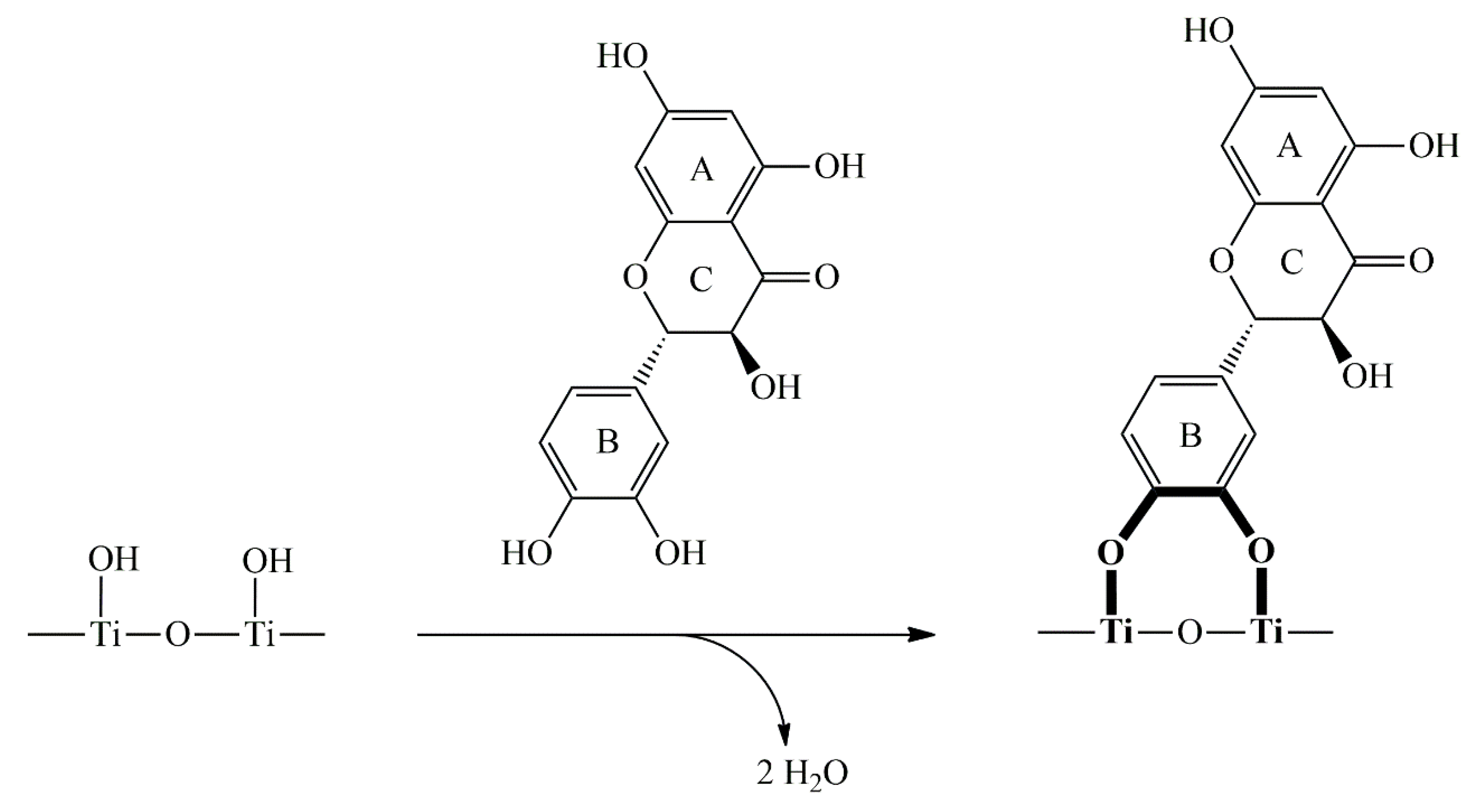
Figure 2.
Left panel: Cytotoxicity of TiO2 in a range of concentrations (1, 2, 5, 10, and 20 mg/mL) determined by MTT assay in MRC-5 cells (A) and HeLa cells (B), expressed as a percentage out of control. Right panel: Cytotoxic effect of TiO2/DHQ in a range of concentrations (1, 2, 5, 10, and 20mg/mL) in MRC-5 cells (C) and HeLa cells (D), determined by MTT assay. The data are expressed as mean + SEM. * p<0.05, ** p<0.01, *** p<0.001, **** p<0.0001 versus control (by one-way Analysis of Variance (ANOVA) with Tukey’s multiple comparison post-hoc test).
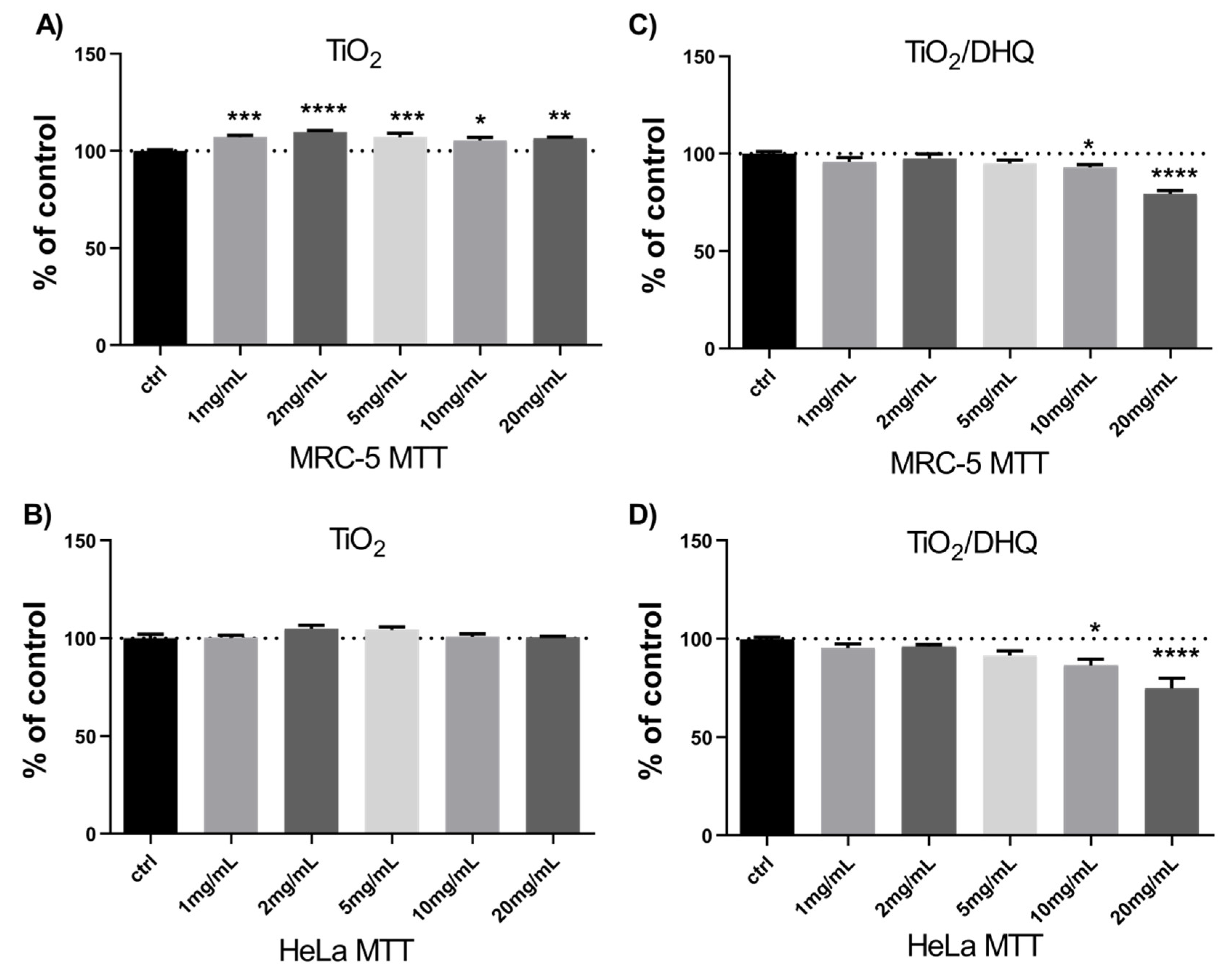
Figure 3.
Left panel: Effect of TiO2 in a range of concentrations (1, 2, 5, 10, and 20 mg/mL) on the production of reactive oxygen species (ROS) in MRC-5 cells (A) and HeLa cells (B), determined by H2DCFDA assay, expressed as relative fluorescence intensity. Right panel: Effect of TiO2DHQ on the production of reactive oxygen species (ROS) in a range of concentrations (1, 2, 5, 10, and 20 mg/mL) in MRC-5 cells (C) and HeLa cells (D), determined by H2DCFDA assay. The data are expressed as mean + SEM. * p<0.05, ** p<0.01, *** p<0.001, **** p<0.0001 versus control (by one-way Analysis of Variance (ANOVA) with Tukey’s multiple comparison post-hoc test).
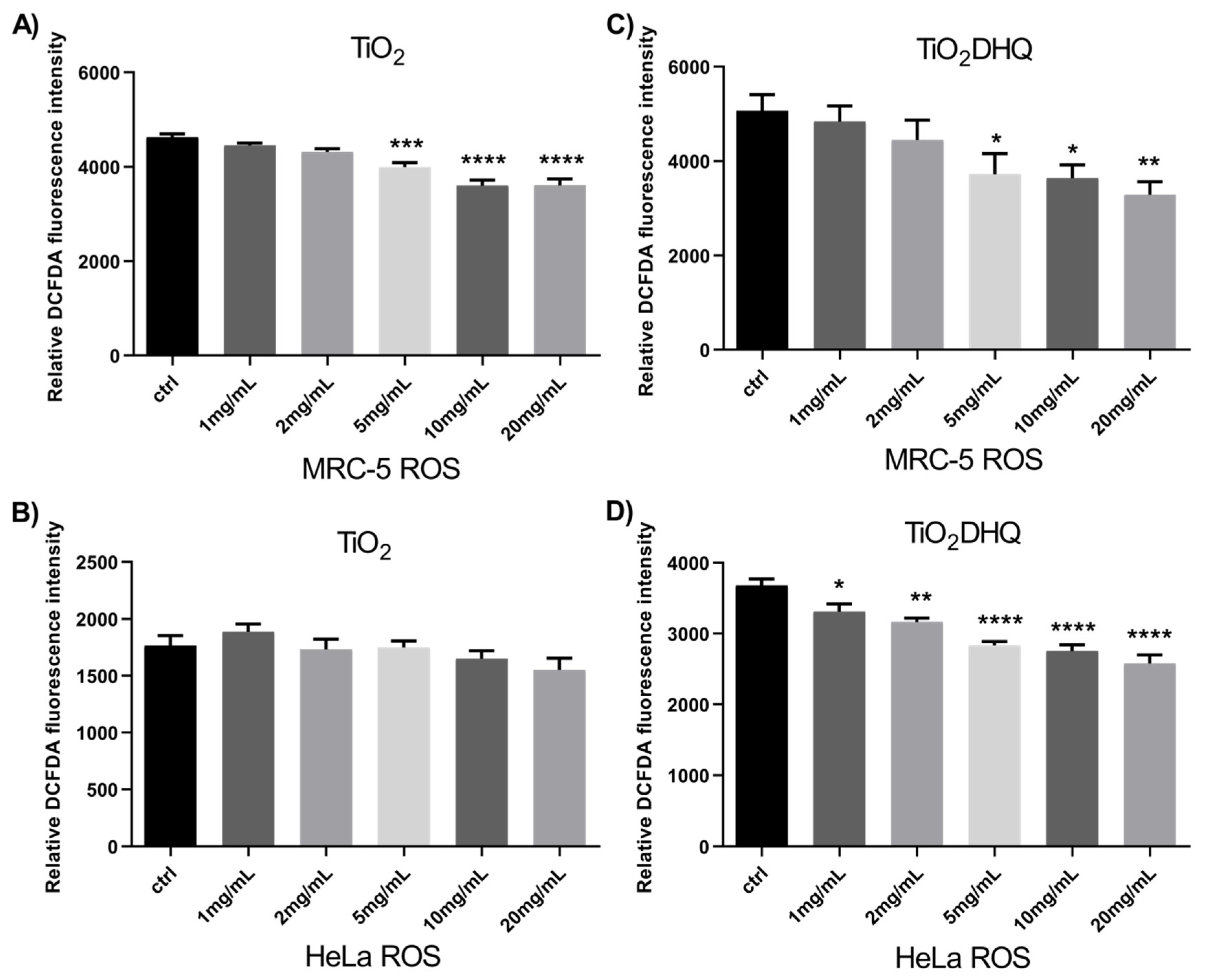
Figure 4.
Genotoxic effect of (A) TiO2 and (B) TiO2/DHQ in MRC-5 cells, and (C) antigenotoxic effect of TiO2/DHQ against DNA damage induced by H2O2 in MRC-5 cells in a range of concentrations (1, 2, 5, 10, and 20 mg/mL) determined by comet assay. The data are expressed as a mean number of cells with DNA damage + SEM. * p<0.05, *** p<0.001, **** p<0.0001 versus control (by one-way Analysis of Variance (ANOVA) with Tukey’s multiple comparison post-hoc test).
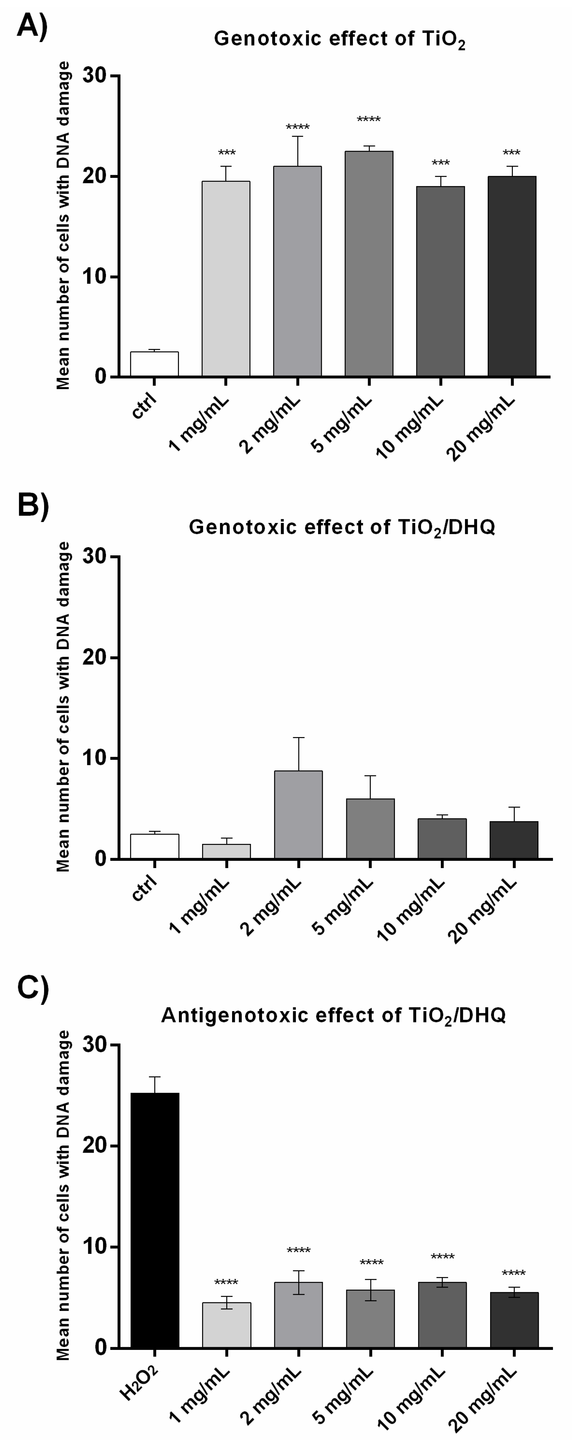
Figure 5.
The time-dependent antimicrobial ability of pristine TiO2 (20 mg/mL) and TiO2/DHQ (1-20 mg/mL) against E. coli under visible light excitation (457 nm); control: experiments without photocatalyst.
Figure 5.
The time-dependent antimicrobial ability of pristine TiO2 (20 mg/mL) and TiO2/DHQ (1-20 mg/mL) against E. coli under visible light excitation (457 nm); control: experiments without photocatalyst.
Disclaimer/Publisher’s Note: The statements, opinions and data contained in all publications are solely those of the individual author(s) and contributor(s) and not of MDPI and/or the editor(s). MDPI and/or the editor(s) disclaim responsibility for any injury to people or property resulting from any ideas, methods, instructions or products referred to in the content. |
© 2025 by the authors. Licensee MDPI, Basel, Switzerland. This article is an open access article distributed under the terms and conditions of the Creative Commons Attribution (CC BY) license (http://creativecommons.org/licenses/by/4.0/).
Copyright: This open access article is published under a Creative Commons CC BY 4.0 license, which permit the free download, distribution, and reuse, provided that the author and preprint are cited in any reuse.


