Submitted:
21 January 2025
Posted:
22 January 2025
You are already at the latest version
Abstract
Accurate segmentation of the liver and liver tumors is crucial for clinical diagnosis and treatment. However, the task poses significant challenges due to the complex morphology of tumors, indistinct features of small targets, and the similarity in grayscale values between the liver and surrounding organs. To address these issues, this paper proposes an enhanced 3D U-Net architecture, named ELANRes-MSCA- UNet. By incorporating a structural re-parameterized residual module (ELANRes) and a multi-scale convolutional attention module (MSCA), the network significantly improves feature extraction and boundary optimization, particularly excelling in segmenting small targets. Additionally, a two-stage strategy is employed, where the liver region is segmented first, followed by the fine-grained segmentation of tumors, effectively reducing false positive rates. Experiments conducted on the LiTS2017 dataset demonstrate that ELANRes-MSCA-UNet achieves Dice scores of 97.2% and 72.9% for liver and tumor segmentation tasks, respectively, significantly outperforming other state-of-the-art methods. These results validate the accuracy and robustness of the proposed method in medical image segmentation and highlight its potential for clinical applications.
Keywords:
1. Introduction
2. Methods
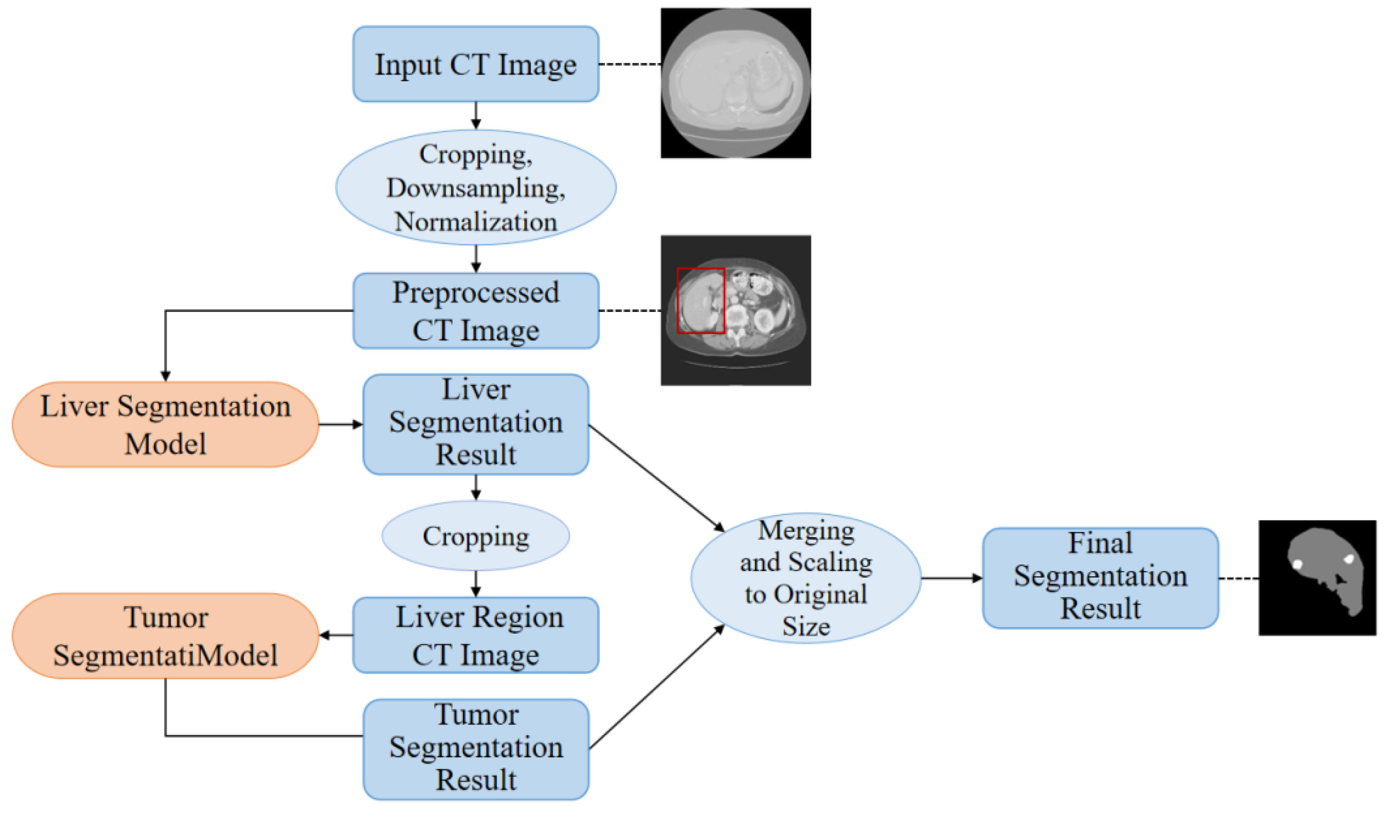
2.1. Methodology Workflow
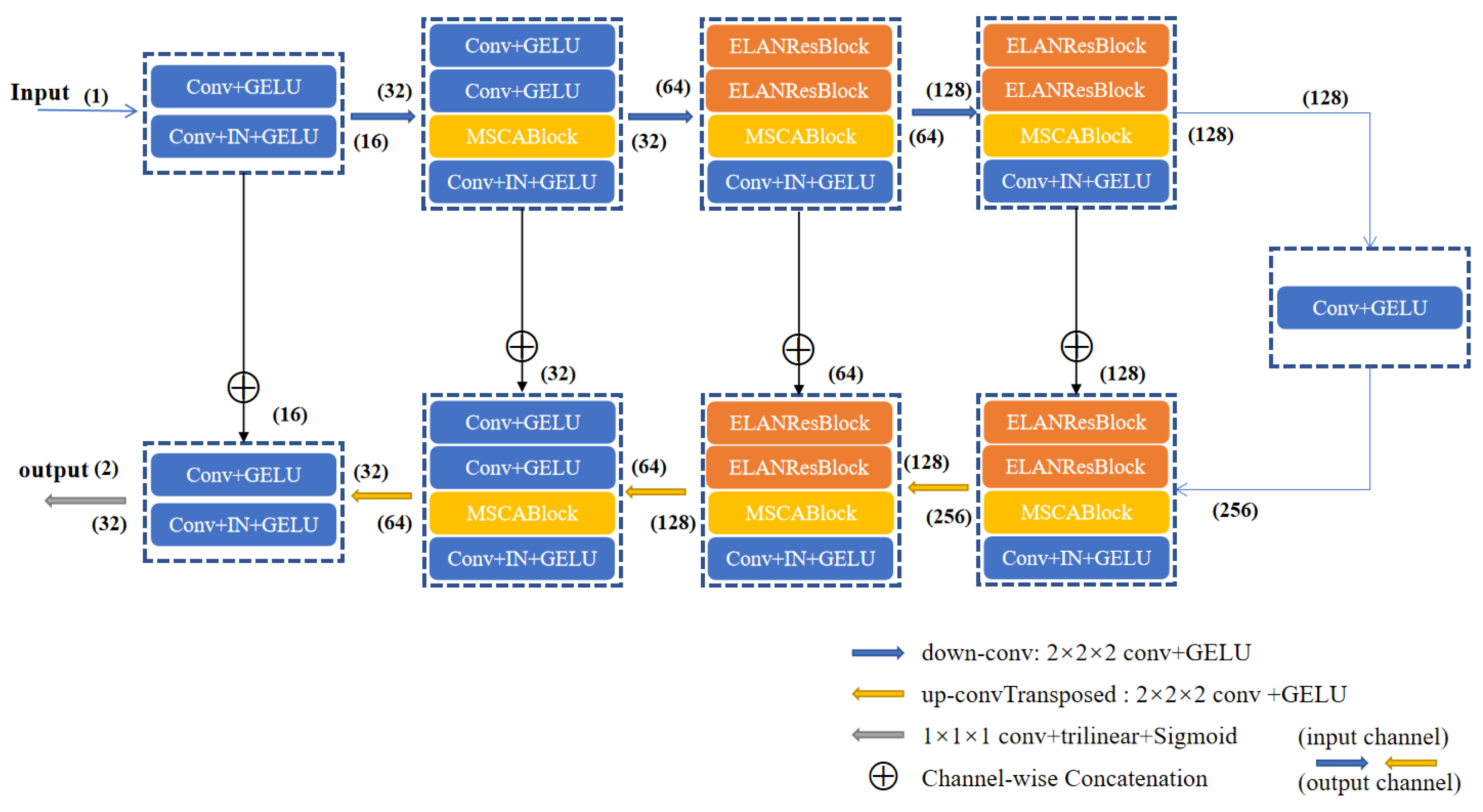
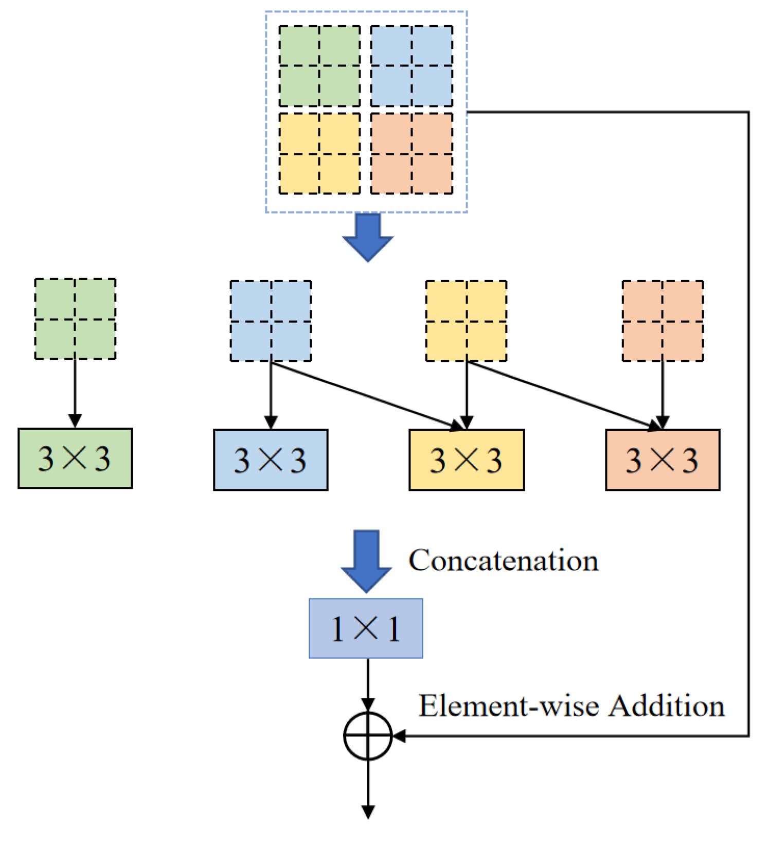
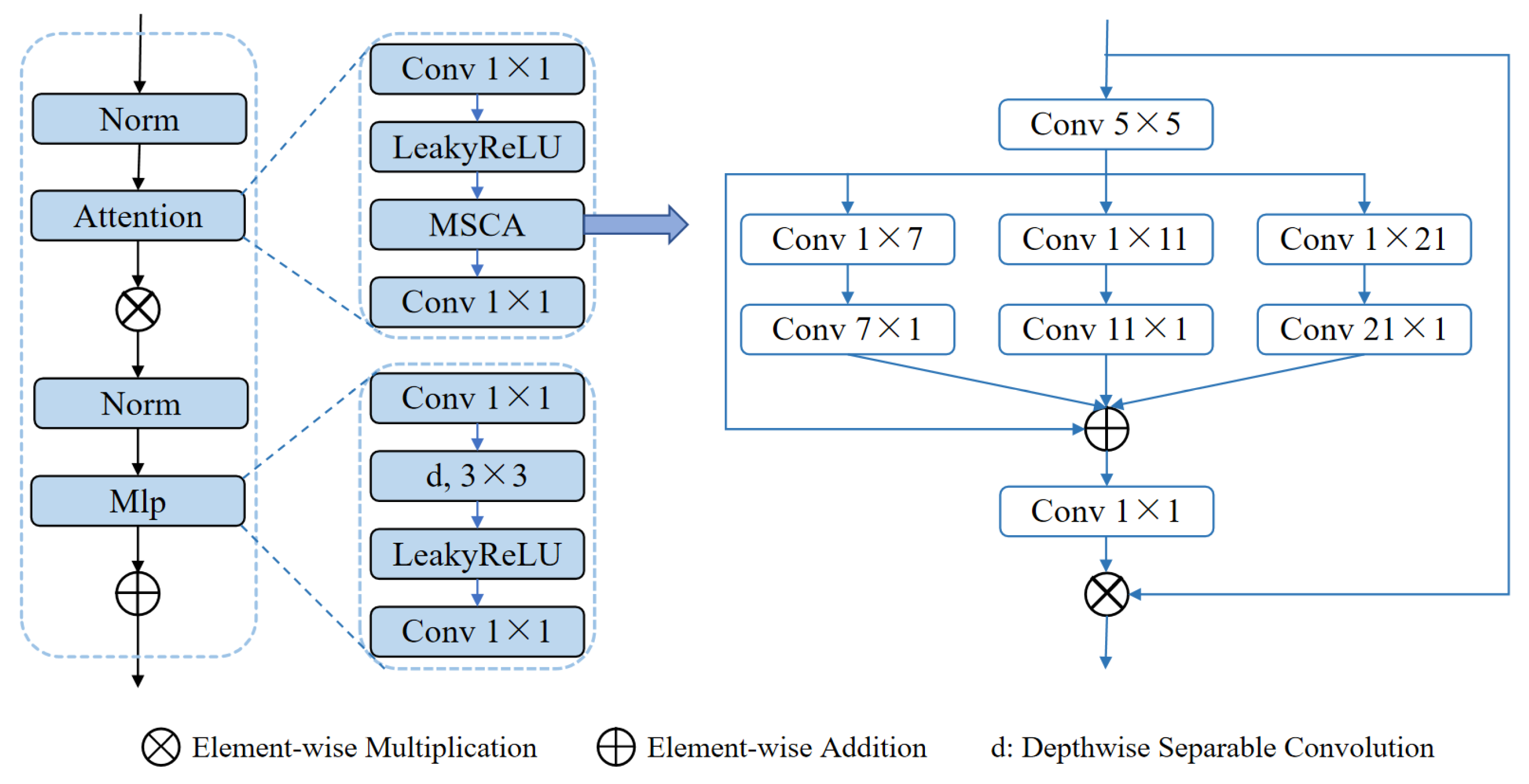
2.2. Proposed ELANRes-MSCA-UNet Network
2.3. Loss Function Combination and Deep Supervision Mechanism
2.4. Evaluation Metrics
3. Experiments and Results
3.1. Data and Implementation
3.2. Ablation
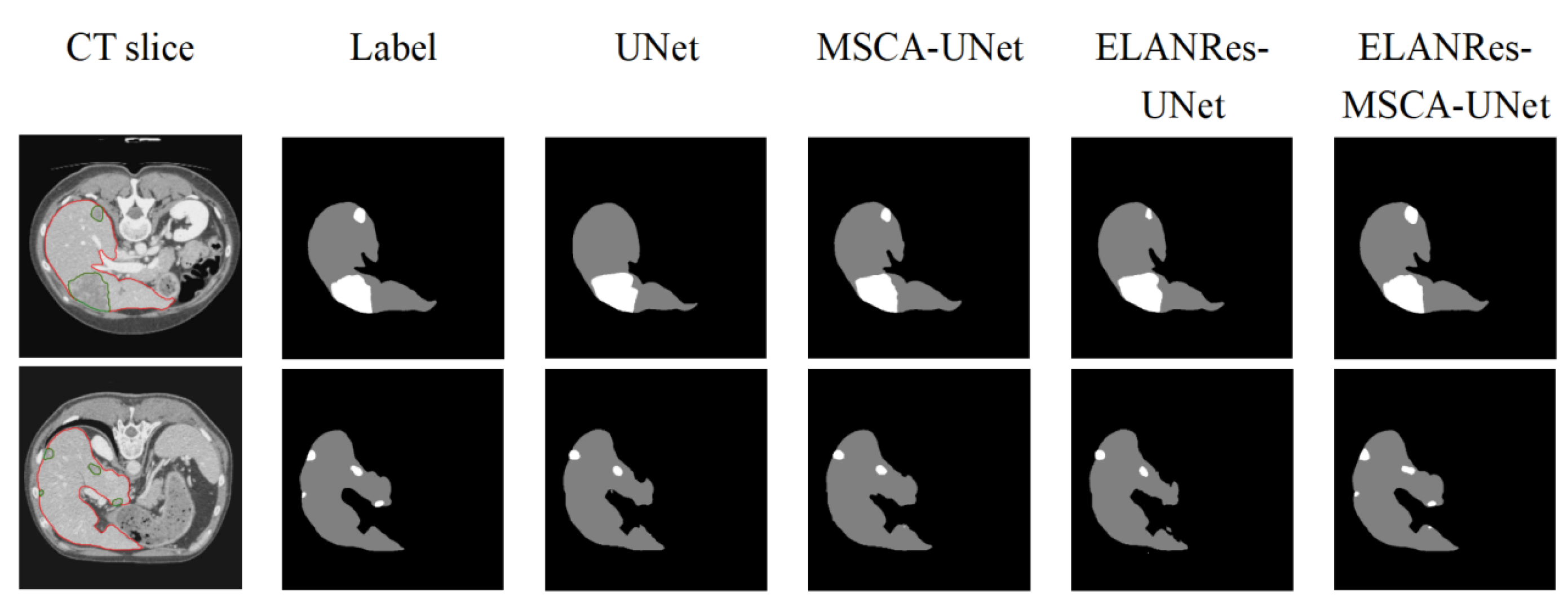
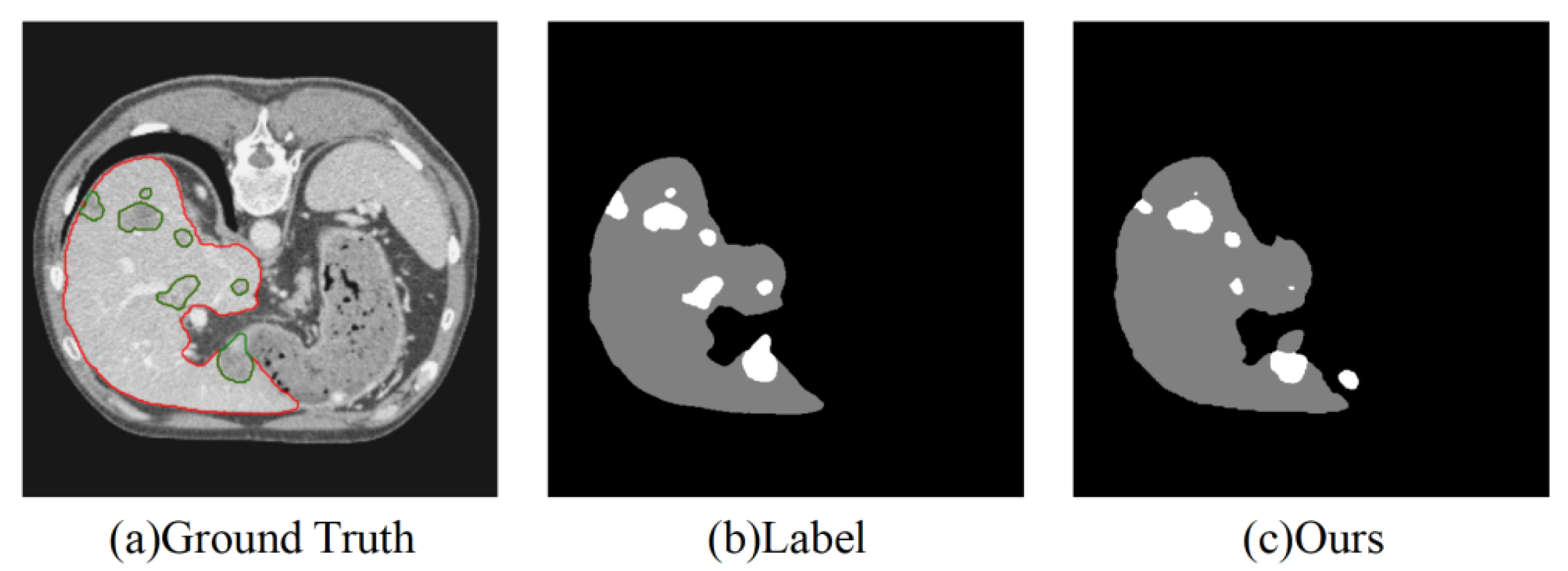
3.3. Comparison of Tumor Segmentation Performance Between Groups
3.4. Comparision with State-of-the-Art Methods
4. Discussion and Conclusions
Author Contributions
Funding Information
Data Availability Statement
Acknowledgments
Conflicts of Interest
References
- Chhikara, B.S.; Parang, K. Global Cancer Statistics 2022: the trends projection analysis[J]. Chemical Biology Letters 2023, 10, 451–451. [Google Scholar]
- Luan, S.; Xue, X.; Ding, Y.; et al. Adaptive attention convolutional neural network for liver tumor segmentation[J]. Frontiers in Oncology 2021, 11, 680807. [Google Scholar] [CrossRef]
- Tomoshige, S.; Oost, E.; Shimizu, A.; et al. A conditional statistical shape model with integrated error estimation of the conditions; application to liver segmentation in non-contrast CT images[J]. Medical image analysis 2014, 18, 130–143. [Google Scholar] [CrossRef] [PubMed]
- Wang X, Yang J, Ai D, et al. Adaptive mesh expansion model (AMEM) for liver segmentation from CT image[J]. PLoS one 2015, 10, e0118064.
- Park, H.; Bland, P.H.; Meyer, C.R. Construction of an abdominal probabilistic atlas and its application in segmentation[J]. IEEE Transactions on medical imaging 2003, 22, 483–492. [Google Scholar] [CrossRef] [PubMed]
- Xu, Z.; Burke, R.P.; Lee, C.P.; et al. Efficient multi-atlas abdominal segmentation on clinically acquired CT with SIMPLE context learning[J]. Medical image analysis 2015, 24, 18–27. [Google Scholar] [CrossRef] [PubMed]
- Wu, W.; Zhou, Z.; Wu, S.; et al. Automatic liver segmentation on volumetric CT images using supervoxel-based graph cuts[J]. Computational and mathematical methods in medicine 2016, 2016, 9093721. [Google Scholar] [CrossRef] [PubMed]
- Soler, L.; Delingette, H.; Malandain, G.; et al. Fully automatic anatomical, pathological, and functional segmentation from CT scans for hepatic surgery[J]. Computer Aided Surgery 2001, 6, 131–142. [Google Scholar] [CrossRef] [PubMed]
- Linguraru, M.G.; Richbourg, W.J.; Liu, J.; et al. Tumor burden analysis on computed tomography by automated liver and tumor segmentation[J]. IEEE transactions on medical imaging 2012, 31, 1965–1976. [Google Scholar] [CrossRef] [PubMed]
- Moltz, J.H.; Bornemann, L.; Kuhnigk, J.M.; et al. Advanced segmentation techniques for lung nodules, liver metastases, and enlarged lymph nodes in CT scans[J]. IEEE Journal of selected topics in signal processing 2009, 3, 122–134. [Google Scholar] [CrossRef]
- Massoptier, L.; Casciaro, S. A new fully automatic and robust algorithm for fast segmentation of liver tissue and tumors from CT scans[J]. European radiology 2008, 18, 1658–1665. [Google Scholar] [CrossRef] [PubMed]
- Conze, P.H.; Noblet, V.; Rousseau, F.; et al. Scale-adaptive supervoxel-based random forests for liver tumor segmentation in dynamic contrast-enhanced CT scans[J]. International journal of computer assisted radiology and surgery 2017, 12, 223–233. [Google Scholar] [CrossRef] [PubMed]
- Christ, P.F.; Elshaer ME, A.; Ettlinger, F.; et al. Automatic liver and lesion segmentation in CT using cascaded fully convolutional neural networks and 3D conditional random fields[C]//Medical Image Computing and Computer-Assisted Intervention–MICCAI 2016: 19th International Conference, Athens, Greece, October 17-21, 2016, Proceedings, Part II 19. Springer International Publishing, 2016: 415-423.
- Bilic, P.; Christ, P.; Li, H.B.; et al. The liver tumor segmentation benchmark (lits)[J]. Medical Image Analysis 2023, 84, 102680. [Google Scholar] [CrossRef] [PubMed]
- Li, X.; Chen, H.; Qi, X.; et al. H-DenseUNet: hybrid densely connected UNet for liver and tumor segmentation from CT volumes[J]. IEEE transactions on medical imaging 2018, 37, 2663–2674. [Google Scholar] [CrossRef] [PubMed]
- Dey, R.; Hong, Y. Hybrid cascaded neural network for liver lesion segmentation[C]//2020 IEEE 17th International Symposium on Biomedical Imaging (ISBI). IEEE, 2020: 1173-1177.
- Han, X. Automatic liver lesion segmentation using a deep convolutional neural network method[J]. arXiv:1704.07239, 2017.
- Gruber N, Antholzer S, Jaschke W; et al. A joint deep learning approach for automated liver and tumor segmentation[C]//2019 13th International conference on Sampling Theory and Applications (SampTA). IEEE, 2019: 1-5.
- Bi, L.; Kim, J.; Kumar, A.; et al. Automatic liver lesion detection using cascaded deep residual networks[J]. arXiv:1704.02703, 2017.
- Kaluva K C, Khened M, Kori A; et al. 2D-densely connected convolution neural networks for automatic liver and tumor segmentation[J]. arXiv preprint arXiv:1802.02182, 2018.
- Deng, Z.; Guo, Q.; Zhu, Z. Dynamic regulation of level set parameters using 3D convolutional neural network for liver tumor segmentation[J]. Journal of healthcare engineering 2019, 2019, 4321645. [Google Scholar] [CrossRef] [PubMed]
- Liu, T.; Liu, J.; Ma, Y.; et al. Spatial feature fusion convolutional network for liver and liver tumor segmentation from CT images[J]. Medical physics 2021, 48, 264–272. [Google Scholar] [CrossRef] [PubMed]
- Isensee, F.; Jaeger, P.F.; Kohl SA, A.; et al. nnU-Net: a self-configuring method for deep learning-based biomedical image segmentation[J]. Nature methods 2021, 18, 203–211. [Google Scholar] [CrossRef] [PubMed]
- Ding, X.; Zhang, X.; Ma, N.; et al. Repvgg: Making vgg-style convnets great again[C]//Proceedings of the IEEE/CVF conference on computer vision and pattern recognition. 2021, 13733–13742.
- Shao, H.; Zeng, Q.; Hou, Q.; et al. MCANet: Medical Image Segmentation with Multi-Scale Cross-Axis Attention[J]. arXiv:2312.08866, 2023.
- Goodfellow, I. Deep learning[J]. 2016.
- Vorontsov, E.; Tang, A.; Pal, C.; et al. Liver lesion segmentation informed by joint liver segmentation[C]//2018 IEEE 15th International Symposium on Biomedical Imaging (ISBI 2018). IEEE, 2018: 1332-1335.
- Liu, T.; Liu, J.; Ma, Y.; et al. Spatial feature fusion convolutional network for liver and liver tumor segmentation from CT images[J]. Medical physics 2021, 48, 264–272. [Google Scholar] [CrossRef] [PubMed]
- Yuan, Y. Hierarchical convolutional-deconvolutional neural networks for automatic liver and tumor segmentation[J]. arXiv:1710.04540, 2017.
- Chlebus G, Meine H, Moltz J H; et al. Neural network-based automatic liver tumor segmentation with random forest-based candidate filtering[J]. arXiv preprint arXiv:1706.00842, 2017.
- Yang D, Xu D, Zhou S K; et al. Automatic liver segmentation using an adversarial image-to-image network[C]//Medical Image Computing and Computer Assisted Intervention−MICCAI 2017: 20th International Conference, Quebec City, QC, Canada, September 11-13, 2017, Proceedings, Part III 20. Springer International Publishing, 2017: 507-515.

| Loss Functions | formula |
|---|---|
| Dice | |
| Tversky | |
| HybridLoss | |
| Jaccard | |
| SSLoss | |
| ELDice |
| Network | Data | Metrics | |
|---|---|---|---|
| Dice | ASD | ||
| UNet | LiTS | 0.937 | 3.242 |
| MSCA-UNet | LiTS | 0.953 | 2.891 |
| ELANRes-UNet | LiTS | 0.949 | 2.369 |
| ELANRes-MSCA-UNet | LiTS | 0.972 | 1.263 |
| Network | Data | Metrics | ||||
|---|---|---|---|---|---|---|
| Dice | ASD | RVD | Precision | Recall | ||
| UNet | LiTS | 0.583 | 1.130 | 0.031 | 0.552 | 0.383 |
| MSCA-UNet | LiTS | 0.631 | 1.197 | 0.170 | 0.426 | 0.431 |
| ELANRes-UNet | LiTS | 0.620 | 1.260 | 0.192 | 0.387 | 0.427 |
| ELANRes-MSCA-UNet | LiTS | 0.729 | 1.012 | 0.021 | 0.564 | 0.508 |
| Network | Data | Average major axis length of liver tumors/mm | |
|---|---|---|---|
| High-performance group | Low-performance group | ||
| ELANRes-MSCA-UNet | LiTS | 41.83 | 18.16 |
| UNet | LiTS | 44.72 | 23.37 |
| Method | Data | Metrics | |
|---|---|---|---|
| Dice | ASD | ||
| L. Bi et al. [19] | LiTS | 0.934 | 258.598 |
| Y. Yuan et al. [29] | LiTS | 0.963 | 1.104 |
| F. Isensee et al. [23] | LiTS | 0.962 | 2.565 |
| Z. Xu et al. [14] | LiTS | 0.959 | 1.342 |
| S. Chen et al. [14] | LiTS | 0.954 | 1.386 |
| Ours | LiTS | 0.972 | 1.263 |
| Method | Data | Metrics | ||||
|---|---|---|---|---|---|---|
| Dice | ASD | RVD | Precision | Recall | ||
| L. Bi et al. [19] | LiTS | 0.645 | 1.006 | 0.016 | 0.316 | 0.431 |
| J. Zou et al. [14] | LiTS | 0.702 | 1.189 | 5.921 | 0.148 | 0.479 |
| J. Li et al. [15] | LiTS | 0.686 | 1.073 | 5.164 | 0.436 | 0.515 |
| X. Han et al. [17] | LiTS | 0.674 | 1.118 | -0.103 | 0.354 | 0.458 |
| G. Chlebus et al. [30] | LiTS | 0.676 | 1.143 | 0.464 | 0.519 | 0.463 |
| D. Xu et al. [31] | LiTS | 0.721 | 0.896 | -0.002 | 0.549 | 0.503 |
| Ours | LiTS | 0.729 | 1.012 | 0.021 | 0.564 | 0.508 |
Disclaimer/Publisher’s Note: The statements, opinions and data contained in all publications are solely those of the individual author(s) and contributor(s) and not of MDPI and/or the editor(s). MDPI and/or the editor(s) disclaim responsibility for any injury to people or property resulting from any ideas, methods, instructions or products referred to in the content. |
© 2025 by the authors. Licensee MDPI, Basel, Switzerland. This article is an open access article distributed under the terms and conditions of the Creative Commons Attribution (CC BY) license (http://creativecommons.org/licenses/by/4.0/).


