

Submitted:
20 January 2025
Posted:
21 January 2025
You are already at the latest version
Abstract
INTRODUCTION: Peripheral prosthetic vascular graft infection represents a very serious complication after lower limb revascularization, with amputation and mortality rates up to 70% and 30%, respectively. The study was designed to determine the incidence of prosthetic graft infection, amputation and mortality rate in our Institution, analyzing different types of treatment.
MATERIALS AND METHODS: A retrospective cohort single institution review of peripheral prosthetic bypass grafts evaluated patient demographics, comorbidities, indications, location of bypass, type of prosthetic material, case urgency and evaluated the incidence of graft infections, amputations, and mortality.
RESULTS: Between January 2016 and December 2021, a total of 516 bypass were recorded: 320 bypass in venous material and 196 prosthetic bypass using Dacron or PTFE.Among patients with a prosthetic bypass, 16 (8.2%) presented a graft infection at a mean follow-up of 39 months. Other 13 patients submitted to prosthetic peripheral bypass in other centres occurred to our Institution with a graft infection, so a total of 29 infected grafts were treated. Infected grafts were removed in 20 patients (68.9%), while a conservative treatment was helpful in 9 cases (31.1%). The germs involved were gram-negative in 27.6% and gram-positive in 41.4%. During follow-up we recorded 5 deaths (17.2%) and 6 amputations (20.7%) directly after bypass excision; another 2 amputations (6.9%) occurred after failure of the new bypass replacing the prosthesis removed.
CONCLUSIONS: Redo-bypass, active infection at the time of bypass and advanced gangrene were associated with a higher risk for prosthetic graft infection and major extremity amputation. Complete graft removal and replacement by venous material or Omniflow II represents the typical treatment. However, aggressive local treatment including drainage, debridement, vacuum assisted closure therapy application and muscle transposition seems to be a better solution in selected patients without the need for graft removal and with rates of limb salvage superior to those obtained with excisional therapy.
Keywords:
critical lower limb ischemia
; bypass
; peripheral prosthetic vascular graft infection
; vacuum assisted closure therapy.
1. Introduction
Peripheral prosthetic vascular graft infection (PVGI) after surgical revascularization for critical lower limb ischemia (CLI) is a serious complication with a reported incidence that ranges from 0.7 to 7% [1,2] and post-operative amputation and mortality rates up to 70% and 30%, respectively. [3] Predictors of surgical site infections after surgical bypass include obesity, diabetes, poor preoperative functional status, a history of smoking, redo-bypass and female gender.[4,5]
Aside from the adverse effect on the patient, postoperative vascular surgical infections significantly contribute to health care costs, as they often require additional procedures and a long hospital stay.[6] The type of treatment depends on the location of the prosthesis, the extent of the infection, the degree of stenosis and/or occlusion, the patient's general condition and his life expectancy.
Excision of the infected prosthetic graft, with or without reconstruction, has been considered conventional therapy [7,8,9,10,11]. It has long been recognized, however, that not all infected bypasses need to be removed. Recent reports have described the treatment of infected lower extremity arterial grafts with aggressive local tissue debridement, administration of local and systemic antibiotics, and coverage by muscle transposition.[12,13,14,15] as well as graft preservation with the use of vacuum assisted closure (VAC) devices, with or without muscle flap coverage.[16,17,18,19,20,21]
In the present study, we report our personal experience about PVGI management after lower extremity bypass, examining the impact of PVGI on post-operative amputation and death rates.
2. Materials and Methods
We performed a retrospective study of all patients presenting a peripheral prosthetic bypass infection in our Institution of Vascular Surgery, a referral centre for CLI treatment.
Between January 2016 and December 2021 a total of 516 bypass were recorded: 320 bypass in venous material and 196 prosthetic bypass using Dacron or PTFE (41 axillo-femoral, 43 femoro-femoral crossover, 27 ilio-femoral, 44 ilio or femoro-tibial, 41 femoro-popliteal bypass).
A patient was considered as having definite PVGI if at least two of the three following criteria were present: 1) positive bacterial culture of intraoperative specimens or blood samples; 2) clinical signs of infection [general (fever, chills, septic shock) or in the area of the prosthesis (e.g. inflammatory signs in the area of the vascular graft: local pain, erythema or tumefaction, sinus tract infection communicating with PVGI, intraoperative gross purulence or failure of graft consolidation]; 3) biological signs of infection (C-reactive protein > 5 mg/L, white blood count > 12 G/L).
Each case of definite infection was classified as early-onset infection (occurring within 3 months after surgery) or as late-onset infection (occurring more than 3 months after surgery).
3. Results
Among 196 prosthetic bypass performed in our Institution, a post-surgical PVGI occurred in 16 cases (8.2%) between 7 days and 3 years after the implantation. Other 13 patients submitted to a prosthetic peripheral bypass in other Institutions, occurred to our Unit presenting a PVGI, so a total of 29 cases of prosthetic peripheral graft infection were treated. The infection occurred in 18 males and 11 females, presenting a mean age of 73.9. Follow-up time ranged from 18 months to 5 years and started from the initial operation. Demographic and clinical characteristics of the patients, germs involved, typology of treatment and outcomes are reported in Table 1. The patients presented the following risk factors and comorbidities: diabetes mellitus (21, 72.4%), hypertension (23, 79.3%), dislipidemia (17, 58.6%), smoke (20, 68.9%), chronic renal failure on hemodialysis (4, 13.8%), chronic obstructive pulmonary disease (11, 37.9%), obesity (9, 31%), coronary artery disease (19, 65.5%). The revascularization procedures performed before the onset of infection are listed in Table 2. The indications were tissue loss (21, 72.4%) and rest pain (8, 27.6%), with 10 (34.5%) redo-bypass after failure of previous multiple attempts of revascularization through a venous or composite bypass. The infected graft was politetrafluoroethylene (PTFE) in 25 cases (86.2%) and Dacron in 4 cases (13.8%). The time from prosthesis implantation to development of PVGI was described as early in 18 cases (62.1%) and late in 11 cases (37.9%). The modes of graft infection presentation were: bleeding (4, 13.8%), acute ischemia of the lower limb (3, 10.3%) of which one with associated distal septic skin lesions (22,23), occluded graft (2, 6.9%), false aneurysm (6, 20.7%); suture dehiscence with persistent discharge (9, 31.1%), abscess (3, 10.3%), seroma (2, 6.9%). Fever > 38°C was recorded in 21 cases (72.4%), while elevation of white blood cells count (range 12-25), C-reactive protein (range: 5-20) and procalcitonin (PCT) values (range 2-28) was observed in all cases. Aerobic and anaerobic cultures were obtained with swabs from the grafts during operations in 22 cases (75.9%) and from dehiscent wounds before operation in 7 patients (24.1%). Broad-spectrum antibiotics were given, both perioperatively (with specific antibiotics to cover any cultured organism) and postoperatively. In cases where the responsible organism was not known preoperatively, cefazolin or vancomycin was started immediately before surgery and continued for 1 week. This was modified to cover any organism grown preoperatively or from cultures taken at operation, on the basis of antibiogram. Bacteriology was available in 23 of 29 cases. Among Gram negative organisms isolated (8 cases, 27.6%), Pseudomonas aeruginosa was cultured in six cases and in two cases in association with Escherichia coli. Two patients with Pseudomonas aeruginosa infection experienced amputation: one patient was impossible to revascularize and amputation followed the bypass excision. One patient underwent to bypass excision and replacement with a femoro-tibial bypass in cephalic vein. Amputation followed the venous graft failure. One patient, after excision of a femoro-popliteal bypass underwent to a new bypass using Omniflow II and muscle flap transposition, with limb salvage. Three patients with a distal bypass infection and dehiscence of a surgical wound were submitted to multiple surgical debridements and various sessions of VAC therapy, with subsequent limb salvage. About the two patients with infection caused by P. aeruginosa and E. coli, the patient with an axillofemoral bypass received the bypass excision and replacement with a new bypass in a clean field, with limb salvage. The other one, with dehiscence of the groin wound underwent to surgical debridement of the wound and various sessions of VAC therapy, experiencing a limb salvage. The majority of germs cultured were gram positive (12 cases, 41.4%): Methicillin-resistent Staphylococcus aureus (MRSA) in four cases; Methicillin-sensitive Staphylococcus aureus (MSSA) in four cases; Staphylococcus warneri, Staphylococcus lugdunensis and Staphylococcus epidermidis respectively, in one case; Enterococcus faecalis in one case. One patient with infection caused by MRSA experienced amputation, after bypass excision and death during follow-up for different causes. One patient underwent to axillo-femoral bypass excision but he did not require revascularization. The patient with an ilio-femoral bypass infection received surgical debridement of the groin wound, VAC therapy application and bypass coverage with a muscle flap transposition. This patient experienced limb salvage. In the case of femoro-popliteal bypass infection and dehiscence of the groin wound, after surgical debridement of the wound and VAC therapy, the patient healed. About patients with MSSA infection, we recorded an amputation after bypass removal and three limb salvage: one patient with prosthetic leg infection of an axillo-femoral bypass, experienced healing after surgical debridement of the groin wound and VAC therapy; one patient with a femoro-femoral crossover bypass, after bypass excision received an axillo-bifemoral bypass; the last patient false aneurysm at the distal anastomosis of a femoro-popliteal bypass, underwent bypass excision and replacement with a femoro-peroneal bypass in GSV followed by limb salvage. Three patients with a femoro-popliteal bypass infection caused by Staphylococcus lugdunensis, Staphylococcus warneri and Enterococcus faecalis respectively, underwent to bypass removal and replacement with a femoro-distal bypass in GSV, with limb salvage. The patient with infection of an axillo-femoral bypass caused by S. epidermidis experienced limb salvage after graft removal and replacement with a new bypass in a clean field. In three cases an infection caused by the association of gram positive and gram negative organisms was recorded. In two cases we observed an infection cause by MRSA and P. aeruginosa: one patient with a femoro-popliteal bypass infection resulted impossible to revascularize and she underwent to amputation and death for sepsis during hospital stay; the other one received a venous femoro-distal bypass after prosthesis excision, with limb salvage. A patient with infection of a femoro-popliteal bypass by Proteus mirabilis and Staphylococcus aureus received graft removal and replacement with Omniflow II associated with a muscle flap transposition. After occlusion of the Omniflow II graft, she underwent to amputation and death during follow-up for comorbidities.
In 6 cases the germ responsible of the graft infection was not identified. Three of these patients presented a femoro-tibial infected bypass: we recorded two amputation after bypass removal in patients impossible to revascularize. One of these patients underwent to surgical debridement of the wound, proximal bypass removal and replacement with Omniflow II and coverage through a muscle flap transposition, with subsequently limb salvage. One patient with ilio-femoral bypass and development of an abscess on the surgical site, received multiple surgical debridement, VAC therapy followed by bypass coverage through a muscle transposition, with subsequently limb salvage. A patient with a late infection of a femoro-femoral crossover bypass underwent to bypass excision without need of revascularization and subsequent limb salvage.
The last patient presented a proximal false aneurysm of a femoro-femoral crossover bypass: he underwent to surgical repair of the false aneurysm with bypass preservation and consequently limb salvage.
In conclusion, the primary mode of treatment was represented by prosthesis excision (20 cases, 68.9%: 17 entire prosthesis removal and 3 partial graft excisions): 9 patients received a new bypass; 6 patients resulted impossible to revascularize and in 2 cases the revascularization was not required. Three patient with false aneurysm on the proximal anastomosis underwent to removal of the proximal segment of the bypass, replaced using Omniflow II. Among the remaining nine cases (31.1%), one patient with femoro-femoral crossover bypass underwent to the surgical repair of a proximal false aneurysm with bypass preservation and limb salvage. Eight patients experienced a successful treatment through multiple surgical wound debridements and VAC therapy application (two femoro-popliteal, one ilio-peroneal, one iliofemoral, one femoro-tibial, one femoro-peroneal and one axillo-femoral bypass infections). (Figure 1, Figure 2 and Figure 3) VAC- therapy was usually started the day after the wound surgical revision: a polyurethane sponge (KCI Medical, San Antonio, TX, USA) was applied with a continuous topical negative pressure of 125 mmHg. Changes of dressings were performed ever three days, with a medium of five sessions for patient (mean duration of treatment: 15 days). Six patients (20.7%) underwent to amputation directly after infected bypass removal; two patients (6.9%) experienced amputation after occlusion of a new bypass replacing the removed infected prosthesis. Five deaths (17.2%) occurred during follow-up, three in patients underwent to amputation: one death occurred during hospital stay for sepsis (blood culture positive for MRSA) while the remaining cases were recorded during follow-up for comorbidities.
4. Discussion
PVGI after surgical revascularization for CLI represents a serious complication with high rates of mortality and amputation.[1,2,3] Most graft infections occur in the groin, usually in the early postoperative period, typically as a result of a progressive surgical site infection.[1,24] Comorbid risk factors associated with PVGI include the presence of ischaemic leg ulcers, diabetes, smoking, infected lymph nodes, and transient bacteraemia from a purulent draining wound or sinus, an abscess, a lymphocele, skin necrosis, pain, septic emboli with petechia, a pulsatile mass, fever, or graft thrombosis. [1,14,24,25,26] Actually, there is no consensus on diagnostic criteria or on best management of PVGI. Antibiotic therapy is the first stage of management of PVGI, followed by prosthesis surgical removal and reconstruction with autologous material. In the absence of appropriate venous graft material, prosthetic grafts with a potentially higher resistance to reinfection (e.g. silver bonded or rifampicin bonded) can be used alternatively, although reinfection rates are higher compared to venous grafts [27] and long-term patency is significantly lower than in venous bypasses. However, experiences with the use of biosynthetic grafts as graft material in elective infrainguinal revascularisation procedures have shown acceptable secondary patency and limb salvage rates [28]. In particular, Omniflow I presented an higher early occlusion rates due to the initial thrombogenicity and the disposition to aneurysmal degeneration that impeded their widespread clinical use. [29] The improved collagen structure of the second generation graft (Omniflow II, Bio Nova International,
Victoria, Australia) is supposed to increase wall strength and stability to reduce aneurysmal changes. Remarkably, the infection rate of biosynthetic grafts in elective femoropopliteal reconstructions is very low. [30]
Topel et al. [31] reported seven cases of infected infrainguinal prosthetic grafts that were explanted and replaced by Omniflow II®, in absence of suitable peripheral vein. They did not recorded early or late reinfection nor amputations, with graft occlusion in three cases and one death for pneumonia 11 months postoperatively.
Chaudhry [32] published a case of a mycotic aneurysm of the common femoral artery successfully reconstructed with an Omniflow II prosthesis in 2008. Another group reported of a patient with an infected prosthetic infrainguinal graft, also replaced with an Omniflow II prosthesis [33]. In both cases no reinfection occurred.
A less invasive alternative to prosthesis removal is represented by the conservative treatment, with surgical debridement of the wound and VAC therapy application, with or without muscle transposition of coverage. As already described in other districts [34,35], VAC-therapy has been reported to have several beneficial effects on wound healing, such as creating a moist wound-healing environment, drainage of superfluous fluid, reduction of tissue oedema, cleansing deep wounds from bacteria, accelerating the formation of vascularised granulation tissue and faster approximation of wound edges.[36,37]
Acosta et al. [40] described 28 synthetic grafts infections treated by VAC-therapy for a mean period of 20 days. Two serious bleeding episodes from the suture lines occurred. The proportion of patent bypass grafts was 91% at a median time of 3.5 months from the start of the treatment. Five patients with seven bypasses had persistent infection or re-infection, and the total graft preservation rate was 76%.
Mayer et al [19] reported the outcomes of 44 polymorbid patients with Szilagyi III infections. Forty grafts (prosthetic = 24, vein = 3, biological = 13) and 9 native arteries were involved. VAC-therapy was applied directly on grafts/arteries after radical debridement of infected tissue with a median duration of 33 days (IQR: 20-78), and an hospital stay of 32 (IQR: 20-82) days. One-year mortality was 16% (7/44). Long-term mortality after a mean follow-up of 43 months (SD: 21) was 41% (18/44). Complete wound healing was achieved in 91% (40/44). In 37 of 44 patients, grafts were preserved long-term without reinfection.
In our Institution, among 196 prosthetic bypass performed between 2016 and 2021, 16 (8.2%) presented a PVGI. In the same period, other 13 patients submitted to prosthetic peripheral bypass in other centres, occurred to our Unit presenting a PVGI. So, a total of 29 patients underwent to multimodal treatments for PVGI in our Institution. The primary mode of treatment was represented by prosthesis excision (20 cases, 68.9%: 17 entire prosthesis removal and 3 partial graft excisions): nine patients received a new bypass; six patients with redo-bypass after multiple attempts of lower limb revascularization resulted impossible to revascularize and underwent directly to amputation; in two cases the revascularization was not required. Three patients with false aneurysm on the proximal anastomosis underwent to removal of the proximal segment of the bypass, replaced using Omniflow II, accompanied by muscle transposition of coverage. More recently, we recurred to VAC therapy after surgical debridements of the dehiscent wound in eight patients (27.6%). During follow-up we recorded five deaths (17.2%), of which only one related to graft infection and eight (27.6%) amputations. The amputation occurred in patients with history of multiple attempts of revascularization: six patients (20.7%) underwent to amputation directly after bypass excision, while two patients (6.9%) after a last attempt of revascularization. In accordance with literature data [41], the analysis of the data collected in our Institution suggests that PVGI in patients with redo-bypasses after failure of multiple attempts of revascularization and the presence of advanced gangrene and active infection at the time of operation (Rutherford 6 class), represent an important risk factor for amputation. [42] In addition, the serious comorbidities recorded in patients affected by CLI are often responsible of the death.
Cherry et al [43] reviewed 39 infected lower extremity bypasses (33 prosthetic bypass,4 venous bypass, and composite in 2). Twenty-eight infected grafts were treated with complete graft removal and 9 new grafts were placed. Recurrent infection developed in five cases, and two patients died of complications of graft infection. Eleven patients with patent bypasses were treated without graft excision: muscle transposition in five cases, incision and drainage of abscesses in five cases, and excision of a persistent sinus tract in one case. One patient underwent major amputation 6.3 years after treatment of graft infection. Limb salvage was significantly higher (p = 0.012, log-rank test) than in patients treated with graft excision. One patient died, and no recurrent infections developed.
Siracuse et al [41] reported their experience about 496 prosthetic grafts with a graft infection rate of 3.8% (19) at a mean follow-up of 27 months. Multivariable analysis showed that redo bypass, active infection at the time of bypass, female gender and diabetes mellitus were significant predictors of graft infection. Graft infection was predictive of major lower extremity amputation (HR, 9.8; 95% CI, 3.5-27.1), as was preoperative tissue loss (HR, 4.7; 95% CI, 1.8-11.9). Infected grafts were removed 79% of the time. In the first 7 years of this study, all grafts except one were removed. However, in the latter 3 years, 50% were preserved by debridement with placement of a VAC device, with or without local rotational muscle flap closure.
Zetrenne et al. [2] described 45 cases of PVGI with 11 amputations (24%) and three perioperative deaths related to graft infection; the other eight deaths were from various unrelated causes. Overall, there was a 6 % perioperative mortality rate in this series. The primary mode of therapy was muscle flap transposition (18 cases, 40%), alone (n = 15) or with in situ graft replacement (n = 3). Ten (67 %) of 15 grafts were preserved with muscle flap transposition. Samson group 3 (n = 30) cases were treated mostly by graft excision (n = 16; 53%), and fewer than half were preserved (n = 14; 46%).The Samson group 4 (n= 9) and group 5 (n = 6) cases were managed with a variety of surgical treatments: ex situ bypass (n = 4; 27 %), graft excision alone (n = 3; 20%),muscle flaps (n = 3; 20%), and in situ bypass with muscle flap (n = 2; 13%). Additionally, irrigation and debridement according to Kwaan and Connolly’s method [12] was performed on three patients (20%).
5. Conclusions
In conclusion, PVGI represents a very serious complication after lower extremity bypass for CLI. Redo-bypass, active infection at the time of bypass and advanced gangrene are associated with a higher risk for PVGI and major extremity amputation. Complete graft removal and replacement by venous material or Omniflow II represents the typical treatment. However, aggressive local treatment including drainage, debridement, VAC-therapy application and muscle transposition seems to be a better solution in selected patients without the need for graft removal and with rates of limb salvage superior to those obtained with excisional therapy. Prospective multicenter well designed randomized clinical trials are needed to better understand for which patient a conservative approach should be recommended.
Author Contributions
Conceptualization, G.D.C, M.M and F.B.; methodology, G.D.C, M.M and F.B..; software, G.D.C. and M.M. ; validation, G.D.C, M.M and F.B.; formal analysis, G.D.C, M.M and F.B.; investigation, G.D.C, M.M and F.B.; resources,G.D.C, M.M and F.B., data curation, G.D.C, M.M and F.B.; writing—original draft preparation, G.D.C, M.M and F.B.; writing—review and editing, G.D.C, M.M and F.B.; visualization, G.D.C, M.M, C.B. and F.B.; supervision, G.D.C, M.M, C.B., and F.B.
Funding
This research received no external funding.
Institutional Review Board Statement
The study was conducted in accordance with the Declaration of Helsinki.
Informed Consent Statement
Written informed consent has been obtained from the patients to publish this paper.
Data Availability Statement
Data is unavailable due to privacy and institutional restrictions.
Conflicts of Interest
The authors declare no conflicts of interest.
References
- Kimmel RM, Murphy RXJ, Chowdary RP. Optimal management of inguinal vascular graft infections. Ann Plast Surg 1994; 32:623 – 629. [CrossRef]
- Zetrenne E, McIntosh BC, McRae MH, et al. Prosthetic vascular graft infection: a multi-center review of surgical management. Yale J Biol Med 2007; 80: 113 – 121.
- Herrera FA, Kohanzadeh S, Nasseri Y, et al. Management of vascular graft infections with soft tissue flap coverage: improving limb salvage rates – a veterans affairs experience. Am Surg 2009; 75: 877 – 881. [CrossRef]
- Giles KA, Hamdan AD, Pomposelli FB, et al. Body mass index: surgical site infections and mortality after lower extremity bypass from the National Surgical Quality Improvement Program 2005–2007. Ann Vasc Surg. 2010 Jan; 24(1):48–56. [CrossRef]
- Greenblatt DY, Rajamanickam V, Mell MW. Predictors of surgical site infection after open lower extremity revascularization. J Vasc Surg. 2011 Aug; 54(2):433–9. [CrossRef]
- Vogel TR, Dombrovskiy VY, Carson JL, et al. Infectious complications after elective vascular surgical procedures. J Vasc Surg. 2010 Jan; 51(1):122–9. [CrossRef]
- Wilson, SE. New alternatives in management of the infected vascular prosthesis. Surg Infect. 2001;2(2):171-7. [CrossRef]
- Williams M, Milling M, Shandall A. Vascularized muscular flaps and arterial graft infection in the groin. Eur J Vasc Surg. 2003;25:390-95.
- Mertens RA, O'Hara PJ, Hertzer NR, et al. Surgical management of infrainguinal arterial prosthetic graft infections: review of a thirty-five-year experience. J Vasc Surg. 1995;21(5):782-91. [CrossRef]
- Henke PK, Bergamini TM, Rose SM, et al. Current options in prosthetic vascular graft infection. Am Surg. 1998;64(1):39-46.
- Morasch MD, Sam AD 2nd, Kibbe MR, et al. Early results with use of gracilis muscle flap coverage of infected groin wounds after vascular surgery. J Vasc Surg. 2004 Jun; 39(6): 1277–83.
- Kwaan JH, Connolly JE. Successful management of prosthetic graft infection with continuous povidone-iodine irrigation. Arch Surg. 1981;116(5):716-20. [CrossRef]
- Calligaro KD, Veith FJ, Gupta SK et al. A modified method for management of prosthetic graft infections involving an anastomosis to the common femoral artery. J Vasc Surg. 1990; 11: 485-492.
- Mixter RC, Turnipseed WD, Smith DJ, et al. Rotational muscle flaps: a new technique for covering infected vascular grafts. J Vasc Surg 1989;9:472–8.
- Meland NB, Arnold PG, Pairolero PC. Infected vascular prosthesis management with muscle transposition. Plast Reconstr Surg. 1994 Apr;93(5):1005-11.
- Edwards WH Jr, Martin RS 3rd, Jenkins JM, et al. Primary graft infections. J Vasc Surg. 1987 Sep; 6(3):235–9.
- Dosluoglu HH, Loghmanee C, Lall P, et al. Management of early (<30 day) vascular groin infections using vacuum-assisted closure alone without muscle flap coverage in a consecutive patient series. J Vasc Surg. 2010 May; 51(5):1160–6. [CrossRef]
- Landry GJ, Carlson JR, Liem TK, et al. The sartorius muscle flap: an important adjunct for complicated femoral wounds involving vascular grafts. Am J Surg. 2009 May; 197(5):655–9.
- Mayer D, Hasse B, Koelliker J, et al. Long-term results of vascular graft and artery preserving treatment with negative pressure wound therapy in Szilagyi grade III infections justify a paradigm shift. Ann Surg. 2011 Nov;254(5):754-59.
- Monsen C, Wann-Hansson C, Wictorsson C, et al. Vacuum-assisted wound closure versus alginate for the treatment of deep perivascular wound infections in the groin after vascular surgery. J Vasc Surg. 2014 Jan;59(1):145-51. [CrossRef]
- Hisata Y, Hashizume K, Tanigawa K, et al. Vacuum-assisted closure therapy for salvaging a methicillin-resistant Staphylococcus aureus-infected prosthetic graft. Asian J Surg. 2014 Jan;37(1):46-8. [CrossRef]
- Massara M, Cascio A, Benedetto F, et al. Septic skin lesions: an uncommon manifestation of peripheral prosthetic vascular graft infection. Acta medica mediterranea 2014; 30: 651-654.
- Massara M, De Caridi G, Barberi G, et al. Peripheral septic arterial embolism of non cardiac source: A systematic review of literature. Reviews in Vascular Medicine. Dec 2014,Vol.2(4):127135. [CrossRef]
- Piano, G. Infections in lower extremity vascular grafts. Surg Clin North Am 1995;74:799–809. [CrossRef]
- Edwards MJ, Richardson JD, Klamer TW. Management of aortic prosthetic infections. Am J Surg 1988;155:327–30. [CrossRef]
- Reilly LM, Ehrenfeld WK, Goldstone J, et al. Gastrointestinal tract involvement by prosthetic graft infection. The significance of gastrointestinal hemorrhage. Ann Surg 1985; 202:342–8. [CrossRef]
- O’Connor S, Andrew P, Batt M, et al. A systematic review and meta-analysis of treatments for aortic graft infection. J Vasc Surg 2006; 44: 38 – 45. [CrossRef]
- Neufang A, Doemland M. Clinical results with the denaturated human umbilical vein (HUV) and the ovine collagen prothesis (Omniflow) as small caliber prosthetic vascular replacement. Gefäßchirurgie 2010; 15: 83 – 89.
- Amman W, Tiesenhausen K, Fruwirth J, et al. Aneurysms in biosynthetic grafts. Z Herz Thorax Gefäßchirurgie 2000; 14: 113 – 116.
- Koch G, Gutschi S, Pascher O, et al. Analysis of 274 Omniflow Vascular Prosthesis implanted over an eight-year period. Aust N Z J Surg. 1997; 67: 637 – 639.
- Töpel I1, Betz T, Uhl C, et al. Use of biosynthetic prosthesis (Omniflow II®) to replace infected infrainguinal prosthetic grafts--first results. Vasa. 2012 May;41(3):215-20.
- Chaudhry A, Shetty R, Crimmin S, et al. Bio-synthetic Graft Repair of Mycotic Aneurysm of the Common Femoral Artery. EJVES Extra 2009; 18: 1 – 2. [CrossRef]
- Ozelci A, Yetkin U, Akyuz M, et al. A clinical application of the Omniflow II collagen- polymer vascular prosthesis in a polytetrafluoroethylene graft infection case. Int J Th or Cardiovasc Surg 2009.
- De Caridi G, Serra R, Massara M, et al. VAC therapy for the treatment of complex wounds after cardio-thoracic surgery. Int Wound J. 2014 Sep 17. [CrossRef]
- De Caridi G, Massara M, Greco M, et al. VAC therapy to promote wound healing after surgical revascularisation for critical lower limb ischaemia. Int Wound J. 2014 May 28. [CrossRef]
- Svensson S, Monsen C, Kölbel T, et al. Predictors for outcome after vacuum assisted closure therapy of peri-vascular surgical site infections in the groin. Eur J Vasc Endovasc 2008;36:84e9. [CrossRef]
- Pinocy J, Albes JM, Wicke C, et al. Treatment of periprosthetic soft tissue infection of the groin following vascular surgical procedures by means of polyvinyl alcohol-vacuum sponge system. Wound Repair Regen 2003;11:104e9. [CrossRef]
- Dee, A. The successful management of a dehisced surgical wound with TNP following femoropopliteal bypass. J Wound Care 2007;16:42e4. [CrossRef]
- Hisata Y, Hashizume K, Tanigawa K, et al. Vacuum-assisted closure therapy for salvaging a methicillin-resistant Staphylococcus aureus-infected prosthetic graft. Asian J Surg. 2014 Jan;37(1):46-8. [CrossRef]
- Acosta S, Monsen C. Outcome after VAC® therapy for infected bypass grafts in the lower limb. Eur J Vasc Endovasc Surg. 2012 Sep;44(3):294-9.
- Siracuse JJ, Nandivada P, Giles KA, et al. Prosthetic graft infections involving the femoral artery. J Vasc Surg. 2013 Mar;57(3):700-5. [CrossRef]
- Massara M, De Caridi G, Serra R, et al. The role of procalcitonin as a marker of diabetic foot ulcer infection. Int Wound J. 2015 Oct 28. [CrossRef]
- Cherry KJ Jr, Roland CF, Pairolero PC, et al. Infected femorodistal bypass: is graft removal mandatory? J Vasc Surg. 1992 Feb;15(2):295-303.
Figure 1.
This is a figure that shows surgical wound dehiscence with prosthesis exposure on the right leg, after femoro-peroneal bypass in PTFE.
Figure 1.
This is a figure that shows surgical wound dehiscence with prosthesis exposure on the right leg, after femoro-peroneal bypass in PTFE.
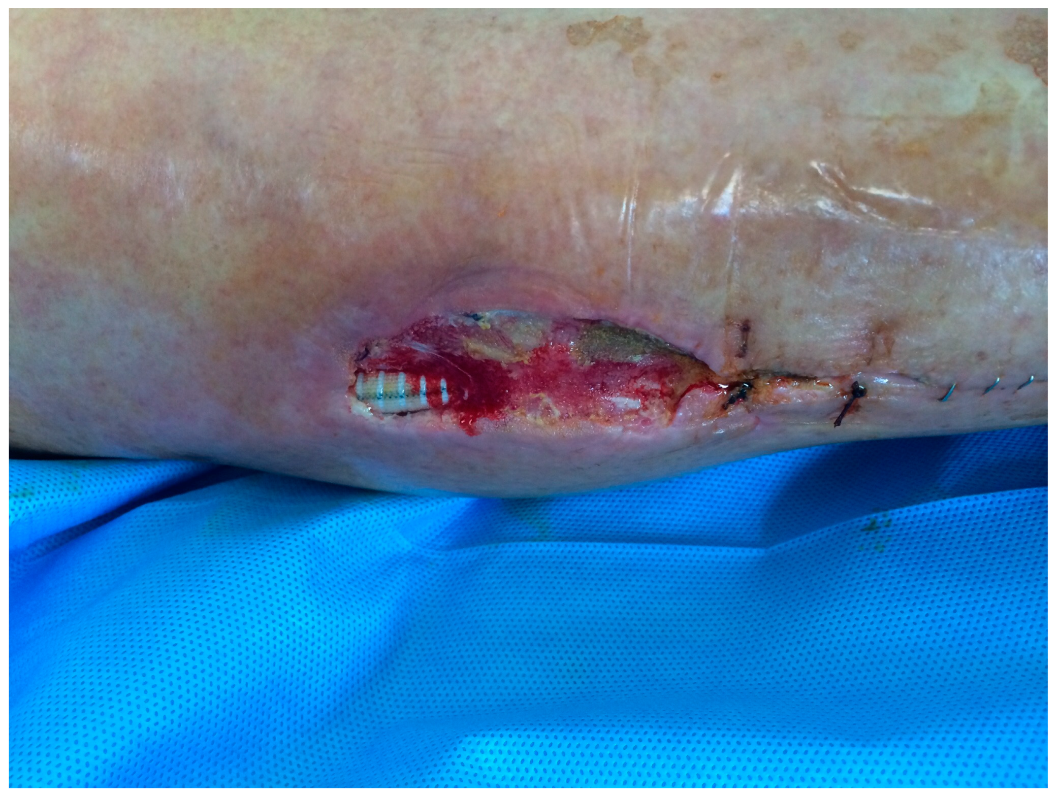
Figure 2.
VAC therapy application on the dehiscent wound after surgical wound debridements.
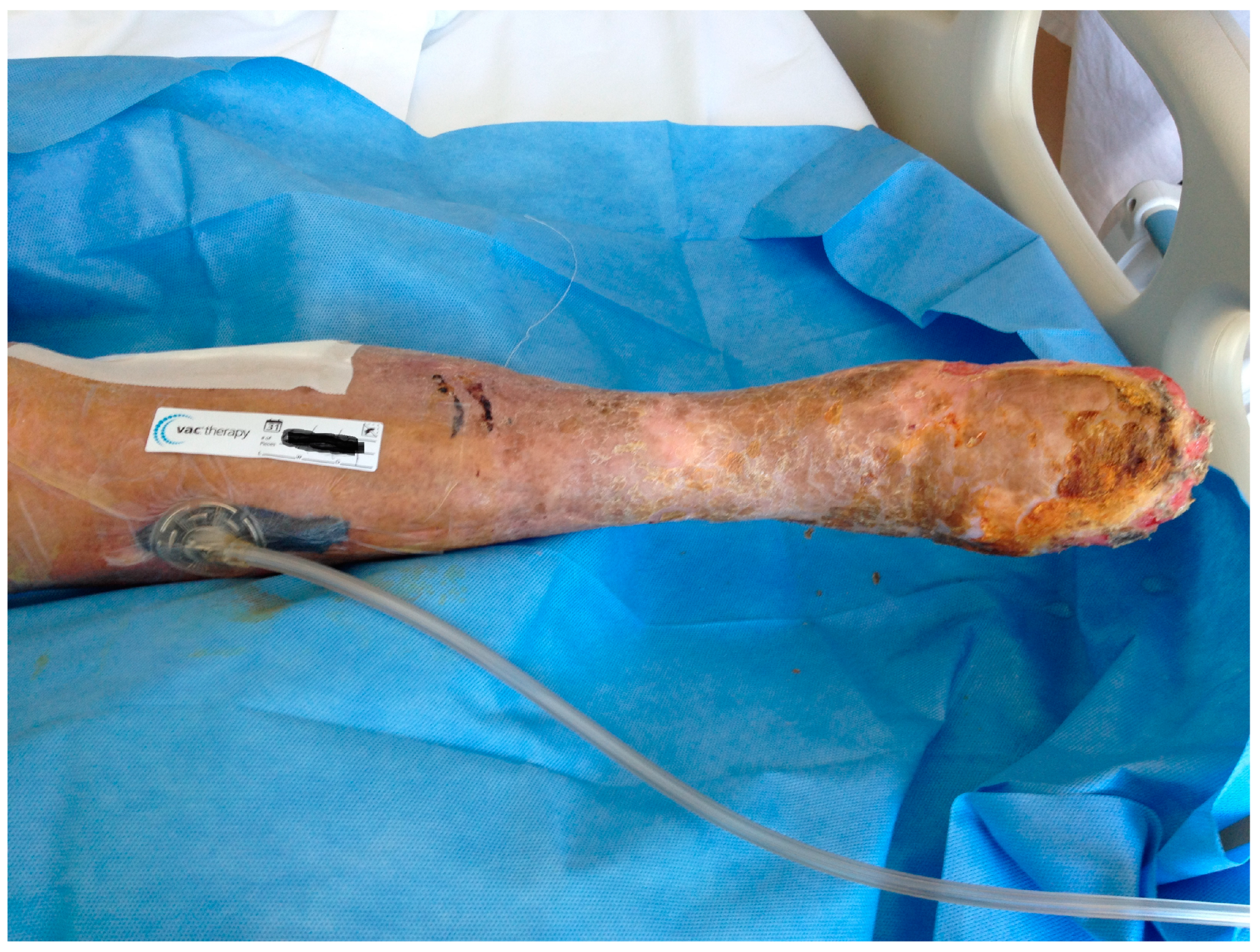
Figure 3.
Outcome after 3 sessions of VAC-therapy application with almost complete coverage and wound healing.
Figure 3.
Outcome after 3 sessions of VAC-therapy application with almost complete coverage and wound healing.
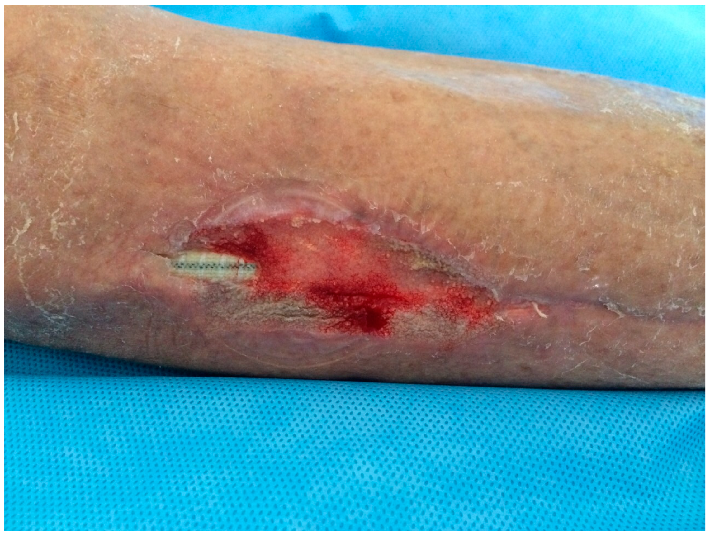

Table 1.
Demographic characteristics, clinical status, germs involved, treatment and outcomes of 29 patients with peripheral prosthetic vascular graft infection.
Table 1.
Demographic characteristics, clinical status, germs involved, treatment and outcomes of 29 patients with peripheral prosthetic vascular graft infection.
| Patient# Sex, age |
Type of bypass | Type of graft | Time, type and location of infection | Organism. Site of bacteriological sampling |
Surgical treatment | Antibiotic treatment | Outcome |
|---|---|---|---|---|---|---|---|
| 1, M 82 y |
Right axillo-femoral bypass |
Dacron | Late infection. Abscess along the abdominal wall |
Staphylococcus epidermidis. Prosthesis |
Bypass excision. New bypass in a clean field |
Tigecycline, gentamicin | Limb salvage |
| 2, F 72 y |
Right ilio-tibial bypass (redo-bypass) | PTFE | Early infection. Abscess and surgical wound dehiscence with pus discharge |
MSSA. Prosthesis, dehiscent groin wound |
Bypass excision. Impossible to revascularize |
Ceftriaxone, amikacin | Amputation |
| 3, M 78 y |
Left ilio-peroneal bypass (redo-bypass) | PTFE | Early infection. Leg abscess with surgical wound dehiscence |
Pseudomonas aeruginosa. Prosthesis, dehiscent leg wound. Negative blood culture |
Surgical debridement of the dehiscent wound. VAC therapy. Surgical closure | Piperacillin-tazobactam | Limb salvage |
| 4, F 81 y |
Right femoro-tibial bypass (redo-bypass) |
PTFE | Early infection. Graft occlusion |
Pseudomonas aeruginosa. Prosthesis, dehiscent groin wound |
Bypass excision. Impossible to revascularize |
Amikacin | Amputation |
| 5, M 61 y |
Left femoro-tibial bypass (redo-bypass) |
PTFE | Early infection. Proximal false aneurysm |
Sterile. Prosthesis |
Proximal bypass excision and replacemnt with Omniflow II | Ceftazidime, gentamicin | Limb salvage |
| 6, F 79 y |
Left femoro-tibial bypass (redo-bypass) |
PTFE | Early infection. Bleeding |
Sterile. Prosthesis. |
Bypass excision. Impossible to revascularize |
Ceftazidime | Amputation |
| 7, M 77 y |
Right femoro-popliteal bypass | PTFE | Late infection. Groin suture dehiscence with persistent pus discharge |
MRSA (dehiscent groin wound); Pseudomonas aeruginosa (prosthesis) |
Bypass excision and replacement in GSV | Imipenem, ciprofloxacin | Limb salvage |
| 8, M 76 y |
Left ilio-femoral bypass | PTFE | Early infection. Abscess |
Sterile (periprosthetic material) | Surgical site debridement and pus evacuation. VAC therapy. Muscle flap transposition |
Ceftriaxone | Limb salvage |
| 9,M 78 y |
Right femoro-popliteal bypass | PTFE | Late infection. Acute ischemia |
Staphylococcus lugdunensis. Prosthesis |
Bypass excision and femoro-peroneal bypass in GSV | Amoxicillin clavulanic acid, Gentamicin | Limb Salvage |
| 10, M 78 y |
Femoro-tibial bypass (redo-bypass) | PTFE | Early infection. Bleeding |
MRSA. Prosthesis and dehiscent groin wound |
Bypass excision. Impossible to revascularize |
Teicoplanin, vancomycin | Amputation. Death during follow-up |
| 11, M 75 |
Femoro-femoral crossover bypass | PTFE | Late infection. Graft occlusion |
Sterile (prosthesis) | Bypass excision. Revascularization not required |
Vancomycin, rifampicin | Limb salvage |
| 12, M 69 y |
Right femoro-tibial bypass (redo- bypass) |
PTFE | Early infection. Surgical wound dehiscence with persistent discharge |
Sterile (prosthesis) | Bypass excision. Impossible to revascularize |
Ceftazidime | Amputation |
| 13, F 78 y |
Left femoro-popliteal bypass | PTFE | Early infection. Bleeding |
Enterococcus faecalis. Prosthesis and dehiscent groin wound |
Bypass excision. Femoro-peroneal bypass in GSV |
Piperacillin-tazobactam, amikacin | Limb salvage |
| 14, M 67 y |
Right femoro-popliteal bypass | PTFE | Late infection. Proximal false aneurysm |
Pseudomonas aeruginosa. Prosthesis, dehiscent groin wound |
Proximal bypass excision and replacement in Omniflow II with muscle flap transposition. | Ampicillin sulbactam | Limb salvage |
| 15, M 67 y |
Left axillo-femoral bypass (Dacron) |
Dacron | Late infection. Seroma |
MRSA. Prosthesis |
Bypass excision and femoral artery patch in GSV. Not required revascularization | Teicoplanin, vancomycin | Limb salvage |
| 16, M 82 y |
Right axillo-femoral bypass (Dacron) | Dacron | Late infection. Seroma |
Escherichia coli (periprosthetic fluid), Pseudomonas aeruginosa (prosthesis) | Bypass excision and replacement in a clean field | Amikacin, colistimethate sodium | Limb salvage. Death during follow-up |
| 17, F 68 y |
Right femoro-popliteal bypass | PTFE | Early infection. Acute ischemia |
MRSA (blood culture). Pseudomonas aeruginosa and Staphylococcus aureus (prosthesis) |
Bypass excision. Impossible to revascularize | Meropenem, colistimethate sodium | Amputation. Death during hospital stay for sepsis |
| 18, F 65 y |
Left femoro-popliteal bypass | PTFE | Late infection. Proximal false aneurysm |
Proteus mirabilis (dehiscent groin wound). MSSA (dehiscent thigh wound) |
Proximal bypass excision and replacement in Omniflow II. Muscle flap transposition. Graft occlusion |
Ertapenem | Amputation after new bypass occlusion. Death during follow-up |
| 19,F 71 y |
Right femoro-popliteal bypass | PTFE | Early infection. Acute ischemia and peripheral septic arterial embolism on the skin |
Staphylococcus warneri (thrombus and prosthesis) | Bypass excision and femoro-peroneal bypass in GSV | Teicoplanin | Limb salvage |
| 20, M 65 y |
Femoro-femoral crossover bypass | PTFE | Early infection. Proximal false aneurysm |
MSSA. Prosthesis |
Bypass excision and axillo-bifemoral bypass | Rifampicin, trimethoprim/sulfamethoxazole, daptomycin | Limb salvage |
| 21, F 48 y |
Left Ilio-tibial bypass (redo-bypass) |
PTFE | Early infection. Surgical wound dehiscence with persistent discharge |
Pseudomonas aeruginosa. Prosthesis |
Bypass excision and replacement in cephalic vein. Graft occlusion |
Amikacin | Amputation after new bypass occlusion |
| 22, F 83 y |
Right axillo-femoral bypass in Dacron | Dacron | Late infection. Groin surgical wound dehiscence with persistent discharge |
MSSA. Dehiscent groin wound |
Surgical debridement of the dehiscent groin wound. VAC therapy. Surgical closure of the wound |
Meropenem | Limb salvage |
| 23, M 80 y |
Right femoro-popliteal bypass | PTFE | Early infection. Groin surgical wound dehiscence with persistent discharge |
Escherichia coli, Pseudomonas aeruginosa. Dehiscent groin wound |
Surgical debridement of the dehiscent groin wound. VAC therapy. Surgical closure of the dehiscent wound |
Amikacin, colistimethate sodium | Limb salvage |
| 24, F 77 y |
Left ilio-femoral bypass | PTFE | Early infection. Surgical wound dehiscence with persistent discharge |
MRSA | Surgical debridement, VAC therapy. Muscle flap transposition. |
Teicoplanin, vancomycin | Limb salvage |
| 25, M 83 y |
Left femoro-popliteal bypass | PTFE | Early infection. Surgical wound dehiscence with persistent discharge |
MRSA | Surgical debridement of the dehiscent groin wound and VAC therapy. Surgical closure of the dehiscent wound | Teicoplanin, vancomycin | Limb salvage. Death during follow-up |
| 26, F 79 y (Fig. 1,2,3) |
Right femoro- peroneal bypass (redo-bypass) |
PTFE | Early infection. Dehiscence of the distal surgical wound with prosthesis exposure |
Pseudomonas aeruginosa. Periprosthetic material |
Surgical debridements and VAC-therapy | Piperacillin-tazobactam | Limb salvage |
| 27, M 73 y |
Femoro-femoral crossover bypass | PTFE | Late infection. Proximal false aneurysm |
Sterile Periprosthetic material |
Surgical repair | Ceftriaxone | Limb salvage |
| 28, M 76 y |
Left femoro-tibial bypass (redo-bypass) |
PTFE | Early infection. Dehiscence of the proximal surgical wound with prosthesis exposure |
Pseudomonas aeruginosa. Periprosthetic fluid |
Surgical debridements and VAC-therapy | Piperacillin-tazobactam | Limb salvage |
| 29, M 74 y |
Right femoro-popliteal bypass | PTFE | Late infection. Proxiaml false aneurysm |
MSSA. Prosthesis |
Bypass excision and replacement with a femoro-peroneal bypass in GSV | Rifampicin, trimethoprim/sulfamethoxazole, daptomycin | Limb salvage |
1 M = male, F = female, MSSA= Methicillin-sensitive Staphylococcus aureus; MRSA = Methicillin-resistant Staphylococcus aureus; PTFE = polytetrafluoroethylene.
Table 2.
Type of infected vascular bypass treated in our Institution.
| Vascular bypass and type of prosthesis implanted | In our Institution (N, %) |
In other Institutions |
|---|---|---|
| Axillo-femoral (Dacron) | 2 (4.9) | 2 |
| Femoro-femoral crossover (PTFE) | 1 (2.3) | 2 |
| Ilio-femoral (PTFE) | 1 (3.7) | 1 |
| Femoro-popliteal (PTFE) | 2 (4.9) | 8 |
| Redo-bypass after failure of multiple attempts of revascularization (PTFE): - Ilio-tibial - Femoro-tibial |
10 (22.7) 3 7 |
|
| TOT = 29 | 16 (8.2) / 196 | 13 |
Disclaimer/Publisher’s Note: The statements, opinions and data contained in all publications are solely those of the individual author(s) and contributor(s) and not of MDPI and/or the editor(s). MDPI and/or the editor(s) disclaim responsibility for any injury to people or property resulting from any ideas, methods, instructions or products referred to in the content. |
© 2025 by the authors. Licensee MDPI, Basel, Switzerland. This article is an open access article distributed under the terms and conditions of the Creative Commons Attribution (CC BY) license (https://creativecommons.org/licenses/by/4.0/).
Copyright: This open access article is published under a Creative Commons CC BY 4.0 license, which permit the free download, distribution, and reuse, provided that the author and preprint are cited in any reuse.