Preprint
Article

This version is not peer-reviewed.

Shifting Outcomes: Superior Functional Recovery in Embolic Stroke of Undetermined Source Compared to Cardioembolic Stroke

A peer-reviewed article of this preprint also exists.

Submitted:

14 January 2025

Posted:

15 January 2025

You are already at the latest version

Abstract

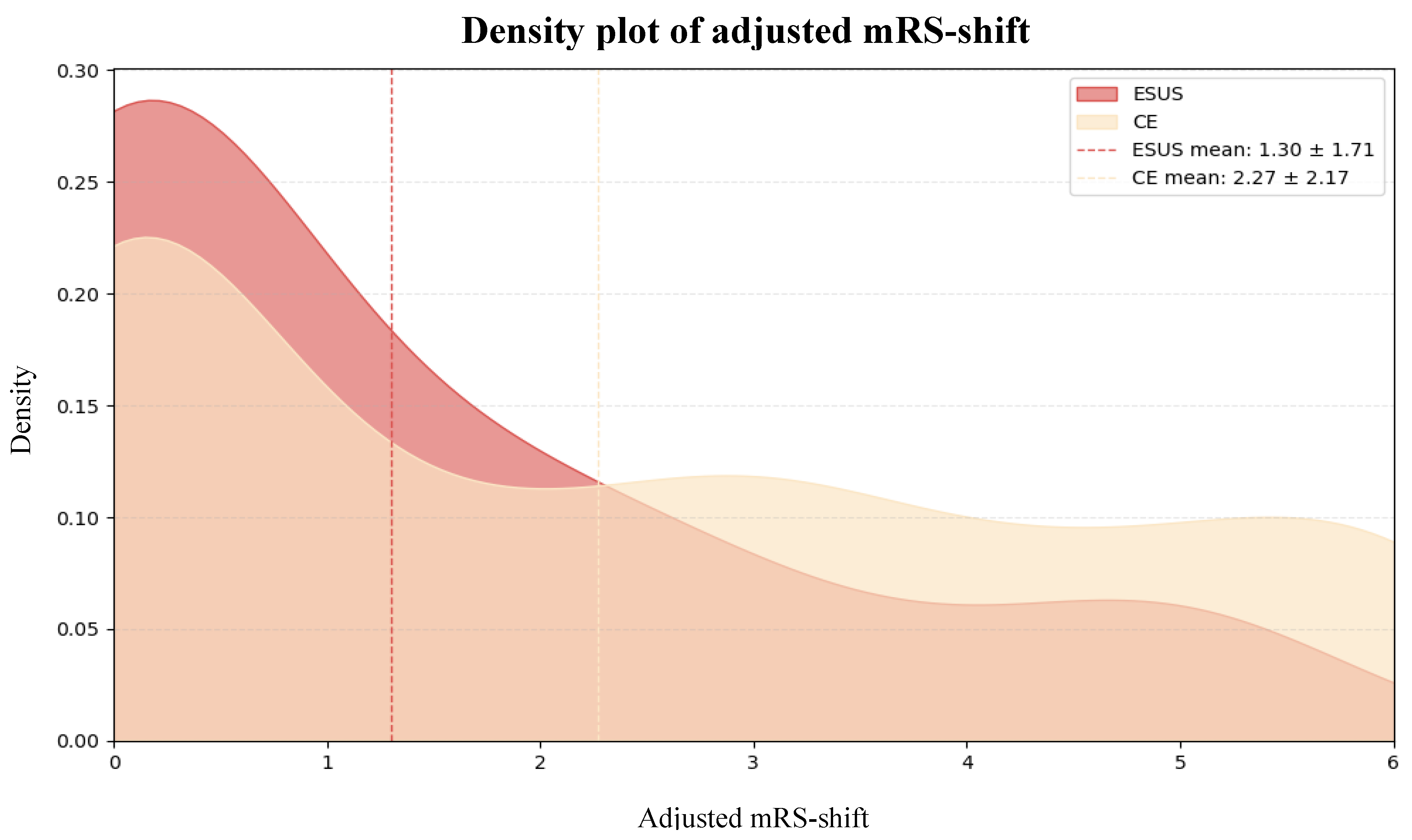
Background/Objectives: Embolic Stroke of Undetermined Source (ESUS) is a subtype of ischemic stroke characterized by a non-lacunar infarct in the absence of a clearly identifiable embolic source, despite comprehensive diagnostic evaluation. While ESUS patients are typically younger, have fewer cardiovascular comorbidities, and experience milder strokes than those with cardioembolic strokes (CE), their long-term functional recovery remains underexplored. Methods: We retrospectively analyzed data from 317 ischemic stroke patients (n = 37 ESUS, n = 280 CE) admitted to the Department of Neurology, University of Pécs, between February 2023 and September 2024. Functional recovery was assessed using the modified Rankin Scale (mRS), adjusted for baseline differences (adjusted mRS-shift). Independent predictors of mRS-shift were identified using Firth penalized regression and extreme gradient boosting (XGBoost). Results: ESUS patients were significantly younger (53.8 ± 13.5 years vs. 75.1 ± 11.3 years, p <0.001), had lower pre-morbid modified Rankin Scale (pre-mRS) scores (0.22 ± 0.75 vs. 0.81 ± 1.23, p <0.001), were less likely to have hypertension (70.3% vs. 86.1%, p = 0.027), and presented with milder strokes at admission (National Institutes of Health Stroke Scale [NIHSS] score 5.5 ± 3.6 vs. 8.1 ± 6.3, p <0.001) and 72 hours post-stroke (2.8 ± 3.7 vs. 6.5 ± 6.3, p <0.001) compared to CE patients. After adjusting for baseline differences, ESUS patients had significantly better functional recovery (adjusted mRS-shift 1.30 ± 1.71 vs. 2.27 ± 2.17, p <0.001). Conclusions: ESUS patients showed superior functional recovery compared to CE patients, even after adjusting for baseline differences. These findings highlight the need for further research into the pathophysiology and optimal treatment for ESUS.

Keywords: 
;  ;  

1. Introduction

Stroke remains one of the leading causes of disability worldwide, with ischemic strokes accounting for approximately 87% of all cases [1]. Within ischemic strokes, embolic stroke of undetermined source (ESUS) and cardioembolic stroke (CE) represent two distinct subtypes that share similar underlying mechanisms but differ in their identifiable causes and clinical management. CE is commonly linked to well-established embolic sources, such as atrial fibrillation (AF), allowing for targeted preventative measures, most notably anticoagulation therapy [2]. In contrast, ESUS, accounting for nearly 17% of all ischemic strokes, presents a diagnostic and therapeutic challenge due to the absence of a clearly identifiable embolic source, despite comprehensive diagnostic evaluations [3].
Emerging evidence suggests that ESUS patients differ significantly from their CE counterparts in terms of baseline characteristics. ESUS patients are generally younger, have fewer cardiovascular comorbidities [4], and typically present with milder neurological deficits upon admission [5,6,7]. Despite these differences, both ESUS and CE patients share similar clinical and radiological features [8], making them comparable groups for evaluating differences in functional recovery.
Previous research has primarily focused on the risk of stroke recurrence and secondary prevention strategies in ESUS patients. Notably, large randomized clinical trials, including the RESPECT-ESUS, NAVIGATE-ESUS, ARCADIA and ATTICUS trials [9,10,11,12], aimed to evaluate the efficacy of anticoagulation in reducing recurrent strokes in ESUS patients, but failed to demonstrate a significant advantage over antiplatelet therapy. Consequently, optimal secondary prevention strategies for ESUS patients remain controversial.
Despite growing recognition of ESUS as a distinct stroke subtype, research focusing on the long-term functional recovery of ESUS patients compared to those with CE remains limited. A more comprehensive understanding of these recovery patterns could guide the development of personalized rehabilitation programs and improve long-term care strategies for ESUS patients.
Moreover, traditional statistical models have limitations in capturing the complex, multifactorial nature of stroke recovery. Recent advances in data analytics, such as machine learning algorithms like Extreme Gradient Boosting (XGBoost), offer more powerful and flexible methods for identifying nuanced predictors of recovery outcomes. XGBoost excels at handling high-dimensional data and uncovering intricate relationships between clinical variables, providing deeper insights into the factors that influence patient recovery.
Therefore, the primary objective of this study is to compare long-term functional recovery between ESUS and CE patients while accounting for baseline differences. By combining traditional statistical models and advanced machine learning approaches, this study aims to identify critical predictors of functional recovery and deepen the understanding of factors influencing outcomes in ESUS patients.

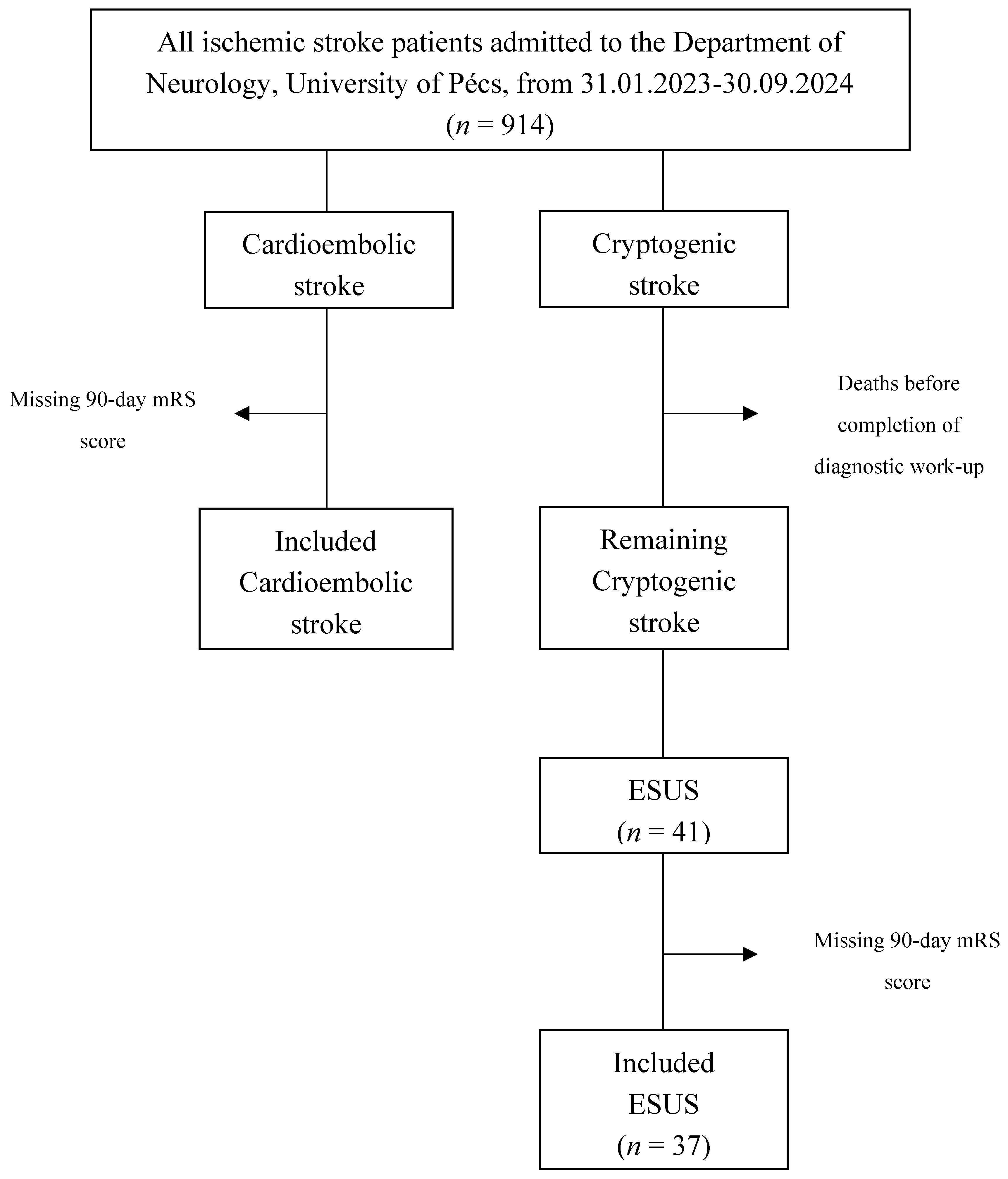
2. Materials and Methods

2.1. Study Design and Patient Population

This retrospective study utilized data from the Transzlációs Idegtudományi Nemzeti Laboratórium (TINL) STROKE-registry, which included patients with acute ischemic stroke (AIS) admitted to the Department of Neurology, University of Pécs, between February 2023 and September 2024.
Adult patients (≥18 years) with imaging-confirmed non-lacunar AIS, classified as either ESUS or CE following comprehensive diagnostic work-up, were included in the study. Patients with incomplete diagnostic evaluations or missing follow-up data were excluded from the analysis.
The study was approved by the local Ethics Committee, and informed consent was waived due to the retrospective nature of the TINL STROKE-registry.

2.2. Definitions

ESUS was defined based on the Cryptogenic Stroke/ESUS International Working Group criteria as a visible non-lacunar infarct in the absence of extracranial or intracranial atherosclerosis causing ≥50% luminal stenosis in arteries supplying the area of ischemia detected by computed tomography angiography (CTA), magnetic resonance angiography (MRA) or carotid ultrasonography, a major-risk cardioembolic source (excluded through transthoracic echocardiography [TTE] and at least 24-hour cardiac Holter monitoring), and any other specific cause of stroke (e.g., arterial dissection, arteritis, migraine/vasospasm, or drug abuse). Lacunar stroke was defined as a subcortical infarct ≤ 1.5 cm (or ≤ 2.0 cm on magnetic resonance tomography [MRT] diffusion-weighted imaging) located in regions supplied by small penetrating cerebral arteries [13].
CE was defined as a stroke caused by emboli originating from confirmed major cardiac sources (e.g. permanent or paroxysmal AF, intracardiac thrombus) according to the ASCOD phenotyping classification system [14].

2.3. Data Collection

Comprehensive data were prospectively collected. Baseline characteristics included demographics (age, sex), medical history (pre-stroke functional status assessed by the pre-morbid modified Rankin Scale [pre-mRS] score, hypertension, diabetes mellitus), cardiovascular risk factors (current smoking, alcohol consumption), and current medications (e.g. anticoagulation therapy). Stroke specific data included interval between stroke onset and hospital admission (onset-door-time), stroke severity assessed using the National Institutes of Health Stroke Scale (NIHSS) [15] at admission and at 72 hours post-stroke, laboratory values (plasma-glucose levels and bedside international normalized ratio [INR] at admission), and treatment modalities (standard care [SC], thrombolysis [TL], mechanical thrombectomy [MT] or combined therapy (TL + MT)].
As part of the initial diagnostic work-up, all patients underwent a non-contrast computed tomography (NCCT) scan, CTA/MRA or carotid duplex ultrasonography, a 12-lead electrocardiogram (ECG), TTE, 24-hour Holter monitoring, and routine blood tests including coagulation profile and vasculitis markers. In selected cases, additional evaluations such as transesophageal echocardiography (TEE) with or without a bubble study, extended Holter monitoring, assessment for prothrombotic conditions, including genetic testing for hypercoagulability, were performed.

2.4. Outcome Measure

Functional recovery was assessed by trained neurology staff using the modified Rankin Scale (mRS) score. A favorable functional outcome was defined as an mRS score of 0-2, while an unfavorable outcome was defined as dependency or death (mRS score of 3-6). Additionally, the mRS-shift was used to evaluate changes in functional status from pre-stroke to 90 days post-stroke.

2.5. Statistical Analyses

Continuous variables were reported as mean ± standard deviation (SD), while categorical variables were expressed as absolute frequencies and percentages. Comparisons between ESUS and CE patients were performed using independent t-tests for continuous variables and chi-square (χ²) or Fisher’s Exact tests for categorical variables. The Cochran-Mantel-Haenszel shift test was used to analyze the distribution of mRS outcomes. Multiple logistic regression was applied to derive adjusted probabilities for functional recovery.
Subgroup analyses assessed the impact of anticoagulation status at admission and treatment modalities on functional recovery. To identify independent predictors and mitigate small sample size bias, both Firth penalized logistic regression analysis and XGBoost with hyperparameter tuning, and k-fold cross-validation were employed. Results were reported as odds ratios (OR) with 95% confidence intervals (CI). A p-value <0.05 was considered statistically significant. Additionally, the XGBoost model generated mean SHapley Additive exPlanations (SHAP) values and percentage contributions, highlighting the relative importance of each feature in predicting outcomes. Together, these models provide complementary insights: Firth regression identifies statistically significant predictors, while the XGBoost model with SHAP analysis quantifies the relative impact of each predictor on functional recovery. All statistical analyses were conducted using Python (version 3.13) and R (version 4.4.2).

3. Results

This retrospective cohort study analyzed data from 914 AIS patients enrolled in the TINL STROKE-registry at the Department of Neurology, University of Pécs, between February 2023 and September 2024. Of these, 317 patients (34.7%) were diagnosed with CE and 235 patients (25.7%) were classified as having cryptogenic stroke.
In the CE group, 37 (11.7%) were excluded due to missing 90-day mRS scores, leaving 280 CE patients for the final analysis. In the cryptogenic stroke group, 11 patients (4.7%) died before completing the diagnostic evaluation and were excluded. Among the remaining 224 patients, 41 (18.3%) met the criteria for ESUS. However, 4 of these (9.8%) lacked 90-day mRS scores and were excluded, resulting in 37 ESUS patients included in the final analysis (Figure 1).

3.1. Baseline Differences

The study cohort consisted of 170 male patients (53.63%) with a mean age of 72.64 13.40 years. ESUS patients were significantly younger than CE patients (53.8 ± 13.5 years vs. 75.1 ± 11.3 years, p <0.001) and had lower pre-mRS scores (0.22 ± 0.75 vs. 0.81 ± 1.23, p <0.001). They were also less likely to have hypertension (70.3% vs. 86.1%, p = 0.027), although smoking was more prevalent among ESUS patients compared to CE patients (32.4% vs. 16.8%, p = 0.040). Additionally, fewer ESUS patients were on anticoagulation therapy at the time of their stroke (13.5% vs. 36.4%, p = 0.005).
In terms of stroke severity, ESUS patients presented with significantly lower NIHSS scores at admission (5.5 ± 3.6 vs. 8.1 ± 6.3, p <0.001) and at 72 hours post-stroke (2.8 ± 3.7 vs. 6.5 ± 6.3, p <0.001), indicating milder strokes. Furthermore, ESUS patients were more likely to receive TL compared to CE patients (35.1% vs. 19.3%, p = 0.033).
Table 1. Baseline Differences between ESUS and CE patients.
ESUS n = 37 CE n = 280 p-Value
Baseline Characteristics
Age, years, mean (SD)
Sex, male, n (%)
Pre-mRS score, mean (SD)
Hypertension, n (%)
Diabetes mellitus, n (%)
Current smoking, n (%)
Alcohol consumption, n (%)
Anticoagulation, n (%)
53.8 (± 13.5)
20 (54.1%)
0.22 (± 0.75)
26 (70.3%)
9 (24.3%)
12 (32.4%)
18 (48.6%)
5 (13.5%)
75.1 (± 11.3)
150 (53.6%)
0.81 (± 1.23)
241 (86.1%)
103 (36.8%)
47 (16.8%)
117 (41.8%)
102 (36.4%)
<0.001*
1.00
<0.001*
0.027
0.148
0.040
0.481
0.005
Stroke Characteristics
Onset-door-time, mean (SD)
NIHSS score, mean (SD)
72hNIHSS score, mean (SD)
Plasma-glucose, mean (SD)
INR, mean (SD)
360 (455)
5.5 (± 3.6)
2.8 (± 3.7)
7.46 (± 2.98)
1.13 (± 0.50)
590 (± 1860)
8.1 (± 6.3)
6.5 (± 6.3)
7.51 (± 2.59)
1.21 (± 0.63)
0.087
<0.001*
<0.001*
0.925
0.394
Treatment Modalities
SC, n (%)
TL, n (%)
MT, n (%)
TL + MT, n (%)
12 (32.4%)
13 (35.1%)
8 (21.6%)
4 (10.8%)
133 (47.5%)
54 (19.3%)
65 (23.2%)
28 (10.0%)
0.113
0.033
1.00
0.777
Abbreviations: ESUS = embolic stroke of undetermined source, CE = cardioembolic stroke, SD = standard deviations, pre-mRS = pre-morbid modified Rankin Scale, NIHSS = National Institutes of Health Stroke Scale at admission, 72hNIHSS = National Institutes of Health Stroke Scale 72 hours post-stroke, INR = international normalized ratio, SC = standard care, TL = thrombolysis, MT = mechanical thrombectomy .

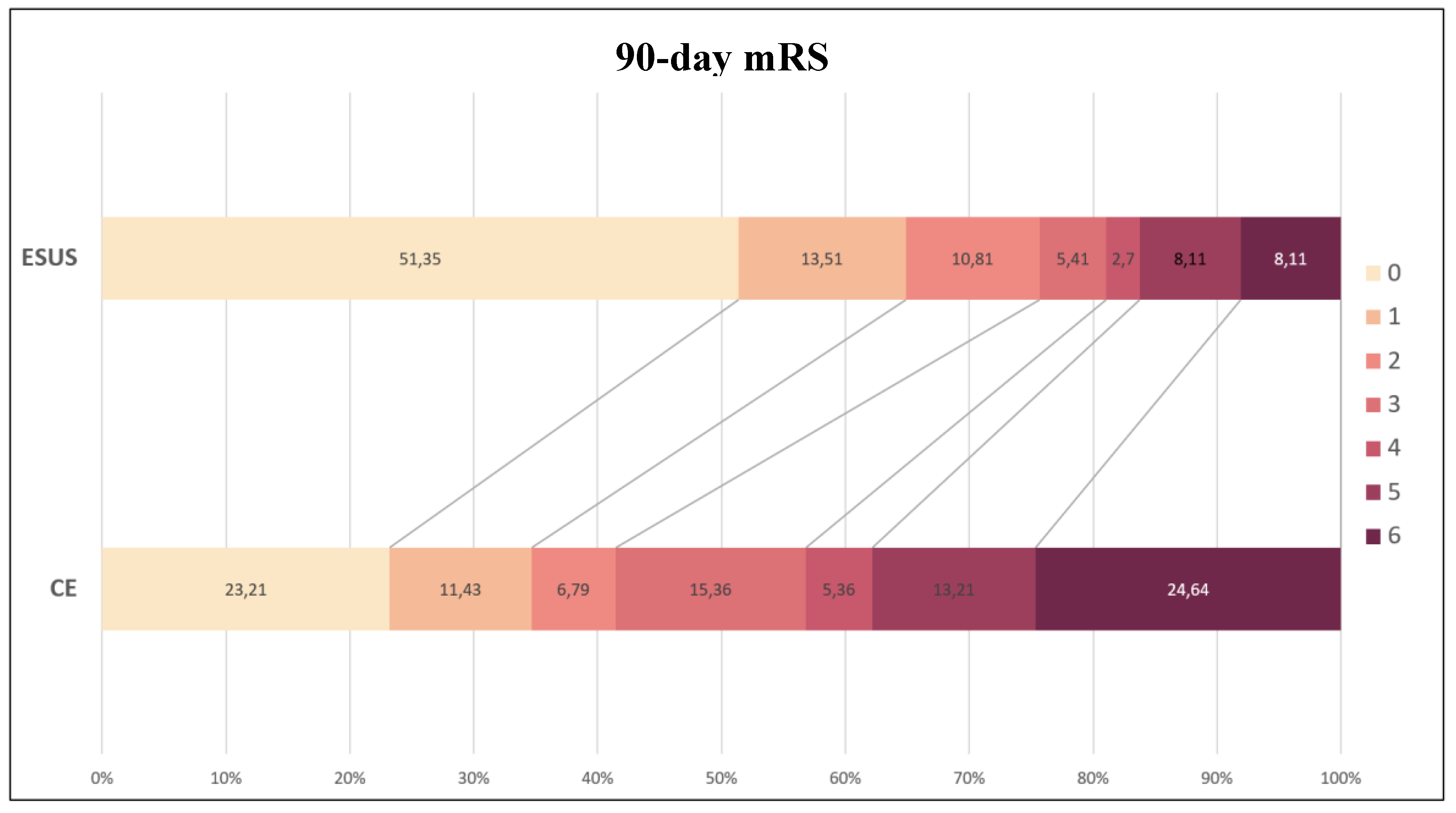
3.2. Functional Recovery

A total of 75.7% of ESUS patients achieved a favorable functional outcome (mRS score 0-2), notably higher than the 41.4% observed in the CE group (OR = 4.40 [95% CI: 2.00-9.67], p <0.001). In contrast, only 24.3% of ESUS patients experienced moderate to severe disability or death (mRS score 3-6), significantly lower than the 58.6% recorded in CE patients (Figure 2).
After adjusting for baseline differences, ESUS patients showed significantly better functional recovery compared to CE patients (adjusted mRS-shift 1.30 ± 1.71 vs. 2.27 ± 2.17, p <0.001) (Figure 3).

3.3. Subgroup Analyses

Anticoagulation Status

Non-anticoagulated CE patients had a better functional recovery compared to anticoagulated CE patients (adjusted mRS-shift 2.09 ± 2.08 vs. 2.59 ± 2.30), although this difference was not statistically significant (p = 0.104). Similarly, ESUS patients showed no significant difference in functional recovery between non-anticoagulated and anticoagulated groups (adjusted mRS-shift 1.16 ± 1.63 vs. 2.20 ± 2.17 p = 0.196).

Treatment Modalities

Across all treatment modalities, ESUS patients consistently demonstrated better functional recovery than CE patients, though the differences were not statistically significant. Specifically, ESUS patients had lower adjusted mRS-shifts compared to CE patients in SC: 2.00 ± 2.00 vs. 2.86 ± 2.15 (p = 0.319), TL: 1.33 ± 1.78 vs. 2.35 ± 2.21 (p = 0.123), MT: 1.08 ± 1.66 vs. 1.48 ± 1.79 (p = 0.352), and TL + MT: 0.50 ± 1.00 vs. 2.04 ± 2.32 (p = 0.213).

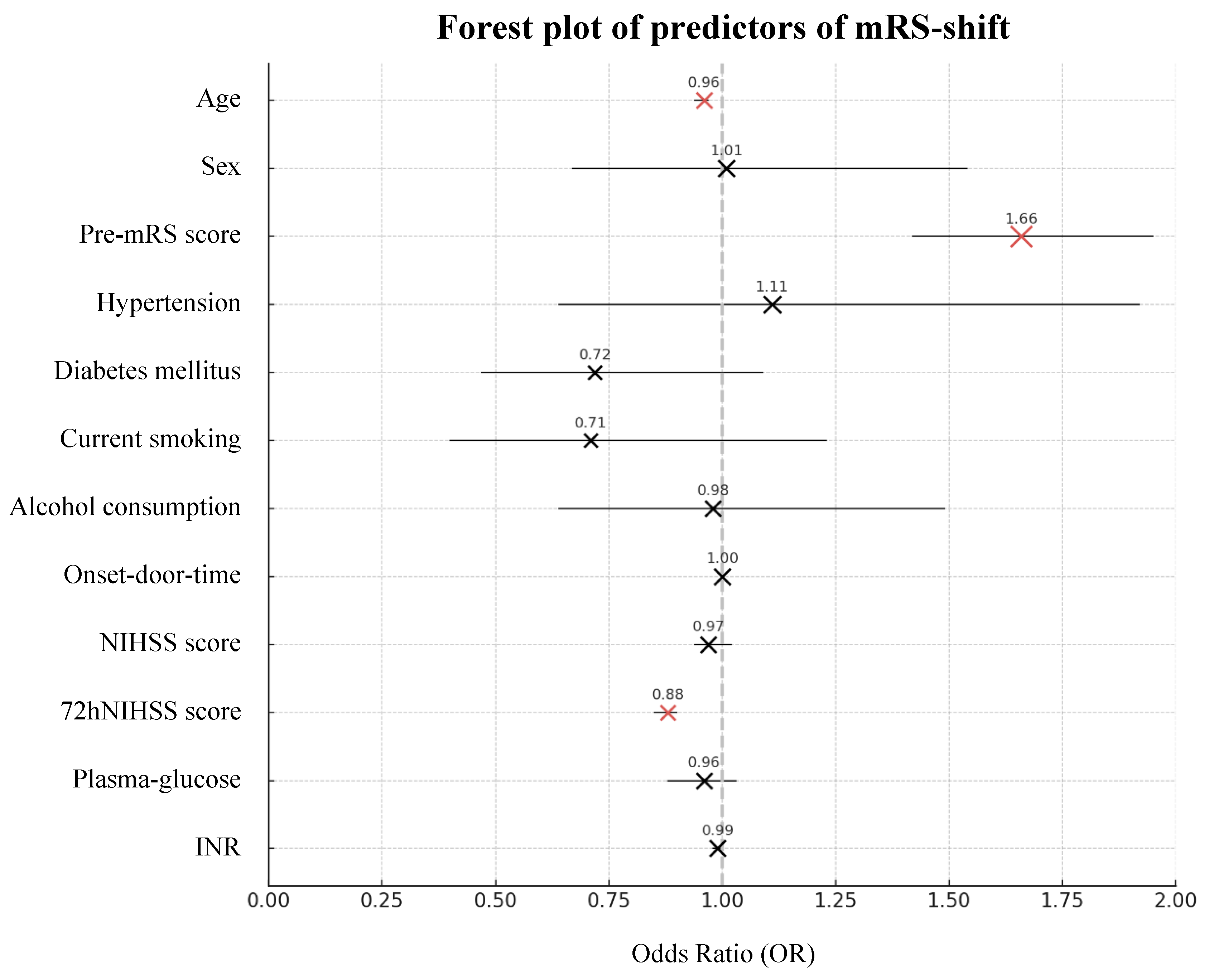
3.4. Predictors of Functional Recovery

Firth Regression and XGBoost

Both Firth regression analysis and XGBoost with hyperparameter tuning and k-fold cross-validation identified age, pre-mRS score and NIHSS score as significant predictors of mRS-shift (Table 2, Figure 4 and 5). Age was negatively associated with recovery, with each additional year reducing the likelihood of better outcomes by 4% (p <0.001). Higher pre-mRS scores were linked to poorer recovery, indicating that patients with greater pre-stroke disability were less likely to recover (p <0.001). Furthermore, stroke severity, measured by NIHSS, strongly influenced recovery - each point increase at admission lowered the odds of improvement by 3%, and by 12% at 72 hours post-stroke (p <0.001). SHAP analysis from the XGBoost model reinforced these findings, highlighting 72-hour NIHSS score (38.97%) as the most impactful predictor, followed by pre-mRS score (10.68%) and age (7.58%).

Model Performance

The Firth penalized logistic regression analysis demonstrated strong statistical significance (² = 321.79, degrees of freedom [df] = 12, p <0.001), indicating that at least one predictor meaningfully contributes to explaining functional recovery. Complementing this, the XGBoost model, optimized through hyperparameter tuning and validated via k-fold-cross-validation, explained 77.8% of the variability in mRS-shift (R2 = 0.778, p <0.001). The SHAP analysis further highlighted the relative importance of key predictors, confirming the model’s strong predictive performance in identifying factors influencing functional recovery.

4. Discussion

This study demonstrated that ESUS patients experienced significantly better functional recovery than CE patients, even after adjusting for baseline differences. While ESUS patients generally presented with more favorable baseline characteristics, these factors alone do not fully account for their better recovery.
Consistent with existing literature, ESUS patients in our cohort were younger, had lower pre-stroke disability, fewer cardiovascular comorbidities, and experienced less severe strokes [5,16,17,18]. Our findings align with previous studies reporting more favorable functional outcomes in ESUS patients compared to CE patients [5,19,20,21,22]. However, some research has reported comparable outcomes between ESUS and CE strokes [23], suggesting that the recovery advantage in ESUS may not be uniform across all populations. Notably, in our cohort, only 24.3% of ESUS patients had poor outcomes at 90 days (mRS score 3-6), which is within the range of previously reported rates of 17.5% [24] to 33.2% [25].

4.1. Impact of Age on Neuroplasticity and Recovery

The younger age of ESUS patients likely contributes to their better recovery. Age significantly impacts neuroplasticity, the brain's ability to reorganize and recover after injury. Younger patients have greater capacity for neuroplasticity, allowing for more effective recovery of lost functions. Additionally, younger patients are generally more physically capable of participating in intensive rehabilitation programs, unlike older CE patients who often have multiple comorbidities that may limit rehabilitation efforts.

4.2. Stroke Pathophysiology and Infarct Patterns

Differences in functional recovery between ESUS and CE patients may be partly attributed to stroke pathophysiology. ESUS often arises from minor-risk embolic sources, such as a patent foramen ovale (PFO), leading to smaller, localized infarcts [13]. In contrast, CE strokes are typically due to high-risk emboli (e.g., from AF), causing larger infarcts that inflict more extensive damage to critical brain regions like the motor cortex, resulting in poorer long-term outcomes [26].

4.3. Complexity of Embolic Sources and Treatment Implications

ESUS patients frequently have multiple potential embolic sources [27,28], often involving minor-risk sources like atrial cardiopathy, PFO, or cancer, leading to red thrombi that respond well to anticoagulation. Conversely, some ESUS cases involve white thrombi from sources like aortic arch or intracranial atherosclerosis, which are more responsive to antiplatelet therapy [29]. This diversity in thrombogenesis complicates optimal treatment strategies.
While CE patients with AF are routinely managed with anticoagulation, identifying embolic sources in ESUS remains challenging. AF was detected in 29% of ESUS patients over 3.2 years in one study [4], with detection rates varying between 7.6% and 33% in larger cohorts [30,31,32].

4.4. Limitations and Future Directions

Despite providing important insights, this study has several limitations. Firstly, the small ESUS sample size (n = 37) limits generalizability and reduces statistical power, particularly in subgroup analyses. Secondly, the retrospective, single-center design introduces potential selection bias, despite rigorous statistical adjustments. Thirdly, excluding patients with missing 90-day mRS scores (n = 41) or incomplete diagnostic work-ups (n = 11) may have further biased the results.
Furthermore, the study's focus on 90-day outcomes may not fully capture long-term functional recovery, underscoring the need for longer follow-up to assess sustained recovery and stroke recurrence. Moreover, the study focused solely on functional outcomes and did not assess patient-reported outcomes or quality of life, which are important indicators of recovery and well-being.
Therefore, these findings should be considered hypothesis-generating. Future research should involve larger, multi-center, prospective studies with extended follow-up to validate and expand upon these results.

5. Conclusions

ESUS patients showed superior functional recovery compared to CE patients, even after adjusting for baseline differences. These findings highlight the need for further research into the distinct pathophysiology and optimal treatment strategies for ESUS, given its diverse underlying mechanisms and implications for personalized stroke care.

List of Abbreviations

ESUS embolic stroke of undetermined source
CE cardioembolic stroke
mRS modified Rankin Scale
XGBoost extreme gradient boosting
pre-mRS pre-morbid modified Rankin Scale
NIHSS National Institues of Health Stroke Scale
AF atrial fibrillation
TINL Transzlációs Idegtudományi Nemzeti Laboratórium
AIS acute ischemic stroke
CTA computed tomography angiography
MRA magnetic resonance angiography
TTE transthoracic echocardiography
MRT magnetic resonance tomography
INR international normalized ratio
SC standard care
TL thrombolysis
MT mechanical thrombectomy
NCCT non-contrast computed tomography
ECG electrocardiogram
TEE transesophageal echocardiography
SD standard deviation
χ2 chi-quare
OR odds ratio
CI confidence interval
SHAP SHapley Additive exPlanations
df degrees of freedom
PFO patent foramen ovale

Author Contributions

Conceptualization, J.S. and B.C.; methodology, J.S. and L.S.; validation, Z.N.K. and E.B.; formal analysis, J.S.; data curation, J.S.; writing—original draft preparation, J.S. and B.C.; writing—review and editing, Z.N.K. and E.B.; visualization, J.S.; supervision, L.S.; project administration, L.S. All authors have read and agreed to the published version of the manuscript.

Funding

This research received no external funding.

Institutional Review Board Statement

This study was conducted in accordance with the Declaration of Helsinki and approved by the Scientific and Research Ethics Committee of the Medical Research Council of the University of Pécs (RRF-2.3.1-21-2022-00011, 01/09/22) and re-approved by the Scientific and Research Ethics Committee of the Medical Research Council of Hungary (BM/22444-1/2024, 01/09/24).

Informed Consent Statement

Informed consent was obtained from all subjects involved in the study.

Data Availability Statement

The original contributions presented in the study are included in the article and further inquiries can be directed to the corresponding author.

Conflicts of Interest

The authors declare no conflicts of interest.

References

  1. Feigin, V.L.; Brainin, M.; Norrving, B.; Martins, S.O.; Pandian, J.; Lindsay, P.; F Grupper, M.; Rautalin, I. World Stroke Organization: Global Stroke Fact Sheet 2025. International Journal of Stroke 2025. [Google Scholar] [CrossRef] [PubMed]
  2. Kamel, H.; Healey, J.S. Cardioembolic Stroke. Circ Res 2017, 120, 514–526. [Google Scholar] [CrossRef]
  3. Ntaios, G. Embolic Stroke of Undetermined Source. J Am Coll Cardiol 2020, 75, 333–340. [Google Scholar] [CrossRef]
  4. Martinez-Majander, N.; Aarnio, K.; Pirinen, J.; Lumikari, T.; Nieminen, T.; Lehto, M.; Sinisalo, J.; Kaste, M.; Tatlisumak, T.; Putaala, J. Embolic Strokes of Undetermined Source in Young Adults: Baseline Characteristics and Long-term Outcome. Eur J Neurol 2018, 25, 535–541. [Google Scholar] [CrossRef]
  5. Ntaios, G.; Papavasileiou, V.; Milionis, H.; Makaritsis, K.; Manios, E.; Spengos, K.; Michel, P.; Vemmos, K. Embolic Strokes of Undetermined Source in the Athens Stroke Registry. Stroke 2015, 46, 176–181. [Google Scholar] [CrossRef] [PubMed]
  6. Li, L.; Yiin, G.S.; Geraghty, O.C.; Schulz, U.G.; Kuker, W.; Mehta, Z.; Rothwell, P.M. Incidence, Outcome, Risk Factors, and Long-Term Prognosis of Cryptogenic Transient Ischaemic Attack and Ischaemic Stroke: A Population-Based Study. Lancet Neurol 2015, 14, 903–913. [Google Scholar] [CrossRef] [PubMed]
  7. Putaala, J.; Nieminen, T.; Haapaniemi, E.; Meretoja, A.; Rantanen, K.; Heikkinen, N.; Kinnunen, J.; Strbian, D.; Mustanoja, S.; Curtze, S.; et al. Undetermined Stroke with an Embolic Pattern—a Common Phenotype with High Early Recurrence Risk. Ann Med 2015, 47, 406–413. [Google Scholar] [CrossRef] [PubMed]
  8. Ntaios, G.; Papavasileiou, V.; Lip, G.Y.H.; Milionis, H.; Makaritsis, K.; Vemmou, A.; Koroboki, E.; Manios, E.; Spengos, K.; Michel, P.; et al. Embolic Stroke of Undetermined Source and Detection of Atrial Fibrillation on Follow-Up: How Much Causality Is There? Journal of Stroke and Cerebrovascular Diseases 2016, 25, 2975–2980. [Google Scholar] [CrossRef] [PubMed]
  9. Hart, R.G.; Sharma, M.; Mundl, H.; Kasner, S.E.; Bangdiwala, S.I.; Berkowitz, S.D.; Swaminathan, B.; Lavados, P.; Wang, Y.; Wang, Y.; et al. Rivaroxaban for Stroke Prevention after Embolic Stroke of Undetermined Source. New England Journal of Medicine 2018, 378, 2191–2201. [Google Scholar] [CrossRef]
  10. Diener, H.-C.; Sacco, R.L.; Easton, J.D.; Granger, C.B.; Bar, M.; Bernstein, R.A.; Brainin, M.; Brueckmann, M.; Cronin, L.; Donnan, G.; et al. Antithrombotic Treatment of Embolic Stroke of Undetermined Source. Stroke 2020, 51, 1758–1765. [Google Scholar] [CrossRef]
  11. Kamel, H.; Longstreth, W.T.; Tirschwell, D.L.; Kronmal, R.A.; Marshall, R.S.; Broderick, J.P.; Aragón García, R.; Plummer, P.; Sabagha, N.; Pauls, Q.; et al. Apixaban to Prevent Recurrence After Cryptogenic Stroke in Patients With Atrial Cardiopathy. JAMA 2024, 331, 573. [Google Scholar] [CrossRef] [PubMed]
  12. Geisler, T.; Keller, T.; Martus, P.; Poli, K.; Serna-Higuita, L.M.; Schreieck, J.; Gawaz, M.; Tünnerhoff, J.; Bombach, P.; Nägele, T.; et al. Apixaban versus Aspirin for Embolic Stroke of Undetermined Source. NEJM Evidence 2023, 3. [Google Scholar] [CrossRef] [PubMed]
  13. Hart, R.G.; Diener, H.-C.; Coutts, S.B.; Easton, J.D.; Granger, C.B.; O’Donnell, M.J.; Sacco, R.L.; Connolly, S.J. Embolic Strokes of Undetermined Source: The Case for a New Clinical Construct. Lancet Neurol 2014, 13, 429–438. [Google Scholar] [CrossRef] [PubMed]
  14. Amarenco, P.; Bogousslavsky, J.; Caplan, L.R.; Donnan, G.A.; Wolf, M.E.; Hennerici, M.G. The ASCOD Phenotyping of Ischemic Stroke (Updated ASCO Phenotyping). Cerebrovascular Diseases 2013, 36, 1–5. [Google Scholar] [CrossRef] [PubMed]
  15. Brott, T.; Adams, H.P.; Olinger, C.P.; Marler, J.R.; Barsan, W.G.; Biller, J.; Spilker, J.; Holleran, R.; Eberle, R.; Hertzberg, V. Measurements of Acute Cerebral Infarction: A Clinical Examination Scale. Stroke 1989, 20, 864–870. [Google Scholar] [CrossRef] [PubMed]
  16. Tsivgoulis, G.; Kargiotis, O.; Katsanos, A.H.; Patousi, A.; Mavridis, D.; Tsokani, S.; Pikilidou, M.; Birbilis, T.; Mantatzis, M.; Zompola, C.; et al. Incidence, Characteristics and Outcomes in Patients with Embolic Stroke of Undetermined Source: A Population-Based Study. J Neurol Sci 2019, 401, 5–11. [Google Scholar] [CrossRef] [PubMed]
  17. Tan, E.S.J.; de Leon, J.; Boey, E.; Chin, H.-K.; Ho, K.-H.; Aguirre, S.; Sim, M.-G.; Seow, S.-C.; Sharma, V.K.; Kojodjojo, P. Stroke Recurrence in Embolic Stroke of Undetermined Source Without Atrial Fibrillation on Invasive Cardiac Monitoring. Heart Lung Circ 2023, 32, 1000–1009. [Google Scholar] [CrossRef]
  18. Marko, M.; Singh, N.; Ospel, J.M.; Uchida, K.; Almekhlafi, M.A.; Demchuk, A.M.; Nogueira, R.G.; McTaggart, R.A.; Poppe, A.Y.; Rempel, J.L.; et al. Symptomatic Non-Stenotic Carotid Disease in Embolic Stroke of Undetermined Source. Clin Neuroradiol 2024, 34, 333–339. [Google Scholar] [CrossRef] [PubMed]
  19. Tsivgoulis, G.; Kargiotis, O.; Katsanos, A.H.; Patousi, A.; Pikilidou, M.; Birbilis, T.; Mantatzis, M.; Palaiodimou, L.; Triantafyllou, S.; Papanas, N.; et al. Clinical and Neuroimaging Characteristics in Embolic Stroke of Undetermined versus Cardioembolic Origin: A Population-Based Study. Journal of Neuroimaging 2019, 29, 737–742. [Google Scholar] [CrossRef] [PubMed]
  20. Arauz, A.; Morelos, E.; Colín, J.; Roldán, J.; Barboza, M.A. Comparison of Functional Outcome and Stroke Recurrence in Patients with Embolic Stroke of Undetermined Source (ESUS) vs. Cardioembolic Stroke Patients. PLoS One 2016, 11, e0166091. [Google Scholar] [CrossRef]
  21. Wang, W.; Tang, X.; Liu, W.; Jia, K.; Zhao, X.; Yu, F. Clinical Features of Embolic Stroke of Undetermined Source. Front Neurol 2020, 11. [Google Scholar] [CrossRef] [PubMed]
  22. Bembenek, J.P.; Karlinski, M.A.; Kurkowska-Jastrzebska, I.; Czlonkowska, A. Embolic Strokes of Undetermined Source in a Cohort of Polish Stroke Patients. Neurological Sciences 2018, 39, 1041–1047. [Google Scholar] [CrossRef] [PubMed]
  23. Scavasine, V.C.; Ribas, G. da C.; Costa, R.T.; Ceccato, G.H.W.; Zétola, V. de H.F.; Lange, M.C. Embolic Stroke of Undetermined Source (ESUS) and Stroke in Atrial Fibrillation Patients: Not so Different after All? International Journal of Cardiovascular Sciences 2021. [Google Scholar] [CrossRef]
  24. Lee, I.H.; Heo, J.; Lee, H.; Jeong, J.; Kim, J.; Han, M.; Yoo, J.; Kim, J.; Baik, M.; Park, H.; et al. Long-Term Outcomes of Patients with Embolic Stroke of Undetermined Source According to Subtype. Sci Rep 2024, 14, 9295. [Google Scholar] [CrossRef] [PubMed]
  25. Lattanzi, S.; Pulcini, A.; Corradetti, T.; Rinaldi, C.; Zedde, M.L.; Ciliberti, G.; Silvestrini, M. Prediction of Outcome in Embolic Strokes of Undetermined Source. Journal of Stroke and Cerebrovascular Diseases 2020, 29, 104486. [Google Scholar] [CrossRef] [PubMed]
  26. Seet, R.C.S. Relationship Between Chronic Atrial Fibrillation and Worse Outcomes in Stroke Patients After Intravenous Thrombolysis. Arch Neurol 2011, 68, 1454. [Google Scholar] [CrossRef] [PubMed]
  27. Ntaios, G.; Perlepe, K.; Lambrou, D.; Sirimarco, G.; Strambo, D.; Eskandari, A.; Karagkiozi, E.; Vemmou, A.; Koroboki, E.; Manios, E.; et al. Prevalence and Overlap of Potential Embolic Sources in Patients With Embolic Stroke of Undetermined Source. J Am Heart Assoc 2019, 8. [Google Scholar] [CrossRef] [PubMed]
  28. Ntaios, G.; Pearce, L.A.; Veltkamp, R.; Sharma, M.; Kasner, S.E.; Korompoki, E.; Milionis, H.; Mundl, H.; Berkowitz, S.D.; Connolly, S.J.; et al. Potential Embolic Sources and Outcomes in Embolic Stroke of Undetermined Source in the NAVIGATE-ESUS Trial. Stroke 2020, 51, 1797–1804. [Google Scholar] [CrossRef] [PubMed]
  29. Kamel, H.; Merkler, A.E.; Iadecola, C.; Gupta, A.; Navi, B.B. Tailoring the Approach to Embolic Stroke of Undetermined Source. JAMA Neurol 2019, 76, 855. [Google Scholar] [CrossRef] [PubMed]
  30. Del Monte, A.; Rivezzi, F.; Giacomin, E.; Peruzza, F.; Del Greco, M.; Maines, M.; Migliore, F.; Zorzi, A.; Viaro, F.; Pieroni, A.; et al. Multiparametric Identification of Subclinical Atrial Fibrillation after an Embolic Stroke of Undetermined Source. Neurological Sciences 2023, 44, 979–988. [Google Scholar] [CrossRef]
  31. Tan, B.Y.Q.; Ho, J.S.Y.; Sia, C.-H.; Boi, Y.; Foo, A.S.M.; Dalakoti, M.; Chan, M.Y.; Ho, A.F.W.; Leow, A.S.; Chan, B.P.L.; et al. Left Atrial Volume Index Predicts New-Onset Atrial Fibrillation and Stroke Recurrence in Patients with Embolic Stroke of Undetermined Source. Cerebrovascular Diseases 2020, 49, 285–291. [Google Scholar] [CrossRef] [PubMed]
  32. Pathan, F.; Sivaraj, E.; Negishi, K.; Rafiudeen, R.; Pathan, S.; D’Elia, N.; Galligan, J.; Neilson, S.; Fonseca, R.; Marwick, T.H. Use of Atrial Strain to Predict Atrial Fibrillation After Cerebral Ischemia. JACC Cardiovasc Imaging 2018, 11, 1557–1565. [Google Scholar] [CrossRef] [PubMed]
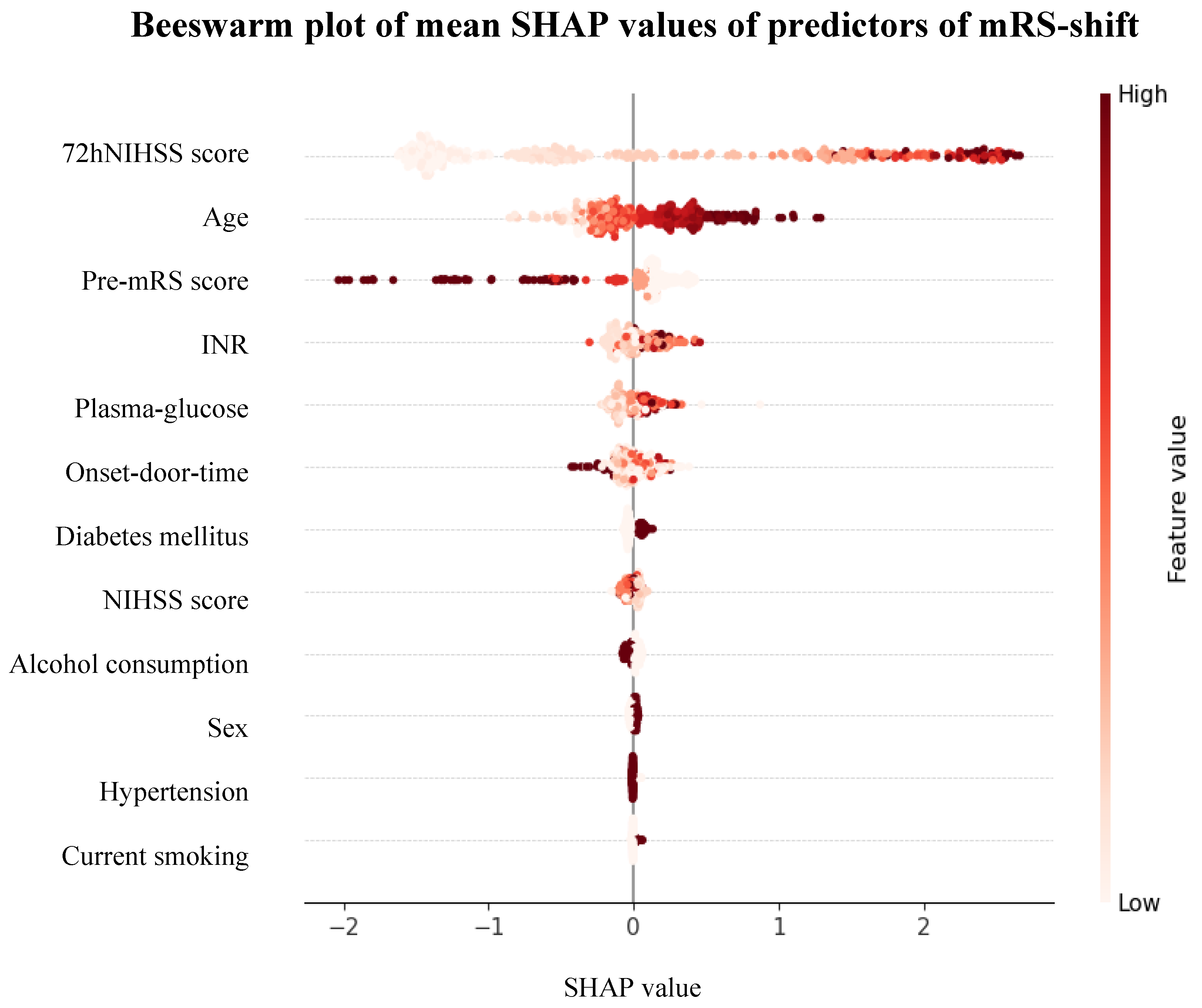
Figure 1. Flowchart of the study. Abbreviations: mRS = modified Rankin Scale, ESUS = embolic stroke of undetermined source.
Preprints 146193 g001
Figure 2. Distribution of 90-day mRS. Abbreviations: mRS = modified Rankin Scale, ESUS = embolic stroke of undetermined source, CE = cardioembolic stroke.
Preprints 146193 g002
Figure 3. Density plot of adjusted mRS-shift. Abbreviations: mRS = modified Rankin Scale, ESUS = embolic stroke of undetermined source, CE = cardioembolic stroke.
Preprints 146193 g003
Figure 4. Forest plot of predictors of mRS-shift. Abbreviations: mRS = modified Rankin Scale, pre-mRS = pre-morbid modified Rankin Scale, NIHSS = National Institutes of Health Stroke Scale at admission, 72hNIHSS = National Institutes of Health Stroke Scale 72 hours post-stroke, INR = international normalized ratio, OR = odds ratio.
Preprints 146193 g004
Figure 5. Beeswarm plot of mean SHAP values of predictors of mRS-shift. Abbreviations: SHAP = SHapley Additive exPlanations, mRS = modified Rankin Scale, 72hNIHSS = National Institutes of Health Stroke Scale 72 hours post-stroke, pre-mRS = pre-morbid modified Rankin Scale, INR = international normalized ratio, NIHSS = National Institutes of Health Stroke Scale at admission.
Preprints 146193 g005
Table 2. Predictors of mRS-shift.
Firth
Coefficient
OR 95% CI p-Value Mean SHAP Value 95% CI Feature
Importance
Baseline Characteristics
Age
Sex
Pre-mRS score
Hypertension
Diabetes mellitus
Current smoking
Alcohol consumption
-0.045
0.014
0.508
0.104
-0.331
-0.348
-0.021
0.96
1.01
1.66
1.11
0.72
0.71
0.98
0.94-0.97
0.67-1.54
1.42-1.95
0.64-1.92
0.47-1.09
0.40-1.23
0.64-1.49
<0.001*
0.948
<0.001*
0.712
0.120
0.223
0.923
0.039
-0.002
-0.017
-0.000
-0.003
-0.001
-0.000
-0.005-0.082
-0.003-0.000
-0.069-0.034
-0.002-0.001
-0.009-0.002
-0.003-(-0.000)
-0.004-0.004
7.58%
4.89%
10.68%
2.96%
5.80%
3.34%
4.79%
Stroke Characteristics
Onset-door-time
NIHSS score
72hNIHSS score
Plasma-glucose
INR
0.000
-0.026
-0.131
-0.046
-0.009
1.00
0.97
0.88
0.96
0.99
1.00-1.00
0.94-1.02
0.85-0.90
0.88-1.03
0.98-1.00
0.407
0.226
<0.001*
0.251
0.110
-0.009
-0.008
0.078
-0.002
0.006
-0.022-0.005
-0.013-(-0.003)
-0.082-0.237
-0.016-0.011
-0.009-0.022
5.91%
4.19%
38.97%
5.25%
5.63%
Abbreviations: mRS = modified Rankin Scale, OR = odds ratio, CI = confidence interval, SHAP = SHapley Additive exPlanations, pre-mRS = pre-morbid modified Rankin Scale, NIHSS = National Institutes of Health Stroke Scale at admission, 72hNIHSS = National Institutes of Health Stroke Scale 72 hours post-stroke, INR = international normalized ratio, SC = standard care, TL = thrombolysis, MT = mechanical thrombectomy.
Disclaimer/Publisher’s Note: The statements, opinions and data contained in all publications are solely those of the individual author(s) and contributor(s) and not of MDPI and/or the editor(s). MDPI and/or the editor(s) disclaim responsibility for any injury to people or property resulting from any ideas, methods, instructions or products referred to in the content.
Copyright: This open access article is published under a Creative Commons CC BY 4.0 license, which permit the free download, distribution, and reuse, provided that the author and preprint are cited in any reuse.
Prerpints.org logo

Preprints.org is a free preprint server supported by MDPI in Basel, Switzerland.

Subscribe

Disclaimer

Terms of Use

Privacy Policy

Privacy Settings

© 2025 MDPI (Basel, Switzerland) unless otherwise stated