Submitted:
12 January 2025
Posted:
13 January 2025
You are already at the latest version
Abstract
The breakdown of dyes poses a considerable problem due to their enduring and resilient characteristics; as a result, creating materials with suitable properties for dye decomposition is a crucial research focus. This investigation involved the production of silver nanoparticles (Ag NPs) using eco-friendly synthesis methods and non-toxic Theobroma cacao extract. The synthesized Ag NPs were examined using various analytical techniques, including scanning electron microscopy (SEM), X-ray diffraction (XRD), Fourier transform infrared (FT-IR) spectroscopy, Brunauer-Emmett-Teller (BET) analysis, transmission electron microscopy (TEM), and UV-visible spectroscopy. XRD analysis indicated that the produced Ag NPs had highly pure crystalline structures. SEM examination revealed that the nanoparticles, observed at different magnifications, displayed a coarse surface texture with imperfections like fissures or cavities. FTIR spectroscopy was utilized to determine the chemical compounds coating the Ag NPs. TEM analysis showed an average particle size of 28.516 nm. The BET method determined that the synthesized nanoparticles had a surface area of 552.638 m²/g and a pore diameter of 2.105 nm. The study also explored the photocatalytic performance of the Ag nanoparticles in degrading indigo carmine dye under UV light exposure. According to the results, Ag NPs exhibited a maximum removal efficiency of 80.2% in 75 min. This demonstrated that the synthesized Ag nanoparticles possess strong potential for application as photocatalysts to rapidly degrade industrial dyes in water treatment.
Keywords:
Green synthesis
; indigo carmine dye
; Photodegradation
; Theobroma cacao
; Water remediation
1. Introduction
Acid Blue 74, also known as Indigo Carmine, is a synthetic colourant widely employed in diverse industries, including textiles, food, and pharmaceuticals [1]. The global issue of water contamination by indigo carmine is of significant concern due to its detrimental effects on human health and aquatic life [2]. This dye's presence in water bodies reduces light penetration, thereby impeding photosynthesis. Human contact with indigo carmine may lead to skin and eye irritation, breathing difficulties [2], and possible neurological damage [3]. The introduction of Indigo Carmine into aquatic environments can result in substantial colour pollution, obstructing light penetration and disrupting aquatic ecosystems [4]. This dye has the potential to harm aquatic organisms, possibly causing disturbances in the food chain and diminishing biodiversity [5]. Insufficiently treated effluents from dye industries can create an imbalance in ecosystem oxygen levels by reducing dissolved oxygen. Moreover, these effluents hinder sunlight penetration, altering the environment's photosynthetic activity. This leads to a deterioration in water quality and subsequent harmful effects on local plant and animal life [6]. Considering the negative impacts of artificial dyes on ecosystems and human well-being, the elimination of these substances from industrial wastewater and polluted water sources has become increasingly vital [7]. Although indigo carmine serves important functions in its intended applications, its release into the environment raises serious concerns due to its persistence and potential to harm aquatic communities and human health [8].
Silver (Ag) stands out amongst the metal oxides and metal nanoparticles (NPs) currently under scientific investigation. Ag NPs exhibit distinctive properties, including catalytic capabilities, photochemical characteristics, therapeutic effects, fungicidal and antimicrobial activities, and UV filtration abilities. These features render them versatile and particularly apt for wastewater treatment [9]. While various techniques exist for Ag NP production, such as substance, snowfall, hydrothermal, microwave, solvothermal, and vibration methods, biosynthetic approaches are gaining traction due to their advantages over traditional techniques [10,11]. Utilising plant materials in biosynthesis is especially advantageous, as it eliminates the need for complex cell culture maintenance and markedly reduces reaction time from days to hours [12]. Moreover, the integration of plant material components with nanoparticles not only stabilises the system but also introduces biocompatible functionalities, enhancing biological interactions. Green-synthesised Ag NPs have wide-ranging applications, serving as optical receptors for solar energy absorption, catalysts in chemical reactions, biolabelling agents, and antimicrobial substances [13]. Prior research has demonstrated that Ag NPs produced using plant-based extracts have potential medical applications. Recently, the bio-components of various plant extracts have been identified as effective agents for synthesising metal nanoparticles. As a result, researchers globally are exploring plant extract biomolecules to control nanoparticle size, shape, and stability. The biosynthesis of Ag NPs has been investigated using several plants, including Malva parviflora [14], Aggregatimonas sanguine [15], and Lantana camara [16]. Antioxidant-containing extracts from different plant parts, such as leaves, roots, seeds, stems, and fruits, have been examined for Ag NP biosynthesis. The biologically inspired green synthesis of Ag NPs is evolving into a distinct and significant branch of nanotechnology [17]. In comparison to conventional physical and chemical methods, green synthesis of nanoparticles using microorganisms, enzymes [18], and plant extracts [19] offers numerous benefits. Biological synthesis routes are cost-effective, environmentally friendly, easily scalable for large-scale production, and do not require toxic chemicals, high pressure, temperature, or energy, thus enhancing their medical applicability [20]. The use of plant materials offers greater advantages compared to other biological methods, as it eliminates the requirement for cell culture maintenance and shortens reaction times from days to mere hours. Integrating plant-derived elements with nanoparticles not only imparts stability to the system but also introduces biocompatible characteristics to these NPs, improving their biological interactions. Significant research has been devoted to the application of plant extracts in producing noble metal nanoparticles, with a particular emphasis on silver nanoparticles (Ag NPs). The attractiveness of plant extract techniques stems from their straightforward nature, cost-efficiency, and environmental friendliness, yielding nanoparticles with unique properties applicable across various sectors including biomedicine, fibre technology, electronics, food preservation, cosmetics, and others [22].
Theobroma cacao L. (T. cacao), a perennial tree crop widely grown in West Africa and South America, boasts numerous industrial uses and is recognised for its polyphenols, which possess strong antioxidant and antimicrobial properties. Preliminary phytochemical analyses have revealed the existence of various compounds, including saponins, tannins, glycosides, triterpenoids, sterols, coumarins, flavonoids, and alkaloids. Processed cocoa leaf extracts are abundant in antioxidants, which can serve as natural antimicrobials and antioxidants [21]. The biochemical composition of T. cacao L. is multifaceted, encompassing alkaloids (theobromine and caffeine), polyphenols (flavonoids and anthocyanins), tannins, amino acids, vitamins (C and E), minerals (magnesium, potassium, calcium), terpenoids, carbohydrates, lipids, proteins, and organic acids. These constituents synergistically deliver health benefits, such as antioxidant, anti-inflammatory, cardiovascular, and neuroprotective effects. Owing to their bioactive characteristics, cocoa leaves are highly valued in the pharmaceutical, nutraceutical, and cosmetic sectors, offering remedies for cardiovascular diseases, neurodegenerative disorders, and metabolic syndromes, as well as functioning as dietary supplements and skincare products. The complex chemical profile of cocoa leaves highlights their potential applications across various industries [23].
To the best of the researchers' knowledge, there have been no reported attempts to make use of the considerable amount of discarded Theobroma cacao leaves. This research seeks to investigate the eco-friendly production of silver nanoparticles (Ag NPs) using these unused leaves. The study implements an uncomplicated technique to create Ag NPs from Theobroma cacao leaf extract and explains the process of nanoparticle formation. The synthesized Ag NPs are extensively characterized using various analytical methods. Based on assessment and application experiments, the study generates relevant conclusions and interpretations. This paper presents a straightforward, scalable, and efficient approach for producing Ag nanoparticles from waste Theobroma cacao leaves. Furthermore, it examines the potential uses of these Ag NPs by evaluating their effectiveness in breaking down indigo carmine dye in artificial wastewater through photodegradation.
2. Materials and Methods
2.1. Chemicals and Plant Material Collection
Theobroma cacao leaves are harvested from farmland in the Igando-Egan area of Alimosho, Lagos State, Nigeria, for various purposes. The harvesting process adheres to stringent quality standards. A botanist from an Agricultural University authenticated and confirmed the leaves' identity. Reagents of analytical grade were obtained and used without further refinement. Silver nitrate (AgNO3), with a purity of ≥99.5%, was sourced from Sigma-Aldrich in India. All solutions were formulated, diluted, and rinsed using double-distilled water.
2.2. Biosynthesis of Ag NPs
The harvested Theobroma cacao underwent a cleaning process to eliminate unwanted contaminants like debris and particles. This involved washing with regular water followed by distilled water. The cleaned leaves were then air-dried in the shade at room temperature for 10 days to remove any remaining moisture. Subsequently, the dried leaves were ground into a fine powder using a sterile electric blender and stored in an airtight container away from sunlight for later use. In a 1000 ml Erlenmeyer flask, 20 g of the leaf powder was mixed thoroughly with 400 ml of distilled water. This mixture was then heated on a magnetic stirrer at 60 °C for 2 hours with constant agitation, resulting in a light yellow liquid at ambient temperature during the boiling process. The yellow extract was further refined using Whatman No. 1 filter paper and stored in a freezer.
A 0.01 M silver nitrate (AgNO3) solution was prepared and heated to 60 °C under stirring conditions on a magnetic stirrer to synthesize silver nanoparticles. Then, 20 mL of the T. cacao extract was introduced to the mixture. After 5 minutes, 1 M of NaOH aqueous solution was added dropwise to adjust the pH to alkaline. The reduction of silver ions occurred quickly, as indicated by the solution turning dark brown within 10 minutes, confirming the formation of Ag NPs. No further color changes were observed [24]. The resulting Ag NPs were purified through multiple rounds of centrifugation at 20,000 rpm for 20 minutes, followed by re-suspension of the pellet in distilled water to remove water-soluble biomolecules such as proteins and secondary metabolites. Finally, the Ag NPs were collected and transferred to a clean bottle for further analysis. Figure 1 shows the graphical representations of the biosynthesis of Ag NPs.
2.3. Photocatalytic Experiments
The degradation of indigo carmine dye (ICD) was evaluated using photocatalytic Ag NPs, with a UV-visible light source (average solar flux of 500 km h−1m−2) and a distance of 21 cm between the light source and the pollutant solution. The reaction was initiated by adding 10 mg of Ag NPs to a 10 ppm solution (10 mg L−1) of the dye and stirring the mixture for 75 minutes in the dark to establish an adsorption-desorption equilibrium. Afterward, the mixture was stirred under UV irradiation, and 2 mL of the suspension was withdrawn every 15 minutes for up to 75 minutes to measure the absorption peak. The absorbance was measured at the maximum absorbance wavelength of the dye, typically at 610 nm which was considered the absorption of the dye at a given time (t) and analyzed using UV-Vis spectroscopy. The following equation was used to determine the dye degradation [25].
Where C0 is the Concentration at time = 0 and Ct is the concentration at time = t.
Kinetic Analysis:
A Plot of the natural logarithm of the concentration ratio of (In Co/Ct) versus time to determine the reaction kinetics, often fitting to a first-order kinetic model:
Where k is the rate constant.
2.4. Characterization of the Synthesized Ag NPs
The synthesized Ag NPs were characterized by various instrumental analyses. XRD (X-ray diffraction) images were captured using Cu K (k = 0.152) illumination and a Siemens D5005 diffractometer. Maximal peak locations were contrasted using standard information to determine the crystallographic stage. Fourier Transform infrared spectroscopy (FT-IR) of the freeze-dried samples was recorded with ATR-FTIR using (Bruker Vertex-80 spectrometer) was employed to identify the chemical molecules that coated the Ag NPs. The average particle size was obtained from TEM analysis using FE- TEM with Tecnai TF30 ST at an accelerating voltage of 300 kV. UV–vis spectroscopy measurements were recorded on a Perkin Elmer Lambda 950 UV–vis-NIR spectrophotometer with a wavelength resolution better than ±0.2 nm. Brunauer-Emmett-Teller (BET) was employed to measure the surface area, pore size, and pore diameter of the synthesized Ag NPs. Employing the field emission Scanning Electron Microscope (SEM), ((FEG-SEM; JEOL/JEM-1230) the specimen’s structure, geography and dimension were determined.
3. Results and Discussion
3.1. UV–Vis Absorption Spectroscopy Analysis
The absorption spectra of green synthetic Ag NPs, as illustrated in Figure 2, exhibit a peak at approximately 420 nm. This observation suggests that the elevated excitation bonding energy of Ag NPs at ambient temperature results in their excitation absorption at this wavelength. Additional evidence indicates a slight blue shift in the Ag NPs' absorption spectrum between 400 and 440 nm. UV-vis spectral analysis confirmed an inverse correlation between extracted quantity and particle size. These findings are consistent with previous research on nanoparticle size variations [26]. The blue shift in the absorption edge is attributed to the quantum confinement effect among individual particle sizes [27]. The calculated band gap of 2.69 eV for Ag NPs synthesized using T. cacao leaf extract indicates semiconductor-like properties. This band gap value enables the Ag NPs to absorb visible light, which is essential for various applications such as photocatalysis, sensors, and optoelectronic devices. The phytochemicals present in T. cacao leaf extract, including flavonoids and phenolic compounds, are hypothesized to play a role in reducing and stabilizing silver ions during synthesis, thereby influencing the electronic properties of the resulting nanoparticles [28].
3.2. X-Ray Diffraction (XRD) Analysis
The XRD analysis reveals prominent Ag NPs peaks at 34.2, 37.9, 44.2, and 64.6°, corresponding to the (100), (111), (200), and (220) planes of the face-centered cubic (FCC) silver crystal structure, as shown in Figure 3. The four planes align perfectly with JCPDS card 04-0783 parameters. These observations confirmed that Ag NPs exhibited a face-centered cubic structure. The (111) orientation exhibits the highest intensity, indicating its predominance. The Ag NP crystallite size is determined using Debye-Scherrer's formula [29].
where D denotes the average crystallite size, K Scherer's constant (K= 0.94), λ X-ray wavelength (0.1546 nm), β full-width at half-maximum of diffraction line in radians, and θ half diffraction angle. Applying this formula, the nanoparticles’ size is 12.78 nm.
3.3. Fourier Transform Infrared Spectroscopy (FT-IR) Analysis
FTIR spectroscopy serves as a valuable analytical tool for examining potential interactions between Ag NPs and various functional groups. The FTIR spectrum reveals the presence of different functional groups at distinct positions (Figure 4). A strong band at 1732 cm−1 is attributed to carbonyl groups involved in nanoparticle formation [30]. Additionally, a sharp peak at 3601 cm−1 corresponds to O–H stretching in alcohol [31]. A less intense band at 3332 cm−1 is assigned to N–H stretching in primary aromatic amines [32]. The sharp peak at 3031 cm-1 indicates C-H stretching of alkene, while a weak peak at 2540 cm-1 is associated with S-H stretching of thiol. The broad peak at 1337 cm-1 can be linked to O-H bending of alcohol. These peaks suggest that phenolic compounds and other phytochemicals in T. cacao contribute to the reduction and stabilization of Ag NPs. The FTIR analysis indicates that hydroxyl and carbonyl groups in carbohydrates, flavonoids, terpenoids, and phenolic compounds function as potent reducing agents, potentially facilitating the bioreduction of Ag+ ions to Ag0 nanoparticles. Furthermore, the analysis confirms that carbonyl groups of amino acid residues and peptides in proteins exhibit a strong affinity for metal ions, possibly encapsulating the nanoparticles and forming a protective membrane to prevent agglomeration, thus stabilizing the nanoparticles in the medium. Notably, the leaf extracts specifically influenced the nanoparticle size, inhibiting oxidation of the Ag NPs. In this process, proteins and secondary metabolites from the extract play a crucial role in nanoparticle formation's reducing and capping mechanisms.
3.4. Brunauer-Emmett-Teller (BET) Analysis
BET revealed that synthesized nanoparticles' surface area, pore volume, and pore diameter were 552.638 m²/g, 0.272 cm3/g, and 2.105 nm respectively. Using the Brunauer-Emmett-Teller (BJH) method, according to the IUPAC porosity classification, the synthesized Ag NPs is mesoporous. Mesophorus particles are particles ranging from 2 to 50 nm in size. Since the synthesized AgNPs has a pore diameter of 2.105 nm, it is therefore classified as a mesoporous particle.
3.5. Scanning Electron Microscope (SEM) Analysis
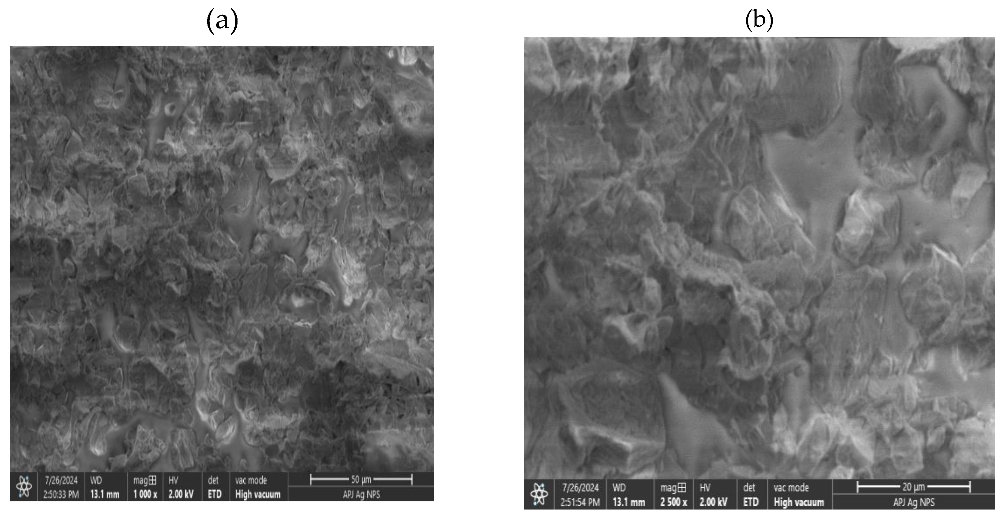
The Scanning Electron Microscopy (SEM) images (Figure 5a and b) display the structure of Ag NPs at various magnifications, showing an uneven surface texture with imperfections like fissures or cavities. The consistent shape observed in both images indicates a uniform distribution of particle size, which is attributed to the magnetic interactions between individual silver nanoparticles [33]. The uniform dispersion formation can also be explained by the presence of biological compounds from theobroma cacao on the nanoparticle surfaces. Additionally, the theobroma cacao biomolecules may contribute to the increased stability of Ag NPs [34].
Figure 5.
BET analysis for synthesized Ag NPs.
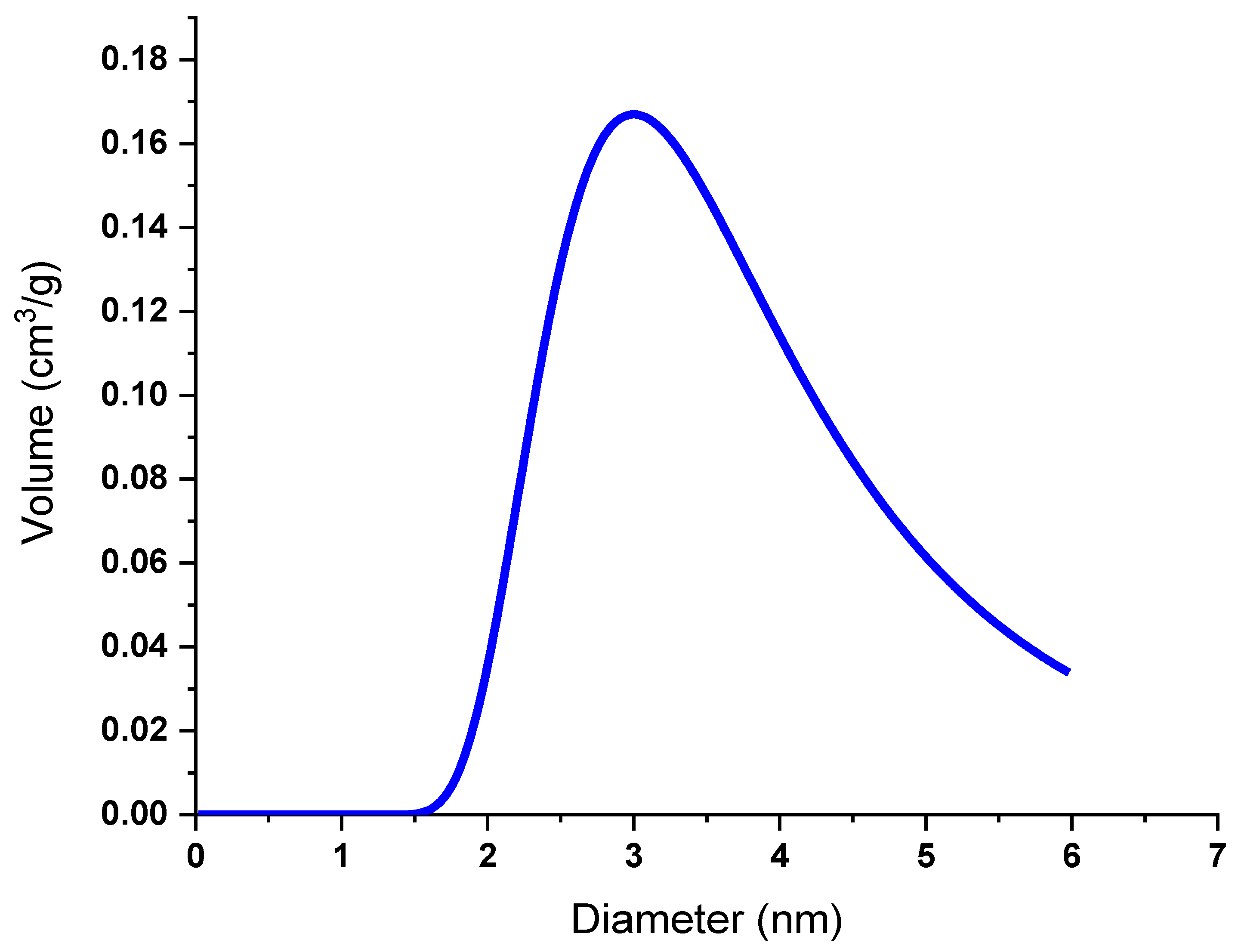
Figure 6.
SEM analysis of biosynthesized Ag NPs: (a) at 1000 × magnification (b) at 2500 × magnification.
Figure 6.
SEM analysis of biosynthesized Ag NPs: (a) at 1000 × magnification (b) at 2500 × magnification.
3.6. Transmission Electron Microscope (TEM) Analysis
Transmission electron microscopy (TEM) was employed to evaluate the dimensions and structure of the produced Ag NPs. The TEM images, displayed in Figure 7 (a-c), revealed the creation of predominantly spherical nanoparticles with uniform size distribution [35]. Various magnifications were used to capture TEM images, enabling the identification of individual particles. The nanoparticles exhibited a range of sizes, with diameters spanning from 9.22 nm to 52.60 nm. Close examination of the TEM images revealed a subtle, thin coating of material on the Ag NPs' surface, potentially attributable to organic capping agents from T. cacao leaf extracts. While the synthesized Ag NPs demonstrated diverse shapes, most were spherical, with a small number of hexagonal formations also observed.
3.7. Analysis of Photocatalytic Activity
The main factors responsible for the decolorization of dyes were hydroxyl and oxy radicals, which degrade toxic contaminants formed when a hole–electron pair was created. Furthermore, the color of dyes simultaneously became lighter with time. Degradation efficiency for IC was 80.2% after 75 min of exposure to Uv-vis in the presence of a photocatalyst, as shown in Figure 8, Table 1. The rate constant differs for the two dyes, as they have different compositions and react differently. The higher the rate constant, the faster the reaction rate and vice versa.
Figure 8.
(a) Absorption spectra of the indigo carmine dye at different time intervals and (b) Degradation rate of indigo carmine (IC) dye.
Figure 8.
(a) Absorption spectra of the indigo carmine dye at different time intervals and (b) Degradation rate of indigo carmine (IC) dye.
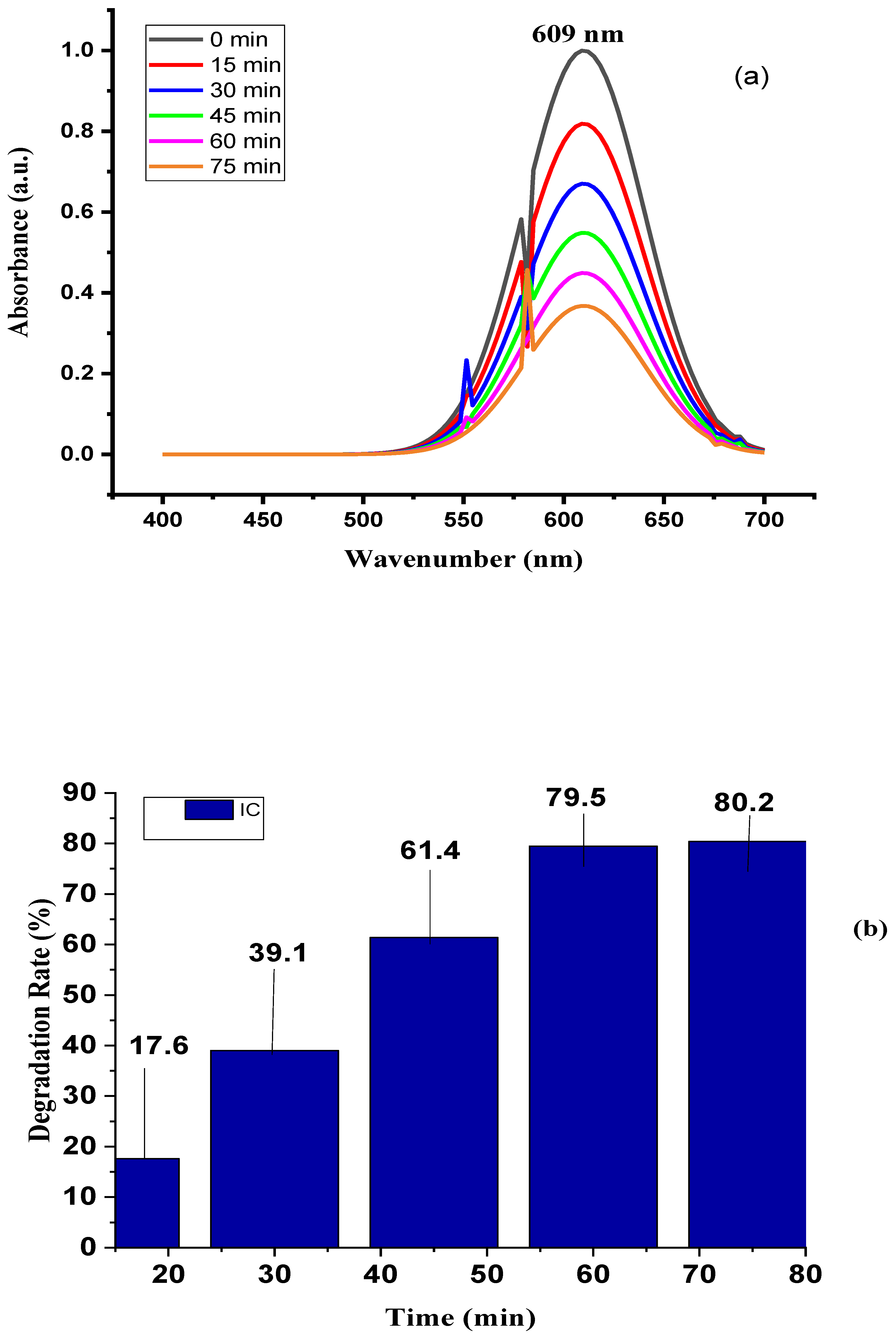
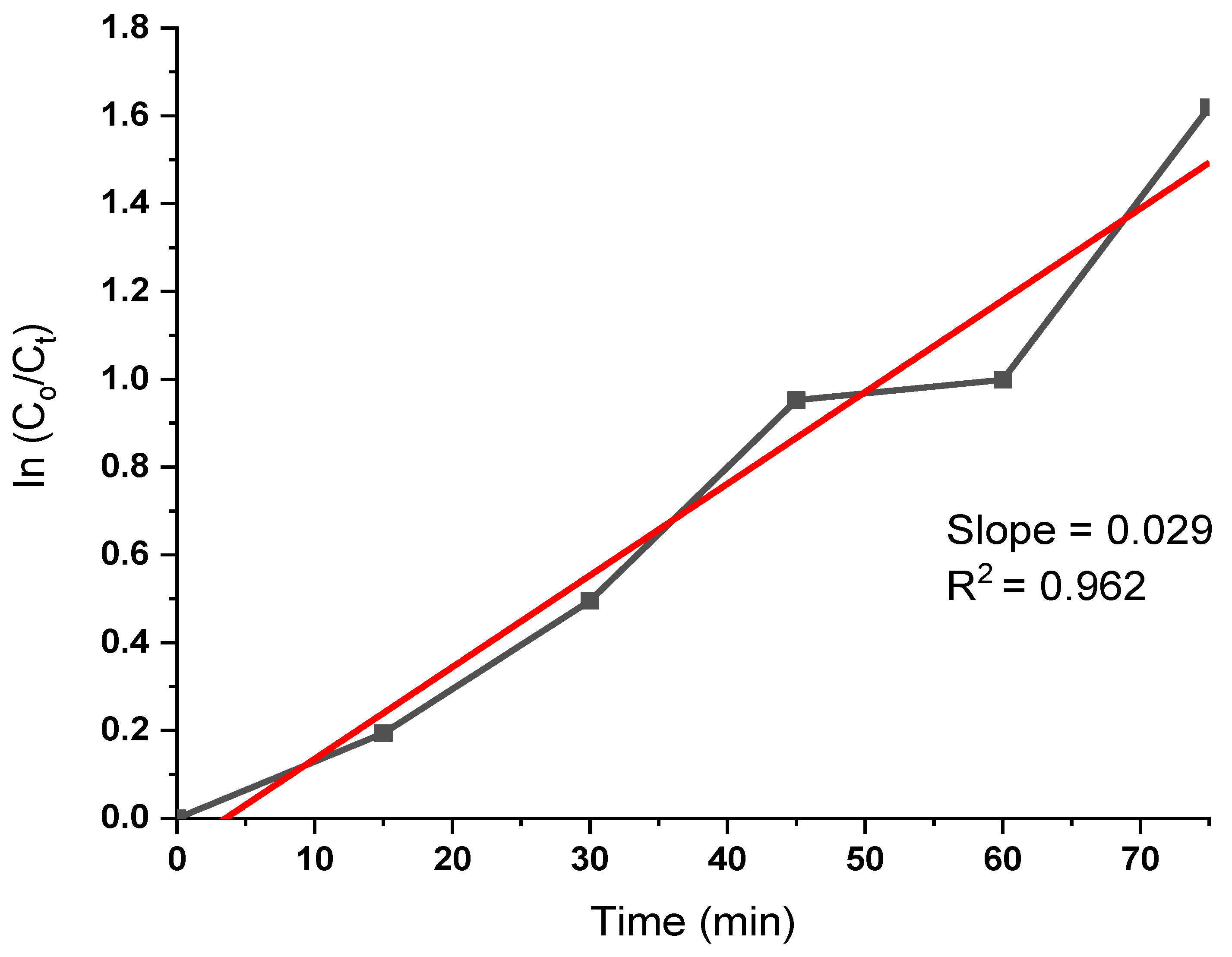
Figure 9.
Kinetic results for indingo carmine dye degradation by Ag NPs.
3.8. Conclusion
The ecologically sound production of nanoparticles of silver employing Theobroma cacao leaf extract is being recognized as an intriguing path with a variety of potential, especially in the fields of sewage treatment and in the search for environmentally conscious and sustainable solutions. Utilizing Theobroma cacao, a plant famous for its many advantages, this novel method creates silver nanoparticles with astounding efficiency and ecological sustainability. Based on the results, the following conclusions were made.
- According to FT-IR studies using an aqueous T. cacao leaf extract, the presence of plant-based nutrients such as proteins, flavonoids, alkaloid compounds, and phenolics, which act as surface-active substances, contributed to the stabilization of nanoparticles. These phytochemicals are associated with the outer layer of silver and play a role in stabilizing silver nanoparticles.
- XRD analysis revealed that the face-centered cubic plane of Ag NPs exhibited characteristic peak intensity patterns. TEM imaging of the Ag nanoparticles showed a size distribution ranging from 9.22 nm to 52.60 nm in diameter, with an average particle size of 28.52 nm determined through TEM analysis. The photocatalytic properties of the silver nanoparticles, specifically their ability to degrade harmful dyes in synthetic water, were evaluated through dye decomposition experiments. The findings indicate that the material achieved an 80.2% degradation efficiency at 75 minutes, with a half-life disintegration rate of 23.9 min-1. These results suggest that the biosynthesis of silver nanoparticles using T. cacao leaf extract is highly suitable for treating industrial effluents, particularly those containing pigments.
To summarize, our research has illuminated the promise of eco-friendly silver nanoparticles derived from Theobroma cacao leaf extract in addressing wastewater treatment challenges. The study demonstrated that these synthesized Ag nanoparticles exhibit significant potential as photocatalysts for the swift degradation of industrial dyes during water purification processes. These nanoparticles could serve as a crucial asset in ongoing efforts to protect environmental and public health, underscoring the importance of sustainable nanotechnology in tackling current global issues. However, realizing this potential will require additional research and advancement.
References
- El-Kammah, M., Elkhatib, E., Gouveia, S., Cameselle, C., Aboukil, E. (2022). Enhanced removal of Indigo Carmine dye from textile effluent using green cost-efficient nanomaterial: Adsorption, kinetics, thermodynamics and mechanisms. Sustainable Chemistry and Pharmacy 29: 100753. [CrossRef]
- Adel, M., Ahmed, M. A., Mohamed, A. A. (2021). Effective removal of indigo carmine dye from wastewaters by adsorption onto mesoporous magnesium ferrite nanoparticles. Environmental Nanotechnology, Monitoring & Management 16:100550. [CrossRef]
- Pereira, P. C. G., Reimão, R. V., Pavesi, T., Saggioro, E. M., Moreira, J. C., Correia, F. V. (2017). Lethal and sub-lethal evaluation of Indigo Carmine dye and byproducts after TiO2 photocatalysis in the immune system of Eisenia andrei earthworms. Ecotoxicology and Environmental Safety 143: 275–282. [CrossRef]
- Assemian, A. S., Kouassi, K. E., Drogui, P., Adouby, K., Boa, D. (2018). Removal of a Persistent Dye in Aqueous Solutions by Electrocoagulation Process: Modeling and Optimization Through. Water Air, and Soil Pollution 229: 184-197. [CrossRef]
- Odogu, A. N., Daouda, K., Keilah, L. P., Tabi, G. A., Rene, L. N., Nsami, N. J., Mbadcam, K. J (2020). Effect of doping activated carbon based Ricinodendron Heudelotti shells with AgNPs on the adsorption of indigo carmine and its antibacterial properties. Arabian Journal of Chemistry 13: 5241–5253. [CrossRef]
- Agnieszka, B, Monika, O-N & Monika, K. (2021). Methods of Dyes Removal from Aqueous Environment. Journal of Ecological Engineering 22(9), 111–118. [CrossRef]
- Devatha CP, Jagadeesh K, Patil M (2018) Effect of Green synthesized iron nanoparticles by Azardirachta Indica in different proportions on antibacterial activity. Environ Nanotechnol Monit Manag 9:85–94. [CrossRef]
- Raghavendra VB, Shankar S, Govindappa M, et al (2022) Green synthesis of zinc oxide nanoparticles (ZnO NPs) for effective degradation of dye, polyethylene and antibacterial performance in waste water treatment. J Inorg Organomet Polym Mater 1–17. [CrossRef]
- Abdelghaffar, F. (2021). Biosorption of anionic dye using nanocomposite derived from chitosan and silver Nanoparticles synthesized via cellulosic banana peel bio-waste. Environmental Technology & Innovation 24 (2021) 101852. [CrossRef]
- Ankita, M., Ashish, T., Srikanta, M., Subhendu, C., Susnata, S. M., Arijit, M., Suddhasattya, D., & Prakash, C. (2024). Silver nanoparticle for biomedical applications. A review Hybrid Advances, Volume 6. [CrossRef]
- Anita, D., Suresh, C., M., Sheetal, S., & Rohini, T.(2023). A review on biological synthesis of silver nanoparticles and their potential applications. Results in Chemistry, Volume 6. [CrossRef]
- Sapana, J., Rizwan, A., Nirmala, K. J., & Rajesh, K. M. (2021). Green synthesis of nanoparticles using plant extracts. Environmental Chemistry Letters 19(1), 355-374. [CrossRef]
- Anita, D., Suresh, C., M., Sheetal, S., & Rohini, T.(2023). A review on biological synthesis of silver nanoparticles and their potential applications. Results in Chemistry, Volume 6. [CrossRef]
- Ong, W. T. J., & Nyam, K. L. (2022). Evaluation of silver nanoparticles in cosmeceutical and potential biosafety complications. Saudi journal of biological sciences, 29(4), 2085–2094. [CrossRef]
- Kwon, Y. M., Cho, E. S., Kim, K. W., Chung, D., Bae, S. S., Yu, W. J., Kim, J. Y. H., & Choi, G. (2023). Synthesis of Silver Nanoparticles Using Aggregatimonas sangjinii F202Z8T and Their BiologicalCharacterization.Microorganisms,11(12),2975. [CrossRef]
- Leena, V. H., Sharanabasava, V. G., Veerabhadragouda, B. P., Sahana, N., & Aishwarya, H. Anticancer potential of biologically synthesized silver nanoparticles using Lantana camara leaf extract. Progress in Biomaterials 12 (2), 155-169. [CrossRef]
- S.S. Shankar, A. Rai, A. Ahmad, M.J. Sastry, Rapid synthesis of Au, Ag and bimetallic Au shell nanoparticles using Neem, J. Colloid Interface Sci. 275 (2004) 496–502. [CrossRef]
- H. Schneidewind, T. Schuler, K.K. Strelau, K. Weber, D. Cialla, M. Diegel, The mor- phology of silver nanoparticles prepared by enzyme-induced reduction, Beilstein J. Nanotechnol. 3 (2012) 404–414. [CrossRef]
- G.M. Sulaiman, W.H. Mohammed, T.R. Marzoog, A.A. Al-Amiery, A.A. Kadhum, A.B. Mohamad, G. Bagnati, Green synthesis, antimicrobial and cytotoxic effects of silver nanoparticles using Eucalyptus chapmaniana leaves extract, Asian Pac. J. Trop Biomed. 3 (2013) 58–63. [CrossRef]
- P. Kouvaris, A. Delimitis, V. Zaspalis, D. Papadopoulos, S.A. Tsipas, N. Michailidis, Green synthesis and characterization of silver nanoparticles produced using Arbutus unedo leaf extract, Mater. Lett. 76 (2012) 18–20. [CrossRef]
- Ashwani, K., Nirmal, P., Mukul, K., Anina, J., Vidisha,T., Emel, O., Charalampos, P., Maomao, Z., Tahra, E., Sneha, K., & Fatih, O. (2023). Major Phytochemicals: Recent Advances in Health Benefits and Extraction Method. [CrossRef]
- S. Patil, R. Sivaraj, P. Rajiv, R. Venckatesh, and R. Seenivasan, “Green synthesis of silver nanoparticle from leaf extract of Aegle marmelos and evaluation of its antibacterial activity,” International Journal of Pharmacy and Pharmaceutical Sciences, vol. 7, no. 6, pp. 169–173, 2015.
- Mahiuddin Md., Saha P and Ochiai B. 2020. “Green Synthesis and Catalytic Activity of Silver Nanoparticles Based on Piper chaba Stem Extracts.” Nanomaterials 10: 1777. [CrossRef]
- De Souza, D. C., Matos, V. A. F., Dos Santos, V. O. A., Medeiros, I. F., Marinho, C. S. R., Nascimento, P. R. P., Dorneles, G. P., Peres, A., Müller, C. H., Krause, M., Costa, E. C., & Fayh, A. P. T. (2018). Effects of high-intensity interval and moderate-intensity continuous exercise on inflammatory, leptin, IgA, and lipid peroxidation responses in obese males. Frontiers in Physiology, 9, 567. [CrossRef]
- Rodríguez-Félix, F., Graciano-Verdugo, A.Z., Moreno-Vásquez, M.J., Lagarda-Díaz, I., Barreras-Urbina, C.G., Armenta-Villegas, L., Olguín-Moreno, A., & Tapia-Hernández, J.A. (2022). Trends in Sustainable Green Synthesis of Silver Nanoparticles Using Agri-Food Waste Extracts and Their Applications in Health. Journal of Nanomaterials. [CrossRef]
- Abbas, R., Luo, J., Qi, X., Naz, A., Khan, I.A., Liu, H., Yu, S., & Wei, J. (2024). Silver Nanoparticles: Synthesis, Structure, Properties and Applications. Nanomaterials, 14. [CrossRef]
- Ghubish, Z., Kamal, R., Mahmoud, H.R., Saif, M., Hafez, H., & El-Kemary, M.A. (2022). Photocatalytic activation of Ag-doped SrSnO3 nanorods under visible light for reduction of p-nitrophenol and methylene blue mineralization. Journal of Materials Science: Materials in Electronics, 33, 24322 - 24339. [CrossRef]
- El-Desouky, N., Shoueir, K.R., El-Mehasseb, I.M., & El-Kemary, M.A. (2022). Synthesis of silver nanoparticles using bio valorization coffee waste extract: photocatalytic flow-rate performance, antibacterial activity, and electrochemical investigation. Biomass Conversion and Biorefinery, 1 - 15. [CrossRef]
- El-Shabasy, R.M., Yosri, N., El-Seedi, H.R., Shoueir, K.R., & El-Kemary, M.A. (2019). A green synthetic approach using chili plant supported Ag/Ag O@P25 heterostructure with enhanced photocatalytic properties under solar irradiation. Optik. [CrossRef]
- Sengupta, A., & Sarkar, A. (2022). Synthesis and characterization of nanoparticles from neem leaves and banana peels: a green prospect for dye degradation in wastewater. Ecotoxicology, 31, 537 - 548. [CrossRef]
- Thatikayala, D., Jayarambabu, N., Banothu, V., Ballipalli, C.B., Park, J., & Rao, K.V. (2019). Biogenic synthesis of silver nanoparticles mediated by Theobroma cacao extract: enhanced antibacterial and photocatalytic activities. Journal of Materials Science: Materials in Electronics, 30, 17303 - 17313. [CrossRef]
- Miri, A., Shahraki Vahed, H.O., & Sarani, M. (2018). Biosynthesis of silver nanoparticles and their role in photocatalytic degradation of methylene blue dye. Research on Chemical Intermediates, 44, 6907-6915. [CrossRef]
- Samuel MS, Jose S, Selvarajan E, Mathimani T, Pugazhendhi A (2020) Biosynthesized silver nanoparticles using Bacillus amyloliquefaciens; application for cytotoxicity effect on A549 cell line and photocatalytic degradation of p-nitrophenol. J Photochem Photobiol B: Biol 202 111642. [CrossRef]
- El-Shabasy R, Yosri N, El-Seedi H, Shoueir K, El-Kemary M (2019) A green synthetic approach using chili plant supported Ag/ Ag2O@ P25 heterostructure with enhanced photocatalytic proper- ties under solar irradiation. Optik 192 162943. [CrossRef]
- Singh J, Mehta A, Rawat M, Basu S (2018) Green synthesis of silver nanoparticles using sun dried tulsi leaves and its catalytic application for 4-nitrophenol reduction. J Environ Chem Eng 6(1):1468–1474. [CrossRef]
- El-Desouky, N., Shoueir, K.R., El-Mehasseb, I.M., & El-Kemary, M.A. (2022). Synthesis of silver nanoparticles using bio valorization coffee waste extract: photocatalytic flow-rate performance, antibacterial activity, and electrochemical investigation. Biomass Conversion and Biorefinery, 1 - 15. [CrossRef]
- Veisi H, Azizi S, Mohammadi P (2018) Green synthesis of the silver nanoparticles mediated by Thymbra spicata extract and its application as a heterogeneous and recyclable nanocatalyst for catalytic reduction of a variety of dyes in water. J Clean Prod 170:1536–1543. [CrossRef]
- El-Desouky, N., Shoueir, K.R., El-Mehasseb, I.M., & El-Kemary, M.A. (2022). Synthesis of silver nanoparticles using bio valorization coffee waste extract: photocatalytic flow-rate performance, antibacterial activity, and electrochemical investigation. Biomass Conversion and Biorefinery, 1 - 15. [CrossRef]
Figure 1.
Biosynthesis process of Ag NPs from T.C leaf.
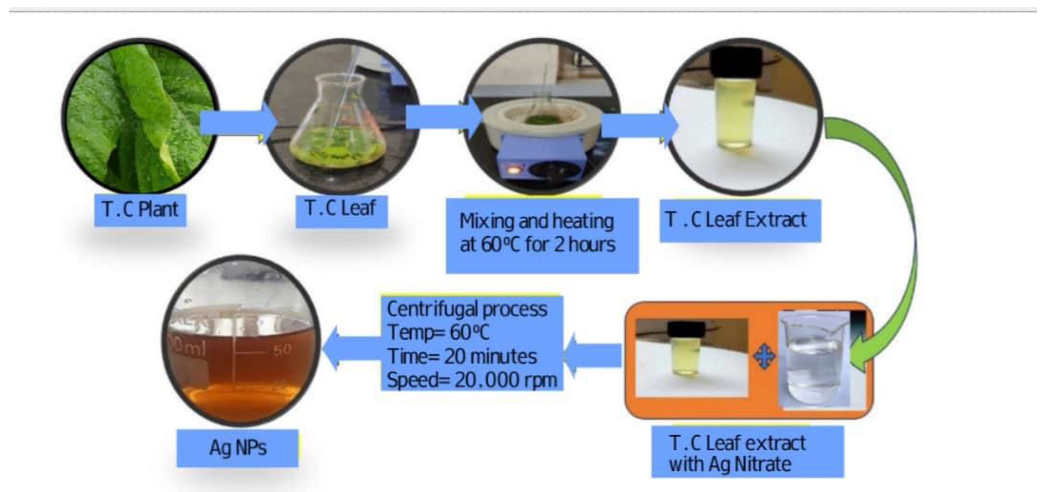
Figure 2.
UV-spectra analysis of biosynthesized Ag NPs and Tauc Plot.
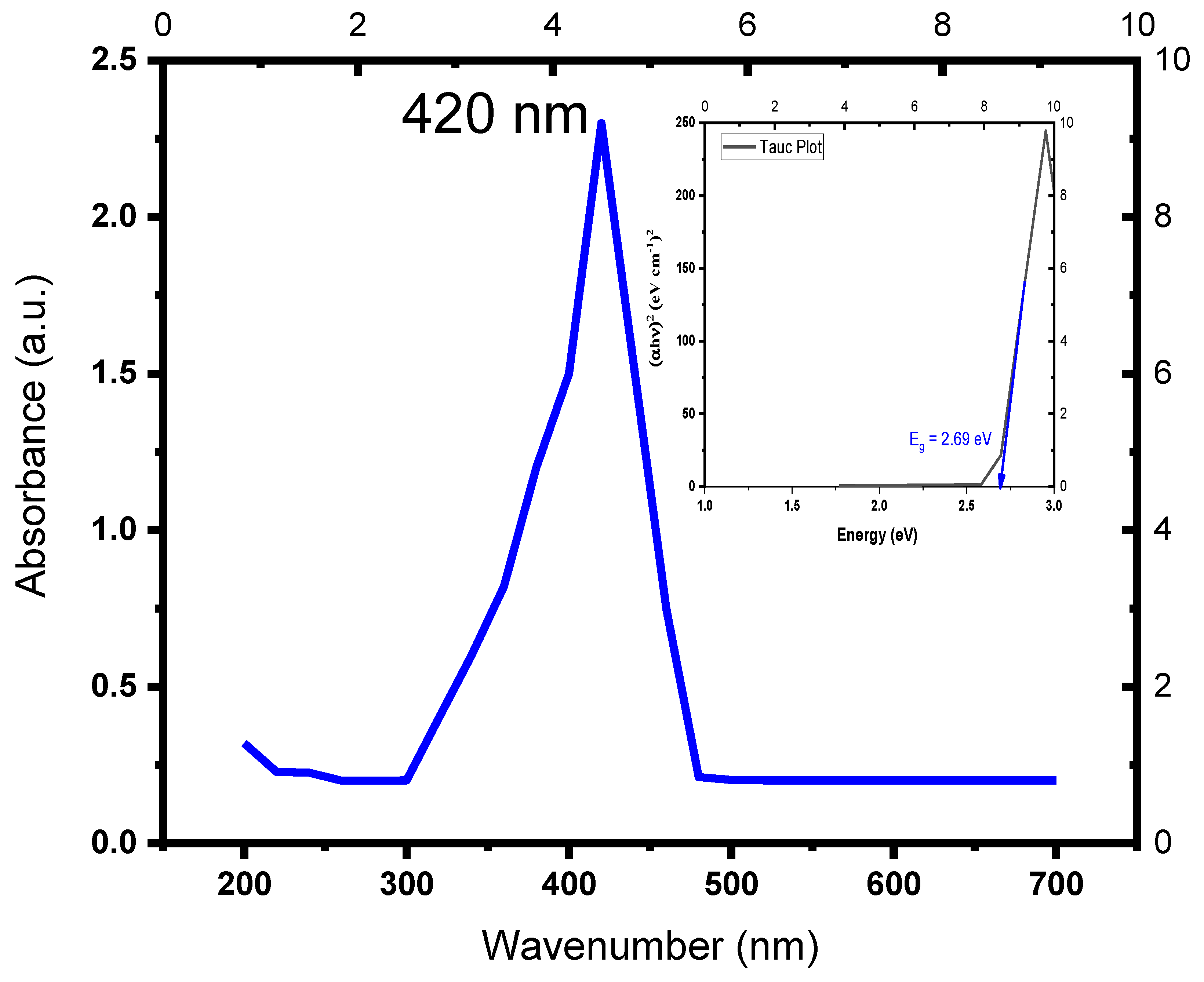
Figure 3.
X-ray diffraction pattern of the biosynthesized silver nanoparticles.
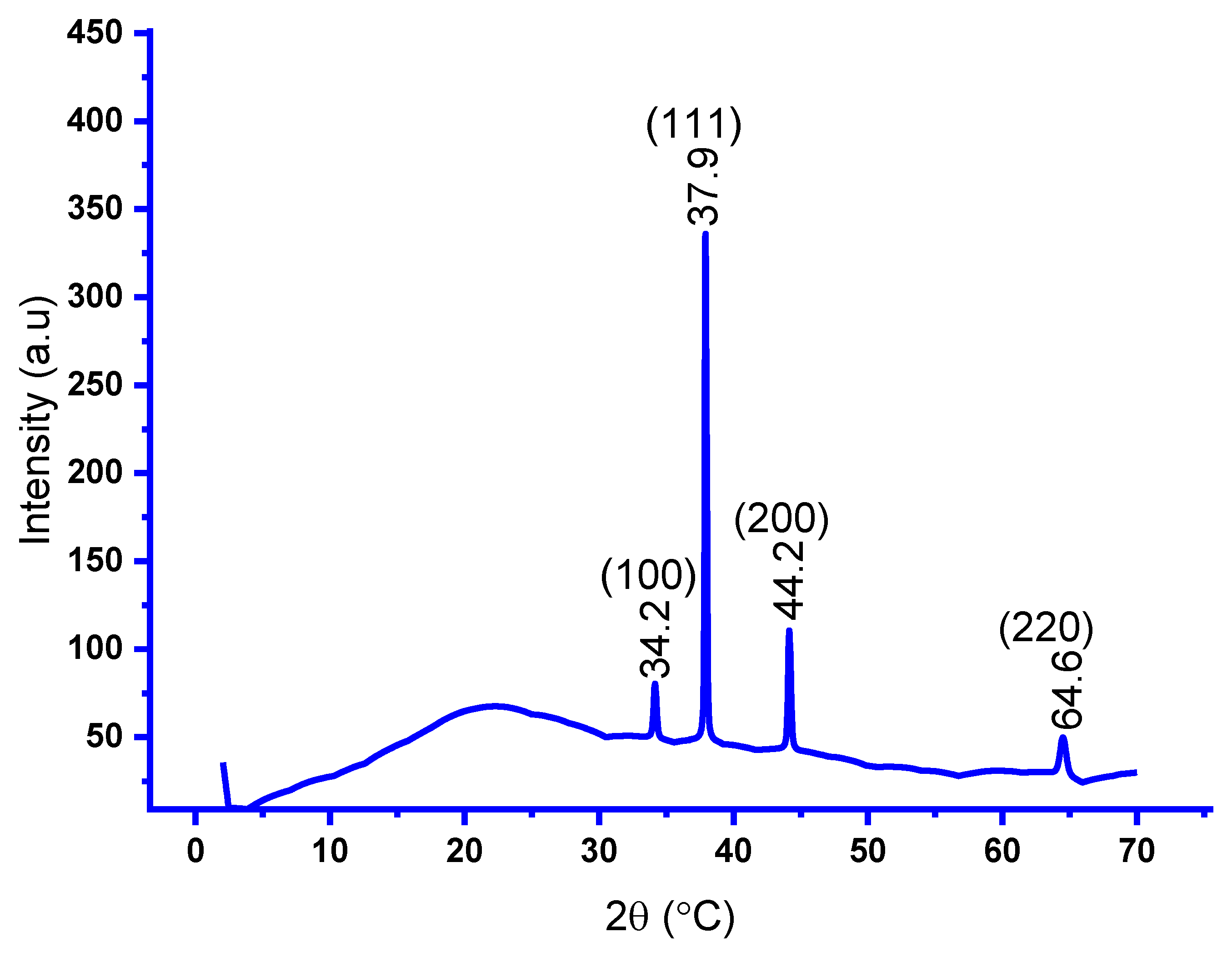
Figure 4.
FT-IR analysis of biosynthesized Ag NPs.
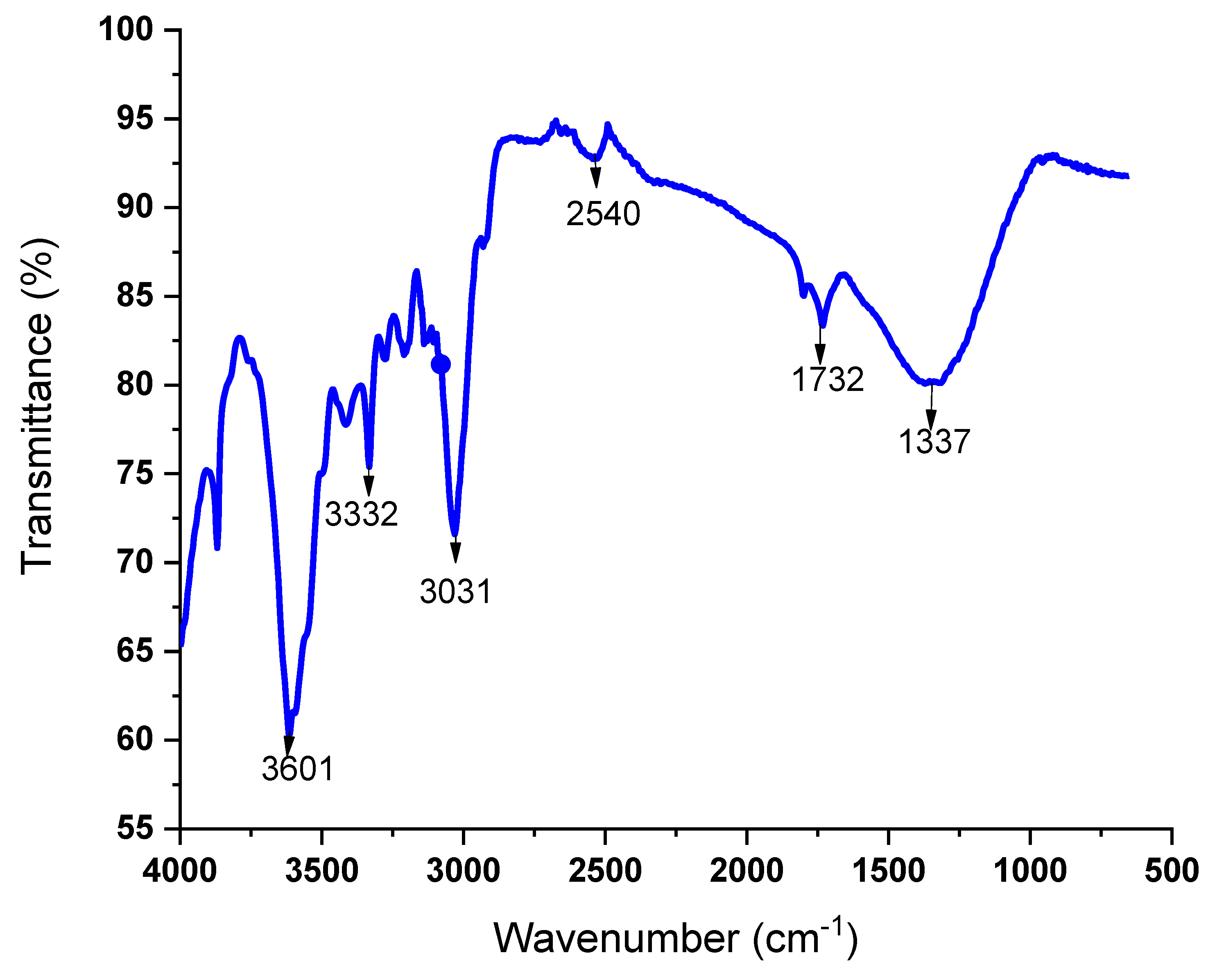
Figure 7.
TEM analysis of synthesized Ag NPs.
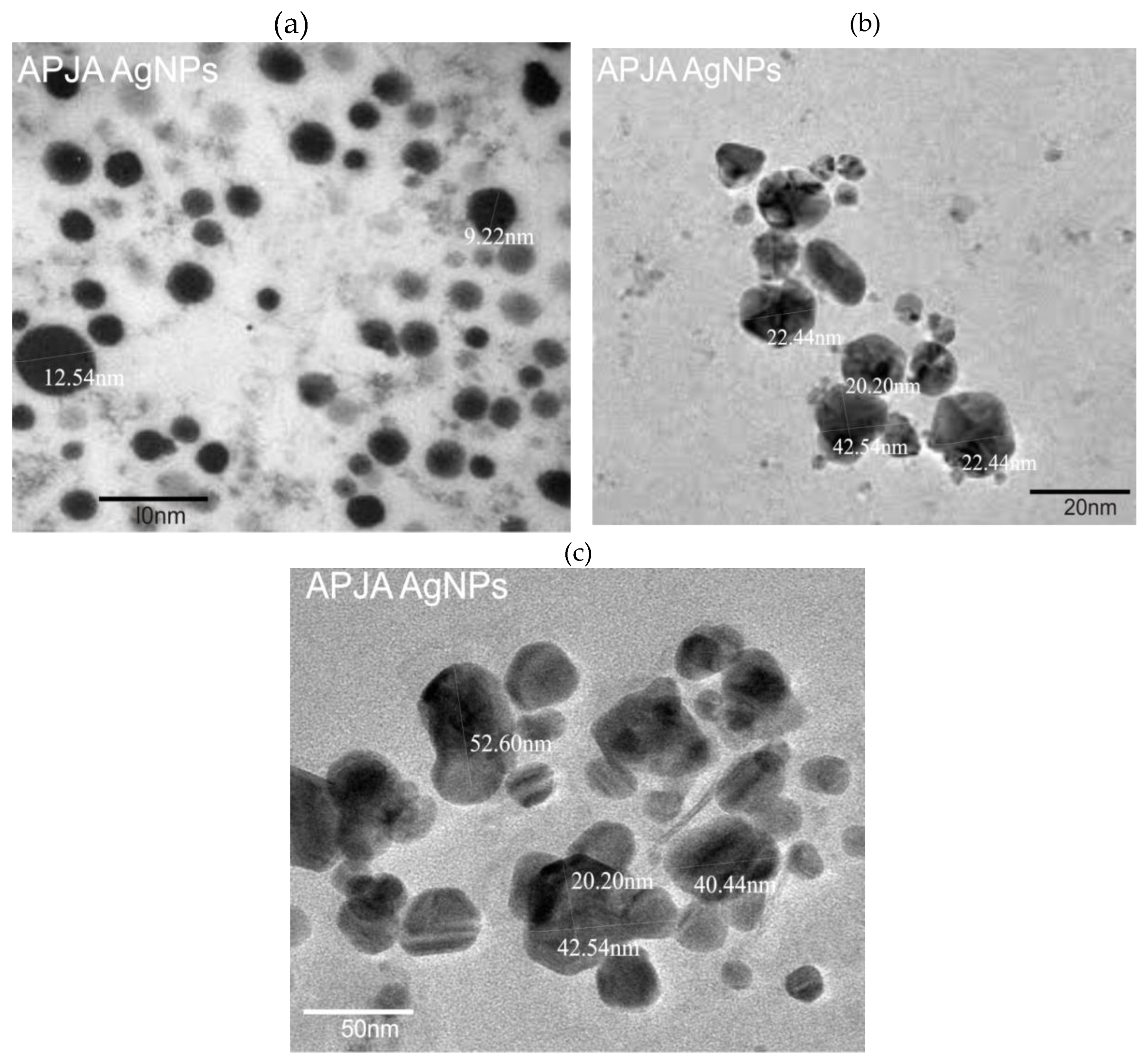

Table 1.
Comparing current work and previous studies for Ag NPs from wastes in photocatalytic activities against different pollutants.
Table 1.
Comparing current work and previous studies for Ag NPs from wastes in photocatalytic activities against different pollutants.
| Photocatalyst type | Pollutant type | Waste type | Time (min) | Efficiency (%) | References |
| Ag NPs | Bacillus amyloliquefaciens (MSR5) | 4- nitrophenol | 15 | 98 | [36] |
| Ag/Ag2O/P25 | Capsicum annuum L (chili) | 2-.4DNA | 60 | 100 | [37] |
| Ag NPs | tulsi leaves | 4- nitrophenol | 30 | 100 | [38] |
| Ag NPs | Coffee waste | Phenol | [39] | ||
| Ag NPs | Thymbra spicata/leaves | 4 nitrophenol | 1 | 96 | [40] |
| Ag NPs | Coffee waste | 2,4 DNA | 30 | 97.7 | [41] |
| Ag NPs | Theobroma cacao | Indigo carmine | 75 | 80.2 | Current work |
Disclaimer/Publisher’s Note: The statements, opinions and data contained in all publications are solely those of the individual author(s) and contributor(s) and not of MDPI and/or the editor(s). MDPI and/or the editor(s) disclaim responsibility for any injury to people or property resulting from any ideas, methods, instructions or products referred to in the content. |
© 2025 by the authors. Licensee MDPI, Basel, Switzerland. This article is an open access article distributed under the terms and conditions of the Creative Commons Attribution (CC BY) license (http://creativecommons.org/licenses/by/4.0/).
Copyright: This open access article is published under a Creative Commons CC BY 4.0 license, which permit the free download, distribution, and reuse, provided that the author and preprint are cited in any reuse.


