

Submitted:
15 December 2024
Posted:
16 December 2024
You are already at the latest version
Abstract
The ectopic thyroid gland is an abnormal development of the embryo. Most of the ectopic thyroid occurs in the path around the thyroglossal duct or on the lateral side of the neck. However, ectopic thyroid occurs in the heart, which is rare. We report a case of right ventricular ectopic thyroid. This case highlights the imaging characteristics of computed tomography (CT) and cardiac magnetic resonance (CMR) and analyzes the underlying mechanisms for a timely diagnosis.
Keywords:
Ectopic thyroid
; cardiac ectopic thyroid
; right ventricular mass
1. Introduction
Ectopic thyroid gland (ETG) refers to thyroid tissue not located in its normal anatomical position. The most common location is the base of the tongue, which accounts for 90% of the cases [1]. Furthermore, ETG may be found in the mediastinum, larynx, trachea, esophagus, and cervical lymphatic system and even in subcutaneous organs, such as the gallbladder, mesentery, and adrenal glands [2]. Intracardial ectopic thyroid is rare, and its prevalence is unknown [3].
2. Case Report
A right ventricular mass was accidently identified in a 56-year-old woman during chest CT for a respiratory infection at a local hospital. Transthoracic echocardiogram (TTE) revealed a cardiac tumor, and the patient was admitted to our hospital for further diagnosis and treatment. The patient had a history of hypertension but no history of coronary artery disease. A grade 2–6 systolic murmur was heard between the second and third intercostal space at the left sternal border. Electrocardiogram showed sinus rhythm with possible right bundle branch block, and chest X-ray revealed a normal-sized heart. TTE indicated that the right ventricle was normal in size. A 43 × 28 mm mass with a medium echo appearance, neither mobile nor pediculated, was found to be adhered to the interventricular septum and extended to the right ventricular outflow tract. In addition, mild tricuspid regurgitation and normal mitral and aortic valves were noted (Figure 1).
Cardiac noncontrast CT confirmed the presence of a round mass slightly high in density within the right ventricle, with a CT value of 70 Hounsfield unit (HU). In the contrast-enhanced scan, early, rapid, and homogeneous enhancement was noted, with a CT value of approximately 131 HU, and further enhancement with a CT value of approximately 213 HU, which was similar in density to the patient’s thyroid gland on noncontrast and contrast-enhanced CT (Figure 2). Coronary computed tomography angiography (CCTA) suggested that the mass was perfused by a branch arising from the left anterior descending artery (Figure 2).
The cine CMR images showed a hypointense mass with regular contours, which adhered to the interventricular septum and protruded into the right ventricular cavity. The signal intensity of the mass was high on T2-weighted images, without any signal decrease on the fat suppression sequence. However, pericardial or pleural effusion was not observed. CMR rest perfusion sequences demonstrated a rapid signal increase in the mass following an augmentation in the left ventricular blood pool signal, which implied that the mass was perfused by the aorta, and then a rapid decrease in signal intensity on early gadolinium enhancement (Figure 3). Moreover, myocardial delayed gadolinium enhancement was considerably lower than the blood pool signal at the same level.
The patient had a structural abnormality of the heart that could not be treated with medications; thus, surgical exploration to remove the mass was recommended. Postsurgical chest X-ray and ultrasound examinations did not show any abnormality. Microscopic findings confirmed the absence of neoplastic cells in the mass, and only typical features of normal thyroid cells were observed. Immunohistochemical staining of the mass tissue revealed a positive reaction for thyroid transcription factor-1 and thyroglobulin (Figure 4). The final pathological diagnosis was intracardial ectopic thyroid.
3. Discussion
The incidence of primary cardiac necropsies is 0.05% [4], and approximately 90% of the cases are benign [5]. The incidence of metastatic tumor in the heart is approximately 1% [6], and intracardial ectopic thyroid is much rarer. This condition could be caused by the presence of thyroid rudiments dragged into the chest during the descent of the heart and great vessels during the early stages of organogenesis [7]. Intracardial ectopic thyroid affects mostly middle-aged women. The most common cardiac sites are the right side of the ventricular septum and the right ventricular outflow tract [7], but it can also be located in the aorta [8,9,10,11] and left ventricular outflow tract [12,13].
The clinical manifestations of intracardial ectopic thyroid are nonspecific and often identified incidentally, sometimes presenting as dyspnea, chest pain, syncope, ventricular arrhythmia, or sudden death due to cardiac obstruction [12]. Patients with thyroid symptoms in the absence of localized thyroid disease or after thyroidectomy in the normal location should be comprehensively investigated for the possibility of ETG [14]. Most patients with intracardial ectopic thyroid exhibit normal thyroid hormone levels, which do not contribute to the diagnosis of the disease. Intracardial ectopic thyroid has been shown to absorb iodine-123 on single-photon emission-CT scans but not fluoride-18-labeled fluorodeoxyglucose uptake on positron emission tomography-CT [14,15]. A study reported possible malignant transformation of cardiac ETG after thyroidectomy in the normal location, probably because increased thyroid stimulating hormone after thyroidectomy could stimulate cardiac thyroid growth and even malignant transformation [16].
Intracardial ectopic thyroid is differentiated primarily from cardiac tumors and thrombus. Cardiac myxomas are attached by a stalk to the atrial septum in the oval fossa, and their range of movement is large. Contrast enhancement on CT often shows heterogeneous enhancement owing to the possible presence of hemorrhage, necrosis, cystic changes, fibrosis, or calcification. Certain rare myxomas may be identified as small, round, sharp, homogenous signals fixed in the cardiac chamber on CMR. Cardiac rhabdomyomas primarily affect infants and children. Approximately 80% of cardiac rhabdomyomas occur in association with tuberous sclerosis [17], and rhabdomyomas are multiple in >60% of the cases [18]. On CMR cine images, rhabdomyomas appear to be isointense or slightly hyperintense on T1-weighted imaging and hyperintense on T2-weighted imaging, without significant enhancement after contrast medium administration. Cardiac hemangiomas have a broad base and no stalk. Contrast enhancement on CT shows centripetal filling from the edge to the center, with a time delay. The hemangioma appears to be isointense on T1-weighted imaging, hyperintense on T2-weighted imaging, and hyperintense on fat-suppressed images. Considerable homogeneous prolonged enhancement is noted after injecting the contrast medium, but heterogeneous areas could also be observed because of calcification or fiber septum. Cardiac lipomas are benign tumors of the heart composed of mature adipose tissue. On CT, the lesion presents as a localized mass with low attenuation. On CMR, both T1- and T2-weighted images present homogeneous hyperintensity, with signal dropout during fat suppression [19]. Cardiac teratomas are germ-cell tumors and contain derivatives of all three germ layers, with mature endodermal, mesodermal, and neuroectodermal elements. On CT, they appear as multicystic, heterogeneous tumors with an associated pericardial effusion [20]. Lipidic or calcific densities are usually present on CT. Cardiac metastatic tumors arise as direct extensions or spread via hematogenous, lymphatic, or intracavitary routes of the primary tumor. Most common metastases to the heart originate from lung carcinoma (35%–40%), followed by hematologic malignancies (10%–20%) and breast cancer (10%) [21]. The most common location of metastasis is the pericardium, followed by epicardium and myocardium. Cardiac metastatic tumors often exhibit pericardial and pleural effusion. Ventricular thrombus occurs in the region of ventricular aneurysm or reduced ventricular motion. Acute thrombus is hyperintense on T1- and T2-weighted imaging. Subacute thrombus is hyperintense on T1-weighted imaging and hypointense on T2-weighted imaging. In the chronic period, the thrombus is characterized by hypointensity on T1- and T2-weighted imaging.
Multimodal imaging is crucial in characterizing cardiac tumors. TTE is the most convenient imaging modality to determine the location of the mass, valve function, and outflow tract obstruction. Cardiac CT and CMR are beneficial in ascertaining the anatomic relationships among the mass, myocardium, and pericardium. In this case, the mass showed transient enhancement on CMR and obvious homogeneous enhancement on contrast-enhanced CT. This difference could be attributed to the different contrast agents used, with the mass absorbing the iodine contrast agent used in the CT contrast enhancement scan but not the gadolinium used in the CMR scan.
4. Conclusions
In summary, if a round, broad mass is found in the right ventricular outflow tract in a middle-aged woman, the imaging findings of the CMR enhancement scan are not consistent with those of the CT enhancement scan, and the mass is similar in density to that of the thyroid gland on noncontrast and contrast-enhanced CT, the possibility of intracardial ectopic thyroid should be considered after ruling out primary and secondary cardiac tumors.
Author Contributions
Conceptualization, methodology, supervision, data curation, project administration, formal analysis, N.Z., Z.S. and Z.W.; formal analysis, validation, writing—original draft preparation, C.W., Y.Z. and Y.S.; writing—review and editing, D.L., J.L. and L.X. All authors have read and agreed to the published version of the manuscript.
Funding
This research received no external funding.
Institutional Review Board Statement
Not applicable.
Informed Consent Statement
Informed consent was obtained from all subjects involved in the study. Written informed consent has been obtained from the patient(s) to publish this paper.
Data Availability Statement
The data that support the findings of this study are not openly available due to reasons of sensitivity and are available from the corresponding author upon reasonable request.
Conflicts of Interest
All authors have completed the ICMJE uniform disclosure form. Z.S. servers as an unpaid associate editor of Journal of Cardiovascular Development and Disease. The other authors have no conflicts of interest to declare.
References
- Jiménez Oliver, V.; Ruiz Rico, R.; Dávila Morillo, A.; Fernández Ruiz, E.; Ruiz del Portal, J.M.; Pérez Arcos, J.A.; Rodríguez Baró, G. Intra-laryngeal ectopic thyroid tissue. Report of one case and review of the literature. Acta. Otorrinolaringol. Esp. 2002, 53, 54–59. [Google Scholar] [CrossRef]
- Paunovic, I.; Rovcanin, B.; Jovanovic, M.; Buzejic, M.; Dundjerovic, D.; Zivaljevic, V. Ectopic thyroid tissue in adrenal gland, case report and review of literature. Gland. Surg. 2020, 9, 1573–1578. [Google Scholar] [CrossRef] [PubMed]
- Kousta, E.; Konstantinidis, K.; Michalakis, C.; Theodoropoulos, G.; Vorias, M.; Georgiou, M.; Sambalis, G. Ectopic thyroid tissue in the lower neck with a coexisting normally located multinodular goiter and brief literature review. Hormones (Athens) 2005, 4, 231–234. [Google Scholar] [CrossRef] [PubMed]
- Shapiro, M.L. Cardiac tumours: diagnosis and management. Heart. 2001, 85, 218–222. [Google Scholar] [CrossRef]
- Mc Allister, H.A. Primary tumors and cysts of the heart and pericardium. Curr. Probl. Cardiol. 1979, 4, 1–51. [Google Scholar] [CrossRef] [PubMed]
- Shapiro, L.M. Cardiac tumours: diagnosis and management. Heart. 2001, 85, 218–222. [Google Scholar] [CrossRef] [PubMed]
- Gackowski, A.; Pietrucha, A.; Miszalski-Jamka, T.; Rostoff, P.; Sadowski, J.; Nessler, J. Intrapericardial ectopic thyroid gland mimicking cardiac tumor. Int. J. Cardiol. 2012, 158, e55–e56. [Google Scholar] [CrossRef]
- Taylor, M.A.; Bray, M.; Roberts, W.C. Aberrant thyroid gland attached to ascending aorta. Am. J. Cardiol. 1986, 57, 708. [Google Scholar] [CrossRef]
- Williams, R.J.; Lindop, G.; Butler, J. Ectopic thyroid tissue on the ascending aorta: an operative finding. Ann. Thorac. Surg. 2002, 73, 1642–1643. [Google Scholar] [CrossRef]
- Özpolat, B.; Doǧan, O.V.; Gökaslan, G.; Erekul, S.; Yücel, E. Ectopic Thyroid Gland on the Ascending Aorta with a Partial Pericardial Defect: Report of a Case. Surg. Today. 2007, 37, 486–488. [Google Scholar] [CrossRef]
- Mesrar, H.; Chatel, D.; Barbey, C.; Biny, J-P. ; Arnould, M-A.; Gouffran, G.; Bar, O.; Chassaing, S. Rare Case of an Intrapericardial Ectopic Thyroid (Struma Cordis) on the Ascending Aorta. Circ. Cardiovasc. Imaging. 2020, 13, e010508. [Google Scholar] [CrossRef] [PubMed]
- Baykut, D.; Fiegen, U.; Krian, A.; Thiel, A. Ectopic thyroid tissue in the left ventricular outflow tract. Ann. Thorac. Surg. 2000, 69, 620–621. [Google Scholar] [CrossRef]
- Kon, N.D.; Headley, R.N.; Cordell, A.R. Successful Operative Management of Struma Cordis Obstructing the Left Ventricular Outflow Tract. Ann. Thorac. Surg. 1988, 46, 244–245. [Google Scholar] [CrossRef]
- Landeta, F.; Hoffmeier, A.; Fuchs, M.; Scheld, H.; Maintz, D.; Stypmann, J.; Schober, O.; Kirchhof, P.; Spieker, T.; Waltenberger, J. Ectopic thyroid mass in the heart. Lancet. 2012, 379, 1762. [Google Scholar] [CrossRef]
- Gu, Y.M.; Fan, Y.Y.; Wang, W.P.; Chen, L.Q. Ectopic thyroid tissue in the heart. QJM. 2019, 112, 927–928. [Google Scholar] [CrossRef] [PubMed]
- Hirnle, T.; Szymczak, J.; Ziotkowski, P.; Lenartowska, L. Ectopic thyroid malignancy in the right ventricle of the heart. Eur. J. Cardiothorac. Surg. 1997, 12, 147–149. [Google Scholar] [CrossRef]
- Groves, A.M.; Fagg, N.L.; Cook, A.C.; Allan, L.D. Cardiac tumours in intrauterine life. Arch Dis Child 1992, 67, 1189–1192. [Google Scholar] [CrossRef]
- Tworetzky, W.; McElhinney, D.B.; Margossian, R.; Moon-Grady, A.J.; Sallee, D.; Goldmuntz, E.; van der Velde, M.E.; Silverman, N.H.; Allan, L.D. Association between cardiac tumors and tuberous sclerosis in the fetus and neonate. Am. J. Cardiol. 2003, 92(4), 487–489. [Google Scholar] [CrossRef]
- Beroukhim, R.S.; Prakash, A.; Valsangiacomo Buechel, E.R.; Cava, J.R.; Dorfman, A.L.; Festa, P.; Hlavacek, A.M.; Johnson, T.R.; Keller, M.S.; Krishnamurthy, R.; Misra, N.; Moniotte, S.; Parks, W.J.; Powell, A.J.; Soriano, B.D.; Srichai, M.B.; Yoo, S-J. ; Zhou, J.; Geva, T. Characterization of Cardiac Tumors in Children by Cardiovascular Magnetic Resonance Imaging. J. Am. Coll. Cardiol. 2011, 58, 1044–1054. [Google Scholar] [CrossRef] [PubMed]
- Taori, K.; Jawale, R.; Sanyal, R.; Sheorain, V.S.; Chandanshive, S. Intrapericardial Teratoma Diagnosed on CT. J. Thorac. Imaging. 2007, 22, 185–187. [Google Scholar] [CrossRef] [PubMed]
- Goldberg, A.D.; Blankstein, R.; Padera, R.F. Tumors Metastatic to the Heart. Circulation. 2013, 128, 1790–1794. [Google Scholar] [CrossRef]
Figure 1.
Transthoracic echocardiography of the lesion. (A) A 43 × 28 mm mass with medium echo appearance, neither mobile or pediculated, adhering to the interventricular septum and extending to the right ventricular outflow tract. (B) Color doppler flow imaging (CDFI): no significant blood flow signal was detected in the mass, and slight aliasing was noted in the right ventricular outflow tract.
Figure 1.
Transthoracic echocardiography of the lesion. (A) A 43 × 28 mm mass with medium echo appearance, neither mobile or pediculated, adhering to the interventricular septum and extending to the right ventricular outflow tract. (B) Color doppler flow imaging (CDFI): no significant blood flow signal was detected in the mass, and slight aliasing was noted in the right ventricular outflow tract.
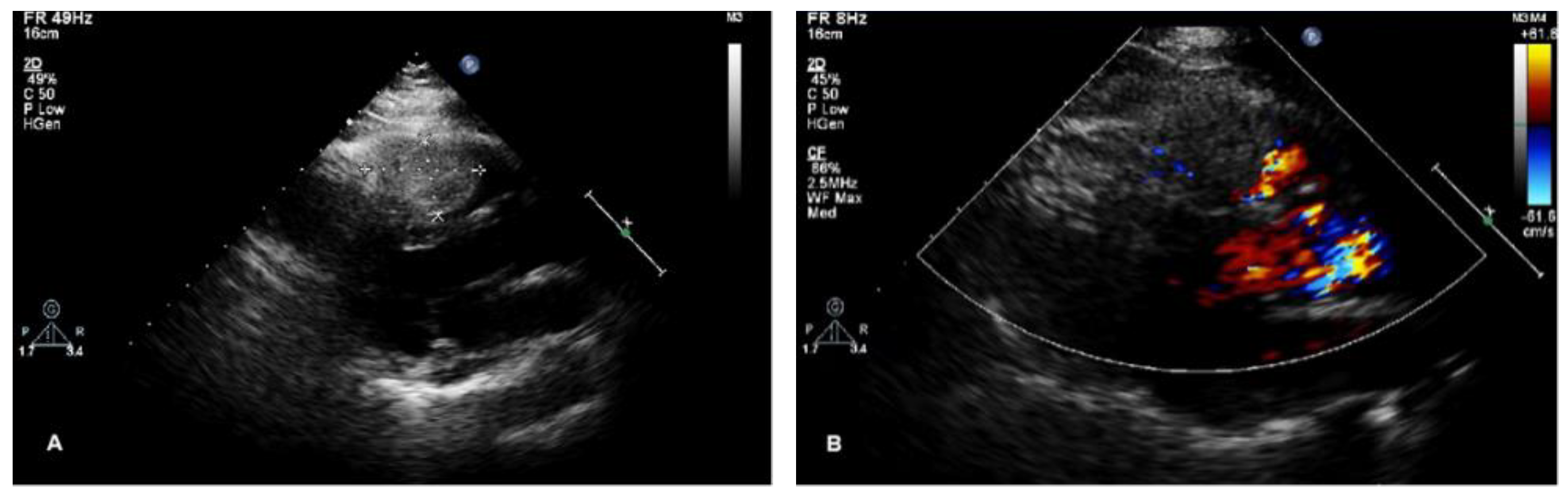
Figure 2.
Cardiac CT imaging of the lesion. (A) The mass was slightly high in density on CT plain scan. (B–E) The mass demonstrated obvious homogeneous enhancement on contrast-enhanced scan, which resembled the patient’s thyroid enhancement pattern. (F) CCTA images showed that the mass was perfused by a branch arising from the left anterior descending artery.
Figure 2.
Cardiac CT imaging of the lesion. (A) The mass was slightly high in density on CT plain scan. (B–E) The mass demonstrated obvious homogeneous enhancement on contrast-enhanced scan, which resembled the patient’s thyroid enhancement pattern. (F) CCTA images showed that the mass was perfused by a branch arising from the left anterior descending artery.
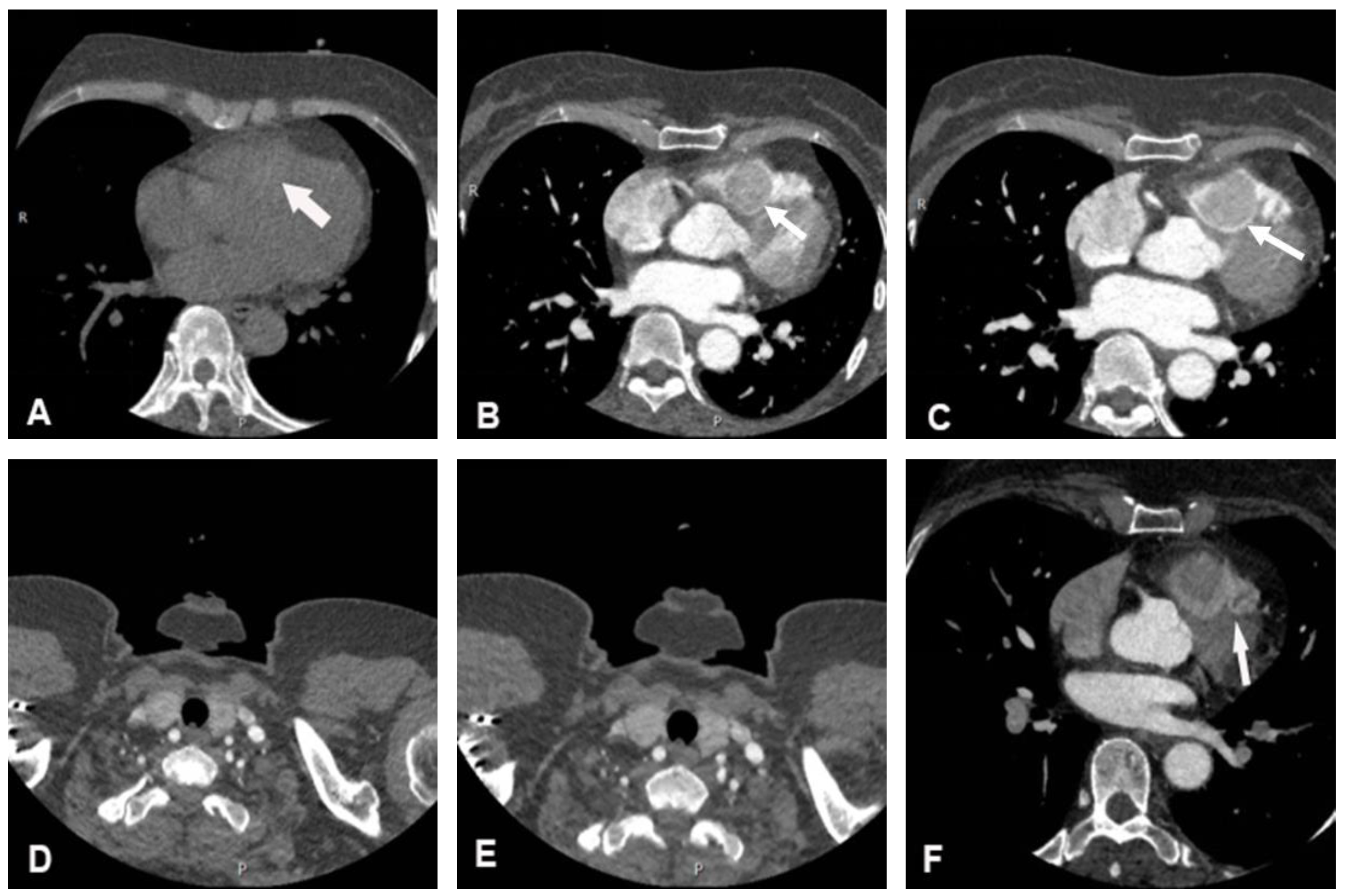
Figure 3.
Cardiac magnetic resonance imaging. (A) Cine images of magnetic resonance imaging showed a hypointense mass. (B) The signal intensity of the mass was high on T2-weighted images. (C,D) The mass showed a rapid enhancement on CMR rest perfusion. (E) The signal intensity of the mass decreased rapidly on early gadolinium enhancement. (F) Myocardial delayed gadolinium enhancement was significantly lower than the blood pool signal at the same level.
Figure 3.
Cardiac magnetic resonance imaging. (A) Cine images of magnetic resonance imaging showed a hypointense mass. (B) The signal intensity of the mass was high on T2-weighted images. (C,D) The mass showed a rapid enhancement on CMR rest perfusion. (E) The signal intensity of the mass decreased rapidly on early gadolinium enhancement. (F) Myocardial delayed gadolinium enhancement was significantly lower than the blood pool signal at the same level.
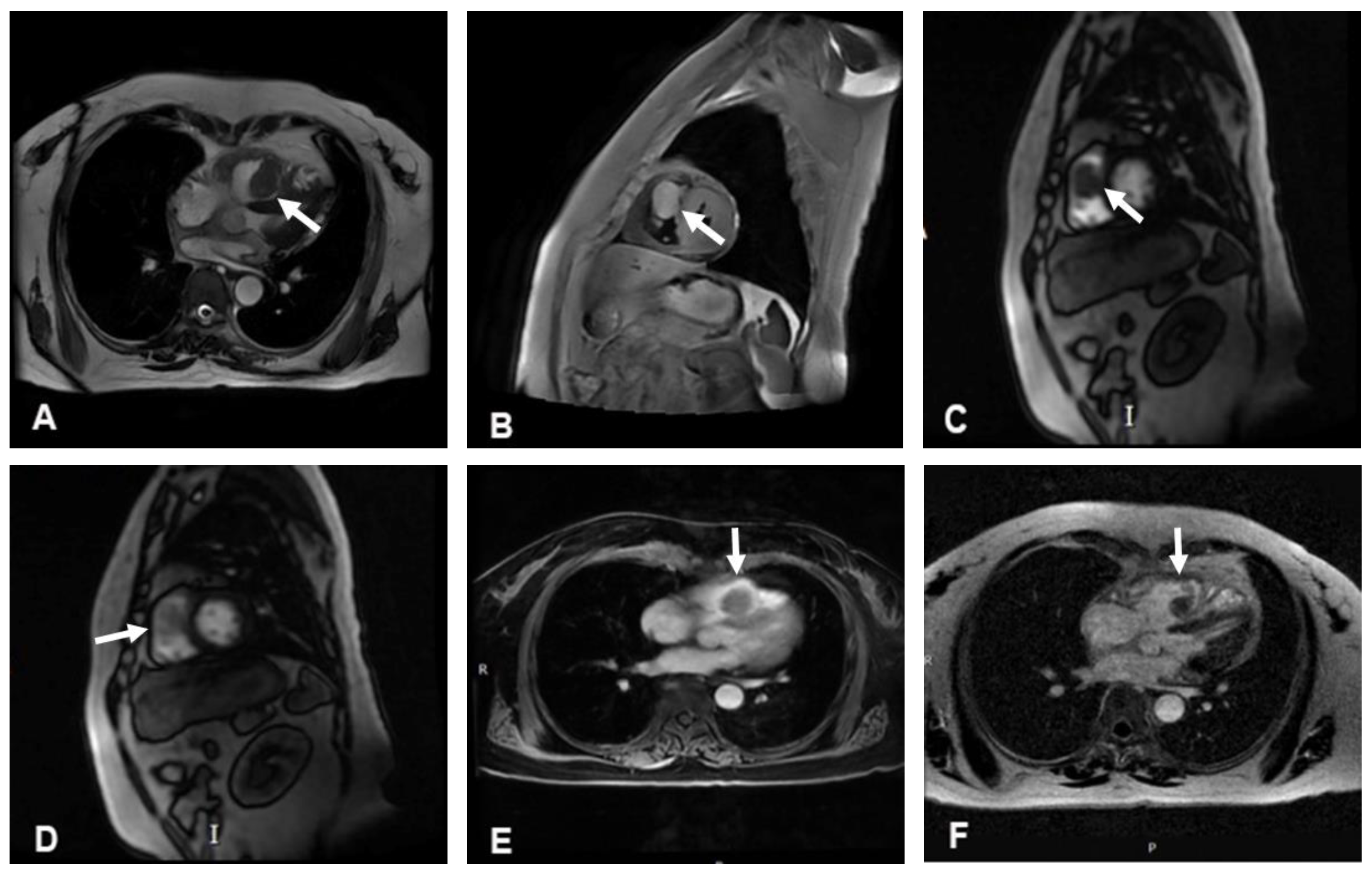
Figure 4.
Postoperative pathology of the cardiac tumor. (A) H&E, ×40. The arrow shows thyroid follicles. (B) thyroid transcription factor-1 (TTF-1), ×40. (C) thyroglobulin (TG), ×40.
Figure 4.
Postoperative pathology of the cardiac tumor. (A) H&E, ×40. The arrow shows thyroid follicles. (B) thyroid transcription factor-1 (TTF-1), ×40. (C) thyroglobulin (TG), ×40.

Disclaimer/Publisher’s Note: The statements, opinions and data contained in all publications are solely those of the individual author(s) and contributor(s) and not of MDPI and/or the editor(s). MDPI and/or the editor(s) disclaim responsibility for any injury to people or property resulting from any ideas, methods, instructions or products referred to in the content. |
© 2024 by the authors. Licensee MDPI, Basel, Switzerland. This article is an open access article distributed under the terms and conditions of the Creative Commons Attribution (CC BY) license (http://creativecommons.org/licenses/by/4.0/).
Copyright: This open access article is published under a Creative Commons CC BY 4.0 license, which permit the free download, distribution, and reuse, provided that the author and preprint are cited in any reuse.


