Preprint
Article

This version is not peer-reviewed.

The Effect of Peri-Implant Therapy on the Expression of Th17-Related Cytokines in Patients with Peri-Implant Mucositis and Peri-Implantitis: A Prospective Longitudinal Study

A peer-reviewed article of this preprint also exists.

Submitted:

14 December 2024

Posted:

16 December 2024

You are already at the latest version

Abstract

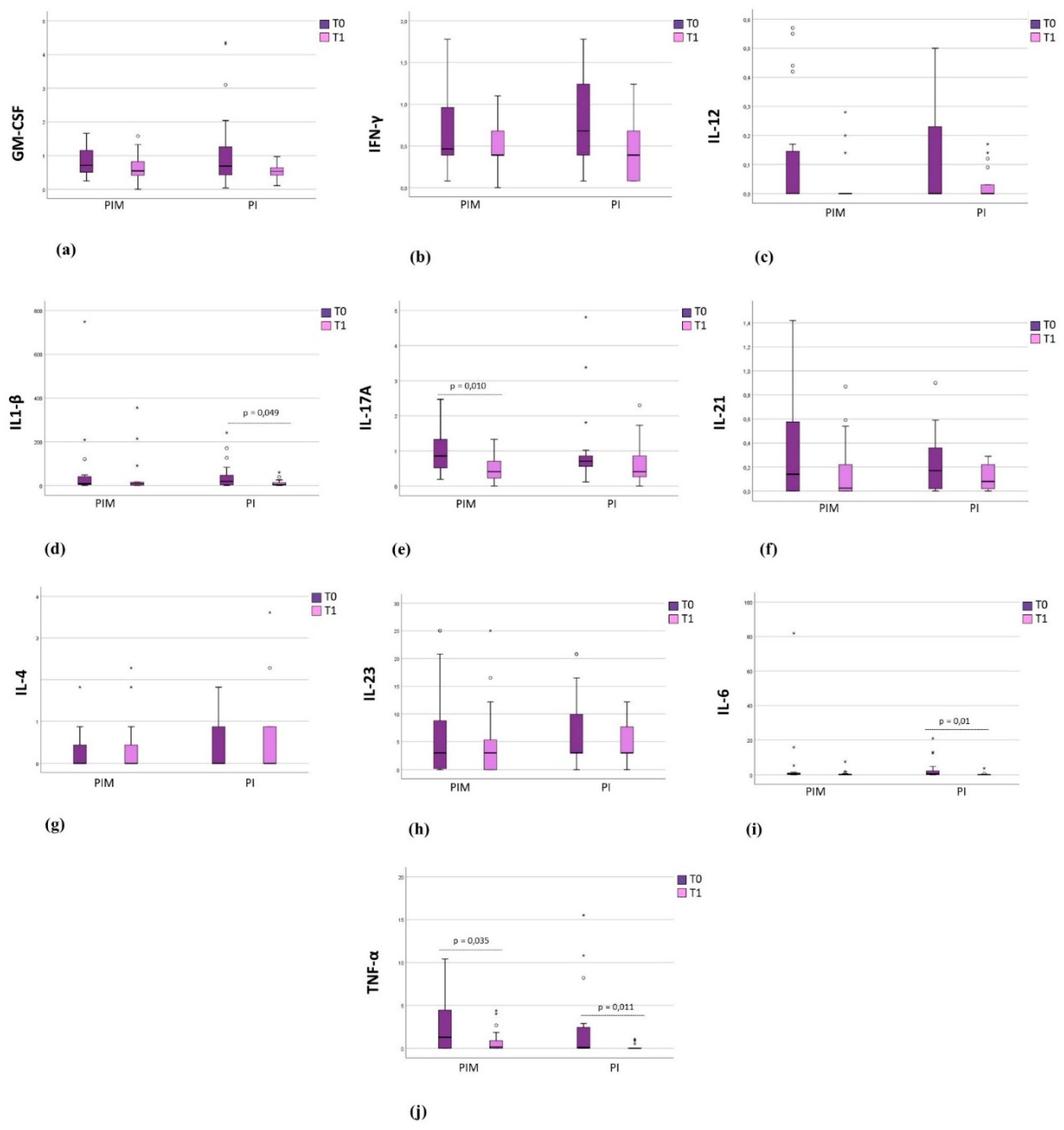
Background: Cytokines related to the Th17 response have been associated with pe-ri-implant diseases, however, the effect of peri-implant therapy on their modulation remains un-derexplored. Objectives: To evaluate the effect of peri-implant therapy on the expression of cyto-kines related to the Th17 response in the peri-implant crevicular fluid (PICF) (GM-CSF, IFN-γ, IL-1β, IL-4, IL-6, IL-10, IL-12(p70), IL-17A, IL-21, IL-23, and TNF-α) of partially edentulous pa-tients with peri-implant disease (PID). Methods: Thirty-seven systemically healthy individuals presenting peri-implant mucositis (PIM) (n = 20) or peri-implantitis (PI) (n = 17) were treated and evaluated at baseline (T0) and three months after therapy (T1). Clinical parameters (probing depth (PD), clinical attachment level (CAL), plaque index, and bleeding on probing index (BoP) were evaluated. The PIM group underwent non-surgical therapy, while the PI group received a surgical approach. PICF was collected with absorbent paper strips and analyzed with a multiplex assay. Results: Eighty-eight implants were treated in 37 patients (56 in the PIM group and 32 in the PI group). After therapy, significant reductions in PD, CAL, plaque index, and BoP were ob-served in the PIM group (p < 0.05). In the PI group, significant reductions in PD, CAL, and BoP were noted (p < 0.05). The PIM group showed a significant reduction of IL-17A and TNF-α after therapy, while the PI group showed a significant reduction of IL-1β, IL-6, and TNF-α (p < 0.05). Conclusions: The peri-implant therapy for patients with PID reduced the expression of cytokines related to the Th17 response in PICF.

Keywords: 
;  ;  ;  ;  ;  

1. Introduction

Peri-implant mucositis (PIM) is an inflammatory condition characterized by inflammation of peri-implant tissues with bleeding and/or suppuration on probing, without accompanying bone loss. Peri-implantitis (PI) is distinguished by progressive bone loss, which may result in implant loss [1,2]. The prevalence of peri-implant diseases (PID) averages 47% for PIM and 20% for PI [3]. As the number of patients undergoing dental implant rehabilitation increases, biological complications also rise, presenting a relevant concern in dentistry [4,5,6,7].
The primary cause of inflammation development is the presence of biofilm in peri-implant tissues, which stimulates an immune host response [1]. The release of pro-inflammatory cytokines plays a crucial role in influencing the onset and progression of PID [7,8]. The precise significance of cytokines in PID remains still not clear, but it is recognized that a complex network of molecular interactions is associated with peri-implant inflammation and bone resorption in response to bacterial factors [9]. The inflammatory response leads to an increase in peri-implant crevicular fluid (PICF) production, enabling the identification of biomarkers through its analysis [10,11]. PICF collection is a simple, reproducible, and non-invasive technique that allows the assessment of specific sites of interest, acting as an important tool for monitoring disease activity [12,13,14].
T helper cells encompass seven subpopulations (Th1, Th2, Th9, Th17, Th22, Th, Tfh e Treg) and are pivotal in orchestrating the host immune response against bacterial aggression [15]. Th17 population is particularly significant in autoimmune and allergic diseases, as well as in host defense against pathogens [16,17]. In this context, understanding the activity of these cells is important to elucidate the immunoinflammatory mechanisms underlying peri-implant tissue destruction [18]. Previous cross-sectional studies have investigated the presence of cytokines from this group in PICF of patients with PIM and PI, establishing an association with disease activity [11,19]. However, the expression of these cytokines seems not to be different in PI and PIM, despite the presence of osteoclastogenesis [19]. As far as we know, prospective studies evaluating the expression of cytokines related to Th17 response and the effects of peri-implant therapy on modulating these biomarkers remain lacking.
Achieving success in peri-implant treatment poses a considerable challenge [20,21]. For patients with PIM, the approach usually involves non-surgical methods aimed at removing biofilm and calculus and providing oral hygiene instructions [22]. However, in cases of PI, non-surgical therapy seems ineffective in reducing probing depth (PD) and bleeding on probing [21,23,24]. As a result, surgical therapy becomes frequently necessary to improve access to the infected site and ensure satisfactory cleaning of the implant, which is crucial to obtain favorable outcomes in PI treatment [25].
Therefore, this study aimed to evaluate the effect of peri-implant therapy in the expression of cytokines related to Th17 response (GM-CSF, IFN-γ, IL-1β, IL-4, IL-6, IL-10, IL-12(p70), IL-17A, IL-21, IL-23, and TNF-α) in the peri-implant crevicular fluid of patients with peri-implant mucositis and peri-implantitis after a three-month follow-up.

2. Materials and Methods

2.1. Participants and Study Setting

This study was conducted at the School of Dentistry, State University of Rio de Janeiro, and obtained approval from the Research Ethics Committee under the number 69156722.5.0000.5259. All research participants were duly informed and signed a free and informed consent form, following the Declaration of Helsinki.
The individuals sampled for this present study were chosen based on the following inclusion criteria [26]:
  • Being systemically healthy or having controlled systemic conditions;
  • Being partially edentulous, with at least two osseointegrated implants affected by peri-implant disease (PID);
  • Having implant prosthetics that have been in function for a minimum of six months.
Exclusion criteria included individuals who [26]:
  • Had received periodontal or peri-implant treatment within six months before the study commencement;
  • Were pregnant or breastfeeding;
  • Were smokers;
  • Had taken antibiotics and anti-inflammatories within the last three months;
  • Had taken antiresorptive drugs within the last two years;
  • Had undergone radiotherapy, chemotherapy, or iodine therapy within the last two years.
The individuals were allocated in groups according to the following criteria:
Peri-implant mucositis group (PIM): clinical signs of inflammation, bleeding and/or suppuration on probing, absence of radiograph bone loss beyond initial levels of bone remodeling [1,27];
Peri-implantitis group (PI): clinical signs of inflammation, bleeding and/or suppuration on probing, accompanied by radiographic bone loss beyond initial levels of bone remodeling, compared to previous radiographs. In the absence of previous exams, criteria included probing depth ≥ 6mm and radiographic bone loss ≥ 3 mm [1,27].

2.2. Clinical Examination

Each participant underwent both a clinical examination and a complete periapical full-mouth X-rays. The clinical examination was further divided into anamnesis and physical examination. During the anamnesis, demographic data and information regarding the time since their last periodontal and peri-implant maintenance therapy (PIMT) were gathered. The intraoral examination involved a complete periodontal chart, performed by a previously calibrated operator.
A standardized millimeter periodontal probe was used and the measurements were rounded to the nearest millimeter (Hu-Friedy® PCP15, Chicago, IL, USA). Probing was performed at six sites per tooth/implant, assessing parameters such as PD (mm), clinical attachment level (CAL) (mm), bleeding on probing index (0 or 1), and visible plaque index (0 or 1) [28]. All clinical parameters and radiographs were evaluated before (baseline) and after three months of therapy (T1).

2.3. Peri-Implant Crevicular Fluid Collection

PICF collection was conducted under relative isolation, with cotton rolls around the implants, and surfaces were gently dried to prevent contamination from plaque or saliva. Two to three sites were selected per group, based on the deepest probing depth. Standardized absorbent paper strips (Periopaper® - Oraflow, Smithtown, NY, USA) were inserted 1-2 mm into the peri-implant sulcus, without traumatizing the tissues, remaining for up to 30 seconds [29]. Any samples contaminated with blood were discarded, while strips containing the fluid were stored in the same Eppendorf-type microtube, containing 200μl of PBS buffer solution and 10μl of protease inhibitor (Sigma-Aldrich, St Louis, MO, USA). After 45 minutes, the paper strips were discarded, and the solution was centrifuged at 8000rpm for five minutes in a laboratory centrifuge (NT800 - Novatécnica, Piracicaba, SP, Brazil). Subsequently, the sample was transferred to a threaded microtube and frozen at -70ºC until analysis. FCPI collection was performed at baseline and T1.

2.4. Peri-Implant Therapy

The treatment of peri-implant diseases adhered to established protocols from prior consensus studies [30,31].
For the PIM group, a non-surgical approach was adopted, which included oral hygiene instruction, plaque control, and scaling of the implants using non-metallic manual curettes (Implacare - Hu-Friedy®, Chicago, IL, USA). Additionally, polishing procedures were conducted with a rubber cup, employing prophylactic paste (Maquira Shine - Maquira, Maringá, PR, Brazil) and bicarbonate jet (Jetlaxis Uno - Schuster, Santa Maria, RS, Brazil).
For the PI group, the same non-surgical approach was applied, followed by a surgical intervention performed by a single experienced periodontist after one month. If the prosthesis was screwed-retained, it was removed before surgery and subsequently reinstalled after the procedure. Surgery was performed using a total flap, with intrasulcular incisions made extending to one tooth or implant adjacent to each side. Granulation tissue was carefully removed with a Gracey curette (Hu-Friedy®, Chicago, IL, USA). The area was irrigated with sterile saline, and biofilm and calculus removal were performed using hand instruments. Polishing was performed with a bicarbonate jet (Jetlaxis Uno - Schuster, Santa Maria, RS, Brazil), a Robinson brush, and prophylactic paste (Maquira Shine - Maquira, Maringá, PR, Brazil). The region was then irrigated with 2% chlorhexidine solution (Chlorhexidine 2% - Maquira, Maringá, PR, Brazil) and the flap was repositioned and sutured. Postoperative medication (Amoxicillin 500 mg - 8 in 8 hours, for 7 days; Nimesulide 100 mg - 12 in 12 hours, for 3 days and Dipyrone - 6 in 6 hours, for 2 days) and mouthwash (chlorhexidine digluconate 0.12% - twice a day, for 14 days) were prescribed, with instructions not to brush the region until sutures were removed. The suture was removed 14 days after the surgical procedure.

2.5. Multiplex Assay

Cytokine levels (GM-CSF, IFN-γ, IL-1β, IL-4, IL-6, IL-10, IL-12(p70), IL-17A, IL-21, IL-23 and TNF- α) were assessed through a multiplex microsphere immunoassay (Bioplex® 200 - Bio-Rad, Hercules, CA, USA). Twenty-five microliters of each fluid sample were analyzed using a commercially available custom kit - Milliplex® Human High Sensitivity T Cell Magnetic Bead Panel Kit (Merck Millipore, Burlington, MA, USA), following the manufacturer's instructions, using a 96-well plate.

2.6. Statistical Analyses

Statistical analyses were conducted using SPSS, version 24 (IBM Corporation, Armonk, NY, USA). Data normality was assessed using the Kolmogorov-Smirnov test. Continuous variables were presented as mean and standard deviation, while categorical variables were presented as frequencies. The Mann-Whitney U test for independent samples was employed to compare continuous variables between groups, while the Chi-square test was utilized to compare frequencies between groups. Within-group analyses at different time points were conducted using the Wilcoxon test adopting a significance level of p < 0.05. Pearson's correlation coefficient was used to assess the correlation between cytokine levels, while Spearman's correlation coefficient was used to assess the relationship between cytokine levels and clinical results. Significance levels for correlation were set at both p < 0.05 and p < 0.01.

3. Results

The study comprised 37 patients, with 15 males (40.54%) and 22 females (59.46%) participating (p = 0.250). The average age was 59.14 years (±10.37). Seventeen patients (45.95%) belonged to the PI group, while 20 (54.05%) were in the PIM group. Among males, seven (46.66%) presented with PIM and eight (53.34%) with PI. Among females, 13 (59.10%) presented PIM and nine (40.90%) PI, with no statistically significant difference observed (p = 0.457).

3.1. Clinical Results

In the evaluation of the 37 patients, it was found that only eight (21.62%) had concomitant periodontitis (p = 0.003). Among these individuals, six (75%) were also diagnosed with PI (p = 0.157).
Regarding the last PIMT, it was observed that eight patients (21.62%) underwent maintenance in less than a year, 17 (45.95%) between one and two years, and 12 (32.43%) at intervals exceeding two years. Notably, only two patients (5.41%) who received maintenance within the first year presented PI, while the other 15 (40.54%) that presented PI had undergone maintenance exceeding one year.
In the analyses of the PIM group, the treated implants showed a significant reduction of PD, CAL, and percentage of PId and BoP values (p = 0.001; p = <0.001; p = 0.003 and p = <0.001, respectively). The outcomes are presented in Table 1.
In the analyses of PI group, the treated implants showed a reduction statistically significant of PD, CAL and % BoP values (p = <0.001; p = 0.004 and p = <0.001, respectively). The outcomes are presented in Table 2.

3.2 Description of Implants

Eighty-eight implants were evaluated: 56 in the PIM group (63.63%) and 32 in the PI group (36.37%). The average functional time of the implants was 88.05 months (±59.53). It was evaluated categorically, divided into less than five years, between five and ten years, and more than ten years. No statistically significant differences were observed among these categories (p = 0.338).
The implants were evaluated according to their location in the arch, type of prosthetic platform, prosthetic connection (cemented or screwed), and whether they were splinted or non-splinted prostheses. The distribution of implants with PIM in the upper arch was significantly greater than those with PI (p = 0.002). In both groups, the prevalence of inflamed sites was significantly higher in the posterior region of the arches (p = <0.001). Additionally, the prevalence of PIM was significantly higher in Morse Taper (MT) platform implants (p = <0.001), while implants with PI had a significantly higher prevalence in the External Hexagon (EH) platform. Complete data are described in Table 3.

3.3. Immunological Results

In the analysis between the PIM and PI groups, no statistically significant differences were observed at baseline and T1 (p > 0.05). The cytokines levels were presented in total quantity (pg).
In the PIM group, a statistically significant reduction in the expression of IL-17A (p = 0.010) and TNF- α (p = 0.035) was observed after therapy.
In the PI group, there was a statistically significant reduction in the expression of IL-1β (p = 0.049), IL-6 (p = 0.01) and TNF- α (p = 0.011), with a trend towards a reduction in IFN-γ (p = 0.059).
The immunological data are described in Figure 1. IL-10 levels were below the detection limit and, therefore, were not included in the presentation.

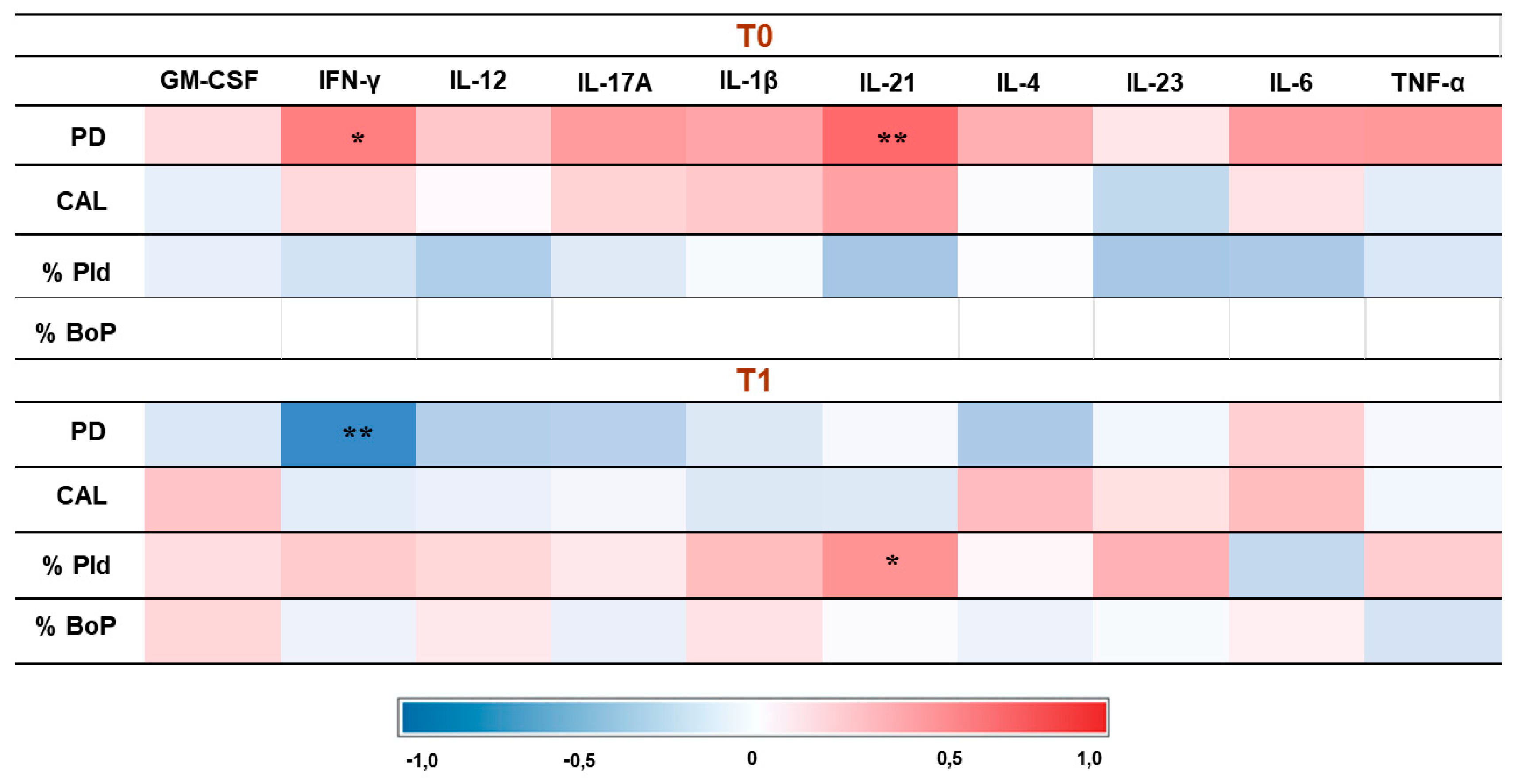
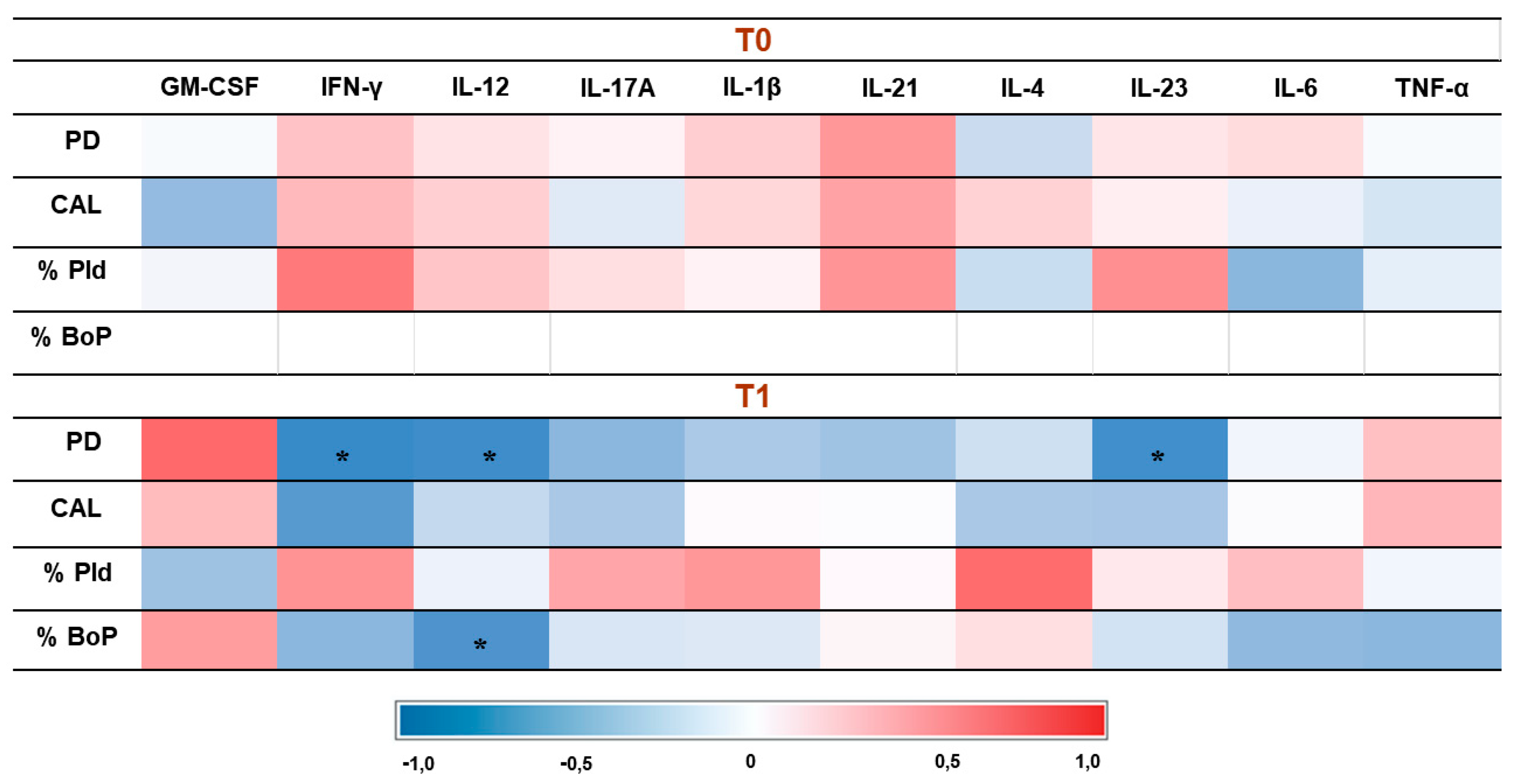
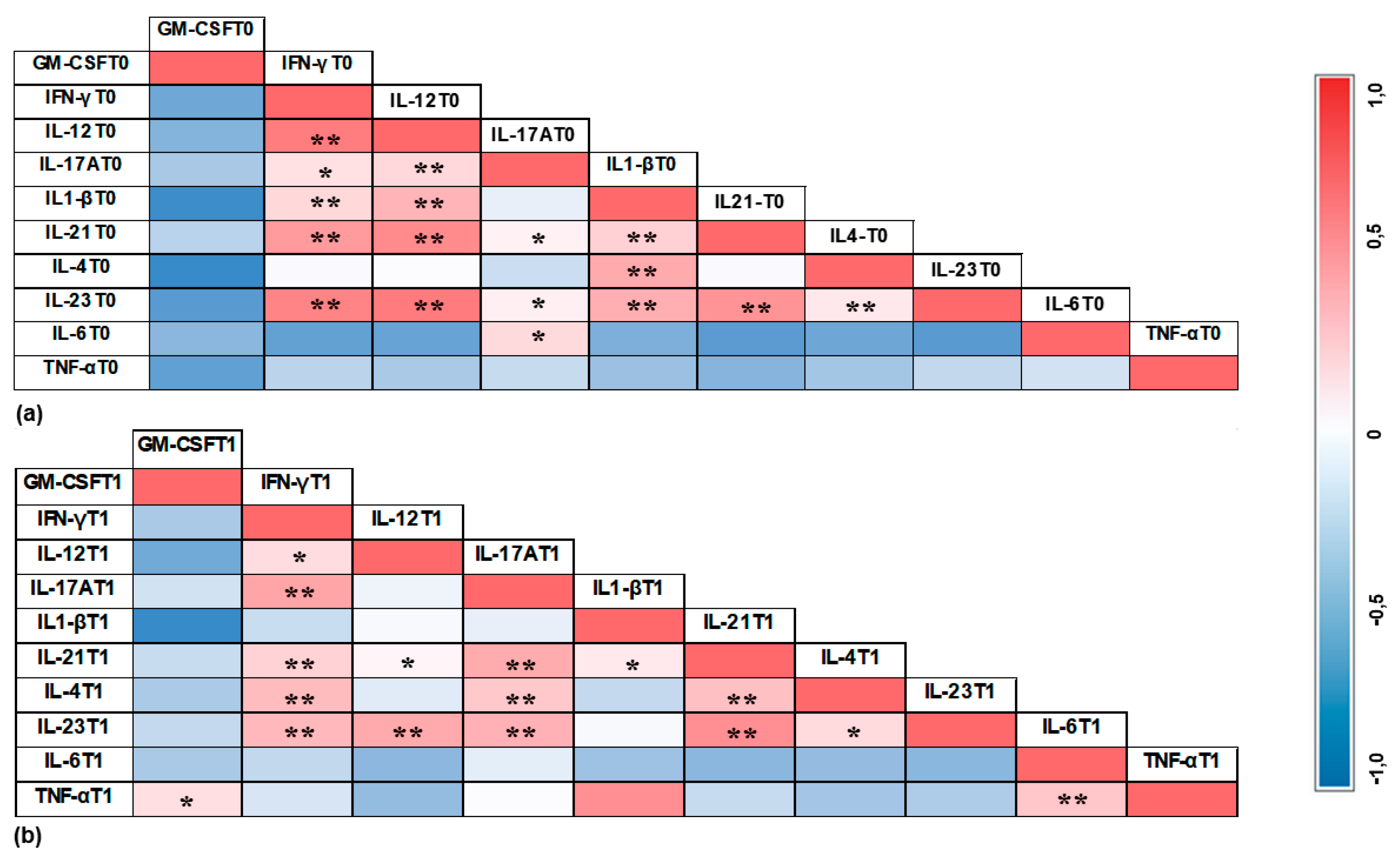
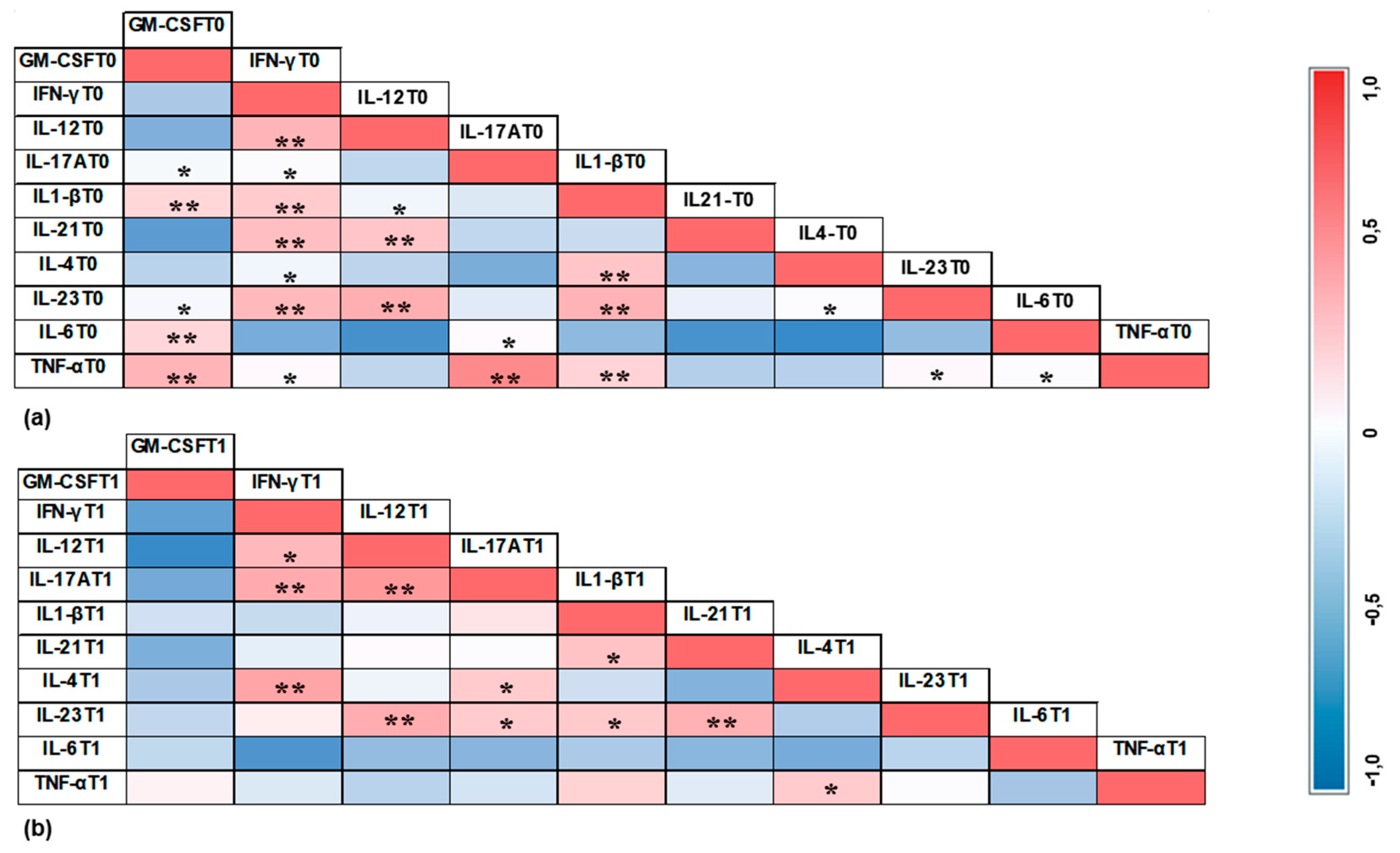
3.4. Correlation Results

In the PIM group, after therapy, a significant negative correlation was observed between IFN-γ, IL-12, and IL-23 with PD. Additionally, IL-12 showed a significant negative correlation with the percentage of bleeding.
In the PI group, before therapy, there was a significant positive correlation between the levels of IFN-γ and IL-21 with PD. After therapy, IFN-γ showed a significant negative correlation with PD, while IL-21 showed a significant positive correlation with the percentage of plaque.
The correlations between cytokines and clinical data at both times (baseline and T1) in PIM group are represented in Figure 2, whereas the correlations between cytokines and clinical data in PI group are represented in Figure 3.
The correlations between cytokines in PIM group in T0 and T1 are illustrated in Figure 4, while the correlations between cytokines in PI group in T0 and T1 are demonstrated in Figure 5.

4. Discussion

In the present study, peri-implant therapy resulted in a reduction of pro-inflammatory biomarker expression in PICF from both the PIM and PI groups. Specifically, the PIM group showed a significant decrease in IL17-A and TNF-α while the PI group exhibited a significant reduction in IL1-β, IL-6, and TNF-α. Additionally, there was a decreased tendency towards IFN-γ levels in the PI group, suggesting that the treatment may modulate this cytokine expression. Health implants were not included in the analysis, as the volume of PICF depends on the level of inflammation and PD [32] which probably would present a lower quantity in non-inflamed sites.
The observed significant reduction in IL-17A levels in patients with PIM after therapy suggests a positive modulation of the Th17 response, as this cytokine is involved in the differentiation of Th17 cells, osteoclast activation, and the recruitment of defense cells and pro-inflammatory cytokines such as IL1-β, IL-6, IL-8, G-CSF, GM-CSF, and TNF-α [33,34,35]. To the best of our knowledge, only one longitudinal study has assessed this cytokine in patients with PIM after therapy [36], also finding a significant reduction level. No prior studies have evaluated the effect of treatment on the modulation of IL-17A in PI. However, in our study, the reduction of IL-17A in the PI group did not reach statistical significance.
This study demonstrated a significant reduction in TNF-α levels in both the PIM and PI groups, and a decrease in IL-1β levels specifically in the PI group after three months of therapy. These findings align with previous research, which also reported a significant reduction in TNF-α levels in the PICF of PIM patients following therapy [37]. Similarly, significant reductions in both TNF-α and IL-1β levels in the PICF of PI patients have been observed [38,39,40,41]. High levels of these biomarkers are typically found in the PICF of patients with PID [13,42,43,44,45,46], and they are considered predictive of disease progression [7]. Furthermore, our study revealed a significant positive correlation between TNF-α and IL-1β levels at baseline in the PI group, suggesting their coordinated activity in more inflamed sites. This positive correlation was not observed after therapy. The observed reduction in these biomarkers indicates a potential decrease in osteoclastic activity and inflammatory response [38,47].
The significant reduction in IL-6 levels in PI patients after therapy highlights the therapy's positive impact on cytokine modulation. IL-6 is crucial in recruiting leukocytes, activating osteoclasts, and producing acute-phase proteins [48]. Consistent with our findings, previous studies have also reported decreased IL-6 levels in the PICF of PIM patients [37] and PI patients after therapy [41]. However, another study did not observe a statistically significant reduction in IL-6 levels six months after surgical therapy of PI, likely due to the initially low baseline levels of cytokine detection [49].
The tendency towards a significant reduction in IFN-γ levels in the PI group after therapy may suggest a potential association between IFN-γ and increased inflammation in PI. Our study also found a significant positive correlation between PD and IFN-γ at baseline in the PI group, with a negative correlation observed after therapy. This may indicate its role in increasing PD. This cytokine is known to promptly recruit macrophages, intensifying the inflammatory response [50] and accelerating periodontal disease progression [51]. However, it is important to note that the role of IFN-γ in the context of PID has been scarcely studied.
When comparing the PIM and PI groups at baseline, no statistically significant differences were found in the expression of any cytokine. This result suggests that once the inflammatory process begins, the expression of biomarkers remains similar regardless the presence of bone loss. This finding is consistent with previous cross-sectional studies on Th17 response biomarker expression in the PICF of partially edentulous patients with PIM and PI [11,19].
IL-10 is a non-inflammatory cytokine that helps to suppress the inflammatory response and to protect the host [52,53]. In PIM and PI groups, IL-10 expression levels were below the detection limit, which can be attributed to the high pro-inflammatory activity present in both groups. The literature presents varied results: higher IL-10 expression in healthy patients compared to those with PI [38], higher IL-10 expression in the PI group compared to healthy controls [54], and no differences between groups [33,55].
The heat maps presented in Figure 4 and Figure 5 illustrate the correlations between cytokines in the two groups at different time points. Interestingly, in the PIM group, IL-17A showed a strong correlation with IL-6 before treatment, but this pattern was not maintained after therapy. Additionally, TNF-α in the PIM group did not show a significant positive correlation with any other cytokine. In contrast, in the PI group, TNF-α exhibited a significant positive correlation with GM-CSF, IL-17A, and IL-1β, which was no longer observed after treatment, suggesting a coordinated action of these cytokines during bone loss. Moreover, the correlation between GM-CSF and other cytokines was not observed in the PIM group but was identified in the PI group, with IL-17A, IL-1β, IL-6, and TNF-α. GM-CSF has the function of recruiting monocytes and dendritic cells in addition to promoting the increase of IL-6 and IL-23, which are involved in the differentiation of Th17 cells. The production of IL-23 enhances GM-CSF secretion, generating a positive feedback loop [56,57]. After treatment, these correlations were not observed, and the heat map reveals a negative tendency in the results. This finding may suggest the relevance of GM-CSF in the bone loss activity in peri-implant disease.
Our study found a higher prevalence of patients with PI when periodontal and peri-implant maintenance therapy (PIMT) was performed for more than one year. This result is consistent with findings from other studies [58,59,60,61,62] and highlights the importance of PIMT in preventing PI. The frequency of maintenance appointments must be established individually for each patient, based on their risk factors. However, a frequency of at least twice a year seems to be ideal for preventing PI [63,64]. Additionally, there was a notable trend of increased PI prevalence among patients with concomitant active periodontitis. Although this outcome was not statistically significant, it is believed that this result is related to the limited number of patients with periodontitis in the study, as this condition is one of the factors most associated with an increased risk of developing PID [4,59,65].
This study observed a higher prevalence of PID in implants located in the posterior regions of the dental arches. This may be attributed to the greater difficulty in patients' biofilm control of these areas, consistent with findings from previous studies [66,67,68,69,70]. However, there is no consensus on this issue, as some studies observed a higher prevalence of PI in implants located in the anterior region [69,70]. Additionally, there was a higher prevalence of PI in splinted prostheses, which can be explained by the increased complexity of biofilm removal in this type of prosthesis, making it more challenging for patients.
HE platform implants showed a higher prevalence of PI compared to CM platform implants. This difference can be attributed to the several advantages of CM over HE platforms, such as a reduced micro gap between the abutment and implant, minimized micro-movements in occlusal forces distribution, lower initial bone loss, improved bacterial sealing, and greater resistance to torque loss [71,72,73,74,75].
After therapy, there was a significant reduction in PD and the percentage of bleeding in the treated implants of PIM and PI groups. However, most of the sites still exhibited bleeding, which may be related to the high plaque index observed. In the PI group, the plaque index was not significantly reduced and, in the PIM group, although it showed a significant reduction, it remained present in almost half of the evaluated implants. The persistence of bleeding after treatment has also been reported in previous studies [49,65,76,77,78]. This finding underscores the patient's difficulty in effectively controlling biofilm on implants and their structures, and the challenge in achieving the elimination of bleeding in all sites in the treatment of PID.
This study presents certain limitations, such as the relatively small sample size and short follow-up period, which may restrict the generalizability of the findings. Future research with more extensive sampling and longer follow-up periods would provide more substantial evidence on the modulation of Th17-related cytokine expression and the clinical outcomes of peri-implant disease treatments. Therefore, the results should be interpreted with caution.

5. Conclusions

In conclusion, our study demonstrates that after three months of therapy, there was a significant reduction in the expression of cytokines in the peri-implant crevicular fluid of patients with peri-implant mucositis (IL-17A and TNF-α) and peri-implantitis (IL -1β, IL-6 and TNF-α). This finding indicates that peri-implant therapy can modulate important cytokines in exacerbating the inflammatory response and osteoclastogenesis.

Author Contributions

Conceptualization, C.M.S.F., M.KS.T. and E.J.V.L.; methodology, L.T.C.G., C.M.S.F., M.K.S.T., and E.J.V.L.; validation, M.K.S.T., D.M.T., and E.J.V.L.; formal analysis, L.T.C.G., M.K.S.T., A.M.P.S., and E.J.V.L.; investigation, L.T.C.G., and G.S.T.N.; writing—original draft preparation, L.T.C.G.; writing—review and editing, L.T.C.G., A.M.P.S., G.S.T.N. and M.K.S.T.; visualization, L.T.C.G., and E.J.V.L.; supervision, M.K.S.T.; project administration, L.T.C.G., D.M.T., and M.K.S.T.; funding acquisition, M.K.S.T. All authors have read and agreed to the published version of the manuscript.

Funding

This research was funded by CARLOS CHAGAS FILHO DE AMPARO FOUNDATION (FAPERJ), grant number 210.072/2018.

Institutional Review Board Statement

The study was conducted in accordance with the Declaration of Helsinki, and approved by the Ethics Committee of PEDRO ERNESTO UNIVERSITY HOSPITAL (HUPE/UERJ) (protocol code, approved on 06/25/2023).

Informed Consent Statement

Informed consent was obtained from all subjects involved in the study.

Acknowledgments

This study was carried out at the School of Dentistry of Rio de Janeiro State University. The authors thank all collaborators of this study.

Conflicts of Interest

The authors declare no conflicts of interest.

References

  1. Berglundh T, Armitage G, Araujo MG et al. Peri-implant diseases and conditions: consensus report of workgroup 4 of the 2017 world workshop on the classification of periodontal and peri-implant diseases and conditions. J Clin Periodontol 2018, 45, 286–291. [Google Scholar] [CrossRef]
  2. Gargallo-Albiol J, Tavelli L, Barootchi S, Monje A, Wang HL. Clinical sequelae and patients' perception of dental implant removal: A cross-sectional study. J Periodontol 2021, 92, 823–832. [Google Scholar] [CrossRef] [PubMed]
  3. Lee CT, Huang YW, Zhu L, Weltman R. Prevalences of peri-implantitis and peri-implant mucositis: systematic review and meta-analysis. J Dent 2017, 62, 1–12. [Google Scholar] [CrossRef]
  4. Derks J, Schaller D, Håkansson J, Wennström JL, Tomasi C, Berglundh T. Peri-implantitis - onset and pattern of progression. J Clin Periodontol 2016, 43, 383–388. [Google Scholar] [CrossRef]
  5. Darby, I. Risk factors for periodontitis & peri-implantitis. Periodontol 2000 2022, 90, 9–12. [Google Scholar] [CrossRef] [PubMed]
  6. Diaz P, Gonzalo E, Villagra LJG, Miegimolle B, Suarez MJ. What is the prevalence of peri-implantitis? A systematic review and meta-analysis. BMC Oral Health 2022. [Google Scholar] [CrossRef]
  7. Chmielewski M, Pilloni A. Current Molecular, Cellular and Genetic Aspects of Peri-Implantitis Disease: A Narrative Review. Dent J (Basel) 2023. [Google Scholar] [CrossRef]
  8. Corrêa MG, Pimentel SP, Ribeiro FV, Cirano FR, Casati MZ. Host response and peri-implantitis. Braz Oral Res 2019. [Google Scholar] [CrossRef]
  9. Troen, BR. Molecular mechanisms underlying osteoclast formation and activation. Exp Gerontol 2003, 38, 605–614. [Google Scholar] [CrossRef]
  10. Markou E, Eleana B, Lazaros T, Antonios K. The influence of sex steroid hormones on gingiva of women. Open Dent J 2009, 5, 114–119. [Google Scholar] [CrossRef]
  11. Severino VO, Beghini M, de Araújo MF et al. Expression of IL-6, IL-10, IL-17 and IL-33 in the peri-implant crevicular fluid of patients with peri-implant mucositis and peri-implantitis. Arch Oral Biol 2016, 72, 194–199. [Google Scholar] [CrossRef] [PubMed]
  12. Petković AB, Matić SM, Stamatović NV, Vojvodić DV, Todorović TM, Lazić ZR, Kozomara RJ. Proinflammatory cytokines (IL-1beta and TNF-alpha) and chemokines (IL-8 and MIP-1alpha) as markers of peri-implant tissue condition. Int J Oral Maxillofac Surg 2010, 39, 478–485. [Google Scholar] [CrossRef] [PubMed]
  13. Faot F, Nascimento GG, Bielemann AM, Campão TD, Leite FR, Quirynen M. Can peri-implant crevicular fluid assist in the diagnosis of peri-implantitis? A systematic review and meta-analysis. J Periodontol 2015, 865, 631–645. [Google Scholar] [CrossRef]
  14. Delucchi F, Canepa C, Canullo L, Pesce P, Isola G, Menini M. Biomarkers from Peri-Implant Crevicular Fluid (PICF) as Predictors of Peri-Implant Bone Loss: A Systematic Review. Int J Mol Sci 2023. [Google Scholar] [CrossRef]
  15. Askar, M. T helper subsets & regulatory T cells: rethinking the paradigm in the clinical context of solid organ transplantation. Int J Immunogenet 2014, 41, 185–194. [Google Scholar] [CrossRef] [PubMed]
  16. Iwakura Y, Nakae S, Saijo S, Ishigame H. The roles of IL-17A in inflammatory immune responses and host defense against pathogens. Immunol Rev 2008, 226, 57–79. [Google Scholar] [CrossRef]
  17. Korn T, Bettelli E, Oukka M, Kuchroo VK. IL-17 and Th17 Cells. Annu Rev Immunol 2009, 27, 485–517. [Google Scholar] [CrossRef] [PubMed]
  18. Giro G, Tebar A, Franco L, Racy D, Bastos MF, Shibli JA. Treg and TH17 link to immune response in individuals with peri-implantitis: a preliminary report. Clin Oral Investig 2021, 25, 1291–1297. [Google Scholar] [CrossRef] [PubMed]
  19. Teixeira MKS, Lira-Junior R, Telles DM, Lourenço EJV, Figueredo CM. Th17-related cytokines in mucositis: is there any difference between peri-implantitis and periodontitis patients? Clin Oral Implants Res 2017, 28, 816–822. [Google Scholar] [CrossRef]
  20. Heitz-Mayfield LJA, Salvi GE, Mombelli A, Loup PJ, Heitz F, Kruger E, Lang NP. Supportive peri-implant therapy following anti-infective surgical peri-implantitis treatment: 5-year survival and success. Clin Oral Implants Res 2018, 29, 1–6. [Google Scholar] [CrossRef] [PubMed]
  21. Schwarz F, Jepsen S, Obreja K, Galarraga-Vinueza ME, Ramanauskaite A. Surgical therapy of peri-implantitis. Periodontol 2000 2022, 88, 145–181. [Google Scholar] [CrossRef] [PubMed]
  22. Verket A, Koldsland OC, Bunaes D, Lie SA, Romandini M. Non-surgical therapy of peri-implant mucositis-Mechanical/physical approaches: A systematic review. J Clin Periodontol 2023, 26, 135–145. [Google Scholar] [CrossRef]
  23. Ramanauskaite A, Daugela P, Juodzbalys G. Treatment of peri-implantitis: Meta-analysis of findings in a systematic literature review and novel protocol proposal. Quintessence Int 2016, 47, 379–393. [Google Scholar] [CrossRef]
  24. Dos Santos Martins BG, Fernandes JCH, Martins AG, de Moraes Castilho R, de Oliveira Fernandes GV. Surgical and Nonsurgical Treatment Protocols for Peri-implantitis: An Overview of Systematic Reviews. Int J Oral Maxillofac Implants 2022, 37, 660–676. [Google Scholar] [CrossRef]
  25. Ichioka Y, Virto L, Nuevo P, Gamonal JD, Derks J, Larsson L, Sanz M, Berglundh T. Decontamination of biofilm-contaminated implant surfaces: An in vitro evaluation. Clin Oral Implants Res 2023, 34, 1058–1072. [Google Scholar] [CrossRef] [PubMed]
  26. Teixeira Neves GS, Elangovan G, Teixeira MKS, Mello-Neto JM, Tadakamadla SK, Lourenço EJV, Telles DM, Figueredo CM. Peri-Implant Surgical Treatment Downregulates the Expression of sTREM-1 and MMP-8 in Patients with Peri-Implantitis: A Prospective Study. Int J Environ Res Public Health 2022. [Google Scholar] [CrossRef]
  27. Renvert S, Persson GR, Pirih FQ, Camargo PM. Peri-implant health, peri-implant mucositis, and peri-implantitis: Case definitions and diagnostic considerations. J Periodontol 2018, 89, 304–312. [Google Scholar] [CrossRef]
  28. Ainamo J, Bay I. Problems and proposals for recording gingivitis and plaque. Int Dent J 1975, 25, 229–235. [Google Scholar]
  29. Wassall RR, Preshaw PM. Clinical and technical considerations in the analysis of gingival crevicular fluid. Periodontol 2000 2016, 70, 65–79. [Google Scholar] [CrossRef] [PubMed]
  30. Lang NP, Berglundh T, Heitz-Mayfield LJ, Pjetursson BE, Salvi GE, Sanz M. Consensus statements and recommended clinical procedures regarding implant survival and complications. Int J Oral Maxillofac Implants 2004, 19, 150–154. [Google Scholar]
  31. Khoury F, Keeve PL, Ramanauskaite A, Schwarz F, Koo KT, Sculean A, Romanos G. Surgical treatment of peri-implantitis - Consensus report of working group 4. Int Dent J 2019, 69, 18–22. [Google Scholar] [CrossRef] [PubMed]
  32. Alassy H, Parachuru P, Wolff L. Peri-Implantitis Diagnosis and Prognosis Using Biomarkers in Peri-Implant Crevicular Fluid: A Narrative Review. Diagnostics (Basel) 2019. [Google Scholar]
  33. Severino VO, Napimoga MH, de Lima Pereira AS. Expression of IL-6, IL-10, IL-17 and IL-8 in the peri-implant crevicular fluid of patients with peri-implantitis. Arch Oral Biol 2011, 56, 823–828. [Google Scholar] [CrossRef]
  34. de Araújo MF, Filho AF, da Silva GP et al. Evaluation of peri-implant mucosa: clinical, histopathological and immunological aspects. Arch Oral Biol 2014, 59, 470–478. [Google Scholar] [CrossRef] [PubMed]
  35. Irie K, Azuma T, Tomofuji T, Yamamoto T. Exploring the Role of IL-17A in Oral Dysbiosis-Associated Periodontitis and Its Correlation with Systemic Inflammatory Disease. Dent J (Basel) 2023. [Google Scholar] [CrossRef]
  36. Kashefimehr A, Pourabbas R, Faramarzi M et al. Effects of enamel matrix derivative on non-surgical management of peri-implant mucositis: a double-blind randomized clinical trial. Clin Oral Investig 2017, 21, 2379–2388. [Google Scholar] [CrossRef] [PubMed]
  37. Pourabbas R, Khorramdel A, Sadighi M, Kashefimehr A, Mousavi SA. Effect of photodynamic therapy as an adjunctive to mechanical debridement on the nonsurgical treatment of peri-implant mucositis: A randomized controlled clinical trial. Dent Res J (Isfahan) 2023. [Google Scholar]
  38. Duarte PM, de Mendonça AC, Máximo MB, Santos VR, Bastos MF, Nociti FH. Effect of anti-infective mechanical therapy on clinical parameters and cytokine levels in human peri-implant diseases. J Periodontol 2009, 80, 234–243. [Google Scholar] [CrossRef] [PubMed]
  39. de Mendonça AC, Santos VR, César-Neto JB, Duarte PM. Tumor necrosis factor-alpha levels after surgical anti-infective mechanical therapy for peri-implantitis: a 12-month follow-up. J Periodontol 2009, 80, 693–699. [Google Scholar] [CrossRef]
  40. Bassetti M, Schär D, Wicki B, Eick S, Ramseier CA, Arweiler NB, Sculean A, Salvi GE. Anti-infective therapy of peri-implantitis with adjunctive local drug delivery or photodynamic therapy: 12-month outcomes of a randomized controlled clinical trial. Clin Oral Implants Res 2014, 25, 279–287. [Google Scholar] [CrossRef]
  41. Elsadek, M.F. Effectiveness of two photosensitizer-mediated photodynamic therapy for treating moderate peri-implant infections in type-II diabetes mellitus patients: A randomized clinical trial. Photodiagnosis Photodyn Ther 2023, 43, 103643. [Google Scholar] [CrossRef] [PubMed]
  42. Ata-Ali J, Flichy-Fernández AJ, Alegre-Domingo T, Ata-Ali F, Palacio J, Peñarrocha-Diago M. Clinical, microbiological, and immunological aspects of healthy versus peri-implantitis tissue in full arch reconstruction patients: a prospective cross-sectional study. BMC Oral Health 2015, 15, 43. [Google Scholar] [CrossRef]
  43. Zani SR, Moss K, Shibli JA, Teixeira ER, de Oliveira Mairink R, Onuma T, Feres M, Teles RP. Peri-implant crevicular fluid biomarkers as discriminants of peri-implant health and disease. J Clin Periodontol 2016, 43, 825–832. [Google Scholar] [CrossRef]
  44. Bhavsar I, Miller CS, Ebersole JL, Dawson DR 3rd, Thompson KL, Al-Sabbagh M. Biological response to peri-implantitis treatment. J Periodontal Res 2019, 54, 720–728. [Google Scholar] [CrossRef]
  45. Hentenaar DFM, De Waal YCM, Vissink A et al. Biomarker levels in peri-implant crevicular fluid of healthy implants, untreated and non-surgically treated implants with peri-implantitis. J Clin Periodontol 2021, 48, 590–601. [Google Scholar] [CrossRef]
  46. Song L, Jiang J, Li J, Zhou C, Chen Y, Lu H, He F. The Characteristics of Microbiome and Cytokines in Healthy Implants and Peri-Implantitis of the Same Individuals. J Clin Med 2022, 11, 5817. [Google Scholar] [CrossRef]
  47. Lumbikananda S, Srithanyarat SS, Mattheos N, Osathanon T. Oral Fluid Biomarkers for Peri-Implantitis: A Scoping Review. Int Dent J 2024, 74, 387–402. [Google Scholar] [CrossRef] [PubMed]
  48. Tanaka T, Narazaki M, Kishimoto T. IL-6 in inflammation, immunity, and disease. Cold Spring Harb Perspect Biol 2014, 6, a016295. [Google Scholar] [CrossRef]
  49. Luengo F, Solonko M, Sanz-Esporrín J, Sanz-Sánchez I, Herrera D, Sanz M. Clinical, Microbiological, and Biochemical Impact of the Surgical Treatment of Peri-Implantitis-A Prospective Case Series. J Clin Med 2022, 11, 4699. [Google Scholar] [CrossRef]
  50. Nagy E, Lei Y, Martínez-Martínez E, Body SC, Schlotter F, Creager M, Assmann A, Khabbaz K, Libby P, Hansson GK, Aikawa E. Interferon-γ Released by Activated CD8+ T Lymphocytes Impairs the Calcium Resorption Potential of Osteoclasts in Calcified Human Aortic Valves. Am J Pathol 2017, 187, 1413–1425. [Google Scholar] [CrossRef] [PubMed]
  51. Danielsen AK, Damgaard C, Massarenti L, Østrup P, Riis Hansen P, Holmstrup P, Nielsen CH. B-cell cytokine responses to Porphyromonas gingivalis in patients with periodontitis and healthy controls. J Periodontol 2023, 94, 997–1007. [Google Scholar] [CrossRef] [PubMed]
  52. Iyer SS, Cheng G. Role of interleukin 10 transcriptional regulation in inflammation and autoimmune disease. Crit Rev Immunol 2012, 32, 23–63. [Google Scholar] [CrossRef] [PubMed]
  53. Abbas AK, Pillai S, Lichtman AH. Imunologia celular e molecular, 9th ed.; Elsevier: Rio de Janeiro, Brazil, 2019; pp. 856–857. [Google Scholar]
  54. Güncü GN, Akman AC, Günday S, Yamalık N, Berker E. Effect of inflammation on cytokine levels and bone remodelling markers in peri-implant sulcus fluid: a preliminary report. Cytokine 2012, 59, 313–316. [Google Scholar] [CrossRef] [PubMed]
  55. Fonseca FJ, Moraes Junior M, Lourenço EJ, Teles DM, Figueredo CM. Cytokines expression in saliva and peri-implant crevicular fluid of patients with peri-implant disease. Clin Oral Implants Res 2014, 25, 68–72. [Google Scholar] [CrossRef]
  56. Huang N, Dong H, Luo Y, Shao B. Th17 Cells in Periodontitis and Its Regulation by A20. Front Immunol 2021, 7, 23–742925. [Google Scholar] [CrossRef]
  57. El-Behi M, Ciric B, Dai H, Yan Y, Cullimore M, Safavi F, Zhang GX, Dittel BN, Rostami A. The encephalitogenicity of T(H)17 cells is dependent on IL-1- and IL-23-induced production of the cytokine GM-CSF. Nat Immunol 2011, 12, 568–575. [Google Scholar] [CrossRef]
  58. Gay IC, Tran DT, Weltman R, Parthasarathy K, Diaz-Rodriguez J, Walji M, Fu Y, Friedman L. Role of supportive maintenance therapy on implant survival: a university-based 17 years retrospective analysis. Int J Dent Hyg 2016, 14, 267–271. [Google Scholar] [CrossRef]
  59. Monje A, Wang HL, Nart J. Association of Preventive Maintenance Therapy Compliance and Peri-Implant Diseases: A Cross-Sectional Study. J Periodontol 2017, 88, 1030–1041. [Google Scholar] [CrossRef] [PubMed]
  60. Frisch E, Vach K, Ratka-Krueger P. Impact of supportive implant therapy on peri-implant diseases: A retrospective 7-year study. J Clin Periodontol 2020, 47, 101–109. [Google Scholar] [CrossRef] [PubMed]
  61. Astolfi V, Ríos-Carrasco B, Gil-Mur FJ, Ríos-Santos JV, Bullón B, Herrero-Climent M, Bullón P. Incidence of Peri-Implantitis and Relationship with Different Conditions: A Retrospective Study. Int J Environ Res Public Health 2022, 19, 4147. [Google Scholar] [CrossRef]
  62. Costa FO, Costa AM, Ferreira SD et al. Long-term impact of patients' compliance to peri-implant maintenance therapy on the incidence of peri-implant diseases: An 11-year prospective follow-up clinical study. Clin Implant Dent Relat Res 2023, 25, 303–312. [Google Scholar] [CrossRef] [PubMed]
  63. Rösing CK, Fiorini T, Haas AN, Muniz FWMG, Oppermann RV, Susin C. The impact of maintenance on peri-implant health. Braz Oral Res 2019. [Google Scholar] [CrossRef]
  64. Leone FD, Blasi G, Amerio E, Valles C, Nart J, Monje A. Influence of the level of compliance with preventive maintenance therapy upon the prevalence of peri-implant diseases. J Periodontol 2024, 95, 40–49. [Google Scholar] [CrossRef]
  65. Schwarz F, Derks J, Monje A, Wang HL. Peri-implantitis. J Periodontol 2018, 89, 267–290. [Google Scholar] [CrossRef]
  66. Jia P, Tang Y, Niu L, Qiu L. Clinical and radiographic outcomes of a combined surgery approach to treat peri-implantitis. Int J Oral Maxillofac Surg 2024, 53, 333–342. [Google Scholar] [CrossRef]
  67. Derks J, Ortiz-Vigón A, Guerrero A et al. Reconstructive surgical therapy of peri-implantitis: A multicenter randomized controlled clinical trial. Clin Oral Implants Res 2022, 33, 921–944. [Google Scholar] [CrossRef] [PubMed]
  68. Romandini M, Laforí A, Pedrinaci I et al. Effect of sub-marginal instrumentation before surgical treatment of peri-implantitis: A multi-centre randomized clinical trial. J Clin Periodontol 2022, 49, 1334–1345. [Google Scholar] [CrossRef] [PubMed]
  69. Moraschini V, Kischinhevsky ICC, Sartoretto SC et al. Does implant location influence the risk of peri-implantitis? Periodontol 2000 2022, 90, 224–235. [Google Scholar] [CrossRef]
  70. Sun JS, Liu KC, Hung MC, Lin HY, Chuang SL, Lin PJ, Chang JZ. A cross-sectional study for prevalence and risk factors of peri-implant marginal bone loss. J Prosthet Dent 0022, S0022-3913(23)00722-9. [Google Scholar] [CrossRef]
  71. Koo KT, Lee EJ, Kim JY, Seol YJ, Han JS, Kim TI, Lee YM, Ku Y, Wikesjö UM, Rhyu IC. The effect of internal versus external abutment connection modes on crestal bone changes around dental implants: a radiographic analysis. J Periodontol 2012, 83, 1104–1109. [Google Scholar] [CrossRef] [PubMed]
  72. Machado LS, Bonfante EA, Anchieta RB, Yamaguchi S, Coelho PG. Implant-abutment connection designs for anterior crowns: reliability and failure modes. Implant Dent 2013, 22, 540–545. [Google Scholar] [CrossRef]
  73. Schmitt CM, Nogueira-Filho G, Tenenbaum HC, Lai JY, Brito C, Döring H, Nonhoff J. Performance of conical abutment (Morse Taper) connection implants: a systematic review. J Biomed Mater Res A 2014, 102, 552–574. [Google Scholar] [CrossRef] [PubMed]
  74. Macedo JP, Pereira J, Vahey BR et al. Morse taper dental implants and platform switching: The new paradigm in oral implantology. Eur J Dent 2016, 10, 148–154. [Google Scholar] [CrossRef]
  75. Teixeira MKS, de Moraes Rego MR, da Silva MFT, Lourenço EJV, Figueredo CM, Telles DM. Bacterial Profile and Radiographic Analysis Around Osseointegrated Implants With Morse Taper and External Hexagon Connections: Split-Mouth Model. J Oral Implantol 2019, 45, 469–473. [Google Scholar] [CrossRef] [PubMed]
  76. Heitz-Mayfield LJA, Salvi GE, Mombelli A, Faddy M, Lang NP. Anti-infective surgical therapy of peri-implantitis. A 12-month prospective clinical study. Clin Oral Implants Res 2012, 23, 205–210. [Google Scholar] [CrossRef] [PubMed]
  77. Carcuac O, Derks J, Charalampakis G, Abrahamsson I, Wennström J, Berglundh T. Adjunctive Systemic and Local Antimicrobial Therapy in the Surgical Treatment of Peri-implantitis: A Randomized Controlled Clinical Trial. J Dent Res 2016, 95, 50–57. [Google Scholar] [CrossRef] [PubMed]
  78. Carcuac O, Derks J, Abrahamsson I, Wennström JL, Petzold M, Berglundh T. Surgical treatment of peri-implantitis: 3-year results from a randomized controlled clinical trial. J Clin Periodontol 2017, 44, 1294–1303. [Google Scholar] [CrossRef]
Figure 1. Representation of biomarker expression results before and after therapy. PIM - Peri-implant mucositis; PI – Peri-implantitis. Results presented in boxplot, with the left side referring to the PIM group (before and after therapy) and the right side referring to the PI group (before and after therapy). When the p-value was < 0.05, the values were included in the figure. The boxplot graphs were created with the SPSS program (version 24).
Preprints 142898 g001
Figure 2. Correlation between cytokines and clinical data in PIM group. PD: probing depth; CAL: clinical attachment level; % PID: plaque index; %BoP: bleeding on probing. Correlation coefficient obtained by Spearman's correlation test. *: p < 0.05 level.
Preprints 142898 g002
Figure 3. Correlation between cytokines and clinical data in PI group. PD: probing depth; CAL: clinical attachment level; % PID: plaque index; %BoP: bleeding on probing. Correlation coefficient obtained by Spearman's correlation test. *: p < 0.05 level.
Preprints 142898 g003
Figure 4. Correlation between cytokines in PIM group in T0 and T1. Correlation coefficient obtained by Pearson's correlation test. **: p <0.01;*: p < 0.05.
Preprints 142898 g004
Figure 5. Correlation between cytokines in PI group in T0 and T1. Correlation coefficient obtained by Pearson's correlation test. **: p <0.01;*: p < 0.05.
Preprints 142898 g005
Table 1. Clinical outcomes of PIM group in baseline and T1.
Baseline T1 P value
PD (mm) 3.90 (±1.34) 3.27 (±1.19) 0.001
CAL (mm) 2.41 (±1.37) 1.50 (±1.31) <0.001
% PId 71.43 (±45.58) 46.43 (±50.32) 0.003
% BoP 100 (±0.00) 51.79 (±50.42) <0.001
PD – Probing depth; CAL – Clinical attachment level; % PId – percentage of plaque index; % BoP – percentage of bleeding on probing. Outcomes were presented as mean and standard deviation (±). P value was evaluated using the Wilcoxon statistical test.
Table 2. Clinical outcomes of PI group in baseline and T1.
Baseline T1 P value
PD (mm) 5.29 (±1.74) 3.00 (±1.00) <0.001
CAL (mm) 4.32 (±1.78) 3.00 (±1.80) 0.004
% PId 65.63 (±48.25) 46.88 (±50.70) 0.083
% BoP 100 (±0.00) 56.25 (±50.40) <0.001
PD – Probing depth; CAL – Clinical attachment level; % PId – percentage of plaque index; % BoP – percentage of bleeding on probing. Outcomes were presented as mean and standard deviation (±). P value was evaluated using the Wilcoxon statistical test.
Table 3. Descriptive data of implants.
Total number of implants
(n = 88)
Implants of PIM group
(n = 56)
Implants of PI group
(n = 32)
P value
Arch, n (%) Upper 40 (45.45%) 30 (34.09%) 10 (11.36%) 0.002
Lower 48 (54.55%) 26 (29.55%) 22 (25%) 0.564
P value 0.394 0.593 0.34
Position, n (%) Anterior (canine-canine) 13 (14.77%) 8 (14.28%) 5 (15.62%) 0.405
Posterior 75 (85.23%) 48 (85.72%) 27 (84.38%) 0.015
P value <0.001 <0.001 <0.001
Type of prosthetic platform Morse Taper 40 (45.45%) 32 (57.14%) 8 (25%) <0.001
External Hexagon 48 (54.55%) 24 (42.86%) 24 (75%) 1.000
P value 0.394 0.285 0.005
Cemented or screwed Cemented 40 (45.45%) 27 (48.21%) 13 (40.62%) 0.027
Screwed 48 (54.55%) 29 (51.79%) 19 (59.38%) 0.149
P value 0.394 0.789 0.289
Splinted or non-splinted Splinted 32 (36.37%) 9 (16.08%) 23 (71.88%) 0.013
Non-splinted 56 (63.63%) 47 (83.92%) 9 (28.12%) <0.001
P value 0.011 <0.001 0.013
Data presented as number of implants and respective percentage. P value was evaluated using the Chi-square statistical test.
Disclaimer/Publisher’s Note: The statements, opinions and data contained in all publications are solely those of the individual author(s) and contributor(s) and not of MDPI and/or the editor(s). MDPI and/or the editor(s) disclaim responsibility for any injury to people or property resulting from any ideas, methods, instructions or products referred to in the content.
Copyright: This open access article is published under a Creative Commons CC BY 4.0 license, which permit the free download, distribution, and reuse, provided that the author and preprint are cited in any reuse.
Prerpints.org logo

Preprints.org is a free preprint server supported by MDPI in Basel, Switzerland.

Subscribe

Disclaimer

Terms of Use

Privacy Policy

Privacy Settings

© 2025 MDPI (Basel, Switzerland) unless otherwise stated