Preprint
Article

This version is not peer-reviewed.

Endothelial Dysfunction And Oxidative Stress In Patients With Severe Coronary Artery Disease: Does Diabetes Play A Contributing Role?

A peer-reviewed article of this preprint also exists.

Submitted:

07 December 2024

Posted:

09 December 2024

You are already at the latest version

Abstract

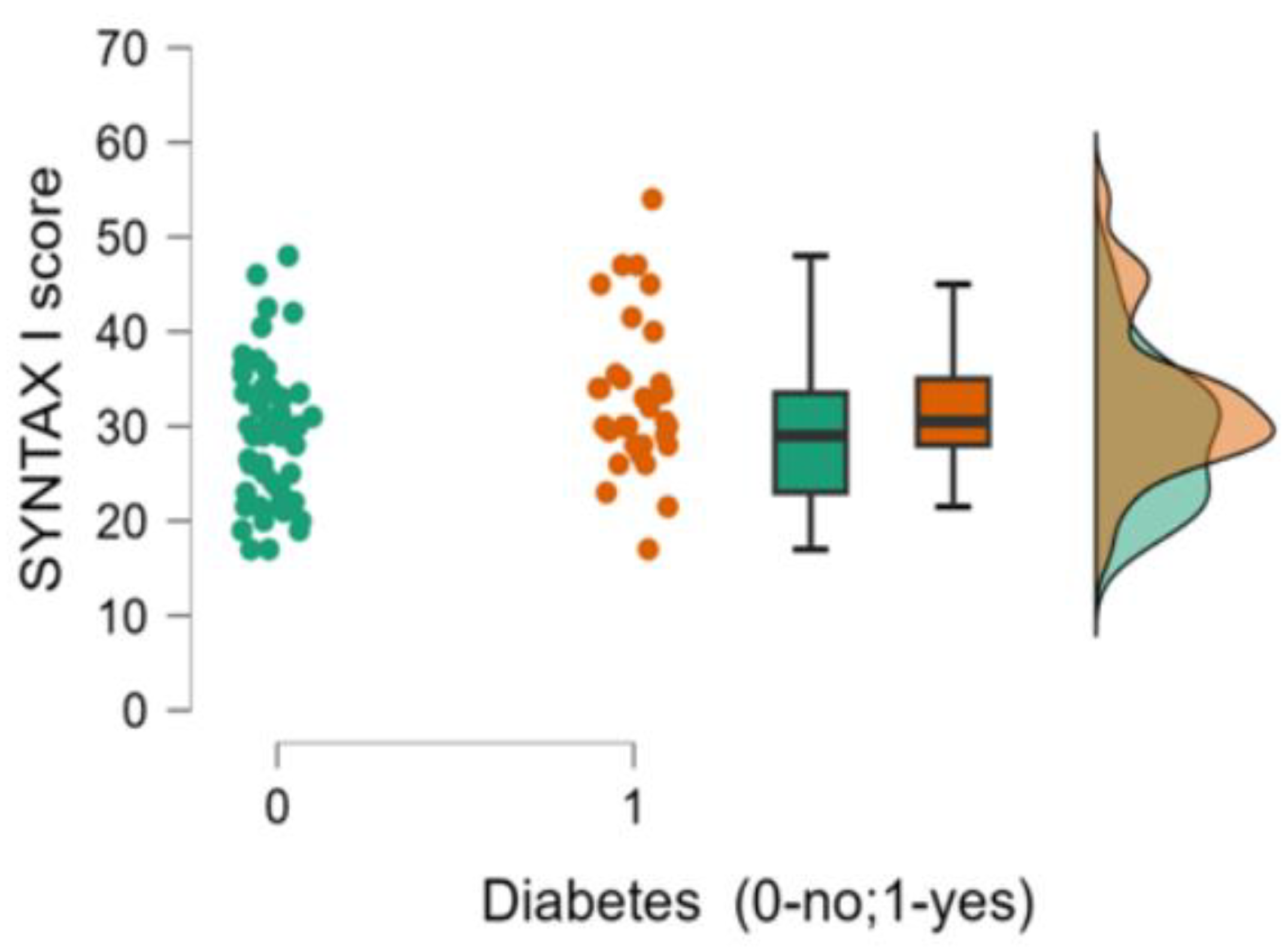
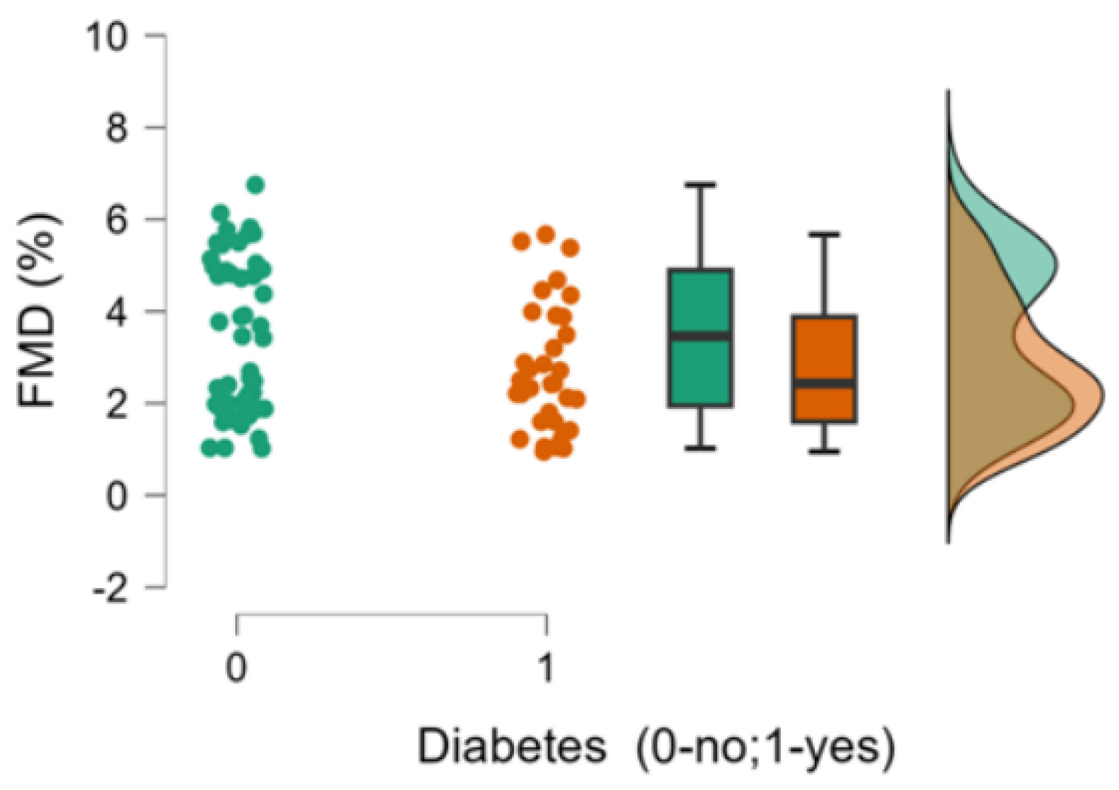
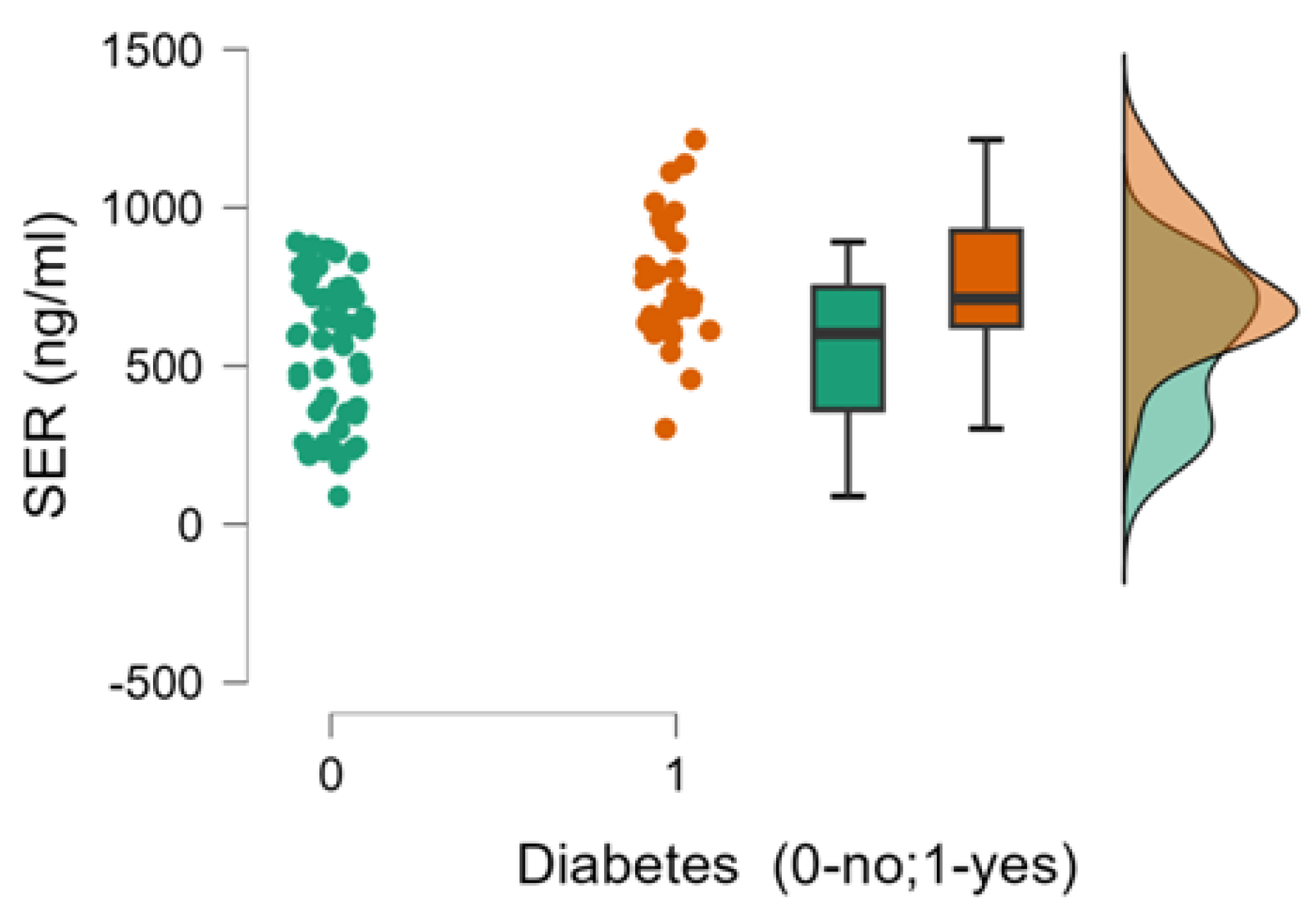
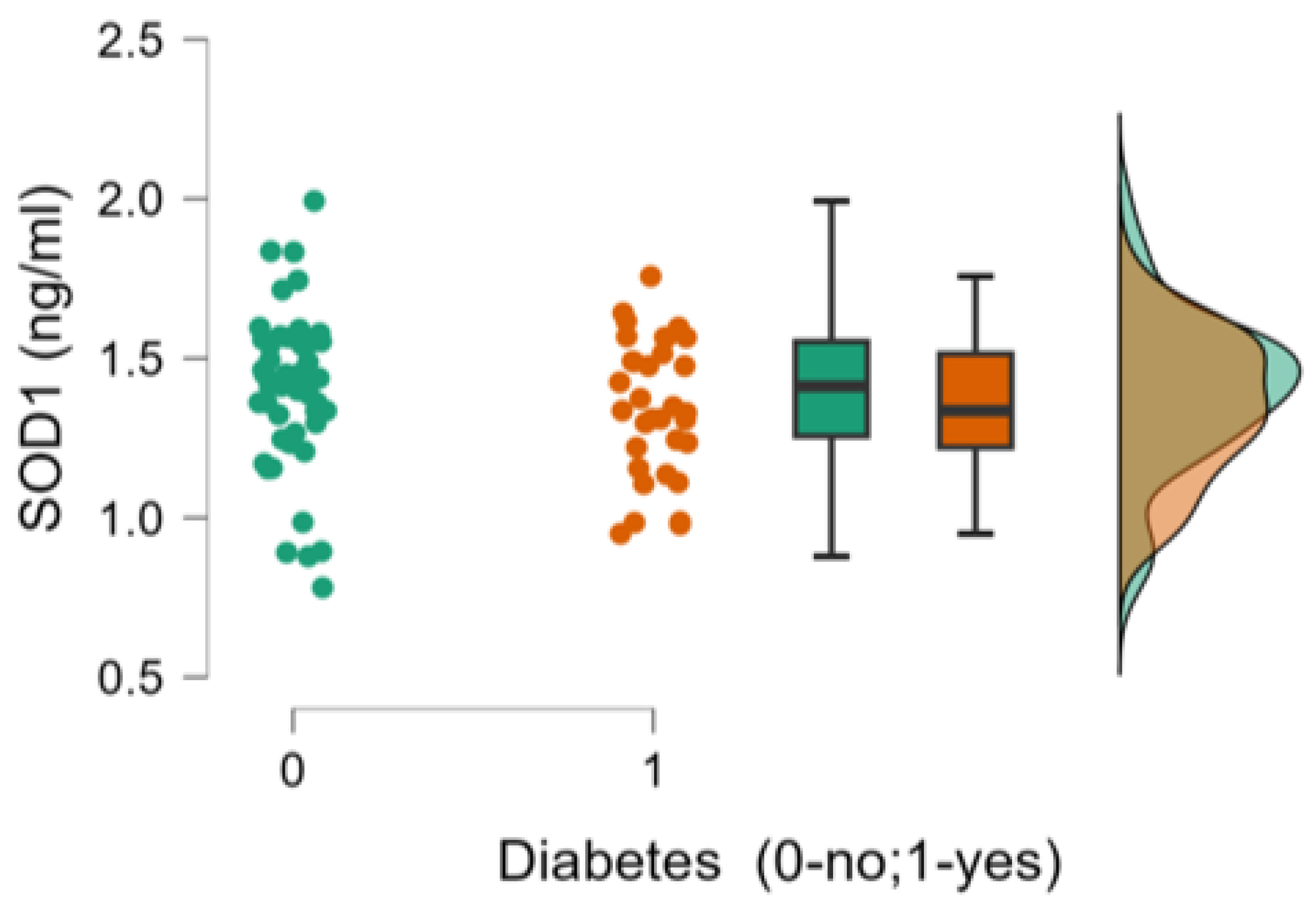
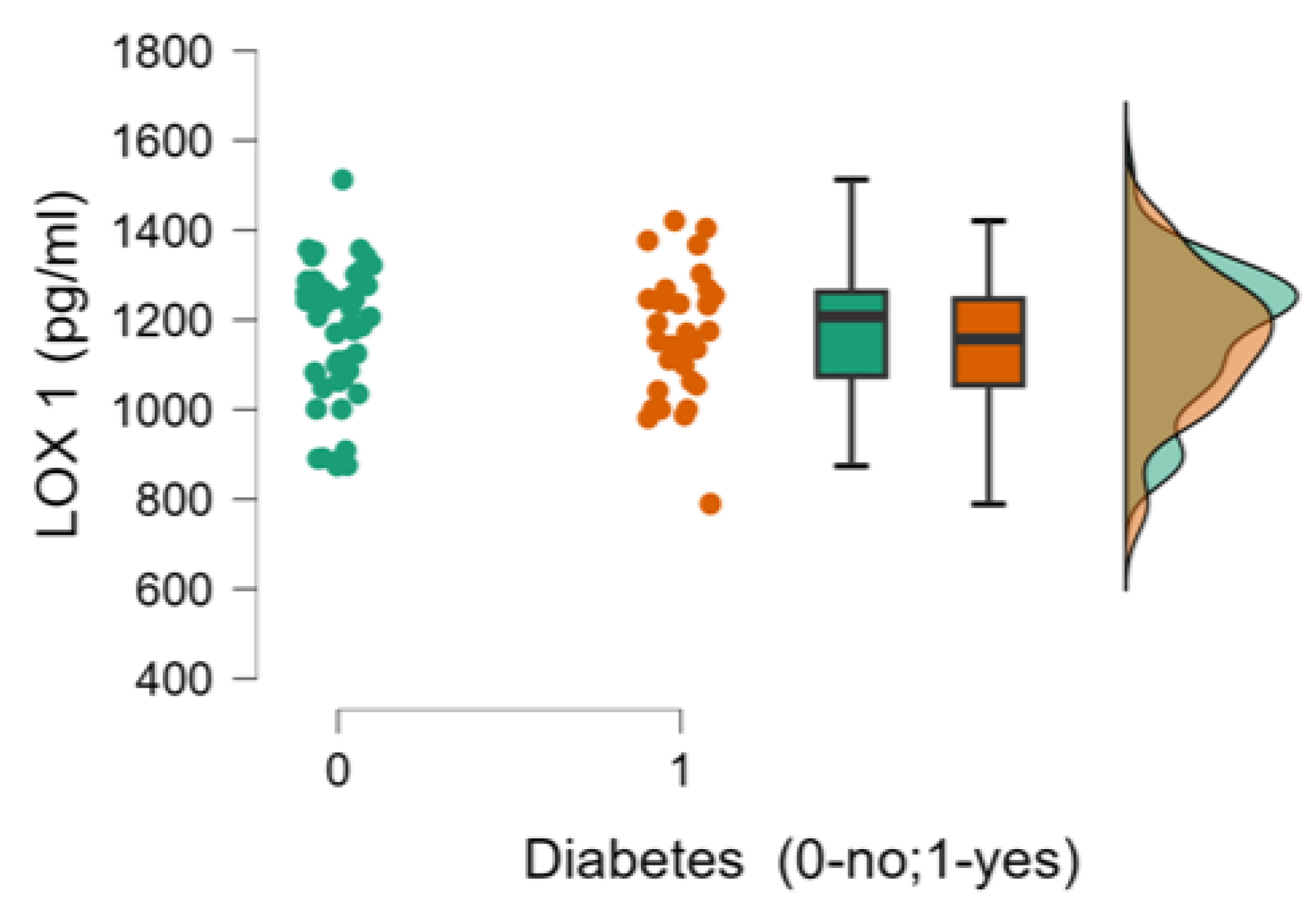
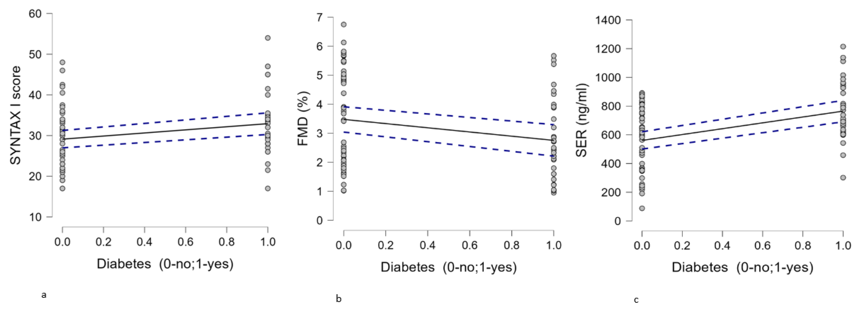
Background and objectives: Endothelial dysfunction (ED) and oxidative stress play major contributions in the initiation and progression of atherosclerosis. Diabetes is a pathological state associated with endothelial damage and enhanced oxidative stress. This study evaluated endothelial dysfunction and oxidative stress in severe coronary artery disease (CAD) patients undergoing coronary artery bypass graft (CABG) surgery, comparing those with and without type 2 diabetes mellitus (T2DM). Materials and methods: We included 84 patients with severe coronary artery disease (33 of whom had type 2 diabetes mellitus) who underwent clinical assessments, ultrasound, and coronary angiography. The SYNTAX I score was calculated from the coronary angiogram. Blood samples were collected to measure plasma serotonin (5-HT; SER) levels, as well as superoxide dismutase 1 (SOD-1) and lectin-like oxidized low-density lipoprotein receptor-1 (LOX-1) to assess oxidative stress. Brachial flow-mediated dilation (FMD) was used as a surrogate for endothelial dysfunction (ED), along with serum concentrations of 5-HT Results: The coronary atherosclerotic burden, assessed using the SYNTAX I score, was more severe in patients with CAD and associated T2DM compared to those with CAD without T2DM (30.5 [17-54] vs. 29 [17-48]; p=0.05). The SYNTAX score was found to be positively correlated with T2DM (p=0.029; r=0.238). ED measured by FMD was associated with T2DM (p=0.042; r=-0.223), with lower FMD measurements in T2DM patients when compared with individuals without this pathology (2.43% (0.95-5.67) vs. 3.46% (1.02-6.75);p=0.079). Also, in the studied population T2DM was correlated with serum 5-HT levels (764.78± 201 ng/ml vs. 561.06±224 ng/ml;p<0.001; r=0.423) with higher plasma circulating levels of 5-HT in patients with T2DM.No statistically significant differences for oxidative stress markers (SOD-1 and LOX-1) were obtained when comparing T2DM and non T2DM patients with severe CAD. Conclusion: ED (as assessed by brachial FMD and serum 5-HT) is more severe in patients in diabetic patients with severe CAD scheduled for CABG surgery, while oxidative stress (as evaluated through serum SOD-1 and LOX-1 concentrations) was not influenced by the presence of T2DM in this specific population. The most important finding of the present study is that circulating 5-HT levels are markedly influenced by T2DM. 5-HT receptor targeted therapy might be of interest in patients undergoing CABG, but further studies are needed to confirm this hypothesis.

Keywords: 
;  ;  ;  ;  

1. Introduction

Endothelial dysfunction is a pathological state present in cardiovascular disease, from subclinical stages of atherosclerosis to overt chronic and acute coronary syndromes [1]. Type 2 diabetes mellitus (T2DM), through mechanisms such as hyperglycemia, insulin resistance, and associated dyslipidemia, contributes to endothelial dysfunction (ED) in both the micro- and macro-vasculature.[2].Glucotoxicity induced ED includes attenuated endothelium-mediated vasomotor function, enhanced endothelial apoptosis, endothelium activation/endothelium–monocyte adhesion [3]. The hallmark of endothelial dysfunction is the alteration of endothelium-dependent vasodilation, primarily mediated by nitric oxide (NO).Therefore, testing for ED mainly involves stimulating the release of NO from the endothelium.ED is a systemic process. Hence, evaluation of peripheral brachial flow mediated dilation (FMD) represents a non invasive method used to quantify ED and its measures correlate well with coronary ED [4,5].
The molecules implicated in platelet aggregation also release NO. In the absence of endothelial barrier, the platelet-derived serotonin (or 5-hydroxytryptamine; 5-HT) diffuses toward the underlying vascular smooth muscle, exerting important vasoconstriction. The vasoconstrictive effect of 5-HT is enhanced inatherosclerosis, a pathological state associated with endothelial dysfunction [6]. High 5-HT serum levels in diabetes have been associated with ED and enhanced thrombogenesis [7].
The generation of reactive oxygen species (ROS) is another key process involved in ED. One such species, cytosolic copper/zinc superoxide dismutase (SOD-1), is an antioxidant enzyme that helps mitigate oxidative damage by scavenging harmful superoxide radicals. This enzyme plays a protective role in safeguarding endothelial cells against oxidative stress. In patients with diabetes, insulin resistance and hyperglycemia-induced apoptosis of the vascular endothelium contribute to the development of ED [8].
Lipid peroxidation of low-density lipoprotein (LDL) plays a crucial role in the process of atherosclerosis, with oxidized LDL being a key factor. Lectin-like oxidized low-density lipoprotein receptor-1 (LOX-1) has been identified as the primary receptor for oxidized LDL in endothelial cells [9]. LOX-1 is upregulated in chronic inflammatory conditions, such as cardiovascular disease and T2DM [10].
This study aimed to evaluate ED through ultrasonography (brachial FMD) and serum serotonin (5-HT; SER) levels, as well as circulating oxidative stress markers (SOD-1 and LOX-1) in patients with severe coronary artery disease (CAD) scheduled for coronary artery bypass graft (CABG) surgery. We also compared the results between individuals with T2DM and those without T2DM.

2. Materials and Methods

2.1. Design, Study Population

This prospective study included 84 adult patients with severe CAD scheduled for CABG, between January 2020 and June 2021. The study protocol received ethical approval from theEthical Committee of Transylvania University (registration number 1/2.03.2019) andadhered to the principles outlined in the Helsinki Declaration and the Code for Good Clinical Practice.Informed consent was obtained in writing from all patients.
Patients with acute coronary syndromes, subclavian artery stenosis, significant valvular disease, severe hepatic or renal failure, recent or active bleeding, coagulation disorders, active malignancy,carcinoid syndrome, and inflammatory diseases (including infections and autoimmune disorders)were excluded from the study. Additionally, patients diagnosed with depression and undergoing treatment with selective serotonin reuptake inhibitors and monoamine oxidase inhibitors were excluded due to the potential impact on 5-HT levels.
At admission, all patients underwent clinical, ultrasound, and coronary angiography evaluations, and blood samples were collected in accordance with the clinic's protocol. The SYNTAX I score was determined using the number of diseased arteries, the location, and the aspect of atherosclerotic plaques , after performing coronary angiograms (https://syntaxscore.org/).

2.2. FMD Measurement

We used the brachial artery for FMD evaluation. FMD measurement was performed after six hours of fasting. Also, physical exertion, caffeine consumption and smoking for the previous twenty-four hourswere restricted [11]. Patients were kept in a quiet room in the supine posture for at least 10 minutes prior to the measurement. To measure FMD we used a vascular linear probe (2D mode; 7,5–12 MHz). In order to induce reactive hyperemia, the occlusion cuff was wrapped around the forearm and inflated for five minutes to a pressure 50 mmHg higher than the systolic blood pressure. The diameter of the brachial artery was measured 3-10 cm above the antecubital fossa, before the cuff was inflated, 60-90 seconds after maximal reactive hyperemia, and 3 minutes after the cuff was deflated. FMD was calculated as percentage index according to current guidelines [12]. In accordance with recent research, we considered a value <6.5% as indicative for endothelial dysfunction [13].

2.3. Measurement of Serum Biomarkers

Peripheral venous blood samples were drawn after a minimum of 8 hours fasting, 3 days prior CABG. Samples were centrifuged and supernatant was frozen at -20 degrees Celsius until the final measurements. 5-HT and LOX-1 concentrations were determined by enzyme-linked immunosorbent assay (ELISA) with commercially available kits (DIAsourceImmunoAssays SA, Louvain-la-Neuve, Belgium-for 5-HT and Elabscience Biotechnology Inc., Houston, Texas, United States-for LOX-1) according to the manufacturer’s specifications. SOD-1 concentrations were measured using colorimetric determinations, with commercially available kits (Elabscience Biotechnology Inc., Houston, Texas, United States) following the manufacturer’s instructions. All measurements were completed by the same technician who had no access to clinical information.

2.4. Statistical Analysis

Categorical variables were expressed as n (%), normally distributed data and skewed data of continuous variables were expressed as mean±standard deviation (SD), and median (minimum-maximum), respectively. The normality of continuous variables was tested by Shapiro-Wilk’s test. To ascertain distinctions between the studied groups (T2DM versus non T2DM) non parametrical tests for small sample sizes were applied (Chi-square and Mann-Whitney tests). To ascertain distinctions in the analyzed data, 2-tailed Pearson correlation was applied. Statistical significance was set at p<0.05.Analysis was performed using Microsoft Excel 2007 and JASP 0.19 software.

3. Results

Out of the 84 patients enrolled, 33 were diagnosed with T2DM. The studied population was predominantly male (26 [78.78 %] with T2DM and 41 [80.39 %] without T2DM), had a mean BMI of approximately 28 kg/m2 (in both groups), and had a small percentage of smokers (9 [27.27 %] in diabetics vs. 8 [15.68 %] in non diabetics). Most of the patients included had dyslipidemia (31 [93.93 %] in T2DM and 49 [96.07%] in non T2DM) and hypertension (all diabetics and 46 [90.19 %] of non diabetics). Patients’ characteristics are depicted in Table 1.
The SYNTAX I score was higher in the diabetes group (p=0.050), reflecting a more severe coronary atherosclerotic burden in this category of patients (Table 1; Figure 1).
We observed lower brachial FMD measurements in patients with diabetes compared to those without diabetes (p = 0.079), though the result only approached statistical significance(Table 1; Figure 2).
The most interesting results of the present study were the higher values of serum serotonin in patients with T2DM compared with patients without T2DM (p<0.001), as depicted in Figure 3.
Oxidative stress was comparable between the groups, as the serum concentrations of SOD 1 and LOX 1 did not differ between patients with diabetes and those without diabetes (p = 0.362 for SOD 1 and p = 0.536 for LOX 1).The results are shown in Figure 4 and Figure 5.
Additionally, we examined whether T2DM correlated with the studied parameters (SYNTAX score, FMD, and circulating levels of 5-HT, SOD 1, and LOX 1). Statistically significant correlations were found between diabetes and the SYNTAX score (Figure 6a), diabetes and FMD (Figure 6b), and diabetes and 5-HT serum concentrations (Figure 6c). A summary of these results is presented in Table 2.

4. Discussion

Diabetes mellitus is a prothrombotic and hypercoagulable state which predisposes to thrombus formation. Platelets play a pivotal role in atherogenesis and its thrombotic complications [14]. Coronary atherosclerosis is more severe and diffuse in diabetic patients. Hence, the SYNTAX score, which evaluates the anatomic complexity of CAD, is higher in patients with T2DM. In the studied population, diabetes and SYNTAX score were correlated (p=0.029; r=0.238), a more severe and complex atherosclerotic burden being identified in patients diagnosed with T2DM. When comparing the two groups (patients with CAD without T2DM, and patients with CAD with T2DM), the difference was also significant (p=0.05).
Endothelial function is an integrated index of all atherogenic and atheroprotective factors present in an individual [15]. The endothelium is the key modulator of vascular tone and vascular homeostasis. A dysfunctional endothelium induces vasoconstriction, mainly through the reduction of NO bioavailability and exerts pro atherogenic and pro thrombotic effects [16].
The techniques for assessing the endothelial function in the clinical setting are mainly based on the vessel’s response during reactive hyperemia, which induces endothelial-dependent relaxation by releasing NO. FMD of the peripheral vasculature is a non-invasive technique widely used in clinical practice for assessing endothelial function. Moreover, several studies have shown that it correlates well with coronary artery ED [4,5]. ED is present in all stages of CAD, promoting atherosclerosis progression and acute coronary events [17]. The present study found that ED, measured by FMD, is severe in the studied population (patients with severe CAD undergoing surgical myocardial revascularization). FMD values were below the established cutoff of 6.5 % in both diabetics and non-diabetics. Hyperglycemia induces repeated alterations of intracellular metabolism and long-term changes in the structure and function of molecules through formation of advanced glycation end products [18]. Thus, T2DM patients had a lower vasodilator response to reactive hyperemia versus patients without T2DM, even if the statistical significance was low for the analyzed population. Our study showed that FMD is influenced by T2DM (p=0.042; r=-0.223), reactive hyperemia being lower in patients with CAD that associate diabetes, and to the best of our knowledge, this research represents the first study to enroll patients with severe CAD, specifically those scheduled for CABG, and to comparatively evaluate FMD based on the presence or absence of T2DM.
ED is present from the early stages of both T2DM and CAD. However, few studies compared ED (assessed by FMD) in patients with CAD and T2DM versus patients with CAD without T2DM [19,20,21,22]. Similar to our findings, two of the studies ([20,21]) observed that the subjects with both CAD and T2DM had lower FMD than subjects with CAD but no T2DM. The studies conducted by Bhargava et al [19] and Simova et al [22] observed ED across different stages of CAD. The first included Indian subjects, different from our Caucasian population. In contrast to our findings, they observed a similar degree of ED in subjects with T2DM and CAD compared with subjects who only had CAD.The latter study found no statistical difference in patients with more extensive CAD, regardless of the presence of T2DM.
Plasma serotonin levels are associated with accelerated atherosclerosis [23]. In patients with diabetes, elevated plasma 5-HT levels have been documented [24], as well as 5-HT –induced platelet aggregation [25], and upregulation of vascular 5-HT2A receptors [26].Moreover, 5-HT appears to act synergistically with T2DM, as it augments hyperglycemia-induced ED [7].
In the population we studied, 5-HT was significantly correlated with T2DM (p<0.001; r=0.423). Also, a statistically significant difference (p<0.001; Figure 3) was obtained when comparing 5-HT concentrations in patients with diabetes versus patients without diabetes. Our study is the first to evaluate serum levels of 5-HT in patients with severe coronary artery disease (CAD) and compare the findings based on the presence of T2DM, and the results may have important clinical implications.
It is now well-established that selective serotonin reuptake inhibitors (SSRIs) are effective in the treatment of depression. Additionally, depression, certain personality traits [27,28], and cardiovascular disease are frequently interconnected, with inflammation and ED being common factors in both. Recent studies have therefore focused on the cardiovascular effects of SSRIs, yielding conflicting results. Some studies report protective cardiovascular effects associated with SSRIs [29,30], while others indicate worse outcomes linked to their use [31].
Sarpogrelate, which is a 5-HT2A and 5-HT 2B receptor antagonist, inhibits serotonin-induced platelet aggregation. In the search for novel therapeutic agents to further diminish the high CAD burden in T2DM, sarpogrelate has emerged as a promising molecule, reducing the total atherosclerotic plaque volume [32]. Furthermore, 5-HT has been implicated in smooth muscle cell proliferation, the major cause of graft failure after CABG. Several in vivo studies conducted on isolated porcine vein [33], human isolated saphenous vein [34], and human isolated internal thoracic artery [35] concluded that sarpogrelate prevents vasospasm and early graft occlusion.Further research is necessary to establish the clinical profile of patients that might benefit the most from this therapy, particularly patients with T2DM undergoing CABG surgery.
Another interesting finding of the present study was the lack of association between SOD-1 and LOX-1, as markers of oxidative stress, and diabetes. Elevated SOD-1 activity has been reported to confer protection against oxidative stress. However, even if elevated serum concentrations are present in CAD, recent studies did not report a significant correlation between plasma SOD 1 and CAD severity [36]. A possible explanation might be that elevated SOD 1 levels might be compensated by elevated ROS formation. In diabetes, vascular injury is induced by a variety of factors, such as hyperglycemia, oxidized LDL, angiotensin II, proinflammatory cytokines, and altered shear stress. Previous studies suggest that LOX-1 cellular signal transduction pathway is important for the onset and development of diabetic vasculopathy [37].A possible explanation for the conflicting result in our study is that oxidative stress levels are at the highest in patients with severe CAD, irrespective of the presence of T2DM.
Several limitationsof our study need mentioning. First, low statistical differences regarding the SYNTAX score and FMD measurements in the T2DM group versus the non T2DM might have been affected by the small study population. Second, even if FMD is a recognized measurement for ED, it still lacks standardization.
ED might be reversible by targeted treatment at every phase of atherosclerosis, even in the setting of established severe CAD andatherothrombotic complications.

5. Conclusions

This study observed that the burden of coronary artery disease (as indicated by the SYNTAX score) is more pronounced in patients with T2DM compared to those without T2DM, even within populations with severe CAD, such as individuals scheduled for CABG.
Although endothelial dysfunction (ED), assessed through FMD and circulating 5-HT levels, is more pronounced in diabetic patients compared to non-diabetic patients, the circulating levels of SOD-1 and LOX-1, which reflect oxidative stress, are not influenced by T2DM in patients with severe CAD.
Further research is needed to determine the role of 5-HT, as its levels may serve as a more reliable serum marker and therapeutic target in patients with severe CAD associated with T2DM, especially considering that 5-HT plasma levels could also contribute to graft vasospasm and early coronary graft occlusion.

Author Contributions

"Conceptualization, A.M.B., and D.T; Methodology, C.D.N. and D.T.; Software, A.M.B, C.D.L.; Validation, A.M.B, C.D.L., A.B. and D.T.; Formal Analysis, A.M.B., C.D.L., and D.T.; Investigation, A.M.B, C.D.L., and D.T.; Resources, D.T.; Data Curation, A.M.B and C.D.N.; Writing – Original Draft Preparation, A.M.B., and D.T.; Writing – Review & Editing, A.M.B., C.D.L, C.D.N., A.B., D.T.; Visualization, D.T., A.B; Supervision, D.T.; Project Administration, D.T.; Funding Acquisition, D.T.” All authors have read and agreed to the published version of the manuscript.

Funding

This research received funding from the “Transylvania Faculty of Medicine”, Braşov and material support from “ICCO Clinics”, Brasov.

Institution review board statement

The study was conducted according to the guidelines of the Declaration of Helsinki, and approved by the Ethics Committee of Transylvania University (registration number 1/2.03.2019).

Informed consent statement

Written informed consent was obtained from all subjects involved in the study.

Acknowledgements

The authors would like to thank all the patients and medical staff who participated in the study, as well as mister Adrian Garcea (for the help provided with laboratory measurements), and mister Alexandru Gaianu (for the statistical analysis).

Conflicts of interest

The authors declare no conflict of interest.

References

  1. Verma, S.; Buchanan, M.R.; Anderson, T.J. Endothelial Function Testing as a Biomarker of Vascular Disease. Circulation 2003, 108, 2054–2059. [Google Scholar] [CrossRef] [PubMed]
  2. Dubsky, M.; Veleba, J.; Sojakova, D.; Marhefkova, N.; Fejfarova, V.; Jude, E.B. Endothelial Dysfunction in Diabetes Mellitus:New Insights. Int J MolSci. 2023, 24, 10705. [Google Scholar] [CrossRef]
  3. Zhang, H.; Dellsperger, K.C.; Zhang, C. The link between metabolic abnormalities and endothelial dysfunction in type 2 diabetes: an update. Basic Res Cardiol. 2012, 107, 237. [Google Scholar] [CrossRef]
  4. Sancheti, S.; Shah, P.; Phalgune, D.S. Correlation of endothelial dysfunction measured by flow-mediated vasodilatation to severity of coronary artery disease. Indian Heart J. 2018, 70, 622–626. [Google Scholar] [CrossRef] [PubMed]
  5. Manganaro, A.; Ciracì, L.; Andrè, L.; et al. Endothelial Dysfunction in Patients With Coronary Artery Disease: Insights From a Flow-Mediated Dilation Study. Clin Appl ThrombHemost. 2014, 20, 583–588. [Google Scholar] [CrossRef]
  6. Vanhoutte, P.M. Endothelial Dysfunction and Atherosclerosis. Eur Heart J 1997, 18 (Suppl E), E19–E29. [Google Scholar] [CrossRef] [PubMed]
  7. Yamada, K.; Niki, H.; Nagai, H.; Nishikawa, M.; Nakagawa, H. Serotonin Potentiates High-Glucose–Induced Endothelial Injury: the Role of Serotonin and 5-HT2A Receptors in Promoting Thrombosis in Diabetes. J PharmacolSci. 2012, 119, 243–250. [Google Scholar] [CrossRef]
  8. Takeda, Y.; Matoba, K.; Sekiguchi, K.; Nagai,Y. ;Yokota,T.; Utsunomiya,K.;Nishimura, R. Endothelial dysfunction in diabetes. Biomedicines. 2020, 8, 182. [Google Scholar] [CrossRef] [PubMed]
  9. Kattoor, A.J.; Goel, A.; Mehta, J.L. LOX-1: Regulation, Signaling and its Role in Atherosclerosis. Antioxidants 2019, 8, 218. [Google Scholar] [CrossRef] [PubMed]
  10. Vavere, A.L.; Sinsakul, M.; Ongstad, E.L.; Yang, Y.; Varma, V.; Jones, C.; et al. Lectin-Like Oxidized Low-DensityLipoprotein Receptor 1 Inhibition in Type 2 diabetes: Phase 1 results. J Am Heart Assoc 2023, 12, e027540. [Google Scholar] [CrossRef]
  11. Thijssen, D.H.J.; Bruno, R.; Van Mil, A.C.C.M.; Holder, S.M.; Faita, F.; Greyling, A.; et al. Expert consensus and evidence-based recommendations for the assessment of flow-mediated dilation in humans. Eur Heart J. 2019, 40, 2534–2547. [Google Scholar] [CrossRef]
  12. Maruhashi, T.; Kajikawa, M.; Kishimoto, S.; Hashimoto, H.; Takaeko, Y.; Yamaji, T.; et al. Diagnostic criteria of flow-mediated vasodilation for normal endothelial function and nitroglycerin-induced vasodilation for normal vascular smooth muscle function of the brachial artery. J Am Heart Assoc. 2020, 9, e013915. [Google Scholar] [CrossRef] [PubMed]
  13. Heiss, C.; Rodriguez-Mateos, A.; Bapir, M.; Skene, S.S.; Sies, H.; Kelm, M. Flow-mediated dilation reference values for evaluation of endothelial function and cardiovascular health. Cardiovasc Res. 2023, 119, 283–293. [Google Scholar] [CrossRef]
  14. Jennings, L.K. Role of platelets in atherothrombosis. Am J Cardiol. 2009, 103, 4A–10A. [Google Scholar] [CrossRef] [PubMed]
  15. Matsuzawa, Y.; Lerman, A. Endothelial dysfunction and coronary artery disease: assessment, prognosis, and treatment. Coron Artery Dis 2014, 25, 713–724. [Google Scholar] [CrossRef]
  16. Godo, S.; Shimokawa, H. Endothelial Functions. Arterioscler Thromb Vasc Biol 2017, 37, e108–e114. [Google Scholar] [CrossRef]
  17. Gutierrez, E.; Flammer, A.J.; Lerman, L.O.; Elizaga1, J.; Lerman, A.; Fernandez-Avile, F. Endothelial dysfunction over the course of coronary artery disease. EurHeartJ. 2013, 34, 3175–3181. [Google Scholar] [CrossRef] [PubMed]
  18. De Vriese, A.S.; Verbeuren, T.J.; Van De Voorde, J.; Lameire, N.H.; Vanhoutte, PM. Endothelial dysfunction in diabetes. Br J Pharmacol. 2000, 130, 963–74. [Google Scholar] [CrossRef]
  19. Bhargava, K.; Hansa, G.; Bansal, M.; Tandon, S.; Kasliwal, R.R. Endothelium-dependent brachial artery flow mediated vasodilatation in patients with diabetes mellitus with and without coronary artery disease. J Assoc Physicians India. 2003, 51, 355–8. [Google Scholar]
  20. Kirma, C.; Akcakoyun, M.; Esen, A.M.; Barutcu, I.; Karakaya, O.; Saglam, M.; et al. Relationship between endothelial function and coronary risk factors in patients with stable coronary artery disease. Circ J. 2007, 71, 698–702. [Google Scholar] [CrossRef]
  21. Reyes-Soffer, G.; Holleran, S.; Di Tullio, M.R.; Homma, S.; Boden-Albala, B.; Ramakrishnan, R.; Elkind, M.S.; Sacco, R.L.; Ginsberg, H.N. Endothelial function in individuals with coronary artery disease with and without type 2 diabetes mellitus. Metabolism. 2010, 59, 1365–71. [Google Scholar] [CrossRef] [PubMed]
  22. Simova, I.I.; Denchev, S.V.; Dimitrov, S.I.; Ivanova, R. Endothelial function in patients with and without diabetes mellitus with different degrees of coronary artery stenosis. J Clin Ultrasound. 2009, 37, 35–9. [Google Scholar] [CrossRef] [PubMed]
  23. Vikenes, K.; Farstad, M.; Nordrehaug, J.E. Serotonin is associated with coronary artery disease and cardiac events. Circulation 1999, 100, 483–489. [Google Scholar] [CrossRef]
  24. Barradas, M.A.; Gill, D.S.; Fonseca, V.A.; Mikhailidis, D.P.; Dandona, P. Intraplatelet serotonin in patients with diabetes mellitus and peripheral vascular disease. Eur J Clin Invest. 1988, 18, 399–404. [Google Scholar] [CrossRef] [PubMed]
  25. Malyszko, J.; Urano, T.; Knofler, R.; Taminato, A.; Yoshimi, T.; Takada, Y.; Takada, A. Daily variations of platelet aggregation in relation to blood and plasma serotonin in diabetes. Thromb Res. 1994, 75, 569–76. [Google Scholar] [CrossRef]
  26. Bir, S.C.; Fujita, M.; Marui, A.; Hirose, K.; Arai, Y.; Sakaguchi, H.; Huang, Y.; Esaki, J.; Ikeda, T.; Tabata, Y.; Komeda, M. New therapeutic approach for impaired arteriogenesis in diabetic mouse hindlimb ischemia. Circ J. 2008, 72, 633–40. [Google Scholar] [CrossRef]
  27. Sherwood, A.; Hinderliter, A.L.; Watkins, L.L.; Waugh, R.A.; Blumenthal, J.A. Impaired endothelial function in coronary heart disease patients with depressive symptomatology. J Am Coll Cardiol. 2005, 46, 656–9. [Google Scholar] [CrossRef]
  28. Enatescu, V.R.; Cozma, D.; Tint, D.; Enatescu, I.; Simu, M.; Giurgi-Oncu, C.; Lazar, M.A.; Mornos, C. The Relationship Between Type D Personality and the Complexity of Coronary Artery Disease. Neuropsychiatr Dis Treat. 2021, 18, 809–820. [Google Scholar] [CrossRef]
  29. Kim, Y.; Lee, Y.S.; Kim, M.G.; Song, Y.K.; Kim, Y.; Jang, H.; Kim, J.H.; Han, N.; Ji, E.; Kim, I.W.; Oh, J.M. The effect of selective serotonin reuptake inhibitors on major adverse cardiovascular events: a meta-analysis of randomized-controlled studies in depression. Int Clin Psychopharmacol. 2019, 34, 9–17. [Google Scholar] [CrossRef]
  30. Delialis, D.; Mavraganis, G.; Dimoula, A.; Patras, R.; Dimopoulou, A. M.; Sianis, A.; Ajdini, E.; Maneta, E.; Kokras, N.; Stamatelopoulos, K.; Georgiopoulos, G. A. A systematic review and meta-analysis on the effect os selective serotonin reuptake inhibitors on endothelial function. J Affect Disord. 2022, 316, 71–75. [Google Scholar] [CrossRef] [PubMed]
  31. Almuwaqqat, Z.; Jokhadar, M.; Norby, F.L.; Lutsey, P.L.; O'Neal, W.T.; Seyerle, A.; Soliman, E.Z.; Chen, L.Y.; Bremner, J.D.; Vaccarino, V.; Shah, A.J.; Alonso, A. Association of Antidepressant Medication Type With the Incidence of Cardiovascular Disease in the ARIC Study. J Am Heart Assoc. 2019 4, e012503. [CrossRef]
  32. Lee, D-H. , Chun, E.J., Hur, J.H., Min, S.H.; Lee, J-E., Oh, T.J.; Kim, K.M.; Jang, H.C.; Han, S.J.; Kang, D.K.; Kim, H.J.; Lim, S. Effect of sarpogrelate, a selective 5-HT2A receptor antagonist, on characteristics of coronary artery disease in patients with type 2 diabetes. Atherosclerosis. 2017, 257, 47–54. [Google Scholar] [CrossRef]
  33. Sharma, S.K.; Del Rizzo, D.F.; Zahradka, P.; Bhangu, S.K.; Werner, J.P.; Takeda, N.; Dhalla, N.S. Sarpogrelate inhibits Serotonin-induced proliferation of porcine coronary artery smooth muscle cells: implications for lon-term graft patency. Ann Thorac Surg. 2001, 71, 1856–64. [Google Scholar] [CrossRef] [PubMed]
  34. Nakamura, E.; Tanaka, N.; Kuwabara, M.; Yamashita, A.; Matsuo, Y.; Kanai, T.; Onitsuka, T.; Asada, Y.; Hisa, H.; Yamamoto, R. Relative contributions of 5-hydroxytryptamine (5-HT) receptor subtypes in 5-HT-induced vasoconstriction of the distended human saphenous vein as a coronary artery bypass graft. Biol Pharm Bull. 2011, 34, 82–6. [Google Scholar] [CrossRef] [PubMed]
  35. Tanak-Totoribe, N.; Hidaka, M.; Gamoh, S.; Yokota, A.; Nakamura, E.; Kuwabara, M.; Tsunezumi, J.; Yamamoto, R. Effects of M-1, a major metabolite of sarpogrelate, on 5-HT-induced constriction of isolated human internal thoracic artery. Biol Pharm Bull. 2020, 43, 1979–1982. [Google Scholar] [CrossRef] [PubMed]
  36. Peng, J.R.; Ting-Ting, L.; Chang, H.T.; Xuan, G.; Bian, H.; Wei-Min, L. Elevated Levels of Plasma Superoxide Dismutases 1 and 2 in Patients with Coronary Artery Disease. BioMed Res Int. 2016, 3708905. [Google Scholar] [CrossRef]
  37. Yan, M.; Mehta, J.L.; Zhang, W.; Hu, C. LOX-1, Oxidative Stress and Inflammation: A Novel Mechanism for Diabetic Cardiovascular Complications. Cardiovasc Drugs Ther. 2011, 25, 451–459. [Google Scholar] [CrossRef]
Figure 1. SYNTAX I score according to the presence of T2DM.
Preprints 142163 g001
Figure 2. FMD measurements in patients with and without diabetes.
Preprints 142163 g002
Figure 3. Serum serotonin (SER) levels in diabetics and non diabetics.
Preprints 142163 g003
Figure 4. SOD 1 serum levels in patients with and without T2DM.
Preprints 142163 g004
Figure 5. LOX 1 circulating levels according to the presence of T2DM.
Preprints 142163 g005
Figure 6. Correlations between T2DM and the studied parameters; a: SYNTAX I score and diabetes (p=0.029; r=0.238); b: FMD and diabetes (p=0.042; r=-0.223); c: FMD and serum serotonin levels (p<0.001; r=0.423).
Preprints 142163 g006
Table 1. Patients’ characteristics according to the presence of T2DM.
Patients’ characteristics T2DM (n=33) Non T2DM (n=51) p
1. Age, mean ±SD (years) 65.24±6.96 65±8.82 0.69
2. Male, n (%) 26 (78.78) 41 (80.39) 0.858
3. BMI ,mean ±SD (kg/m2) 28.48±4.4 28.28±4.17 0.759
4. Smoking status, n (%) 9 (27.27) 8 (15.68) 0.197
5. Dyslipidemia, n (%) 31 (93.93) 49 (96.07) 0.653
6. Hypertension, n (%) 33 (100) 46 (90.19) 0.064
7. SYNTAX I score, median (min-max) 30.5 (17-54) 29 (17-48) 0.050
8. FMD %, median (min-max) 2.43 (0.95-5.67) 3.46 (1.02-6.75) 0.079
9. 5-HT (ng/ml),mean ±SD 764.78±201.44 561.06±224.3 <0.001
10. SOD 1 (ng/ml), mean ±SD 1.34±0.21 1.39±0.24 0.362
11. LOX 1(pg/ml), median (min-max) 1152.33±153.63 1167.11±152.53 0.536
Abbreviations: BMI: body mass index; FMD: flow mediated dilation; SD: standard deviation; T2DM:type 2 diabetes mellitus; Italics: statistical significance; 5-HT: 5-hydroxytryptamine.
Table 2. Correlation coefficients and statistical significance for the studied parameters.
Pearson’s correlations
Variable SYNTAX I score FMD (%) 5-HT (ng/ml) SOD 1(ng/ml) LOX 1(pg/ml) Diabetes (0-no;1-yes)
1.SYNTAX I score Pearson’s r -
p-value -
2. FMD (%) Pearson’s r -0.786*** -
p-value <.001 -
3. 5-HT (ng/ml) Pearson’s r 0.159 -0.141 -
p-value 0.149 0.200 -
4.SOD 1(ng/ml) Pearson’s r -0.067 0.099 0.038 -
p-value 0.547 0.372 0.731 -
5.LOX 1(pg/ml) Pearson’s r -0.104 0.105 -0.035 0.185 -
p-value 0.348 0.341 0.751 0.092 -
6. Diabetes (0-no;1-yes) Pearson’s r 0.238* -0.223* 0.423*** -0.106 -0.048 -
p-value 0.029 0.042 <.001 0.338 0.666 -
* p < .05, ** p < .01, *** p < .001
Disclaimer/Publisher’s Note: The statements, opinions and data contained in all publications are solely those of the individual author(s) and contributor(s) and not of MDPI and/or the editor(s). MDPI and/or the editor(s) disclaim responsibility for any injury to people or property resulting from any ideas, methods, instructions or products referred to in the content.
Copyright: This open access article is published under a Creative Commons CC BY 4.0 license, which permit the free download, distribution, and reuse, provided that the author and preprint are cited in any reuse.
Prerpints.org logo

Preprints.org is a free preprint server supported by MDPI in Basel, Switzerland.

Subscribe

Disclaimer

Terms of Use

Privacy Policy

Privacy Settings

© 2025 MDPI (Basel, Switzerland) unless otherwise stated