Preprint
Review

This version is not peer-reviewed.

Novel Insight of N6-Methyladenosine in Cardiovascular System

A peer-reviewed article of this preprint also exists.

Submitted:

03 December 2024

Posted:

04 December 2024

You are already at the latest version

Abstract

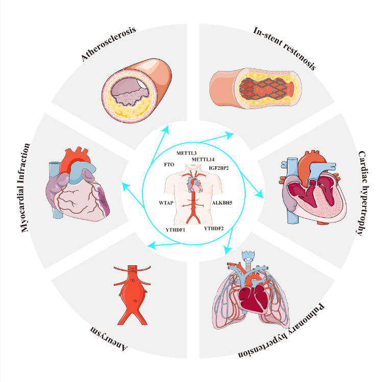
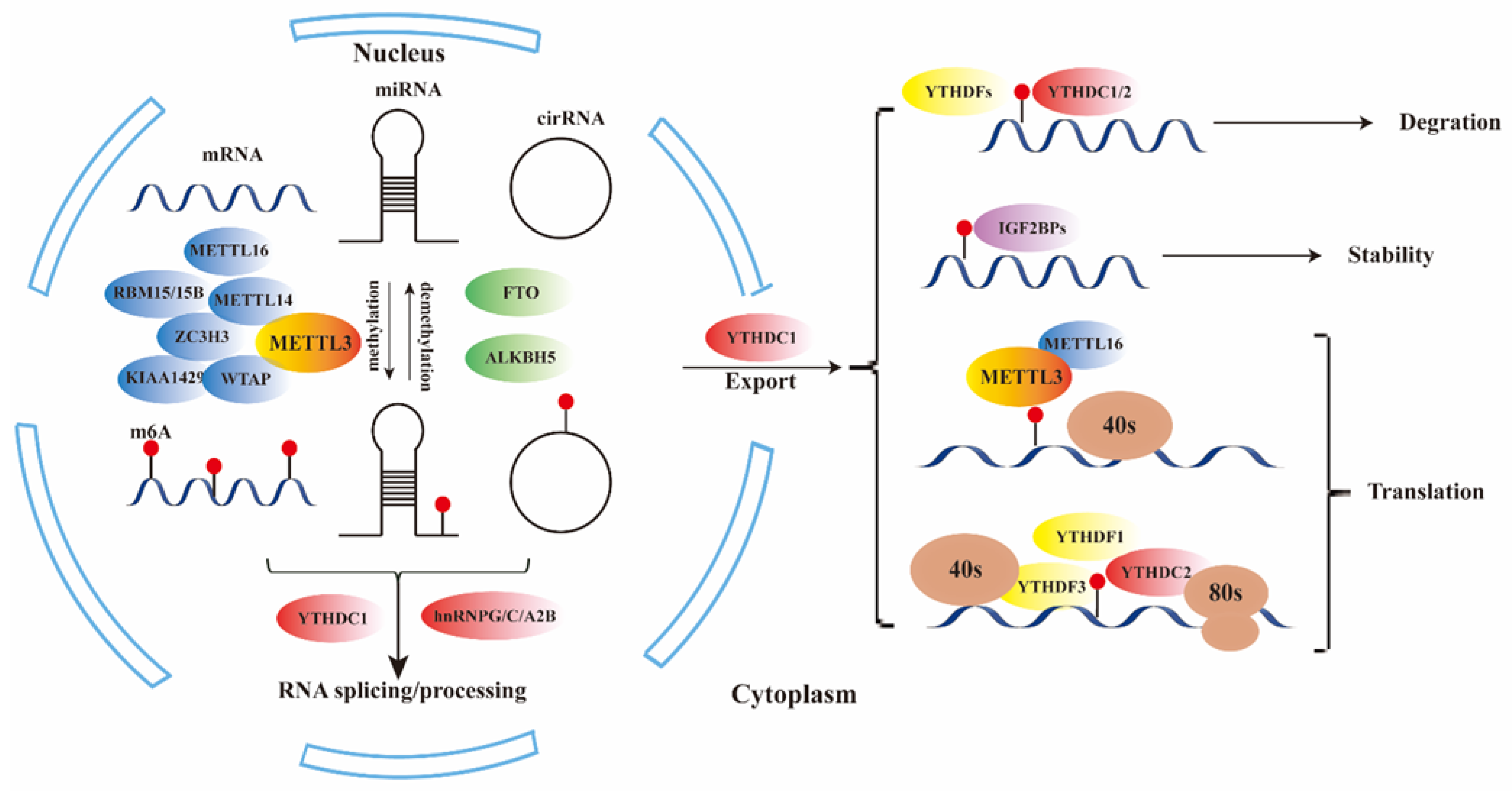
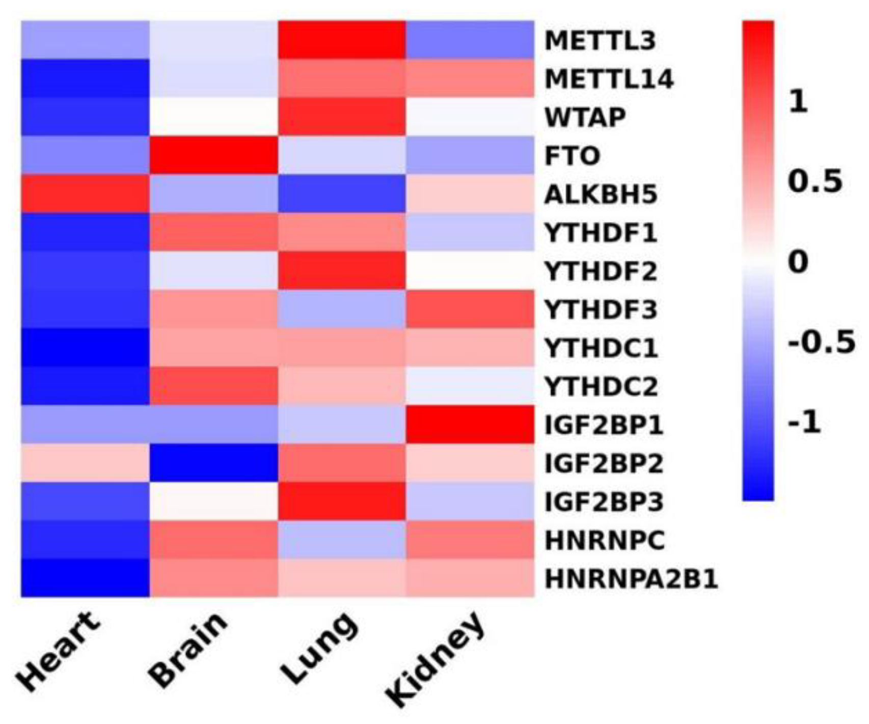
N6-methyladenosine (m6A) is the most common and abundant internal co-transcriptional modification in eukaryotic RNAs. This modification is catalyzed by m6A methyltransferases, known as "writers," including METTL3/14 and WTAP, and removed by demethylases, or "erasers," such as FTO and ALKBH5. It is recognized by m6A-binding proteins, or "readers," such as YTHDF1/2/3, YTHDC1/2, IGF2BP1/2/3, and HNRNPA2B1. Cardiovascular diseases (CVDs) are the leading cause of morbidity and mortality worldwide. Recent studies indicate that m6A RNA modification plays a critical role in both the physiological and pathological processes involved in the initiation and progression of CVDs. In this review, we will explore how m6A RNA methylation impacts both normal and disease states of the cardiovascular system. Our focus will be on recent advancements in understanding the biological functions, molecular mechanisms, and regulatory factors of m6A RNA methylation, along with its downstream target genes in various CVDs, such as atherosclerosis, ischemic diseases, metabolic disorders, and heart failure. We propose that the m6A RNA methylation pathway holds promise as a potential therapeutic target in cardiovascular disease.

Keywords: 
;  ;  ;  ;  

1. Introduction

More than 140 types of chemical modifications have been discovered in RNA[1]. Among these modifications, RNA methylation involves the addition of methyl groups to specific nucleotide residues in RNA, such as N1-methyladenosine (m1A), N6-methyladenosine (m6A), N6,2’-O-dimethyladenosine (m6Am), and 2’-O’-methylation (2’-OMe)[2,3]. Among these modifications, the methylation of mRNA at the N-6th position of the adenosine residue (m6A) has been extensively studied since its discovery in the 1970s. It is the most common methylation modification found in the internal sequence of mRNA in eukaryotes, and it is the most abundant and evolutionarily conserved reversible post-transcriptional modification[4,5,6]. Functionally, m6A is involved in various aspects of RNA biology, including RNA stability, nuclear export, translation efficiency, and mRNA degradation[2,3,7]. In addition to regulating RNA coding sequences (mRNA), m6A methylation also plays a regulatory role in non-coding RNAs such as microRNAs, long non-coding RNAs, and circular RNAs, thereby influencing gene expression in diverse physiological processes[8,9,10].
Cardiovascular disease (CVDs) is the leading cause of morbidity and mortality worldwide, and its pathological mechanisms are complex[11]. Despite significant advances in the diagnosis, treatment, and prognosis of CVDs, there is an urgent need for new diagnostic biomarkers and treatments to reduce mortality and improve therapeutic outcomes for patients with CVDs.
2. m6A RNA methylation
The rapid advancement of bioinformatic analyses and high-throughput sequencing technologies has revealed that m6A modification primarily occurs at the consensus motif RR (m6A) CH (R = A or G, H = A, C, or U) in long internal exons, near stop codons, or in the 3' untranslated region (UTR) [7,12](Figure 1). Importantly, m6A modification is dynamic and its methylation levels undergo dynamic changes, regulated by various methyltransferases ("writers"), m6A binding proteins ("readers"), and demethylases ("erasers")[13,14]. These three groups of proteins are responsible for the addition, removal, and recognition of m6A RNA modification.
Studies have shown that m6A modification can alter RNA secondary structures or be recognized by "readers" to regulate the metabolism of methylated mRNAs, thereby exerting various functions[15]. The process of RNA modification by m6A begins during transcription. The addition and removal of m6A mostly occur in the nucleus, where specific nuclear "readers" can bind to m6A and potentially affect mRNA splicing and other nuclear processes. After export to the cytoplasm, m6A interacts with specific cytoplasmic reader proteins that influence RNA stability, translation efficiency, and localization[2] (Table 1).
M6A "writers" consist of METTL3/14, KIAA1429, WTAP, zinc finger CCCH domain-containing protein 13 (ZC3H13), and RBM15/15B[31,32]. Among these, METTL3 and METTL14 are considered the most critical core components of the m6A methyltransferase complex[16]. METTL3 acts as the catalytic subunit, while METTL14 plays a structural role and activates METTL3 through allosteric and RNA substrate recognition[16,33]. WTAP interacts with METTL3-METTL14 and is the third subunit of the complex[17,18]. RBM15/15B is responsible for recruiting the methylation complex to XIST[19]. ZC3H13 plays a critical role in anchoring WTAP, Virilizer, and Hakai in the nucleus for m6A methylation regulation and mESC self-renewal[20]. KIAA1429, as a top interactor with WTAP, recruits specific cleavage and polyadenylation specificity factors, leading to longer 3'UTR selection[21]. ZC3H13, KIAA1429, and RBM15/RBM15B play key roles in localizing the methyltransferase complex in nuclear speckles and U-rich regions adjacent to m6A sites in mRNAs.
FTO (FAT mass and obesity-associated protein) and ALKBH5 (ALKB homologue 5 protein) belong to the ferric divalent/α-ketoglutarate dependent dioxygenase ALKB protein family and exhibit efficient oxidative demethylation activity targeting N6-methyladenosine residues in RNA[2,23]. Both FTO and ALKBH5 are located in the nuclear speckle area, bind to m6A-modified RNA, catalyze the oxidation of m6A to adenine, and produce intermediate products hm6A (N6 hydroxymethyl adenosine) and further oxidation product f6A (N6 formyl adenosine).
"Readers" are proteins that recognize m6A methylation sites on RNAs, bind to mRNA, and influence nuclear export, RNA stability, or degradation. The YTH domain-containing proteins, including YTHDC1, YTHDC2 (DC2), YTHDF1 (DF1), DF2, and DF3, were the first m6A-binding proteins to be discovered[14]. YTHDF1 promotes mRNA translation, while YTHDF2 accelerates the decay of m6A-modified transcripts. The role of YTHDF3 is complex, as it facilitates YTHDF1's role in promoting translation, and knockdown of YTHDF3 results in reduced translation efficiency of mRNA targets of both YTHDF3 and YTHDF1[24,25]. A recent study showed that YTHDC1 blocks SRSF10 mRNA binding by selectively recruiting the SRSF3 mRNA pre-splicing factor, thereby promoting exon inclusion in the target transcript[34]. Nuclear export of m6A-methylated transcripts may be facilitated by YTHDC1 through interaction with nuclear transport receptors[27]. YTHDC2 is an N6-methyladenosine binding protein that regulates mammalian spermatogenesis, by enhancing the translation efficiency of its targets and also decreasing their mRNA abundance[28]. IGFBP proteins, including IGF2BP1/2/3, can regulate RNA localization, translation, and stability[29]. The heterogeneous nuclear ribonucleoprotein (hnRNP) family, including hnRNPG, hnRNPC, and hnRNPA2B1, is another group of m6A "reader" proteins. Wu et al. found that m6A can enhance the ability of hnRNP A2/B1 to enhance nuclear events, such as pri-miRNA processing[30].

3. Role of m6A in cardiovascular disease

3.1. Risk factors associated with CVDs

Unhealthy diet, obesity, and diabetes are well-recognized risk factors involved in the pathophysiology of numerous chronic cardiovascular diseases (CVDs). In this comprehensive review, we aim to summarize the current state of research regarding the mechanisms underlying m6A methylation and its role in the development of these CVD risk factors. By examining the existing literature, we can gain insights into the molecular processes driving m6A methylation and its contribution to the pathogenesis of CVDs. Importantly, these findings offer valuable information that can guide future therapeutic interventions and help identify potential targets for the prevention and management of CVDs. Table 2 provides a summary of the key findings and future therapeutic prospects in this field.

3.1.1. Glucose Metabolism

The regulation of glucose metabolism is crucial in maintaining cardiovascular health, as diabetes or hyperglycemia can have detrimental effects on the cardiovascular system. Correcting or preventing abnormalities in glucose metabolism can significantly reduce the incidence of cardiovascular disease. Yang et al. demonstrated that high glucose stimulation enhanced the expression of FTO, which resulted in decreased m6A methylation. FTO then triggered the mRNA expression of FOXO1 and G6PC, which are associated with gluconeogenesis[35]. This finding suggests that controlling blood glucose levels by regulating FTO expression could be a potential method for managing glucose metabolism.
Additionally, FTO plays a regulatory role in improving glucose levels and may have implications in protecting the injured heart or improving heart function in heart failure patients. In a recent study, a team created a model of transverse aortic constriction-induced heart failure (HF) in mice and found that cardiac fibrosis and hypertrophy were ameliorated in mice overexpressing FTO. They further observed that 18F-FDG uptake, an indicator of glucose uptake, was significantly increased in mice overexpressing FTO, confirming FTO's role in regulating glucose uptake and glycolysis for the attenuation of cardiac dysfunction[41].
Depletion of m6A levels in pancreatic β-cell was found to induce cell-cycle arrest and impair insulin secretion by decreasing AKT phosphorylation and PDX1 protein levels, as reported by De Jesus et al.[36]. Another study by Krüger et al. in 2019 supported this concept, demonstrating that the loss of endothelial FTO protected mice from high-fat diet-induced glucose intolerance and insulin resistance by increasing AKT phosphorylation in endothelial cells and skeletal muscle[37].

3.1.2. Adipogenesis and Obesity

The deposition of lipids in the arterial intima and the formation of foam cells are closely associated with adipogenesis. Obesity, being a major risk factor for coronary heart disease, necessitates effective control of adipogenesis to prevent atherosclerosis and subsequent coronary syndromes such as myocardial infarction.
Current evidence strongly supports the role of FTO in promoting adipogenesis. KLF4, an essential early regulator of adipogenesis, is normally suppressed by C/EBPβ through a negative feedback loop involving Krox20 and KLF4 expression[42]. Wu et al. found that FTO depletion in porcine and mouse preadipocytes inhibited adipogenesis through the JAK2-STAT3-C/EBPβ signaling pathway[38]. Zhao et al. revealed that FTO controls the exonic splicing of RUNX1T1, an adipogenesis-related transcription factor, by regulating m6A levels around splice sites, thereby promoting adipogenesis[39]. The relationship between FTO and autophagy remains controversial. Wang et al. demonstrated that FTO knockdown inhibited autophagy and adipogenesis by decreasing the expression of ATG5 and ATG7, providing a new mechanism to inhibit adipogenesis[40]. Furthermore, in the study conducted by Song et al., it was verified that Zinc finger protein 217 (Zfp217), a known oncogenic protein, promotes adipogenesis through m6A mRNA methylation via FTO and YTHDF2. Interestingly, the study also highlighted that YTHDF2 blocks the demethylase activity of FTO, leading to increased m6A levels, while Zfp217 functions as a regulator to maintain FTO's m6A demethylation activity[43]. These findings suggest that FTO knockdown or YTHDF2 overexpression could serve as potential targets for obesity therapy aimed at the prevention of cardiovascular diseases.

3.2. Function of m6a in CVDs

Currently, numerous studies have investigated the role of m6A in cardiovascular diseases (CVDs), including ischemia/hypoxia injury, atherosclerosis, acute myocardial infarction (AMI), and heart failure (Table 3, Figure 2). The effects of m6A modification vary across different CVDs. In this review, we aim to explore the relationship between m6A and CVDs in order to identify potential promising therapeutic targets.

3.2.1. m6A and Ischemia/Hypoxia Injury

Ischemia and hypoxia are recognized as common causes of vascular injury. TFEB is a master regulator of lysosomal biogenesis and autophagy genes. In cardiomyocytes subjected to hypoxia/reoxygenation, increased expression of METTL3 regulates the m6A modification of TFEB mRNA by promoting HNRNPD association with TFEB pre-mRNA[46]. This dysregulation impairs autophagic flux and enhances apoptosis in hypoxia/reoxygenation-treated cardiomyocytes. Conversely, ALKBH5 reverses the hypoxia/reoxygenation-mediated m6A modification of TFEB mRNA in cardiomyocytes, providing a potential mechanism for cellular prognosis regulation by METTL3 through autophagy modulation under ischemic and hypoxic conditions. Apart from TFEB, METTL3 has also been shown to promote DGCR8 binding to pri-miR-143-3p, thereby enhancing miR-143-3p expression and inhibiting the transcription of PRKCE, a gene implicated in autophagy, which further exacerbates cardiomyocyte pyroptosis and myocardial ischemia-reperfusion injury[69].
Interestingly, there is evidence suggesting contrasting roles of METTL3 and ALKBH5 in response to ischemia/hypoxia injury. While METTL3 exacerbates injury to cells under ischemic/hypoxic conditions, ALKBH5 contributes to angiogenesis maintenance in endothelial cells following acute ischemic stress by reducing SPHK1 m6A methylation and downstream eNOS-AKT signaling[47]. In addition, ALKBH5 overexpression in cardiomyocytes leads to increased translation of YAP, which undergoes YTHDF1-mediated m6A modification, enhancing the proliferative capacity of human and mouse cardiomyocytes[48]. However, it appears that ALKBH5 may sometimes hinder angiogenesis. Zhao et al. demonstrated that ALKBH5 overexpression attenuates blood flow recovery and angiogenesis post-ischemic injury by promoting WNT5A mRNA decay and decreasing its half-life[49]. These contradictory findings suggest that ALKBH5 may have different roles in distinct stages of disease progression or during different periods of human development. Further investigation is needed to elucidate the role of m6A in cardiovascular diseases, particularly in vascular injury.
ULK1, serving as a protein kinase activated upon autophagy stimulation, plays a critical role in recruiting other autophagy-related proteins to the autophagosome formation site[70]. Jin et al. demonstrated in a 2018 study that FTO demethylates ULK1 transcripts, thereby prolonging the half-life of ULK1 transcripts and promoting autophagy while inhibiting cellular apoptosis[71]. Similarly, FTO overexpression inhibits apoptosis in hypoxia/reoxygenation-treated myocardial cells through the regulation of m6A modification in Mhrt[50]. Additionally, Tao Yin et al. found that exosome-based WTAP siRNA delivery ameliorated myocardial ischemia-reperfusion injury by influencing the m6A modification of TXNIP mRNA[51].

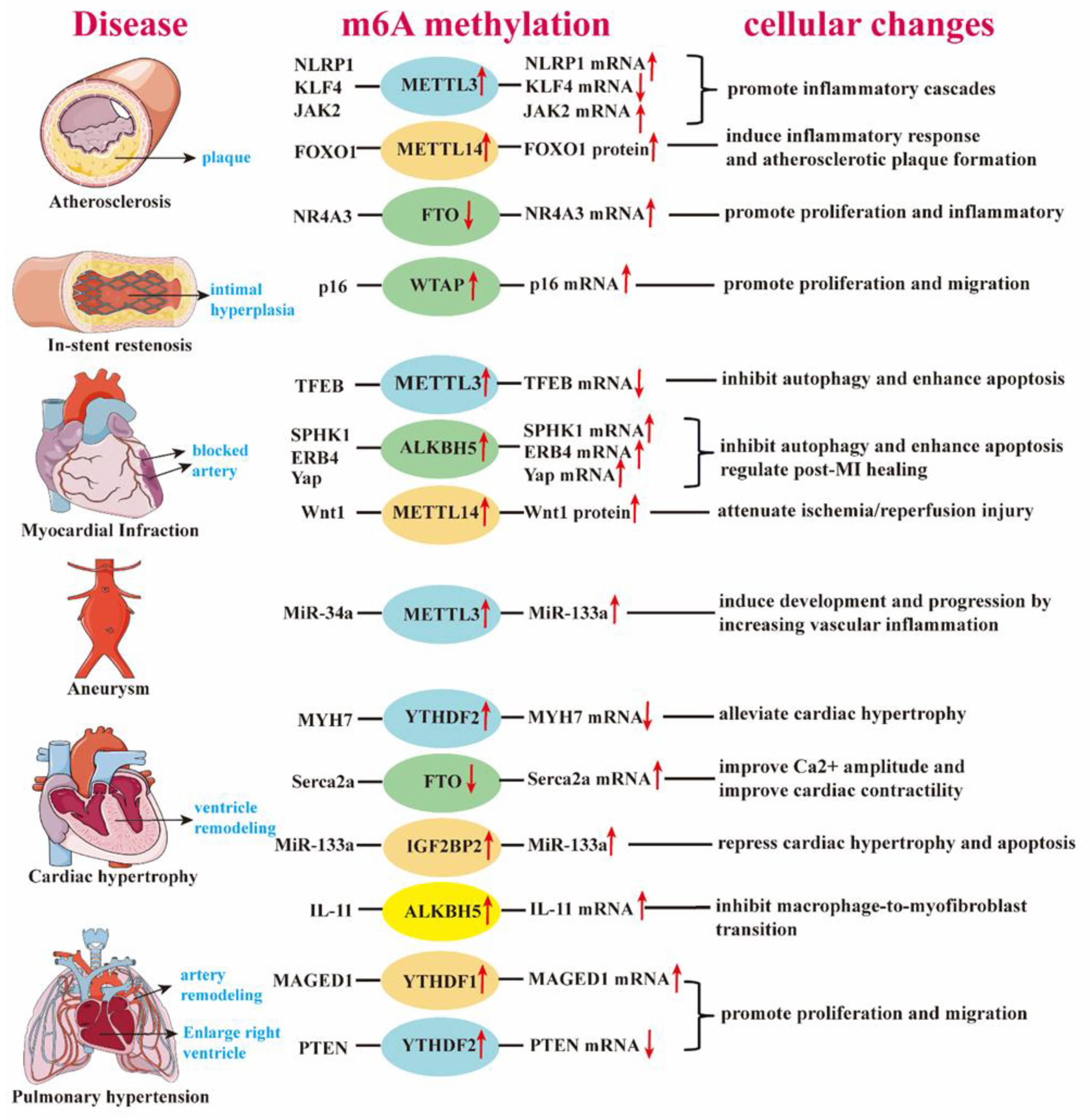
3.2.2. m6A and Atherosclerosis

3.2.2.1. m6A in Vascular Smooth Muscle Cell (VSMC) Differentiation and Angiogenesis

VSMC differentiation is a common phenotype observed in various cardiac vascular diseases, particularly in atherosclerosis[72]. One study investigated the promotion of differentiation of adipose-derived stem cells (ADSC) into VSMCs under hypoxic stress. The expression pattern of Mettl3 in their experiment was consistent with that of VSMC-specific markers, such as α-SMA, SM22α, and calponin, suggesting an important role of METTL3 in VSMC differentiation and potential changes in VSMCs in CVDs[73]. Additionally, circYTHDC2, a non-coding endogenous RNA, has been shown to promote the dedifferentiation of VSMCs into a "synthetic type" through the regulation of TET2 expression. The m6A modification mediated by YTHDC2 stabilizes circYTHDC2, highlighting the potential of the YTHDC2/circYTHDC2/TET2 pathway as an important target to inhibit atherosclerosis through dedifferentiation methods[74].
Apart from its role in differentiation, m6A can also mediate the process of angiogenesis. Parial et al. observed that METTL3 activates the phosphorylation of PHLPP2-mTOR-AKT signaling to enhance angiogenesis[75]. Furthermore, METTL3 enhances the translation of LRP6 and DVL1 in a YTHDF1-dependent manner, thereby regulating the Wnt signaling pathway to exert its angiogenic role[76]. These studies suggest that targeting m6A modification holds promise as a strategy for the treatment of angiogenic diseases.

3.2.2.2. m6A and Calcification

Calcification is frequently observed in atherosclerosis and valvular diseases, often leading to plaque rupture, thrombosis, valve sclerosis, and valve insufficiency, which can result in serious complications like heart failure. Zhou et al. reported that METTL3 is highly expressed in human calcified aortic valves compared to normal valves. Mechanistically, METTL3 promotes osteogenic differentiation of human aortic valve interstitial cells by suppressing TWIST1 expression in an m6A-YTHDF2-dependent manner[44]. Additionally, Chen et al. demonstrated that decreasing METTL14 expression in calcified arteries attenuates indoxyl sulfate-induced m6A modification and reduces human artery smooth muscle cell (HASMC) calcification. Knockdown of METTL14 may enhance vascular repair function by reducing calcification, presenting a potential therapeutic approach for atherosclerosis and valvular diseases[45].

3.2.2.3. m6A in Atherosclerosis

Atherosclerosis, the leading cause of CVD, has a high mortality rate among the population. The involvement of inflammation in the pathogenesis of atherosclerosis and its complications has gained considerable attention[77]. Chien et al. discovered that oscillatory stress (OS) upregulates METTL3 expression, leading to increased NF-κB p65 Ser536 phosphorylation and enhanced monocyte adhesion, thus promoting atherosclerosis. Mechanistically, METTL3-mediated hypermethylation stabilizes NLRP1 mRNA while inducing the degradation of KLF4 mRNA under OS. The m6A hypermethylation is recognized by YTHDF1 and YTHDF2 reader proteins, respectively[52]. Another study investigating the effect of m6A methylation on endothelial cells showed that METTL3 is highly expressed in oxidized low-density lipoprotein (ox-LDL)-induced dysregulated human umbilical vein endothelial cells (HUVECs). METTL3 knockdown prevents the progression of atherosclerosis by inhibiting the JAK2/STAT3 pathway via IGF2BP1[54]. These findings provide a basis for targeting METTL3 molecules and their downstream pathways to delay the progression of atherosclerosis.
Inflammation increases blood monocyte adhesion and migration into the subendothelial space, which is a critical event in the development of atherosclerosis[78]. METTL14, induced by TNF-α, promotes FOXO1 expression, leading to increased transcription of VCAM-1 and ICAM-1 by enhancing m6A modification and inducing endothelial cell inflammatory response and atherosclerotic plaque formation[53]. Moreover, METTL14 promotes DGCR8-mediated processing of pri-miR-19a, leading to the formation of mature miR-19a in atherosclerotic vascular endothelial cells (ASVEC), thereby promoting the proliferation and invasion of ASVEC[55]. Therefore, METTL14 plays a major role in endothelial cell activation and represents a novel therapeutic target.
The role of VSMCs in atherosclerosis has been extensively studied, highlighting their capability to switch to transitional, multipotential cells adopting various cellular states, including inflammation, ossification, and collagen matrix deposition[79]. Recently, it has been shown that m6A modification is involved in the regulation of VSMC behavior. For instance, dihydroartemisinin alleviates angiotensin II-induced VSMC proliferation and inflammatory response by blocking the FTO/NR4A3 axis[56]. TPNS inhibits VSMC proliferation, migration, and intimal hyperplasia by regulating the WTAP/p16 signaling pathway[57].

3.2.3. m6A and Acute Myocardial Infarction

Coronary atherosclerosis is a chronic disease characterized by stable and unstable periods. During unstable periods, when inflammation is activated in the vascular wall, patients may experience myocardial infarction (MI). Inflammation plays a fundamental role in atherogenesis and the pathophysiology of ischemic events[80].
M1 macrophages exhibit robust antimicrobial and antitumoral activity, but they also mediate tissue damage induced by reactive oxygen species (ROS), impairing tissue regeneration and wound healing[81]. Liu et al. suggested that METTL3 directly methylates STAT1, the master transcription factor controlling M1 macrophage polarization[82]. The inflammatory pathways mediated by METTL3 are not limited to a single mechanism. In Wang's study, Mettl3-mediated m6A modification of CD40, CD80, and TLR4 signaling adaptor Tirap transcripts enhanced their translation in dendritic cells, strengthening TLR4/NF-κB signaling-induced cytokine production[83]. Therefore, METTL3 can serve as an anti-inflammatory target during myocardial infarction to reduce tissue damage. Regarding tissue recovery after an ischemic event, METTL3 knockdown may be a feasible approach. Loss of METTL3/m6A impairs the maturation of pri-miR-143, resulting in low expression of miR-143-3p, which in turn leads to high expression of Yap and Ctnnd1. This promotes cardiomyocyte proliferation and endogenous heart regeneration after MI[58].
We must not overlook tissue damage caused by ischemia-reperfusion (I/R) injury, which is a significant cause of death in MI. Such damage occurs when blood supply is restored, and excessive free radicals attack the tissues. Chen's study showed a positive correlation between the expression of ALKBH5 in the infarct area and the levels of CK-MB and LDH in peripheral blood. Additionally, ALKBH5 was found to regulate the content of amino acids involved in the tricarboxylic acid (TCA) cycle and mediate the activity of key enzymes, thereby affecting cell metabolism and survival[59] . Moreover, ALKBH5 has been shown to activate the EGFR-PI3K-AKT-mTOR signaling pathway, enhancing the stability of BCL-2 mRNA and promoting the interaction between Bcl-2 and Beclin1, thereby inhibiting autophagy[84]. Kun Yang et al. demonstrated that ALKBH5 promotes the stability of ErbB4 mRNA and the degradation of ST14 mRNA through m6A demethylation, leading to improved fibroblast-to-myofibroblast transformation and better post-MI repair[60]. Considering the role of ALKBH5 in MI, inhibiting ALKBH5 could be a potential method to improve heart dysfunction after MI.
m6A is also involved in tissue repair after ischemic events. Reduced expression of METTL14 in human artery smooth muscle cells (HASMCs) decreases calcification and enhances vascular repair function[45]. However, Pang et al. found that Mettl14 attenuates cardiac ischemia-reperfusion injury by increasing m6A modification of Wnt1 mRNA, leading to upregulation of Wnt1 protein and subsequent activation of the Wnt1/β-catenin signaling pathway[61]. In fact, N6-methyladenosine and the Wnt protein family have close interactions. In a recent study in 2021, ALKBH5 was found to decrease the stability of WNT5A, thereby impeding angiogenesis in hypoxic cardiac microvascular endothelial cells[49].

3.2.4. m6A and Heart Failure (HF)

Heart failure occurs as a consequence of abnormalities in cardiac structure, function, rhythm, or conduction[85]. Despite improvements in survival rates, the absolute mortality rates for HF remain around 50% within 5 years of diagnosis[86]. Researchers have begun exploring the potential of m6A-mediated approaches for improving heart failure. Zhang et al. found that the expression of METTL3, METTL4, KIAA1429, FTO, and YTHDF2 was significantly upregulated in patients with heart failure with preserved ejection fraction (HFpEF) compared to healthy controls, suggesting that m6A methylation may be a therapeutic target for HFpEF interventions[87].
In addition to restoring contractile protein expression, such as SERCA2, which can alleviate cardiac insufficiency, Mathiyalagan et al. provided experimental evidence that FTO overexpression decreased cardiac fibrosis and enhanced angiogenesis in the ischemic myocardium[62]. This conclusion aligns with the results of Berulova et al., who analyzed m6A RNA methylation through next-generation sequencing and found alterations in the m6A landscape in heart hypertrophy and heart failure[88]. Furthermore, they demonstrated that cardiomyocyte-specific FTO knockdown mice exhibited impaired cardiac function compared to control mice. Hence, the role of FTO is closely associated with recovering cardiac function. In a recent 2024 study, macrophage-specific knockout of ALKBH5 inhibited angiotensin II-induced macrophage-to-myofibroblast transition, subsequently ameliorating cardiac fibrosis and dysfunction. This mechanism involves the targeting of interleukin-11 (IL-11), leading to increased IL-11 mRNA stability and protein levels[65].
Contrary to demethylation, although METTL3 is positively related to angiogenesis[75,76], it serves as a negative regulator in HF. Hinger et al. found increased levels of m6A and METTL3 in human non-ischemic failing hearts[89]. In a recent study, silencing METTL3 reduced m6A modification levels in fibrosis-related genes and decreased myocardial fibrosis in mice with myocardial infarction. This was achieved by inhibiting proliferation, fibroblast-to-myofibroblast transition, and collagen accumulation[90]. Maslinic acid has been demonstrated to protect against pressure-overload-induced cardiac hypertrophy by blocking METTL3-mediated m6A methylation[91]. A study by Dorn et al. identified enhanced METTL3-mediated methylation of mRNA on N6-adenosines in response to hypertrophic stimuli, driving cardiomyocyte hypertrophy in vitro and in vivo[92]. However, Kmietczyk's experiments with Mettl3-overexpressing mice and control mice subjected to transverse aortic constriction surgery to induce pathological hypertrophy led to a contrasting finding – pathological hypertrophic cellular growth was attenuated in hearts of Mettl3-overexpressing mice[93]. Kmietczyk explained this contradiction by suggesting that Dorn et al. used a transgenic model in the FVB background, while the in vivo studies utilized a C57Bl6/N background, and various methods were employed to overexpress METTL3.
In Xu et al.'s study, YTHDF2 was also found to increase in human HF samples. Moreover, YTHDF2 protein levels increased in cell and animal models of cardiac hypertrophy stimulated by isoproterenol or phenylephrine. Mechanistically, YTHDF2 recognizes the m6A site on Myh7 (beta-myosin heavy chain) mRNA, promoting its degradation and thereby alleviating cardiac hypertrophy[63]. MiR-133a represses cardiac hypertrophy and hypoxia-induced apoptosis[94,95]. Qian et al. found that IGF2BP2 binding to the m6A-modified site could enhance the accumulation of the miR-133a-AGO2-RISC complex on its targets, augmenting the repression effect of miR-133a and ultimately inhibiting cardiac hypertrophy[64].

3.2.5. m6a and other CVDs

FTO gene is strongly expressed in the hypothalamus, a brain structure involved in blood pressure regulation. Marcadenti et al. demonstrated that common genetic variants of FTO rs9939609 are negatively associated with diastolic and mean blood pressure in men with hypertension[96]. Through large-scale genome-wide association studies, Mo et al. recently revealed the crucial role of m6A in blood pressure regulation. They identified 1,236 m6A-SNPs, such as rs9847953 and rs197922, that are potentially associated with blood pressure levels[97]. However, the underlying mechanism of this association still needs further exploration.
In a study on hypoxia-mediated pulmonary hypertension (HPH), researchers identified the transcriptome-wide map of m6A circRNAs and found a reduction in m6A levels in circRNAs in lungs exposed to hypoxia. Furthermore, m6A influenced the circRNA-miRNA-mRNA co-expression network in hypoxia, involving circXpo6 and circTmtc3, indicating the important role of m6A in HPH[98]. In a recent study using a rat model exposed to hypoxic conditions, increased YTHDF2 recognition of METTL3-mediated m6A modification in PTEN mRNA promoted PTEN degradation via PI3K/Akt signaling, leading to excessive proliferation of pulmonary artery smooth muscle cells (PASMCs)[66]. Additionally, YTHDF1 was found to promote pulmonary hypertension by increasing the translational efficiency of MAGED1, which is involved in the hypoxia-induced proliferation of PASMCs through upregulating PCNA[67]. These pieces of evidence suggest that downregulating the level of N6-methyladenosine could be useful in the treatment of HPH.
In the context of aneurysms, similar to how METTL14 promotes DGCR8-mediated processing of pri-miR-19a in atherosclerosis[69], METTL3-dependent m6A methylation promotes primary miR-34a maturation through DGCR8, leading to decreased SIRT1 expression and an aggravated aneurysm formation[68]. This pathway represents a novel therapeutic target and diagnostic biomarker for aneurysm treatment. Vascular smooth muscle FTO promotes aortic dissecting aneurysms via m6A modification of Klf5, enhancing the GSK3β signaling pathway[99]. Moreover, Tan Li et al.'s genome-wide approaches revealed that aneurysm-associated m6A-SNPs might be linked to aneurysm pathogenesis by influencing local gene expression through m6A modification. They specifically identified two m6A-SNPs, NECTIN2 rs6859 and HPCAL1 rs10198139[100].

4. Discussion

This review provides a comprehensive summary of the pathophysiological effects of m6A methylation in cardiovascular risk factors, ischemia/hypoxia injury, atherosclerosis, myocardial infarction (MI), heart failure, and other cardiovascular diseases (Table 2 and Table 3). We have also explored potential mechanisms of m6A methylation, including glucose metabolism, adipogenesis, VSMC differentiation and angiogenesis, autophagy, macrophage response, and inflammation, in the regulation of cardiovascular disease development.
However, m6A research is still in its early stages, and many unanswered questions remain. Currently, research on m6A methylation detection in cardiovascular diseases primarily focuses on the expression of METTL3 and FTO. Limited attention has been given to the effects of m6A on cardiovascular system through LncRNA, circRNA, and miRNA. This could be attributed to the relatively lower expression of methylated regulatory enzymes in the cardiovascular system compared to other systems (Figure 3). Future research should aim to investigate how other methylases and demethylases regulate the expression of downstream proteins under various pathological factors. Additionally, the role and mechanism of m6A binding proteins in mediating the activity of m6A methylation in cardiovascular diseases should be explored.
Furthermore, CVDs are complex and multifactorial, involving multiple mechanisms. It is important to explore the synergistic effects of multiple methylated mRNA pathways on pathological progression. Dorn and Kmietczyk's conflicting conclusions regarding the inhibitory effect of METTL3 on cardiomyocyte hypertrophy suggest that changes in methylation patterns at different stages of disease occurrence and development may lead to distinct pathological processes. This highlights the need for further research on m6A-mediated methods for disease control, prevention, and treatment. Additionally, the effective clinical translation of m6A methylation findings is a topic that warrants comprehensive investigation[92,93].

Funding

The study was supported by the National Natural Science Foundation of China (NSFC 81970307, NSFC 82100357, NSFC 82200385 and NSFC 82270344), and was jointly supported by Jiangsu Provincial Medical Innovation Center (CXZX202215), Jiangsu Natural Science Foundation (BK20221173), and Nanjing Health Committee (JQX22007, and ZKX23030).

References

  1. Kottakis, F.; Nicolay, B.N.; Roumane, A.; Karnik, R.; Gu, H.; Nagle, J.M.; Boukhali, M.; Hayward, M.C.; Li, Y.Y.; Chen, T.; et al. LKB1 loss links serine metabolism to DNA methylation and tumorigenesis. Nature 2016, 539, 390–395. [Google Scholar] [CrossRef] [PubMed]
  2. Zaccara, S.; Ries, R.J.; Jaffrey, S.R. Reading, writing and erasing mRNA methylation. Nat Rev Mol Cell Biol 2019, 20, 608–624. [Google Scholar] [CrossRef]
  3. Wang, C.; Hou, X.; Guan, Q.; Zhou, H.; Zhou, L.; Liu, L.; Liu, J.; Li, F.; Li, W.; Liu, H. RNA modification in cardiovascular disease: implications for therapeutic interventions. Signal Transduct Target Ther 2023, 8, 412. [Google Scholar] [CrossRef] [PubMed]
  4. Motorin, Y.; Helm, M. RNA nucleotide methylation. Wiley Interdiscip Rev RNA 2011, 2, 611–631. [Google Scholar] [CrossRef] [PubMed]
  5. Desrosiers, R.; Friderici, K.; Rottman, F. Identification of methylated nucleosides in messenger RNA from Novikoff hepatoma cells. Proc Natl Acad Sci U S A 1974, 71, 3971–3975. [Google Scholar] [CrossRef]
  6. Davalos, V.; Blanco, S.; Esteller, M. SnapShot: Messenger RNA Modifications. Cell 2018, 174, 498–498 e491. [Google Scholar] [CrossRef]
  7. Dominissini, D.; Moshitch-Moshkovitz, S.; Schwartz, S.; Salmon-Divon, M.; Ungar, L.; Osenberg, S.; Cesarkas, K.; Jacob-Hirsch, J.; Amariglio, N.; Kupiec, M.; et al. Topology of the human and mouse m6A RNA methylomes revealed by m6A-seq. Nature 2012, 485, 201–206. [Google Scholar] [CrossRef]
  8. Han, J.; Wang, J.Z.; Yang, X.; Yu, H.; Zhou, R.; Lu, H.C.; Yuan, W.B.; Lu, J.C.; Zhou, Z.J.; Lu, Q.; et al. METTL3 promote tumor proliferation of bladder cancer by accelerating pri-miR221/222 maturation in m6A-dependent manner. Mol Cancer 2019, 18, 110. [Google Scholar] [CrossRef] [PubMed]
  9. Zhou, C.; Molinie, B.; Daneshvar, K.; Pondick, J.V.; Wang, J.; Van Wittenberghe, N.; Xing, Y.; Giallourakis, C.C.; Mullen, A.C. Genome-Wide Maps of m6A circRNAs Identify Widespread and Cell-Type-Specific Methylation Patterns that Are Distinct from mRNAs. Cell Rep 2017, 20, 2262–2276. [Google Scholar] [CrossRef]
  10. Yang, D.; Qiao, J.; Wang, G.; Lan, Y.; Li, G.; Guo, X.; Xi, J.; Ye, D.; Zhu, S.; Chen, W.; et al. N6-Methyladenosine modification of lincRNA 1281 is critically required for mESC differentiation potential. Nucleic Acids Res 2018, 46, 3906–3920. [Google Scholar] [CrossRef] [PubMed]
  11. Noale, M.; Limongi, F.; Maggi, S. Epidemiology of Cardiovascular Diseases in the Elderly. Adv Exp Med Biol 2020, 1216, 29–38. [Google Scholar] [CrossRef] [PubMed]
  12. Meyer, K.D.; Saletore, Y.; Zumbo, P.; Elemento, O.; Mason, C.E.; Jaffrey, S.R. Comprehensive analysis of mRNA methylation reveals enrichment in 3' UTRs and near stop codons. Cell 2012, 149, 1635–1646. [Google Scholar] [CrossRef] [PubMed]
  13. Yue, Y.; Liu, J.; He, C. RNA N6-methyladenosine methylation in post-transcriptional gene expression regulation. Genes Dev 2015, 29, 1343–1355. [Google Scholar] [CrossRef] [PubMed]
  14. Meyer, K.D.; Jaffrey, S.R. Rethinking m(6)A Readers, Writers, and Erasers. Annu Rev Cell Dev Biol 2017, 33, 319–342. [Google Scholar] [CrossRef]
  15. Yun, S.; He, X.; Zhang, W.; Chu, D.; Feng, C. Alleviation Effect of Grape Seed Proanthocyanidins on Neuronal Apoptosis in Rats with Iron Overload. Biol Trace Elem Res 2020, 194, 210–220. [Google Scholar] [CrossRef] [PubMed]
  16. Liu, J.; Yue, Y.; Han, D.; Wang, X.; Fu, Y.; Zhang, L.; Jia, G.; Yu, M.; Lu, Z.; Deng, X.; et al. A METTL3-METTL14 complex mediates mammalian nuclear RNA N6-adenosine methylation. Nat Chem Biol 2014, 10, 93–95. [Google Scholar] [CrossRef] [PubMed]
  17. Ping, X.L.; Sun, B.F.; Wang, L.; Xiao, W.; Yang, X.; Wang, W.J.; Adhikari, S.; Shi, Y.; Lv, Y.; Chen, Y.S.; et al. Mammalian WTAP is a regulatory subunit of the RNA N6-methyladenosine methyltransferase. Cell Res 2014, 24, 177–189. [Google Scholar] [CrossRef] [PubMed]
  18. Schwartz, S.; Mumbach, M.R.; Jovanovic, M.; Wang, T.; Maciag, K.; Bushkin, G.G.; Mertins, P.; Ter-Ovanesyan, D.; Habib, N.; Cacchiarelli, D.; et al. Perturbation of m6A writers reveals two distinct classes of mRNA methylation at internal and 5' sites. Cell Rep 2014, 8, 284–296. [Google Scholar] [CrossRef] [PubMed]
  19. Patil, D.P.; Chen, C.K.; Pickering, B.F.; Chow, A.; Jackson, C.; Guttman, M.; Jaffrey, S.R. m(6)A RNA methylation promotes XIST-mediated transcriptional repression. Nature 2016, 537, 369–373. [Google Scholar] [CrossRef]
  20. Wen, J.; Lv, R.; Ma, H.; Shen, H.; He, C.; Wang, J.; Jiao, F.; Liu, H.; Yang, P.; Tan, L.; et al. Zc3h13 Regulates Nuclear RNA m(6)A Methylation and Mouse Embryonic Stem Cell Self-Renewal. Mol Cell 2018, 69, 1028–1038 e1026. [Google Scholar] [CrossRef]
  21. Yue, Y.; Liu, J.; Cui, X.; Cao, J.; Luo, G.; Zhang, Z.; Cheng, T.; Gao, M.; Shu, X.; Ma, H.; et al. VIRMA mediates preferential m(6)A mRNA methylation in 3'UTR and near stop codon and associates with alternative polyadenylation. Cell Discov 2018, 4, 10. [Google Scholar] [CrossRef]
  22. Jia, G.; Fu, Y.; Zhao, X.; Dai, Q.; Zheng, G.; Yang, Y.; Yi, C.; Lindahl, T.; Pan, T.; Yang, Y.G.; He, C. N6-methyladenosine in nuclear RNA is a major substrate of the obesity-associated FTO. Nat Chem Biol 2011, 7, 885–887. [Google Scholar] [CrossRef] [PubMed]
  23. Zheng, G.; Dahl, J.A.; Niu, Y.; Fedorcsak, P.; Huang, C.M.; Li, C.J.; Vagbo, C.B.; Shi, Y.; Wang, W.L.; Song, S.H.; et al. ALKBH5 is a mammalian RNA demethylase that impacts RNA metabolism and mouse fertility. Mol Cell 2013, 49, 18–29. [Google Scholar] [CrossRef]
  24. Wang, X.; Zhao, B.S.; Roundtree, I.A.; Lu, Z.; Han, D.; Ma, H.; Weng, X.; Chen, K.; Shi, H.; He, C. N(6)-methyladenosine Modulates Messenger RNA Translation Efficiency. Cell 2015, 161, 1388–1399. [Google Scholar] [CrossRef]
  25. Shi, H.; Wang, X.; Lu, Z.; Zhao, B.S.; Ma, H.; Hsu, P.J.; Liu, C.; He, C. YTHDF3 facilitates translation and decay of N(6)-methyladenosine-modified RNA. Cell Res 2017, 27, 315–328. [Google Scholar] [CrossRef]
  26. Wang, X.; Lu, Z.; Gomez, A.; Hon, G.C.; Yue, Y.; Han, D.; Fu, Y.; Parisien, M.; Dai, Q.; Jia, G.; et al. N6-methyladenosine-dependent regulation of messenger RNA stability. Nature 2014, 505, 117–120. [Google Scholar] [CrossRef] [PubMed]
  27. Roundtree, I.A.; Luo, G.Z.; Zhang, Z.; Wang, X.; Zhou, T.; Cui, Y.; Sha, J.; Huang, X.; Guerrero, L.; Xie, P.; et al. YTHDC1 mediates nuclear export of N(6)-methyladenosine methylated mRNAs. Elife 2017, 6. [Google Scholar] [CrossRef] [PubMed]
  28. Hsu, P.J.; Zhu, Y.; Ma, H.; Guo, Y.; Shi, X.; Liu, Y.; Qi, M.; Lu, Z.; Shi, H.; Wang, J.; et al. Ythdc2 is an N(6)-methyladenosine binding protein that regulates mammalian spermatogenesis. Cell Res 2017, 27, 1115–1127. [Google Scholar] [CrossRef]
  29. Degrauwe, N.; Suva, M.L.; Janiszewska, M.; Riggi, N.; Stamenkovic, I. IMPs: an RNA-binding protein family that provides a link between stem cell maintenance in normal development and cancer. Genes Dev 2016, 30, 2459–2474. [Google Scholar] [CrossRef] [PubMed]
  30. Wu, B.; Su, S.; Patil, D.P.; Liu, H.; Gan, J.; Jaffrey, S.R.; Ma, J. Molecular basis for the specific and multivariant recognitions of RNA substrates by human hnRNP A2/B1. Nat Commun 2018, 9, 420. [Google Scholar] [CrossRef]
  31. Tang, Y.; Chen, K.; Song, B.; Ma, J.; Wu, X.; Xu, Q.; Wei, Z.; Su, J.; Liu, G.; Rong, R.; et al. m6A-Atlas: a comprehensive knowledgebase for unraveling the N6-methyladenosine (m6A) epitranscriptome. Nucleic Acids Res 2021, 49, D134–D143. [Google Scholar] [CrossRef] [PubMed]
  32. Li, W.; Gao, Y.; Jin, X.; Wang, H.; Lan, T.; Wei, M.; Yan, W.; Wang, G.; Li, Z.; Zhao, Z.; Jiang, X. Comprehensive analysis of N6-methylandenosine regulators and m6A-related RNAs as prognosis factors in colorectal cancer. Mol Ther Nucleic Acids 2022, 27, 598–610. [Google Scholar] [CrossRef]
  33. Wang, Y.; Li, Y.; Toth, J.I.; Petroski, M.D.; Zhang, Z.; Zhao, J.C. N6-methyladenosine modification destabilizes developmental regulators in embryonic stem cells. Nat Cell Biol 2014, 16, 191–198. [Google Scholar] [CrossRef] [PubMed]
  34. Xiao, W.; Adhikari, S.; Dahal, U.; Chen, Y.S.; Hao, Y.J.; Sun, B.F.; Sun, H.Y.; Li, A.; Ping, X.L.; Lai, W.Y.; et al. Nuclear m(6)A Reader YTHDC1 Regulates mRNA Splicing. Mol Cell 2016, 61, 507–519. [Google Scholar] [CrossRef] [PubMed]
  35. Yang, Y.; Shen, F.; Huang, W.; Qin, S.; Huang, J.T.; Sergi, C.; Yuan, B.F.; Liu, S.M. Glucose Is Involved in the Dynamic Regulation of m6A in Patients With Type 2 Diabetes. J Clin Endocrinol Metab 2019, 104, 665–673. [Google Scholar] [CrossRef] [PubMed]
  36. De Jesus, D.F.; Zhang, Z.; Kahraman, S.; Brown, N.K.; Chen, M.; Hu, J.; Gupta, M.K.; He, C.; Kulkarni, R.N. m(6)A mRNA Methylation Regulates Human beta-Cell Biology in Physiological States and in Type 2 Diabetes. Nat Metab 2019, 1, 765–774. [Google Scholar] [CrossRef]
  37. Kruger, N.; Biwer, L.A.; Good, M.E.; Ruddiman, C.A.; Wolpe, A.G.; DeLalio, L.J.; Murphy, S.; Macal, E.H., Jr.; Ragolia, L.; Serbulea, V.; et al. Loss of Endothelial FTO Antagonizes Obesity-Induced Metabolic and Vascular Dysfunction. Circ Res 2020, 126, 232–242. [Google Scholar] [CrossRef]
  38. Wu, R.; Guo, G.; Bi, Z.; Liu, Y.; Zhao, Y.; Chen, N.; Wang, F.; Wang, Y.; Wang, X. m(6)A methylation modulates adipogenesis through JAK2-STAT3-C/EBPbeta signaling. Biochim Biophys Acta Gene Regul Mech 2019, 1862, 796–806. [Google Scholar] [CrossRef] [PubMed]
  39. Zhao, X.; Yang, Y.; Sun, B.F.; Shi, Y.; Yang, X.; Xiao, W.; Hao, Y.J.; Ping, X.L.; Chen, Y.S.; Wang, W.J.; et al. FTO-dependent demethylation of N6-methyladenosine regulates mRNA splicing and is required for adipogenesis. Cell Res 2014, 24, 1403–1419. [Google Scholar] [CrossRef]
  40. Wang, X.; Wu, R.; Liu, Y.; Zhao, Y.; Bi, Z.; Yao, Y.; Liu, Q.; Shi, H.; Wang, F.; Wang, Y. m(6)A mRNA methylation controls autophagy and adipogenesis by targeting Atg5 and Atg7. Autophagy 2020, 16, 1221–1235. [Google Scholar] [CrossRef] [PubMed]
  41. Zhang, B.; Jiang, H.; Wu, J.; Cai, Y.; Dong, Z.; Zhao, Y.; Hu, Q.; Hu, K.; Sun, A.; Ge, J. m6A demethylase FTO attenuates cardiac dysfunction by regulating glucose uptake and glycolysis in mice with pressure overload-induced heart failure. Signal Transduct Target Ther 2021, 6, 377. [Google Scholar] [CrossRef] [PubMed]
  42. Birsoy, K.; Chen, Z.; Friedman, J. Transcriptional regulation of adipogenesis by KLF4. Cell Metab 2008, 7, 339–347. [Google Scholar] [CrossRef] [PubMed]
  43. Song, T.; Yang, Y.; Wei, H.; Xie, X.; Lu, J.; Zeng, Q.; Peng, J.; Zhou, Y.; Jiang, S.; Peng, J. Zfp217 mediates m6A mRNA methylation to orchestrate transcriptional and post-transcriptional regulation to promote adipogenic differentiation. Nucleic Acids Res 2019, 47, 6130–6144. [Google Scholar] [CrossRef] [PubMed]
  44. Zhou, T.; Han, D.; Liu, J.; Shi, J.; Zhu, P.; Wang, Y.; Dong, N. Factors influencing osteogenic differentiation of human aortic valve interstitial cells. J Thorac Cardiovasc Surg 2021, 161, e163–e185. [Google Scholar] [CrossRef] [PubMed]
  45. Chen, J.; Ning, Y.; Zhang, H.; Song, N.; Gu, Y.; Shi, Y.; Cai, J.; Ding, X.; Zhang, X. METTL14-dependent m6A regulates vascular calcification induced by indoxyl sulfate. Life Sci 2019, 239, 117034. [Google Scholar] [CrossRef]
  46. Song, H.; Feng, X.; Zhang, H.; Luo, Y.; Huang, J.; Lin, M.; Jin, J.; Ding, X.; Wu, S.; Huang, H.; et al. METTL3 and ALKBH5 oppositely regulate m(6)A modification of TFEB mRNA, which dictates the fate of hypoxia/reoxygenation-treated cardiomyocytes. Autophagy 2019, 15, 1419–1437. [Google Scholar] [CrossRef]
  47. Kumari, R.; Dutta, R.; Ranjan, P.; Suleiman, Z.G.; Goswami, S.K.; Li, J.; Pal, H.C.; Verma, S.K. ALKBH5 Regulates SPHK1-Dependent Endothelial Cell Angiogenesis Following Ischemic Stress. Front Cardiovasc Med 2021, 8, 817304. [Google Scholar] [CrossRef]
  48. Han, Z.; Wang, X.; Xu, Z.; Cao, Y.; Gong, R.; Yu, Y.; Yu, Y.; Guo, X.; Liu, S.; Yu, M.; et al. ALKBH5 regulates cardiomyocyte proliferation and heart regeneration by demethylating the mRNA of YTHDF1. Theranostics 2021, 11, 3000–3016. [Google Scholar] [CrossRef] [PubMed]
  49. Zhao, Y.; Hu, J.; Sun, X.; Yang, K.; Yang, L.; Kong, L.; Zhang, B.; Li, F.; Li, C.; Shi, B.; et al. Loss of m6A demethylase ALKBH5 promotes post-ischemic angiogenesis via post-transcriptional stabilization of WNT5A. Clin Transl Med 2021, 11, e402. [Google Scholar] [CrossRef]
  50. Shen, W.; Li, H.; Su, H.; Chen, K.; Yan, J. FTO overexpression inhibits apoptosis of hypoxia/reoxygenation-treated myocardial cells by regulating m6A modification of Mhrt. Mol Cell Biochem 2021, 476, 2171–2179. [Google Scholar] [CrossRef] [PubMed]
  51. Yin, T.; Wang, N.; Jia, F.; Wu, Y.; Gao, L.; Zhang, J.; Hou, R. Exosome-based WTAP siRNA delivery ameliorates myocardial ischemia-reperfusion injury. Eur J Pharm Biopharm 2024, 197, 114218. [Google Scholar] [CrossRef] [PubMed]
  52. Chien, C.S.; Li, J.Y.; Chien, Y.; Wang, M.L.; Yarmishyn, A.A.; Tsai, P.H.; Juan, C.C.; Nguyen, P.; Cheng, H.M.; Huo, T.I.; et al. METTL3-dependent N(6)-methyladenosine RNA modification mediates the atherogenic inflammatory cascades in vascular endothelium. Proc Natl Acad Sci U S A 2021, 118. [Google Scholar] [CrossRef]
  53. Jian, D.; Wang, Y.; Jian, L.; Tang, H.; Rao, L.; Chen, K.; Jia, Z.; Zhang, W.; Liu, Y.; Chen, X.; et al. METTL14 aggravates endothelial inflammation and atherosclerosis by increasing FOXO1 N6-methyladeosine modifications. Theranostics 2020, 10, 8939–8956. [Google Scholar] [CrossRef] [PubMed]
  54. Dong, G.; Yu, J.; Shan, G.; Su, L.; Yu, N.; Yang, S. N6-Methyladenosine Methyltransferase METTL3 Promotes Angiogenesis and Atherosclerosis by Upregulating the JAK2/STAT3 Pathway via m6A Reader IGF2BP1. Front Cell Dev Biol 2021, 9, 731810. [Google Scholar] [CrossRef]
  55. Zhang, B.Y.; Han, L.; Tang, Y.F.; Zhang, G.X.; Fan, X.L.; Zhang, J.J.; Xue, Q.; Xu, Z.Y. METTL14 regulates M6A methylation-modified primary miR-19a to promote cardiovascular endothelial cell proliferation and invasion. Eur Rev Med Pharmacol Sci 2020, 24, 7015–7023. [Google Scholar] [CrossRef] [PubMed]
  56. Huo, Y.B.; Gao, X.; Peng, Q.; Nie, Q.; Bi, W. Dihydroartemisinin alleviates AngII-induced vascular smooth muscle cell proliferation and inflammatory response by blocking the FTO/NR4A3 axis. Inflamm Res 2022, 71, 243–253. [Google Scholar] [CrossRef]
  57. Zhu, B.; Gong, Y.; Shen, L.; Li, J.; Han, J.; Song, B.; Hu, L.; Wang, Q.; Wang, Z. Total Panax notoginseng saponin inhibits vascular smooth muscle cell proliferation and migration and intimal hyperplasia by regulating WTAP/p16 signals via m(6)A modulation. Biomed Pharmacother 2020, 124, 109935. [Google Scholar] [CrossRef]
  58. Gong, R.; Wang, X.; Li, H.; Liu, S.; Jiang, Z.; Zhao, Y.; Yu, Y.; Han, Z.; Yu, Y.; Dong, C.; et al. Loss of m(6)A methyltransferase METTL3 promotes heart regeneration and repair after myocardial injury. Pharmacol Res 2021, 174, 105845. [Google Scholar] [CrossRef] [PubMed]
  59. Cheng, P.; Han, H.; Chen, F.; Cheng, L.; Ma, C.; Huang, H.; Chen, C.; Li, H.; Cai, H.; Huang, H.; et al. Amelioration of acute myocardial infarction injury through targeted ferritin nanocages loaded with an ALKBH5 inhibitor. Acta Biomater 2022, 140, 481–491. [Google Scholar] [CrossRef]
  60. Yang, K.; Zhao, Y.; Hu, J.; Gao, R.; Shi, J.; Wei, X.; Chen, J.; Hu, K.; Sun, A.; Ge, J. ALKBH5 induces fibroblast-to-myofibroblast transformation during hypoxia to protect against cardiac rupture after myocardial infarction. J Adv Res 2024, 61, 193–209. [Google Scholar] [CrossRef]
  61. Pang, P.; Qu, Z.; Yu, S.; Pang, X.; Li, X.; Gao, Y.; Liu, K.; Liu, Q.; Wang, X.; Bian, Y.; et al. Mettl14 Attenuates Cardiac Ischemia/Reperfusion Injury by Regulating Wnt1/beta-Catenin Signaling Pathway. Front Cell Dev Biol 2021, 9, 762853. [Google Scholar] [CrossRef] [PubMed]
  62. Mathiyalagan, P.; Adamiak, M.; Mayourian, J.; Sassi, Y.; Liang, Y.; Agarwal, N.; Jha, D.; Zhang, S.; Kohlbrenner, E.; Chepurko, E.; et al. FTO-Dependent N(6)-Methyladenosine Regulates Cardiac Function During Remodeling and Repair. Circulation 2019, 139, 518–532. [Google Scholar] [CrossRef]
  63. Xu, H.; Wang, Z.; Chen, M.; Zhao, W.; Tao, T.; Ma, L.; Ni, Y.; Li, W. YTHDF2 alleviates cardiac hypertrophy via regulating Myh7 mRNA decoy. Cell Biosci 2021, 11, 132. [Google Scholar] [CrossRef] [PubMed]
  64. Qian, B.; Wang, P.; Zhang, D.; Wu, L. m6A modification promotes miR-133a repression during cardiac development and hypertrophy via IGF2BP2. Cell Death Discov 2021, 7, 157. [Google Scholar] [CrossRef]
  65. Zhuang, T.; Chen, M.H.; Wu, R.X.; Wang, J.; Hu, X.D.; Meng, T.; Wu, A.H.; Li, Y.; Yang, Y.F.; Lei, Y.; et al. ALKBH5-mediated m6A modification of IL-11 drives macrophage-to-myofibroblast transition and pathological cardiac fibrosis in mice. Nat Commun 2024, 15, 1995. [Google Scholar] [CrossRef] [PubMed]
  66. Qin, Y.; Qiao, Y.; Li, L.; Luo, E.; Wang, D.; Yao, Y.; Tang, C.; Yan, G. The m(6)A methyltransferase METTL3 promotes hypoxic pulmonary arterial hypertension. Life Sci 2021, 274, 119366. [Google Scholar] [CrossRef]
  67. Hu, L.; Wang, J.; Huang, H.; Yu, Y.; Ding, J.; Yu, Y.; Li, K.; Wei, D.; Ye, Q.; Wang, F.; et al. YTHDF1 Regulates Pulmonary Hypertension through Translational Control of MAGED1. Am J Respir Crit Care Med 2021, 203, 1158–1172. [Google Scholar] [CrossRef] [PubMed]
  68. Zhong, L.; He, X.; Song, H.; Sun, Y.; Chen, G.; Si, X.; Sun, J.; Chen, X.; Liao, W.; Liao, Y.; Bin, J. METTL3 Induces AAA Development and Progression by Modulating N6-Methyladenosine-Dependent Primary miR34a Processing. Mol Ther Nucleic Acids 2020, 21, 394–411. [Google Scholar] [CrossRef]
  69. Wang, X.; Li, Y.; Li, J.; Li, S.; Wang, F. Mechanism of METTL3-Mediated m(6)A Modification in Cardiomyocyte Pyroptosis and Myocardial Ischemia-Reperfusion Injury. Cardiovasc Drugs Ther 2023, 37, 435–448. [Google Scholar] [CrossRef] [PubMed]
  70. Kroemer, G. Autophagy: a druggable process that is deregulated in aging and human disease. J Clin Invest 2015, 125, 1–4. [Google Scholar] [CrossRef]
  71. Jin, S.; Zhang, X.; Miao, Y.; Liang, P.; Zhu, K.; She, Y.; Wu, Y.; Liu, D.A.; Huang, J.; Ren, J.; Cui, J. m(6)A RNA modification controls autophagy through upregulating ULK1 protein abundance. Cell Res 2018, 28, 955–957. [Google Scholar] [CrossRef]
  72. Frismantiene, A.; Philippova, M.; Erne, P.; Resink, T.J. Smooth muscle cell-driven vascular diseases and molecular mechanisms of VSMC plasticity. Cell Signal 2018, 52, 48–64. [Google Scholar] [CrossRef] [PubMed]
  73. Lin, J.; Zhu, Q.; Huang, J.; Cai, R.; Kuang, Y. Hypoxia Promotes Vascular Smooth Muscle Cell (VSMC) Differentiation of Adipose-Derived Stem Cell (ADSC) by Regulating Mettl3 and Paracrine Factors. Stem Cells Int 2020, 2020, 2830565. [Google Scholar] [CrossRef] [PubMed]
  74. Yuan, J.; Liu, Y.; Zhou, L.; Xue, Y.; Lu, Z.; Gan, J. YTHDC2-Mediated circYTHDC2 N6-Methyladenosine Modification Promotes Vascular Smooth Muscle Cells Dysfunction Through Inhibiting Ten-Eleven Translocation 2. Front Cardiovasc Med 2021, 8, 686293. [Google Scholar] [CrossRef] [PubMed]
  75. Parial, R.; Li, H.; Li, J.; Archacki, S.; Yang, Z.; Wang, I.Z.; Chen, Q.; Xu, C.; Wang, Q.K. Role of epigenetic m(6) A RNA methylation in vascular development: mettl3 regulates vascular development through PHLPP2/mTOR-AKT signaling. FASEB J 2021, 35, e21465. [Google Scholar] [CrossRef]
  76. Yao, M.D.; Jiang, Q.; Ma, Y.; Liu, C.; Zhu, C.Y.; Sun, Y.N.; Shan, K.; Ge, H.M.; Zhang, Q.Y.; Zhang, H.Y.; et al. Role of METTL3-Dependent N(6)-Methyladenosine mRNA Modification in the Promotion of Angiogenesis. Mol Ther 2020, 28, 2191–2202. [Google Scholar] [CrossRef] [PubMed]
  77. Geovanini, G.R.; Libby, P. Atherosclerosis and inflammation: overview and updates. Clin Sci (Lond) 2018, 132, 1243–1252. [Google Scholar] [CrossRef]
  78. Chiu, J.J.; Chien, S. Effects of disturbed flow on vascular endothelium: pathophysiological basis and clinical perspectives. Physiol Rev 2011, 91, 327–387. [Google Scholar] [CrossRef]
  79. Miano, J.M.; Fisher, E.A.; Majesky, M.W. Fate and State of Vascular Smooth Muscle Cells in Atherosclerosis. Circulation 2021, 143, 2110–2116. [Google Scholar] [CrossRef] [PubMed]
  80. Libby, P. The changing landscape of atherosclerosis. Nature 2021, 592, 524–533. [Google Scholar] [CrossRef] [PubMed]
  81. Shapouri-Moghaddam, A.; Mohammadian, S.; Vazini, H.; Taghadosi, M.; Esmaeili, S.A.; Mardani, F.; Seifi, B.; Mohammadi, A.; Afshari, J.T.; Sahebkar, A. Macrophage plasticity, polarization, and function in health and disease. J Cell Physiol 2018, 233, 6425–6440. [Google Scholar] [CrossRef]
  82. Liu, Y.; Liu, Z.; Tang, H.; Shen, Y.; Gong, Z.; Xie, N.; Zhang, X.; Wang, W.; Kong, W.; Zhou, Y.; Fu, Y. The N(6)-methyladenosine (m(6)A)-forming enzyme METTL3 facilitates M1 macrophage polarization through the methylation of STAT1 mRNA. Am J Physiol Cell Physiol 2019, 317, C762–C775. [Google Scholar] [CrossRef] [PubMed]
  83. Wang, H.; Hu, X.; Huang, M.; Liu, J.; Gu, Y.; Ma, L.; Zhou, Q.; Cao, X. Mettl3-mediated mRNA m(6)A methylation promotes dendritic cell activation. Nat Commun 2019, 10, 1898. [Google Scholar] [CrossRef]
  84. Zhu, H.; Gan, X.; Jiang, X.; Diao, S.; Wu, H.; Hu, J. ALKBH5 inhibited autophagy of epithelial ovarian cancer through miR-7 and BCL-2. J Exp Clin Cancer Res 2019, 38, 163. [Google Scholar] [CrossRef]
  85. Writing Committee, M.; Yancy, C.W.; Jessup, M.; Bozkurt, B.; Butler, J.; Casey, D.E., Jr.; Drazner, M.H.; Fonarow, G.C.; Geraci, S.A.; Horwich, T.; et al. 2013 ACCF/AHA guideline for the management of heart failure: a report of the American College of Cardiology Foundation/American Heart Association Task Force on practice guidelines. Circulation 2013, 128, e240–327. [Google Scholar] [CrossRef]
  86. Roger, V.L.; Weston, S.A.; Redfield, M.M.; Hellermann-Homan, J.P.; Killian, J.; Yawn, B.P.; Jacobsen, S.J. Trends in heart failure incidence and survival in a community-based population. JAMA 2004, 292, 344–350. [Google Scholar] [CrossRef]
  87. Zhang, B.; Xu, Y.; Cui, X.; Jiang, H.; Luo, W.; Weng, X.; Wang, Y.; Zhao, Y.; Sun, A.; Ge, J. Alteration of m6A RNA Methylation in Heart Failure With Preserved Ejection Fraction. Front Cardiovasc Med 2021, 8, 647806. [Google Scholar] [CrossRef] [PubMed]
  88. Berulava, T.; Buchholz, E.; Elerdashvili, V.; Pena, T.; Islam, M.R.; Lbik, D.; Mohamed, B.A.; Renner, A.; von Lewinski, D.; Sacherer, M.; et al. Changes in m6A RNA methylation contribute to heart failure progression by modulating translation. Eur J Heart Fail 2020, 22, 54–66. [Google Scholar] [CrossRef] [PubMed]
  89. Hinger, S.A.; Wei, J.; Dorn, L.E.; Whitson, B.A.; Janssen, P.M.L.; He, C.; Accornero, F. Remodeling of the m(6)A landscape in the heart reveals few conserved post-transcriptional events underlying cardiomyocyte hypertrophy. J Mol Cell Cardiol 2021, 151, 46–55. [Google Scholar] [CrossRef]
  90. Li, T.; Zhuang, Y.; Yang, W.; Xie, Y.; Shang, W.; Su, S.; Dong, X.; Wu, J.; Jiang, W.; Zhou, Y.; et al. Silencing of METTL3 attenuates cardiac fibrosis induced by myocardial infarction via inhibiting the activation of cardiac fibroblasts. FASEB J 2021, 35, e21162. [Google Scholar] [CrossRef]
  91. Fang, M.; Deng, J.; Zhou, Q.; Hu, Z.; Yang, L. Maslinic acid protects against pressure-overload-induced cardiac hypertrophy by blocking METTL3-mediated m(6)A methylation. Aging (Albany NY) 2022, 14, 2548–2557. [Google Scholar] [CrossRef]
  92. Dorn, L.E.; Lasman, L.; Chen, J.; Xu, X.; Hund, T.J.; Medvedovic, M.; Hanna, J.H.; van Berlo, J.H.; Accornero, F. The N(6)-Methyladenosine mRNA Methylase METTL3 Controls Cardiac Homeostasis and Hypertrophy. Circulation 2019, 139, 533–545. [Google Scholar] [CrossRef]
  93. Kmietczyk, V.; Riechert, E.; Kalinski, L.; Boileau, E.; Malovrh, E.; Malone, B.; Gorska, A.; Hofmann, C.; Varma, E.; Jurgensen, L.; et al. m(6)A-mRNA methylation regulates cardiac gene expression and cellular growth. Life Sci Alliance 2019, 2. [Google Scholar] [CrossRef] [PubMed]
  94. Xu, C.; Hu, Y.; Hou, L.; Ju, J.; Li, X.; Du, N.; Guan, X.; Liu, Z.; Zhang, T.; Qin, W.; et al. beta-Blocker carvedilol protects cardiomyocytes against oxidative stress-induced apoptosis by up-regulating miR-133 expression. J Mol Cell Cardiol 2014, 75, 111–121. [Google Scholar] [CrossRef]
  95. Liu, N.; Bezprozvannaya, S.; Williams, A.H.; Qi, X.; Richardson, J.A.; Bassel-Duby, R.; Olson, E.N. microRNA-133a regulates cardiomyocyte proliferation and suppresses smooth muscle gene expression in the heart. Genes Dev 2008, 22, 3242–3254. [Google Scholar] [CrossRef] [PubMed]
  96. Marcadenti, A.; Fuchs, F.D.; Matte, U.; Sperb, F.; Moreira, L.B.; Fuchs, S.C. Effects of FTO RS9939906 and MC4R RS17782313 on obesity, type 2 diabetes mellitus and blood pressure in patients with hypertension. Cardiovasc Diabetol 2013, 12, 103. [Google Scholar] [CrossRef] [PubMed]
  97. Mo, X.B.; Lei, S.F.; Zhang, Y.H.; Zhang, H. Examination of the associations between m(6)A-associated single-nucleotide polymorphisms and blood pressure. Hypertens Res 2019, 42, 1582–1589. [Google Scholar] [CrossRef]
  98. Su, H.; Wang, G.; Wu, L.; Ma, X.; Ying, K.; Zhang, R. Transcriptome-wide map of m(6)A circRNAs identified in a rat model of hypoxia mediated pulmonary hypertension. BMC Genomics 2020, 21, 39. [Google Scholar] [CrossRef]
  99. Ma, D.; Liu, X.; Zhang, J.J.; Zhao, J.J.; Xiong, Y.J.; Chang, Q.; Wang, H.Y.; Su, P.; Meng, J.; Zhao, Y.B. Vascular Smooth Muscle FTO Promotes Aortic Dissecting Aneurysms via m6A Modification of Klf5. Front Cardiovasc Med 2020, 7, 592550. [Google Scholar] [CrossRef] [PubMed]
  100. Li, T.; Wu, Y.; Yang, J.; Jing, J.; Ma, C.; Sun, L. N6-methyladenosine-associated genetic variants in NECTIN2 and HPCAL1 are risk factors for abdominal aortic aneurysm. iScience 2024, 27, 109419. [Google Scholar] [CrossRef]
Figure 1. The Regulation of m6A Modification. m6A modification is established by m6A methyltransferases ("writers") and removed by m6A demethylases ("erasers"). m6A-binding proteins ("readers") recognize and bind to m6A-modified RNA, playing essential roles in RNA metabolism. METTL3, in collaboration with METTL14/16, KIAA1429, WTAP, RBM15/15B, and ZC3H13, forms the core methylation complex. This modification is reversible, with demethylases such as ALKBH5 and FTO serving as m6A erasers. The modified transcripts are recognized by readers, including YTHDF1/2/3, YTHDC1/2, IGF2BP1/2/3, and hnRNPG/C/A2B, which subsequently influence various aspects of RNA function such as translation promotion, stability, localization, splicing, and nuclear export.
Preprints 141671 g001
Figure 2. The Role of m6A Modulators in Cardiovascular Diseases and Biological Processes. METTL3-mediated m6A modification contributes to atherosclerosis by affecting inflammatory pathways, including NLRP1, KLF4, and JAK2. METTL14 upregulation induces inflammation and plaque formation by enhancing FOXO1 translation. Reduced FTO expression promotes smooth muscle cell proliferation and inflammation by stabilizing NR4A3 mRNA, accelerating atherosclerosis. In stent restenosis, WTAP promotes smooth muscle cell proliferation and migration by increasing P16 mRNA via m6A modification. In myocardial ischemic disease, METTL3 upregulation decreases TFEB expression, impairing autophagic flux and enhancing cell apoptosis. During myocardial infarction repair, ALKBH5 upregulation inhibits autophagy by destabilizing mRNAs like SPHK1, ERB4, and YAP, promoting infarct repair. METTL3 also mediates miR-34a maturation, which downregulates SIRT1 and promotes inflammatory infiltration in abdominal aortic aneurysm. Decreased FTO expression is linked to reduced Serca2a levels, leading to impaired cardiac contractility and heart failure. In heart failure, m6A-modified MYH7 mRNA and miR-133a play protective roles in ventricular remodeling. Additionally, ALKBH5 increases IL-11 mRNA, inhibiting macrophage-to-myofibroblast transition. In hypoxic pulmonary hypertension, upregulated MAGED1 and downregulated PTEN, both m6A-modified, contribute to smooth muscle cell proliferation, inflammation, and pulmonary vascular remodeling.
Preprints 141671 g002
Figure 3. Relative amounts of methylated regulatory enzymes in the cardiovascular system and other systems. Heatmap shows differentially expressed methylated regulatory enzymes in different tissues. Data derives from PRJEB4337 of HPA RNA-seq normal tissues, in which RNA-seq was performed of tissue samples from 95 human individuals representing 27 different tissues in order to determine tissue-specificity of all protein-coding genes.
Preprints 141671 g003
Table 1. writers ,erasers and readers in m6a.
type regulator Function of RNA modification reference
writers METTL3 main catalytic subunit of m6A [16]
METTL14/16 activate METTL3 through allosteric and RNA substrate recognition [16]
WTAP the third subunit of METTL3-METTL14 complex [17,18]
ZC3H3 assist the localization of the methyltransferase complex in nuclear speckles and U-rich regions adjacent to the m6A sites in mRNAs [19]
RBM15/15B [20]
KIAA1429 [21]
erasers FTO demethylation of m6a [2,22,23]
ALKBH5
readers YTHDF1 promote mRNA translation [24,25]
YTHDF2 accelerates the decay of m6A-modified transcripts
YTHDF3 promote mRNA translation or enhance RNA decay
YTHDC1 promote mRNA translation and splicing and nuclear export [26,27]
YTHDC2 enhance translation [28]
IGF2BP1/2/3 regulate RNA localization, translation, and stability [29]
hnRNPG/C/A2B Promote RNA stability and mediate RNA splicing and microRNA process [30]
Abbreviations: ALKBH5: ALKB homolog 5, RNA demethylase; FTO: FTO alpha-ketoglutarate dependent dioxygenase; KIAA1429: vir like m6A methyltransferase associated;METTL3:methyltransferase3;METTL14:methyltransferase14;METTL16:methyltransferase16; RBM15/15B: RNA binding motif protein 15B;WTAP: WT1associated protein; YTHDF1: YTH N6-methyladenosine RNA binding protein F1; YTHDF2: YTH N6-methyladenosine RNA binding protein F2; YTHDF3: YTH N6-methyladenosine RNA binding protein F3; YTHDC1: YTH N6-methyladenosine RNA binding protein C1; YTHDC2: YTH N6-methyladenosine RNA binding protein C2; IGF2BP1/2/3: insulin like growth factor 2 mRNA binding protein 1/2/3; hnRNPG/C/A2B: RNA binding motif protein X-linked G/C/A2B; ZC3H3: zinc finger CCCH-type containing3.
Table 2. molecular mechanisms of m6a in risk factors in CVDs.
Risk factors regulators Cell Regulation signaling function reference
Glucose metabolism FTO↑ hepatoellular cell up-regulate mRNA FOXO1/FASN/
G6PC/ DGAT2
improve the production of serum glucose and lipids [35]

diabetes
METTL14↓ β-cell promote mRNA translation AKT/ PDX1 induce cell-cycle arrest and impair insulin secretion [36]
obesity FTO↑ endothelial cell down-regulate mRNA AKT/
prostaglandinD2
aggravate vascular dysfunction [37]
FTO↑ preadipocyte up-regulate mRNA JAK2-STAT3-C/EBPβ promote adipogenesis [38]
FTO↑ preadipocyte control exonic splicing RUNX1T1 modulate differentiation to promote adipogenesis [39]
FTO↑ preadipocytes improve mRNA stability Atg5/Atg7 promote autophagy and adipogenesis [40]
Table 3. The roles of m6A methylation in CVDs.
Diseases regulators Cell Regulation signaling function reference
calcification METTL3↑ valve interstitial cell up-regulate mRNA TWIST1 promote osteogenic differentiation process [44]
calcification METTL14↑ smooth muscle cell down-regulate mRNA Klotho promote calcification [45]
hypoxia/reoxygenation METTL3↑ cardiomyocyte down-regulate mRNA TFEB inhibit autophagy and enhance apoptosis [46]
ischemic injury ALKBH5↑ endothelial cell up-regulate mRNA SPHK1/eNOS-AKT maintain angiogenesis [47]
heart regeneration ALKBH5↑ cardiomyocyte improve mRNA stability YAP promote proliferation [48]
post-ischemic ALKBH5↑ endothelial cell decrease mRNA stability WNT5A exacerbate dysfunction of CMECs [49]
hypoxia/reoxygenation FTO↑ cardiomyocyte up-regulate mRNA Mhrt inhibit apoptosis [50]
hypoxia/reoxygenation WTAP↑ cardiomyocyte up-regulate mRNA TXNIP enhance apoptosis [51]
atherosclerosis METTL3↑ endothelial cell up/down-regulate mRNA NLRP1/KLF4 promote inflammatory cascades [52]
atherosclerosis METTL14↑ endothelial cell promote translation FOXO1/VCAM-1/ICAM-1 induce inflammatory response and promote atherosclerotic plaque formation [53]
atherosclerosis METTL3↑ endothelial cell up-regulate mRNA JAK2/STAT3
promote atherosclerosis progression [54]
atherosclerosis METTL14↑ endothelial cell up-regulate miRNA pri-miR-19a
/DGCR8
promote proliferation and invasion of ASVEC [55]
atherosclerosis FTO↓ smooth muscle cell up-regulate mRNA NR4A3 promote proliferation and inflammatory [56]
intimal hyperplasia WTAP↓ smooth muscle cell up-regulate mRNA p16 promote proliferation and migration of VSMC [57]
heart regeneration METTL3↓ cardiomyocyte up-regulate miRNA miR-143
/Yap/Ctnnd1
Inhibit heart regeneration [58]
AML ALKBH5↑ cardiomyocyte not mention TCA cycle affect cell metabolism and survival [59]
AML ALKBH5↑ fibroblast improve mRNA stability ErbB4 regulate post-MI healing [60]
ischemia/reperfusion Injury METTL14↑ cardiomyocyte promote translation efficiency Wnt1 attenuate ischemia/reperfusion Injury [61]
Heart failure FTO↓ cardiomyocyte improve mRNA stability Serca2a improve Ca2+ amplitude [62]
Heart failure YTHDF2↑ cardiomyocyte promote mRNA
degradation
Myh7 alleviate cardiac hypertrophy [63]
Heart failure IGF2BP2↑ cardiomyocyte promote miRNA accumulation on its target site miR-133a repress cardiac hypertrophy and apoptosis [64]
Heart failure FTO↓ cardiomyocytes improve mRNA stability Pgam2 regulating glucose uptake [41]
Heart failure ALKBH5↑ macrophage improve mRNA stability IL-11 Inhibit macrophage-to-myofibroblast transition [65]
pulmonary hypertension YTHDF2↑ smooth muscle cell promote mRNA
degradation
PTEN /PI3K/Akt enhance proliferation [66]
pulmonary hypertension YTHDF1↑ smooth muscle cell promote translation efficiency MAGED1 promote proliferation [67]
aneurysm METTL3↑ smooth muscle cell up-regulate miRNA DGCR8/
miR-34a
induce development and progression [68]
Abbreviations: ALKBH5: ALKB homolog 5, RNA demethylase;;FTO: FTO alpha-ketoglutarate dependent dioxygenase; FOXO1: forkhead box O1; ICAM-1: intercellular adhesion molecule 1; JAK2: Janus kinase 2; METTL3: methyltransferase3; METTL14:methyltransferase14; Myh7: myosin heavy chain 7; MAGED1: MAGE family member D1; NLRP1: NLR family pyrin domain containing 1; KLF4: KLF transcription factor 4; NR4A3: nuclear receptor subfamily 4 group A member 3; SPHK1: sphingosine kinase 1; STAT3: signal transducer and activator of transcription 3; Serca2a: Sarco/endoplasmic reticulum Ca(2+)-ATPase; TFEB: transcription factor; WTAP: WT1 associated protein; Wnt1: Wnt family member 1; YAP: Yes associated protein; YTHDF1/2: YTH N6-methyladenosine RNA binding protein F1/2.
Disclaimer/Publisher’s Note: The statements, opinions and data contained in all publications are solely those of the individual author(s) and contributor(s) and not of MDPI and/or the editor(s). MDPI and/or the editor(s) disclaim responsibility for any injury to people or property resulting from any ideas, methods, instructions or products referred to in the content.
Copyright: This open access article is published under a Creative Commons CC BY 4.0 license, which permit the free download, distribution, and reuse, provided that the author and preprint are cited in any reuse.
Prerpints.org logo

Preprints.org is a free preprint server supported by MDPI in Basel, Switzerland.

Subscribe

Disclaimer

Terms of Use

Privacy Policy

Privacy Settings

© 2026 MDPI (Basel, Switzerland) unless otherwise stated