
Submitted:
27 November 2024
Posted:
28 November 2024
You are already at the latest version
Abstract
This review examines recent methodologies for fabricating nonwoven polymer materials through electrospinning, focusing on the underlying physical principles, including the effects of external parameters, experimental conditions, material selection, and primary operational mechanisms. Potential applications of electrospun polymer matrices in tissue engineering are analyzed, with particular emphasis on their utility in biomedical context. Key challenges in incorporating new materials into biomedical devices are discussed, along with recent advances in electrospinning techniques driving innovation in this field.
Keywords:
electrospinning
; biodegradable polymers
; nanofibers
; tissue engineering
; regenerative medicine
; polymer scaffold
1. Introduction
Electrospinning, a method for producing polymer nonwoven materials, was first developed in the early 20th century [1], but has only recently gained widespread attention in biomedical applications [2,3,4,5]. This technique is used to fabricate polymeric matrices, or scaffolds, consisting of micro– or nanofibers. An electrospinning setup primarily consists of three core components: a syringe with a pump system for dispensing a polymer solution or melt, a high–voltage power supply, and a collector for gathering the formed fibers. Under the influence of the applied high voltage, the liquid forms a Taylor cone at the needle tip, from which fine polymer jets are ejected. These jets travel toward the collector, solidifying into fibers during their trajectory [6].
One of the most prominent applications of electrospinning lies in the fabrication of nonwoven materials for biomedical uses. The resulting structures consist of entangled micro– or nanofibers with varied surface morphologies. For tissue–engineering scaffolds, biodegradable polymers are typically used, such as polylactic acid (PLA) [7], poly(lactic–co–glycolic acid) (PLGA) [7], poly(ε–caprolactone) (PCL) [8], and polyurethanes (PUs) [9,10]. These materials provide a supportive environment for cell growth and tissue regeneration, making electrospinning a valuable tool in the development of next–generation biomedical solutions [11].
The tissue engineering scaffold, produced using electrospun nanofibrous materials, closely mimics the native extracellular matrix (ECM) through its mechanical and functional properties, providing a biomimetic structure that matches the architecture of natural protein fibers [12]. Key factors such as fiber diameter, porosity, and pore diameter influence cell attachment, distribution, proliferation, and differentiation [13,14]. However, challenges remain, such as uneven cell distribution and limited migration within the polymer scaffold, often due to the structural geometry, which may not be conducive to certain cell types [15,16].
To improve the bioactive properties of polymer nonwoven materials, various polymer combinations and biologically active components are employed to promote cellular proliferation and differentiation while reducing tissue scarring. Polymer scaffolds embedded with drugs or bioactive compounds are widely used in tissue engineering for treating burn wounds and preventing scarring [4]. Nanofiber–based biosensors, known for their enhanced sensitivity, broad detection range, and cost efficiency, are another promising application [5]. Electrospun materials are also utilized in drug delivery [17,18], biosensing [19,20], and tissue engineering [15,21,22,23]. The versatility of electrospinning in selecting polymer components, forming fiber structures, and functionalizing them with bioactive molecules has led to the development of nanofibrous scaffolds with optimized mechanical properties and biological characteristics [15,21,22,23].
This review will explore the design of electrospinning setups, the conditions for forming polymeric matrices, methods for producing nanofibers, polymers used in tissue engineering, and the latest trends in the development of polymeric matrices for biomedical applications.
2. Instrumentation and Technical Setup of the Electrospinning Process
Several factors influence the electrospinning process, which can be categorized into three main groups: (1) the rheological and dielectric properties of the polymer solution used, (2) processing parameters, and (3) environmental conditions [24,25].
2.1. Electrospinning Apparatus for Fiber Fabrication
A standard electrospinning setup comprises a high–voltage power supply, a syringe pump for the polymer solution or melt, a needle, an electrode, and a collector (Figure 1). There are two main geometric configurations for these setups: horizontal and vertical [26,27,28]. The choice of configuration is determined by the user’s specific requirements and the parameters set prior to operation (discussed in Section 2.2 and Section 2.3). Additionally, the configuration depends on whether the electrospinning process is solution– or melt–based.
A voltage is applied to the needle from a high–voltage power supply, allowing the polymer solution or melt to be drawn into fine fibers. For more complex tissue engineering constructs, a coaxial needle can be employed instead of the conventional needle. The coaxial needle consists of two or more concentric needles, forming a "needle within a needle" configuration, enabling the creation of core–shell or multi–layered fibers (further discussed in Section 4.3) [29].
The collector can take various forms, such as flat plates, rotating drums, or other configurations [30]. When the collector remains static, the fibers are deposited randomly. However, at higher rotational speeds (e.g., 2,000 rpm), the fibers become more aligned [31]. To achieve highly aligned fibers, rotating rollers or disks are often used. Figure 2 illustrates how the alignment of fibers is affected by the rotational speed of the collector [32].
2.2. Properties of the Polymer Solution
The properties of the resulting polymer matrix are significantly influenced by the initial concentration of the polymer in the solution. An increase in concentration generally leads to larger fiber diameters, while insufficient concentration can hinder jet formation due to insufficient polymer chain entanglement. Therefore, careful consideration of both polymer concentration and molecular weight is critical for optimizing the characteristics of the electrospun fibers [33].
Another crucial factor affecting the properties of the electrospun nonwoven materials is the electric charge of the polymer solution, which is essential for the electrospinning process. To ensure high conductivity, solvents with elevated electrical conductivity must be selected. Polymer solutions with zero conductivity cannot undergo electrospinning, as an uncharged droplet is formed during delivery through the syringe pump, which cannot be influenced by the electric field. This lack of charge prevents the formation of a Taylor cone, effectively stopping the electrospinning process [34]. When selecting a solvent, it is also important to consider the dielectric constant, that influences the electrostatic interactions. However, it is secondary to conductivity in terms of its immediate impact on the electrospinning process. It is commonly accepted that a solvent with a higher dielectric constant exhibits a greater charge density in solution. The charge density in the solution affects the elongation of the jet under the influence of an electric field, resulting in the production of finer fibers [35,36,37].
Various polymeric systems can be utilized in formulating polymer solutions, making it essential to account for the molecular weight and specific properties of each polymer. Figure 3 demonstrates the impact of the PSF/PLA polymer ratio and concentration on fiber diameter [38].
Fong et al. [39] investigated the effect of surface tension on electrospinning and found that reducing surface tension resulted in larger fiber diameters. In addition, varying solvent mixtures can also influence the surface characteristics of the fibers. For example, by selecting the ratio of solvents and polymer concentration, porous polymer matrices can be produced. For instance, Liu et al. utilized a solvent mixture of high vapor pressure (DCM) and low vapor pressure (DMAC) in a 10:1 ratio, successfully creating fibers with enhanced adsorption properties due to the increased surface area [38].
2.3. Optimizing Electrospinning Parameters
The parameters of the electrospinning process play a critical role in determining the formation of the Taylor cone, as well as the fiber diameter, orientation, and structural organization. One of the primary factors is the applied voltage [24], which directly influences the electrospinning mechanism and, consequently, the fiber morphology. While it is generally believed that higher voltage produces finer fibers, some studies suggest that increased voltage may actually result in larger fiber diameters [40,41,42].
Another crucial factor is the solvent evaporation rate, as the jet requires sufficient time to dry, which is influenced by the distance between the needle and the collector. Increasing this distance can promote jet elongation, resulting in smaller fiber diameters [43]. However, an excessive flow rate can lead to larger fiber diameters and bead formation, reducing fiber uniformity [44].
2.4. Influence of Environmental Conditions on Electrospinning
Environmental conditions, such as temperature and relative humidity, also play a crucial role in influencing fiber diameter, pore distribution, and overall uniformity of the fibers produced during electrospinning [45]. Relative humidity, in particular, has a significant effect on the fiber formation process. As humidity increases, the average fiber diameter typically decreases. However, when humidity levels are too high, the stability of the polymer jet is compromised, leading to fiber merging and, paradoxically, an increase in fiber diameter. Additionally, high humidity can hinder solvent evaporation due to condensation on the fiber surface, which further affects the final structure.
Temperature also exerts a notable influence on the viscosity, surface tension, and electrical conductivity of the polymer solution or melt [46]. The formation of thin fibers is possible at both high and low temperatures, but higher temperatures generally reduce the solution’s viscosity, facilitating easier jet flow and allowing for the production of finer fibers. These relationships highlight the importance of precise temperature control to optimize the electrospinning process and achieve the desired fiber characteristics [47].
3. Electrospinning
3.1. Polymer Fiber Formation Process
Electrospinning relies on electrohydrodynamics, where charges induce movement in an electric field [48]. The standard process consists of four key stages: Taylor cone formation, jet formation, jet splitting, and fiber collection (Figure 4) [49]. It begins with the creation of a Taylor cone. Initially, polymer solution droplets adopt a spherical shape due to surface tension, which minimizes surface area. When voltage is applied, the droplet stretches, forming a cone. A Taylor cone arises when the localized charges effectively counterbalance surface tension, and the jet is ejected once these charges surpass the surface tension. This balance, maintained by electrostatic forces, stabilizes the cone, enabling jet formation (Figure 4) [50,51]. Upon applying high voltage, the elongated droplet forms a Taylor cone, and Rayleigh instability becomes a crucial factor in determining fiber uniformity and quality. Rayleigh instability refers to the tendency of a fluid filament to break into droplets based on surface tension and inertia [51]. If a perturbation wavelength in the filament exceeds a critical value, the balance tips toward instability, leading to filament break–up.
For stable electrospinning, the polymer concentration in the solution must exceed a certain threshold. This critical concentration, at which molecular entanglements start to form, is typically proportional to the ratio of the polymer’s molecular weight to its effective volume [52]. When the polymer chains exceed this critical concentration, the jet becomes stable, allowing it to elongate, stretch, and thin as it travels from the cone tip to the collector. During this process, the jet splits into multiple fibers that overlap on the collector, forming a continuous polymer matrix [40,53].
4. Methods for the Production of Polymer Nonwoven Materials
There are several methods for producing nanofibers, including both solution–based [29,54,55], and melt electrospinning techniques. The selection of a specific method depends on the research objectives, the apparatus design, and the required characteristics of the final product (Figure 5). Each spraying technique is carefully chosen based on the desired fiber structure, experimental conditions, and application goals.
4.1. Melt Electrospinning
Melt electrospinning is a widely utilized technique for generating micro– and nanofibers, where fiber formation is achieved under high temperatures and applied voltage. One of its primary advantages is its cost–effectiveness and environmental sustainability, as it eliminates the need for toxic solvents [28].
In this method, the polymer melt is drawn into fibers via a charged jet. The high viscosity of the melt allows for precise control of the jet, facilitating the creation of defined patterns and even three–dimensional structures—an advantage over solution electrospinning [57,58,59]. To further enhance fiber properties, additives such as nanoparticles can be incorporated into the polymer melt [60,61]. However, challenges arise when incorporating biologically active components, as high temperatures may lead to their degradation [62]. Despite this, the inclusion of proteins remains feasible for low–melting–point polymers [63]. The diameter of fibers produced via melt electrospinning typically ranges from 0.1 to 5 mm [64].
4.2. Electrospinning of Multicomponent Systems
Multicomponent system electrospinning combines polymers with biologically active components, including drugs [55,65], plant–based substances [66,67], amino acids [68], inorganic materials [69], and various other additives. In this process, biologically active agents or nanoparticles are either dissolved or dispersed within the polymer solution, allowing for the encapsulation of active components within the polymer fibers to enable controlled release. This method is particularly effective for small molecule delivery and has been successfully used for encapsulating antimicrobial peptides, among other applications [70,71].
4.3. Coaxial Electrospinning: Advanced Fiber Formation Technique
In coaxial electrospinning, two distinct solutions are employed: one forms the core, while the other forms the shell of the fiber. Achieving optimal complementarity between these solutions is crucial, particularly regarding their viscosity, miscibility, conductivity, and flow rates [72]. A coaxial nozzle, usually comprising two or more concentric needles, facilitates this process (Figure 6) [29]. The core solution is dispensed through the inner needle, while the shell solution is delivered through the outer needle [73].
A key aspect of successful coaxial fiber formation is ensuring that the shell solution has higher viscosity [74], and that the flow rates and conductivity of both solutions are appropriately matched. The use of non–volatile solvents for the shell solution is essential to ensure the formation of a stable and uniform shell layer [75]. When volatile solvents or low polymer concentrations are utilized, hollow fibers may form due to rapid evaporation of the core solvent during fiber formation [76,77].
Both miscible and immiscible solutions can be used in coaxial electrospinning [78]. Immiscible solutions prevent interactions between the core and shell components, thus avoiding conflicts between solvents and solutes. In the case of miscible solutions, distinct core–shell fibers can still form, but the diffusion and evaporation rates must be precisely controlled to maintain effective separation [52].
4.4. Emulsion–Based Electrospinning Techniques
Emulsion electrospinning employs two or more immiscible phases to create fibers. In this process, the continuous phase forms the fiber shell, while the dispersed droplet phase becomes the fiber core [54]. Unlike coaxial electrospinning, emulsion electrospinning does not require a specialized coaxial needle, making it a more environmentally sustainable option. This is achieved by minimizing the use of organic solvents, often substituting them with water [79]. Surfactants are commonly utilized to reduce surface tension between the immiscible phases, promoting the emulsification of the aqueous phase into the solvent phase, which enhances the fiber formation process [80].
5. Polymers Utilized in Electrospinning for Tissue Engineering Applications
Polymers used in electrospinning for tissue engineering can be broadly categorized into two groups: natural polymers derived from animal and plant sources, and synthetic or semi–synthetic polymers. Natural polymers include proteins and polysaccharides, while synthetic and semi–synthetic polymers, though artificially produced, often demonstrate properties such as biocompatibility and biodegradability that make them suitable for biomedical applications, akin to their natural counterparts [81].
5.1. Applications of Natural and Plant–Based Polymers in Electrospinning for Tissue Engineering Applications
Natural and plant–based polymers have attracted considerable interest in tissue engineering because of their intrinsic biocompatibility, biodegradability, and their ability to closely replicate the structural features of native tissues. These materials support enhanced cellular interactions and promote better tissue integration, offering sustainable alternatives to synthetic polymers. Their use represents a promising path toward innovative solutions in regenerative medicine, combining environmental sustainability with advanced biomedical functionality [82].
5.1.1. Silk Fibroin
Silk fibroin is a natural protein polymer with exceptional properties, traditionally sourced from insect cocoons, predominantly from the silkworm (Bombyx mori) [83]. Its biocompatibility, biodegradability, high mechanical strength, and low immunogenicity make it a highly versatile material for various biomedical applications, such as vascular grafts [84,85,86], wound dressings [87,88], sutures [89] and other medical uses. Ongoing research continues to explore the modification of silk fibroin, particularly in the development of composite materials that combine fibroin with other biocompatible polymers or bioactive additives to enhance functionality [90,91,92]. For example, incorporating hydroxyapatite into fibroin scaffolds has been shown to improve osteointegration in bone implants [93], while the addition of silver nanoparticles offers antibacterial properties to prevent infections on biomaterial surfaces [94].
In a study by Phamornnak et al. [95], two–layer electrospun fibroin scaffolds were developed, with an aligned upper layer and a randomly oriented lower layer. Investigations using the NG108–15 hybrid cell line demonstrated that these scaffolds were biocompatible, enhanced metabolic activity, and promoted axonal growth, highlighting their potential in nerve tissue engineering.
5.1.2. Collagen
Collagen, a fundamental component of the ECM, is the most abundant protein in human and animal tissues, playing a key role in maintaining structural integrity [96]. Its low antigenicity, excellent biocompatibility, and biodegradability promote cell proliferation and tissue regeneration [97,98,99,100]. Sources of collagen are diverse, including bovine skin, tendons 101], and porcine bladder tissue [102], making it easily accessible for both research and practical applications. Collagen–based biomaterials have been extensively utilized in tissue engineering, particularly in areas such as burn treatment, wound healing [103,104,105], and bone tissue engineering [106,107].
In a study by García–Hernández et al. [108], electrospun polymer matrices were fabricated from a blend of PVA (polyvinyl alcohol), HC (collagen), and EEHP (ethanol extract of St. John’s wort). These matrices exhibited optimal porosity (67–90%), essential for supporting cell growth, interaction, and proliferation. Furthermore, the fibrous scaffolds demonstrated significant antimicrobial activity against S. aureus, indicating their potential for applications in wound healing and tissue regeneration.
5.1.3. Gelatin
Gelatin, a biopolymer derived through partial acid or alkaline hydrolysis of collagen from sources such as skin, white connective tissues, and bones [109], is classified as a derivative protein since it does not exist in nature in its pure form and is produced through hydrolysis [110]. Gelatin is recognized for its key properties: biocompatibility, biodegradability, and low toxicity, which support cell proliferation. Additionally, it does not elicit immune responses, making it suitable for biomedical applications [111]. Despite these advantages, gelatin is thermally unstable, which can limit its applications.
Gelatin can be employed to fabricate hydrogels and nanofibers that promote cell attachment, proliferation, and differentiation, showing promise in regenerating skin, bones, cartilage, and even nerve tissues [112,113,114]. One of the emerging applications is in nerve tissue engineering, a field of growing interest.
In a study by Ahmadi et al. [114], poly(ε–caprolactone) (PCL)/gelatin (Gel) scaffolds incorporating layered double hydroxides (LDH) were evaluated for their effects on SH–SY5Y human neuroblastoma cells. The study demonstrated that the scaffold composition significantly improved cell attachment and proliferation. After three days of seeding, the scaffolds supported notable neuronal growth, with the microstructure similar to the ECM and promoting cell migration and proliferation (Figure 7) [114].
5.1.4. Chitosan
Chitosan, a linear, semi–crystalline polysaccharide, is derived from the deacetylation of chitin [115], the second most abundant biopolymer after cellulose. Chitin is commonly sourced from the exoskeletons of crustaceans, insects, and certain fungi species [116,117]. Chitosan exhibits limited solubility in neutral and alkaline conditions and is insoluble in most organic solvents [118]. Chitosan offers numerous advantages, including wide availability, hydrophilicity, biocompatibility, biodegradability, and non–toxicity [118,119,120,121]. These properties make it an ideal candidate for bioactive materials used in tissue engineering applications, such as bone, vascular, and skin tissue development [122,123,124].
The performance of chitosan in tissue engineering can be significantly enhanced by combining it with other materials, improving both its bioactive and mechanical properties. One successful example is the combination of silk fibroin with chitosan, which enhances cell proliferation and adhesion to the scaffold [123]. Additionally, chitosan–based systems incorporating nanohydroxyapatite and polyethylene glycol have shown marked improvements in mechanical strength, promoting bone tissue regeneration [125].
In a study by Pezeshki–Modaress et al. [126] gelatin/chitosan (Chi) scaffolds in ratios of 100/0, 70/30, 60/40, and 50/50 were evaluated using human dermal fibroblasts (HDF). The in–vitro assessments demonstrated biocompatibility, adhesion, differentiation, and proliferation across all scaffold formulations. DAPI staining results confirmed effective cell attachment and proliferative behavior on all studied scaffolds (Figure 8) [126].
As depicted in Figure 9, the number of cells increased consistently over the 14–day cultivation period on all polymer matrices. On days 7 and 14, the Chi 30 and Chi 40 matrices demonstrated the highest cell proliferation, while the sample without chitosan (Chi 0) exhibited the lowest proliferation rate.
Table 180. to 196 nm, 92% porosity, and a tensile strength of 1.1 MPa. The MTS assay results demonstrated that the presence of chitosan positively affected HDF cell cultures. Based on these findings and the scaffold’s mechanical properties, the authors suggested that the gelatin/chitosan (70/30) scaffold holds significant promise for tissue engineering applications[126].
5.1.4. Alginate and Its Derivatives
Alginate is a natural polysaccharide valued for its biocompatibility, gel–forming capability, stimulation of cell proliferation, non–toxicity, biodegradability, high absorption capacity, and ease of processing [127,128]. It is primarily derived from brown seaweeds such as Laminaria, Sargassum, and Lessonia, as well as certain bacteria like Azotobacter and Pseudomonas [129,130]. In algae, alginate is typically found in the form of calcium, magnesium, and sodium salts.
To enhance its efficiency, researchers have developed various alginate–based composites. For instance, Wang et al. [131] synthesized sodium alginate with different oxidation levels to improve spinnability for electrospinning, incorporating zinc oxide nanoparticles to create fibrous membranes with optimized morphology, mechanical properties, and biocompatibility. Membranes containing up to 3% zinc oxide nanoparticles exhibited antibacterial activity against E. coli and S. aureus in vitro, reduced inflammatory responses, and promoted granulation tissue formation [131].
Recent studies have increasingly focused on developing polymer composites to improve the functional characteristics of biomaterials. Hajiabbas et al. [132] created a scaffold composed of oxidized alginate (OAL), gelatin (G), and silk fibroin (SF), which provided the appropriate porosity and structure for mesenchymal stem cell (AMSC) adhesion and distribution within the scaffold. Both the OAL–G and OAL–G–SF scaffolds were non–toxic and effectively promoted AMSC proliferation.
5.2. Applications of Synthetic Polymers in Electrospinning for Tissue Engineering
Synthetic polymers have become indispensable in tissue engineering, offering customizable properties and exceptional versatility that facilitate the design of scaffolds for a wide range of applications. Their ability to precisely regulate mechanical strength, degradation rates, and bioactivity makes them crucial in promoting cell proliferation and tissue regeneration across various clinical contexts. These attributes enable the development of advanced biomaterials that meet the specific demands of diverse therapeutic applications.
5.2.1. Polylactic Acid (PLA)
Polylactic acid (PLA) is a thermoplastic, biodegradable aliphatic polyester derived from lactic acid (2–hydroxypropionic acid) [133]. It undergoes biodegradation easily and decomposes in physiological environments, with its breakdown products excreted through the kidneys [134]. PLA exists in two main stereoisomeric forms: poly(L–lactic acid) (PLLA) and poly(D–lactic acid) (PDLA). The combination of these two forms in various proportions influences the polymer’s structural, thermal, barrier, and mechanical properties [135].
PLA’s solubility is influenced by its molecular weight and isomer content. Common solvents for PLA include dichloromethane (DCM) [136], hexafluoroisopropanol (HFIP) [137], acetone (AC) [138], chloroform (CF) [139], and tetrahydrofuran (THF) [140], among others. A common method is to use solvent mixtures [38], as combining volatile and less volatile solvents allows for the production of porous fibers (Figure 10) [141]. The size and number of pores can be regulated by adjusting the solvent concentration [142]. The choice of solvent or solvent system also impacts the solution’s properties and conductivity [143]. Yin et al. successfully produced porous PLA fibers using a CF/DMF mixture in 90/10 and 80/20 ratios [141].
PLA is extensively utilized in various medical applications, including drug delivery systems [144], maxillofacial surgery [145,146], and tissue engineering [147]. Recent studies have focused on the development of composite scaffolds. For instance, Samokhin et al. [148] investigated the incorporation of polyethylene glycol (PEG) into nanofiber membranes composed of PLA/chitosan, fabricated using electrospinning. The addition of PEG led to the production of thinner fibers with reduced surface porosity, as well as increased metabolic activity in seeded cells, suggesting enhanced hydrophilicity of the scaffold [148]. Similarly, Abdullah et al. [149] utilized coaxial electrospun fibers for partial bone tissue replacement, with cellulose acetate (CA) serving as the core and PLA as the shell. The coaxially spun fibers demonstrated greater strength and stiffness compared to pure CA fibers. By controlling flow rates, defect–free fibers were successfully fabricated. In–vitro tests revealed superior distribution and attachment of human osteoblasts on the matrix, along with higher cell proliferation, compared to monolithic PLA fibers and mixed PLA/CA fibers [149].
Polymer matrices composed of copolymers of polylactic acid and glycolic acid (PLGA) were utilized by Stachewicz et al. [150] to assess biocompatibility with osteoblast-like cells, including MC3T3-E1 and UMR106. The results demonstrated that the gene expression of UMR106 cells cultured on PLGA fibers was comparable to that of cells grown on conventional plastic substrates, indicating the preservation of their phenotypic characteristics. Additionally, the study found that randomly oriented polymer fibers enhanced cell migration due to increased porosity, attributed to the presence of free spaces between fibers.
5.2.2. Poly(ε–Caprolactone) (PCL)
Poly(ε–caprolactone) (PCL) is a semi–crystalline aliphatic polyester, with crystallinity reaching up to 50%, produced through the ring–opening polymerization of ε–caprolactone. Its key attributes include biocompatibility, high mechanical strength, and biodegradability [151,152]. PCL finds extensive use in tissue engineering [153,154,155], drug delivery systems [156,157,158,159], and bone regeneration applications [160], among other fields.
To produce nanofibers and minimize bead formation, Li et al. [161] introduced water into a PCL solution in glacial acetic acid. Water enhanced the conductivity of the solution and ionized the acetic acid, leading to the formation of ultrathin fibers. The proportion of fibers with a diameter of 500 nm diminished as the water content increased, and at a water concentration of 9%, the fibers became more uniform [161].
Research on pore formation in fibers has been a focus for many groups. For example, Katsogiannis et al. [162] investigated the effects of different solvents, including chloroform, dichloromethane, tetrahydrofuran (THF), and formic acid (FA). These solvents were identified as optimal for producing electrospinnable PCL solutions. Chloroform and dichloromethane were preferred due to their low boiling points, while THF’s miscibility with water and FA’s high dielectric constant contributed to their suitability. Additionally, dimethyl sulfoxide (DMSO), which has a low evaporation rate, was used to promote good phase separation. The study found that pores of various sizes and depths formed, with considerable variability in fiber diameter. Increasing DMSO concentration led to a reduction in fiber diameter from 2270 nm to 1470 nm, corresponding to increased solution conductivity from 0.42 to 0.67 μS/cm. However, the benefits of DMSO were limited to concentrations between 10% and 20%. At concentrations exceeding 30%, ribbon–like fibers were produced, and the pores disappeared [162].
Gao and Callanan [163] explored PCL–based scaffolds for HepG2 liver cells, creating fibers with varied surface morphologies using different solvent combinations. The resulting fibers featured surface depressions or pores, which significantly influenced cell viability and DNA content (Figure 11).
The HepG2 liver cells demonstrated optimal viability and DNA levels on scaffolds with small surface depressions or pores (Figure 12). This research underscores the potential of polymer scaffolds for developing platforms that mimic liver tissue.
5.2.3. Polyamide–6 (PA6)
Polyamide–6 (PA6) is known for its biocompatibility, biodegradability, excellent thermomechanical properties, and high wear resistance [164,165]. Due to these properties, PA6 is widely used in biomedicine [166,167], as well as in the automotive, electrical, and packaging industries. PA6 is obtained through the hydrolytic polymerization of caprolactone, with a structure similar to the collagen backbone [166].
Zhang and his research team [168] developed composite matrices using PA6 for biomedical applications. Cerium oxide (CeO2) was added to enhance biological activity. At CeO2 concentrations ranging from 0 to 5%, the fiber diameter decreased, but increased at 7 to 9%. The optimal concentration of CeO2 was found to be 5%, which resulted in minimal fiber diameter variability. Tests on mouse macrophages and osteoblasts demonstrated that the matrices exhibited the required biocompatibility and were non–toxic, with osteoblast proliferation outpacing that of macrophages [168].
5.2.4. Polyhydroxyalkanoates (PHA)
Polyhydroxyalkanoates (PHA) are a family of linear, thermoplastic, aliphatic polyesters, classified as biopolymers [169]. These polymers are synthesized by microorganisms as nutrient reserves [170]. The chemical structure of PHA depends on the metabolic capabilities of the microorganism [171,172]. PHAs can be classified based on chain length—short, medium, or long—each with distinct mechanical properties. For example, short–chain PHAs have poor mechanical properties, while medium–chain PHAs exhibit improved tensile strength and mechanical performance [173].
PHAs possess desirable traits such as renewability, biocompatibility, mechanical strength, and biodegradability under physiological conditions without producing toxic byproducts. Additionally, they have piezoelectric properties that stimulate bone tissue growth and wound healing [173,174,175].
PHA–based matrices are commonly used in wound healing. For instance, Li et al. [176] employed PHA in coaxial electrospinning to create wound dressings. The inner layer consisted of dodecyltrimethylammonium chloride and polyvinylpyrrolidone (PVP), while the outer layer was a PHA/polyethersulfone (PES) blend. Single fibers were also produced for comparison. The authors noted that the PHA/PES outer layer was hydrophobic, effectively preventing unwanted biocide release in physiological conditions. The single and coaxial nanofibers reduced the viability of P. aeruginosa bacteria by 97.4% and 86.9%, respectively, after 2 hours of contact, and further reduced viability to 98.9% and 98.0% after 4 hours. These findings suggest that the matrices could be effective for treating localized wound surfaces [176].
7. Tissue Engineering Using Electrospun Polymeric Matrices
The application of polymeric matrices in tissue engineering has emerged as a critical area of research, offering innovative solutions for repairing and regenerating various types of tissues [177,178]. These matrices play a significant role in wound healing, facilitating fast recovery while enhancing the integration of engineered tissues with host structures. Additionally, their utility extends into nerve tissue engineering, where they provide support for neuronal growth and repair, while also serving as scaffolds in bone tissue engineering to promote osteoconduction and facilitate the regeneration of bone tissue, thereby addressing the challenges associated with skeletal injuries. The following subsections examine key studies in tissue engineering, with a focus on applications in wound healing, nerve tissue engineering, and bone tissue engineering. These investigations highlight the use of multi–component systems based on biodegradable polymers for specific biomedical applications.
7.1. Wound healing
Wound healing electrospinning is a cutting–edge technique that produces nanofibrous scaffolds, providing a supportive environment for tissue regeneration.
Ahn et al. [179] described the development of a polymer nonwoven material composed of a mixture of cellulose acetate (CA) and soy protein hydrolysate (SPH), using pure PCL and CA as comparative matrices. The addition of SPH to CA increased surface roughness and hydrophilicity. In–vitro studies demonstrated that the CA/SPH matrices enhanced the proliferation, growth, migration, and infiltration of fibroblasts, while exhibiting low cytotoxicity in comparison to PCL and CA nanofibers. The authors suggest that such tissue–engineered constructs will represent the next generation of regenerative dressings, broadening the potential of nanofiber technology and the wound care market.
Chronic skin ulcers are a common occurrence in individuals with diabetes, highlighting the need for bioactive dressings. In this context, Chouhan et al. [180] investigated polymer matrices derived from various types of silk fibroin (SF) combined with polyvinyl alcohol (PVA). The polymer matrices included compositions of PVA, PVAAA = (PVA + Antheraea assama silk fibroin), PVABM = (PVA + Bombyx mori silk fibroin), and PVAPR = (PVA + Philosamia ricini silk fibroin). The hybrid matrices based on PVA/SF demonstrated superior performance when compared to pure PVA. Based on their findings, the authors concluded that the produced polymer matrices represent a reliable wound dressing material with significant potential for the treatment of chronic wounds, such as diabetic foot ulcers.
7.2. Nerve Tissue Engineering
Nerve еissue engineering (NTE) represents one of the most promising approaches to the restoration of the central nervous system. A key aspect of NTE is the three–dimensional distribution and growth of cells within a porous scaffold, which has significant clinical implications. Currently, there is no ideal strategy for nerve tissue restoration; however, this field demonstrates a high potential for further development.
Hu et al. [181] investigated multi–component systems of PCL, nerve growth factor (NGF), and Bovine Serum Albumin (BSA) on rat pheochromocytoma (PC12) cells. The authors produced aligned and randomly oriented fibers using an emulsified electrospinning technique, in configurations of PCL, PCL/NGF, PCL/BSA, and PCL/NGF/BSA. The release rate study indicated that there was almost no discernible difference between the randomly oriented and aligned fibers. The release profile of BSA after 56 days for the randomly oriented R–PCL–BSA and the aligned A–PCL–BSA was 92.57% ± 0.41% and 94.72% ± 1.94%, respectively, while stable release of NGF from (R/A)–PCL–NGF/BSA was observed for 28 days. All polymeric matrices demonstrated high biocompatibility and showed no adverse effects on cell viability. The sustained release of NGF facilitated enhanced neuronal differentiation of the cells. The authors assert that the obtained results could be directed toward the development of improved constructs for nerve repair.
Biocomposite polymer matrices replicate the morphology of the ECM and can thus serve as nerve grafts in tissue engineering. Kijeńska et al. [182] developed matrices composed of poly(L–lactic acid)–co–poly(ε–caprolactone) or P(LLA–CL), collagen I, and collagen III, with P(LLA–CL) serving as a reference. Cellular assays were conducted using C17.2 nerve stem cells. The authors successfully produced aligned fibers of P(LLA–CL)/collagen I/collagen III, exhibiting an average diameter of 253 ± 102 nm. Notably, cell proliferation was found to be 22% higher for the matrix comprising P(LLA–CL)/collagen I/collagen III compared to the pure P(LLA–CL). Based on the findings, the authors conclude that composite matrices possess significant potential for enhancing nerve regeneration.
7.3. Bone Tissue Engineering
Bone tissue engineering has established itself as one of the most promising therapeutic approaches for the treatment of bone defects. The materials employed in the construction of scaffolds intended for bone tissue regeneration must possess a high specific surface area, significant porosity, and an optimal surface structure that promotes cell adhesion, proliferation, and differentiation [183].
Rethinam et al. [184] developed nanobiomembranes using the electrospinning method, employing PVA, nano–Demineralized Bone Matrix (nano–DBM), and incorporating carbon nanoparticles (CNP) to impart additional strength. To evaluate biocompatibility, the authors utilized the MG 63 osteoblast cell line. According to the obtained data, the polymer matrix composed of PVA, nano–DBM, and CNP (0.6%) exhibited the best mechanical properties, with a tensile strength of 14.58 ± 0.13 MPa and an elongation at break of 13.87 ± 0.05%. In–vitro testing demonstrated higher cellular proliferation compared to the control. The antibacterial properties were assessed against both gram–negative bacteria (E. coli) and gram–positive bacteria (S. aureus), confirming the antibacterial activity of the resulting polymer matrices. Based on their findings, the authors concluded that the scaffolds comprising PVA, nano–DBM, and CNP show promising potential for bone tissue regeneration.
8. Conclusions
Electrospinning is a straightforward and adaptable technique extensively used to fabricate nonwoven materials, particularly in tissue engineering. The morphology of electrospun nanofibers is influenced by multiple factors, including polymer concentration, viscosity, molecular weight, applied voltage, needle–to–collector distance, and solvent choice. Adjustments to humidity and solvent type can yield polymer fibers with a porous surface, and specialized collectors, like rotating drums or disks, can produce aligned fibers, which promote greater cell proliferation and differentiation than randomly oriented matrices.
Various electrospinning methods are available for creating polymer matrices, including melt extrusion and solution electrospinning, which encompasses blended, coaxial, and emulsion techniques. Blended electrospinning, being straightforward and widely used, is advantageous for its simplicity, requiring fewer parameter adjustments and less specialized equipment.
Polymers selected for tissue engineering applications must exhibit biocompatibility, biodegradability, and non–toxicity upon degradation. Polycaprolactone (PCL) and polylactic acid (PLA) are among the most commonly utilized polymers due to their biocompatibility and biodegradability, with PLA, in particular, valued for its mechanical strength and suitability in medical applications.
Despite these advantages, electrospinning faces limitations in efficiency that pose challenges for scaling up in industrial applications. Further research and technological advancements are required to improve production yields and facilitate the integration of electrospinning into larger–scale processes.
Electrospun nanofibrous scaffolds effectively replicate the extracellular matrix’s microstructure, enhancing cell adhesion, differentiation, and proliferation. These characteristics position electrospun polymer scaffolds as promising materials for current and future biomedical applications in tissue engineering.
Author Contributions
Conceptualization, Tsareva Anastasiia D., Klinov Dmitriy V. and Ivanov Dimitri A.; Writing—Original draft preparation, Tsareva Anastasiia D.; Review & Editing, Ivanov Dimitri A.; Visualization, Shtol Valeriia S. All authors have read and agreed to the published version of the manuscript.
Funding
The research was conducted as part of the implementation of the state program for the federal territory of “Sirius” entitled “Scientific and Technological Development of the Federal Territory” “Sirius” (NRB–BFT–2406).
Institutional Review Board Statement
N/A.
Conflicts of Interest
The authors declare no conflict of interest.
References
- Cooley, J.F. Apparatus for Electrically Dispersing Fluids. U.S. Patent No. 692,631, 4 February 1902. [Google Scholar]
- Ding, H.; Cheng, Y.; Niu, X.; Hu, Y. Application of Electrospun Nanofibers in Bone, Cartilage and Osteochondral Tissue Engineering. J Biomater Sci Polym Ed 2020, 32, 536–561. [Google Scholar] [CrossRef]
- Jiffrin, R.; Razak, S.I.A.; Jamaludin, M.I.; Hamzah, A.S.A.; Mazian, M.A.; Jaya, M.A.T.; Nasrullah, M.Z.; Majrashi, M.; Theyab, A.; Aldarmahi, A.A.; et al. Electrospun Nanofiber Composites for Drug Delivery: A Review on Current Progresses. Polymers (Basel) 2022, 14, 3725. [Google Scholar] [CrossRef] [PubMed]
- Mulholland, E.J. Electrospun Biomaterials in the Treatment and Prevention of Scars in Skin Wound Healing. Front Bioeng Biotechnol 2020, 8, 481. [Google Scholar] [CrossRef] [PubMed]
- Liu, Y.; Hao, M.; Chen, Z.; Liu, L.; Liu, Y.; Yang, W.; Ramakrishna, S. A Review on Recent Advances in Application of Electrospun Nanofiber Materials as Biosensors. Curr Opin Biomed Eng 2020, 13, 174–189. [Google Scholar] [CrossRef]
- Kretov, E.I.; Zapolotsky, E.N.; Tarkova, A.R.; Prokhorikhin, A.A.; Boykov, A.A.; Malaev, D.U. Electrospinning for the Design of Medical Supplies. Bulletin of Siberian Medicine 2020, 19, 153–162. [Google Scholar] [CrossRef]
- You, Y.; Min, B.M.; Lee, S.J.; Lee, T.S.; Park, W.H. In Vitro Degradation Behavior of Electrospun Polyglycolide, Polylactide, and Poly(Lactide–Co–Glycolide). J Appl Polym Sci 2005, 95, 193–200. [Google Scholar] [CrossRef]
- Yoshimoto, H.; Shin, Y.M.; Terai, H.; Vacanti, J.P. A Biodegradable Nanofiber Scaffold by Electrospinning and Its Potential for Bone Tissue Engineering. Biomaterials 2003, 24, 2077–2082. [Google Scholar] [CrossRef]
- Jaganathan, S.K.; Prasath Mani, M.; Ayyar, M.; Rathanasamy, R. Biomimetic Electrospun Polyurethane Matrix Composites with Tailor Made Properties for Bone Tissue Engineering Scaffolds. Polym Test 2019, 78, 105955. [Google Scholar] [CrossRef]
- Gabriel, L.P.; Rodrigues, A.A.; Macedo, M.; Jardini, A.L.; Maciel Filho, R. Electrospun Polyurethane Membranes for Tissue Engineering Applications. Materials Science and Engineering C 2017, 72, 113–117. [Google Scholar] [CrossRef]
- Rahmati, M.; Mills, D.K.; Urbanska, A.M.; Saeb, M.R.; Venugopal, J.R.; Ramakrishna, S.; Mozafari, M. Electrospinning for Tissue Engineering Applications. Prog Mater Sci 2021, 117, 100721. [Google Scholar] [CrossRef]
- Sell, S.A.; Wolfe, P.S.; Garg, K.; McCool, J.M.; Rodriguez, I.A.; Bowlin, G.L. The Use of Natural Polymers in Tissue Engineering: A Focus on Electrospun Extracellular Matrix Analogues. Polymers (Basel) 2010, 2, 522–553. [Google Scholar] [CrossRef]
- Nam, J.; Huang, Y.; Agarwal, S.; Lannutti, J. Improved Cellular Infiltration in Electrospun Fiber via Engineered Porosity. Tissue Eng 2007, 13, 2249–2257. [Google Scholar] [CrossRef] [PubMed]
- Şimşek, M. Tuning Surface Texture of Electrospun Polycaprolactone Fibers: Effects of Solvent Systems and Relative Humidity. J Mater Res 2020, 35, 332–342. [Google Scholar] [CrossRef]
- Lannutti, J.; Reneker, D.; Ma, T.; Tomasko, D.; Farson, D. Electrospinning for Tissue Engineering Scaffolds. Materials Science and Engineering C 2007, 27, 504–509. [Google Scholar] [CrossRef]
- Huang, W.Y.; Suye, S.I.; Fujita, S. Cell Trapping via Migratory Inhibition within Density–Tuned Electrospun Nanofibers. ACS Appl Bio Mater 2021, 4, 7456–7466. [Google Scholar] [CrossRef]
- Sill, T.J.; von Recum, H.A. Electrospinning: Applications in Drug Delivery and Tissue Engineering. Biomaterials 2008, 29, 1989–2006. [Google Scholar] [CrossRef]
- Pant, B.; Park, M.; Park, S.J. Drug Delivery Applications of Core–Sheath Nanofibers Prepared by Coaxial Electrospinning: A Review. Pharmaceutics 2019, 11, 305. [Google Scholar] [CrossRef]
- Sawicka, K.; Gouma, P.; Simon, S. Electrospun Biocomposite Nanofibers for Urea Biosensing. In Proceedings of the Sensors and Actuators, B: Chemical 2005; 108, 585–588. [CrossRef]
- Halicka, K.; Cabaj, J. Electrospun Nanofibers for Sensing and Biosensing Applications—a Review. Int J Mol Sci 2021, 22. [Google Scholar] [CrossRef]
- Zhao, G.; Zhang, X.; Lu, T.J.; Xu, F. Recent Advances in Electrospun Nanofibrous Scaffolds for Cardiac Tissue Engineering. Adv Funct Mater 2015, 25, 5726–5738. [Google Scholar] [CrossRef]
- Nguyen, T.H.; Bao, T.Q.; Park, I.; Lee, B.T. A Novel Fibrous Scaffold Composed of Electrospun Porous Poly(ε–Caprolactone) Fibers for Bone Tissue Engineering. J Biomater Appl 2013, 28, 514–528. [Google Scholar] [CrossRef]
- Abrigo, M.; McArthur, S.L.; Kingshott, P. Electrospun Nanofibers as Dressings for Chronic Wound Care: Advances, Challenges, and Future Prospects. Macromol Biosci 2014, 14, 772–792. [Google Scholar] [CrossRef] [PubMed]
- Reyes, C.G.; Lagerwall, J.P.F. Disruption of Electrospinning Due to Water Condensation into the Taylor Cone. ACS Appl Mater Interfaces 2020, 12, 26566–26576. [Google Scholar] [CrossRef] [PubMed]
- Scheideler, W.J.; Chen, C.H. The Minimum Flow Rate Scaling of Taylor Cone–Jets Issued from a Nozzle. Appl Phys Lett 2014, 104, 2. [Google Scholar] [CrossRef]
- Suresh, S.; Becker, A.; Glasmacher, B. Impact of Apparatus Orientation and Gravity in Electrospinning—a Review of Empirical Evidence. Polymers (Basel) 2020, 12, 1–15. [Google Scholar] [CrossRef]
- Alghoraibi, I.; Alomari, S. Different Methods for Nanofiber Design and Fabrication. In Handbook of Nanofibers. Springer International Publishing 2018, 1–46. [Google Scholar] [CrossRef]
- Waqas Munir, M.; Ali, U. Classification of Electrospinning Methods. Nanorods and nanocomposites 2020, 229. [Google Scholar] [CrossRef]
- Maleki, M.; Latifi, M.; Amani–Tehran, M.; Mathur, S. Electrospun Core–Shell Nanofibers for Drug Encapsulation and Sustained Release. Polym Eng Sci 2013, 53, 1770–1779. [Google Scholar] [CrossRef]
- Sheikhi, S.; Ghassemi, A.; Sajadi, S.M.; Hashemian, M. Comparison of the Mechanical Characteristics of Produced Nanofibers by Electrospinning Process Based on Different Collectors. Heliyon 2024, 10, 1. [Google Scholar] [CrossRef]
- Alfaro De Prá, M.A.; Ribeiro–do–Valle, R.M.; Maraschin, M.; Veleirinho, B. Effect of Collector Design on the Morphological Properties of Polycaprolactone Electrospun Fibers. Mater Lett 2017, 193, 154–157. [Google Scholar] [CrossRef]
- DN, M.; A, S.; Mavrilas, D. Tuning Fiber Alignment to Achieve Mechanical Anisotropy on Polymeric Electrospun Scaffolds for Cardiovascular Tissue Engineering. Journal of Material Science & Engineering 2018, 07, 466. [Google Scholar] [CrossRef]
- Ramakrishna, S. An Introduction to Electrospinning and Nanofibers; Singapore: World Scientific, 2005. [Google Scholar]
- Angammana, C.J.; Jayaram, S.H. Analysis of the Effects of Solution Conductivity on Electrospinning Process and Fiber Morphology. IEEE Trans Ind Appl 2011, 47, 1109–1117. [Google Scholar] [CrossRef]
- Luo, C.J.; Stride, E.; Edirisinghe, M. Mapping the Influence of Solubility and Dielectric Constant on Electrospinning Polycaprolactone Solutions. Macromolecules 2012, 45, 4669–4680. [Google Scholar] [CrossRef]
- You, Y.; Lee, S.J.; Min, B.M.; Park, W.H. Effect of Solution Properties on Nanofibrous Structure of Electrospun Poly(Lactic–Co–Glycolic Acid). J Appl Polym Sci 2006, 99, 1214–1221. [Google Scholar] [CrossRef]
- Sun, Z.; Deitzel, J.M.; Knopf, J.; Chen, X.; Gillespie, J.W. The Effect of Solvent Dielectric Properties on the Collection of Oriented Electrospun Fibers. J Appl Polym Sci 2012, 125, 2585–2594. [Google Scholar] [CrossRef]
- Liu, L.G.; He, J.H. Solvent Evaporation in a Binary Solvent System for Controllable Fabrication of Porous Fibers by Electrospinning. Thermal Science 2017, 21, 1821–1825. [Google Scholar] [CrossRef]
- Fong, H.; Chun, I.; Reneker, D.H. Beaded Nanofibers Formed during Electrospinning. Polymers 1999, 40, 4585–4592. [Google Scholar] [CrossRef]
- Wu, C.M.; Chiou, H.G.; Lin, S.L.; Lin, J.M. Effects of Electrostatic Polarity and the Types of Electrical Charging on Electrospinning Behavior. J Appl Polym Sci 2012, 126, 89–97. [Google Scholar] [CrossRef]
- Tong, H. –W.; wang, M. Negative voltage electrospinning and positive voltage electrospinning of tissue engineering scaffolds: a comparative study and charge retention on scaffolds. Nano Life 2012, 02, 1250004. [Google Scholar] [CrossRef]
- Yarin, A.L.; Kataphinan, W.; Reneker, D.H. Branching in Electrospinning of Nanofibers. J Appl Phys 2005, 98. [Google Scholar] [CrossRef]
- Xue, J.; Wu, T.; Dai, Y.; Xia, Y. Electrospinning and Electrospun Nanofibers: Methods, Materials, and Applications. Chem Rev 2019, 119, 5298–5415. [Google Scholar] [CrossRef]
- Tang, X.P.; Si, N.; Xu, L.; Liu, H.Y. Effect of Flow Rate on Diameter of Electrospun Nanoporous Fibers. Thermal Science 2014, 18, 1447–1449. [Google Scholar] [CrossRef]
- Rogina, A. Electrospinning Process: Versatile Preparation Method for Biodegradable and Natural Polymers and Biocomposite Systems Applied in Tissue Engineering and Drug Delivery. Appl Surf Sci 2014, 296, 221–230. [Google Scholar] [CrossRef]
- Mit–Uppatham, C.; Nithitanakul, M.; Supaphol, P. Ultrafine Electrospun Polyamide–6 Fibers: Effect of Solution Conditions on Morphology and Average Fiber Diameter. Macromol Chem Phys 2004, 205, 2327–2338. [Google Scholar] [CrossRef]
- De Vrieze, S.; Camp, T.; Nelvig, A.; Hagström, B.; Westbroek, P.; Clerck, K. The Effect of Temperature and Humidity on Electrospinning. J Mater Sci 2009, 44, 1357–1362. [Google Scholar] [CrossRef]
- Collins, G.; Federici, J.; Imura, Y.; Catalani, L.H. Charge Generation, Charge Transport, and Residual Charge in the Electrospinning of Polymers: A Review of Issues and Complications. J Appl Phys 2012, 111. [Google Scholar] [CrossRef]
- Reneker, D.H.; Yarin, A.L.; Fong, H.; Koombhongse, S. Bending Instability of Electrically Charged Liquid Jets of Polymer Solutions in Electrospinning. J Appl Phys 2000, 87, 4531–4547. [Google Scholar] [CrossRef]
- Uhljar, L.É.; Ambrus, R. Electrospinning of Potential Medical Devices (Wound Dressings, Tissue Engineering Scaffolds, Face Masks) and Their Regulatory Approach. Pharmaceutics 2023, 15, 417. [Google Scholar] [CrossRef]
- Reneker, D.H.; Yarin, A.L. Electrospinning Jets and Polymer Nanofibers. Polymer (Guildf) 2008, 49, 2387–2425. [Google Scholar] [CrossRef]
- Han, D.; Steckl, A.J. Coaxial Electrospinning Formation of Complex Polymer Fibers and Their Applications. Chempluschem 2019, 84, 1453–1497. [Google Scholar] [CrossRef]
- He, J.H.; Wu, Y.; Zuo, W.W. Critical Length of Straight Jet in Electrospinning. Polymer (Guildf) 2005, 46, 12637–12640. [Google Scholar] [CrossRef]
- Buzgo, M.; Mickova, A.; Doupnik, M.; Rampichova, M. Blend Electrospinning, Coaxial Electrospinning, and Emulsion Electrospinning Techniques. In Core–Shell Nanostructures for Drug Delivery and Theranostics: Challenges, Strategies and Prospects for Novel Carrier Systems. Elsevier 2018; 325–347. ISBN 9780081021989. [CrossRef]
- Cornejo Bravo, J.M.; Villarreal Gómez, L.J.; Serrano Medina, A. Electrospinning for Drug Delivery Systems: Drug Incorporation Techniques. In Electrospinning – Material, Techniques, and Biomedical Applications. InTech 2016, 14. [Google Scholar] [CrossRef]
- Dayan, C.B.; Afghah, F.; Okan, B.S.; Yıldız, M.; Menceloglu, Y.; Culha, M.; Koc, B. Modeling 3D Melt Electrospinning Writing by Response Surface Methodology. Mater Des 2018, 148, 87–95. [Google Scholar] [CrossRef]
- Zhang, L.H.; Duan, X.P.; Yan, X.; Yu, M.; Ning, X.; Zhao, Y.; Long, Y.Z. Recent Advances in Melt Electrospinning. RSC Adv 2016, 6, 53400–53414. [Google Scholar] [CrossRef]
- Brown, T.D.; Dalton, P.D.; Hutmacher, D.W. Melt Electrospinning Today: An Opportune Time for an Emerging Polymer Process. Prog Polym Sci 2016, 56, 116–166. [Google Scholar] [CrossRef]
- Bachs–Herrera, A.; Yousefzade, O.; Del Valle, L.J.; Puiggali, J. Melt Electrospinning of Polymers: Blends, Nanocomposites, Additives and Applications. Applied Sciences (Switzerland) 2021, 11, 1–39. [Google Scholar] [CrossRef]
- Li, X.; Liu, H.; Wang, J.; Li, C. Preparation and Properties of PET/SiO 2 Composite Micro/Nanofibers by a Laser Melt–Electrospinning System. J Appl Polym Sci 2012, 125, 2050–2055. [Google Scholar] [CrossRef]
- Hengsawas Surasarang, S.; Keen, J.M.; Huang, S.; Zhang, F.; McGinity, J.W.; Williams, R.O. Hot Melt Extrusion versus Spray Drying: Hot Melt Extrusion Degrades Albendazole. Drug Dev Ind Pharm 2017, 43, 797–811. [Google Scholar] [CrossRef]
- Hewitt, E.; Mros, S.; McConnell, M.; Cabral, J.D.; Ali, A. Melt–Electrowriting with Novel Milk Protein/PCL Biomaterials for Skin Regeneration. Biomedical Materials (Bristol) 2019, 14, 055013. [Google Scholar] [CrossRef]
- Hutmacher, D.W.; Dalton, P.D. Melt Electrospinning. Chem Asian J 2011, 6, 44–56. [Google Scholar] [CrossRef]
- Vlachou, M.; Siamidi, A.; Kyriakou, S. Electrospinning and Drug Delivery. Electrospinning and electrospraying–techniques and applications 2019, 1–22, www.intechopen.com.
- Suganya, S.; Senthil Ram, T.; Lakshmi, B.S.; Giridev, V.R. Herbal Drug Incorporated Antibacterial Nanofibrous Mat Fabricated by Electrospinning: An Excellent Matrix for Wound Dressings. J Appl Polym Sci 2011, 121, 2893–2899. [Google Scholar] [CrossRef]
- Liu, H.; Bai, Y.; Huang, C.; Wang, Y.; Ji, Y.; Du, Y.; Xu, L.; Yu, D.G.; Bligh, S.W.A. Recent Progress of Electrospun Herbal Medicine Nanofibers. Biomolecules 2023, 13, 184. [Google Scholar] [CrossRef] [PubMed]
- Ji, Y.; Song, W.; Xu, L.; Yu, D.G.; Bligh, S.W.A. A Review on Electrospun Poly(Amino Acid) Nanofibers and Their Applications of Hemostasis and Wound Healing. Biomolecules 2022, 12, 794. [Google Scholar] [CrossRef] [PubMed]
- El–Aassar, M.R.; Ibrahim, O.M.; Fouda, M.M.G.; El–Beheri, N.G.; Agwa, M.M. Wound Healing of Nanofiber Comprising Polygalacturonic/Hyaluronic Acid Embedded Silver Nanoparticles: In–Vitro and in–Vivo Studies. Carbohydr Polym 2020, 238, 116175. [Google Scholar] [CrossRef] [PubMed]
- Zehetmeyer, G.; Meira, S.; Scheibel, J.; Silva, C.; Rodembusch, F.; Brandelli, A.; Soares, R. Biodegradable and Antimicrobial Films Based on Poly(Butylene Adipate–Co–Terephthalate) Electrospun Fibers. Polymer Bulletin 2017, 74, 1–26. [Google Scholar] [CrossRef]
- Wang, X.; Yue, T.; Lee, T. ching Development of Pleurocidin–Poly(Vinyl Alcohol) Electrospun Antimicrobial Nanofibers to Retain Antimicrobial Activity in Food System Application. Food Control 2015, 54, 150–157. [Google Scholar] [CrossRef]
- Dias, F.T.G.; Rempel, S.P.; Agnol, L.D.; Bianchi, O. The Main Blow Spun Polymer Systems: Processing Conditions and Applications. Journal of Polymer Research 2020, 27, 205. [Google Scholar] [CrossRef]
- Heseltine, P.L.; Hosken, J.; Agboh, C.; Farrar, D.; Homer–Vanniasinkam, S.; Edirisinghe, M. Fiber Formation from Silk Fibroin Using Pressurized Gyration. Macromol Mater Eng 2019, 304, 1800577. [Google Scholar] [CrossRef]
- Sun, Z.; Zussman, E.; Yarin, A.L.; Wendorff, J.H.; Greiner, A. Compound Core–Shell Polymer Nanofibers by Co–Electrospinning. Advanced Materials 2003, 15, 1929–1932. [Google Scholar] [CrossRef]
- Chen, R.; Huang, C.; Ke, Q.; He, C.; Wang, H.; Mo, X. Preparation and Characterization of Coaxial Electrospun Thermoplastic Polyurethane/Collagen Compound Nanofibers for Tissue Engineering Applications. Colloids Surf B Biointerfaces 2010, 79, 315–325. [Google Scholar] [CrossRef]
- Pelipenko, J.; Kocbek, P.; Kristl, J. Critical Attributes of Nanofibers: Preparation, Drug Loading, and Tissue Regeneration. Int J Pharm 2015, 484, 57–74. [Google Scholar] [CrossRef]
- Khalf, A.; Madihally, S.V. Recent Advances in Multiaxial Electrospinning for Drug Delivery. European Journal of Pharmaceutics and Biopharmaceutics 2017, 112, 1–17. [Google Scholar] [CrossRef] [PubMed]
- Yan, K.; Le, Y.; Mengen, H.; Zhongbo, L.; Zhulin, H. Effect of Solution Miscibility on the Morphology of Coaxial Electrospun Cellulose Acetate Nanofibers. Polymers (Basel) 2021, 13, 4419. [Google Scholar] [CrossRef] [PubMed]
- Avossa, J.; Herwig, G.; Toncelli, C.; Itel, F.; Rossi, R.M. Electrospinning Based on Benign Solvents: Current Definitions, Implications and Strategies. Green Chemistry 2022, 24, 2347–2375. [Google Scholar] [CrossRef]
- J. X. Zhang D. Chen, S.J.W.; Zhu, K.J. Optimizing Double Emulsion Process to Decrease the Burst Release of Protein from Biodegradable Polymer Microspheres. J Microencapsul 2005, 22, 413–422. [CrossRef]
- Huang, Z.M.; Zhang, Y.Z.; Kotaki, M.; Ramakrishna, S. A Review on Polymer Nanofibers by Electrospinning and Their Applications in Nanocomposites. Compos Sci Technol 2003, 63, 2223–2253. [Google Scholar] [CrossRef]
- Chinatangkul, N.; Limmatvapirat, C.; Limmatvapirat, S. Electrospun Nanofibers from Natural Polymers and Their Application. Science, Engineering and Health Studies. Science, Engineering and Health Studies 2021, 21010005–21010005.
- Wray, L.S.; Hu, X.; Gallego, J.; Georgakoudi, I.; Omenetto, F.G.; Schmidt, D.; Kaplan, D.L. Effect of Processing on Silk–Based Biomaterials: Reproducibility and Biocompatibility. J Biomed Mater Res B Appl Biomater 2011, 99 B, 89–101. [Google Scholar] [CrossRef]
- Zhang, X.; Baughman, C.B.; Kaplan, D.L. In Vitro Evaluation of Electrospun Silk Fibroin Scaffolds for Vascular Cell Growth. Biomaterials 2008, 29, 2217–2227. [Google Scholar] [CrossRef]
- Zhou, J.; Cao, C.; Ma, X.; Lin, J. Electrospinning of Silk Fibroin and Collagen for Vascular Tissue Engineering. Int J Biol Macromol 2010, 47, 514–519. [Google Scholar] [CrossRef]
- Wang, D.; Liu, H.; Fan, Y. Silk Fibroin for Vascular Regeneration. Microsc Res Tech 2017, 80, 280–290. [Google Scholar] [CrossRef]
- Çalamak, S.; Erdoǧdu, C.; Özalp, M.; Ulubayram, K. Silk Fibroin Based Antibacterial Bionanotextiles as Wound Dressing Materials. Materials Science and Engineering C 2014, 43, 11–20. [Google Scholar] [CrossRef]
- Patil, P.P.; Reagan, M.R.; Bohara, R.A. Silk Fibroin and Silk–Based Biomaterial Derivatives for Ideal Wound Dressings. Int J Biol Macromol 2020, 164, 4613–4627. [Google Scholar] [CrossRef]
- Choudhury, A.J.; Gogoi, D.; Chutia, J.; Kandimalla, R.; Kalita, S.; Kotoky, J.; Chaudhari, Y.B.; Khan, M.R.; Kalita, K. Controlled Antibiotic–Releasing Antheraea Assama Silk Fibroin Suture for Infection Prevention and Fast Wound Healing. Surgery (United States) 2016, 159, 539–547. [CrossRef]
- Tuwalska, A.; Grabska–Zielińska, S.; Sionkowska, A. Chitosan/Silk Fibroin Materials for Biomedical Applications— A Review. Polymers (Basel) 2022, 14, 1343. [Google Scholar] [CrossRef] [PubMed]
- Yonesi, M.; Garcia–Nieto, M.; Guinea, G.V.; Panetsos, F.; Pérez–Rigueiro, J.; González–Nieto, D. Silk Fibroin: An Ancient Material for Repairing the Injured Nervous System. Pharmaceutics 2021, 13, 429. [Google Scholar] [CrossRef] [PubMed]
- Asadpour, S.; Kargozar, S.; Moradi, L.; Ai, A.; Nosrati, H.; Ai, J. Natural Biomacromolecule Based Composite Scaffolds from Silk Fibroin, Gelatin and Chitosan toward Tissue Engineering Applications. Int J Biol Macromol 2020, 154, 1285–1294. [Google Scholar] [CrossRef] [PubMed]
- Farokhi, M.; Mottaghitalab, F.; Samani, S.; Shokrgozar, M.A.; Kundu, S.C.; Reis, R.L.; Fatahi, Y.; Kaplan, D.L. Silk Fibroin/Hydroxyapatite Composites for Bone Tissue Engineering. Biotechnol Adv 2018, 36, 68–91. [Google Scholar] [CrossRef]
- Khan, R.S.; Rather, A.H.; Wani, T.U.; Rather, S. ullah; Abdal–hay, A.; Sheikh, F.A. A Comparative Review on Silk Fibroin Nanofibers Encasing the Silver Nanoparticles as Antimicrobial Agents for Wound Healing Applications. Mater Today Commun 2022, 32, 103914. [Google Scholar] [CrossRef]
- Phamornnak, C.; Han, B.; Spencer, B.F.; Ashton, M.D.; Blanford, C.F.; Hardy, J.G.; Blaker, J.J.; Cartmell, S.H. Instructive Electroactive Electrospun Silk Fibroin–Based Biomaterials for Peripheral Nerve Tissue Engineering. Biomaterials Advances 2022, 141, 213094. [Google Scholar] [CrossRef]
- Shoulders, M.; Raines, R. Collagen Structure and Stability. Annu Rev Biochem 2009, 78, 929–958. [Google Scholar] [CrossRef]
- Schmitt, F. 0; Levine, L.; Drake, M.P.; Rubin, A.L.; Pfahl, $ D.; Davison, P.F. The antigenicity of tropocollagen. Bull. Soc. Chim. Biol. (France) 1964, 51; 493–497, https://www.pnas.org.
- Parenteau–Bareil, R.; Gauvin, R.; Berthod, F. Collagen–Based Biomaterials for Tissue Engineering Applications. Materials 2010, 3, 1863–1887. [Google Scholar] [CrossRef]
- Lynn, A.K.; Yannas, I.V.; Bonfield, W. Antigenicity and Immunogenicity of Collagen. J Biomed Mater Res B Appl Biomater 2004, 71, 343–354. [Google Scholar] [CrossRef]
- Lee, C.H.; Singla, A.; Lee, Y. Biomedical Applications of Collagen. International Journal of Pharmaceutics 2001, 221, 1–22. [Google Scholar] [CrossRef]
- Rodrigues, C.V.M.; Serricella, P.; Linhares, A.B.R.; Guerdes, R.M.; Borojevic, R.; Rossi, M.A.; Duarte, M.E.L.; Farina, M. Characterization of a Bovine Collagen–Hydroxyapatite Composite Scaffold for Bone Tissue Engineering. Biomaterials 2003, 24, 4987–4997. [Google Scholar] [CrossRef] [PubMed]
- Chen, F.; Yoo, J.J.; Atala, A. ACELLULAR COLLAGEN MATRIX AS A POSSIBLE “OFF THE SHELF” BIOMATERIAL FOR URETHRAL REPAIR. Urology 1999, 54, 407–410. [Google Scholar] [CrossRef] [PubMed]
- Chattopadhyay, S.; Raines, R.T. Review Collagen–Based Biomaterials for Wound Healing. Biopolymers 2014, 101, 821–833. [Google Scholar] [CrossRef]
- Doillon, C.J.; Whyne, C.F.; Brandwein, S.; Silver, F.H. Collagen-based Wound Dressings: Control of the Pore Structure and Morphology. J Biomed Mater Res 1986, 20, 1219–1228. [Google Scholar] [CrossRef]
- Brett, D.W. A Review of Collagen and Collagen–Based Wound Dressings. Wounds 2008, 20, 347–356. Available online: https://www.researchgate.net/publication/281848414.
- Kon, E.; Delcogliano, M.; Filardo, G.; Busacca, M.; Di Martino, A.; Marcacci, M. Novel Nano–Composite Multilayered Biomaterial for Osteochondral Regeneration: A Pilot Clinical Trial. American Journal of Sports Medicine 2011, 39, 1180–1190. [Google Scholar] [CrossRef]
- Pankajakshan, D.; Voytik–Harbin, S.L.; Nör, J.E.; Bottino, M.C. Injectable Highly Tunable Oligomeric Collagen Matrices for Dental Tissue Regeneration. ACS Appl Bio Mater 2020, 3, 859–868. [Google Scholar] [CrossRef]
- García–Hernández, A.B.; Morales–Sánchez, E.; Berdeja–Martínez, B.M.; Escamilla–García, M.; Salgado–Cruz, M.P.; Rentería–Ortega, M.; Farrera–Rebollo, R.R.; Vega–Cuellar, M.A.; Calderón–Domínguez, G. PVA–Based Electrospun Biomembranes with Hydrolyzed Collagen and Ethanolic Extract of Hypericum Perforatum for Potential Use as Wound Dressing: Fabrication and Characterization. Polymers (Basel) 2022, 14, 1981. [Google Scholar] [CrossRef]
- Kodjo Boady Djagny, Z.W.; Xu, S. Gelatin: A Valuable Protein for Food and Pharmaceutical Industries: Review. Crit Rev Food Sci Nutr 2001, 41, 481–492. [Google Scholar] [CrossRef]
- Elzoghby, A.O. Gelatin–Based Nanoparticles as Drug and Gene Delivery Systems: Reviewing Three Decades of Research. Journal of Controlled Release 2013, 172, 1075–1091. [Google Scholar] [CrossRef]
- Echave, M.C.; Sánchez, P.; Pedraz, J.L.; Orive, G. Progress of Gelatin–Based 3D Approaches for Bone Regeneration. J Drug Deliv Sci Technol 2017, 42, 63–74. [Google Scholar] [CrossRef]
- Yazdanpanah, A.; Madjd, Z.; Pezeshki–Modaress, M.; Khosrowpour, Z.; Farshi, P.; Eini, L.; Kiani, J.; Seifi, M.; Kundu, S.C.; Ghods, R.; et al. Bioengineering of Fibroblast–Conditioned Polycaprolactone/Gelatin Electrospun Scaffold for Skin Tissue Engineering. Artif Organs 2022, 46, 1040–1054. [Google Scholar] [CrossRef] [PubMed]
- Gautam, S.; Sharma, C.; Purohit, S.D.; Singh, H.; Dinda, A.K.; Potdar, P.D.; Chou, C.F.; Mishra, N.C. Gelatin–Polycaprolactone–Nanohydroxyapatite Electrospun Nanocomposite Scaffold for Bone Tissue Engineering. Materials Science and Engineering C 2021, 119, 111588. [Google Scholar] [CrossRef]
- Ahmadi, S.; Shafiei, S.S.; Sabouni, F. Electrospun Nanofibrous Scaffolds of Polycaprolactone/Gelatin Reinforced with Layered Double Hydroxide Nanoclay for Nerve Tissue Engineering Applications. ACS Omega 2022, 7, 28351–28360. [Google Scholar] [CrossRef] [PubMed]
- Wang, J.; Zhuang, S. Chitosan–Based Materials: Preparation, Modification and Application. J Clean Prod 2022, 355, 131825. [Google Scholar] [CrossRef]
- Kim, S.K. Chitin, Chitosan, Oligosaccharides and Their Derivatives: Biological Activities and Applications. 2010. [CrossRef]
- Arbia, W.; Arbia, L.; Adour, L.; Amrane, A. Chitin Extraction from Crustacean Shells Using Biological Methods –A Review. Food Technol Biotechnol 2013, 51. [Google Scholar] [CrossRef]
- Wang, W.; Meng, Q.; Li, Q.; Liu, J.; Zhou, M.; Jin, Z.; Zhao, K. Chitosan Derivatives and Their Application in Biomedicine. Int J Mol Sci 2020, 21, 487. [Google Scholar] [CrossRef]
- Pavoni, J.M.F.; Luchese, C.L.; Tessaro, I.C. Impact of Acid Type for Chitosan Dissolution on the Characteristics and Biodegradability of Cornstarch/Chitosan Based Films. Int J Biol Macromol 2019, 138, 693–703. [Google Scholar] [CrossRef]
- Kaczmarek, B.; Owczarek, A.; Nadolna, K.; Sionkowska, A. The Film–Forming Properties of Chitosan with Tannic Acid Addition. Mater Lett 2019, 245, 22–24. [Google Scholar] [CrossRef]
- Kritchenkov, A.S.; Egorov, A.R.; Kurasova, M.N.; Volkova, O.V.; Meledina, T.V.; Lipkan, N.A.; Tskhovrebov, A.G.; Kurliuk, A.V.; Shakola, T.V.; Dysin, A.P.; et al. Novel Non–Toxic High Efficient Antibacterial Azido Chitosan Derivatives with Potential Application in Food Coatings. Food Chem 2019, 301, 125247. [Google Scholar] [CrossRef]
- Balagangadharan, K.; Dhivya, S.; Selvamurugan, N. Chitosan Based Nanofibers in Bone Tissue Engineering. Int J Biol Macromol 2017, 104, 1372–1382. [Google Scholar] [CrossRef] [PubMed]
- Islam, M.M.; Shahruzzaman, M.; Biswas, S.; Nurus Sakib, M.; Rashid, T.U. Chitosan Based Bioactive Materials in Tissue Engineering Applications–A Review. Bioact Mater 2020, 5, 164–183. [Google Scholar] [CrossRef] [PubMed]
- Zhang, X.; Jia, C.; Qiao, X.; Liu, T.; Sun, K. Silk Fibroin Microfibers and Chitosan Modified Poly (Glycerol Sebacate) Composite Scaffolds for Skin Tissue Engineering. Polym Test 2017, 62, 88–95. [Google Scholar] [CrossRef]
- Vishwanath, V.; Pramanik, K.; Biswas, A. Optimization and Evaluation of Silk Fibroin–Chitosan Freeze–Dried Porous Scaffolds for Cartilage Tissue Engineering Application. J Biomater Sci Polym Ed 2016, 27, 657–674. [Google Scholar] [CrossRef] [PubMed]
- Pezeshki–Modaress, M.; Zandi, M.; Rajabi, S. Tailoring the Gelatin/Chitosan Electrospun Scaffold for Application in Skin Tissue Engineering: An in Vitro Study. Prog Biomater 2018, 7, 207–218. [Google Scholar] [CrossRef]
- Sahoo, D.R.; Biswal, T. Alginate and Its Application to Tissue Engineering. SN Appl Sci 2021, 3, 50. [Google Scholar] [CrossRef]
- Sudarsan, S.; Franklin, D.S.; Guhanathan, S. Imbibed Salts and PH–Responsive Behaviours of Sodium Alginate Based Eco–Friendly Biopolymeric Hydrogels–A Solventless Approach. MMAIJ 2015, 11, 24–29. [Google Scholar]
- Clementi, F. Alginate Production by Azotobacter Vinelandii. Crit Rev Biotechnol 1997, 17. [Google Scholar] [CrossRef]
- Govan, J.R.W.; Fyfe, J.A.M.; Jarman, T.R. Isolation of Alginate–Producing Mutants of Pseudomonas Fluorescens, Pseudomonas Putida and Pseudomonas Mendocina. J Gen Microbiol 1981, 125, 217–220. [Google Scholar] [CrossRef]
- Wang, W.; Liu, M.Y.; Shafiq, M.; Li, H.Y.; Hashim, R.; EL–Newehy, M.; EL–Hamshary, H.; Morsi, Y.; Mo, X. Synthesis of Oxidized Sodium Alginate and Its Electrospun Bio–Hybrids with Zinc Oxide Nanoparticles to Promote Wound Healing. Int J Biol Macromol 2023, 232, 123480. [Google Scholar] [CrossRef]
- Hajiabbas, M.; Alemzadeh, I.; Vossoughi, M. A Porous Hydrogel–Electrospun Composite Scaffold Made of Oxidized Alginate/Gelatin/Silk Fibroin for Tissue Engineering Application. Carbohydr Polym 2020, 245, 116465. [Google Scholar] [CrossRef]
- Lim, L.T.; Auras, R.; Rubino, M. Processing Technologies for Poly(Lactic Acid). Progress in Polymer Science (Oxford) 2008, 33, 820–825. [Google Scholar] [CrossRef]
- Pawar, R.P.; Tekale, S.U.; Shisodia, S.U.; Totre, J.T.; Domb, A.J. Biomedical Applications of Poly(Lactic Acid). Recent Pat Regen Med 2014, 4, 40–51. [Google Scholar] [CrossRef]
- Ahmed, J.; Zhang, J.X.; Song, Z.; Varshney, S.K. Thermal Properties of Polylactides: Effect of Molecular Mass and Nature of Lactide Isomer. In Proceedings of the Journal of Thermal Analysis and Calorimetry. Journal of Thermal Analysis and Calorimetry 2009, 95, 957–964. [Google Scholar] [CrossRef]
- Cao, X.; Wang, W.; Hu, J.; Wan, J.; Cui, L. Effect of Mixed Solvents on the Structure and Properties of PLLA/PDLA Electrospun Fibers. Fibers and Polymers 2020, 21, 970–977. [Google Scholar] [CrossRef]
- Chen, S.C.; Huang, X.B.; Cai, X.M.; Lu, J.; Yuan, J.; Shen, J. The Influence of Fiber Diameter of Electrospun Poly(Lactic Acid) on Drug Delivery. Fibers and Polymers 2012, 13, 1120–1125. [Google Scholar] [CrossRef]
- Wang, P.; Mele, E. Effect of Antibacterial Plant Extracts on the Morphology of Electrospun Poly(Lactic Acid) Fibres. Materials 2018, 11, 923. [Google Scholar] [CrossRef]
- Arrieta, M.P.; López, J.; López, D.; Kenny, J.M.; Peponi, L. Development of Flexible Materials Based on Plasticized Electrospun PLA–PHB Blends: Structural, Thermal, Mechanical and Disintegration Properties. Eur Polym J 2015, 73, 433–446. [Google Scholar] [CrossRef]
- Natarajan, L.; New, J.; Dasari, A.; Yu, S.; Manan, M.A. Surface Morphology of Electrospun PLA Fibers: Mechanisms of Pore Formation. RSC Adv 2014, 4, 44082–44088. [Google Scholar] [CrossRef]
- Yin, J.; Xu, L.; Ahmed, A. Batch Preparation and Characterization of Electrospun Porous Polylactic Acid–Based Nanofiber Membranes for Antibacterial Wound Dressing. Advanced Fiber Materials 2022, 4, 832–844. [Google Scholar] [CrossRef]
- Nguyen, T.T.T.; Ghosh, C.; Hwang, S.G.; Chanunpanich, N.; Park, J.S. Porous Core/Sheath Composite Nanofibers Fabricated by Coaxial Electrospinning as a Potential Mat for Drug Release System. Int J Pharm 2012, 439, 296–306. [Google Scholar] [CrossRef] [PubMed]
- Casasola, R.; Thomas, N.L.; Trybala, A.; Georgiadou, S. Electrospun Poly Lactic Acid (PLA) Fibres: Effect of Different Solvent Systems on Fibre Morphology and Diameter. Polymer (Guildf) 2014, 55, 4728–4737. [Google Scholar] [CrossRef]
- Fattahi, F.S.; Khoddami, A.; Avinc, O. Poly (Lactic Acid) Nano–Fibers as Drug–Delivery Systems: Opportunities and Challenges. Nanomedicine Research Journal 2019, 4, 130–140. [Google Scholar] [CrossRef]
- Matsumine, H.; Sasaki, R.; Yamato, M.; Okano, T.; Sakurai, H. A Polylactic Acid Non–Woven Nerve Conduit for Facial Nerve Regeneration in Rats. J Tissue Eng Regen Med 2014, 8, 454–462. [Google Scholar] [CrossRef]
- Murgia, D.; Mauceri, R.; Campisi, G.; De Caro, V. Advance on Resveratrol Application in Bone Regeneration: Progress and Perspectives for Use in Oral and Maxillofacial Surgery. Biomolecules 2019, 9, 94. [Google Scholar] [CrossRef]
- Maleki, H.; Azimi, B.; Ismaeilimoghadam, S.; Danti, S. Poly(Lactic Acid)–Based Electrospun Fibrous Structures for Biomedical Applications. Applied Sciences (Switzerland) 2022, 12, 3192. [Google Scholar] [CrossRef]
- Samokhin, Y.; Varava, Y.; Diedkova, K.; Yanko, I.; Husak, Y.; Radwan–Pragłowska, J.; Pogorielova, O.; Janus, Ł.; Pogorielov, M.; Korniienko, V. Fabrication and Characterization of Electrospun Chitosan/Polylactic Acid (CH/PLA) Nanofiber Scaffolds for Biomedical Application. J Funct Biomater 2023, 14, 414. [Google Scholar] [CrossRef]
- Abdullah, M.F.; Andriyana, A.; Muhamad, F.; Ang, B.C. Fabrication of Poly(Lactic Acid)–Cellulose Acetate Core–Shell Electrospun Fibers with Improved Tensile Strength and Biocompatibility for Bone Tissue Engineering. Journal of Polymer Research 2023, 30, 257. [Google Scholar] [CrossRef]
- Stachewicz, U.; Qiao, T.; Rawlinson, S.C.F.; Almeida, F.V.; Li, W.Q.; Cattell, M.; Barber, A.H. 3D Imaging of Cell Interactions with Electrospun PLGA Nanofiber Membranes for Bone Regeneration. Acta Biomater 2015, 27, 88–100. [Google Scholar] [CrossRef]
- Sisson, A.L.; Ekinci, D.; Lendlein, A. The Contemporary Role of ε–Caprolactone Chemistry to Create Advanced Polymer Architectures. Polymer 2013, 54, 4333–4350. [Google Scholar] [CrossRef]
- Neppalli, R.; Marega, C.; Marigo, A.; Bajgai, M.P.; Kim, H.Y.; Causin, V. Poly(ε–Caprolactone) Filled with Electrospun Nylon Fibres: A Model for a Facile Composite Fabrication. Eur Polym J 2010, 46, 968–976. [Google Scholar] [CrossRef]
- Ghomi, E.R.; Lakshminarayanan, R.; Chellappan, V.; Verma, N.K.; Chinnappan, A.; Neisiany, R.E.; Amuthavalli, K.; Poh, Z.S.; Wong, B.H.S.; Dubey, N.; et al. Electrospun Aligned PCL/Gelatin Scaffolds Mimicking the Skin ECM for Effective Antimicrobial Wound Dressings. Advanced Fiber Materials 2023, 5, 968–976. [Google Scholar] [CrossRef]
- Shie Karizmeh, M.; Poursamar, S.A.; Kefayat, A.; Farahbakhsh, Z.; Rafienia, M. An in Vitro and in Vivo Study of PCL/Chitosan Electrospun Mat on Polyurethane/Propolis Foam as a Bilayer Wound Dressing. Biomaterials Advances 2022, 135, 112667. [Google Scholar] [CrossRef] [PubMed]
- Lin, M.; Liu, Y.; Gao, J.; Wang, D.; Xia, D.; Liang, C.; Li, N.; Xu, R. Synergistic Effect of Co–Delivering Ciprofloxacin and Tetracycline Hydrochloride for Promoted Wound Healing by Utilizing Coaxial PCL/Gelatin Nanofiber Membrane. Int J Mol Sci 2022, 23, 1895. [Google Scholar] [CrossRef]
- Mitxelena–Iribarren, O.; Riera–Pons, M.; Pereira, S.; Calero–Castro, F.J.; Castillo Tuñón, J.M.; Padillo–Ruiz, J.; Mujika, M.; Arana, S. Drug–Loaded PCL Electrospun Nanofibers as Anti–Pancreatic Cancer Drug Delivery Systems. Polymer Bulletin 2023, 80, 7763–7778. [Google Scholar] [CrossRef]
- Haroosh, H.J.; Dong, Y.; Jasim, S.; Ramakrishna, S. Morphological Structures and Drug Release Effect of Multiple Electrospun Nanofibre Membrane Systems Based on PLA, PCL, and PCL/Magnetic Nanoparticle Composites. J Nanomater 2022, 5190163. [Google Scholar] [CrossRef]
- Eskitoros–Togay, M.; Bulbul, Y.E.; Tort, S.; Demirtaş Korkmaz, F.; Acartürk, F.; Dilsiz, N. Fabrication of Doxycycline–Loaded Electrospun PCL/PEO Membranes for a Potential Drug Delivery System. Int J Pharm 2019, 565, 83–94. [Google Scholar] [CrossRef]
- Darbasizadeh, B.; Mortazavi, S.A.; Kobarfard, F.; Jaafari, M.R.; Hashemi, A.; Farhadnejad, H.; Feyzi–barnaji, B. Electrospun Doxorubicin–Loaded PEO/PCL Core/Sheath Nanofibers for Chemopreventive Action against Breast Cancer Cells. J Drug Deliv Sci Technol 2021, 64, 102576. [Google Scholar] [CrossRef]
- Heydari, Z.; Mohebbi–Kalhori, D.; Afarani, M.S. Engineered Electrospun Polycaprolactone (PCL)/Octacalcium Phosphate (OCP) Scaffold for Bone Tissue Engineering. Materials Science and Engineering C 2017, 81, 127–132. [Google Scholar] [CrossRef]
- Li, W.; Shi, L.; Zhang, X.; Liu, K.; Ullah, I.; Cheng, P. Electrospinning of Polycaprolactone Nanofibers Using H2O as Benign Additive in Polycaprolactone/Glacial Acetic Acid Solution. J Appl Polym Sci 2018, 135, 45578. [Google Scholar] [CrossRef]
- Katsogiannis, K.A.G.; Vladisavljević, G.T.; Georgiadou, S. Porous Electrospun Polycaprolactone (PCL) Fibres by Phase Separation. Eur Polym J 2015, 69, 284–295. [Google Scholar] [CrossRef]
- Gao, Y.; Callanan, A. Influence of Surface Topography on PCL Electrospun Scaffolds for Liver Tissue Engineering. J Mater Chem B 2021, 9, 8081–8093. [Google Scholar] [CrossRef] [PubMed]
- Heikkilä, P.; Harlin, A. Parameter Study of Electrospinning of Polyamide–6. Eur Polym J 2008, 44, 3067–3079. [Google Scholar] [CrossRef]
- Tsou, S.Y.; Lin, H.S.; Cheng, P.J.; Huang, C.L.; Wu, J.Y.; Wang, C. Rheological Aspect on Electrospinning of Polyamide 6 Solutions. Eur Polym J 2013, 49, 3619–3629. [Google Scholar] [CrossRef]
- Niu, X.; Wang, L.; Xu, M.; Qin, M.; Zhao, L.; Wei, Y.; Hu, Y.; Lian, X.; Liang, Z.; Chen, S.; et al. Electrospun Polyamide–6/Chitosan Nanofibers Reinforced Nano–Hydroxyapatite/Polyamide–6 Composite Bilayered Membranes for Guided Bone Regeneration. Carbohydr Polym 2021, 260, 117769. [Google Scholar] [CrossRef]
- Fatma Nur, P.; Pınar, T.; Uğur, P.; Ayşenur, Y.; Murat, E.; Kenan, Y. Fabrication of Polyamide 6/Honey/Boric Acid Mats by Electrohydrodynamic Processes for Wound Healing Applications. Mater Today Commun 2021, 29, 102921. [Google Scholar] [CrossRef]
- Zhang, X.; Chen, X. Preparation of Polyamide 6/CeO2 Composite Nanofibers through Electrospinning for Biomedical Applications. Int J Polym Sci 2019, 2494586. [Google Scholar] [CrossRef]
- Patel, S.K.S.; Sandeep, K.; Singh, M.; Singh, G.P.; Lee, J.K.; Bhatia, S.K.; Kalia, V.C. Biotechnological Application of Polyhydroxyalkanoates and Their Composites as Anti–Microbials Agents. In Biotechnological Applications of Polyhydroxyalkanoates 2019, 207–225. [Google Scholar] [CrossRef]
- Urtuvia, V.; Villegas, P.; Fuentes, S.; González, M.; Seeger, M. Burkholderia Xenovorans LB400 Possesses a Functional Polyhydroxyalkanoate Anabolic Pathway Encoded by the Pha Genes and Synthesizes Poly(3–Hydroxybutyrate) under Nitrogen–Limiting Conditions. International Microbiology 2018, 21, 47–57. [Google Scholar] [CrossRef]
- Jia, K.; Cao, R.; Hua, D.H.; Li, P. Study of Class i and Class III Polyhydroxyalkanoate (PHA) Synthases with Substrates Containing a Modified Side Chain. Biomacromolecules 2016, 17, 1477–1485. [Google Scholar] [CrossRef]
- Mudhoo, A.; Sharma, S.K. A Handbook of Applied Biopolymer Technology: Synthesis, Degradation and Applications. RSC Green Chemistry 2011; 12.
- Chen, G.Q.; Wu, Q. The Application of Polyhydroxyalkanoates as Tissue Engineering Materials. Biomaterials 2005, 26, 6565–6578. [Google Scholar] [CrossRef]
- Rai, R.; Keshavarz, T.; Roether, J.A.; Boccaccini, A.R.; Roy, I. Medium Chain Length Polyhydroxyalkanoates, Promising New Biomedical Materials for the Future. Materials Science and Engineering R: Reports 2011, 72, 29–47. [Google Scholar] [CrossRef]
- Sudesh, K.; Lee, Y. –F.; Sridewi, N.; Ramanathan, S. The Influence of Electrospinning Parameters and Drug Loading on Polyhydroxyalkanoate (PHA) Nanofibers for Drug Delivery. Int J Biotechnol Wellness Ind 2016, 4, 103–113. [Google Scholar] [CrossRef]
- Li, W.; Cicek, N.; Levin, D.B.; Logsetty, S.; Liu, S. Bacteria–Triggered Release of a Potent Biocide from Core–Shell Polyhydroxyalkanoate (PHA)–Based Nanofibers for Wound Dressing Applications. J Biomater Sci Polym Ed 2020, 31, 394–406. [Google Scholar] [CrossRef]
- Dhandayuthapani, B.; Yoshida, Y.; Maekawa, T.; Kumar, D.S. Polymeric Scaffolds in Tissue Engineering Application: A Review. Int J Polym Sci 2011, 290602. [Google Scholar] [CrossRef]
- Ozdil, D.; Aydin, H.M. Polymers for Medical and Tissue Engineering Applications. Journal of Chemical Technology and Biotechnology 2014, 89, 1793–1810. [Google Scholar] [CrossRef]
- Ahn, S.; Chantre, C.O.; Gannon, A.R.; Lind, J.U.; Campbell, P.H.; Grevesse, T.; O’Connor, B.B.; Parker, K.K. Soy Protein/Cellulose Nanofiber Scaffolds Mimicking Skin Extracellular Matrix for Enhanced Wound Healing. Adv Healthc Mater 2018, 7, 1701175. [Google Scholar] [CrossRef]
- Chouhan, D.; Janani, G.; Chakraborty, B.; Nandi, S.K.; Mandal, B.B. Functionalized PVA–Silk Blended Nanofibrous Mats Promote Diabetic Wound Healing via Regulation of Extracellular Matrix and Tissue Remodelling. J Tissue Eng Regen Med 2018, 12, e1559–e1570. [Google Scholar] [CrossRef]
- Hu, J.; Tian, L.; Prabhakaran, M.P.; Ding, X.; Ramakrishna, S. Fabrication of Nerve Growth Factor Encapsulated Aligned Poly(ε–Caprolactone) Nanofibers and Their Assessment as a Potential Neural Tissue Engineering Scaffold. Polymers (Basel) 2016, 8, 54. [Google Scholar] [CrossRef]
- Kijeńska, E.; Prabhakaran, M.P.; Swieszkowski, W.; Kurzydlowski, K.J.; Ramakrishna, S. Electrospun Bio–Composite P(LLA–CL)/Collagen I/Collagen III Scaffolds for Nerve Tissue Engineering. J Biomed Mater Res B Appl Biomater 2012, 100 B, 1093–1102. [Google Scholar] [CrossRef]
- Meng, C.; Tang, D.; Liu, X.; Meng, J.; Wei, W.; Gong, R.H.; Li, J. Heterogeneous Porous PLLA/PCL Fibrous Scaffold for Bone Tissue Regeneration. Int J Biol Macromol 2023, 235, 123781. [Google Scholar] [CrossRef] [PubMed]
- Rethinam, S.; Basaran, B.; Vijayan, S.; Mert, A.; Bayraktar, O.; Aruni, A.W. Electrospun Nano–Bio Membrane for Bone Tissue Engineering Application– a New Approach. Mater Chem Phys 2020, 249, 123010. [Google Scholar] [CrossRef]
Figure 1.
Vertical electrospinning setup for fiber formation from solutions.
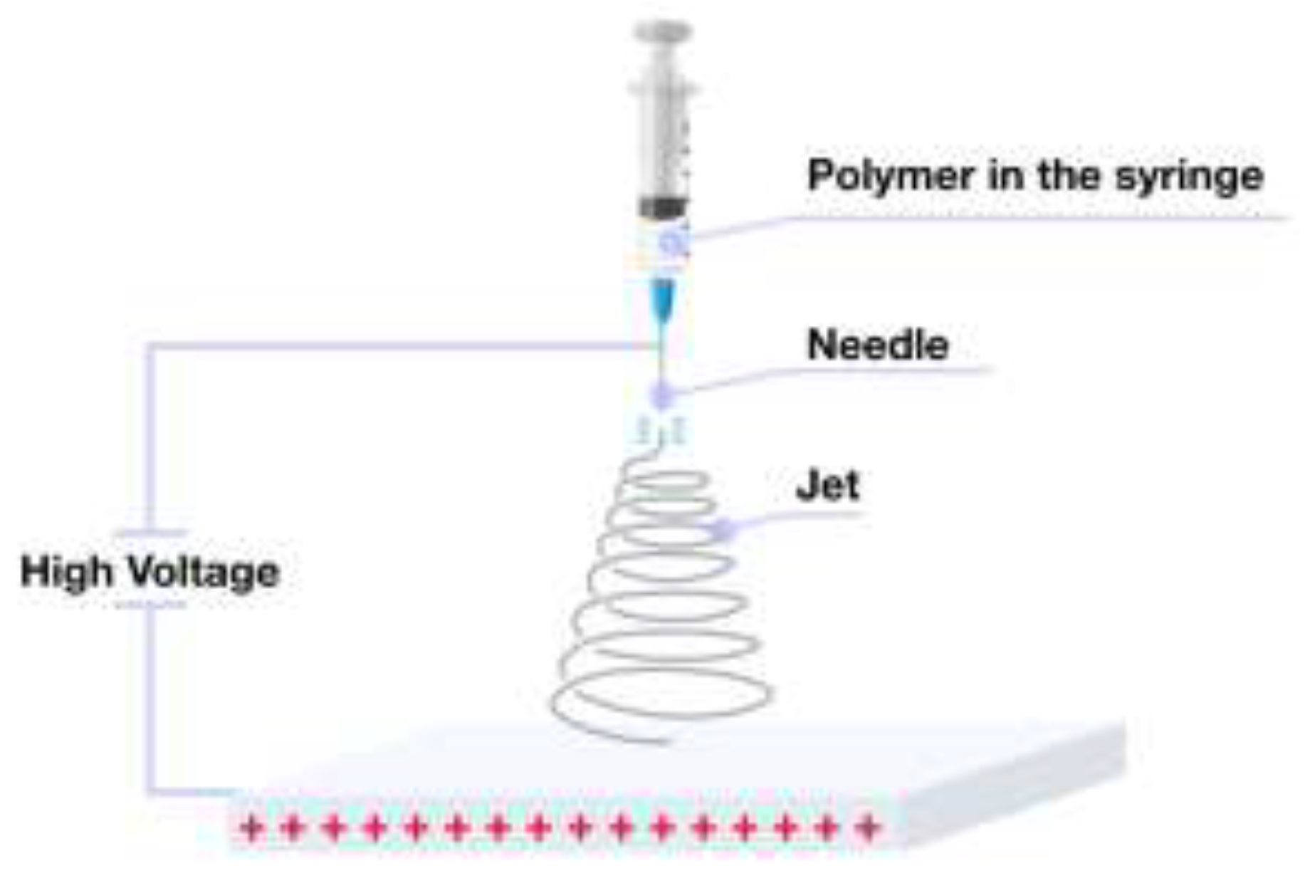
Figure 2.
SEM images illustrating morphologies of electrospun polyvinyl alcohol (PVA) fibers collected on a rotating drum, with speed of rotation of (a) 500 (b) 1000 (c) 1500 and (d) 2150 RPM [32].
Figure 2.
SEM images illustrating morphologies of electrospun polyvinyl alcohol (PVA) fibers collected on a rotating drum, with speed of rotation of (a) 500 (b) 1000 (c) 1500 and (d) 2150 RPM [32].
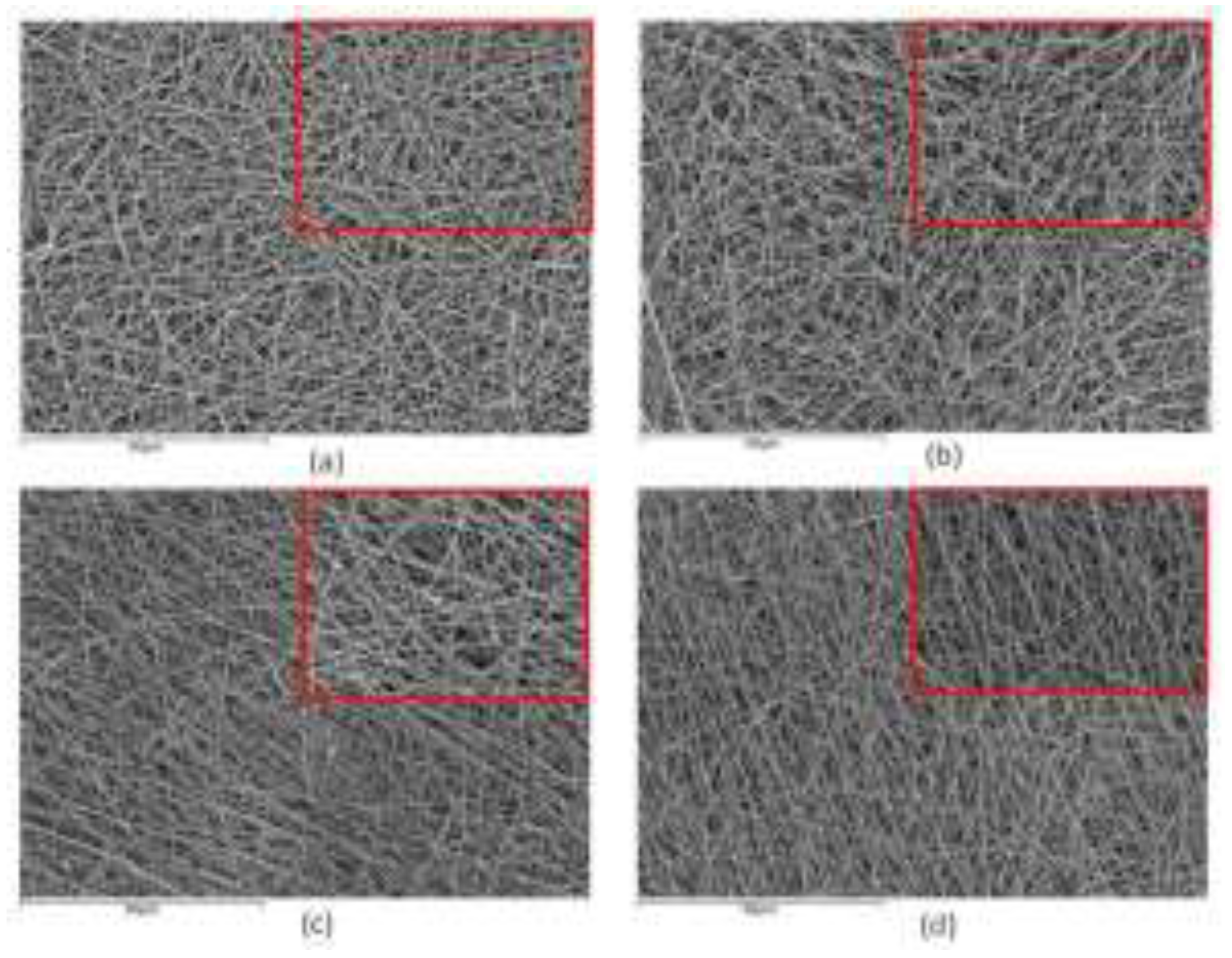
Figure 3.
Morphologies of electrospun PSF/PLA fibers [38] with varying weight ratios: 9:1, 7:3, 6:4, 4:6, 3:7, 1:9, and 0:1. The fiber diameter corresponding to each of the blend solutions is shown.
Figure 3.
Morphologies of electrospun PSF/PLA fibers [38] with varying weight ratios: 9:1, 7:3, 6:4, 4:6, 3:7, 1:9, and 0:1. The fiber diameter corresponding to each of the blend solutions is shown.
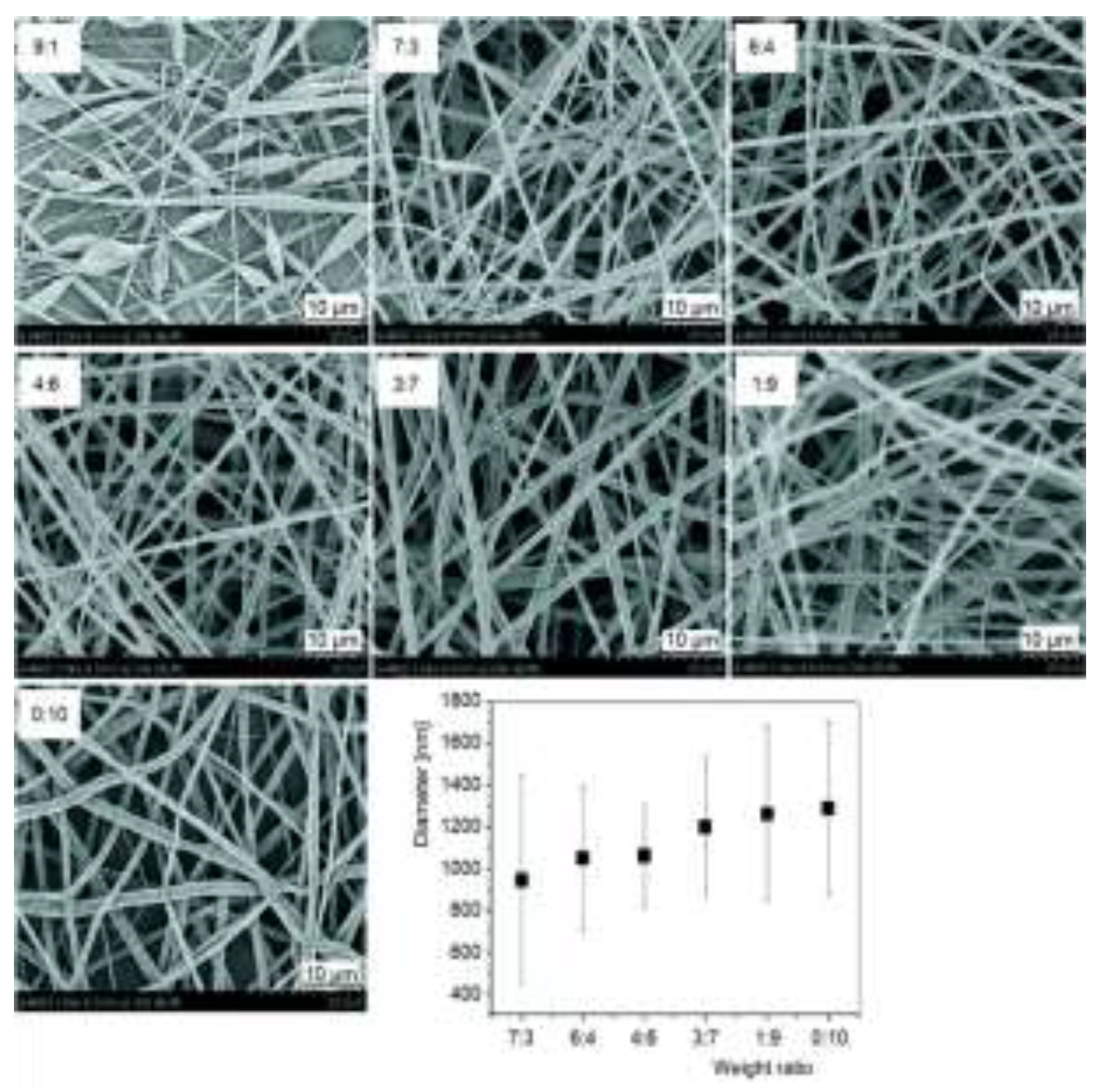
Figure 4.
Formation of a Taylor cone: (a) Polymer solution droplet shaped by surface tension; (b) Localized charges develop under applied voltage, stretching the droplet into a cone; (c) Taylor cone forms when localized charges counterbalance surface tension; (d) Jet ejection occurs as the charges surpass surface tension.
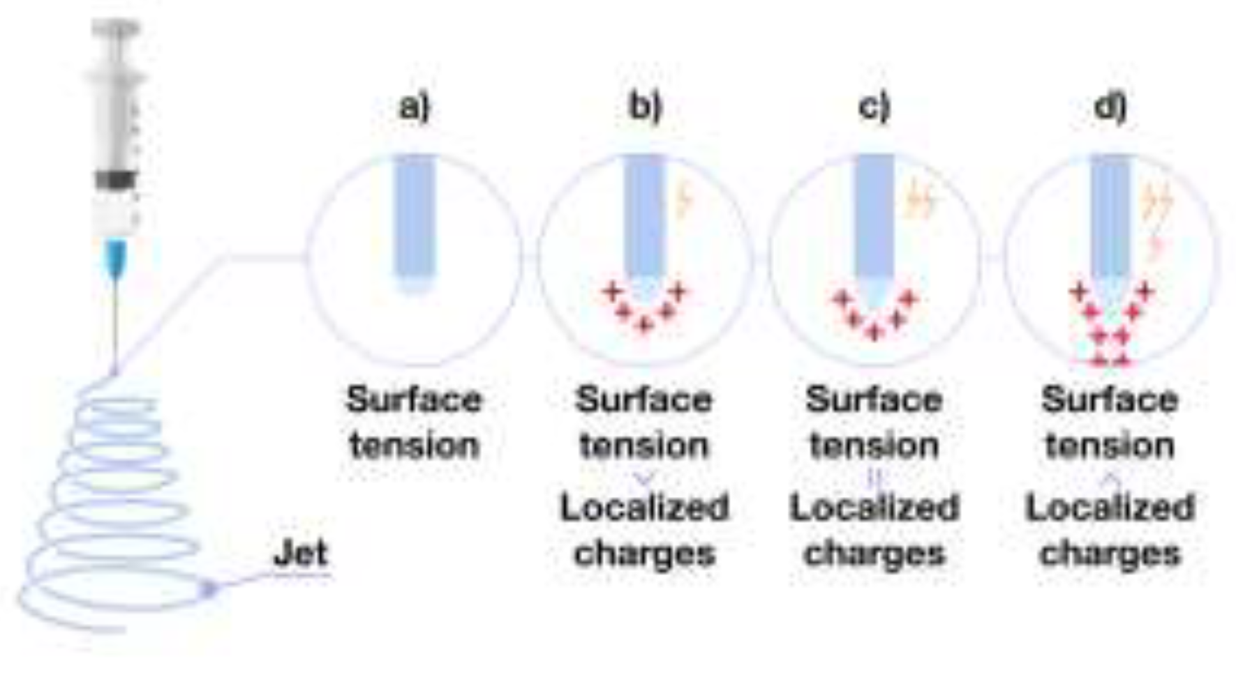
Figure 5.
Schematic illustration of conventional electrospinning techniques for polymer fiber fabrication.
Figure 5.
Schematic illustration of conventional electrospinning techniques for polymer fiber fabrication.
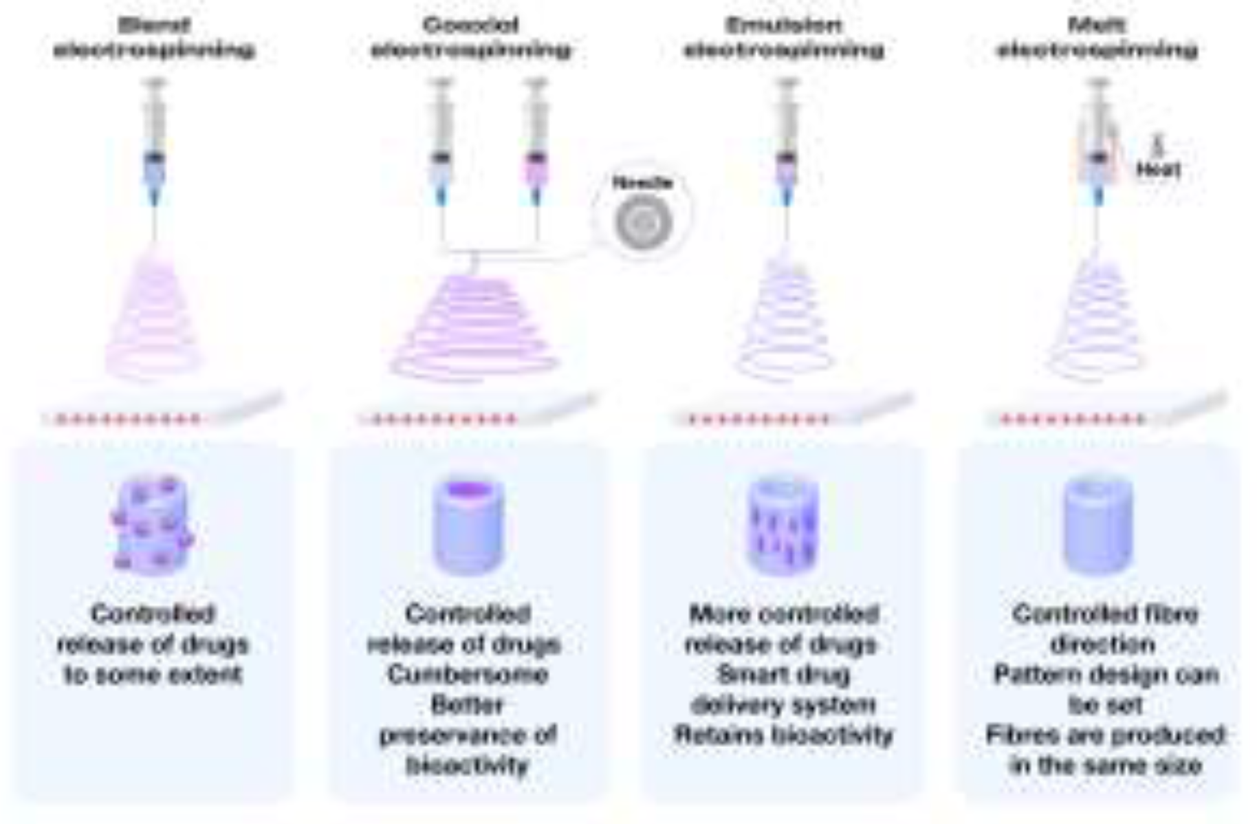
Figure 6.
Schematic representation of fiber formation through coaxial electrospinning.
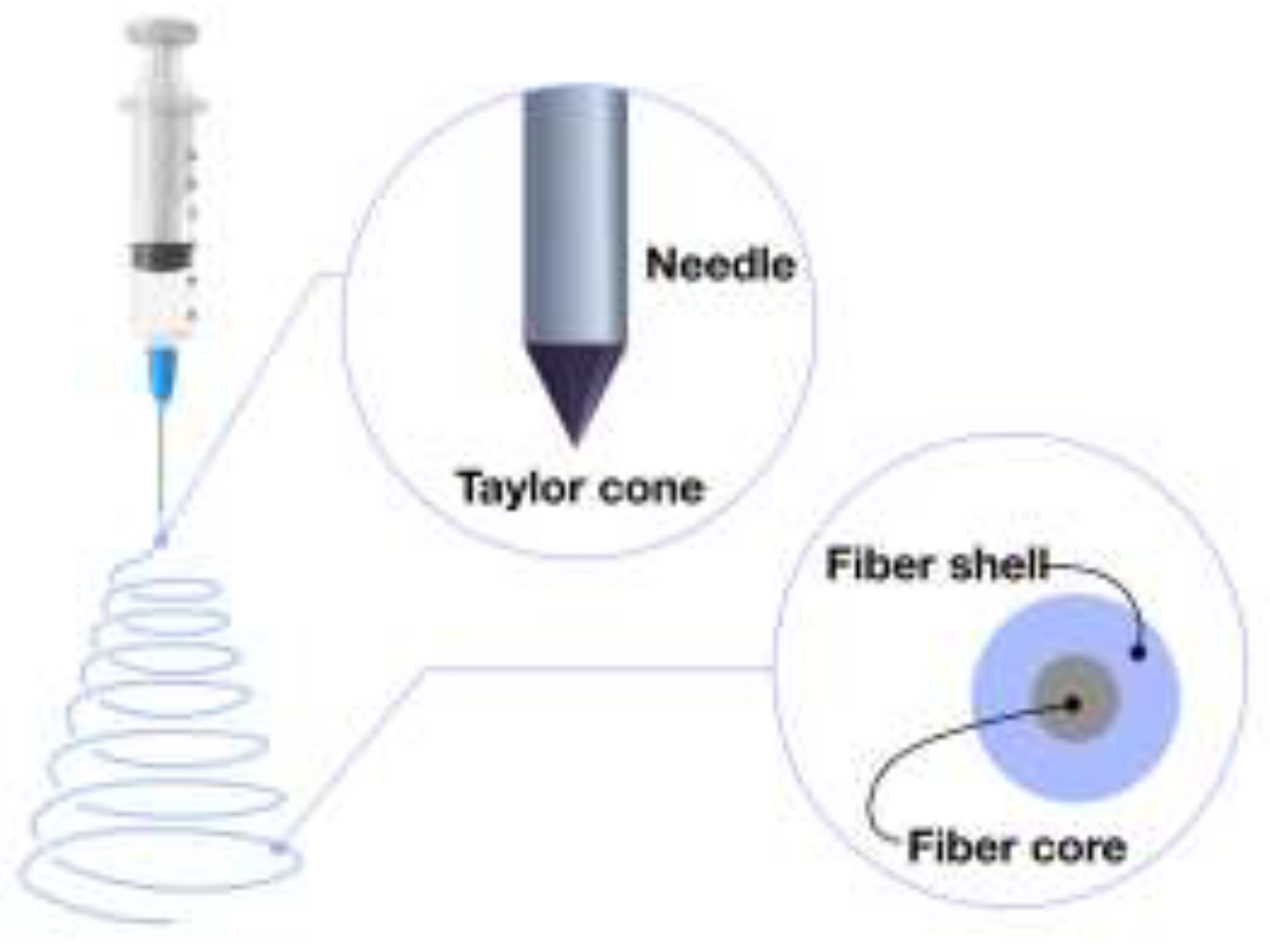
Figure 7.
SEM micrographs of SH–SY5Y neuroblastoma cells cultured on electrospun scaffolds for 1 and 3 days. (a, b) PCL/Gel, (c, d) PCL/Gel 0.1% LDH, (e, f) PCL/Gel 1% LDH, and (g, h) PCL/Gel 10% LDH scaffolds [114].
Figure 7.
SEM micrographs of SH–SY5Y neuroblastoma cells cultured on electrospun scaffolds for 1 and 3 days. (a, b) PCL/Gel, (c, d) PCL/Gel 0.1% LDH, (e, f) PCL/Gel 1% LDH, and (g, h) PCL/Gel 10% LDH scaffolds [114].
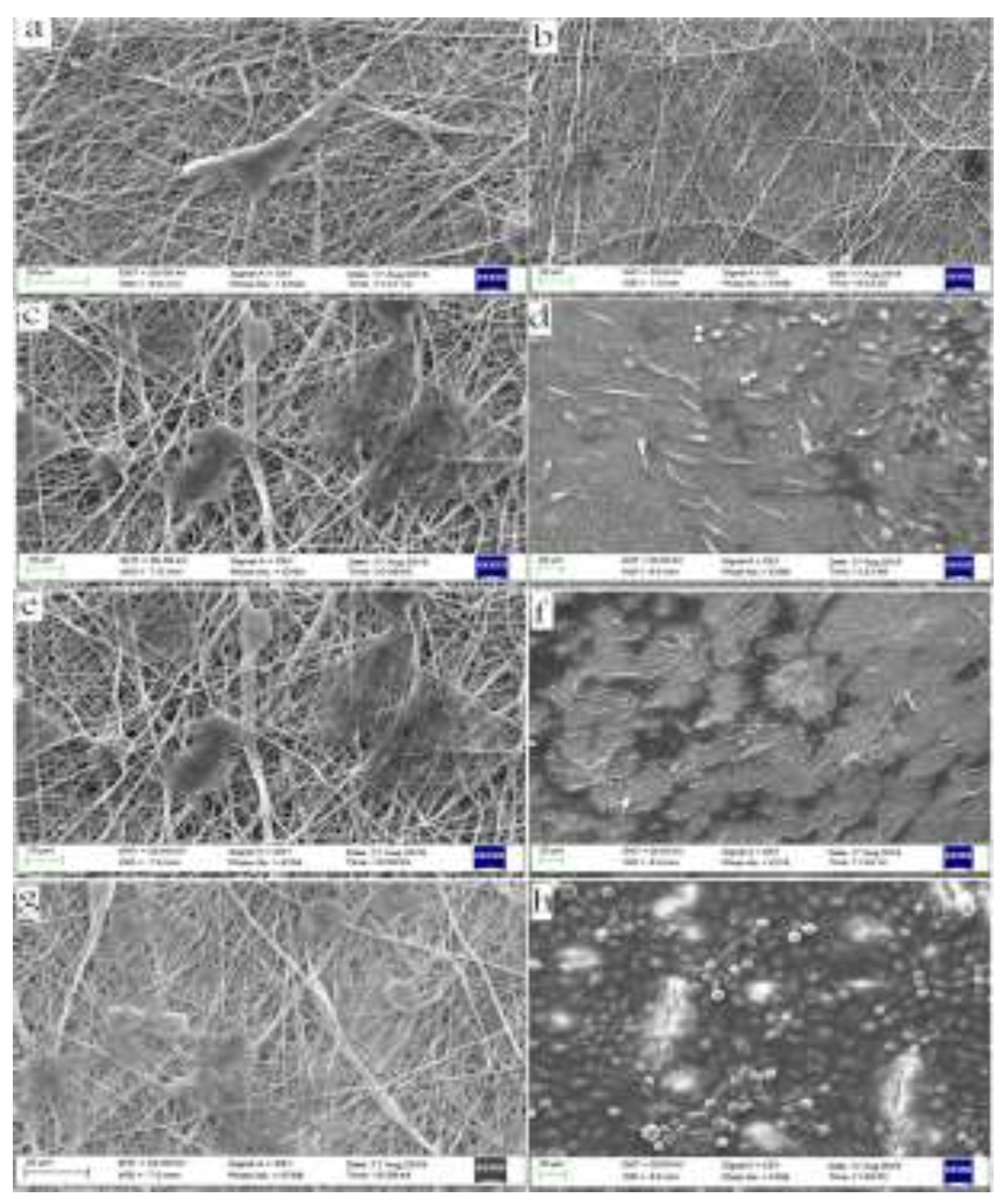
Figure 8.
DAPI staining of HDF cells on electrospun gelatin/chitosan scaffold after 1 and 7 days of culturing. (a) Chi 50; (b) Chi 40; (c) Chi 30 [126].
Figure 8.
DAPI staining of HDF cells on electrospun gelatin/chitosan scaffold after 1 and 7 days of culturing. (a) Chi 50; (b) Chi 40; (c) Chi 30 [126].
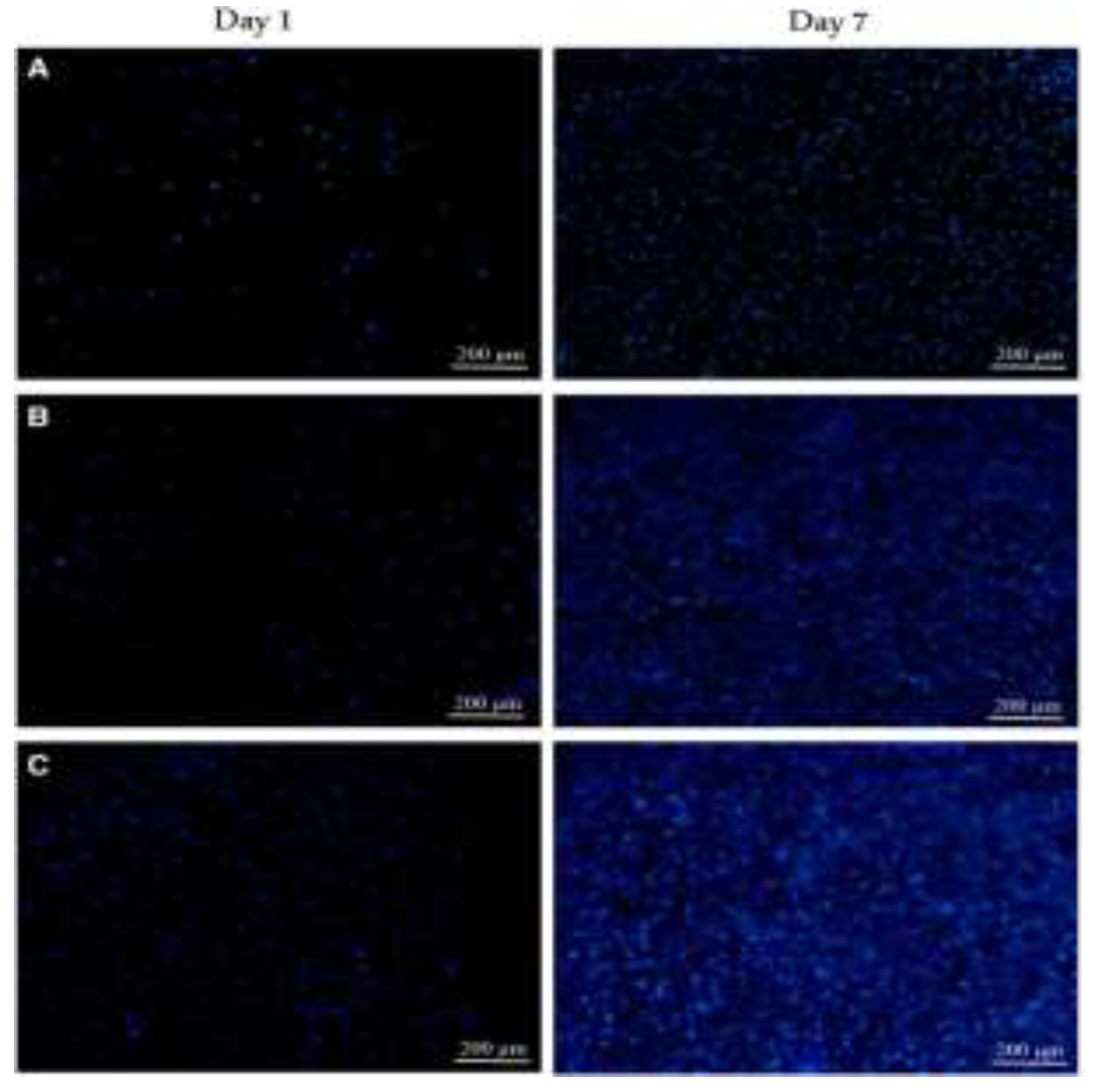
Figure 9.
MTS assay after HDF cells were cultured on nanofibrous scaffolds. Formazan absorbance expressed as a function of culture time [126].
Figure 9.
MTS assay after HDF cells were cultured on nanofibrous scaffolds. Formazan absorbance expressed as a function of culture time [126].
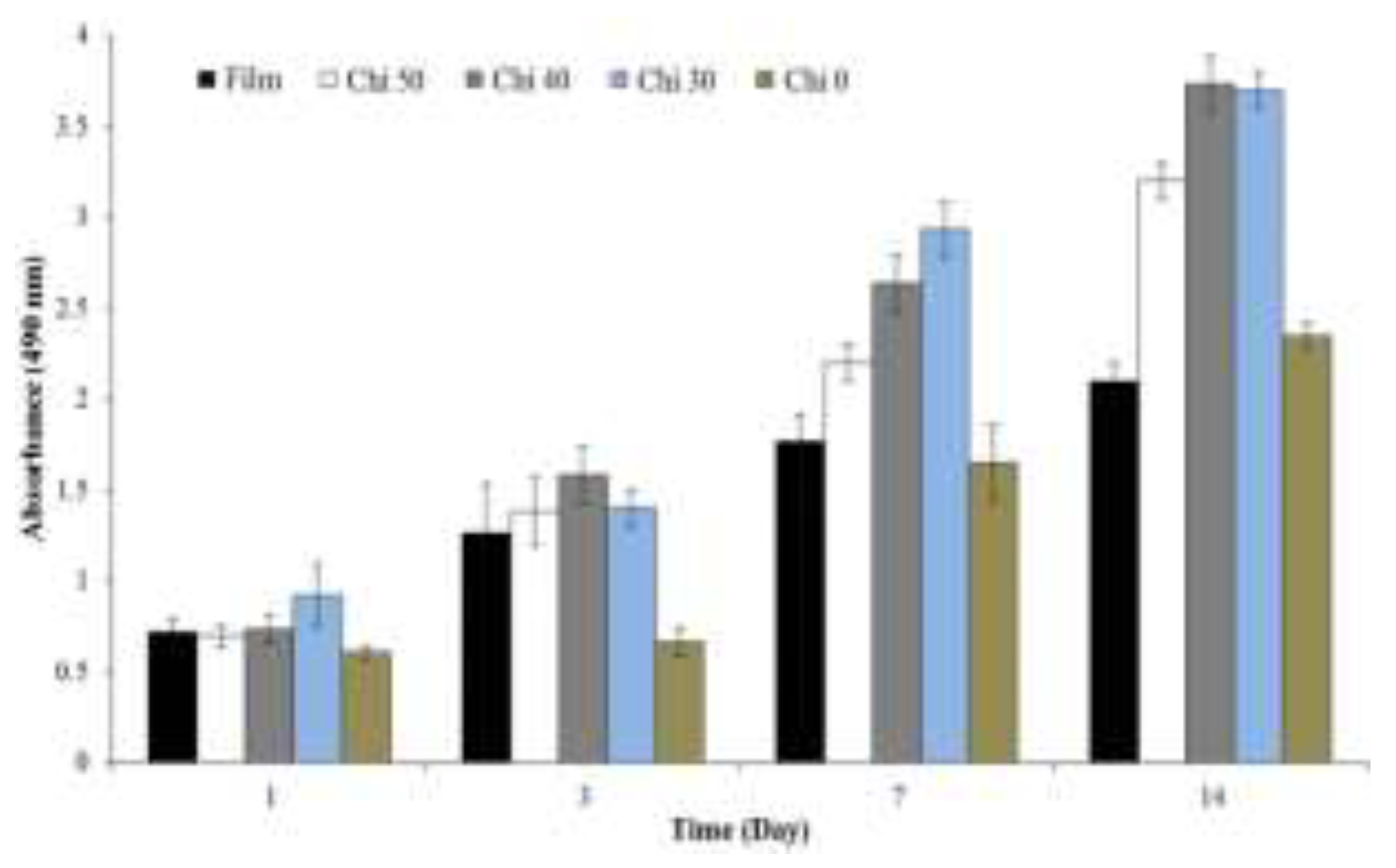
Figure 10.
SEM images of cross–sections of pure PLA fibers using CF/DMF with the weight ratio of 90/10 (a), 80/20 (b) [141].
Figure 10.
SEM images of cross–sections of pure PLA fibers using CF/DMF with the weight ratio of 90/10 (a), 80/20 (b) [141].
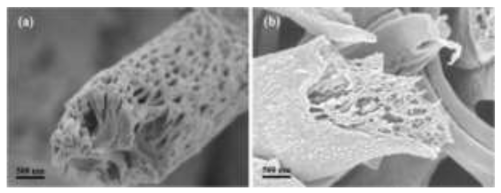
Figure 11.
SEM images of electrospun fibers reveal PCL scaffolds displaying diverse surface topographies (A); cross–sectional SEM images illustrate fibers with prominent, minor, and no surface depressions (B) [163].
Figure 11.
SEM images of electrospun fibers reveal PCL scaffolds displaying diverse surface topographies (A); cross–sectional SEM images illustrate fibers with prominent, minor, and no surface depressions (B) [163].
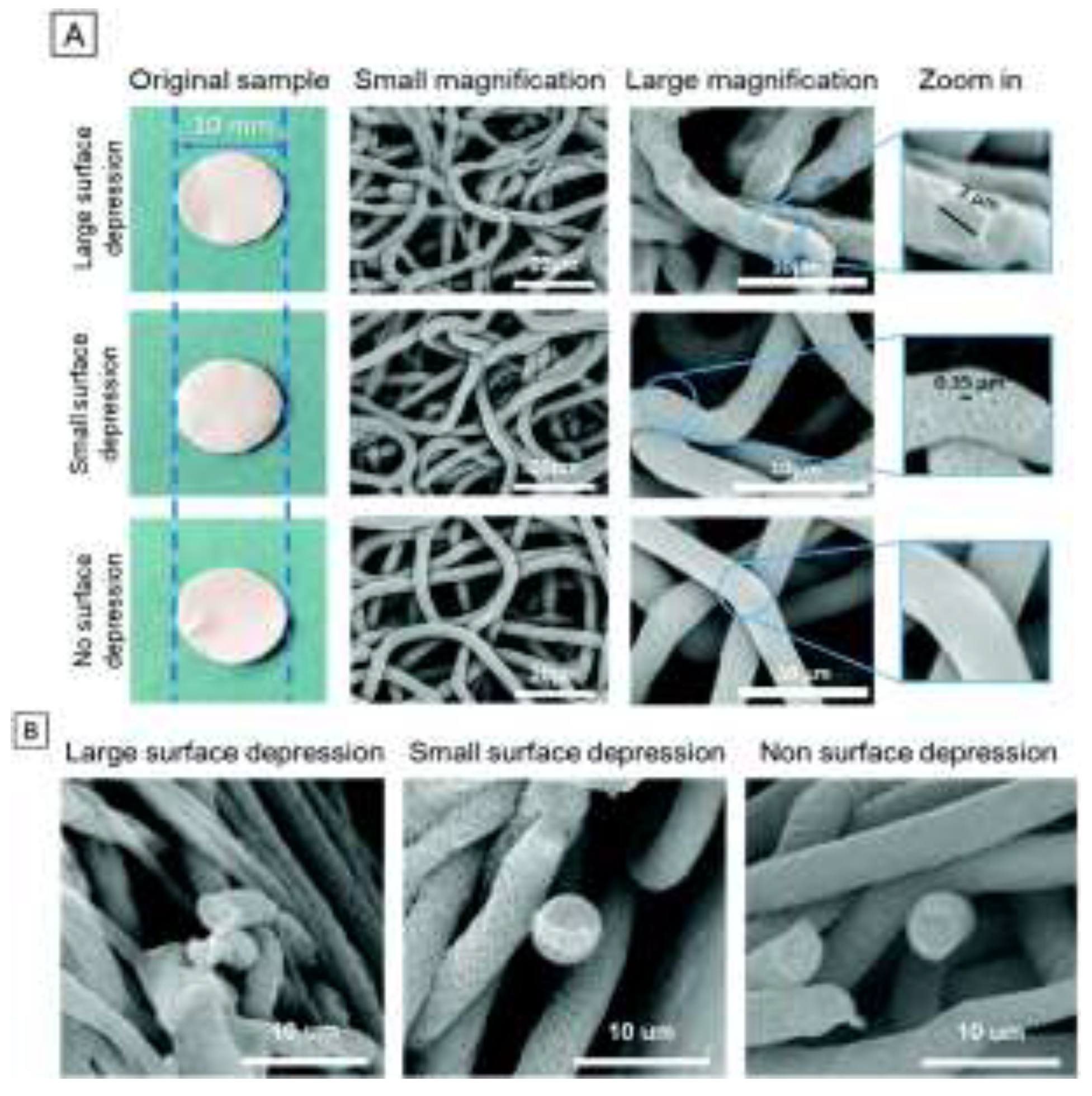
Figure 12.
Cell Titre Blue assay. (B) HepG2 dsDNA quantity, measured via Picogreen assay, N = 5, error bars ± SD. (C) Cell viability normalized to DNA content [163].
Figure 12.
Cell Titre Blue assay. (B) HepG2 dsDNA quantity, measured via Picogreen assay, N = 5, error bars ± SD. (C) Cell viability normalized to DNA content [163].
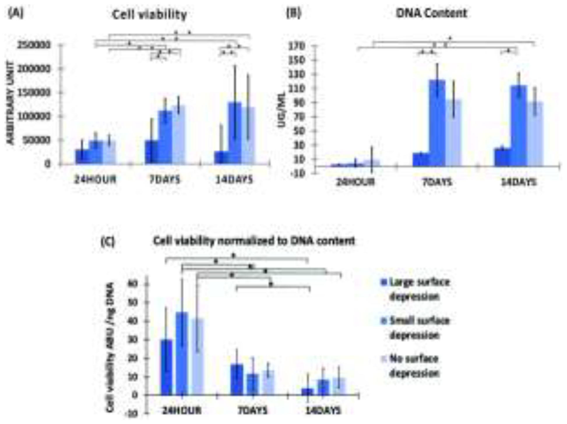
Disclaimer/Publisher’s Note: The statements, opinions and data contained in all publications are solely those of the individual author(s) and contributor(s) and not of MDPI and/or the editor(s). MDPI and/or the editor(s) disclaim responsibility for any injury to people or property resulting from any ideas, methods, instructions or products referred to in the content. |
© 2024 by the authors. Licensee MDPI, Basel, Switzerland. This article is an open access article distributed under the terms and conditions of the Creative Commons Attribution (CC BY) license (http://creativecommons.org/licenses/by/4.0/).
Copyright: This open access article is published under a Creative Commons CC BY 4.0 license, which permit the free download, distribution, and reuse, provided that the author and preprint are cited in any reuse.


