Submitted:
13 September 2024
Posted:
14 September 2024
You are already at the latest version
Abstract
This review aims to show, by an analysis of the literature on the topic, that hyperinsulinemia associated with insulin resistance is a multiple risk factor for type 2 diabetes, cardiovascular diseases, cellular senescence and cancer, and neurodegenerative diseases. This condition is progressively increasing in developed and developing countries, and its prevalence has now exceeded 50% of the general population. Since it is asymptomatic or poorly symptomatic, it can last for many years before manifesting itself in the form of type 2 diabetes mellitus, cardiovascular disease, neoplasm, cognitive impairment or dementia, therefore determining enormous social and health care costs. For these reasons, we strongly believe that a screening plan for this pathology should be implemented, in order to identify affected subjects and promptly start them on preventive treatment.
Keywords:
insulin resistance
; hyperinsulinemia
; type 2 diabetes
; treatments
; adverse effects
; risk factor
; cardiovascular disease
; cellular senescence
; cancer
; neurodegenerative disease
1. Background
Despite the notable and continuous scientific advances made in the prevention and treatment of cardiovascular pathologies and tumors, they are still today the main causes of hospital admissions and deaths [1]. Furthermore, the aging of the population appears to be associated with a notable increase in neurodegenerative diseases. This entails significant social and health care costs. We must therefore ask ourselves a question: "We accidentally overlooked something?". Well, the answer is certainly “Yes”! Increased levels of circulating insulin (hyperinsulinemia, Hyperin) secondary to Insulin resistance (IR) represent a condition that predisposes to many diseases. This determines a silent pandemic, producing enormous healthcare costs, and an increase in hospitalizations and deaths.
Unfortunately, IR/Hyperin is a condition rapidly growing worldwide and its prevalence has now exceeded 50% in the general population and is continuously growing [2,3].
This review of the literature on the topic aims to demonstrate how Hyperin associated with IR, if neglected, can cause serious and, often, irreversible damage to our body. The manuscripts used for the review were searched on medical literature databases such as PubMed, Scopus, Web of Science, etc, using these keywords: Insulin resistance, hyperinsulinemia, type 2 diabetes, treatments, adverse effects, risk factor, cardiovascular disease, cellular senescence, cancer, neurodegenerative disease.
2. Definition and Causes of IR
IR can be defined as a condition in which a given quantity of insulin produces a reduced metabolic result, in terms of glycemic control, compared to that expected [3]. For this reason, in order to maintain normal glycemic values, the pancreas of subjects affected by IR is forced to secrete greater quantities of insulin, with the result that Hyperin turns out to be a constant and fundamental characteristic of IR. The mechanisms through which IR can develop in our body, and those linked to Hyperin damage of some organs are very complex and, still today, not completely clarified. There may be a defect at the receptor or, more likely, post- receptor level. However, there are also some certainties. We know well enough two of the pathways triggered by the binding of insulin to its receptors, their mechanisms and what happens to these pathways at the post-receptor level in case of IR. They are the phosphoinositide 3 kinase (PI3K) and the mitogen-activated protein kinase (MAPK) pathways.
Insulin determines its multiple metabolic and non-metabolic actions through binding to its transmembrane receptors located on the target cells of its action. The intracellular domain of the insulin receptor belongs to the tyrosine kinase receptor protein family. The interaction of insulin and its receptors determines the activation of numerous protein kinases and gene transcription factors. Among these, the most important and best known, as already mentioned, are the PI3K pathway, which mainly regulates metabolic effects and the secretion of nitric oxide (NO), and the MAPK pathway, responsible, instead, for gene expression effects, cell growth and differentiation, and the production of endothelin-1 (ET-1) at vascular level [4].
In the presence of an IR condition, we have mostly a malfunction at the PI3K post receptor level which mainly regulates the metabolic actions and the formation of NO, while the functioning of MAPK is little or not altered at all, so that the non-metabolic actions of insulin and, in particular, the stimulating action on cell proliferation and on the secretion of ET-1 are exalted due to the chronic effects of Hyperin. Therefore, the different behavior of these two pathways, resulting from IR, determines, over time, very important alterations in the target organs of insulin [3].
There are many causes that can produce IR, although probably not all of them are known. There are certainly genetic causes but they are the minority. Among these, type A IR syndrome is a rare genetic disorder characterized by severe IR. In affected women, the main features of the condition appear during adolescence. Many affected women do not start menstruating by age 16 (primary amenorrhea) or their cycles are scanty and irregular. They develop ovarian cysts and excessive growth of body hair (hirsutism). Acanthosis nigricans is also often present. Unlike most people with IR, these individuals are generally not overweight. The characteristics of type A IR syndrome are more subtle in males. Some of them have only low blood sugar as the only initial sign, others may also have acanthosis nigricans. In most cases, males with this condition reach medical attention only in adulthood, when diabetes appears. IR syndrome type A is estimated to affect 1 in 100,000 people worldwide. Because women have more evident health problems associated with this condition, it is diagnosed more often in women than in men [5].
Although it is not yet perfectly clear whether the chicken or the egg came first, i.e. whether the IR appears first and then the hyperinsulinemia (as one might think), these two conditions are, in the vast majority of cases, chronically associated, and they are rapidly and constantly growing throughout the world. Furthermore, since it is asymptomatic or poorly symptomatic, it lasts unrecognized for years, constituting what could be defined as a silent pandemic [3]. Over the centuries, there has been a notable change in our lifestyle, characterized by a progressive increase in caloric intake in favor of foods rich in carbohydrates and highly processed, with a simultaneous reduction in physical activity, and this already begins in children and persists into adulthood. This notable change in lifestyle, also associated with the increased stress and competitiveness of modern life, which, as is known, stimulates the secretion of diabetogenic hormones (above all cortisol and GH), has produced a progressive increase and spread of IR and Hyperin [6].
3. Diagnosis
The euglycemic-hyperinsulinemic clamp technique gives us the diagnostic certainty of IR, but it is not imaginable that it should be employed for mass screening purpose, so it is used almost exclusively for scientific research purposes [7]. However, there are many surrogate indices of IR that can be used for this purpose. Although they are many and all useful, I have chosen 3 indices that I believe are simple to obtain and reliable, [Table 1] namely: the homeostatic model assessment index (HOMA-IR), which is calculated through the simultaneous fasting measurement of glycemia and insulinemia, with a cut-off of 2.5 in adults and 3.6 in children; the triglyceride-glucose index (TyG), which is obtained by simultaneously measuring triglycerides and fasting blood sugar, with a cut-off value of 8; the ratio between triglycerides and HDLc with a cut-off of 2.75 in men and 1.65 in women. These 3 surrogate indices of IR have also been shown to be excellent independent markers of adverse cardiovascular events [7,8,9].
Table 1. Table 1 shows the formulas to obtain the three indices: HOMA:IR, TyG, and Triglycerides/HDLc, and their respective normal values.
HOMA-IR= fasting blood glucose (mg/dl) x fasting insulin (mU/L) / 405, n.v. 0.23-2.5
TyG= Ln [fasting triglycerides(mg/dL) x fasting blood glucose (mg/dL)] / 2, n.v.<8.0
Triglycerides/HDLc =fasting triglycerides (mg/dL): fasting HDL cholesterol (mg/dL), n.v. <2.75 for men; <1.65 for women.
4. Effects of Hyperinsulinemia Associated with Insulin Resistance
4.1. Cardiovascular Effects
Hyperinsulinemia associated with IR determines an alteration of circulatory homeostasis defined as endothelial dysfunction. It is caused by the prevailing synthesis and secretion of ET-1, compared to a reduced availability of NO, by the cells of the wall of the arteries and arterioles [Figure 1]. This causes vasoconstriction with reduction of circulatory flows to the tissues. Furthermore, hyperinsulinemia, acting as a growth stimulus, determines increase in vascular thickness and parietal stiffening, a phenomenon which is prodromal of development and worsening of atherosclerosis [10,11].
In addition to this, which is already very important in itself, in IR conditions there is solid evidence of the fact that Hyperin is an important cause of arterial hypertension.
Hyperin produces reabsorption of sodium and water at the renal level. Both endogenous and exogenous Hyperin have been found to be associated with blood pressure enhancement. Insulin receptors are located in the renal tubules, and it has been seen that their stimulation by insulin determines increased Na+ and water reabsorption. In addition to this, a close relationship has been demonstrated between the increased levels of circulating insulin and the enhanced activity of sympathetic nervous system. Arterial hypertension resulted associated with both increased insulin levels and the enhanced sympathetic activity, with a relationship demonstrated both in the whole population and after correction for BMI and body fat. Furthermore, it has been assessed that, by lowering insulin levels in obese subjects, it can be obtained decreases in plasma norepinephrine and blood pressure values [12,13].
A fairly recent study, performed in 88 hypertensive Sub-Saharan African patients with myocardial hypertrophy, has shown that obesity and IR/Hyperin predicted the increase in left ventricular mass. Therefore, the authors suggested that could be particularly important to correct the obesity and IR/Hyperin to counteract the development of LVH in these patients [14]. Another study was performed in Japan: 210 normotensive subjects and 180 patients with mild or moderate hypertension were studied by echocardiography and measurement of glycemic metabolic parameters . The sum of glucose or Hb A1c levels in the whole group of subjects and the sum of insulin levels (or insulin values 2 hours post load) in non-diabetic subjects were highly related to the relative LV wall thickness values, independently of age, systolic blood pressure and BMI. Therefore, the authors concluded that hyperglycemia and Hyperin could stimulate LV concentric remodeling in normotensive subjects and in patients with mild or moderate hypertension. Concentric remodeling of the LV is a recognized predictor of heart failure with preserved ejection fraction (HFpEF) [15]. In a study by our Group performed by Doppler-echocardiography, 59 patients with IR/Hyperin showed both increased LV mass and relative wall thickness, together with LV diastolic dysfunction [16] [Figure 2].
4.2. Effects on Cellular Senescence and Cancer
Hyperinsulinemia associated with IR has many other negative actions. Among these, the action on cellular senescence and the development of tumors should not be overlooked. Senescent cells are characterized by the fact that they stop dividing and undergo specific changes, both in their appearance and activity. They produce specific molecules, contributing to the aging state of the entire organism. Cellular senescence is due to several factors, including, above all, the presence of DNA damage. In particular, replicative senescence is linked to the so-called telomere attrition, a process that leads to chromosomal instability and promotes the onset of tumors [17].
It should be noted that increased cellular senescence is present in adult obese subjects, type 2 diabetes and non-alcoholic fatty liver, regardless of age. In particular, IR/Hyperin stimulates cellular senescence in metabolic target organs such as adipose tissue, muscle, liver and brain, in humans [Figure 3].
Among various published studies, one, carried out on cultured human hepatocytes under chronic hyperinsulinemia and in knockout mice for insulin receptors in the liver (LIRKO mice) demonstrating a direct relation between hyperinsulinemia and senescence of hepatocytes, is very interesting. This study also demonstrated that the dangerous effects of chronic Hyperin on cellular senescence of hepatocytes can be blocked by reducing the number of insulin receptors or by the senolytic substances desatinib and quercetin [18,19,20].
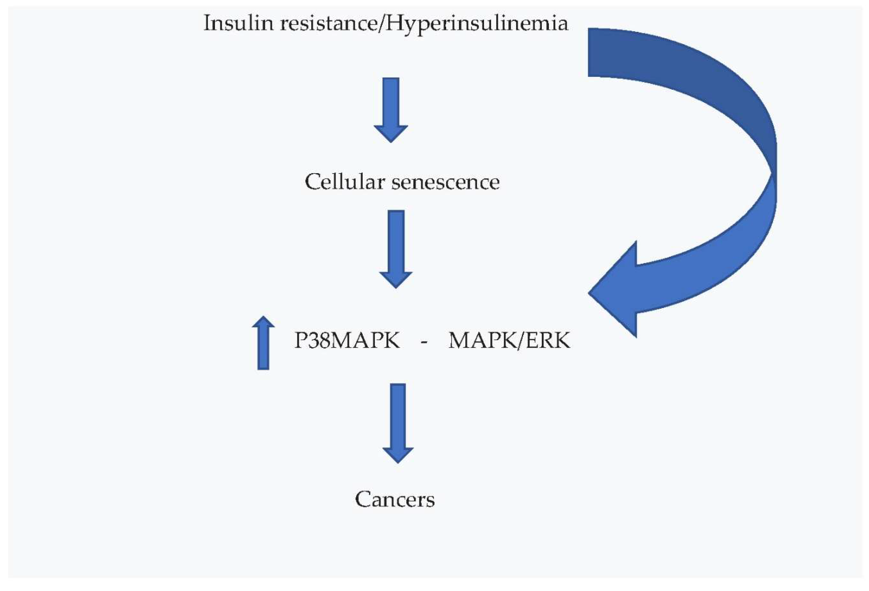
Age predisposes to the development of many types of cancer and, in fact, the incidence of numerous tumors increases with age, even if the underlying relation has not yet been fully clarified. However, there is growing scientific demonstration that the increase in senescent cells in our body contributes to the advancement of tumors [21,22,23]. Recently, it has been shown that senescence acts as a tumor promoter and that stimulates skin cancer by upregulating p38MAPK and MAPK/ERK signaling, on which the high insulin levels associated with IR also act [24]. Insulin binds not only to its own receptors, but also to those of Insulin like growth factor-1 (IGF-1), thus acting as a growth factor with pathologic consequences on the development of tumors. Diabetic patients and /or subjects with metabolic syndrome have a doubled risk of developing cancer and cancer-related deaths [25,26,27]. Furthermore, it has also been reported, in non-diabetic and non-obese subjects, that Hyperin itself is related to increased cancer deaths and, therefore, the authors underline that a treatment to reduce circulating insulin levels could be an important therapeutic approach to the prevention of cancer [28,29].
4.3. Effects on Brain
Another problem that should not be underestimated regarding the damage caused by hyperinsulinemia associated with IR is the very close relationship that exists between it and neurodegenerative diseases, although the mechanisms that link the two pathologies are not yet fully clarified. There are numerous pathophysiological hypotheses, partly also well supported by scientific literature, which try to explain the intricate mechanisms that associate Hyperin/IR with brain damage [30,31,32].
As regards the relationship between insulin and the brain, it must be remembered that, until a few years ago, it was believed that the brain was an organ insensitive to insulin actions. Instead, over the last 20 years, a considerable amount of scientific literature has accumulated which demonstrates that insulin penetrates the brain crossing the blood-brain barrier, where it binds to its specific receptors and regulates some important functions of the central nervous system such as the stimulation of appetite, cognitive behavior and depression. By this way, it also controls some important systemic functions such as the production of glucose by the liver, lipogenesis and lipolysis, and the response of the sympathetic system to episodes of hypoglycemia [33]. Insulin binds to its receptors localized in different regions of the brain and initiates a series of phosphorylation reactions using two different receptor substrates (IRS-1 and -2) which, in turn, activate subsequent metabolic pathways. PI3K and protein kinase B (Akt) are kinases activated at post-receptor level when insulin binds to its receptors and, by this way, acts on neuronal plasticity and survival, and neurotransmitter trafficking. Insulin activates also MAPK, which controls cell growth and proliferation. It should be underlined that hippocampus, which is involved in cognitive function, and hypothalamus, which controls peripheral metabolism, are characteristically rich in insulin receptors [34,35,36,37].
Let's try to understand what could happen at a brain level in the case of Iperin/IR. There are numerous epidemiological studies that highlight how the prevalence of this condition is very high in patients with cognitive deficits or Alzheimer's disease, reaching and exceeding the value of 81% of cases, overall. Going into more detail regarding the close relationship between cognitive deficits, AD and type 2 diabetes mellitus, it has been seen that these pathologies have in common multiple risk factors, comorbidities and hypothetical pathophysiological mechanisms, so much so that, provocatively, it has been proposed to call AD with the name of type 3 diabetes mellitus [38,39,40,41]. Studies based on post-mortem examinations of brains with cognitive deficits and Down syndrome have highlighted clear signs of IR in them, such as a significant reduction in receptors for insulin in the hippocampus, cortex and hypothalamus [42].
Furthermore, a very interesting study, carried out in Vervet monkeys, has shown that, going from a state of health to a stage of prediabetes and, subsequently, to frank diabetes, the cerebral metabolism is altered with an increase in glucose and a decrease in amino acids and acylcarnitine. This alteration of metabolism determined a stimulation of the production and aggregation of amyloid β in a similar way in type 2 diabetes and AD, clarifying how some mechanisms present in diabetes can lead to cognitive deficits and dementia [43].
4.4. Possibilities of Treatment
What therapeutic possibilities do we have for Hyperin/IR? First of all, we need to completely change our approach compared to what we do now, because now we really do very little. We must therefore move from an watch and wait approach to a more rapid preventive intervention, because, by waiting, the least that can happen is that diabetes develops. In most cases, Hyperin associated with IR precedes the development of the described events by several years, even up to 15, and this can happen already in children [3]. For this reason, we must use a broad-based strategy, educating and acting effectively. We must educate the general population, already as children, to a more correct lifestyle, with a balanced and more adequate caloric intake, accompanied by moderate, but constant over time, physical activity (walking at a normal pace 4000/5000 steps per day).
Unfortunately, this type of intervention may, in many cases, not be sufficient for various reasons, including the fact that many people are unable to constantly correct their lifestyle. At this point, therefore, we are forced to intervene by helping the patient with the administration of insulin sensitizing substances. There are many substances to choose from, both drugs and natural substances, depending on the tastes and characteristics of the patient.
4.5. Drugs
One of the best known is certainly the biguanide metformin. It is used as oral anti-diabetic drug for many years. It has to its credit many experimental and clinical studies which demonstrate its effectiveness in reducing IR with secondary Hyperin. The mechanisms of metformin in improving insulin sensitivity are several, different and, probably, not all of them are known yet. They include increased insulin receptor tyrosine kinase activity, increased glycogen synthesis and, more downstream end, increased GLUT4 recruitment and activity [44]. In addition, metformin can activate adenosine 5′-monophosphate-activated protein kinase (AMPK), a cellular energy sensor [45,46].
There is, among others, a good review manuscript on the effects of metformin on all-cause mortality and the incidence of aging diseases in diabetic subjects compared to the non-diabetic people and to diabetic subjects in treatment with anti-diabetic drugs other than metformin. This study showed that metformin significantly reduced mortality in diabetics, in comparison with the group of non-diabetics and that of diabetic people treated with drugs other than metformin. Furthermore, it was verified that the group of diabetic patients treated with metformin also had a reduction in cancers, compared to the other two groups of subjects and a lower number of cardiovascular events compared to diabetics who did not take this drug [47].
Metformin therapy showed its benefits also in patients with HFpEF. A recent meta regression analysis of observational and randomized studies shows that metformin significantly reduces deaths in patients with HFpEF [48]. Metformin has also been shown to be beneficial for the heart of individuals with HF. In fact, it improves the energy state of the myocardium due to the modulation of glucose and lipid metabolism, the reduction of oxidative stress and inflammation and the improvement of concentric remodeling process of the LV. Another meta-analysis was recently published showing that metformin treatment reduced LVM and improved EF, both in subjects with and without pre-existing cardiopathy [49,50].
Other very interesting drugs, which have been on the market for the treatment of diabetes for a relatively short time and which have an important action in reducing insulinemia and IR, are the inhibitors of the sodium-glucose cotransporter 2 (SGLT2 I). These include canaglifozin, dapaglifozin and empaglifozin. Vast scientific literature demonstrates that these drugs reduce the number of hospitalizations, cardiovascular deaths and deaths from any cause in HF patients [51,52].
6263 patients aged >40 years with an EF >40%, were randomized to receive dapaglifozin 10 mg daily or placebo in addition to standard HF therapy. The results of this study showed a significant decrease in the risk of HF worsening and in cardiovascular deaths. Another recent large meta-regression analysis, whose purpose was to establish the effects of these drugs on all-cause death, demonstrated that SGLT2 I significantly reduced this in randomized trials [53,54].
The exact mechanisms by which these drugs produce their beneficial effects are complex, multiple and not entirely known. What is certain is that treatment with SGLT2 I in subjects with IR/Hyperin causes a reduction in glycemic levels due to enhanced glucose excretion at the tubular level of kidney. In consequence of this, the insulin required to maintain daily glycemic balance is clearly reduced. Thus, SGLT2 I reduce circulating insulin levels and the consequent development over the years of cardiovascular and non-cardiovascular damage [55,56].
Other very interesting drugs, not to be overlooked when dealing with this topic, are the glucagon-like peptide 1 receptor agonists (GLP-1 a). These are a class of drugs that are in use for the treatment of type 2 diabetes and obesity. They are administered subcutaneously, usually once a week, and are quite expensive. They include semaglutide, dulaglutide, lixisenotide, liraglutide, xenatide, and tirzepatide. They act through multiple complex mechanisms, including that of reducing body weight and, above all, mechanisms involved in contrasting IR/Hyperin, such as the increase in the expression of glucose transporters in target tissues, the anti-inflammatory and anti-oxidative stress effect, and the modulation of lipid metabolism [57,58].
A meta-analysis of 54,092 patients from 7 randomized controlled trials on the use of GLP-1 agonists in patients with type 2 diabetes has been recently published. 16% of these patients had also a history of HF. The results of the study nave shown that these drugs prevented the onset of HF in the diabetic population, but that, in subjects with pre-existing HF, they did not decrease the number of episodes of worsening HF and the resulting hospitalizations, nor deaths [59]. A further meta-analysis of randomized and controlled studies, performed to understand whether treatment with these drugs in diabetic or non-diabetic subjects led to decrease in morbidity and deaths, also demonstrated that this therapy does not lower the number of cardiovascular events, including cardiovascular deaths, hospital admissions for HF, and does not improve HF grade or 6-minute walking test [60].
Unfortunately, on these drugs, in addition to positive scientific reports, there is literature reporting serious adverse events, such as severe pancreatitis. Patients taking semaglutide or liraglutide have been associated with nine-fold higher risk of pancreatitis, but also with high risk of developing intestinal and, in some cases, gastroparetic obstruction. Furthermore, the doubt that this class of drugs can stimulate thoughts of suicide and self-harm has not yet been completely dispelled [61,62].
For all the reasons listed above, these drugs, even though they produced great progress in the treatment of metabolic pathologies, should be mainly intended for diabetic patients with high degree of obesity, at least until the doubts are completely clarified and their costs will not be reduced.
4.6. Natural Substances
A very interesting natural substance for its action to counteract IR, which has mechanisms of action in part overlapping with those of metformin, is berberine. It is a plant alkaloid, used for over 2000 years in traditional Chinese medicine and Indian Ayurvedic medicine, which has shown, over the years, many beneficial effects on human health. A large amount of scientific literature shows that berberine improves IR, as well as other risk factors, such as hypercholesterolemia, hyperglycemia and arterial hypertension. In a double-blind, randomized and placebo-controlled study lasting 4 months, 59 patients with metabolic syndrome were treated with nutraceutics, among which berberine 500 mg or placebo. The results showed that the berberine group had a significant reduction in IR/Hyperin, as documented by a reduction in the HOMA-IR index and fasting insulin levels [16]. Furthermore, in these same patients, using Doppler-echocardiography, a significant decrease in LVM and RWT was documented, accompanied by a lower LV diastolic dysfunction [63].
In another study, 145 patients with metabolic syndrome and LV hypertrophy (LVH), were randomized to berberine 500 mg or placebo for 6 months. Echocardiography showed that treatment with berberine reduced significantly the LVM. Therefore, the authors concluded that berberine could represent an effective strategy to lower LVM, which is an important predictive factor of cardiovascular events [64]. Another study, performed in 2 groups of patients with HF, also showed very interesting results. One group of 79 patients was treated with berberine in addition to standard therapy and the other of 76 patients was treated with standard therapy plus placebo. During a 2-year follow-up, 7 patients died in the berberine group and 13 patients in the placebo group, demonstrating that the addition of berberine to standard HF treatment significantly reduced mortality [65].
It has also recently been suggested that berberine could produce benefits in patients with cognitive impairment and dementia. It has documented beneficial effects on neurotransmitters, inflammation, oxidative stress, metabolism, and other pathways in the brain. However, the mechanisms are numerous, complex and still not fully understood.
A rather recent study, carried out on human neurons, has demonstrated that berberine protects these cells from the negative effects produced by β amyloid42. In fact, berberine, added to neuronal cells, determined protection against the damage induced by β amyloid42 [66].
A further study, carried out in a transgenic mouse model of AD, demonstrated that treatment with berberine in these mice, between 2 and 6 months of age, significantly improved cognitive deficits, long-term spatial memory and the accumulation of amyloid plaques compared to vehicle-only control mice. In particular, an inhibition of the activity of glycogen synthase kinase 3B was observed [67,68]. Berberine has also demonstrated a positive action on the microbiota-intestine-brain axis. The results of a very recent study show that treatment with berberine reduces the formation of β-amyloid plaques, mitigates inflammatory processes and improves spatial memory dysfunction in a mouse model with major features of AD. This occurred simultaneously with a reduction in inflammation and intestinal permeability, and an improvement in the composition of their intestinal microbiota [69].
Another natural substance that is very interesting for its multiple beneficial actions is quercetin. It is a flavonoid present in many edible vegetables, such as red onions, capers, broccoli, chicory, lettuce, apples, etc. Among the various beneficial effects of quercetin on human health, much scientific literature supports a significant IR reduction effect. Schematically it is possible to group the effects of quercetin on IR as resulting from 3 different mechanisms: its antioxidant action, the regulation of protein phosphorylation chains and its anti-inflammatory action [70].
Bile duct closure is a surgical model performed in rodents to determine IR, it is a procedure associated with increased oxidative stress and consequent liver fibrosis. The molecular mechanism underlying this type of liver damage is also consequent to the activation of superoxide production via NAPDH oxidase (NOX1). Quercetin treatment significantly reduced the liver damage caused by bile duct ligation [71]. In the same model, the study by Khodarahami et al. demonstrated that the antidiabetic effect of quercetin was associated with increased insulin receptor substrate 1 and decreased expression of NADPH oxidase [72]. Furthermore, it has been seen that quercetin can inhibit the expression or function of NAPDH oxidase also in a rat model with IR related to polycystic ovaries and in the cardiomyocytes of rats with type 2 diabetes induced by a high calorie diet and streptozocin [73,74].
Some studies have also shown that quercetin hinders IR through the activation of protein kinase B (Akt), essential in many physiological processes but, in particular, in cellular metabolism, and of AMP-dependent protein kinases (AMPK), which act as energy sensor of the cells. In addition to this, quercetin stimulates the translocation of GLUT-4 to the cytoplasmic membrane, which, when stimulated by insulin, determines the passage of glucose into the cells of the target organs [75].
Other authors have caused IR in cultured liver cells through the addition of palmitic acid and verified that the addition of quercetin significantly increased glucose uptake in these cells and the expression of glucose transporters GLUT-2 and -4. Another nice observation is that quercetin would suppress the phosphorylation of insulin receptor substrate 1 at the level of Serine 612, which is a well-known inhibitory signal and, instead, would promote that at the level of tyrosine, therefore, increasing the expression of PI3K [76].
Another very interesting natural substance in the treatment of IR is silymarin. It is a flavonoid extracted from milk thistle (cardus marianus), composed of several components, of which sililybin is the most effective. Oral absorption is relatively poor, but improves significantly when administered in the form of nanoparticles. Its peak plasma concentration is obtained 6 hours after intake and its elimination occurs mainly via the liver. Silymarin was put on the market many years ago for its effectiveness as a hepatoprotector, in particular to avoid the occurrence of non-alcoholic fatty liver [77].
It is known, as already mentioned, that Hyperin associated with IR determines a state of chronic inflammation of the organism which over time causes damage to various organs and districts. The study by Guo et al., among others, demonstrated how silymarin determined a reduction in TNFα, IL-6, and IL-1b in the serum of obese and insulin-resistant mice, compared to untreated control mice. At the same time, the treatment significantly reduced IR in the treated group of mice, as demonstrated by the significant reduction in fasting insulin levels and the improved insulin tolerance tests [78].
Another important mechanism through which silymarin determines its protective action against Hyperin/IR is its anti-oxidant action. It increases the expression of Sirtuin-1 (SIRT-1). This latter plays a key role counteracting the development of IR, in fact, it not only regulates glucose-dependent insulin secretion, but also stimulates the insulin signaling pathway in target tissues. Furthermore, it is known that adiponectin counteracts IR, and silymarin improves the secretion of adiponectin, increasing the expression of genes that control its secretion by adipocytes [79,80].
Elgarf et al. demonstrated, through a randomized, controlled trial, that silymarin, added to standard diabetes therapy, improved glycemic indices in diabetic patients. In fact, after 3 months of treatment, fasting glycemia, HbA1c, fasting insulinemia and HOMA-IR index significantly lowered in the group treated with silymarin. These results were subsequently confirmed by an Iranian group, which found that all glycemic indices evaluated significantly improved already after 45 days of treatment with silymarin, compared to the control group. A more recent observational study, carried out in 200 patients with new diagnosis of type 2 diabetes, confirmed that the addition of silymarin to standard therapy for 3 months resulted in a significant reduction in fasting blood sugar, HbA1c, HOMA-IR and fasting insulinemia [81,82,83].
Two recent meta-analysis studies were carried out to establish the effects of silymarin treatment in randomized controlled trials carried out in subjects with type 2 diabetes. In the first, 7 studies including 350 patients were analyzed, and it was seen that all glycemic indices, taken into consideration, improved. In the second, larger, of 16 studies including 1358 patients, it was concluded that the addition of silymarin to standard therapy reduced fasting blood sugar, HOMA-IR index, HbA1c and lipid profile compared to the control group [84,85].
In addition to the beneficial actions already described and the many that, for reasons of time and in order not to make the discussion heavier, we have left out, silymarin is known to inhibit P-glycoprotein at intestinal level. This is the best-known of a family of transport proteins called ABCs, which use energy obtained from the hydrolysis of ATP to transport molecules across the cell membrane. Berberine, which we talked about previously, unfortunately, is a substrate of P-glycoprotein, so that its absorption at the intestinal level would be very variable and uncertain without the intervention of silymarin. For this reason, in addition to the advantages of silymarin already described, its association with berberine would lead to an increase and more constant absorption of the latter at the intestinal level, making it possible not to have to increase the daily doses of berberine to obtain an effective dose [86].
Another natural substance that is very interesting for its beneficial actions on human health and, in particular, on IR is L-arginine. It is an amino acid defined as conditionally essential, because it is essential for growth and for malnourished adults who suffer from serious illnesses or who have suffered trauma or significant burns, but not for healthy adults. It can be synthesized by our body. and a quantity between 3.5 and 5.0 grams is regularly assumed with the daily diet. In our body, L-arginine is converted into NO by nitric oxide synthase, stimulating vasodilation and improving district blood flow. Among other things, L-arginine has shown to improve insulin sensitivity and endothelial dysfunction in obese and diabetic patients [87].
IR, as we have seen previously, is strongly associated with a reduction in NO availability and endothelial dysfunction, a prodromal situation for the development of atherosclerosis. It has also been demonstrated that NO administered externally, by inhalation, is able to stimulate the altered transport of insulin, caused by an experimental IR, through endothelial cells. It has also been shown that L-arginine supplementation in subjects fed with a high-fat diet resulted in a significant improvement in insulin sensitivity, as demonstrated by lower circulating insulin levels and HOMA-IR indices and this results, at least in part, was related to enhanced adiponectin concentration [88,89].
In another study, Luccotti et al. found that treatment with L-arginine for 21 days, together with a low-calorie diet and exercise training in obese patients with type 2 diabetes, improved significantly the anthropometric parameters and IR indices, such as lower fasting insulinemia and HOMA-IR. Many other parameters improved too. In fact, increases in adiponectin and flow-mediated dilation, which is a proven index of endothelial function, were shown [90]. Therefore, L-arginine, having many beneficial effects on our body and, among these, also that of improving IR, should not be forgotten when we want to implement a cardiovascular disease prevention plan.
Another natural substance that has recently come to the fore for its beneficial properties is pyrolloquinoline quinone. This is an aromatic heterocyclic orthoquinone, discovered in 1964 by the Norwegian biochemist Jens G. Houge in bacteria. However, only in 2003, the working group coordinated by the Japanese neuroscientist Tadafumi Koto discovered that this substance was also present in humans. It is physiologically contained in the mitochondria, near the site of free radical formation, where it is able to intercept and inactivate them. Recent scientific studies have shown that, precisely at the mitochondrial level, it would be able to carry out its main functions: improving cellular energy processes and activating important mitochondrial DNA repair mechanisms. For these reasons, most scientists who deal intensively with these topics see an indication for this molecule in preventive and anti-aging medicine [91].
AMP kinase (AMPK) is a key regulator of cellular energy. It plays a crucial role in maintaining cellular energy homeostasis. In addition, for its effects on AMPK and mitochondrial function, PQQ has considerable potential in the treatment of diabetes, non-alcoholic fatty liver disease, and chronic kidney disease. Furthermore, PQQ influences insulin signaling through numerous pathways and increases glucose uptake by stimulating glucose transporter translocation. In addition to this, some clinical studies have documented the usefulness of PQQ against IR and type 2 diabetes [92,93].
5. Conclusions
From the analysis of the available literature, it is clear that Hyperin/IR is an important risk factor for many pathologies. Therefore, we strongly believe that an early screening for IR in the general population should be recommended by medical societies and national health authorities, in order to identify individuals affected by this condition and promptly begin a preventive treatment with the aim of improving IR and reduce hyperinsulinemia. Lifestyle interventions, through a balanced diet, low in carbohydrates, and constant physical exercise, and, in case of insufficient results, the addition of insulin sensitizing substances should be applied in the identified subjects in order to counteract the high risk of diabetes, cardiovascular diseases, cellular senescence and neoplasms, and neurodegenerative diseases.
Back in 2012, we provocatively published a manuscript entitled “Insulin resistance: Is it time for primary prevention?”, in the hope of inducing medical societies and national health authorities to take the problem into consideration [94]. But, since then, nothing has changed and little has been done. Now we're trying again, because are obviously late!
References
- World Health Organization. The Top Ten Causes of Death. Available online: https://www.who.int/news-room/fact-sheets/detail/the-top-10-causes-of-death (accessed on 22 March 2024).
- Bermudez, V.; Salazar, J.; Martínez, M.S. ; Chávez-Castillo, M, Olivar, L.C.; Calvo, M.J.; Palmar, J.; Bautista, J.; Ramos, E.; Cabrera, M.; Pachano, F.; Rojas, J. Prevalence and Associated Factors of Insulin Resistance in Adults from Maracaibo City, Venezuela. Adv Prev Med. 2016;2016:9405105. [CrossRef] [PubMed] [PubMed Central]
- Freeman, A.M.; Pennings, N. Insulin Resistance. In StatPearls; StatPearls Publishing: Treasure Island, FL, USA, 2023. [Google Scholar]
- Belfiore, A.; Malaguarnera, R.; Vella, V.; Lawrence, M.C.; Sciacca, L.; Frasca, F.; Morrione, A.; Vigneri, R. Insulin Receptor Isoforms in Physiology and Disease: An Updated View. Endocr Rev. 2017 Oct 1;38(5):379-431. [CrossRef] [PubMed] [PubMed Central]
- NIH National Library of Medicine. Medline Plus, Trusted Health Information for you. Type A Insulin Resistance Syndrome. Available online: https://medlineplus.gov/download/genetics/condition/type-a-insulin-resistance-syndrome.pdf (accessed on 15 July 2024).
- Schernthaner-Reiter, M.H.; Wolf, P.; Vila, G.; Luger, A. The Interaction of Insulin and Pituitary Hormone Syndromes. Front Endocrinol (Lausanne). 2021 Apr 28;12:626427. [CrossRef] [PubMed] [PubMed Central]
- So, A.; Sakaguchi, K.; Okada, Y.; Morita, Y.; Yamada, T.; Miura, H.; Otowa-Suematsu, N.; Nakamura, T.; Komada, H.; Hirota, Y.; Tamori; Ogawa, W. Relation between HOMA-IR and insulin sensitivity index determined by hyperinsulinemic-euglycemic clamp analysis during treatment with a sodium-glucose cotransporter 2 inhibitor. Endocr J. 2020 ;67(5):501-507. [CrossRef] [PubMed]
- Simental-Mendía, L.E.; Rodríguez-Morán, M.; Guerrero-Romero, F. The product of fasting glucose and triglycerides as surrogate for identifying insulin resistance in apparently healthy subjects. Metab Syndr Relat Disord. 2008 Dec;6(4):299-304. [CrossRef] [PubMed]
- Azarpazhooh, M.R.; Najafi, F.; Darbandi, M.; Kiarasi, S.; Oduyemi, T.; Spence, J.D. Triglyceride/High-Density Lipoprotein Cholesterol Ratio: A Clue to Metabolic Syndrome, Insulin Resistance, and Severe Atherosclerosis. Lipids. 2021 Jul;56(4):405-412. [CrossRef] [PubMed]
- Hill, M.A.; Yang, Y.; Zhang, L.; Sun, Z.; Jia, G.; Parrish, A.R.; Sowers, J.R. Insulin resistance, cardiovascular stiffening and cardiovascular disease. Metabolism. 2021 Jun;119:154766. [CrossRef] [PubMed]
- Cao, W.; Ning, J.; Yang, X.; Liu, Z. Excess exposure to insulin is the primary cause of insulin resistance and its associated atherosclerosis. Curr Mol Pharmacol. 2011 Nov;4(3):154-66. [CrossRef] [PubMed]
- Landsberg, L. Insulin and the sympathetic nervous system in the pathophysiology of hypertension. Blood Press Suppl. (1996) 1:25–9.
- Brosolo, G.; Da Porto, A.; Bulfone, L.; Vacca, A.; Bertin, N.; Scandolin, L.; Catena, C.; Sechi, L.A. Insulin Resistance and High Blood Pressure: Mechanistic Insight on the Role of the Kidney. Biomedicines. 2022 Sep 23;10(10):2374. [CrossRef] [PubMed]
- Kianu Phanzu, B.; Nkodila Natuhoyila, A.; Kintoki Vita, E.; M'Buyamba Kabangu, J.R.; Longo-Mbenza, B. Association between insulin resistance and left ventricular hypertrophy in asymptomatic, Black, sub-Saharan African, hypertensive patients: a case-control study. BMC Cardiovasc Disord. 2021 Jan 2;21(1):1. [CrossRef] [PubMed] [PubMed Central]
- Ohya, Y.; Abe, I.; Fujii, K.; Ohmori, S.; Onaka, U.; Kobayashi, K.; Fujishima, M. Hyperinsulinemia and left ventricular geometry in a work-site population in Japan. Hypertension. 1996 Mar;27(3 Pt 2):729-34. [CrossRef] [PubMed]
- Affuso, F.; Ruvolo, A.; Micillo, F.; Saccà, L.; Fazio, S. Effects of a nutraceutical combination (berberine, red yeast rice and policosanols) on lipid levels and endothelial function randomized, double-blind, placebo-controlled study. Nutr Metab Cardiovasc Dis. 2010 Nov;20(9):656-61. [CrossRef] [PubMed]
- Lucente, C. UPO Aging Project, Università del Piemonte Orientale. La teoria della senescenza cellulare. Dic 3, 2021. https://www.agingproject.uniupo.it/per-i-professionisti/teorie-invecchiamento/teoria-senescenza-cellulare/; accessed on , 2024. 11 July.
- Spinelli, R.; Baboota, R.K.; Gogg, S.; Beguinot, F.; Blüher, M.; Nerstedt, A.; Smith, U. Increased cell senescence in human metabolic disorders. J Clin Invest. 2023 Jun 15;133(12):e169922. [CrossRef] [PubMed]
- Janssen, J.A.M.J.L. Hyperinsulinemia and Its Pivotal Role in Aging, Obesity, Type 2 Diabetes, Cardiovascular Disease and Cancer. Int J Mol Sci. 2021 Jul 21;22(15):7797. [CrossRef] [PubMed]
- Baboota, R.K. ; Spinelli. R.; Erlandsson, M.C.; Brandao, B.B.; Lino, M.; Yang, H.; Mardinoglu, A.; Bokarewa, M.I.; Boucher, J.; Kahn, C.R.; Smith, U. Chronic hyperinsulinemia promotes human hepatocyte senescence. Mol Metab. 2022 Oct;64:101558. [CrossRef] [PubMed]
- Calcinotto, A.; Kohli, J.; Zagato, E.; Pellegrini, L.; Demaria, M.; Alimonti, A. Cellular Senescence: Aging, Cancer, and Injury. Physiol Rev. 2019 Apr 1;99(2):1047-1078. [CrossRef] [PubMed]
- Campisi, J. Aging, cellular senescence, and cancer. Annu Rev Physiol. 2013;75:685-705. [CrossRef] [PubMed]
- Schmitt, C.A.; Wang, B.; Demaria, M. Senescence and cancer - role and therapeutic opportunities. Nat Rev Clin Oncol. 2022 Oct;19(10):619-636. [CrossRef] [PubMed] [PubMed Central]
- Alimirah, F.; Pulido, T.; Valdovinos, A.; Alptekin, S. ; Chang E, Jones, E.; Diaz, D.A.; Flores, J.; Velarde, M.C.; Demaria, M.; Davalos, A.R.; Wiley, C.D.; Limba, C.; Desprez, P.Y.; Campisi, J. Cellular Senescence Promotes Skin Carcinogenesis through p38MAPK and p44/42MAPK Signaling. Cancer Res. 2020 Sep 1;80(17):3606-3619. [CrossRef] [PubMed]
- Chiefari, E.; Mirabelli, M.; La Vignera, S.; Tanyolaç, S.; Foti, D.P.; Aversa, A.; Brunetti, A. Insulin Resistance and Cancer: In Search for a Causal Link. Int J Mol Sci. 2021 Oct 15;22(20):11137. [CrossRef] [PubMed]
- Lawrence, M.C.; McKern, N.M.; Ward, C.W. Insulin receptor structure and its implications for the IGF-1 receptor. Curr Opin Struct Biol. 2007 Dec;17(6):699-705. [CrossRef] [PubMed]
- Cohen, D.H.; LeRoith, D. Obesity, type 2 diabetes, and cancer: The insulin and IGF connection. Endocr. Relat. Cancer 2012, 19, F27–F45. [Google Scholar] [CrossRef] [PubMed]
- Lee, J.S.; Cho, S.I.; Park, H.S. Metabolic syndrome and cancer-related mortality among Korean men and women. Ann Oncol. 2010 Mar;21(3):640-645. [CrossRef] [PubMed]
- Tsujimoto, T.; Kajio, H.; Sugiyama, T. Association between hyperinsulinemia and increased risk of cancer death in nonobese and obese people: A population-based observational study. Int J Cancer. 2017 Jul 1;141(1):102-111. [CrossRef] [PubMed]
- Chow, H.M.; Shi, M.; Cheng, A.; Gao, Y.; Chen, G.; Song, X.; So, R.W.L.; Zhang, J.; Herrup, K. Age-related hyperinsulinemia leads to insulin resistance in neurons and cell-cycle-induced senescence. Nat Neurosci. 2019 Nov;22(11):1806-1819. [CrossRef] [PubMed]
- Sędzikowska, A.; Szablewski, L. Insulin and Insulin Resistance in Alzheimer's Disease. Int J Mol Sci. 2021 Sep 15;22(18):9987. [CrossRef] [PubMed]
- Chen, W.; Cai, W.; Hoover, B.; Kahn, C.R. Insulin action in the brain: cell types, circuits, and diseases. Trends Neurosci. 2022 May;45(5):384-400. [CrossRef] [PubMed] [PubMed Central]
- Agrawal, R.; Reno, C.M.; Sharma, S.; Christensen, C.; Huang, Y.; Fisher, S.J. Insulin action in the brain regulates both central and peripheral functions. Am J Physiol Endocrinol Metab. 2021 Jul 1;321(1):E156-E163. [CrossRef] [PubMed]
- Kleinridders, A.; Ferris, H.A.; Cai, W.; Kahn, C.R. Insulin action in brain regulates systemic metabolism and brain function. Diabetes. 2014 Jul;63(7):2232-43. [CrossRef] [PubMed]
- Goodner, C.J.; Hom, F.G.; Berrie, M.A. Investigation of the effect of insulin upon regional brain glucose metabolism in the rat in vivo. Endocrinology. 1980 Dec;107(6):1827-32. [CrossRef] [PubMed]
- Van der Heide, L.P.; Kamal, A.; Artola, A.; Gispen, W.H.; Ramakers, G.M. Insulin modulates hippocampal activity-dependent synaptic plasticity in a N-methyl-d-aspartate receptor and phosphatidyl-inositol-3-kinase-dependent manner. J Neurochem. 2005 Aug;94(4):1158-66. [CrossRef] [PubMed]
- Ghasemi, R.; Haeri, A.; Dargahi, L.; Mohamed, Z.; Ahmadiani, A. Insulin in the brain: sources, localization and functions. Mol Neurobiol. 2013 Feb;47(1):145-71. [CrossRef] [PubMed]
- Butterfield, D.A.; Di Domenico, F.; Barone, E. Elevated risk of type 2 diabetes for development of Alzheimer disease: a key role for oxidative stress in brain. Biochim Biophys Acta. 2014 Sep;1842(9):1693-706. [CrossRef] [PubMed]
- Janson, J.; Laedtke, T.; Parisi, J.E.; O'Brien, P.; Petersen, R.C.; Butler, P.C. Increased risk of type 2 diabetes in Alzheimer disease. Diabetes. 2004 Feb;53(2):474-81. [CrossRef] [PubMed]
- de la Monte, S.M. The Full Spectrum of Alzheimer's Disease Is Rooted in Metabolic Derangements That Drive Type 3 Diabetes. Adv Exp Med Biol. 2019;1128:45-83. [CrossRef] [PubMed]
- Talbot, K.; Wang, H.Y.; Kazi, H.; Han, L.Y. ; Bakshi. K.P.; Stucky, A.; Fuino, R.L.; Kawaguchi, K.R.; Samoyedny, A.J.; Wilson, R.S.; Arvanitakis, Z.; Schneider, J.A.; Wolf, B.A.; Bennett, D.A.; Trojanowski, J.Q.; Arnold, S.E. Demonstrated brain insulin resistance in Alzheimer's disease patients is associated with IGF-1 resistance, IRS-1 dysregulation, and cognitive decline. J Clin Invest. 2012 Apr;122(4):1316-38. [CrossRef] [PubMed]
- Tramutola, A.; Lanzillotta, C.; Di Domenico, F.; Head, E.; Butterfield, D.A.; Perluigi, M.; Barone, E. Brain insulin resistance triggers early onset Alzheimer disease in Down syndrome. Neurobiol Dis. 2020 Apr;137:104772. [CrossRef] [PubMed]
- Kavanagh, K.; Day, S.M.; Pait, M.C.; Mortiz, W.R.; Newgard, C.B.; Ilkayeva, O.; Mcclain, D.A.; Macauley, S.L. Type-2-Diabetes Alters CSF but Not Plasma Metabolomic and AD Risk Profiles in Vervet Monkeys. Front Neurosci. 2019 Aug 28;13:843. [CrossRef] [PubMed]
- Herman, R.; Kravos, N.A.; Jensterle, M.; Janež, A.; Dolžan, V. Metformin and Insulin Resistance: A Review of the Underlying Mechanisms behind Changes in GLUT4-Mediated Glucose Transport. Int J Mol Sci. 2022 Jan 23;23(3):1264. [CrossRef] [PubMed]
- Giannarelli, R.; Aragona, M.; Coppelli, A.; Del Prato, S. Reducing insulin resistance with metformin: the evidence today. Diabetes Metab. 2003 Sep;29(4 Pt 2):6S28-35. [CrossRef] [PubMed]
- Lv, Z.; Guo, Y. Metformin and Its Benefits for Various Diseases. Front Endocrinol (Lausanne). 2020 Apr 16;11:191. [CrossRef] [PubMed]
- Campbell, J.M.; Bellman, S.M.; Stephenson, M.D.; Lisy, K. Metformin reduces all-cause mortality and diseases of ageing independent of its effect on diabetes control: A systematic review and meta-analysis. Ageing Res Rev. 2017 Nov;40:31-44. [CrossRef] [PubMed]
- Halabi, A.; Sen, J.; Huynh, Q.; Marwick, T.H. Metformin treatment in heart failure with preserved ejection fraction: a systematic review and meta-regression analysis. Cardiovasc Diabetol. 2020 Aug 5;19(1):124. [CrossRef] [PubMed]
- Salvatore, T.; Galiero, R.; Caturano, A.; Vetrano, E.; Rinaldi, L.; Coviello, F.; Di Martino, A.; Albanese, G.; Marfella, R.; Sardu, C.; Sasso, F.C. Effects of Metformin in Heart Failure: From Pathophysiological Rationale to Clinical Evidence. Biomolecules. 2021 Dec 4;11(12):1834. [CrossRef] [PubMed]
- Kamel, A.M.; Sabry, N.; Farid, S. Effect of metformin on left ventricular mass and functional parameters in non-diabetic patients: a meta-analysis of randomized clinical trials. BMC Cardiovasc Disord. 2022 Sep 10;22(1):405. [CrossRef] [PubMed]
- Kelsey, M.D.; Nelson, A.J.; Green, J.B.; Granger, C.B. ; Peterson. E.D.; McGuire, D.K.; Pagidipati, N.J. Guidelines for Cardiovascular Risk Reduction in Patients With Type 2 Diabetes: JACC Guideline Comparison. J Am Coll Cardiol. 2022 ;79(18):1849-1857. [CrossRef] [PubMed]
- Butler, J.; Usman, M.S.; Khan, M.S.; Greene, S.J.; Friede, T.; Vaduganathan, M.; Filippatos, G.; Coats, A.J.S.; Anker, S.D. Efficacy and safety of SGLT2 inhibitors in heart failure: systematic review and meta-analysis. ESC Heart Fail. 2020 Dec;7(6):3298-3309. Erratum in: ESC Heart Fail. 2021 Jun;8(3):2362. doi: 10.1002/ehf2.13338. [CrossRef] [PubMed]
- Solomon, S.D.; McMurray, J.J.V.; Claggett, B.; de Boer, R.A.; DeMets, D. ; Hernandez,A.F.; Inzucchi, S.E.; Kosiborod, M.N.; Lam, C.S.P.;Martinez, F. et al. DELIVER Trial Committees and Investigators. Dapagliflozin in Heart Failure with Mildly Reduced or Preserved Ejection Fraction. N Engl J Med. 2022 Sep 22;387(12):1089-1098. [CrossRef] [PubMed]
- Silverii, G.A.; Monami, M.; Mannucci, E. Sodium-glucose co-transporter-2 inhibitors and all-cause mortality: A meta-analysis of randomized controlled trials. Diabetes Obes Metab. 2021 Apr;23(4):1052-1056. [CrossRef] [PubMed]
- Pabel, S.; Hamdani, N.; Luedde, M.; Sossalla, S. SGLT2 Inhibitors and Their Mode of Action in Heart Failure-Has the Mystery Been Unravelled? Curr Heart Fail Rep. 2021 Oct;18(5):315-328. [CrossRef] [PubMed]
- Hosokawa, Y.; Ogawa, W. SGLT2 inhibitors for genetic and acquired insulin resistance: Considerations for clinical use. J Diabetes Investig. 2020 Nov;11(6):1431-1433. [CrossRef] [PubMed]
- Bednarz, K.; Kowalczyk, K.; Cwynar, M.; Czapla, D.; Czarkowski, W.; Kmita, D.; Nowak, A.; Madej, P. The Role of Glp-1 Receptor Agonists in Insulin Resistance with Concomitant Obesity Treatment in Polycystic Ovary Syndrome. Int J Mol Sci. 2022 Apr 14;23(8):4334. [CrossRef] [PubMed]
- Jiang, Y.; Wang, Z.; Ma, B.; Fan, L.; Yi, N.; Lu, B.; Wang, Q.; Liu, R. GLP-1 Improves Adipocyte Insulin Sensitivity Following Induction of Endoplasmic Reticulum Stress. Front Pharmacol. 2018 Oct 16;9:1168. [CrossRef] [PubMed]
- Ferreira, J.P.; Saraiva, F.; Sharma, A.; Vasques-Nóvoa, F.; Angélico-Gonçalves, A.; Leite, A.R.; Borges-Canha, M.; Carvalho, D.; Packer, M.; Zannad, F.; Leite-Moreira, A.; Neves, J.S. Glucagon-like peptide 1 receptor agonists in patients with type 2 diabetes with and without chronic heart failure: A meta-analysis of randomized placebo-controlled outcome trials. Diabetes Obes Metab. 2023 Jun;25(6):1495-1502. [CrossRef] [PubMed]
- Merza, N.; Akram, M.; Mengal, A.; Rashid, A.M.; Mahboob, A.; Faryad, M.; Fatima, Z.; Ahmed, M.; Ansari, S.A. The Safety and Efficacy of GLP-1 Receptor Agonists in Heart Failure Patients: A Systematic Review and Meta-Analysis. Curr Probl Cardiol. 2023 May;48(5):101602. [CrossRef] [PubMed]
- Sodhi, M.; Rezaeianzadeh, R.; Kezouh, A.; Etminan, M. Risk of Gastrointestinal Adverse Events Associated With Glucagon-Like Peptide-1 Receptor Agonists for Weight Loss. JAMA. 2023 Nov 14;330(18):1795-1797. [CrossRef] [PubMed]
- European medicines agency. Science Medicines Health. EMA Statement on Ongoing Review of GLP-1 Receptor Agonists. News 11/07/2023 (2023). Available online at: news">https://www.ema.europa.eu>news (accessed March 7, 2024).
- Carlomagno, G.; Pirozzi, C.; Mercurio, V.; Ruvolo, A.; Fazio, S. Effects of a nutraceutical combination on left ventricular remodeling and vasoreactivity in subjects with the metabolic syndrome. Nutr Metab Cardiovasc Dis. 2012 May;22(5):e13-4. [CrossRef] [PubMed]
- Mercurio, V.; Pucci, G.; Bosso, G.; Fazio, V.; Battista, F.; Iannuzzi, A.; Brambilla, N.; Vitalini, C.; D'Amato, M.; Giacovelli, G.; et al. A nutraceutical combination reduces left ventricular mass in subjects with metabolic syndrome and left ventricular hypertrophy: A multicenter, randomized, double-blind, placebo-controlled trial. Clin Nutr. 2020 May;39(5):1379-1384. [CrossRef] [PubMed]
- Zeng, X.H.; Zeng, X.J.; Li, Y.Y. Efficacy and safety of berberine for congestive heart failure secondary to ischemic or idiopathic dilated cardiomyopathy. Am J Cardiol. 2003 Jul 15;92(2):173-6. [CrossRef] [PubMed]
- Zhang, N.; Gao, Y.; Yu, S.; Sun, X.; Shen, K. Berberine attenuates Aβ42-induced neuronal damage through regulating circHDAC9/miR-142-5p axis in human neuronal cells. Life Sci. 2020 Jul 1;252:117637. [CrossRef] [PubMed]
- Durairajan, S.S.; Liu, L.F.; Lu, J.H.; Chen, L.L.; Yuan, Q.; Chung, S.K.; Huang, L.; Li, X.S.; Huang, J.D.; Li, M. Berberine ameliorates β-amyloid pathology, gliosis, and cognitive impairment in an Alzheimer's disease transgenic mouse model. Neurobiol Aging. 2012 Dec;33(12):2903-19. [CrossRef] [PubMed]
- Sun, C.; Dong, S.; Chen, W.; Li, J.; Luo, E.; Ji, J. Berberine alleviates Alzheimer's disease by regulating the gut microenvironment, restoring the gut barrier and brain-gut axis balance. Phytomedicine. 2024 Jul;129:155624. [CrossRef] [PubMed]
- Hooper, C.; Killick, R.; Lovestone, S. The GSK3 hypothesis of Alzheimer's disease. J Neurochem. 2008 Mar;104(6):1433-9. [CrossRef] [PubMed]
- Bellavite, P.; Fazio, S.; Affuso, F. A Descriptive Review of the Action Mechanisms of Berberine, Quercetin and Silymarin on Insulin Resistance/Hyperinsulinemia and Cardiovascular Prevention. Molecules. 2023 Jun 1;28(11):4491. [CrossRef] [PubMed]
- Kabirifar, R.; Ghoreshi, Z.A.; Safari, F.; Karimollah, A.; Moradi, A.; Eskandari-Nasab, E. Quercetin protects liver injury induced by bile duct ligation via attenuation of Rac1 and NADPH oxidase1 expression in rats. Hepatobiliary Pancreat Dis Int. 2017 Feb;16(1):88-95. [CrossRef] [PubMed]
- Khodarahmi, A.; Eshaghian, A.; Safari, F.; Moradi, A. Quercetin Mitigates Hepatic Insulin Resistance in Rats with Bile Duct Ligation Through Modulation of the STAT3/SOCS3/IRS1 Signaling Pathway. J Food Sci. 2019 Oct;84(10):3045-3053. [CrossRef] [PubMed]
- Wang, Z.; Zhai, D.; Zhang, D.; Bai, L.; Yao, R.; Yu, J.; Cheng, W.; Yu, C. Quercetin Decreases Insulin Resistance in a Polycystic Ovary Syndrome Rat Model by Improving Inflammatory Microenvironment. Reprod Sci. 2017 May;24(5):682-690. [CrossRef] [PubMed]
- Gorbenko, N.I.; Borikov, O.Y.; Kiprych, T.V.; Ivanova, O.V.; Taran, K.V.; Litvinova, T.S. Quercetin improves myocardial redox status in rats with type 2 diabetes. Endocr Regul. 2021 Sep 13;55(3):142-152. [CrossRef] [PubMed]
- Tang,P.; Tang, Y.; Liu, Y.; He, B.; Shen, X.; Zhang, Z.J.; Qin, D.L.; Tian, J. Quercetin-3-O-α-L-arabinopyranosyl-(1→2)-β-D-glucopyranoside Isolated from Eucommia ulmoides Leaf Relieves Insulin Resistance in HepG2 Cells via the IRS-1/PI3K/Akt/GSK-3β Pathway. Biol Pharm Bull. 2023 Feb 1;46(2):219-229. [CrossRef] [PubMed]
- Guo, X.D.; Zhang, D.Y.; Gao, X.J.; Parry, J.; Liu, K.; Liu, B.L.; Wang, M. Quercetin and quercetin-3-O-glucuronide are equally effective in ameliorating endothelial insulin resistance through inhibition of reactive oxygen species-associated inflammation. Mol Nutr Food Res. 2013 Jun;57(6):1037-45. [CrossRef] [PubMed]
- MacDonald-Ramos, K.; Michán, L.; Martínez-Ibarra, A.; Cerbón, M. Silymarin is an ally against insulin resistance: A review. Ann Hepatol. 2021 Jul-Aug;23:100255. [CrossRef] [PubMed]
- Guo, Y.; Wang, S.; Wang, Y.; Zhu, T. Silymarin improved diet-induced liver damage and insulin resistance by decreasing inflammation in mice. Pharm Biol. 2016 Dec;54(12):2995-3000. [CrossRef] [PubMed]
- Feng, B.; Huang, B.; Jing, Y.; Shen, S.; Feng, W.; Wang, W.; Meng, R.; Zhu, D. Silymarin ameliorates the disordered glucose metabolism of mice with diet-induced obesity by activating the hepatic SIRT1 pathway. Cell Signal. 2021 Aug;84:110023. [CrossRef] [PubMed]
- Qiao, L.; Shao, J. SIRT1 regulates adiponectin gene expression through Foxo1-C/enhancer-binding protein alpha transcriptional complex. J Biol Chem. 2006 Dec 29;281(52):39915-24. [CrossRef] [PubMed]
- Elgarf, A.T.; Mahdy, M.M.; Ali Sabri, N. Effect of Silymarin Supplementation on Glycemic Control, Lipid Profile and Insulin Resistance in Patients with Type 2 Diabetes Mellitus. Int. J. Adv. Res. 2015;3:812–821.
- Ebrahimpour-Koujan, S.; Gargari, B.P.; Mobasseri, M.; Valizadeh, H.; Asghari-Jafarabadi, M. Lower glycemic indices and lipid profile among type 2 diabetes mellitus patients who received novel dose of Silybum marianum (L.) Gaertn. (silymarin) extract supplement: A Triple-blinded randomized controlled clinical trial. Phytomedicine. 2018 ;44:39-44. [CrossRef] [PubMed]
- Memon, A.; Siddiqui, S.S.; Ata, M.A.; Shaikh, K.R.; Soomro, U.A.; Shaikh, S. Silymarin improves glycemic control through reduction of insulin resistance in newly diagnosed patients of type 2 diabetes mellitus. Professional Med J 2022; 29(3):362-366. [CrossRef]
- Ravari, S.S.; Talaei, B.; Gharib, Z. The effects of silymarin on type 2 diabetes mellitus: A systematic review and meta-analysis. Obes. Med. 2021;26:100368.
- Xiao, F.; Gao, F.; Zhou, S.; Wang, L. The therapeutic effects of silymarin for patients with glucose/lipid metabolic dysfunction: A meta-analysis. Medicine (Baltimore). 2020 Oct 2;99(40):e22249. [CrossRef] [PubMed]
- Mirhadi, E.; Rezaee, M.; Malaekeh-Nikouei, B. Nano strategies for berberine delivery, a natural alkaloid of Berberis. Biomed Pharmacother. 2018 Aug;104:465-473. [CrossRef] [PubMed]
- Piatti, P.M.; Monti, L.D.; Valsecchi, G.; Magni, F.; Setola, E.; Marchesi, F.; Galli-Kienle, M.; Pozza, G.; Alberti, K.G. Long-term oral L-arginine administration improves peripheral and hepatic insulin sensitivity in type 2 diabetic patients. Diabetes Care. 2001 May;24(5):875-80. [CrossRef] [PubMed]
- Wang, H.; Wang, A.X.; Aylor, K.; Barrett, E.J. Nitric oxide directly promotes vascular endothelial insulin transport. Diabetes. 2013 Dec;62(12):4030-42. [CrossRef] [PubMed]
- Miczke, A.; Suliburska, J.; Pupek-Musialik, D.; Ostrowska, L.; Jabłecka, A.; Krejpcio, Z.; Skrypnik, D.; Bogdański, P. Effect of L-arginine supplementation on insulin resistance and serum adiponectin concentration in rats with fat diet. Int J Clin Exp Med. 2015 Jul 15;8(7):10358-66. [PubMed] [PubMed Central]
- Lucotti, P.; Setola, E.; Monti, L.D.; Galluccio, E.; Costa, S.; Sandoli, E.P.; Fermo, I.; Rabaiotti, G.; Gatti, R.; Piatti, P. Beneficial effects of a long-term oral L-arginine treatment added to a hypocaloric diet and exercise training program in obese, insulin-resistant type 2 diabetic patients. Am J Physiol Endocrinol Metab. 2006 Nov;291(5):E906-12. [CrossRef] [PubMed]
- Jonscher, K.R.; Chowanadisai, W.; Rucker, R.B. Pyrroloquinoline-Quinone Is More Than an Antioxidant: A Vitamin-like Accessory Factor Important in Health and Disease Prevention. Biomolecules. 2021 Sep.
- Grahame Hardie, D. AMP-activated protein kinase: a key regulator of energy balance with many roles in human disease. J Intern Med. 2014 Dec;276(6):543-59. [CrossRef] [PubMed]
- Akagawa, M.; Nakano, M.; Ikemoto, K. Recent progress in studies on the health benefits of pyrroloquinoline quinone. Biosci Biotechnol Biochem. 2016;80(1):13-22. [CrossRef] [PubMed]
- Mercurio, V.; Carlomagno, G.; Fazio, V.; Fazio, S. Insulin resistance: Is it time for primary prevention? World J Cardiol. 2012 Jan 26;4(1):1-7. [CrossRef] [PubMed]
Figure 1.
Insulin resistance with associated hyperinsulinemia increases ET-1 secretion and decreases NO availability, therefore producing vasoconstriction, reduced district blood flow and endothelial dysfunction.
Figure 1.
Insulin resistance with associated hyperinsulinemia increases ET-1 secretion and decreases NO availability, therefore producing vasoconstriction, reduced district blood flow and endothelial dysfunction.
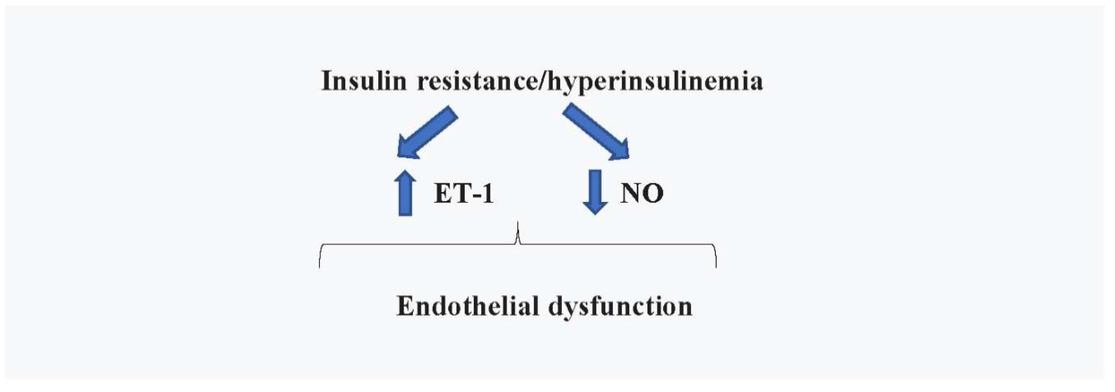
Figure 2.
Insulin resistance with associated hyperinsulinemia, by producing endothelial dysfunction, increased sympathetic tone and anti-natriuretic action, causes arterial hypertension, which, together with the stimulation of myocardiocyte growth by insulin, produce pathologic concentric remodeling of LV. LV: left ventricle; HFpEF: Heart failure with preserved ejection fraction.
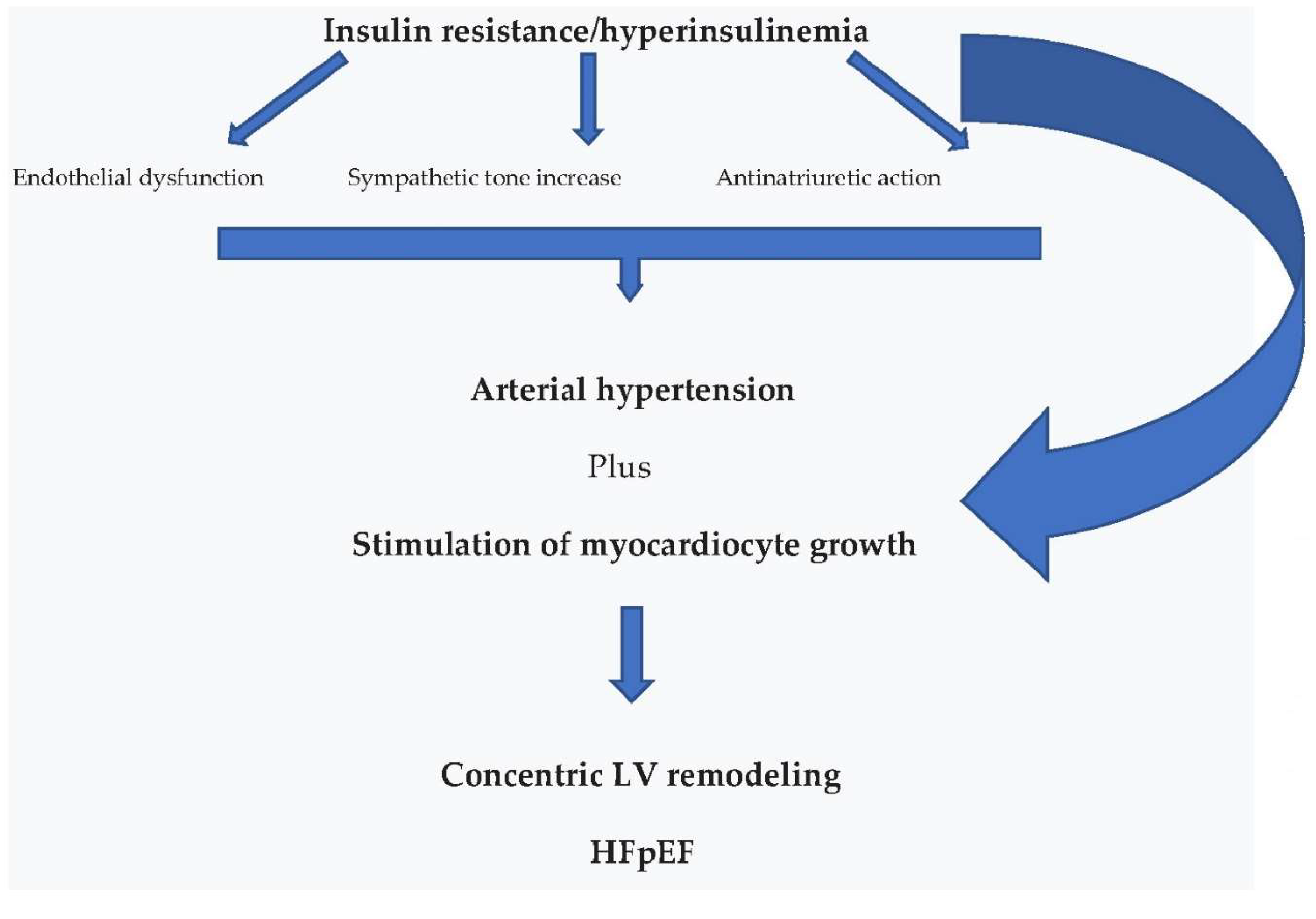
Figure 3.
Hyperinsulinemia associated with IR causes cellular senescence in target organs of insulin actions. Both increased circulating levels of insulin and cellular senescence stimulate cancer development by upregulating p38MAPK and MAPK/ERK signaling. P38MAPK: P38 mitogen activated protein kinases; MAPK: Mitogen activated protein kinase; ERK: Extracellular signal-regulated kinase.
Disclaimer/Publisher’s Note: The statements, opinions and data contained in all publications are solely those of the individual author(s) and contributor(s) and not of MDPI and/or the editor(s). MDPI and/or the editor(s) disclaim responsibility for any injury to people or property resulting from any ideas, methods, instructions or products referred to in the content. |
© 2024 by the authors. Licensee MDPI, Basel, Switzerland. This article is an open access article distributed under the terms and conditions of the Creative Commons Attribution (CC BY) license (http://creativecommons.org/licenses/by/4.0/).
Copyright: This open access article is published under a Creative Commons CC BY 4.0 license, which permit the free download, distribution, and reuse, provided that the author and preprint are cited in any reuse.


