Submitted:
09 September 2024
Posted:
10 September 2024
You are already at the latest version
Abstract
Osteomyelitis is a significant cause of total carcass condemnation in pigs at the slaughterhouse. The decision for total condemnation of a pig carcass for osteomyelitis is often based on traditional perceptions of the risk of pyemia, leading to controversy among Official Veterinarians (OV) in the industry. This review aims to provide a more comprehensive understanding of the etiopatho-genesis of osteomyelitis in pigs, the microorganisms involved, and the risk factors. It also high-lights the urgent need for a more uniform method to evaluate osteomyelitis cases, which could significantly reduce economic losses in the industry. Lesions originating from tail-biting, tail docking, castration, teeth resection, and raw management are described as risk factors for oste-omyelitis. Osteomyelitis is caused by the entry of pathogens into the bloodstream of the animal through an open wound. The most described pathogens are Trueperella monocytogenes, Staphylo-coccus aureus, and Streptococcus spp. At slaughter, OVs condemn carcasses with osteomyelitis due to pyemia. Signs of acute disease are essential to identify pyemia cases. In chronic cases, total carcass condemnation can be avoided depending on the number of lesions and number of vertebrae af-fected. A clear overall image of the problem would help authorities in various countries adopt a more homogenous approach.
Keywords:
osteomyelitis
; meat inspection
; swine
; food safety
1. Introduction
Pork is the most consumed meat worldwide [1]. Its impact on the food industry and society increases the demand by consumers for safe meat [2]. The consumption of contaminated meat is still a serious food safety risk globally [3]. Meat inspection at the slaughterhouse determines if the meat is fit for human consumption [3,4]. Its importance extends to the identification, by assessment of pathological findings, of animal health and welfare problems [4,5]. Traditional post-mortem inspection consists of the incision and palpation of the carcass and organs to arrive at a sanitary decision [4]. The inspection focused on detecting zoonotic diseases, such as brucellosis and tuberculosis, which at present are rare to observe at the slaughterhouse [3]. Furthermore, traditional meat inspection now poses a modern food safety risk [3]. Incision and palpation of several carcasses promote the spread of pathogens (such as Salmonella, Campylobacter and Yersinia), leading to cross-contamination [3,4]. The European Union now promotes a more modern meat inspection, where OVs make a sanitary decision through risk assessment, risk management and risk communication [4]. This involves visual inspection and assessing food chain information [4].
During post-mortem inspection of pigs at the slaughterhouse, many findings can lead to total carcass condemnation [6,7]. Pneumonia, multiple abscesses, arthritis and osteomyelitis are all important causes of total carcass condemnation in pigs [6,7]. Osteomyelitis has been the most impactful condemnation cause in countries such as Portugal, leading to high economic losses [7]. Osteomyelitis is an inflammatory process caused by pathogenic bacteria that affects the bone [8,9,10]. Osteomyelitis may affect long bones, physis, and vertebrae, causing possible fractures and spinal cord compression consecutive to vertebral osteomyelitis [11,12]. Due to the high vascularisation of the bone marrow, osteomyelitis is associated with the risk of pyemia [10]. In the European Union, Regulation EC 2019/627 specifies that meat derived from animals affected by a generalised disease must be declared unfit for human consumption. This includes septicemia, pyemia, toxaemia, and viremia [13]. Thus, if signs of pyemia are present in pig carcasses with osteomyelitis, then the entire carcass must be condemned.
Nevertheless, pig carcasses affected by osteomyelitis are still a problem up for debate between OVs. Research on the risk factors and characteristics of the disease is severely lacking [7]. Detecting osteomyelitis lesions and characterising them as acute or chronic continues to be difficult [10]. This shortage of information can be detrimental for OVs to apply a uniform and correct approach to osteomyelitis cases. The criteria for sanitary decision-making are unclear, leading to controversial discussions among OVs. Unnecessary condemnations can lead to avoidable economic losses and minor food waste. On the other hand, carcasses with pyemia mistakenly deemed fit for human consumption can represent a serious health risk for consumers [10].
This review aims to provide more clarity to the OVs on making the correct decision based on a more in-depth knowledge of the pathogenesis, the risk factors, and microorganisms involved in osteomyelitis, combined with a summary of the various sanitary decision options. It also aims to promote the need for food safety authorities to develop more unambiguous, homogeneous and uniform guidelines regarding decision-making regarding osteomyelitis.
2. Pathogenesis of Osteomyelitis
Osteomyelitis is an inflammatory process of the bone and bone marrow that leads to bone destruction [14,15,16]. An accumulation of pus at the lesion location often occurs [10]. It is primarily associated with bacterial infection and can be accompanied by secondary infections [14,17]. Bacteria infect the bone through three possible routes: hematogenous, local direct extension from adjacent soft tissues, and inoculation [14,17]. The pathogenesis can follow acute, subacute, or chronic courses [15].
At the early stage, after bacterial contamination and adhesion, the response is characterised by acute inflammation and oedema [17]. Following this, purulent material can spread widely through the medullary canal, and on some occasions, it can destroy the cortical bone, burst through the periosteum, and lead to infection in the surrounding soft tissues or drain externally [14]. Bone necrosis may occur when the purulent material increases pressure in the medullary cavity and thrombosis blood vessels [14]. Necrotic debris and a mixed population of inflammatory cells may fill the marrow spaces between necrotic and partly reabsorbed bone trabeculae, and a thick layer of granulation tissue may surround larger areas of necrotic bone [17].
Usually, in red meat animals, osteomyelitis is secondary to trauma, such as castrated wounds and umbilical infection in neonates [14]. Iatrogenic or spontaneous inoculation of infectious agents into traumatic or surgical wounds results in osteomyelitis [18] and may develop hematogenous spread. Hematogenous osteomyelitis is reported chiefly in young animals; it is commonly found in young horses and ruminants and affects mainly the metaphyseal area of the bone due to the vascular pattern [16,17,18]. In neonatal foals, hematogenous osteomyelitis can be associated with immunocompromise after failure of passive transfer of immunity [14]. Osteomyelitis in cows is commonly associated with actinomycosis and brucellosis infections [14]. In adult horses, the risk of osteomyelitis is higher after trauma or following internal fixation of fractures [14,16]. In domestic ruminants, maxillomandibular osteomyelitis is frequently associated with bacteria penetration due to sharp and hard forage through the primary trauma to the oral cavity [16]. Actinomyces bovis infection, a common cause of mandibular osteomyelitis in cattle, can occasionally affect pigs [17].
Vertebral osteomyelitis (VO) is relatively frequent in cattle, sheep, goats, horses, and pigs [16,17]. It leads to severe pain and neurological symptoms, such as tetraparesis [10,19]. VO is an inflammation that affects the vertebrae and involves the medullar cavity [20]. VO in horses is considered a result of hematogenous spread of infection [17]. In pigs, cervical osteomyelitis may occur by direct invasion of bacteria from muscular abscesses, which may result from contaminated injections [10]. Coccygeal and sacral VOs usually result from tail biting [10,17]. Identification of two or more VOs in a pig carcass is a sign that a haematogenous spread of pathogenic organisms has occurred [10].
Osteomyelitis in the oral cavity of pigs can result from dental pulp infection after clipping the teeth of piglets, which exposes the dental pulp [21]. Teiga-Teixeira et al. (2024) reported that 94.78% of osteomyelitis in the anterior region of slaughtered pigs was mandibular osteomyelitis, representing 34.28% of the Total Condemnations. In this study, teeth resection was the factor that most contributed to mandibular osteomyelitis in pigs [7]. Other entry sites of infectious agents associated with VO in pigs are the umbilical vein, bite wounds, and infections after tail docking [19]. Defining the entry site in live animals remains difficult, as when clinical signs of osteomyelitis become noticeable, primary infection is usually resolved [19].
3. Microorganisms Associated with Osteomyelitis in Pigs
Osteomyelitis seems to be caused by various pathogens. Staphylococcus aureus is a bacterium prone to affecting bones [17]. Pigs of all ages are susceptible to the septicemic form of infection of this pathogen [22]. Staphylococcus aureus is a Gram-positive, bio-film-forming bacterium commonly found on the skin of animals and humans and associated with skin and wound infections, abscesses, joint infections, endocarditis, pneumonia, and bacteremia [23,24]. Additionally, it is the seventh major cause of foodborne outbreaks in the European Union, representing 8.41% of the outbreaks linked to meat and meat product consumption in 2022 [25]. Staphylococcus aureus produces various enterotoxins and may cause nausea and diarrhoea [8]. It is resistant to commonly used antibiotics [26,27]. Antimicrobial-resistant Staphylococcus aureus, such as Methicillin-Resistant Staphylococcus aureus (MRSA) and Vancomycin-resistant Staphylococcus aureus (VRSA), is currently a significant challenge to public health [26,27]. The abuse and misuse of antibiotics in animal husbandry and treatment of human infections are the leading causes of the emergence of MRSA in animal production environments [28]. The routes of dissemination of livestock-associated MRSA to humans may include direct contact with animals, incorrect food handling, and consumption of unproperly cooked meat [28]. The study of Santos et al. (2020) on the occurrence of MRSA CC398 in purulent lesions of piglets and fattening pigs in Portugal concluded that domestic pigs act as a reservoir for MRSA and transmit it to humans and animals. The same study states that from all 141 purulent samples collected from pig carcasses, 44 were from osteomyelitis cases. Seven of these samples were Staphylococcus aureus-positive; three were MRSA, representing 6.8% of the total osteomyelitis sampled material [29]. These results raise the concern that osteomyelitis may be associated with the transmission of resistant strains by management and consuming contaminated meat, but further investigations must occur.
Trueperella pyogenes and Streptococcus spp. are also described as bacteria related to pyemia cases associated with osteomyelitis [8,10]. Despite belonging to commensal microflora, both are Gram-positive bacteria that can behave as opportunistic pathogens and lead to infections [30,31]. Trueperella pyogenes is the most common causative agent of osteomyelitis [17]. This agent is an opportunistic animal pathogen mainly associated with various suppurative infections in wild and domestic animals [32]. In cases where the risk of exposure is not mitigated, both Trueperella pyogenes and Streptococcus spp. should be considered as a potential occupational risk for people who work in the slaughterhouse [8,10].
Actinobacillus pleuropneumonia (APP) was also once described as a cause of necrotising osteomyelitis in 8—to 12-week-old pigs [33,34]. A case of osteomyelitis in a vertebral fracture due to Actinobacillus pleuropneumoniae was documented in an 8-week-old female weaned domestic pig [19]. Actinobacillus pleuropneumoniae is a Gram-negative bacterium that resides primarily on the tonsils and mucous membranes of the respiratory tract and, due to stress or other external or internal factors, may lead to necrotising pneumonia, pulmonary oedema, fibrinous pleuritis and also multifocal abscesses [35,36,37]. Although Actinobacillus pleuropneumonia leads to pleuropneumonia and respiratory symptoms, it has been sporadically described as a cause of swine osteomyelitis and fibrinopurulent arthritis [19,37]. Furthermore, it has already been confirmed to be related to multifocal granulomatous hepatitis, nephritis, and meningitis [37]. Teiga-Teixeira et al. (2024) also linked pleurisies, usually connected with Actinobacillus pleuropneumonia infections [38], with a higher occurrence of osteomyelitis in swine carcasses at the abattoir.
Other reported osteomyelitis-leading pathogens are Erysipelothrix rushiopathie, Pseudomonas, E.coli, and Pasteurella haemolytica [19].
4. Risk Factors for Osteomyelitis in Pigs
Research on the causative factors for osteomyelitis is still very scarce [7]. Tail-biting lesions have long been described as a possible predisposing factor [7,10,20]. The presence of tail lesions is common in condemned carcasses due to generalised disease at the slaughterhouse [39,40,41]. Tail-biting outbreaks are characterised by the pigs' behaviour of biting others' tails, increasing the risk of infection and carcass condemnation [42]. It is often associated with reduced animal welfare and may promote the spread of disease during production [20,43]. A tail lesion is an entry site for pathogens to the bloodstream of the animal. This promotes the dissemination of infection to several organs and tissues of the body [40]. This results in the formation of abscesses, lung lesions, arthritis and osteomyelitis in the carcass that are detectable during post-mortem inspection [7]. Bacterial spread to vascular loops beneath growth plate cartilage results in embolic osteomyelitis and often pyemia [44]. Associated with osteomyelitis, the presence of suppurative lesions such as purulent foci in kidneys, lungs, and spleen during post-mortem inspection also suggests pyemia [10,44]. Acute pyemia may be associated with non-capsulated purulent lesions in lymph nodes, muscle, and spleen, hyperemia, and haemorrhages in the liver and lungs [10,45].
An interesting association between swine production management and the occurrence of osteomyelitis was found. Alban et al. (2015) concluded that osteomyelitis was more frequent among batches produced under organic/free-range management than in conventional indoor pig production. According to these authors, tail lesions were more frequent among these pigs, which may explain these results. They also associated their findings with the fact that there is more accessible access to antimicrobials in conventional production than in organic/free-range production, and pigs with tail lesions are more likely to be treated sooner by veterinary practitioners [46].
To reduce tail-biting, pig farmers often resort to tail docking [39,40,47]. As an invasive procedure, tail docking may also play a role in infections that lead to bone infections [10]. Docking consists of amputating a part of the tail, aiming to reduce the attractiveness of the tail for biting [48]. It is usually performed by the farmer or employees without anaesthesia or analgesia, using teeth clippers and scissors to gas or electrical cautery iron [48]. However, the efficiency of tail docking is limited and does not entirely solve tail biting [42,48]. Moreover, not having hygienic tail docking procedures also represents a potential risk of infection and may lead to spinal abscesses, arthritis and osteomyelitis [10,42].
Another described risk factor for osteomyelitis in pigs is teeth resection. Teiga-Teixeira et al. (2024) found an association between osteomyelitis and carcasses with clipped teeth. Teeth resection is a controversial invasive procedure still commonly used in piglets despite being highly discouraged in the European Union. The incisor and canine teeth of the mandible and upper jaw are clipped or ground on the first days of piglets to prevent lesions in the sows' teats and lesions caused by littermates [7]. The advantages of teeth resection are disputable because, alongside severe pain and stress, it may lead to damage and infection of the dental pulp, gums and roots of the teeth [7]. Bacteria may also access the bloodstream and disseminate to other body parts [7,49].
Castration wounds, brucellosis, and atrophic/necrotic rhinitis may be linked with osteomyelitis in pigs [10,14].
Fertner et al. (2017) described a link between herd size and the occurrence of osteomyelitis. Osteomyelitis among finishers affected by tail lesions was more likely to occur in medium-sized herds than in large ones, and some of the author's possible explanations are the better infrastructure conceived for large herds and more experienced staff that works in these herds [50].
5. Osteomyelitis as a Cause of Carcass Condemnation and Sanitary Decision Criteria
Osteomyelitis is one of the leading causes of carcass total carcass condemnations in several countries [6,7,10,51,52,53]. However, it is difficult to compare the prevalence of osteomyelitis cases among different countries. Some recent studies on causes for swine carcass condemnations don't even mention osteomyelitis [54,55,56,57,58]. This can be explained by the fact that national regulatory authorities use different codes to record post-mortem findings. Osteomyelitis can be registered as simply “osteomyelitis” or, in other cases, be defined as “septicemia” or “multiple abscesses” cases in epidemiological databases and documents [59]. The latter makes it practically impossible to use these data to assess osteomyelitis, as many other factors can result in septicemia.
Carcasses affected by osteomyelitis are considered not suitable for human consumption because, in addition to having a disgusting appearance, there is the traditional perception of pyemia occurrence risk, as bone marrow is highly vascularised and has an important role in hematopoiesis [60].
Although some cases of osteomyelitis can relate to neurological and septicaemic clinical signs, tail-biting lesions and abscesses [7,10], the ante-mortem inspection may not be effective in detecting suspected osteomyelitis cases [10]. Osteomyelitis cases are generally detected at post-mortem inspection. At this stage, osteomyelitis is evident as a bone abscess, a deforming inflammatory process with an accumulation of pus [7].
According to Ninios et al. (2014), acute single osteomyelitis should lead to total condemnation as acute lesions are more associated with pyemia before slaughter, while chronic single lesions may lead to partial condemnation [61]. Some authors describe that if a fibrous capsule does not surround the osteomyelitis, the best decision is total condemnation [45].
In Denmark, lesions that may reflect an acute and generalised stage of infection lead to a rejected carcass. In contrast, carcasses with chronic purulent lesions (encapsulated abscesses in bones and organs) are sent to de-boning [8]. De-boning aims to detect abscesses connected to prior septicemia that could not be detected in the rework area (Baekbo et al., 2016). The result is primarily partial condemnation [59].
However, according to Baekbo et al. (2016), de-boning was unnecessary to ensure food safety. These authors proceeded to a bacteriological exam of samples (including from abscesses and muscle) taken from 102 finishing pig carcasses sent for de-boning due to lesions indicative of prior septicemia. They showed that 6% of the abscesses and 83% of the muscle samples were sterile or below detection level. Furthermore, the only potential foodborne pathogen they isolated was Staphylococcus aureus, found in 15 abscesses and one muscle sample [8].
In Portugal, a country where osteomyelitis is the most impactful cause for carcass condemnation, most osteomyelitis cases are not differentiated as acute and chronic. Due to the perceived risk of pyemia and the gap in objective criteria to evaluate osteomyelitis, OVs generally decide the total condemnation of the affected carcass [10]. This lack of criteria leads to inefficient resources for making a correct sanitary decision. As such, many carcasses fit for human consumption may be incorrectly condemned.
Vieira-Pinto et al. (2020) conducted a study on 40 carcasses that were totally condemned due to Vertebral Osteomyelitis (VO) in an undifferentiated way. The same authors registered that half the sampling carcasses presented were chronic lesions. A histological analysis was performed to validate the macroscopic classification of acute or chronic osteomyelitis, revealing a significant association between macroscopic and histological classifications. A microbiological analysis aimed at detecting pyemia cases [10]. Pyemia cases were associated with 46.8% of the generalised cases, which led to total condemnation. The study found that the acute characteristic is essential in identifying pyemia cases. Moreover, after conceiving and applying a decision-making criteria scheme based on the study findings, the authors concluded that 14 out of 40 osteomyelitis cases (11 were single chronic lesions and 3 were two chronic lesions located in different places) could have been possibly spared from total condemnation. However, further studies should be conducted to support these findings.
According to the scheme designed by Vieira-Pinto et al. (2020), the decision of total condemnation in the case of VO must be taken if: a) there is extensive contamination with purulent exudate; b) an acute lesion is present (even if only in one vertebra); c) more than three purulent lesions in different locations (including vertebrae and pyemia-related lesions). According to these authors, in chronic cases, partial condemnation (by trimming/cutting/deboning affected areas) is suitable if, after additional post-mortem procedures, a single vertebra is affected by a chronic purulent lesion or if two chronic purulent lesions were in different locations.
In 2022, a European survey on post-mortem inspection of finishing pigs assessed the total condemnation criteria to declare meat unfit for human consumption [60]. This study concluded that nine out of forty respondents' answers (22.5%) selected the option that specified total condemnation in the case of osteomyelitis. In contrast, eighteen out of forty answers chose the option of total condemnation in case of other post-mortem inspection findings, such as abscesses (for 50% of these answers) and acute stage of osteomyelitis (for the other 50%). Secondary to osteomyelitis, abscesses are commonly associated with pyemia. However, osteomyelitis may also be a unique lesion [60]. In summary, it is essential to evaluate the concomitant abscesses and their distribution, to establish the source of infection, and to assess whether an osteomyelitis lesion indicates an acute stage.
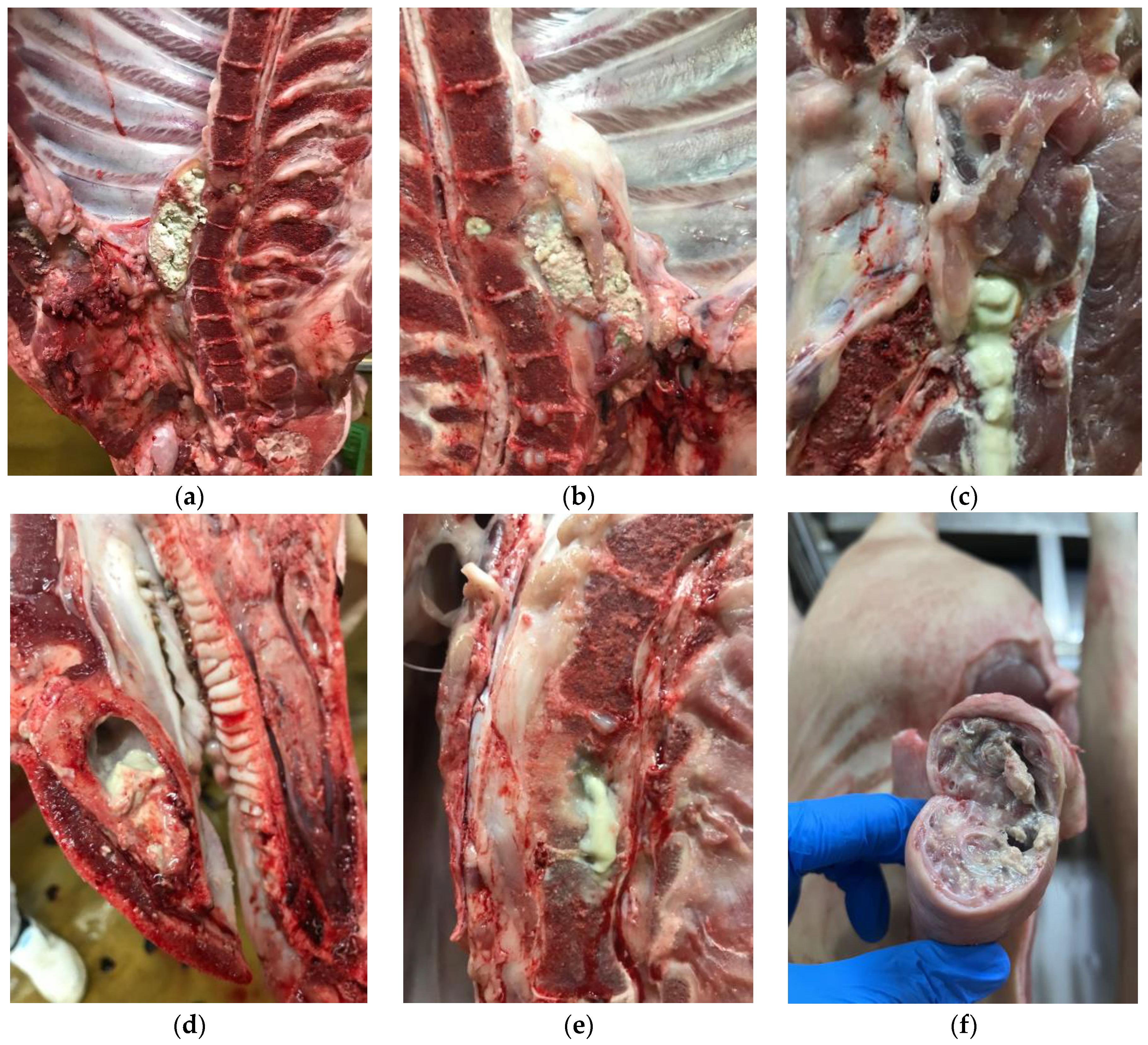
During the post-mortem inspection procedure, osteomyelitis classification according to its chronicity may be challenging. Macroscopically, acute osteomyelitis may be represented by shiny, moist lesions with congested areas, evident bone destruction not circumscribed by adjacent remodelling tissue, and fluid purulent exudate [10]. Chronic lesions may present remodelling tissue that circumscribes moderate bone destruction without visible congested areas and thickened exudate [7]. In Figure 1, osteomyelitis lesions in the mandible and vertebrae are macroscopically classified according to the chronicity of the process.
Additional post-mortem inspection (APMI) procedures are crucial to differentiate localised and systemic cases [62]. If necessary, an incision of the iliopsoas musculature and the shoulder and topside can be performed for suspected cases of osteomyelitis and pyemia [62]. Laukkanen-Ninios et al. (2022) studied the use and variability of APMI procedures and laboratory methods as supplements for visual meat inspection of finishing pigs in Europe. They found that osteomyelitis was one of the most minor conditions associated with required APMI procedures. According to these authors, thanks to the traditional splitting of carcasses, vertebral osteomyelitis is easily visible without APMI procedures [62]. However, this statement raises a question. For market reasons, all carcasses are cut longitudinally in two portions. This permits an easy vertebral osteomyelitis assessment, but other bones are difficult to evaluate during post-mortem inspection [43]. Furthermore, the sagittal cut of the head for meat inspection is not mandatory and not applied in many (if not in the majority) of European swine slaughterhouses due to commercial reasons, and this may be a limitation in detecting mandibular osteomyelitis in pig carcasses [7]. Considering that the whole pig's head is a piece sold, exported and consumed in several countries, this can become an issue of quality and food safety.
6. Conclusions
Osteomyelitis causes considerable economic losses to the pork industry. However, studies about the disease and its assessment during post-mortem inspection at the slaughterhouse remain scarce. This condition is often caused by Staphylococcus aureus, Trueperella pyogenes and Streptococcus spp. infections. Invasive farm procedures, open wounds and compromised welfare are all risk factors for osteomyelitis. Bone infections by Staphylococcus aureus are considered a major food safety risk in pig carcasses. Osteomyelitis is a common cause of total carcass condemnation in pig carcasses. The most affected locations are the lumbar and thoracic vertebrae. Cases of osteomyelitis usually indicate a generalised infection in the carcass, which deems it unfit for human consumption. The recognition of signs of pyemia is vital to a correct sanitary judgement by the OV during post-mortem inspection of pigs. However, the identification of such signs is difficult, and information about their assessment is unclear. This lack of information and research leads to different opinions and sanitary decisions among OVs. A very important step in the inspection process is to identify the lesion as acute or chronic. Acute osteomyelitis cases are more likely for pyemia to be present in the carcass. Acute lesions contain a shiny and moist purulent exudate often associated with congestion. Chronic lesions present remodelling tissue that clearly contains the purulent exudate in the lesion. Bone destruction is moderate, and the purulent material is thick and more caseous. Congestion is rare in older osteomyelitis cases. Thus, carcasses with chronic osteomyelitis may be spared from total condemnation. However, more studies are needed to confirm this hypothesis as clear evidence is lacking.
A more informed decision making by OVs in osteomyelitis cases promotes uniformity and correctness in food safety inspection of pig carcasses. The development of guidelines for this specific inspection finding is essential to avoid further economic losses.
Author Contributions
Conceptualization and writing—original draft preparation, M.A.R; writing—review and editing, M.A.R, A.E., P.T.-T., F.S.; supervision, A.E.; funding acquisition, A.E. All authors have read and agreed to the published version of the manuscript.
Funding
This study was supported by the projects UIDB/00772/2020 and LA/P/0059/2020, funded by the Portuguese Foundation for Science and Technology (FCT).
Data Availability Statement
Not applicable.
Acknowledgments
We would like to thank the support of the Food and Veterinary Division of Vila Real and Douro Sul, the Directorate of Food and Veterinary of the North Region (DSAVRN), and the Directorate-General for Food and Veterinary Affairs (DGAV).
Conflicts of Interest
The authors declare no conflict of interest.
References
- Markets, R.a. (2020). Global pork market forecast (2018 to 2026) - by production, consumption, import, export & company. from https://www.globenewswire.com/n.
- Tao, F., & Peng, YA method for nondestructive prediction of pork meat quality and safety attributes by hyperspectral imaging technique. J. Food Eng. 2014, 126, 98-106. [CrossRef]
- Riess, L. E., & Hoelzer, K. Implementation of visual-only swine inspection in the European Union: Challenges, opportunities, and lessons learned. J. Food Prot. 2020, 83(11), 1918-1928. [CrossRef]
- Li, T. T., Langforth, S., Isbrandt, R., Langkabel, N., Sotiraki, S., Anastasiadou, S., ... & Meemken, D. Food chain information for pigs in Europe: A study on the status quo, the applicability and suggestions for improvements. Food Control 2024, 157, 110174. [CrossRef]
- Vecerek, V.; Voslarova, E.; Semerad, Z.; Passantino, A. The health and welfare of pigs from the perspective of post mortem findings in slaughterhouses. Animals 2020, 10, 825. [CrossRef]
- Garcia-Diez, J.; Coelho, A.C. Causes and factors related to pig carcass condemnation. Vet. Med. 2014, 59, 194–201. https://doi.org./10.17221/7480-VETMED.
- Teiga-Teixeira, P., Alves Rodrigues, M., Moura, D., Teiga-Teixeira, E., & Esteves, A. Osteomyelitis in Pig Carcasses at a Portuguese Slaughterhouse: Association with Tail-Biting and Teeth Resection. Animals 2024, 14(12), 1794. [CrossRef]
- Baekbo, A.K.; Petersen, J.V.; Larsen, M.H.; Alban, L. The food safety value of de-boning finishing pig carcasses with lesions indicative of prior septicemia. Food Control 2016, 69, 177–184. [CrossRef]
- Patel, M.; Rojavin, Y.; Jamali, A.A.; Wasielewski, S.J.; Salgado, C.J. Animal models for the study of osteomyelitis. Semin. Plast. Surg. 2009, 23, 148–154. [CrossRef]
- Vieira-Pinto, M., Azevedo, J., Poeta, P., Pires, I., Ellebroek, L., Lopes, R., ... & Alban, L. Classification of vertebral osteomyelitis and associated judgment applied during post-mortem inspection of swine carcasses in Portugal. Foods 2020, 9(10), 1502. [CrossRef]
- Jensen, L. K., Johansen, A. S., & Jensen, H. E. Porcine models of biofilm infections with focus on pathomorphology. Front. Microbiol. 2017, 8, 1961. [CrossRef]
- Madson, D. M., Arruda, P. H., & Arruda, B. L. Nervous and locomotor system. In Diseases of swine, 11th ed. Jeffrey, J.Z., Lock, A.K., Ramirez, A., Kent, J.S., Gregory, W.S., Zhang, J., Eds.; John Wiley & Sons, Inc.: Hoboken, NJ, USA, 2019; pp. 339–372. [CrossRef]
- Commission Implementing Regulation (EU) 2019/627 of 15 March 2019, laying down uniform practical arrangements for the performance of official controls on products of animal origin intended for human consumption in accordance with Regulation (EU) 2017/625 of the European Parliament and of the Council and amending Commission Regulation (EC) No 2074/2005 as regards official controls. Off. J. Eur. Union 2019, 131, 51–100.
- Clegg, P. D. Osteomyelitis in the veterinary species. In Biofilms and veterinary medicine. Percival, S., Knottenbelt, D., Cochrane, C. Berlin, Eds; Springer Series on Biofilms, vol 6. Springer, Heidelberg, 2011; pp. 175-190. [CrossRef]
- Roy, M., Somerson, J. S., Kerr, K. G., & Conroy, J. L. Pathophysiology and pathogenesis of osteomyelitis. INTECH Open Access Publisher 2012, 1-26.
- González-Martín, M., Silva, V., Poeta, P., Corbera, J. A., & Tejedor-Junco, M. T. Microbiological aspects of osteomyelitis in veterinary medicine: drawing parallels to the infection in human medicine. Vet. Q. 2022, 42(1), 1-11. [CrossRef]
- Craig, L.E.; Dittmer, K.E.; Thompson, K.G. Bones and Joints. In Jubb, Kennedy, and Palmer’s Pathology of Domestic Animals, 6th ed.; Maxie, G., Ed.; Academic Press: Cambridge, MA, USA, 2016; pp. 16–163.
- Gieling, F., Peters, S., Erichsen, C., Richards, R. G., Zeiter, S., & Moriarty, T. F. Bacterial osteomyelitis in veterinary orthopedics: pathophysiology, clinical presentation and advances in treatment across multiple species. Vet. J. 2019, 250, 44-54. [CrossRef]
- Giebels, F., Geissbühler, U., Oevermann, A., Grahofer, A., Olias, P., Kuhnert, P., ... & Stein, V. M. Vertebral fracture due to Actinobacillus pleuropneumoniae osteomyelitis in a weaner. BMC Vet. Res. 2020, 16, 1-7. [CrossRef]
- García-Díez, J., Saraiva, S., Moura, D., Grispoldi, L., Cenci-Goga, B. T., & Saraiva, C. The importance of the slaughterhouse in surveilling animal and public health: a systematic review. Vet. Sci. 2023, 10(2), 167. [CrossRef]
- Thomson, J.R.; Friendship, R.M. Digestive system. In Disease of Swine, 11th ed.; Zimmerman, J.J., Karriker, L.A., Ramirez, A., Schwartz, K.J., Stevenson, G.W., Zhang, J., Eds.; Wiley-Blackwell: Hoboken, NJ, USA, 2019; pp. 234–263. [CrossRef]
- Serbessa, T. A., Geleta, Y. G., & Terfa, I. O. Review on diseases and health management of poultry and swine. Int. j. avian wildl. biol. 2023, 7(1), 27-38. [CrossRef]
- Kawanishi, M., Matsuda, M., Abo, H., Ozawa, M., Hosoi, Y., Hiraoka, Y., ... & Sekiguchi, H. Prevalence and Genetic Characterization of Methicillin-Resistant Staphylococcus aureus Isolated from Pigs in Japan. Antibiotics 2024, 13(2), 155. [CrossRef]
- Smith, R. P., Sharma, M., Gilson, D., Anjum, M., & Teale, C. J. Livestock-associated methicillin-resistant Staphylococcus aureus in slaughtered pigs in England. Epidemiol. Infect. 2021, 149, e236. [CrossRef]
- EFSA - Monitoring of foodborne diseases. https://www.efsa.europa.eu/en/microstrategy/FBO-dashboard (accessed on 1st august 2024).
- Pal, M., Shuramo, M. Y., Tewari, A., Srivastava, J. P., & HD, C. Staphylococcus aureus from a Commensal to Zoonotic Pathogen: A Critical Appraisal. Int. J. Clin. Exp. Med. Res 2023, 7, 220-228. [CrossRef]
- Gungor, C., Onmaz, N. E., Gundog, D. A., Yavas, G. T., Koskeroglu, K., & Gungor, G. Four novel bacteriophages from slaughterhouse: Their potency on control of biofilm-forming MDR S. aureus in beef model. Food Control 2024, 156, 110146. [CrossRef]
- Bouchami, O., Fraqueza, M. J., Faria, N. A., Alves, V., Lawal, O. U., de Lencastre, H., & Miragaia, M. Evidence for the dis-semination to humans of methicillin-resistant Staphylococcus aureus ST398 through the pork production chain: a study in a Portuguese slaughterhouse. Microorganisms 2020, 8(12), 1892. [CrossRef]
- Santos, V., Gomes, A., Ruiz-Ripa, L., Mama, O. M., Sabença, C., Sousa, M., ... & Poeta, P. A. C. Q. D. Methicillin-resistant Staphylococcus aureus CC398 in purulent lesions of piglets and fattening pigs in Portugal. Microb. Drug Resist. 2020, 26(7), 850-856. [CrossRef]
- Zhang, Z., Liang, Y., Yu, L., Chen, M., Guo, Y., Kang, Z., ... & Liu, M. TatD DNases contribute to biofilm formation and virulence in Trueperella pyogenes. Front. Microbiol. 2021, 12, 758465. [CrossRef]
- Costinar, L., Badea, C., Marcu, A., Pascu, C., & Herman, V. Multiple Drug Resistant Streptococcus Strains—An Actual Problem in Pig Farms in Western Romania. Antibiotics 2024, 13(3), 277. [CrossRef]
- Tamai, I. A., Mohammadzadeh, A., Mahmoodi, P., Pakbin, B., & Salehi, T. Z. Antimicrobial susceptibility, virulence genes and genomic characterization of Trueperella pyogenes isolated from abscesses in dairy cattle. Res. Vet. Sci. 2023, 154, 29-36. [CrossRef]
- Jensen TK, Boye M, Hagedorn-Olsen T, Riising HJ, Angen O. Actinobacillus pleuropneumoniae osteomyelitis in pigs demonstrated by fluorescent in situ hybridization. Vet Pathol. 1999 36:258–61. [CrossRef]
- Hennig-Pauka, I., Hartmann, M., Merkel, J., & Kreienbrock, L. Coinfections and Phenotypic Antimicrobial Resistance in Actinobacillus pleuropneumoniae Strains Isolated From Diseased Swine in North Western Germany—Temporal Patterns in Samples From Routine Laboratory Practice From 2006 to 2020. Front. Vet. Sci. 2022, 8, 802570. [CrossRef]
- Maes, D., Sibila, M., Pieters, M., Haesebrouck, F., Segalés, J., & de Oliveira, L. G. Review on the methodology to assess respiratory tract lesions in pigs and their production impact. Vet. Res. 2023, 54(1), 8. [CrossRef]
- Seakamela, E. M., Henton, M. M., Jonker, A., Kayoka-Kabongo, P. N., & Matle, I. Temporal and Serotypic Dynamics of Actinobacillus pleuropneumoniae in South African Porcine Populations: A Retrospective Study from 1985 to 2023. Pathogens 2024, 13(7), 599. [CrossRef]
- Tenk, M., Tóth, G., Márton, Z., Sárközi, R., Szórádi, A., Makrai, L., ... & Fodor, L. Examination of the Virulence of Actinobacillus pleuropneumoniae Serovar 16 in Pigs. Vet. Sci. 2024, 11(2), 62. [CrossRef]
- Meyns, T., Van Steelant, J., Rolly, E., Dewulf, J., Haesebrouck, F., & Maes, D. A cross-sectional study of risk factors as-sociated with pulmonary lesions in pigs at slaughter. Vet. J. 2011, 187(3), 388-392. [CrossRef]
- Vom Brocke, A.L.; Karnholz, C.; Madey-Rindermann, D.; Gauly, M.; Leeb, C.; Winckler, C.; Schrader, L.; Dippel, S. Tail Lesions in Fattening Pigs: Relationships with Post-mortem Meat Inspection and Influence of a Tail biting Management Tool. Animal 2019, 13, 835–844. [CrossRef]
- Schrøder-Petersen, D.L.; Simonsen, H.B. Tail biting in pigs. Vet. J. 2001, 162, 196–210. [CrossRef]
- Kritas, S.K.; Morrison, R.B. Relationships between tail biting in pigs and disease lesions and condemnations at slaughter. Vet. Rec. 2007, 160, 149–152. [CrossRef]
- Valros, A., & Heinonen, M. Save the pig tail. Porc. Health Manag. 2015, 1, 1-7. [CrossRef]
- Martínez, J., Jaro, P. J., Aduriz, G., Gómez, E. A., Peris, B., & Corpa, J. M. Carcass condemnation causes of growth retarded pigs at slaughter. Vet. J. 2007, 174(1), 160-164. [CrossRef]
- Lindén, J.; Pohjola, L.; Rossow, L.; Tognetti, D. Meat Inspection Lesions. In Meat Inspection and Control in the Slaughterhouse, 1st ed.; Ninios, T., Lundén, J., Korkeala, H., Fredriksson-Ahomaa, M., Eds.; Wiley Blackwell: West Sussex, UK, 2014; Chapter 8; pp. 163–199. [CrossRef]
- Jensen, H.E.; Leifsson, P.S.; Nielsen, O.L.; Agerholm, J.S.; Iburg, T. Meat Inspection: The Pathoanatomic Basis; Bifolia: Frederiksberg, Denmark, 2017; pp. 661–663.
- Alban, L., Petersen, J. V., & Busch, M. E. A comparison between lesions found during meat inspection of finishing pigs raised under organic/free-range conditions and conventional, indoor conditions. Porc. Health Manag. 2015, 1, 1-11. [CrossRef]
- Harley, S., More, S., Boyle, L., Connell, N. O., & Hanlon, A. Good animal welfare makes economic sense: potential of pig abattoir meat inspection as a welfare surveillance tool. Ir. Vet. J. 2012, 65, 1-12.
- Nannoni, E., Valsami, T., Sardi, L., & Martelli, GTail docking in pigs: a review on its short-and long-term consequences and effectiveness in preventing tail biting. Ital. J. Anim. Sci. 2014, 13(1), 3095.
- Reese, D.; Straw, B.E. Teeth Clipping—Have You Tried to Quit? Neb. Swine Rep. 2005, 33, 12–13. Available online: https://digitalcommons.unl.edu/coopext_swine/33 (accessed on 23 July 2024).
- Fertner, M., Denwood, M., Birkegård, A. C., Stege, H., & Boklund, A. Associations between antibacterial Treatment and the Prevalence of Tail-Biting-related sequelae in Danish Finishers at slaughter. Front. vet. sci. 2017, 4, 182. [CrossRef]
- Gomes-Neves, E., Müller, A., Correia, A., Capas-Peneda, S., Carvalho, M., Vieira, S., & Cardoso, M. F. Food chain information: Data quality and usefulness in meat inspection in Portugal. J. Food Prot. 2018, 81(11), 1890-1896. [CrossRef]
- DGAV. Análise Exploratória dos Dados de Abate de Ungulados Para Consumo Humano em Portugal Entre Janeiro de 2011 e Dezembro de 2019. 2020. Available online: https://www.dropbox.com/s/26p3sukb6vx6koc/Dados%20de%20abates%20e%20reprova%C3%A7%C3%B5es%20Ungulados%202011%20a%202019.pdf?dl=0 (accessed on 6th july 2024).
- Franco, R., Gonçalves, S., Cardoso, M. F., & Gomes-Neves, E. Tail-docking and tail biting in pigs: Findings at the slaughterhouse in Portugal. Livest. Sci. 2021, 254, 104756. [CrossRef]
- Bueno, L. S., Caldara, F. R., Nääs, I. A., Salgado, D. D., García, R. G., & Almeida Paz, I. C. Swine carcass condemnation in commercial slaughterhouses. Rev. MVZ Cordoba 2013, 18(3), 3836-3842.
- Ceccarelli, M., Leprini, E., Sechi, P., Iulietto, M. F., Grispoldi, L., Goretti, E., & Cenci-Goga, B. T. Analysis of the causes of the seizure and destruction of carcasses and organs in a slaughterhouse in central Italy in the 2010-2016 period. Ital. J. Food Saf. 2018, 7(1). [CrossRef]
- Guardone, L., Vitali, A., Fratini, F., Pardini, S., Cenci Goga, B. T., Nucera, D., & Armani, A. A retrospective study after 10 years (2010–2019) of meat inspection activity in a domestic swine abattoir in tuscany: The slaughterhouse as an epidemiological observatory. Animals 2020, 10(10), 1907. [CrossRef]
- Akkina, J., Burkom, H., Estberg, L., Carpenter, L., Hennessey, M., & Meidenbauer, K. Feral Swine Commercial Slaughter and Condemnation at Federally Inspected Slaughter Establishments in the United States 2017–2019. Front. vet. sci. 2021, 8, 690346. [CrossRef]
- Rosamilia, A., Galletti, G., Benedetti, S., Guarnieri, C., Luppi, A., Capezzuto, S., ... & Marruchella, G. Condemnation of Porcine Carcasses: A Two-Year Long Survey in an Italian High-Throughput Slaughterhouse. Vet. Sci. 2023, 10(7), 482. [CrossRef]
- Alban, L., Vieira-Pinto, M., Meemken, D., Maurer, P., Ghidini, S., Santos, S., ... & Langkabel, N. Differences in code terminology and frequency of findings in meat inspection of finishing pigs in seven European countries. Food Control 2022, 132, 108394. [CrossRef]
- Vieira-Pinto, M., Langkabel, N., Santos, S., Alban, L., Laguna, J. G., Blagojevic, B., ... & Laukkanen-Ninios, R. A European survey on post-mortem inspection of finishing pigs: Total condemnation criteria to declare meat unfit for human consumption. Res. Vet. Sci. 2022, 152, 72-82. [CrossRef]
- Ninios, T. Judgment of meat. In Meat Inspection and Control in the Slaughterhouse, 1st ed.; Ninios, T., Lundén, J., Korkeala, H., Fredriksson-Ahomaa, M., Eds.; Wiley Blackwell: West Sussex, UK, 2014; Chapter 10; pp. 219–224.
- Laukkanen-Ninios, R., Ghidini, S., Gomez Laguna, J., Langkabel, N., Santos, S., Maurer, P., ... & Vieira-Pinto, M. Additional post-mortem inspection procedures and laboratory methods as supplements for visual meat inspection of finishing pigs in Europe—Use and variability. J. Consum. Prot. Food S. 2022, 17(4), 363-375. [CrossRef]
Figure 1.
Macroscopical classification of osteomyelitis lesions in pigs' carcasses: (a) and (b) are chronic osteomyelitis lesions located in the thoracic vertebrae, characterised by the presence of caseous material and a capsulated abscess at the ventral level; (c) acute osteomyelitis in a coccygeal vertebra, with fluid purulent exudate; (d) intermediate to chronic mandibular osteomyelitis presenting a more thickened pus, but still evident bone destruction and congestion; (e) thoracic vertebra with acute osteomyelitis with fluid pus; (f) chronic osteomyelitis in the caudal vertebrae associated with caseous material and tail tip necrosis.
Disclaimer/Publisher’s Note: The statements, opinions and data contained in all publications are solely those of the individual author(s) and contributor(s) and not of MDPI and/or the editor(s). MDPI and/or the editor(s) disclaim responsibility for any injury to people or property resulting from any ideas, methods, instructions or products referred to in the content. |
© 2024 by the authors. Licensee MDPI, Basel, Switzerland. This article is an open access article distributed under the terms and conditions of the Creative Commons Attribution (CC BY) license (http://creativecommons.org/licenses/by/4.0/).
Copyright: This open access article is published under a Creative Commons CC BY 4.0 license, which permit the free download, distribution, and reuse, provided that the author and preprint are cited in any reuse.


