Submitted:
27 August 2024
Posted:
28 August 2024
You are already at the latest version
Abstract
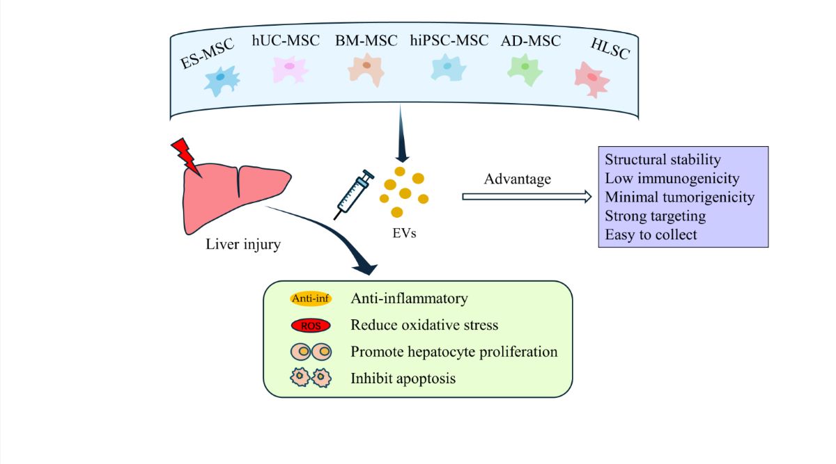
Liver injury caused by various factors significantly impacts human health. Stem cell transplantation has potential for enhancing liver functionality, but safety concerns such as immune rejection, tumorigenesis, and the formation of emboli in the lungs remain. Recent studies have shown that stem cells primarily exert their effects through the secretion of extracellular vesicles (EVs). EVs have been shown to play crucial roles in reducing inflammation, preventing cell death, and promoting liver cell proliferation. Additionally, they can function as carriers to deliver targeted drugs to the liver, thereby exerting specific physiological effects. EVs possess several advantages, including structural stability, low immunogenicity, minimal tumorigenicity targeting capabilities, and convenient collection,. Consequently, these factors have garnered significant attention from researchers and are expected to become alternative therapeutic agents to stem cell therapy. This article provides a comprehensive review of the current research progress on the use of stem cell-derived EVs in the treatment of liver injury.
Keywords:
Extracellular vesicles
; Stem cells
; Liver injury
; Animal models
; Clinical research
1. Introduction
Liver injury can arise from various causes, such as drug toxicity, surgical resection, oxidative stress, and inflammatory reactions after liver transplantation. These injuries often result in severe liver dysfunction, thereby exerting significant effects on the quality of life experienced by affected patients [1,2].
The use of stem cells, which are characterized by their pluripotent differentiation potential and self-renewal ability, has emerged as a promising approach in the field of liver disease therapy. Extensive animal experiments and clinical studies have demonstrated the therapeutic efficacy of stem cell transplantation in conditions such as liver ischemia‒reperfusion injury (IRI) [3], liver fibrosis [4,5], and liver cancer [6]. Commonly used stem cell types include embryonic stem cells, hematopoietic stem cells, mesenchymal stem cells, liver stem cells, and induced pluripotent stem cells [7]. The mechanisms underlying the therapeutic effects of stem cell therapy include the induction of endogenous cell proliferation, the inhibition of cell apoptosis, and immune regulation [8]. However, several challenges need to be addressed in stem cell therapy, such as the limited in vivo survival time of transplanted stem cells [9], low homing efficiency [10], immune rejection [11], and the risk of pulmonary embolism following intravenous injection [12,13].
In recent years, research has highlighted the crucial role of extracellular vesicles (EVs) secreted by stem cells in mediating therapeutic effects rather than relying solely on the differentiation of stem cells into functional cells [14,15,16,17,18]. Stem cell-derived EVs (SC-EVs) retain similar contents as their parent cells, endowing them with biological functions akin to those of the original stem cells [18,19,20]. Compared to stem cells, SC-EVs offer several advantages: (1) smaller size, thereby preventing entrapment and thrombus formation within the microvasculature [21]; (2) precise localization to the liver after intravenous injection [22]; (3) flexible dosage adjustments [22]; (4) lower immunogenicity due to decreased levels of membrane-bound proteins [23]; (5) minimal tumorigenicity due to the absence of cellular components [24]; and (6) a relatively simple structure that allows for modifications to confer specific biological functions for precise therapies [25]. Consequently, the use of SC-EVs has emerged as a promising alternative therapeutic strategy to conventional stem cell therapy. This review provides a comprehensive summary of the current research progress on the application of SC-EVs in the treatment of liver injury.
2. Extracellular Vesicles
EVs are membrane-bound vesicles released by various cell types that contain a diverse array of biomolecules, such as nucleic acids (DNA, mRNA, lncRNA, microRNA), proteins, peptides, lipids, and other bioactive molecules [26,27]. EVs play crucial roles in intercellular communication, cell growth, apoptosis inhibition, angiogenesis, and immune regulation by transferring their cargo to target cells by fusing with the target cell membrane, endocytosis, or binding to surface receptors on target cells [28]. Based on their biogenesis mechanisms, EVs can be classified into three main types: exosomes, microvesicles, and apoptotic bodies [29]. Exosomes are formed through the invagination of the plasma membrane, leading to the creation of early endosomes that mature into late endosomes, eventually developing into multivesicular bodies (MVBs). These MVBs then fuse with the plasma membrane, releasing exosomes with diameters of approximately 30-150 nm [30,31]. Microvesicles, ranging from 50-1000 nm in diameter, are directly budded and released from the plasma membrane of live cells. Apoptotic bodies, which are the largest type of EV with diameters of approximately 800-5000 nm, originate from apoptotic cells [32].
While most studies classify vesicles containing exosome-like proteins as exosomes [33,34,35], there is currently no standardized method to fully distinguish exosomes, microvesicles, and apoptotic bodies [36]. Moreover, other types of extracellular vesicles may exist beyond the known classifications [37]. Therefore, to avoid controversy, this article collectively refers to exosomes and other extracellular vesicles as EVs.
3. Therapeutic Studies of SC-EVs In Vitro and in Animal Models of Liver Injury
Liver damage caused by ischemia‒reperfusion injury (IRI) is a significant consequence of liver resection and transplantation. During hepatic ischemia, hepatocytes experience hypoxia, which leads to cell death. When the extent of hepatocyte death surpasses the regenerative capacity of the liver, severe impairment of liver function occurs [38]. In response to reperfusion, reactive oxygen species (ROS) and inflammatory factors are produced, promoting hepatocyte necrosis and apoptosis and thereby aggravating liver injury [39]. In recent years, numerous studies have provided preclinical evidence supporting the efficacy of SC-EVs in treating liver injury. Six types of SC-EVs have been shown to have therapeutic effects on liver injury in vitro and in rodent models (Figure 1 and Table 1). Different tissues-derived SCs and their secreted EVs exhibit similar significant therapeutic effects in alleviating liver injury. Moreover, the therapeutic efficacy of SC-EVs can be enhanced through specific preconditioning methods [40,41,42]. Then this article primarily focuses on the use of six specific types of SC-EVs to treat liver injury.
3.1. EVs Derived from Embryonic Stem Cell-Derived Mesenchymal Stem Cells (ES-MSCs)
Tan et al. [2] demonstrated the protective effect of EVs derived from human embryonic stem cell-derived mesenchymal stem cells (hES-MSCs) in acute liver injury induced by carbon tetrachloride (CCl4) in mice, which is a classic liver toxicity model. After intrasplenic injection of EVs, the levels of proliferating cell nuclear antigen (PCNA) and cyclin D1 significantly increased in the liver, and the expression of the antiapoptotic protein Bcl-xl increased. These results suggest that EVs derived from hES-MSCs can promote hepatocyte proliferation and inhibit apoptosis to restore liver regeneration. However, no significant effect on oxidative stress was observed when liver injury was treated with EVs derived from hES-MSCs, which may be related to the rapid degradation and significant reduction in GPX1 in embryonic stem cells during early differentiation [70]. Moreover, cells cultured in a 3D environment exhibit stronger regenerative abilities than those cultured in a 2D environment [43]. Moreover, do EVs derived from 3D culture models possess greater potential? Wang et al. [43] conducted relevant studies and confirmed that EVs derived from human embryonic stem cells cultured in a 3D model (3D-hESC-EVs) accumulated more efficiently in the liver and exhibited more significant therapeutic potential in a mouse model of liver injury than those cultured in 2D conditions. The main mechanism may involve the transfer of miR-6766-3p, which is abundant in 3D-cultured hESC-EVs, to activated hepatic stellate cells (HSCs). This transfer inhibits the expression of TGFβRII and downstream SMAD proteins, including the phosphorylated proteins p-SMAD2/3 and SMAD4, preventing their oligomerization. Consequently, HSC activation is reduced, inhibiting the progression of liver fibrosis.
Compared to EVs secreted by bone marrow- and adipose-derived MSCs, EVs derived from ES-MSCs have a more robust ability to secrete anti-inflammatory cytokines and inhibit the proliferation of peripheral blood mononuclear cells (PBMCs) [18]. Therefore, in terms of regulating immune cell activity and reducing inflammation, EVs derived from ES-MSCs may be a more advantageous choice.
3.2. EVs Derived from Human Umbilical Cord Mesenchymal Stem Cells (hUC-MSCs)
EVs derived from hUC-MSCs possess protective effects against liver injury induced by ischemia-reperfusion [40]. CD4+ T cells are critical for the initiation of the inflammatory response in liver IRI. Protein mass spectrometry analysis showed that EVs derived from hUC-MSCs were rich in TCP1 subunit 2 (CCT2). CCT2 in EVs acted on CD4+ T cells by targeting the Ca2+-calmodulin-NFAT1 signaling pathway, inhibiting the expression of inflammatory factors and CD154, which initiates liver inflammation; thus, the initiation of inflammation was blocked [44]. The balance between proinflammatory Th17 cells and anti-inflammatory Treg cells is a key factor in the development of liver IRI [71]. Xie et al. [45] demonstrated that miR-1246 in hUC-MSC-derived EVs could promote the transformation of Th17 cells to Treg cells by mediating the IL-6-gp130-STAT3 axis in CD4+ T cells, thereby reducing inflammation and improving liver IRI. Consistent with these animal experimental results, EVs also significantly reduced the levels of proinflammatory cytokines (TNF-α, IL-6a, and IL-1β) in cell model of hypoxia/reoxygenation (H/R) by delivering miR-1246, thereby alleviating inflammation [15].
Autophagy and apoptosis are also important pathological mechanisms of liver IRI. Fas is thought to induce liver cell apoptosis [72], and Beclin-1 is a key factor in autophagy [73]. In a liver IRI model, miR-20a expression was inhibited, and hUC-MSC-derived EVs contained high levels of miR-20a. It was found that hUC-MSC-derived EVs could inhibit Fas and Beclin-1 expression by delivering miR-20a to target the 3’UTR, significantly reducing liver cell apoptosis and alleviating liver injury [46]. In addition, miRNAs in hUC-MSC-derived EVs can promote liver cell proliferation after partial hepatectomy. Song et al. [47] confirmed that miR-124 in EVs targeted the transcription factor Foxg1 in liver cells and inhibited its expression, thereby promoting liver regeneration. In contrast, EVs with miR-124 deficiency exhibited a weakened ability to promote cell regeneration. hUC-MSC-EVs contain abundant GPX1 and can exert their effects on liver cells by secreting GPX1 and inducing ERK1/2 phosphorylation, thereby alleviating liver oxidative damage and reducing cell apoptosis [50]. Subsequently, Jiang et al. once again verified the antioxidant capacity and anti-apoptosis ability of EVs in the identical experimental model[74].
In the early stage of liver IRI, neutrophil infiltration in the liver increases, leading to an increase in neutrophil extracellular traps (NETs) in the liver [75]. It has been proven that hUC-MSC-derived EVs can reduce neutrophil infiltration, thereby reducing the inflammatory response [48]. Lu et al. [49] proposed that hUC-MSC-EVs exert protective effects on neutrophils in the liver and further explored the underlying mechanism, suggesting that hUC-MSC-EVs could induce mitochondrial fusion in neutrophils by transferring functional mitochondria, thus restoring mitochondrial function and reducing the formation of local NETs to play a therapeutic role.
In summary, hUC-MSC-derived EVs can be used to treat liver injury by preventing inflammation, reducing autophagy and apoptosis, and promoting liver cell regeneration.
3.3. EVs Derived from Bone Marrow Mesenchymal Stem Cells (BM-MSCs)
EVs derived from BM-MSCs mainly achieve therapeutic effect and treat liver injury by alleviating inflammation and diminishing cell apoptosis, and different mechanisms of action have been observed in various liver injury models. In a mouse liver ischemia‒reperfusion injury (IRI) model [51], EVs derived from mouse BM-MSCs were shown to exert anti-inflammatory effects primarily by targeting NACHT, LRR, and PYD domain-containing protein 12 (Nlrp12). Nlrp12 is a negative regulator of inflammatory activity in the immune system. After EVs targeted Nlrp12, the mRNA expression of Nlrp12 and chemokine (C-X-C motif) ligand 1 (CXCL1) increased, while the mRNA expression of several inflammatory factors, such as IL-6, was reduced during liver injury. This regulation of inflammatory factors occurs at the transcriptional level rather than by directly modulating their protein levels[52]. Zhang et al. [41] have demonstrated that BM-MSC-derived EVs ameliorate the degree of liver inflammation by inducing the upregulation of hepatocyte FGF21 expression, which inhibits the JAK2/STAT3 pathway. Moreover, the hepatoprotective effect of the EVs secreted by BM-MSC can be improved by pretreating the BM-MSC with baicalin. Additionally, miRNA sequencing of BM-MSC-derived EVs has revealed an enrichment of miR-25-3p. Through a mouse model of hepatic ischemia-reperfusion injury (HIRI) and hypoxia/reoxygenation (H/R) cell models, it has been determined that miR-25-3p reduces hepatocyte apoptosis by downregulating the target gene PTEN, inhibiting the p53 signaling pathway, and promoting cell proliferation in vitro models [53]. In addition to hepatocytes, BM-MSC-derived EVs can also target Kupffer cells [76]. Zhang et al. [54] confirmed that mouse BM-MSC-derived EVs interacted with liver macrophages and promoted their anti-inflammatory polarization by delivering endogenous IL-10, thereby reducing liver inflammation.
In recent years, ferroptosis has been associated with acute liver injury (ALI) [77]. In a mouse model of ALI induced by D-galactosamine and lipopolysaccharide (D-GaIN/LPS), BM-MSC-derived EVs could inhibit ROS and lipid peroxidation-induced ferroptosis by activating the P62 protein-mediated Keap1-NRF2 pathway [42]. Furthermore, pretreatment of BM-MSC-derived EVs with baicalin increased the protein levels of P62 within EVs, resulting in increased inhibition of cell death and anti-inflammatory effects in vivo. Similarly, hBM-MSC-derived EVs pretreated with glycyrrhizic acid exhibited could more robustly regulate abnormal protein levels in vivo than hBM-MSC-derived EVs alone [55]. The level of the antiapoptotic protein Bcl-2 significantly increased, while the levels of inflammatory factors such as IL-1β and TNF-α decreased significantly. Additionally, Tamura et al. simulated the continuous release of EVs in vivo by administering multiple doses of mBM-MSC-derived EVs [22]. The authors demonstrated that multiple administrations of mBM-MSC-derived EVs were more effective than single administrations of either mBM-MSC-derived EVs or mBM-MSCs alone. Multiple administrations resulted in smaller liver necrotic areas, reduced levels of the liver injury marker ALT, and increased numbers of anti-inflammatory Treg cells, indicating improved therapeutic outcomes.
Furthermore, Yang et al. [35] induced the differentiation of mBM-MSCs into hepatocytes and extracted EVs from them. After the EVs were intravenously injected into mice, the levels of autophagy activity markers, such as LC3-II and Beclin-1, which are components of the PI3K complex required for autophagy, were increased, confirming an increase in cellular autophagy. Moreover, concentrated EVs was obtained by ultracentrifugation of EVS-rich MSC-CM, and then injected into rat hepatic IRI model through hepatic portal vein. Concentrated EVs showed stronger anti-oxidative stress and anti-apoptotic cell ability[56]. These findings indicate an improvement in the ability of liver cells to remove damaged mitochondria, leading to reduced ROS production, the inhibition of liver cell apoptosis, and the alleviation of liver injury.
3.4. EVs Derived from Adipose-Derived Mesenchymal Stem Cells (AD-MSCs)
In a rat liver IRI model, the therapeutic effect of EVs secreted by AD-MSCs was similar to that of the two types of stem cell-derived EVs mentioned above. The mechanism may be related to the prostaglandin E2 (PGE2) protein contained in rAD-MSC-EVs. After PGE2 acts on target cells, it activates the second messenger cyclic adenosine monophosphate (cAMP), which in turn promotes the phosphorylation of extracellular regulated protein kinase (ERK) ERK1/2 and glycogen synthase kinase (GSK) GSK-3β, inhibits the production of ROS, and simultaneously increases the level of the antiapoptotic protein Bcl-2 and decreases the level of the proapoptotic protein Bax, thereby reducing oxidative stress and cell apoptosis [58]. Cellular homeostasis after ischemia‒reperfusion is closely related to mitochondrial homeostasis [78]. rAD-MSC-derived EVs have been shown to promote mitochondrial fusion, inhibit mitochondrial fission, and enhance mitochondrial biogenesis, thereby regulating mitochondrial homeostasis and inhibiting cell apoptosis, which is beneficial for alleviating liver IRI [1].
In addition, mAD-MSC-EVs alleviate acute liver injury induced by CCL4, as indicated by decreases in the liver injury markers ALT, AST, and γ-GT and an increase in serum ALB levels [60]. Subsequently, the authors pretreated mAD-MSC-EVs with quercetin and vitamin A, and the reduction in liver injury markers was even more significant than that in control cells, indicating that pretreatment with EVs could increase their therapeutic efficacy and lead to new ideas for clinical disease treatment. Moreover, hAD-MSC-EVs could also promote liver cell proliferation, thus restoring liver function [59,61]. Additionally, in large animal models, AD-MSC-EVs have also demons trated similar therapeutic effects on liver regeneration [62,63]. In a mini-pig model of hepatectomy combined with IRI, mini-pig AD-MSC-EVs, when administered intravenously, can target liver cells and ameliorate liver damage by inhibiting oxidative stress, promoting anti-apoptotic effects, and alleviating endoplasmic reticulum stress reactions [57,64]. They also reduce the secretion of inflammatory factors and foster liver regeneration, thereby mitigating hepatic IRI [65]. Interestingly, a substantial body of experiments has confirmed that AD-MSC-EVs possess protective effects in vivo and in vitro that are similar to those of AD-MSC [63,64,66]. These findings indicate that EVs hold significant potential as a novel cell-free therapeutic strategy and provide an experimental basis in animal models for the use of AD-MSC-EVs as an alternative to AD-MSCs for the treatment of hepatic IRI.
3.5. EVs Derived from Human Induced Pluripotent Stem Cell-Derived Mesenchymal Stromal Cells (hiPSC-MSCs)
In the liver IRI model, Nong et al. [34] showed that EVs secreted by hiPSC-MSCs could alleviate liver damage caused by liver IRI, which was mainly characterized by significant inhibition of liver injury markers (ALT, AST), apoptotic markers (caspase-3, bax), and inflammatory factors (TNF-α, IL-6, and HMGB1), while the protein levels of the antiapoptotic protein Bcl-2 and the antioxidant markers glutathione peroxidase (GPX) and superoxide dismutase (SOD) were significantly increased. These results indicate that EVs secreted by hiPSC-MSCs can alleviate liver damage by inhibiting liver cell apoptosis, reducing the inflammatory response after liver injury, and relieving oxidative stress. In addition, in an in vivo experiment, it was found that EVs secreted by hiPSC-MSCs could promote liver cell proliferation, possibly through activation of the sphingosine kinase and sphingosine-1-phosphate pathways by EVs in the liver, thereby promoting the expression of proliferating cell nuclear antigen (PCNA) and liver cell regeneration [33].
3.6. EVs Derived from Human Liver Stem Cells (HLSCs)
EVs derived from HLSCs can alleviate liver damage and promote liver cell proliferation. The optimal method for preserving transplanted tissue before liver transplantation is static cold storage (SCS). However, this method is not effective for preserving suboptimal transplants (such as livers donated after circulatory death); moreover, normothermic machine perfusion (NMP) can maintain the transplant at 37 °C, provide nutrients and oxygen, and has been proven to be an effective alternative method for preserving transplants [79,80]. Giving HLSC-EVs during the first 15 minutes of NMP can effectively reduce the release of the liver injury marker ALT and promote liver cell proliferation in a dose-dependent manner [67,68]. In contrast, in a mouse liver IRI model [69], after intravenous injection of HLSC-EVs, immunofluorescence analysis confirmed that the labeled HLSC-EVs were internalized by liver cells, leading to significant decreases in liver enzymes (such as ALT and LDH), the necrotic area, and certain cytokines (such as TNF-α). After the administration of higher doses of EVs, the changes in cytokine levels were more significant, indicating that the protective effect of HLSC-EVs against liver damage may be dose dependent.
4. Clinical Research Progress on SC-EVs in Liver Diseases
A search for “liver diseases and stem cell” on the U.S. Clinical Trials website (clinicaltrials.gov) returned a total of 268 registered trials as of June 6, 2024. Of these, 31 are currently recruiting, and 95 have been completed. However, the number of registered clinical trials on the use of stem cell-derived EVs for liver disease treatment is limited, and there are only three trials currently recorded in the database (Table 2). While two of these trials were registered by Sun Yat-sen University, they were later withdrawn due to challenges with EV supplies. The remaining clinical trial, which was registered in Iran, is still ongoing and has not yet published any results. In contrast, a search for “extracellular vesicles” and “exosomes” in the Chinese Clinical Trial Registry (chictr.org.cn) yielded 222 registered trials, 22 of which involved stem cell-derived EVs. There are three studies specifically focused on liver diseases (Table 2). Two of these trials aimed to evaluate the efficacy and safety of EVs for treating liver diseases, while the other focused on using tumor stem cell-derived EVs as drug carriers. These findings suggest that clinical research on the use of stem cell-derived EVs for the treatment of liver injury is still in its early stages. However, with further advances in animal experiments and the development of highly active/engineered EVs, additional clinical studies are likely to be conducted in the future.
5. Conclusions
The use of SC-EVs has shown great potential as an alternative strategy for the treatment of liver injury, particularly liver ischemia‒reperfusion injury (IRI), and is supported by extensive animal experiments. Different types of stem cells have been used to produce these EVs, and various studies have demonstrated promising results in the treatment of liver damage. Mesenchymal stem cell (MSC)-derived EVs have shown significant therapeutic effects on reducing liver inflammation, enhancing hepatocyte survival, and promoting liver regeneration. EVs derived from embryonic stem cells (ESCs), induced pluripotent stem cells (iPSCs), hepatic stem cells and adipose-derived stem cells (ADSCs) have also exhibited therapeutic benefits and alleviated liver injury by suppressing oxidative stress and apoptosis and enhancing liver function.
However, there is limited clinical research on the use of SC-EVs in liver disease treatment, and the ongoing clinical trials have small sample sizes and are conducted at single centers. To improve the reliability of SC-EV therapy, extensive cellular and animal experiments are needed to determine the underlying mechanisms of action, standardize and scale the production of EVs, and determine the optimal administration route and dose for different types of EVs. Further multicenter, large-sample randomized controlled clinical trials are needed to validate the safety and efficacy of EVs. In summary, SC-EVs have enormous potential in the treatment of liver injury and may be a safer alternative to stem cell transplantation, as indicated by further research and validation.
Author Contributions
Writing—original draft preparation, J.J.D.; writing—review and editing, L.Y. and Y.T.G.; Funding Acquisition, Y.T.G.; All authors have read and agreed to the published version of the manuscript.
Funding
This work was supported by Natural Science Foundation of Tianjin Science and Technology Bureau, China, No. 21JCZDJC01050; Tianjin Key Medical Discipline (Specialty) Construction Project, China, No. TJYXZDXK-047A; and Tianjin Municipal Health Science and Technology Project, China, No. TJWJ2021ZD003.
Institutional Review Board Statement
Not applicable.
Informed Consent Statement
Not applicable.
Data Availability Statement
No new data were created or analyzed in this study. Data sharing is not applicable to this article.
Conflicts of Interest
The authors declare no conflict of interest.
References
- Zhang, Q.; Piao, C.; Ma, H.; Xu, J.; Wang, Y.; Liu, T.; Liu, G.; Wang, H. Exosomes from Adipose-Derived Mesenchymal Stem Cells Alleviate Liver Ischaemia Reperfusion Injury Subsequent to Hepatectomy in Rats by Regulating Mitochondrial Dynamics and Biogenesis. J. Cell. Mol. Med. 2021, 25, 10152–10163. [Google Scholar] [CrossRef]
- Tan, C. Y.; Lai, R. C.; Wong, W.; Dan, Y. Y.; Lim, S.-K.; Ho, H. K. Mesenchymal Stem Cell-Derived Exosomes Promote Hepatic Regeneration in Drug-Induced Liver Injury Models. Stem Cell Res. Ther. 2014, 5, 76. [Google Scholar] [CrossRef] [PubMed]
- Liu, J.; Feng, B.; Xu, Y.; Zhu, J.; Feng, X.; Chen, W.; Sheng, X.; Shi, X.; Pan, Q.; Yu, J.; Zeng, X.; Cao, H.; Li, L. Immunomodulatory Effect of Mesenchymal Stem Cells in Chemical-Induced Liver Injury: A High-Dimensional Analysis. Stem Cell Res. Ther. 2019, 10, 262. [Google Scholar] [CrossRef]
- Feng, Y.; Li, Y.; Xu, M.; Meng, H.; Dai, C.; Yao, Z.; Lin, N. Bone Marrow Mesenchymal Stem Cells Inhibit Hepatic Fibrosis via the AABR07028795.2/Rno-miR-667-5p Axis. Stem Cell Res. Ther. 2022, 13, 375. [Google Scholar] [CrossRef] [PubMed]
- Liu, P.; Mao, Y.; Xie, Y.; Wei, J.; Yao, J. Stem Cells for Treatment of Liver Fibrosis/Cirrhosis: Clinical Progress and Therapeutic Potential. Stem Cell Res. Ther. 2022, 13, 356. [Google Scholar] [CrossRef] [PubMed]
- Elkhenany, H.; Shekshek, A.; Abdel-Daim, M.; El-Badri, N. Stem Cell Therapy for Hepatocellular Carcinoma: Future Perspectives. Adv. Exp. Med. Biol. 2020, 1237, 97–119. [Google Scholar] [CrossRef] [PubMed]
- Li, T.-T.; Wang, Z.-R.; Yao, W.-Q.; Linghu, E.-Q.; Wang, F.-S.; Shi, L. Stem Cell Therapies for Chronic Liver Diseases: Progress and Challenges. Stem Cells Transl. Med. 2022, 11, 900–911. [Google Scholar] [CrossRef]
- Hass, R.; Kasper, C.; Böhm, S.; Jacobs, R. Different Populations and Sources of Human Mesenchymal Stem Cells (MSC): A Comparison of Adult and Neonatal Tissue-Derived MSC. Cell Commun. Signal. CCS 2011, 9, 12. [Google Scholar] [CrossRef]
- Saat, T. C.; van den Engel, S.; Bijman-Lachger, W.; Korevaar, S. S.; Hoogduijn, M. J.; IJzermans, J. N. M.; de Bruin, R. W. F. Fate and Effect of Intravenously Infused Mesenchymal Stem Cells in a Mouse Model of Hepatic Ischemia Reperfusion Injury and Resection. Stem Cells Int. 2016, 2016, 5761487. [Google Scholar] [CrossRef]
- Yuan, M.; Hu, X.; Yao, L.; Jiang, Y.; Li, L. Mesenchymal Stem Cell Homing to Improve Therapeutic Efficacy in Liver Disease. Stem Cell Res. Ther. 2022, 13, 179. [Google Scholar] [CrossRef]
- Lou, G. H.; Chen, Z.; Zheng, M.; Liu, Y. N. Mesenchymal Stem Cell-Derived Exosomes as a New Therapeutic Strategy for Liver Diseases. Exp. Mol. Med. 2017, 49, e346. [Google Scholar] [CrossRef]
- Furlani, D.; Ugurlucan, M.; Ong, L.; Bieback, K.; Pittermann, E.; Westien, I.; Wang, W.; Yerebakan, C.; Li, W.; Gaebel, R.; Li, R.; Vollmar, B.; Steinhoff, G.; Ma, N. Is the Intravascular Administration of Mesenchymal Stem Cells Safe? Mesenchymal Stem Cells and Intravital Microscopy. Microvasc. Res. 2009, 77, 370–376. [Google Scholar] [CrossRef] [PubMed]
- Lee, R. H.; Pulin, A. A.; Seo, M. J.; Kota, D. J.; Ylostalo, J.; Larson, B. L.; Semprun-Prieto, L.; Delafontaine, P.; Prockop, D. J. Intravenous hMSCs Improve Myocardial Infarction in Mice Because Cells Embolized in Lung Are Activated to Secrete the Anti-Inflammatory Protein TSG-6. Cell Stem Cell 2009, 5, 54–63. [Google Scholar] [CrossRef]
- Zhao, K.; Lou, R.; Huang, F.; Peng, Y.; Jiang, Z.; Huang, K.; Wu, X.; Zhang, Y.; Fan, Z.; Zhou, H.; Liu, C.; Xiao, Y.; Sun, J.; Li, Y.; Xiang, P.; Liu, Q. Immunomodulation Effects of Mesenchymal Stromal Cells on Acute Graft-versus-Host Disease after Hematopoietic Stem Cell Transplantation. Biol. Blood Marrow Transplant. J. Am. Soc. Blood Marrow Transplant. 2015, 21, 97–104. [Google Scholar] [CrossRef] [PubMed]
- Xie, K.; Liu, L.; Chen, J. M.; Liu, F. B. Exosomes Derived from Human Umbilical Cord Blood Mesenchymal Stem Cells Improve Hepatic Ischemia Reperfusion Injury via Delivering miR-1246. Cell Cycle Georget. Tex 2019, 18, 3491–3501. [Google Scholar] [CrossRef]
- Camussi, G.; Deregibus, M. C.; Cantaluppi, V. Role of Stem-Cell-Derived Microvesicles in the Paracrine Action of Stem Cells. Biochem. Soc. Trans. 2013, 41, 283–287. [Google Scholar] [CrossRef] [PubMed]
- Cheng, Y.-H.; Chen, K.-H.; Sung, Y.-T.; Yang, C.-C.; Chien, C.-T. Intrarenal Arterial Transplantation of Dexmedetomidine Preconditioning Adipose Stem-Cell-Derived Microvesicles Confers Further Therapeutic Potential to Attenuate Renal Ischemia/Reperfusion Injury through miR-122-5p/Erythropoietin/Apoptosis Axis. Antioxidants 2022, 11, 1702. [Google Scholar] [CrossRef] [PubMed]
- Li, F.; Wu, J.; Li, D.; Hao, L.; Li, Y.; Yi, D.; Yeung, K. W. K.; Chen, D.; Lu, W. W.; Pan, H.; Wong, T. M.; Zhao, X. Engineering Stem Cells to Produce Exosomes with Enhanced Bone Regeneration Effects: An Alternative Strategy for Gene Therapy. J. Nanobiotechnology 2022, 20, 135. [Google Scholar] [CrossRef]
- Mardpour, S.; Hassani, S.-N.; Mardpour, S.; Sayahpour, F.; Vosough, M.; Ai, J.; Aghdami, N.; Hamidieh, A. A.; Baharvand, H. Extracellular Vesicles Derived from Human Embryonic Stem Cell-MSCs Ameliorate Cirrhosis in Thioacetamide-Induced Chronic Liver Injury. J. Cell. Physiol. 2018, 233, 9330–9344. [Google Scholar] [CrossRef]
- Kim, H.; Lee, M. J.; Bae, E.-H.; Ryu, J. S.; Kaur, G.; Kim, H. J.; Kim, J. Y.; Barreda, H.; Jung, S. Y.; Choi, J. M.; Shigemoto-Kuroda, T.; Oh, J. Y.; Lee, R. H. Comprehensive Molecular Profiles of Functionally Effective MSC-Derived Extracellular Vesicles in Immunomodulation. Mol. Ther. J. Am. Soc. Gene Ther. 2020, 28, 1628–1644. [Google Scholar] [CrossRef]
- Penders, J.; Nagelkerke, A.; Cunnane, E. M.; Pedersen, S. V.; Pence, I. J.; Coombes, R. C.; Stevens, M. M. Single Particle Automated Raman Trapping Analysis of Breast Cancer Cell-Derived Extracellular Vesicles as Cancer Biomarkers. ACS Nano 2021, 15, 18192–18205. [Google Scholar] [CrossRef] [PubMed]
- Rong, X.; Liu, J.; Yao, X.; Jiang, T.; Wang, Y.; Xie, F. Human Bone Marrow Mesenchymal Stem Cells-Derived Exosomes Alleviate Liver Fibrosis through the Wnt/β-Catenin Pathway. Stem Cell Res. Ther. 2019, 10, 98. [Google Scholar] [CrossRef]
- Tamura, R.; Uemoto, S.; Tabata, Y. Immunosuppressive Effect of Mesenchymal Stem Cell-Derived Exosomes on a Concanavalin A-Induced Liver Injury Model. Inflamm. Regen. 2016, 36, 26. [Google Scholar] [CrossRef] [PubMed]
- Shi, M.; Liu, H.; Zhang, T.; Zhang, M.; Tang, X.; Zhang, Z.; Lu, W.; Yang, S.; Jiang, Z.; Cui, Q.; Li, Z. Extracellular Vesicles Derived from Adipose Mesenchymal Stem Cells Promote Peritoneal Healing by Activating MAPK-ERK1/2 and PI3K-Akt to Alleviate Postoperative Abdominal Adhesion. Stem Cells Int. 2022, 2022, 1940761. [Google Scholar] [CrossRef]
- Gu, Z.; Yin, Z.; Song, P.; Wu, Y.; He, Y.; Zhu, M.; Wu, Z.; Zhao, S.; Huang, H.; Wang, H.; Tong, C.; Qi, Z. Safety and Biodistribution of Exosomes Derived from Human Induced Pluripotent Stem Cells. Front. Bioeng. Biotechnol. 2022, 10, 949724. [Google Scholar] [CrossRef] [PubMed]
- Lin, Y.; Yan, M.; Bai, Z.; Xie, Y.; Ren, L.; Wei, J.; Zhu, D.; Wang, H.; Liu, Y.; Luo, J.; Li, X. Huc-MSC-Derived Exosomes Modified with the Targeting Peptide of aHSCs for Liver Fibrosis Therapy. J. Nanobiotechnology 2022, 20, 432. [Google Scholar] [CrossRef]
- Yeo, R. W. Y.; Lai, R. C.; Zhang, B.; Tan, S. S.; Yin, Y. J.; Teh, B. J.; Lim, S. K. Mesenchymal Stem Cell: An Efficient Mass Producer of Exosomes for Drug Delivery. Adv. Drug Deliv. Rev. 2013, 65, 336–341. [Google Scholar] [CrossRef]
- Tkach, M.; Théry, C. Communication by Extracellular Vesicles: Where We Are and Where We Need to Go. Cell 2016, 164, 1226–1232. [Google Scholar] [CrossRef]
- Wang, G.; Jin, S.; Huang, W.; Li, Y.; Wang, J.; Ling, X.; Huang, Y.; Hu, Y.; Li, C.; Meng, Y.; Li, X. LPS-Induced Macrophage HMGB1-Loaded Extracellular Vesicles Trigger Hepatocyte Pyroptosis by Activating the NLRP3 Inflammasome. Cell Death Discov. 2021, 7, 337. [Google Scholar] [CrossRef]
- Phan, J.; Kumar, P.; Hao, D.; Gao, K.; Farmer, D.; Wang, A. Engineering Mesenchymal Stem Cells to Improve Their Exosome Efficacy and Yield for Cell-Free Therapy. J. Extracell. Vesicles 2018, 7, 1522236. [Google Scholar] [CrossRef]
- Auger, C.; Brunel, A.; Darbas, T.; Akil, H.; Perraud, A.; Bégaud, G.; Bessette, B.; Christou, N.; Verdier, M. Extracellular Vesicle Measurements with Nanoparticle Tracking Analysis: A Different Appreciation of Up and Down Secretion. Int. J. Mol. Sci. 2022, 23, 2310. [Google Scholar] [CrossRef] [PubMed]
- Tang, B.; Zeng, W.; Song, L. L.; Wang, H. M.; Qu, L. Q.; Lo, H. H.; Yu, L.; Wu, A. G.; Wong, V. K. W.; Law, B. Y. K. Extracellular Vesicle Delivery of Neferine for the Attenuation of Neurodegenerative Disease Proteins and Motor Deficit in an Alzheimer’s Disease Mouse Model. Pharmaceuticals 2022, 15, 83. [Google Scholar] [CrossRef]
- Pugholm, L. H.; Revenfeld, A. L. S.; Søndergaard, E. K. L.; Jørgensen, M. M. Antibody-Based Assays for Phenotyping of Extracellular Vesicles. BioMed Res. Int. 2015, 2015, 524817. [Google Scholar] [CrossRef]
- Du, Y. D.; Li, D. W.; Han, C. H.; Wu, H. Y.; Xu, L.; Zhang, M.; Zhang, J. J.; Chen, X. S. Exosomes from Human-Induced Pluripotent Stem Cell-Derived Mesenchymal Stromal Cells (hiPSC-MSCs) Protect Liver against Hepatic Ischemia/ Reperfusion Injury via Activating Sphingosine Kinase and Sphingosine-1-Phosphate Signaling Pathway. Cell. Physiol. Biochem. Int. J. Exp. Cell. Physiol. Biochem. Pharmacol. 2017, 43, 611–625. [Google Scholar] [CrossRef]
- Nong, K.; Wang, W. W.; Niu, X.; Hu, B.; Ma, C.; Bai, Y. Q.; Wu, B.; Wang, Y.; Ai, K. X. Hepatoprotective Effect of Exosomes from Human-Induced Pluripotent Stem Cell-Derived Mesenchymal Stromal Cells against Hepatic Ischemia-Reperfusion Injury in Rats. Cytotherapy 2016, 18, 1548–1559. [Google Scholar] [CrossRef] [PubMed]
- Yang, B.; Duan, W.; Wei, L.; Zhao, Y.; Han, Z.; Wang, J.; Wang, M.; Dai, C.; Zhang, B.; Chen, D.; Chen, Z. Bone Marrow Mesenchymal Stem Cell-Derived Hepatocyte-Like Cell Exosomes Reduce Hepatic Ischemia/Reperfusion Injury by Enhancing Autophagy. Stem Cells Dev. 2020, 29, 372–379. [Google Scholar] [CrossRef]
- Sajidah, E. S.; Lim, K.; Yamano, T.; Nishide, G.; Qiu, Y.; Yoshida, T.; Wang, H.; Kobayashi, A.; Hazawa, M.; Dewi, F. R. P.; Hanayama, R.; Ando, T.; Wong, R. W. Spatiotemporal Tracking of Small Extracellular Vesicle Nanotopology in Response to Physicochemical Stresses Revealed by HS-AFM. J. Extracell. Vesicles 2022, 11, 12275. [Google Scholar] [CrossRef]
- Abels, E. R.; Breakefield, X. O. Introduction to Extracellular Vesicles: Biogenesis, RNA Cargo Selection, Content, Release, and Uptake. Cell. Mol. Neurobiol. 2016, 36, 301–312. [Google Scholar] [CrossRef] [PubMed]
- Bi, J.; Zhang, J.; Ren, Y.; Du, Z.; Li, Q.; Wang, Y.; Wei, S.; Yang, L.; Zhang, J.; Liu, C.; Lv, Y.; Wu, R. Irisin Alleviates Liver Ischemia-Reperfusion Injury by Inhibiting Excessive Mitochondrial Fission, Promoting Mitochondrial Biogenesis and Decreasing Oxidative Stress. Redox Biol. 2019, 20, 296–306. [Google Scholar] [CrossRef]
- Li, J.; Li, R.-J.; Lv, G.-Y.; Liu, H.-Q. The Mechanisms and Strategies to Protect from Hepatic Ischemia-Reperfusion Injury. Eur. Rev. Med. Pharmacol. Sci. 2015, 19, 2036–2047. [Google Scholar]
- Sameri, M. J.; Savari, F.; Hoseinynejad, K.; Danyaei, A.; Mard, S. A. The Hepato-Protective Effect of H2S-Modified and Non-Modified Mesenchymal Stem Cell Exosomes on Liver Ischemia-Reperfusion Injury in Mice: The Role of MALAT1. Biochem. Biophys. Res. Commun. 2022, 635, 194–202. [Google Scholar] [CrossRef]
- Zhang, B.; Su, L.; Chen, Z.; Wu, M.; Wei, J.; Lin, Y. Exosomes Derived from Baicalin-Pretreated Bone Mesenchymal Stem Cells Improve Th17/Treg Imbalance after Hepatic Ischemia-Reperfusion via FGF21 and the JAK2/STAT3 Pathway. IUBMB Life 2024. [Google Scholar] [CrossRef] [PubMed]
- Zhao, S.; Huang, M.; Yan, L.; Zhang, H.; Shi, C.; Liu, J.; Zhao, S.; Liu, H.; Wang, B. Exosomes Derived from Baicalin-Pretreated Mesenchymal Stem Cells Alleviate Hepatocyte Ferroptosis after Acute Liver Injury via the Keap1-NRF2 Pathway. Oxid. Med. Cell. Longev. 2022, 2022, 8287227. [Google Scholar] [CrossRef] [PubMed]
- Wang, N.; Li, X. J.; Zhong, Z. Y.; Qiu, Y. Q.; Liu, S.; Wu, H.; Tang, X.; Chen, C.; Fu, Y.; Chen, Q.; Guo, T.; Li, J.; Zhang, S.; Zern, M. A.; Ma, K.; Wang, B.; Ou, Y.; Gu, W.; Cao, J.; Chen, H.; Duan, Y. 3D hESC Exosomes Enriched with miR-6766-3p Ameliorates Liver Fibrosis by Attenuating Activated Stellate Cells through Targeting the TGFβRII-SMADS Pathway. J. Nanobiotechnology 2021, 19, 437. [Google Scholar] [CrossRef] [PubMed]
- Zheng, J.; Lu, T. Y.; Zhou, C. R.; Cai, J.; Zhang, X. M.; Liang, J. L.; Sui, X.; Chen, X. Y.; Chen, L.; Sun, Y.; Zhang, J. B.; Chen, W. J.; Zhang, Y. C.; Yao, J.; Chen, G. H.; Yang, Y. Extracellular Vesicles Derived from Human Umbilical Cord Mesenchymal Stem Cells Protect Liver Ischemia/Reperfusion Injury by Reducing CD154 Expression on CD4+ T Cells via CCT2. Adv. Sci. Weinh. Baden-Wurtt. Ger. 2020, 7, 1903746. [Google Scholar] [CrossRef]
- Xie, K.; Liu, L.; Chen, J. M.; Liu, F. B. Exosomal miR-1246 Derived from Human Umbilical Cord Blood Mesenchymal Stem Cells Attenuates Hepatic Ischemia Reperfusion Injury by Modulating T Helper 17/Regulatory T Balance. IUBMB Life 2019, 71, 2020–2030. [Google Scholar] [CrossRef]
- Zhang, L.; Song, Y. L.; Chen, L.; Li, D.; Feng, H.; Lu, Z.; Fan, T.; Chen, Z.; Livingston, M. J.; Geng, Q. MiR-20a-Containing Exosomes from Umbilical Cord Mesenchymal Stem Cells Alleviates Liver Ischemia/Reperfusion Injury. J. Cell. Physiol. 2020, 235, 3698–3710. [Google Scholar] [CrossRef]
- Song, X.-J.; Zhang, L.; Li, Q.; Li, Y.; Ding, F.-H.; Li, X. hUCB-MSC Derived Exosomal miR-124 Promotes Rat Liver Regeneration after Partial Hepatectomy via Downregulating Foxg1. Life Sci. 2021, 265, 118821. [Google Scholar] [CrossRef] [PubMed]
- Yao, J.; Zheng, J.; Cai, J. Y.; Zeng, K. N.; Zhou, C.; Zhang, J. B.; Li, S. H. ui; Li, H.; Chen, L.; He, L. Y.; Chen, H. X.; Fu, H. Y.; Zhang, Q.; Chen, G. H.; Yang, Y.; Zhang, Y. C. Extracellular Vesicles Derived from Human Umbilical Cord Mesenchymal Stem Cells Alleviate Rat Hepatic Ischemia-Reperfusion Injury by Suppressing Oxidative Stress and Neutrophil Inflammatory Response. FASEB J. Off. Publ. Fed. Am. Soc. Exp. Biol. 2019, 33, 1695–1710. [Google Scholar] [CrossRef]
- Lu, T. Y.; Zhang, J. B.; Cai, J. Y.; Xiao, J. Q.; Sui, X.; Yuan, X. F.; Li, R.; Li, Y.; Yao, J.; Lv, G.; Chen, X. Y.; Chen, H. T.; Zeng, K. N.; Liu, Y. S.; Chen, W. J.; Chen, G. H.; Yang, Y.; Zheng, J.; Zhang, Y. C. Extracellular Vesicles Derived from Mesenchymal Stromal Cells as Nanotherapeutics for Liver Ischaemia-Reperfusion Injury by Transferring Mitochondria to Modulate the Formation of Neutrophil Extracellular Traps. Biomaterials 2022, 284, 121486. [Google Scholar] [CrossRef]
- Yan, Y.; Jiang, W.; Tan, Y.; Zou, S.; Zhang, H.; Mao, F.; Gong, A.; Qian, H.; Xu, W. hucMSC Exosome-Derived GPX1 Is Required for the Recovery of Hepatic Oxidant Injury. Mol. Ther. 2017, 25, 465–479. [Google Scholar] [CrossRef]
- Haga, H.; Yan, I. K.; Borrelli, D. A.; Matsuda, A.; Parasramka, M.; Shukla, N.; Lee, D. D.; Patel, T. Extracellular Vesicles from Bone Marrow-Derived Mesenchymal Stem Cells Protect against Murine Hepatic Ischemia/Reperfusion Injury. Liver Transplant. Off. Publ. Am. Assoc. Study Liver Dis. Int. Liver Transplant. Soc. 2017, 23, 791–803. [Google Scholar] [CrossRef] [PubMed]
- Anger, F.; Camara, M.; Ellinger, E.; Germer, C.-T.; Schlegel, N.; Otto, C.; Klein, I. Human Mesenchymal Stromal Cell-Derived Extracellular Vesicles Improve Liver Regeneration After Ischemia Reperfusion Injury in Mice. Stem Cells Dev. 2019, 28, 1451–1462. [Google Scholar] [CrossRef] [PubMed]
- Li, H.; Lin, W.; Zhang, G.; Liu, R.; Qu, M.; Zhang, J.; Xing, X. BMSC-Exosomes miR-25-3p Regulates the P53 Signaling Pathway Through PTEN to Inhibit Cell Apoptosis and Ameliorate Liver Ischemia‒reperfusion Injury. Stem Cell Rev. Rep. 2023, 19, 2820–2836. [Google Scholar] [CrossRef]
- Zhang, Y.; Zhang, X.; Zhang, H.; Song, P.; Pan, W.; Xu, P.; Wang, G.; Hu, P.; Wang, Z.; Huang, K.; Zhang, X.; Wang, H.; Zhang, J. Mesenchymal Stem Cells Derived Extracellular Vesicles Alleviate Traumatic Hemorrhagic Shock Induced Hepatic Injury via IL-10/PTPN22-Mediated M2 Kupffer Cell Polarization. Front. Immunol. 2022, 12, 811164. [Google Scholar] [CrossRef]
- Wei, X.; Zheng, W.; Tian, P.; Liu, H.; He, Y.; Peng, M.; Liu, X.; Li, X. Administration of Glycyrrhetinic Acid Reinforces Therapeutic Effects of Mesenchymal Stem Cell-Derived Exosome against Acute Liver Ischemia-Reperfusion Injury. J. Cell. Mol. Med. 2020, 24, 11211–11220. [Google Scholar] [CrossRef]
- Damania, A.; Jaiman, D.; Teotia, A. K.; Kumar, A. Mesenchymal Stromal Cell-Derived Exosome-Rich Fractionated Secretome Confers a Hepatoprotective Effect in Liver Injury. Stem Cell Res. Ther. 2018, 9, 31. [Google Scholar] [CrossRef]
- Sun, C.-K.; Chen, C.-H.; Chang, C.-L.; Chiang, H.-J.; Sung, P.-H.; Chen, K.-H.; Chen, Y.-L.; Chen, S.-Y.; Kao, G.-S.; Chang, H.-W.; Lee, M. S.; Yip, H.-K. Melatonin Treatment Enhances Therapeutic Effects of Exosomes against Acute Liver Ischemia-Reperfusion Injury. Am. J. Transl. Res. 2017, 9, 1543–1560. [Google Scholar]
- Zhang, Y.; Li, Y.; Wang, Q.; Zheng, D.; Feng, X.; Zhao, W.; Cai, L.; Zhang, Q.; Xu, H.; Fu, H. Attenuation of Hepatic Ischemia-Reperfusion Injury by Adipose Stem Cell-Derived Exosome Treatment via ERK1/2 and GSK-3β Signaling Pathways. Int. J. Mol. Med. 2022, 49, 13. [Google Scholar] [CrossRef]
- Gupta, S.; Pinky, null; Vishal, null; Sharma, H.; Soni, N.; Rao, E. P.; Dalela, M.; Yadav, A.; Nautiyal, N.; Kumar, A.; Nayak, B.; Banerjee, A.; Dinda, A. K.; Mohanty, S. Comparative Evaluation of Anti-Fibrotic Effect of Tissue Specific Mesenchymal Stem Cells Derived Extracellular Vesicles for the Amelioration of CCl4 Induced Chronic Liver Injury. Stem Cell Rev. Rep. 2022, 18, 1097–1112. [Google Scholar] [CrossRef] [PubMed]
- Fang, J.; Liang, W. L. ASCs -Derived Exosomes Loaded with Vitamin A and Quercetin Inhibit Rapid Senescence-like Response after Acute Liver Injury. Biochem. Biophys. Res. Commun. 2021, 572, 125–130. [Google Scholar] [CrossRef] [PubMed]
- Gong, Y.; Dai, H.; Liu, W.; Liao, R.; Chen, H.; Zhang, L.; Wang, X.; Chen, Z. Exosomes Derived from Human Adipose-Derived Stem Cells Alleviate Hepatic Ischemia-Reperfusion (I/R) Injury through the miR-183/ALOX5 Axis. FASEB J. Off. Publ. Fed. Am. Soc. Exp. Biol. 2023, 37, e22782. [Google Scholar] [CrossRef]
- Piao, C.; Sang, J.; Kou, Z.; Wang, Y.; Liu, T.; Lu, X.; Jiao, Z.; Wang, H. Effects of Exosomes Derived from Adipose-Derived Mesenchymal Stem Cells on Pyroptosis and Regeneration of Injured Liver. Int. J. Mol. Sci. 2022, 23, 12065. [Google Scholar] [CrossRef] [PubMed]
- Wang, Y.; Piao, C.; Liu, T.; Lu, X.; Ma, Y.; Zhang, J.; Liu, G.; Wang, H. Effects of the Exosomes of Adipose-Derived Mesenchymal Stem Cells on Apoptosis and Pyroptosis of Injured Liver in Miniature Pigs. Biomed. Pharmacother. 2023, 169, 115873. [Google Scholar] [CrossRef]
- Zhang, Q.; Piao, C.; Xu, J.; Wang, Y.; Liu, T.; Ma, H.; Wang, H. ADSCs-Exo Attenuates Hepatic Ischemia-Reperfusion Injury after Hepatectomy by Inhibiting Endoplasmic Reticulum Stress and Inflammation. J. Cell. Physiol. 2023, 238, 659–669. [Google Scholar] [CrossRef]
- Wang, Y.; Piao, C.; Liu, T.; Lu, X.; Ma, Y.; Zhang, J.; Ma, H.; Wang, H. Exosomes Derived from Adipose Mesenchymal Stem Cells Promote Regeneration of Injured Liver in Minipigs. Int. J. Mol. Sci. 2024, 25, 6604. [Google Scholar] [CrossRef]
- Wang, Y.; Liu, T.; Jiao, G.; Lv, Y.; Piao, C.; Lu, X.; Ma, H.; Wang, H. Exosomes from Adipose-Derived Mesenchymal Stem Cells Can Attenuate Liver Injury Caused by Minimally Invasive Hemihepatectomy Combined with Ischemia-Reperfusion in Minipigs by Modulating the Endoplasmic Reticulum Stress Response. Life Sci. 2023, 321, 121618. [Google Scholar] [CrossRef]
- De Stefano, N.; Navarro-Tableros, V.; Roggio, D.; Calleri, A.; Rigo, F.; David, E.; Gambella, A.; Bassino, D.; Amoroso, A.; Patrono, D.; Camussi, G.; Romagnoli, R. Human Liver Stem Cell-Derived Extracellular Vesicles Reduce Injury in a Model of Normothermic Machine Perfusion of Rat Livers Previously Exposed to a Prolonged Warm Ischemia. Transpl. Int. Off. J. Eur. Soc. Organ Transplant. 2021, 34, 1607–1617. [Google Scholar] [CrossRef] [PubMed]
- Rigo, F.; De Stefano, N.; Navarro-Tableros, V.; David, E.; Rizza, G.; Catalano, G.; Gilbo, N.; Maione, F.; Gonella, F.; Roggio, D.; Martini, S.; Patrono, D.; Salizzoni, M.; Camussi, G.; Romagnoli, R. Extracellular Vesicles from Human Liver Stem Cells Reduce Injury in an Ex Vivo Normothermic Hypoxic Rat Liver Perfusion Model. Transplantation 2018, 102, e205–e210. [Google Scholar] [CrossRef]
- Calleri, A.; Roggio, D.; Navarro-Tableros, V.; De Stefano, N.; Pasquino, C.; David, E.; Frigatti, G.; Rigo, F.; Antico, F.; Caropreso, P.; Patrono, D.; Bruno, S.; Romagnoli, R. Protective Effects of Human Liver Stem Cell-Derived Extracellular Vesicles in a Mouse Model of Hepatic Ischemia-Reperfusion Injury. Stem Cell Rev. Rep. 2021, 17, 459–470. [Google Scholar] [CrossRef]
- Wang, Q.-Y.; Liu, Z.-S.; Wang, J.; Wang, H.-X.; Li, A.; Yang, Y.; Wang, X.-Z.; Zhao, Y.-Q.; Han, Q.-Y.; Cai, H.; Liang, B.; Song, N.; Li, W.-H.; Li, T. Glutathione Peroxidase-1 Is Required for Self-Renewal of Murine Embryonic Stem Cells. Biochem. Biophys. Res. Commun. 2014, 448, 454–460. [Google Scholar] [CrossRef] [PubMed]
- Zhang, Y.; Wang, X. F.; Mao, L. W.; Yang, D.; Gao, W.; Tian, Z.; Zhang, M.; Yang, X.; Ma, K.; Wu, Y.; Ni, B. Dual Roles of IL-22 at Ischemia-Reperfusion Injury and Acute Rejection Stages of Rat Allograft Liver Transplantation. Oncotarget 2017, 8, 115384–115397. [Google Scholar] [CrossRef] [PubMed]
- Feldstein, A. E.; Canbay, A.; Angulo, P.; Taniai, M.; Burgart, L. J.; Lindor, K. D.; Gores, G. J. Hepatocyte Apoptosis and Fas Expression Are Prominent Features of Human Nonalcoholic Steatohepatitis. Gastroenterology 2003, 125, 437–443. [Google Scholar] [CrossRef] [PubMed]
- Valentim, L.; Laurence, K. M.; Townsend, P. A.; Carroll, C. J.; Soond, S.; Scarabelli, T. M.; Knight, R. A.; Latchman, D. S.; Stephanou, A. Urocortin Inhibits Beclin1-Mediated Autophagic Cell Death in Cardiac Myocytes Exposed to Ischaemia/Reperfusion Injury. J. Mol. Cell. Cardiol. 2006, 40, 846–852. [Google Scholar] [CrossRef]
- Jiang, W.; Tan, Y.; Cai, M.; Zhao, T.; Mao, F.; Zhang, X.; Xu, W.; Yan, Z.; Qian, H.; Yan, Y. Human Umbilical Cord MSC-Derived Exosomes Suppress the Development of CCl 4 -Induced Liver Injury through Antioxidant Effect. Stem Cells Int. 2018, 2018, 1–11. [Google Scholar] [CrossRef]
- Huang, H.; Tohme, S.; Al-Khafaji, A. B.; Tai, S.; Loughran, P.; Chen, L.; Wang, S.; Kim, J.; Billiar, T.; Wang, Y.; Tsung, A. Damage-Associated Molecular Pattern-Activated Neutrophil Extracellular Trap Exacerbates Sterile Inflammatory Liver Injury. Hepatol. Baltim. Md 2015, 62, 600–614. [Google Scholar] [CrossRef]
- Wang, Q.-L.; Zhuang, X.-Y.; Sriwastva, M. K.; Mu, J.; Teng, Y.; Deng, Z.; Zhang, L.; Sundaram, K.; Kumar, A.; Miller, D.; Yan, J.; Zhang, H.-G. Blood Exosomes Regulate the Tissue Distribution of Grapefruit-Derived Nanovector via CD36 and IGFR1 Pathways. Theranostics 2018, 8, 4912–4924. [Google Scholar] [CrossRef]
- Yamada, N.; Karasawa, T.; Kimura, H.; Watanabe, S.; Komada, T.; Kamata, R.; Sampilvanjil, A.; Ito, J.; Nakagawa, K.; Kuwata, H.; Hara, S.; Mizuta, K.; Sakuma, Y.; Sata, N.; Takahashi, M. Ferroptosis Driven by Radical Oxidation of N-6 Polyunsaturated Fatty Acids Mediates Acetaminophen-Induced Acute Liver Failure. Cell Death Dis. 2020, 11, 144. [Google Scholar] [CrossRef]
- Go, K. L.; Lee, S.; Zendejas, I.; Behrns, K. E.; Kim, J.-S. Mitochondrial Dysfunction and Autophagy in Hepatic Ischemia/Reperfusion Injury. BioMed Res. Int. 2015, 2015, 183469. [Google Scholar] [CrossRef]
- Nasralla, D.; Coussios, C. C.; Mergental, H.; Akhtar, M. Z.; Butler, A. J.; Ceresa, C. D. L.; Chiocchia, V.; Dutton, S. J.; García-Valdecasas, J. C.; Heaton, N.; Imber, C.; Jassem, W.; Jochmans, I.; Karani, J.; Knight, S. R.; Kocabayoglu, P.; Malagò, M.; Mirza, D.; Morris, P. J.; Pallan, A.; Paul, A.; Pavel, M.; Perera, M. T. P. R.; Pirenne, J.; Ravikumar, R.; Russell, L.; Upponi, S.; Watson, C. J. E.; Weissenbacher, A.; Ploeg, R. J.; Friend, P. J.; Consortium for Organ Preservation in Europe. A Randomized Trial of Normothermic Preservation in Liver Transplantation. Nature 2018, 557, 50–56. [Google Scholar] [CrossRef]
- Ravikumar, R.; Jassem, W.; Mergental, H.; Heaton, N.; Mirza, D.; Perera, M. T. P. R.; Quaglia, A.; Holroyd, D.; Vogel, T.; Coussios, C. C.; Friend, P. J. Liver Transplantation After Ex Vivo Normothermic Machine Preservation: A Phase 1 (First-in-Man) Clinical Trial. Am. J. Transplant. Off. J. Am. Soc. Transplant. Am. Soc. Transpl. Surg. 2016, 16, 1779–1787. [Google Scholar] [CrossRef] [PubMed]
Figure 1.
The reparative mechanisms of six types of stem cell-derived extracellular vesicles (SC-EVs) on liver injury.
Figure 1.
The reparative mechanisms of six types of stem cell-derived extracellular vesicles (SC-EVs) on liver injury.
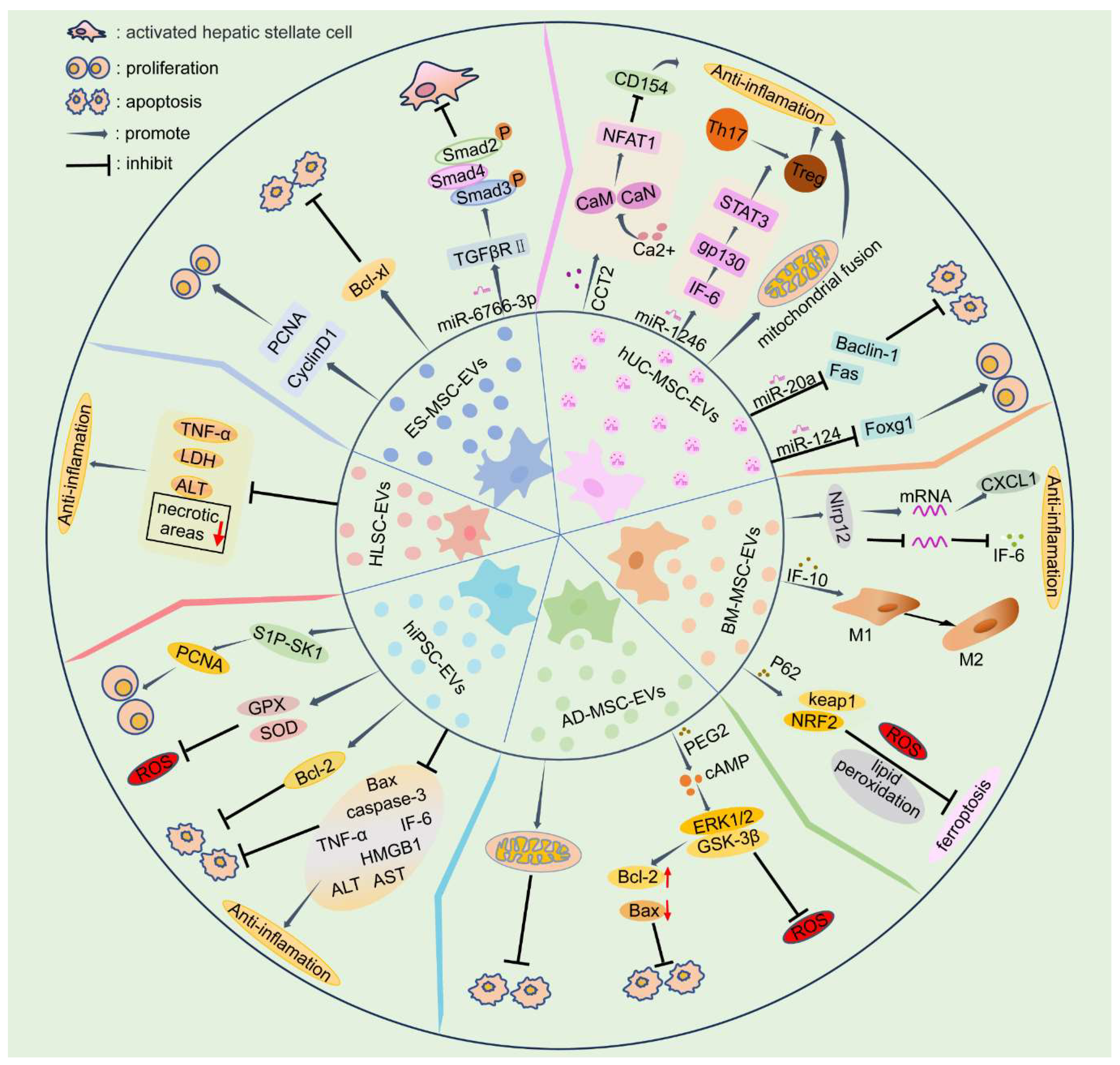

Table 1.
Experimental study on the effects of EVs from different sources of stem cells on liver injury.
Table 1.
Experimental study on the effects of EVs from different sources of stem cells on liver injury.
| Reference | Animal model | In vitro | EV source | Dose | Mode of administration | Signaling pathway/mechanism | Therapeutic effect |
|---|---|---|---|---|---|---|---|
| Tan et al. 2014[2] | mouse | + | ES-MSC | 0.4 μg/dose | Splenic injection | Increase hepatocyte proliferation (PCNA elevation); inhibit hepatocyte apoptosis | |
| Wang et al. 2021[43] | mouse | + | 3D-hESC | Vein | TGFβRII/SMADS pathway | reduce HSC activation | |
| Mardpour et al. 2018[18] | rat | + | ES-MSC | 350 µg/dose | Splenic injection | Reduce inflammation; reduce apoptosis | |
| Sameri et al. 2022[40] | mouse | + | hUC-MSC | 100 μg/dose | Caudal vein | Reduce inflammation; reduce apoptosis; Inhibit oxidative stress | |
| Zheng et al. 2020[44] | mouse | + | hUC-MSC | 4 mg/kg | Caudal vein | The Ca2+-calcineurin-NFAT1 signaling pathway | Inhibit the initiation of inflammatory responses |
| Xie et al. 2019[45] | mouse | + | hUC-MSC | 10 μg/dose | Vein | Transfer miR-1246 targeting the IL-6-gp130-STAT3 pathway | Reduce inflammation (decreased Th17/Treg ratio among CD4+ T cells) |
| Xie et al. 2019[15] | mouse | + | hUC-MSC | 2.5 × 10¹² particles/dose | Portal vein | Transfer miR-1246 targeting the GSK3β/Wnt/β-catenin pathway | Inhibit apoptosis and inflammation (the inflammatory factors TNF-α, IL-6a, and IL-1β are significantly reduced) |
| Zhang et al. 2020[46] | rat | + | hUC-MSC | By secreting miR-20a, it targets Fas and Beclin-1 and inhibits their expression | Inhibit autophagy and apoptosis | ||
| Song et al. 2021[47] | rat | hUC-MSC | 0.4 μg/dose | Caudal vein | The expression of Foxg1 is downregulated by the secretion of miR-124 | Promote hepatocyte proliferation (the proliferation marker PCNA was elevated) | |
| Yao et al. 2019[48] | rat | + | hUC-MSC | 10 mg/kg | Caudal vein | Inhibit oxidative stress (increased levels of the mitochondrial antioxidant enzyme MnSOD); inhibit inflammation (prevent neutrophils from entering the inflammatory microenvironment) | |
| Lu et al. 2022[49] | mouse | hUC-MSC | 100 μg/dose | Caudal vein | Transfer of mitochondria to modulate the formation of NETs |
Reduce autophagy and apoptosis | |
| Yan et al. 2017[50] | mouse | + | hUC-MSC | 16 mg/kg | Caudal vein/gavage | Phosphorylation of ERK1/2 is induced by the secretion of GPX1 | Inhibit oxidative stress; reduce apoptosis |
| Haga et al. 2017[51] | mouse | + | mBM-MSC | 2 ×10¹⁰ particles/dose | Caudal vein | Targeting Nlrp12 | Inhibit inflammation (decreased expression of inflammatory cytokines IL-6 and IL-1β); reduce apoptosis (reduction of caspase-3 positive cells and apoptotic cells) |
| Anger et al.2019[52] | mouse | hBM-MSC | 1 ×10⁹ particles/dose | Vena cava inferior | Reduce inflammation; reduce liver damage | ||
| Zhang et al. 2024[41] | mouse | + | mBM-MSC | Caudal vein | Targeting FGF21 and the JAK2/STAT3 pathway | Ba-EVs can improve Th17/Treg imbalance | |
| Li et al. 2023[53] | mouse | + | mBM-MSC | 50 μg/dose | Caudal vein | Regulates the p53 signaling pathway through PTEN | Inhibit cell apoptosis |
| Yang et al. 2020[35] | mouse | + | mBM-MSC-Heps | 100 μg/dose | Caudal vein | Enhanced autophagy | Reduce apoptosis; reduce liver damage |
| Zhang et al. 2022[54] | mouse | + | mBM-MSC | 20 μg/dose | Arteria femoralis | By delivering IL-10 and interacting with Kupffer cells, it causes Kupffer cells to change to an anti-inflammatory phenotype (M2) | Inhibit inflammation |
| Zhao et al. 2022[42] | mouse | + | BM-MSC | 150 µg/dose | Caudal vein | Ba-EVs inhibits iron death by activating the Keap1-NRF2 pathway through P62 | Inhibit ROS production and lipid peroxide-induced iron death |
| Wei et al. 2020[55] | rat | + | hBM-MSC | Intraperitoneal injection | Inhibit inflammation and apoptosis | ||
| Tamura et al. 2016[22] | mouse | + | mBM-MSC | 10 μg/dose | Vein | Inhibit inflammation; reduce apoptosis | |
| Damania et al.2018[56] | rat | + | rBM-MSC | 50 μg/dose | Portal vein | Reduce oxidative stress and apoptosis | |
| Sun et al. 2017[57] | rat | + | Mini-pig AD-MSC | 100 µg/dose | Vein | Inhibit inflammation (the inflammation markers MIF, MMP-9, L-1β, TNF-α, COX-2 are decreased); inhibit oxidative stress (NOX-1, NOX-2 levels decreased; HO-1, NQO-1 levels increased); inhibit apoptosis (reduced caspase 3 and PARP) | |
| Zhang et al. 2022[58] | rat | + | rAD-MSC | 30 μg/dose | Portal vein | Phosphorylation of ERK1/2 and GSK-3β by prostaglandin E2 (PGE2) | Reduce apoptosis and oxidative stress; reduce inflammation |
| Zhang et al. 2021[1] | rat | rAD-MSC | 100 µg/dose | Caudal vein | Reduce mitochondrial division, promote mitochondrial fusion and improve mitochondrial biosynthesis | Inhibit oxidative stress (MDA, ROS oxidation index decreased, antioxidant enzymes SOD, CAT, GSH-px content increased), reduce cell apoptosis (inhibition of Caspase-3 and Caspase-9 activities, decrease Bax mRNA and protein expression, increase Bcl-2 mRNA and protein expression) | |
| Gupta et al.2022[59] | mouse | + | hAD-MSC | 250 µg/dose | Caudal vein | Promote hepatocyte proliferation | |
| Fang and Liang, 2021[60] | mouse | + | mAD-MSC | 200 μL | Caudal vein | EVs loaded with vitamin A and quercetin were more effective in reducing liver damage | |
| Gong et al.2023[61] | rat | + | hAD-MSC | Caudal vein | Through the miR-183/ALOX5 axis | Reduce liver injury | |
| Piao et al.2022[62] | rat | hAD-MSC | 100 µg/dose | Caudal vein | Inhibit the NF-κB pathway and activate the Wnt/β-catenin pathway | Inhibit cell pyroptosis; Promote hepatocyte proliferation | |
| Wang et al.2023[63] | mini-pig | + | mini-pig AD-MSC | 5 × 10⁶ particles/g of liver | Portal vein | Inhibit apoptosis, pyroptosis and inflammatory responses | |
| Zhang et al.2023[64] | rat | rAD-MSC | 100 µg/dose | Caudal vein | Inhibit ERS and inflammation | ||
| Wang et al.2024[65] | mini-pig | + | mini-pig AD-MSC | 5 × 10⁶ particles/g of liver | Portal vein | Inhibit inflammation; Promote hepatocyte proliferation | |
| Wang et al.2023[66] | mini-pig | mini-pig AD-MSC | 5 × 10⁶ particles/g of liver | Portal vein | Modulat the ERS response | ||
| Nong et al. 2016[34] | rat | hiPSC-MSC | 2 mg/kg | Vena cava inferior | Inhibit inflammation (TNF-α, IL-6 and HMGB1 decreased significantly), reduce oxidative stress (increased GSH, GSH-PX and SOD levels), reduce apoptosis (significantly decreased caspase-3 and bax levels), promote hepatocyte proliferation | ||
| Du et al. 2017[33] | mouse | + | hiPSC-MSC | 2.5 × 10¹² particles/dose | Vena cava inferior | The sphingosine kinase and sphingosine-1-phosphate-dependent pathway | Reduce liver damage (significantly decreased AST and ALT levels) and promote hepatocyte proliferation (significantly increased expression of the proliferation markers PCNA and PHH3) |
| De Stefano et al. 2021[67] | rat | + | HLSC | 5 or 25 × 108 particles/g of liver | Promote hepatocyte regeneration, damage mitigation | ||
| Rigo et al. 2018[68] | rat | HLSC | 5x 10⁸ HLSC-EV/g liver tissue | Reduce liver damage | |||
| Calleri et al. 2021[69] | mouse | HLSC | 3×10⁹ particles/dose | Vein | Reduce liver damage, reduce inflammation |
Note: hESC-MSC: human embryonic stem cell-mesenchymal stem cell; PCNA: proliferating cell nuclear antigen; 3D-hESC: 3D-human embryonic stem cell; TGFβRII: Transforming Growth Factor-β type II receptor; HSC: Hepatic stellate cell; hUC-MSC: human umbilical cords-derived mesenchymal stem cell; NFAT1: nuclear factor of activated T cells 1; STAT3: Signal transducer and activator of transcription 3; Th17: T helper 17 cells; Treg: regulatory T cells; GSK3β: glycogen synthase kinase 3β; TNF-α: tumor necrosis factor alpha; IL-6: interleukin 6; MnSOD: manganese superoxide dismutase; NETs: neutrophil extracellular traps; GPX1: glutathione peroxidase 1; ERK1/2: Recombinant Extracellular Signal Regulated Kinase 1/2; Nlrp12: NACHT, LRR and PYD domains-containing protein 12; mBM-MSC: mouse bone marrow derived mesenchymal stem cell; mBM-MSC-Heps: mouse bone marrow-mesenchymal stem cells derived Hepatocyte-like cells; Ba-EVs: extracellular vesicles derived from baicalin-pretreated MSCs; ROS: reactive oxygen species; hBM-MSC: human bone marrow-mesenchymal stem cells; AD-MSC: adipose mesenchymal stem cell; rAD-MSC: rat adipose mesenchymal stem cell; hAD-MSC: human adipose mesenchymal stem cell; mAD-MSC: mouse adipose mesenchymal stem cell; mini-pig AD-MSC:mini-pig adipose mesenchymal stem cell; ERS: endoplasmic reticulum stress; MIF: migration inhibitory factor; MMP-9: matrix metallopeptidase 9; IL-1β: interleukin-1 beta; COX-2: cyclooxygenase-2; NOX-1: NADPH oxidase 1; NOX-2: NADPH oxidase 2; PARP: Poly ADP ribose polymerase; PGE2: prostaglandin E2; GSK - 3β: glycogen synthase kinase-3b; MDA: malondialdehyde; SOD: superoxide dismutase; CAT: catalase; GSH- px: glutathione peroxidase; hiPSC-MSC: human induced pluripotent stem cell–derived mesenchymal stromal cells; HMGB1: high mobility group box 1; AST: aspartate aminotransferase; ALT: alanine aminotransferase; PHH3: phosphohistone-H3; HLSC: human liver stem cell.
Table 2.
Clinical research progress on the use of EVs derived from stem cells to treat liver injury.
Table 2.
Clinical research progress on the use of EVs derived from stem cells to treat liver injury.
| Registration number | Title | Country | Year | Status | Study type | Phase | EVs source |
|---|---|---|---|---|---|---|---|
| NCT05940610 | The Safety and Efficacy of MSC-EVs in Acute/Acute-on-Chronic Liver Failure | China | 2023 | Withdrawn | Interventional | 1、2 | hMSC |
| NCT05881668 | MSC-EV in Acute-on-Chronic Liver Failure After Liver Transplantation | China | 2023 | Withdrawn | Interventional | 1 | MSC |
| NCT05871463 | Effect of Mesenchymal Stem Cells-derived Exosomes in Decompensated Liver Cirrhosis | Iran | 2023 | Recruiting | Interventional | 2 | hUC-MSC |
| ChiCTR-INR-17010677 | Study on the effect of MSCs-HNF4α exosomes combined with normal mechanical perfusion on liver transplantation of fatty liver | China | 2017 | Not yet recruiting | Interventional | New Treatment Measure Clinical | hMSCs-HNF4α |
| ChiCTR2300075676 | A small sample clinical study of the safety and initial efficacy of exosomes in the treatment of cirrhosis | China | 2023 | Recruiting | Interventional | New Treatment Measure Clinical Study | MB-MSC |
| ChiCTR1800020076 | A clinical study for cancer stem cells exosome loaded dendritic cells vaccine and its activated CTL injection in the treatment of hepatic cell cancer and other solid tumors | China | 2018 | Not yet recruiting | Interventional | 1、2 | cancer stem cells |
Note: hMSCs-HNF4α: human Mesenchymal Stem Cells-Hepatocyte Nuclear Factor 4 Alpha; MB-MSC: Menstrual Blood Mesenchymal Stem Cell.
Disclaimer/Publisher’s Note: The statements, opinions and data contained in all publications are solely those of the individual author(s) and contributor(s) and not of MDPI and/or the editor(s). MDPI and/or the editor(s) disclaim responsibility for any injury to people or property resulting from any ideas, methods, instructions or products referred to in the content. |
© 2024 by the authors. Licensee MDPI, Basel, Switzerland. This article is an open access article distributed under the terms and conditions of the Creative Commons Attribution (CC BY) license (http://creativecommons.org/licenses/by/4.0/).
Copyright: This open access article is published under a Creative Commons CC BY 4.0 license, which permit the free download, distribution, and reuse, provided that the author and preprint are cited in any reuse.


