Submitted:
01 August 2024
Posted:
02 August 2024
You are already at the latest version
Abstract
We have synthesized and examined a series of calix[4]arene-based chemosensors containing thiosemicarbazone or thiourea as a binding site and a pyridine-chromophore unit in various conformations. These conformations include pinched cone (I), cone (III and IV), and 1,3-alternate (II and V). Our objective was to assess the binding abilities of these sensors towards anions and cations using different spectroscopic techniques. All receptors demonstrate the ability to recognize cations, with notable distinctions between them. Receptor I demonstrates a broad spectrum of ion recognition, encompassing Ag+, Cu2+, Co2+, Ni2+, and Zn2+. Receptors IV and V also exhibit complex formation with Co2+, Ni2+, and Cu2+ ions. Sensors II and III demonstrate exceptional selectivity for recognizing the copper (II) ion. When studying anions, only receptor I yielded satisfactory results. This is due to the thiosemicarbazone bridge and its spatial orientation in the pinched cone conformation, which allows for an interaction to occur. It is worth noting that sensors IV and V, which are fluorescent molecules due to the triazolo pyridine unit in their structure, exhibit particularly noteworthy behavior. The highest association constant is observed for cobalt (II) complexes, with values of 6.59 and 2.38 (x1012 mol/L)-2 for receptors IV and V, respectively. Our findings indicate that there are no significant differences in selectivity and sensitivity between ca-lix[4]arene receptors in 1,3-alternate and cone conformations.
Keywords:
Calix[4]arene
; Conformations
; Receptor
; Bifunctional
1. Introduction
Calix[4]arenes are macrocyclic oligomers composed of phenolic units linked by methylene bridges. This structural arrangement allows calix[4]arenes to serve as molecular scaffolds in the design of artificial receptors. These molecules have a three-dimensional cavity and a pre-organized core [1], making them effective building blocks for the design of fluorescent receptors used in molecular recognition [2,3,4,5]. The ability of calix[4]arene molecules to form complexes with ions is influenced by several factors, including the size and conformation (such as cone, partial cone, 1,2-alternate, and 1,3-alternate) of the calixarene cavity, as well as the type of solvent used in the studies [6,7].
The development of molecular systems for the detection of anions [8,9], cations [10,11], or neutral molecules [12] is an important area of research within supramolecular chemistry. These systems have a wide range of applications in various fields, including chemistry, medicine, biology, and environmental science. The broad applicability of these systems makes them a major focus of investigation for researchers. In recent decades, considerable effort has been devoted to the development of molecular probes capable of dual sensing, which involves the detection of multiple ions using different binding sites within a single receptor molecule. Research has led to the development of numerous sensors designed to detect different combinations of anions and cations [13,14,15].
The selection of the ion recognition moiety is a critical aspect in the development of new artificial chemosensors because it affects the selectivity and binding efficiency of the sensor for target ions. To achieve effective ion recognition, various functional groups are incorporated into calixarenes. These include amide, urea, and thiourea [16,17,18], which introduce interaction sites with atoms such as nitrogen (N), oxygen (O), and sulfur (S). The presence of these functional groups creates opportunities for specific interactions with ions, enhancing the sensor's ability to detect and recognize molecules. For example, the thiosemicarbazone group, which includes hydrogen bond donors, thiocarbonyl groups, and imine groups, contains several key interaction sites (N, O, S atoms). This structural arrangement has been shown to improve the binding efficiency and selectivity of the sensor towards various metal cations and anions [19,20,21,22,23].
The most basic fluorescent groups used in the design of chemosensors are polycyclic aromatic compounds such as anthracene, pyrene, phenanthrene, and naphthalene. These groups have been incorporated into calixarenes, either at the top or bottom edge, to facilitate ion detection [24,25,26,27]. In our research, we focused on two pyridine-based chromophores, incorporating either naphthalene or triazolopyridine.
For the first chromophore, we used the synthesis method described in the literature for N-2-pyridinyl-2-naphthalenecarboxamide. This method involved catalysis and microwave irradiation [28,29]. Due to its fluorescent properties, this compound was considered a suitable candidate for our applications [30]. In the second case, we used the compound 3-(2-pyridyl)-[1,2,3]triazolo[1,5-a]pyridine obtained from the researcher Belen Abarca. The synthesis of this compound is described in her various publications [31,32,33]. In addition, we have exploited its properties as a fluorescent molecule in studies of cation detection in the presence of cyclodextrin [34].
Building on this background, our study advances previous work in the development of new sensors based on calix[4]arenes [17,35,36,37,38,39,40]. In this project, we have introduced pyridinyl chromophore moieties into a calix[4]arene scaffold using a thiosemicarbazone or thiourea bridge at either the lower or upper edge. The new receptors, based on calix[4]arene in alternate, pinched cone, and cone conformations, are designed to detect both cations and anions using UV-visible and fluorescence techniques.
2. Materials and Methods
2.1. General Procedures
The chemicals, including perchlorate salts, tetrabutylammonium salts, and other substances, were procured from Sigma-Aldrich. All reagents and solvents were of analytical grade and used without further purification. The UV-Vis absorption spectra of the samples were recorded on an Agilent 8453 spectrophotometer using standard 1.00 cm quartz cells in accordance with standard operating procedures. Melting points (uncorrected) were obtained using a Kofler hot-stage microscope. Nuclear magnetic resonance (NMR) spectra were obtained using a Bruker AVANCE III HD (300 MHz) spectrometer with tetramethylsilane as the internal reference at room temperature. Infrared spectra were obtained using a FT-IR spectrometer (Thermo iS50) with a germanium attenuated total reflection (ATR) accessory. The range was 4000-600 cm-1, with an average of 20 scans per analysis. Prior to taking any measurements, a blank analysis was conducted to account for any air signals. The mass spectra (LC/MSD-TOF) were obtained at the University of Valencia in Spain. The carbon, hydrogen, and nitrogen content were determined using an Elemental LECO TRUSPEC CHNS Micro analyzer at the University of Seville in Spain. All experiments were conducted in triplicate.
2.2. Synthesis of Receptors
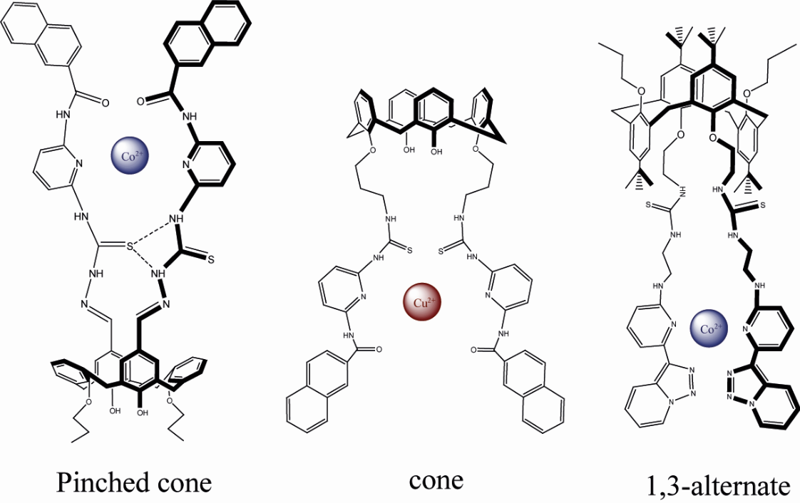
Figure 1 shows the structures of the calixarene-based receptors. The synthesis of the receptors and their characterization by IR, NMR and elemental analysis are described below, while spectra figures or images are provided in Supplementary Information.
Synthesis of 5,17-N-(pyridin-2-yl)-2-naphthamide-thiosemicarbazone-25,27-dipropoxy-26,28-dihydroxycalix[4]arene, pinched cone (I)
To a solution of 5,17-dimethylenehydrazine-25,27-dipropoxy-26,28-dihydroxycalix[4]arene (76 mg, 0.131 mmol) in CHCl3 (8 mL), N-(6-isothiocyanatopyridin-2-yl)-2-naphthamide (100 mg, 0.328 mmol) was added and left stirring for one hour. Once the requisite time has elapsed, a precipitate will form. This should be filtered and washed with CHCl3 (3 × 5 mL), resulting in the desired product (I). Yield: 57.9% (90 mg). Melting point: 244-247 °C. 1H-NMR: (300 MHz; DMSO-d6, TMS, 25 °C) δ 11.16 (s, 2H, C=N-NH-C=S), 11.02 (s, 2H, Ar-NH-C=O), 10.84 (s, 2H, Ar-NH-C=S), 10.07 (s, 2H, Ar-OH), 9.06 (s, 2H, -HC=N), 8.74 (s, 2H, Naf-H), 8, 11 - 6.75 (m, 28H, Ar-H), 4.19 (d, 4H, J = 12 Hz, Ar-CH2-Ar), 3.98 (t, 4H, O-CH2-CH2-), 3.58 (d, 4H, J = 12 Hz, Ar-CH2-Ar), 2.00 (m, 4H, CH2CH2CH3), 1.32 (t, 6H, -CH2-CH3). FT-IR (cm-1): 3412 (ν O-H), 3315 (ν N-H amide), 3053 (ν C-H aromatic), 2929 (ν C-H aliphatic), 2868 (ν C-H aliphatic), 1665 (ν C=O amide), 1659 (δ N-H amide), 1605 (ν C=N imine), 1481 (δ C-H aliphatic), 1441 (δ C-H aromatic), 1388 (δ O-H phenol), 1220 (ν C=S thiourea), 792 (δ C-H aromatic), 776 (δ C-H aromatic). Elem. Anal.: C, 69.86; H, 5.19; N, 11.64; O, 7.98; S, 5.33.
Synthesis of 25,26,27,28-tetra[3-(3-(6-(2-naphthamido)pyridin-2-yl)thioureido) propyloxy]calix[4]arene, 1,3-alternate (II).
A 50-ml balloon was prepared with 64.11 mg (0.098 mmol) of 25,26,27,28-tetra[(3-aminopropyl)oxy]calix[4]arene, 1,3-alternate, and 180 mg (0.59 mmol) of N-(6-isothiocyanato pyridin-2-yl)-2-naphthamide (10). Both solids were dissolved in 30 ml of chloroform (CHCl3). Four drops of triethylamine were added to the reaction, which was then stirred for a period of 24 hours at room temperature. During this time, the formation of a yellow precipitate was observed. Upon completion of the allotted reaction period, the solid was filtered and washed with an ample quantity of hot chloroform. A total of 120 mg of product (II) was obtained, representing a yield of 65.33%. The melting point was determined to be 199-201 °C, with melting occurring at the point of decomposition. 1H-RMN (DMSO-d6) δ: 11.56 (t, J = 5.57 Hz, 4H, CH2-NH-CS-), 10.92 (s, 4H, CS-NH-Py), 10.52 (s, 2H, -NH-CO), 8.39 (s, 4H, Ar-H), 7.87 (m, 16 H, Ar-H), 7.75 (t, J = 8 Hz, 4H, Py-H), 7.56 (m, 8H, Ar-H), 7.47 (d, J = 8 Hz, 4H, Py-H), 6.88 (d, 8 Hz, 4H, Py-H), 6.76 (d, J = 7.3 Hz, 8H, Ar-H), 6.45 (t, J = 7.3 Hz, 4H, Ar-H), 3.70 (s, 8H, Ar-CH2-Ar), 3.44 (t, 8H, O-CH2-), 3.34 (t, 8H, NH-CH2-), 1.82 (q, 8H, -CH2-CH2-CH2-CH2-). 13C-NMR (DMSO-d6) δ: 179 (C=S), 166 (C=O), 156 (=C-O-C), 152 (HN-C=N), 149 (HN-C=N), 140 (O=C-C=), 134-107 (C aromatics), 69 (O-CH2), 42 (N-CH2), 36 (Ar-CH2-Ar), 29 (C-CH2-C). FT-IR (cm-1): 3397 (ν N-H amide), 3192 (ν N-H thiourea), 3052 (ν C-H aromatic), 2938 (ν C-H aliphatic), 1680 (ν C=O amide), 1659 (δ N-H thiourea and amide), 1613 (ν C=N aromatic), 1543 (ν C=S thiocarbonyl), 1523 (ν C=C aromatic), 1501 (ν C=C aromatic), 1443 (ν C=C aromatic), 1232 (ν C-O aromatic), 1158 (ν C-N aliphatic) 1152 (ν C-O aliphatic), 791 (δ C-H aromatic). ESI-TOF: m/z calculated: 1872.65 measured: 1872.6433. Elem. Anal.: C, 69.13; H, 5.26; N, 11.94; O, 6.82; S, 6.84.
Synthesis of 25,27-bis[3-(6-(2-naphthamido)pyridin-2-thioureido)propyloxy]-26,28-dihydroxychalix[4]arene, cone (III).
In a 50 mL balloon, 80 mg (0.15 mmol) of 25,27-bis[(3-aminopropyl)oxy] calix[4]arene, 180 mg (0.59 mmol) of N-(6-isothiocyanatopyridin-2-yl)-2-naphthamide (10), and 30 ml of chloroform (CHCl3) were added. Four drops of triethylamine were also added, and the mixture was stirred for a period of 16 hours at reflux. During this time, the formation of a white precipitate was observed. Upon completion of the reaction period, the solid was filtered and washed with an ample quantity of hot chloroform. The yield of product (III) was 109 mg (63.2%), with a melting point of 179-181 °C. 1H-NMR (DMSO-d6) δ 11.73 (t, J = 5.22 Hz, 2H, -CH2-NH-CS-), 10.85 (s, 2H, Py-NH-CS), 10.49 (s, 2H, Py-NH-CO), 8.44 (s, 2H, Ar-H), 8.26 (s, 2H, Ar-OH), 7. 90 (m, 8H, Ar-H), 7.76 (t, J = 8 Hz, 2H, Py-H), 7.59 (m, 4H, Ar-H), 7.45 (d, J = 8 Hz, 2H, Py-H), 6.92 (d, J = 7.6 Hz, 4H, Ar-H), 6.86 (d, J = 8 Hz, 2H, Py-H), 6.84 (d, J = 7.6 Hz, 4H, Ar-H), 6.61 (d, J = 7.6 Hz, 2H, Ar-H), 6.45 (t, J = 7.6 Hz, 2H, Ar-H) 4.04 (m, 12H, Ar-CH2-Ar; O-CH2-; NH-CH2-), 3.15 (d, 4H, J = 13 Hz, Ar-CH2-Ar), 2.54 (m, 4H, CH2-CH2-CH2). 13C-NMR: 179 (C=S), 166 (C=O), 152.98 (HN-C=N), 152.23 and 152.17 (CAr-O-), 149 (HN-C=N), 140 (O=C-C=), 134-107 (C aromatics), 75 (O-CH2), 42 (N-CH2), 31 (Ar-CH2-Ar), 29 (C-CH2-C). FT-IR (cm-1): 3400 (ν O-H alcohol), 3397 (ν N-H amide), 3192 (ν N-H thiourea), 3052 (ν C-H aromatic), 2938 (ν C-H aliphatic), 1680 (ν C=O amide), 1659 (δ N-H thiourea and amide), 1613 (ν C=N aromatic), 1543 (ν C=S thiocarbonyl), 1523 (ν C=C aromatic), 1501 (ν C=C aromatic), 1443 (ν C=C thiocarbonyl), 1232 (ν C-O aromatic), 1158 (ν C-N aliphatic), 1152 (ν C-O aliphatic), 791 (δ C-H aromatic). TOF-MS: calculated: 1148.41, measured: 1148.4066. Elem. Anal.: C, 71.06; H, 5.26; N, 9.75; O, 8.35; S, 5.58.
Synthesis of 5,11,17,23-tetra-tert-butyl-25,27-bis[3-(6-(N-diethylamino)-2-pyridinyl)-1,2,3-triazolo[1,5-a]pyridine)ethoxy]-26,28-dipropoxycalix[4]arene, cone (IV).
To a 25-ml balloon, add 10 ml of ethyl acetate, 101.1 mg (0.398 mmol) of 3-(6-(N-diethylamino)-2-pyridinyl)-1,2,3-triazolo[1,5-a]pyridine, and 163.7 mg (0.181 mmol) of 5,11,17,23-tetra-tert-butyl-25,27-di(isothiocyanatoethoxy)-26,28-dipropoxy calix[4]arene. Three drops of triethylamine are then added to the aforementioned reaction mixture. The reaction mixture is stirred for 24 hours on a magnetic stirrer and monitored by TLC using ethyl acetate as the mobile phase. Following a 24-hour period, the product is purified on a chromatographic column using the aforementioned mixture as the mobile phase. A total of 141.8 mg was recovered. Yield: 56.1%; 1H-NMR (300 Mhz, CDCl3, TMS, 25 °C) δ 8.65 (d, 2H, J = 7 Hz, ArH (e)), 8.47 (d, 2H, J = 8.9 Hz, ArH (d)), 7.49-7.47 (m, 2H, ArH (g)), 7.47-7. 44 (m, 2H, ArH (b)), 7.33 (ddd, 2H, J = 8.9, J = 6.6, J = 0.9 Hz, ArH (f)), 7.08 (s, 4H, ArH), 6.98 (td, 2H, J = 6.7, J = 1.2 Hz, ArH (c)) 6.41 (c, 2H, J = 3.7, J = 1.7 Hz, ArH (a)), 6.38 (s, 4H, ArH), 5.37-5.26 (s broad, 2H, ArNHCH2), 4.23 (d, 4H, J = 12.6 Hz, ArCH2Ar), 4.16 (m, 4H, OCH2CH2NH), 4.15-4.05 (s broad, 4H, Hz, OCH2CH2NH), 3.93-3.85 (s broad, 4H, OCH2CH2CH3), 3.70 (t, 4H, J = 7 Hz, CSNHCH2CH2CH2NH), 3.65-3.60 (s broad, 4H, CSNHCHCH2CH2NH), 3.09 (d, 4H, J = 12.8 Hz, ArCH2Ar), 1.64 (m, 4H, OCH2CH2CH2CH3), 1.33 (s, 18H, C(CH3)3), 0.78 (s, 18H, C(CH3)3), 0.68 (t, 6H, J = 7.4 Hz, OCH2CH2CH2CH3). FT-IR (cm-1): 3290(ν N-H thiourea), 2968 (ν C-H aliphatic), 1475 (δ C-H aliphatic), 1193 (ν C=S thiourea). Elem. Anal.: C, 69.81; H, 7.48; N, 13.73; O, 4.48; S, 4.49.
Synthesis of 5,11,17,23-tetra-tert-butyl-25,27-bis[3-(6-(N-diethylamino)-2-pyridinyl)-1,2,3-triazolo[1,5-a]pyridine)ethoxy]-26,28-dipropoxycalix[4]arene, 1,3-alternate (V).
To a 25-ml balloon, add 8 ml of ethyl acetate, 51.2 mg (0.201 mmol) of 3-(6-(N-diethylamino)-2-pyridinyl)-1,2,3-triazolo[1,5-a]pyridine, and 82. Please add 1 mg (0.091 mmol) of 5,11,17,23-tetra-tert-butyl-25,27-di(isothiocyanatoethoxy)-26,28-dipropoxychalix[4]arene 1,3 alternating. Three drops of triethylamine are then added to the aforementioned reaction mixture. The reaction mixture is stirred for 24 hours on a magnetic stirrer and monitored by TLC using ethyl acetate as the mobile phase. Following a 24-hour period, the product is purified on a chromatographic column using the aforementioned mixture as the mobile phase. A total of 60.9 mg was recovered. Yield: 60.2%. 1H-NMR (300 Mhz, CDCl3, TMS, 25 °C) δ 8.67 (d, 2H, J = 6.9 Hz, ArH (e)), 8.38 (d, 2H, J = 7.9 Hz, ArH (d)), 7.73 (t, J = 7.7, J = 7.9, Hz, ArH (g)), 7.38-7.36 (m, 2H, ArH (b)), 7.31 (m, 2H, ArH (f)), 7.00 (s, 4H, ArH), 6.93 (m, 2H, ArH (c)), 6.33 (d, 2H, J = 8.2 Hz, ArH (a)), 5.27 (s broad, 2H, ArNHCH2), 3.86 (d, 4H, J = 16.2 Hz, ArCH2Ar), 3.77 (d, 4H, J = 16.2 Hz, Ar-CH2Ar), 3.74 (m, 4H, OCH2CH2CH3), 3.62 (s broad, 4H, OCH2CH2NH), 3.30 (t, 4H, J = 8, J = 8.1 Hz, OCH2CH2NH), 3.20 (s broad, 8H, NHCH2CH2NH), 1.25 (s, 18H, C(CH3)3), 1.14 (s, 18H, C(CH3)3), 1.03 (m, 4H, OCH2CH2CH2CH3), 0.67 (t, 6H, J = 7.3, J = 7.6 Hz, OCH2CH2CH2CH3). FT-IR (cm-1): 3296 (ν N-H thiourea), 2968 (ν C-H aliphatic), 1469(δ C-H aliphatic), 1204 (ν C=S thiourea). Elem. Anal. C, 69.71; H, 7.61; N, 13.71; O, 4.48; S, 4.48.
2.3. Spectrophotometric Measurements
The receptors were dissolved in DMSO (stock solution 1.5 mmol L-1) and stock solutions (15 mmol L-1) of cations and anions were prepared in CH3CN. All anions were countered with tetrabutylammonium, and all cations were countered with perchlorate. UV absorption measurements were conducted on an Agilent 8453 spectrophotometer using standard 1.00 cm quartz cells. The titrations were conducted by varying the equivalents of the ion between 0 and 5, using stock solutions, with the receptor concentration fixed at 11.5 μmol L-1. All solutions were diluted to 2.6 mL. The screening studies were conducted with the receptor concentration fixed at 11.5 μmol L-1, and 10 equivalents of the stock solutions of ions were added accordingly. Job’s plot experiments were conducted with the total concentration (receptor + ion) maintained at 11.5 μmol L-1, in accordance with the methodology described in reference [41].
Fluorescence spectra were measured on a Perkin Elmer LS55, with excitation at 350 nm and bandwidths of 5 and 10 nm, respectively, for both excitation and emission. The emission spectra were recorded within the 380-560 nm range. All measurements were conducted in a 1 cm quartz cell with a final receptor concentration of 0.158 μmol L-1. During the titration, the concentration of cations was varied from 0 to 3 equivalents. The appropriate amounts of ion stock solutions were added to prepare the screening response experiments.
2.4. Determination of Association Constants
The association constants (Ka) of receptors with all ions were calculated from the gradual changes in absorbance at selected wavelengths of receptors (I-III) upon stepwise addition of each ion to the solution. This was achieved through a linear regression described by the Benesi-Hildebrand equation (1) [42].
where Ao is the absorbance of receptor L in absence of a guest, A is the absorbance recorded in the presence of an added guest, A∞ is absorbance in presence of added [Ion]∞ and Ka is the association constant.
In order to ascertain the experimental fluorescence association constant of the receptors (IV and V), the Benesi-Hildebrand equation (Equation 2) was employed for complexes of stoichiometry calix[4]arene:ion, 1:2 [43].
where F∞ represents the fluorescence intensity when all the calix[4]arene has associated with the ion, F0 represents the fluorescence intensity of calix[4]arene in the absence of the ion, [I]t is the total ion concentration. The association constant is determined by Equation (1) by linear regression [I]t2 vs (F − F0).
3. Results and Discussion
3.1. Synthetic Procedures
The synthesis of the receptors was based on experimental procedures that involved a convergent synthesis, whereby the calixarene in the upper and lower rims, as well as in the cone and 1,3-alternate conformations, was modified. Additionally, the pyridines were modified with chromophore or fluorophore groups.
In the synthesis of receptor (I), the initial step involves preparing the di-methylhydrazine calix[4]arene compound and pyridine isothiocyanate derivatives as precursors. Subsequently, a coupling reaction based on the nucleophilic attack of the amino group on the isothiocyanate in chloroform was employed to obtain the thiosemicarbazone group of (I) [44]. In order to obtain the precursor dimethylhydrazine, it was first necessary to perform a retro Friedel-Crafts reaction on the p-tert-butylcalix[4]arene [45]. Two hydroxyl groups of calix[4]arene were transformed into an ether linkage via a lower rim di-alkylation with propyl bromide and potassium carbonate (K2CO3) [46]. A Duff reaction, comprising a di-formylation of the activated rings with hydroxyl groups, was conducted using hexamethylenetetramine (HMTA) as a source of formyl groups on the upper rim. [47] Thus, the distally di-formylated calix[4]arene was obtained. Subsequently, the aldehyde groups of the molecule were nucleophilically attacked by an excess of hydrazine monohydrate, resulting in the formation of Schiff bases derived from the calix[4]arene.
The compound pyridine isothiocyanate bearing naphthalene was obtained by starting with a symmetrical reagent (2,6-diaminopyridine). A controlled synthetic route has been described where only one of the amino groups reacts with an equivalent of 2-naphthoyl chloride. This reaction was carried out in tetrahydrofuran at 0 °C. The reaction was conducted with an excess of 2,6-diaminopyridine (2 equivalents), to which the acid chloride was added dropwise [48], thereby achieving kinetic control of the reaction [49] and preventing the synthesis of the disubstituted secondary product. Subsequently, the formation of the isothiocyanate group was achieved through the reaction of the aromatic primary amine and thiophosgene (CSCl2) in a 1:1 molar ratio. In the case of receptors (II and III), the precursors were obtained by analogous reactions, wherein the base utilized in the alkylation was altered to induce a change in the cone conformation to 1,3-alternate. The remaining steps are as follows: The synthesis route commenced with the removal of the tert-butyl groups from the ρ-tert-butylcalix[4]arene through a retro Friedel-Crafts reaction (de-tert-butylation) [45]. The synthetic plan proceeded in accordance with the process described by Chrisstoffels et al. [50], which permits dialkylation at distal 1,3 positions with N-(3-bromopropyl)phthalimide, utilizing potassium carbonate, which abstracts solely the protons of the opposite phenols. The synthesis proceeded with the process described by Erdemir et al. [51], which resulted in the synthesis of calix[4]arene in a 1,3-alternate conformation. The deprotection of the phthalimide groups was then carried out to form the primary amines based on the Gabriel reaction [52]. The synthetic plan continued with the synthesis of chromophores as described above. The receptors (IV and V) were based on the reaction between the triazolopyridine derivatives obtained by Belen et al [31,32,33]. The modification of the calixarene in cone and 1,3-alternate conformation was performed by our usual isothiocyanate synthesis procedure [17].
It is noteworthy that the characterization of these sensors revealed the formation of thiosemicarbazone or thiourea bridges, which were clearly identified by the thiocarbonyl signal in infrared spectroscopy, occurring at approximately 1200 cm-1. It is noteworthy that the receptors adopt distinct conformations, with molecules II and V exhibiting a cone conformation. This is evidenced by the separation of the geminal hydrogens of the calixarene bridge, which range from 1 to 0.8 ppm. In the case of compounds (III and IV) in the 1,3-alternate conformation, only one signal was observed for the aforementioned hydrogens, which are characteristic of this conformation.
Ultimately, receptor I is in the pinched cone conformation, likely due to the hydrogen bridge type interaction between the thioureas groups of the spacers, which causes these fragments to approach and rotate the aromatic units of the calix[4]arene that are not substituted outwards. This results in a separation of 0.6 ppm of the signals of the hydrogens of the calixarene bridge.
3.2. Screening with Ions
The UV-Visible responses of calixarenes towards different cations were investigated in acetonitrile. With the addition of 5 equivalents of Ag+, Ca2+, Cd2+, Co2+, Cu2+, K+, Li+, Mg2+, Mn2+, Na+, Ni2+, Zn2+ and Fe2+ to the solutions of I-V. The Figure 2 illustrat.es that the behavior of I in the presence of cations is capable of recognizing a diverse range of cations, including nickel (II), copper (II), cobalt (II), zinc (II) and silver (I). The absorbance band demonstrates a notable decline, ranging from 16% to 32%. In comparison to receptors II and III, these demonstrate a higher degree of selectivity, exhibiting recognition of only copper (II). Receptors IV and V were examined using fluorescence spectroscopy, revealing a notable decrease in fluorescence intensity (approximately 90%) in the presence of copper (II), nickel (II), and cobalt (II) ions.
Figure 2 shows the change of the absorption peak of the I-V compounds with and without the different anions (F-, HSO4-, I-, N3-, NO3-, NO2-, SCN-, ClO4-, Br-, Cl-, CH3COO-, CF3SO3- and CN-). It is evident that for receptors II-V, the observed behavior is practically identical, and no interaction with anions is present. Upon analysis with an excess of anions in acetonitrile, it was observed that the absorption spectra of receptor I exhibited a decrease in the band between 22 and 6% when interacting with fluoride, cyanide, and acetate anions.
3.2. UV-Visible Studies
In accordance with the screening results, the corresponding titration and job-plot studies were conducted, and the ensuing results are presented below. The images obtained are included in the supplementary information.
Figure 3 illustrates the titration of compound I with copper (II) ion. As the cation concentration increases from 0 to 5 equivalents, a decrease in the maximum at 300 nm and the appearance of an isosbestic point at 360 nm are observed. The appearance of an isosbestic point in the titrations indicates equilibrium between the free receptor and the complex formed. In this case, it allows confirmation of the formation of the complex between the Cu2+ ion and I. Furthermore, in the specific case of copper (II), the band at 300 nm is broadened, coupled with the band at 310 nm into a single large band, as shown in Figure 3. This behavior was observed to be similar for the interaction with other cations. All presented a decrease in the maximum at 300 nm, an increase in the band around 380 nm, and the appearance of an isosbestic point at 360 nm.
To illustrate, the study of receptor I with the F- ion is presented. As the concentration of the fluoride ion increases, the band around 335 nm gradually decreases, and in turn, a band at lower energy appears at 390 nm, with a clear isosbestic point at 360 nm (Figure S2). This behavior can be explained by the fact that these ions may produce a strong interaction with the N-H of the thiosemicarbazone groups, resulting in the deprotonation of the latter due to the basic character of both ions in acetonitrile [53,54,55]. Consequently, hydrogen bridges are formed between the molecules and the anions, with the thionyl groups facilitating a closer approach between the thiosemicarbazone group and the fluoride and cyanide ions, thereby enhancing the interaction and complex formation.
It has been reported by other research groups that the inclusion of functional groups such as those present in calixarenes II and III has enabled the formation of complexes with copper (II) perchlorate salts due to an interaction with sulfur and oxygen atoms that exist in the structure [56,57]. The absorption spectra of calixarenes synthesized in the presence of copper (II) exhibit the formation of a new absorption band at 390 nm, which can be attributed to interactions between sulfur or nitrogen atoms with the cation due to a charge transfer process. This observation is in accordance with Pearson's hard-soft acid-base (HSAB) theory, which postulates that sulfur and nitrogen atoms of the pyridine moiety exhibit affinity for the copper(II) ion.
The association constants were determined by utilizing data from UV-visible spectrophotometric titrations through Equation (1) (Figure S2) and concurrently identifying the stoichiometry using the continuous variation method (Figure S3).
The values of all constants determined by Equation (1) are presented in Table 1. The association constant for receptor (I) for the cation complex with cobalt(II) is the highest observed among all the studied cations. The observed values for both anions are analogous to those observed for the change in the absorbance spectrum when present. With regard to receptors II and III, it is evident that they exhibit a high degree of selectivity for copper(II), with the values of their respective constants exhibiting notable similarity.
3.3. Fluorescence Studies
Compounds IV and V could be studied by fluorescence spectroscopy, where both receptors IV and V show a strong fluorescence centered at 450 nm upon excitation at 350 nm, and based on the screening results, the corresponding titration and job plot studies were performed. (Figures S4 and S5).
The binding of copper, nickel and cobalt to photophysically active supramolecular sensors can lead to fluorescence enhancement, fluorescence quenching and/or other changes in the fluorescence emission spectrum of the host, depending on the host structure and the mechanism of electronic interaction between the host and the cation analyte [58]. The strong fluorescence intensity decreases after the gradual addition of cobalt, the interaction of thiourea bridge and triazolepyridine with the cation avoids the fluorescence process and a decrease in emission can be observed, Figure 4. This observation clearly indicates that the triazolepyridine nitrogens interact with cobalt to form the complex.
Due to this decrease in fluorescence, the association constant, Ka, was determined using the Benesi-Hildebrand [43] equation for 1:2 steochemistry determined for job plot experiments and considering the gradual changes in fluorescence intensity at 350 nm upon stepwise addition of cations to the solution, the results of these constants are tabulated in Table 2.
The association constants of receptor IV are slightly higher compared to receptor V, probably due to the conformations of calixarene. It should also be noted that the highest constant for both receptors corresponds to the cobalt (II) ion.
3.4. Colorimetric Studies
During the titrations, a color change was observed in the solutions of receptor I in the presence of copper (II), associated with changes in the absorption spectra of the calix[4]arene-based compounds. As shown in Figure 5, the solution of receptor I at a concentration of 7.88 µM in acetonitrile is colorless. By adding aliquots of 0.4 mM copper(II) solution, the solution becomes increasingly yellow. These results indicate that a colorimetric sensor has been obtained that changes color in the presence of anions and cations when complexes are formed [54,59].
For the absorbance spectra of the complexes with molecule I with copper (II), the appearance of a new band at lower energy (around 400 nm) was observed, very pronounced compared to other cations, which can be related to the color change observed in the cuvettes. Based on the above, it is concluded that this compound could be used as a naked eye sensor for the detection of copper (II) cations.
4. Conclusions
In the present work, five derivatives of calix[4]arene were synthesized, each in the 1,3-alternate, pinched cone, and cone conformations. These derivatives were functionalized at the lower or upper rim with a thiosemicarbazone or thiourea group attached to pyridinyl chromophore moieties. The synthesized receptors were evaluated for their ability to sense ions using UV-visible and fluorescence techniques.
Based on the results obtained, the receptors can be classified into three distinct groups: i) Receptor I: This receptor has the ability to detect both anions and cations, making it a bifunctional receptor. It effectively detects ions such as copper (II), cobalt (II), zinc (II), nickel (II), silver (I), fluoride and cyanide. Notably, Receptor I also allows the colorimetric detection of copper (II) ion. ii) Receptors II and III: These receptors are highly selective and recognize only the copper (II) ion through a 1:1 stoichiometric interaction. This selectivity is probably due to the spatial orientation of the 1,3-alternate conformation during complex formation, which provides two possible binding sites for the copper (II) ion. iii) Receptors IV and V: These receptors are fluorescent calixarenes due to the presence of the triazolopyridine fluorophore fragment. They interact exclusively with cations, especially cobalt (II), copper (II) and nickel (II). The interaction between these receptors and the ions is characterized by a 1:2 stoichiometric ratio and these receptors show the highest association constant among all the complexes studied. For the cobalt (II) ion, the association constant is 6.59 × 10¹² (mol/L)-2. No significant differences in selectivity and sensitivity were observed between calix[4]arene receptors with 1,3-alternate and cone conformations.
Supplementary Materials
The following supporting information can be downloaded at the website of this paper posted on Preprints.org, Characterization of molecules, determination of the association constant by absorbance or fluorescent titration and determination of the stoichiometry de each receptor (I-V) with respective ions studied.
Author Contributions
Conceptualization, C.S., B.H., J.H. and A.O.; methodology, C.S.; validation, B.H., J.H. and A.O.; formal analysis, B.H., J.H. and A.O.; investigation, B.H., J.H. and A.O.; resources, C.S.; writing—original draft preparation, H.G.; writing—review and editing, H.G.; supervision, C.S.; project administration, C.S.; funding acquisition, C.S. All authors have read and agreed to the published version of the manuscript.
Funding
This research received no external funding.
Acknowledgments
Our thanks are to FONDECYT Grants: 1151310 for support. Dr. Horacio Gomez-Machuca thanks are to ANID for postdoctoral project FONDECYT grants: 3210140.
Conflicts of Interest
The authors declare no conflicts of interest.
References
- Gutsche, C.D. Calixarenes Revisited. Royal Society of Chemistry, 1998. [Google Scholar]
- Kim, J.S.; Quang, D.T. Calixarene-Derived Fluorescent Probes. Chem. Rev. 2007, 107, 3780–3799. [Google Scholar] [CrossRef] [PubMed]
- Kumar, N.; Pham-Xuan, Q.; Depauw, A.; Hemadi, M.; Ha-Duong, N.T.; Lefevre, J.P.; Ha-Thi, M.H.; Leray, I. New Sensitive and Selective Calixarene-Based Fluorescent Sensors for the Detection of Cs+ in an Organo-Aqueous Medium. N. J. Chem. 2017, 41, 7162–7170. [Google Scholar] [CrossRef]
- Depauw, A.; Kumar, N.; Ha-Thi, M.H.; Leray, I. Calixarene-Based Fluorescent Sensors for Cesium Cations Containing BODIPY Fluorophore. J. Phys. Chem. A. 2015, 119, 6065–6073. [Google Scholar] [CrossRef]
- Hosseinzadeh, R.; Nemati, M.; Zadmard, R.; Mohadjerani, M. Amidofluorene-Appended Lower Rim 1,3-Diconjugate of Calix[4]arene: Synthesis, Characterization and Highly Selective Sensor for Cu2+. Beilstein J. Org. Chem. 2016, 12, 1749–1757. [Google Scholar] [CrossRef]
- Patra, S.; Maity, D.; Gunupuru, R.; Agnihotri, P.; Paul, P. Calixarenes: Versatile Molecules as Molecular Sensors for Ion Recognition Study. J. Chem. Sci. 2012, 124, 1287–1299. [Google Scholar] [CrossRef]
- Maity, D.; Chakraborty, A.; Gunupuru, R.; Paul, P. Calix[4]arene Based Molecular Sensors with Pyrene as Fluorogenic Unit: Effect of Solvent in Ion Selectivity and Colorimetric Detection of Fluoride. Inor. Chim. Acta 2011, 372, 126–135. [Google Scholar] [CrossRef]
- Busschaert, N.; Caltagirone, C.; Van Rossom, W.; Gale, P.A. Applications of Supramolecular Anion Recognition. Chem. Rev. 2015, 115, 8038–8155. [Google Scholar] [CrossRef] [PubMed]
- Gale, P.A.; Howe, N.W.E.; Wu, X. Anion Receptor Chemistry. Chem. 2016, 1, 351–422. [Google Scholar] [CrossRef]
- Yeung, M.C.-L.; Yam, V.W.-W. Luminescent Cation Sensors: From Host-Guest Chemistry, Supramolecular Chemistry to Reaction-Based Mechanisms. Chem. Soc. Rev. 2015, 44, 4192–4202. [Google Scholar] [CrossRef]
- Lo, P.; Wong, M. Extended Calix[4]arene-Based Receptors for Molecular Recognition and Sensing. Sensors 2008, 8, 5313. [Google Scholar] [CrossRef]
- Bell, T.W.; Hext, N.M. Supramolecular Optical Chemosensors for Organic Analytes. Chem. Soc. Rev. 2004, 33, 589–598. [Google Scholar] [CrossRef] [PubMed]
- Qu, W.-J.; Guan, J.; Wei, T.-B.; Yan, G.-T.; Lina, Q.; Zhang, Y.-M. A Turn-On Fluorescent Sensor for Relay Recognition of Two Ions: From a F- Selective Sensor to Highly Zn2+ Selective Sensor by Tuning Electronic Effects. RSC Adv. 2016, 6, 35804–35808. [Google Scholar] [CrossRef]
- Nikolaeva, O.G.; Shepelenko, E.N.; Tikhomirova, K.S.; Revinskii, Y.V.; Dubonosov, A.D.; Bren, V.A.; Minkin, V.I. Bifunctional Fluorescent and Colorimetric ‘Naked Eye’ Aroylhydrazone Chemosensors for Hg2+ and F– Ions Detection. Mendeleev Commun. 2016, 26, 402–404. [Google Scholar] [CrossRef]
- Uahengo, V.; Zhang, Y.; Xiong, B.; Zhao, P.; Cai, P.; Rhyman, L.; Ramasami, P.; Hu, K.; Cheng, G. A Fluoro-Chromogenic Sensor Based on Organic Molecular Framework for Cu2+ and F− in Aqueous Soluble DMSO. J. Fluoresc. 2017, 27, 191–197. [Google Scholar] [CrossRef] [PubMed]
- Park, G.J.; You, G.R.; Choi, Y.W.; Kim, C. A Naked-Eye Chemosensor for Simultaneous Detection of Iron and Copper Ions and Its Copper Complex for Colorimetric/Fluorescent Sensing of Cyanide. Sens. Actuators B Chem. 2016, 229, 257–271. [Google Scholar] [CrossRef]
- Ferreira, J.F.; Bagatin, I.A. A Cr(VI) Selective Probe Based on a Quinoline-Amide Calix[4]arene. Spectrochim. Acta A 2018, 189, 44–50. [Google Scholar] [CrossRef] [PubMed]
- Quiroga-Campano, C.; Gómez-Machuca, H.; Moris, S.; Jara, P.; De la Fuente, J.R.; Pessoa-Mahana, H.; Jullian, C.; Saitz, C. Synthesis of Bifunctional Receptor for Fluoride and Cadmium Based on Calix[4]arene with Thiourea Moieties. J. Mol. Struct. 2017, 1141, 133–141. [Google Scholar] [CrossRef]
- Athar, M.; Lone, M.Y.; Jha, P.C. Recognition of Anions Using Urea and Thiourea Substituted Calixarenes: A Density Functional Theory Study of Non-Covalent Interactions. Chem. Phys. 2018, 501, 68–77. [Google Scholar] [CrossRef]
- Bhowmick, R.; Alam, R.; Mistri, T.; Das, K.K.; Katarkar, A.; Chaudhuri, K.; Ali, M. A Thiosemicarbazone Based Chemo and Fluorogenic Sensor for Zn2+ with CHEF and ESIPT Behaviour: Computational Studies and Cell Imaging Application. RSC Adv. 2016, 6, 11388–11399. [Google Scholar] [CrossRef]
- Tang, L.; Huang, Z.; Zheng, Z.; Zhong, K.; Bian, Y. A New Thiosemicarbazone-Based Fluorescence “Turn-On” Sensor for Zn2+ Recognition with a Large Stokes Shift and Its Application in Live Cell Imaging. J. Fluoresc. 2016, 26, 1535–1540. [Google Scholar] [CrossRef]
- Tang, L.; Zhou, P.; Zhang, Q.; Huang, Z.; Zhao, J.; Cai, M. A Simple Quinoline Derivatized Thiosemicarbazone as a Colorimetric and Fluorescent Sensor for Relay Recognition of Cu2+ and Sulfide in Aqueous Solution. Inorg. Chem. Commun. 2013, 36, 100–104. [Google Scholar] [CrossRef]
- Kim, J.S.; Kim, H.J.; Kim, H.M.; Kim, S.H.; Lee, J.W.; Kim, S.K.; Cho, B.R. Metal Ion Sensing Novel Calix[4]Crown Fluoroionophore with a Two-Photon Absorption Property. J. Org. Chem. 2006, 71, 8016–8022. [Google Scholar] [CrossRef] [PubMed]
- Kumar, M.; Nagendra Babu, J.; Bhalla, V. Fluorescent Chemosensor for Cu2+ Ion Based on Iminoanthryl Appended Calix[4]arene. J. Incl. Phenom. Macrocycl. Chem. 2010, 66, 139–145. [Google Scholar] [CrossRef]
- Sahin, O.; Yilmaz, M. Synthesis and Fluorescence Sensing Properties of Novel Pyrene-Armed Calix[4]arene Derivatives. Tetrahedron 2011, 67, 3501–3508. [Google Scholar] [CrossRef]
- Sahin, O.; Akceylan, E. A Phenanthrene-Based Calix[4]arene as a Fluorescent Sensor for Cu2+ and F−. Tetrahedron 2014, 70, 6944–6950. [Google Scholar] [CrossRef]
- Bhatti, A.A.; Oguz, M.; Memon, S.; Yilmaz, M. Dual Fluorescence Response of Newly Synthesized Naphthalene Appended Calix[4]arene Derivative Towards Cu2+ and I. J. Fluoresc. 2017, 27, 263–270. [Google Scholar] [CrossRef] [PubMed]
- Ragupathi, A.; Sagadevan, A.; Lin, C.-C.; Hwua, J.-R.; Hwang, K.C. Design, Synthesis, and Evaluation of a Bifunctional Receptor Based on Calix[4]arene Bearing Benzoxazolinone Group. Chem. Commun. 2016, 52, 11756–11759. [Google Scholar] [CrossRef] [PubMed]
- Fu, R.; Yang, Y.; Jin, W.; Gu, H.; Zeng, X.; Chai, W.; Yunsheng, M.; Wang, Q.; Yi, J.; Yuan, R. Microwave-Assisted Heteropolyanion-Based Ionic Liquid Promoted Sustainable Protocol to N-Heteroaryl Amides via N-Directing Dual Catalyzed Oxidative Amidation of Aldehydes. RSC Adv. 2016, 6, 107699–107707. [Google Scholar] [CrossRef]
- Yamaji, M.; Tomonari, K.; Ikuma, K.; Shiotari, A.; Fujisawa, Y. Blue Fluorescence from N,O-Coordinated BF2 Complexes Having Aromatic Chromophores in Solution and the Solid State. Photochem. Photobiol. Sci. 2019, 18, 2884–2892. [Google Scholar] [CrossRef]
- Abarca, B.; Ballesteros, R.; Blanco, F.; Bouillon, A.; Collot, V.; Dominguez, J.R.; Lancelot, J.C.; Rault, S. Synthesis of Novel Triazolopyridylboronic Acids and Esters. Study of Potential Application to Suzuki-Type Reactions. Tetrahedron 2004, 60, 4887–4893. [Google Scholar] [CrossRef]
- Abarca, B.; Ballesteros, R.; Elmasnaouy, M. A Facile Route to New Potential Helicating Ligands. Tetrahedron 1998, 54, 15287–15292. [Google Scholar] [CrossRef]
- Abarca, B.; Aucejo, R.; Ballesteros, R.; Blanco, F.; García-España, E. Synthesis of Novel Fluorescent 3-Aryl- and 3-Methyl-7-Aryl-[1,2,3]Triazolo[1,5-a]Pyridines by Suzuki Cross-Coupling Reactions. Tetrahedron Lett. 2006, 47, 8101–8103. [Google Scholar] [CrossRef]
- Jullian, C.; Fernández-Sandoval, S.; Rojas-Aranguiz, M.; Gómez-Machuca, H.; Salgado-Figueroa, P.; Celis-Barros, C.; Zapata-Torres, G.; Adam, R.; Abarca, B. Detecting Ni(II) in Aqueous Solution by 3-(2-Pyridyl)-[1,2,3]Triazolo[1,5-a]Pyridine and Dimethyl-β-Cyclodextrin. Carbohydr. Polym. 2014, 107, 124–131. [Google Scholar] [CrossRef]
- Quiroga-Campano, C.; Gómez-Machuca, H.; Jullian, C.; De la Fuente, J.; Pessoa-Mahana, H.; Saitz, C. Study by Fluorescence of Calix[4]Arenes Bearing Heterocycles with Divalent Metals: Highly Selective Detection of Pb(II). J. Incl. Phenom. Macrocycl. Chem. 2014, 80, 369–375. [Google Scholar] [CrossRef]
- Gómez-Machuca, H.; Quiroga-Campano, C.; Jullian, C.; De la Fuente, J.; Pessoa-Mahana, H.; Escobar, C.A.; Dobado, J.A.; Saitz, C. Study by Fluorescence of Calix[4]Arenes Bearing Heterocycles with Anions: Highly Selective Detection of Iodide. J. Incl. Phenom. Macrocycl. Chem. 2013, 79, 161–169. [Google Scholar] [CrossRef]
- Quiroga-Campano, C.; Gómez-Machuca, H.; Moris, S.; Pessoa-Mahana, H.; Jullian, C.; Saitz, C. Synthesis of Calix[4]Arenes Bearing Thiosemicarbazone Moieties with Naphthalene Groups: Highly Selective Turn Off/On Fluorescent Sensor for Cu(II) Recognition. J. Mol. Struct. 2021, 1225, 129125. [Google Scholar] [CrossRef]
- Gómez-Machuca, H.; Quiroga-Campano, C.; Jullian, C.; Saitz, C. Bifunctional Receptor Based on Calix[4]Arene with Chromone Groups as an Efficient Colorimetric Sensor for Co2+, Cu2+, CN−, and F−. ChemistrySelect 2022, 7, e202202581. [Google Scholar] [CrossRef]
- Gómez-Machuca, H.; Quiroga-Campano, C.; Jullian, C.; Saitz, C. Design, Synthesis, and Evaluation of a Bifunctional Receptor Based on Calix[4]Arene Bearing Benzoxazolinone Group. J. Mol. Struct. 2024, 1298, 137062. [Google Scholar] [CrossRef]
- Gómez-Machuca, H.; Quiroga-Campano, C.; Pessoa-Mahana, H.; Saitz, C. Ion Sensing with a Calix[4]Arene Bifunctional Receptor with Thiosemicarbazone Moieties and Naphthalene Chromophore. J. Incl. Phenom. Macrocycl. Chem. 2024, 1–18. [Google Scholar] [CrossRef]
- Job, P. Formation and Stability of Inorganic Complexes in Solution. Ann. Chim. Appl. 1928, 9, 113–203. [Google Scholar]
- Benesi, H.A.; Hildebrand, J.H. A Spectrophotometric Investigation of the Interaction of Iodine with Aromatic Hydrocarbons. J. Am. Chem. Soc. 1949, 71, 2703–2707. [Google Scholar] [CrossRef]
- Akram, D.; Elhaty, I.A.; AlNeyadi, S.S. Synthesis and Antibacterial Activity of Rhodanine-Based Azo Dyes and Their Use as Spectrophotometric Chemosensor for Fe3+ Ions. Chemosensors 2020, 8, 16. [Google Scholar] [CrossRef]
- Santos-Figueroa, L.E.; Moragues, M.E.; Raposo, M.M.; Batista, R.M.F.; Costa, S.P.G.; Ferreira, R.C.M.; Sancenón, F.; Martínez-Máñez, R.; Ros-Lis, J.V.; Soto, J. Synthesis and Evaluation of Thiosemicarbazones Functionalized with Furyl Moieties as New Chemosensors for Anion Recognition. Org. Biomol. Chem. 2012, 10, 7418–7428. [Google Scholar] [CrossRef] [PubMed]
- Gutsche, C.D.; Levine, J.A.; Sujeeth, P.K. Calixarenes. 17. Functionalized Calixarenes: The Claisen Rearrangement Route. J. Org. Chem. 1985, 50, 5802. [Google Scholar] [CrossRef]
- Zhang, W.-C.; Huang, Z.-T. Synthesis of 4-tert-Butylcalix[4]Arenes Bearing Two Schiff-Base Units at the Lower Rim. Synthesis 1997, 9, 1073–1076. [Google Scholar] [CrossRef]
- Mocerino, M.; Ogden, M.I.; Pettersen, J.K.; Skelton, B.W.; White, A.H. One-Pot Selective Formylation and Claisen Rearrangement on Calix[4]Arenes. Supramol. Chem. 2006, 18, 91–95. [Google Scholar] [CrossRef]
- McGrath, J.M.; Pluth, M.D. Understanding the Effects of Preorganization, Rigidity, and Steric Interactions in Synthetic Barbiturate Receptors. J. Org. Chem. 2014, 79, 711–719. [Google Scholar] [CrossRef] [PubMed]
- Sykes, P. A Guidebook to Mechanism in Organic Chemistry, 6th ed.; Pearson Education Limited: Prentice-Hall, 1986. [Google Scholar]
- Chrisstoffels, L.A.J.; de Jong, F.; Reinhoudt, D.N.; Sivelli, S.; Gazzola, L.; Casnati, A.; Ungaro, R. Facilitated Transport of Hydrophilic Salts by Mixtures of Anion and Cation Carriers and by Dytopic Carriers. J. Am. Chem. Soc. 1999, 121, 10142–10151. [Google Scholar] [CrossRef]
- Erdemir, S.; Yilmaz, M. Novel Triphenylamine-Appended 1,3-Alternate-Calix[4]Arenes: Synthesis and Characterization. Synth. Commun. 2013, 43, 1668–1675. [Google Scholar] [CrossRef]
- Gibson, M.S.; Bradshaw, R.W. The Gabriel Synthesis of Primary Amines. Angew. Chem. Int. Ed. Engl. 1968, 7, 919–930. [Google Scholar] [CrossRef]
- Sarkar, A.; Bhattacharyya, S.; Mukherjee, A. Colorimetric Detection of Fluoride Ions by Anthraimidazoledione-Based Sensors in the Presence of Cu(II) Ions. Dalton Trans. 2016, 45, 1166–1175. [Google Scholar] [CrossRef] [PubMed]
- Hung, H.-C.; Chang, Y.-Y.; Luo, L.; Hung, C.-H.; Diau, E.W.-G.; Chung, W.-S. Different Sensing Modes of Fluoride and Acetate Based on a Calix[4]Arene with 25,27-Bistriazolylmethylpyrenylacetamides. Photochem. Photobiol. Sci. 2014, 13, 370–379. [Google Scholar] [CrossRef] [PubMed]
- Udhayakumari, D.; Nahaa, S.; Velmathi, S. Colorimetric and Fluorescent Chemosensors for Cu2+: A Comprehensive Review from the Years 2013–15. Anal. Methods 2017, 9, 552–578. [Google Scholar] [CrossRef]
- Arnaud-Neu, F.; Barrett, G.; Corry, D.; Cremin, S.; Ferguson, G.; Gallager, J.F.; Harris, S.J.; McKervey, M.A.; Schwing-Weill, M. Cation Complexation by Chemically Modified Calixarenes. Part 10. Thioamide Derivatives of p-tert-Butylcalix[4]-, [5]-, and [6]-Arenes with Selectivity for Copper, Silver, Cadmium, and Lead. X-Ray Molecular Structures of Calix[4]Arene Thioamide–Lead(II) and Calix[5]Arene Thioamide–Silver(I). J. Chem. Soc. Perkin Trans. 2 1997, 3, 575–580. [Google Scholar]
- Lee, J.Y.; Kim, S.K.; Jung, J.H.; Kim, J.S. Bifunctional Fluorescent Calix[4]Arene Chemosensor for Both a Cation and an Anion. J. Org. Chem. 2005, 70, 1463–1466. [Google Scholar] [CrossRef] [PubMed]
- Mako, T.L.; Racicot, J.M.; Levine, M. Supramolecular Luminescent Sensors. Chem. Rev. 2019, 119, 322–477. [Google Scholar] [CrossRef]
- Duke, R.M.; Veale, E.B.; Pfeffer, F.M.; Kruger, P.E.; Gunnlaugsson, T. Colorimetric and Fluorescent Anion Sensors: An Overview of Recent Developments in the Use of 1,8-Naphthalimide-Based Chemosensors. Chem. Soc. Rev. 2010, 39, 3936–3953. [Google Scholar]
Figure 1.
Structure and conformations of the various calixarene-based receptors modified at the lower or upper rim (I-V).
Figure 1.
Structure and conformations of the various calixarene-based receptors modified at the lower or upper rim (I-V).

Figure 2.
UV-Visible or fluorescent ratiometric behavior receptors I-V after addition of 5 equivalent of different perchlorates salts of cations: 1 = Li+, 2 = Na+, 3 = K+, 4 = Ag+, 5 = Ca2+, 6 = Mg2+, 7 = Cd2+, 8 = Zn2+, 9 = Mn2+, 10 = Fe2+, 11 = Ni2+-, 12 = Co2+ and 13 = Cu2+ and tetrabutylammonium salts of anions: 14 = F-, 15 = HSO4-, 16 = I-, 17 = N3-, 18 = NO3-, 19 = NO2-, 20 = SCN-, 21 = CN-, 22 = ClO4-, 23 = Br-, 24 = Cl-, 25 = CH3COO- and 26 = CF3SO3- in acetonitrile solution.

Figure 3.
Titration of receptor I in presence of copper (II) a) Change in absorbance spectrum of receptor I after titration of 0-5 equivalents of copper (II) ion. b) linear curve fitting according to Benesi-Hildebrand equation.
Figure 3.
Titration of receptor I in presence of copper (II) a) Change in absorbance spectrum of receptor I after titration of 0-5 equivalents of copper (II) ion. b) linear curve fitting according to Benesi-Hildebrand equation.
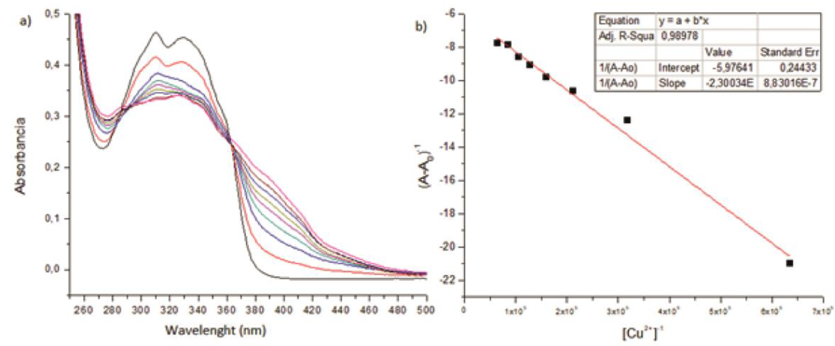
Figure 4.
Titration of receptor IV and V in presence of cobalt (II) a) Change in fluorescence spectrum of receptor IV after titration of 0-5 equivalents of cobalt (II) ion. b) linear curve fitting according to Benesi-Hildebrand equation. c) Change in fluorescence spectrum of receptor V after titration of 0-5 equivalents of cobalt (II) ion. d) linear curve fitting according to Benesi-Hildebrand equation.
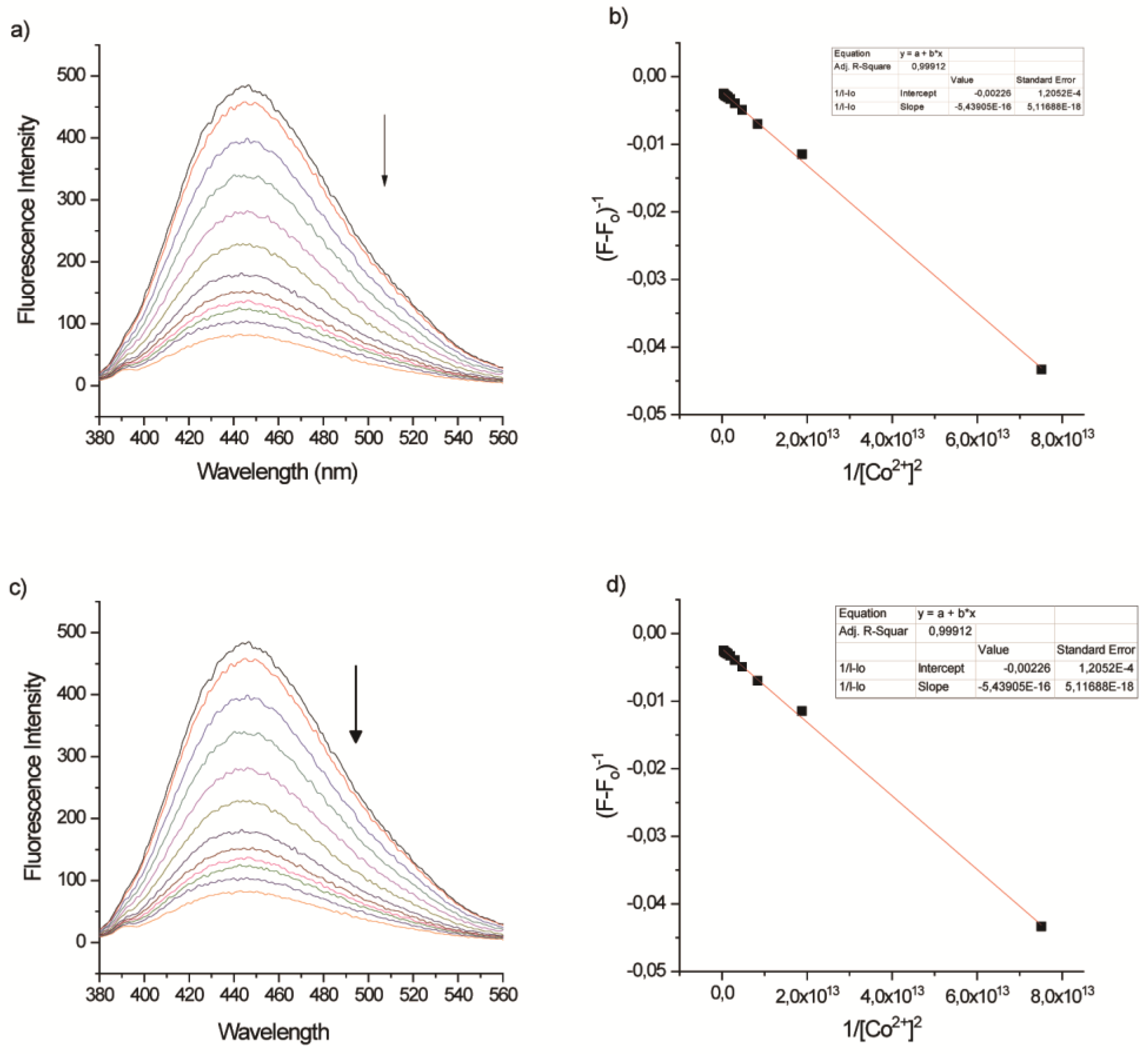
Figure 5.
Colorimetric studies of receptors of I (7.88 mM) with aliquots 0.4 mM copper (II) ions.


Table 1.
Binding constant of receptor (I-III) determined in UV-visible spectroscopy in acetonitrile.
Table 1.
Binding constant of receptor (I-III) determined in UV-visible spectroscopy in acetonitrile.
| Title 1 | Ag+ | Ni2+ | Co2+ | Zn2+ | Cu2+ | CN- | F- | |
|---|---|---|---|---|---|---|---|---|
| I | Ka (105 M-1) | 7.15 | 3.75 | 9.12 | 1.13 | 2.59 | 4.31 | 4.52 |
| stoichiometry | 1:1 | 1:1 | 1:1 | 1:1 | 1:1 | 1:1 | 1:1 | |
| II | Ka (104 M-1) | - | - | - | - | 3.07 | - | - |
| stoichiometry | - | - | - | - | 1:1 | - | - | |
| III | Ka (104 M-1) | - | - | - | - | 2.67 | - | - |
| stoichiometry | - | - | - | - | 1:1 | - | - |
Table 2.
Binding constant of receptor (IV and V) determined in fluorescence spectroscopy in acetonitrile.
Table 2.
Binding constant of receptor (IV and V) determined in fluorescence spectroscopy in acetonitrile.
| Title 2 | Ni2+ | Co2+ | Cu2+ | |
|---|---|---|---|---|
| IV | Ka (×1012(mol/l)-2) | 3.62 | 6.59 | 3.96 |
| stoichiometry | 1:2 | 1:2 | 1:2 | |
| V | Ka (×1012(mol/l)-2) | 1.63 | 2.38 | 1.09 |
| stoichiometry | 1:2 | 1:2 | 1:2 |
Disclaimer/Publisher’s Note: The statements, opinions and data contained in all publications are solely those of the individual author(s) and contributor(s) and not of MDPI and/or the editor(s). MDPI and/or the editor(s) disclaim responsibility for any injury to people or property resulting from any ideas, methods, instructions or products referred to in the content. |
© 2024 by the authors. Licensee MDPI, Basel, Switzerland. This article is an open access article distributed under the terms and conditions of the Creative Commons Attribution (CC BY) license (http://creativecommons.org/licenses/by/4.0/).
Copyright: This open access article is published under a Creative Commons CC BY 4.0 license, which permit the free download, distribution, and reuse, provided that the author and preprint are cited in any reuse.


